General anesthesia selectively disrupts astrocyte calcium signaling
Upload
duongkhanhCategory
view
223download
0
Astrocyte-derived interleukin-15 exacerbates ischemicbrain injury via propagation of cellular immunityMinshu Lia,b, Zhiguo Lib, Yang Yaoa, Wei-Na Jina,b, Kristofer Woodb, Qiang Liua,b, Fu-Dong Shia,b, and Junwei Haoa,1
aDepartment of Neurology, Tianjin Neurological Institute, Tianjin Medical University General Hospital, Tianjin 300052, China; and bDepartment ofNeurology, Barrow Neurological Institute, St. Joseph’s Hospital and Medical Center, Phoenix, AZ 85013
Edited by Gregg L. Semenza, Johns Hopkins University School of Medicine, Baltimore, MD, and approved November 28, 2016 (received for review August3, 2016)
Astrocytes are believed to bridge interactions between infiltratinglymphocytes and neurons during brain ischemia, but the mechanismsfor this action are poorly understood. Herewe found that interleukin-15 (IL-15) is dramatically up-regulated in astrocytes of postmortembrain tissues from patients with ischemic stroke and in a mousemodel of transient focal brain ischemia. We generated a glial fibrillaryacidic protein (GFAP) promoter-controlled IL-15–expressing transgenicmouse (GFAP–IL-15tg) line and found enlarged brain infarcts, exacer-bated neurodeficits after the induction of brain ischemia. In addition,knockdown of IL-15 in astrocytes attenuated ischemic brain injury.Interestingly, the accumulation of CD8+ T and natural killer (NK) cellswas augmented in these GFAP–IL-15tg mice after brain ischemia. Ofnote, depletion of CD8+ T or NK cells attenuated ischemic brain injuryin GFAP–IL-15tg mice. Furthermore, knockdown of the IL-15 recep-tor α or blockade of cell-to-cell contact diminished the activationand effector function of CD8+ T and NK cells in GFAP–IL-15tg mice,suggesting that astrocytic IL-15 is delivered in trans to target cells.Collectively, these findings indicate that astrocytic IL-15 could ag-gravate postischemic brain damage via propagation of CD8+ T andNK cell-mediated immunity.
astrocyte | interleukin-15 | lymphocytes | inflammation |ischemic brain injury
Infiltrating leukocytes such as lymphocytes are major effectorsof postischemic brain inflammation (1–6). The phenotype and
function of infiltrating lymphocytes are largely dictated by organ-specific intrinsic factors during inflammatory responses (7–9), andsuch factors in the brain are unique in terms of cellular constituents,blood–brain barrier (10–12), and microenvironment (1–3, 7). As themost abundant cell type in the CNS, astrocytes constitute nearly50% of the human brain’s volume. Astrocytes contribute to theregulation of neural transmission, survival of neurons and other gliacells, and integrity of the blood–brain barrier. In the inflamed CNS,astrocytes engage in significant cross-talk with CNS-infiltrating im-mune cells by providing a major source of the proinflammatorycytokines and chemokines, thereby activating infiltrating lymphocytes.Evidence has shown that astrocytes can exert potent proinflammatoryfunctions by producing factors including monocyte chemotacticprotein-1 (MCP-1/CCL2), interleukin 1 beta (IL-1β), interleukin-6(IL-6), etc., as their primary mode of action after CNS injury. Inaddition, astrocytes are considered as important nonprofessionalantigen-presenting cells. Depending on the stage of brain pathology,astrocytes also possess antiinflammatory properties such as scar for-mation and restriction of inflammation by producing transforminggrowth factor-β (13, 14). Recent studies have shown that the in-hibition of astrocytes correlates with decreased infarct size (15, 16)and that treatments capable of decreasing infarct size are oftenaccompanied by attenuated astrocyte responses. These findingssuggest a detrimental role for astrocytes after brain ischemia (15–18). However, still unknown are whether and how astrocytes shapeacute CNS immune responses in the context of a postischemicbrain and whether this process has any clinical significance.IL-15 belongs to a family of cytokines using the common γ-chain
as a component of their receptors (19, 20). IL-15 interacts specifically
with the high-affinity IL-15 receptor α (IL-15Rα) and binds toIL-2/IL-15Rβ and a common γ-chain expressed by target cells(21–23). In the periphery, monocytes and dendritic cells are themain sources of IL-15 (24, 25). IL-15 maintains homeostasisand cytotoxic activities of lymphocytes that bear its receptor [i.e.,natural killer (NK) and CD8+ T cells] (19, 20). Some studies havedemonstrated that IL-15 contributes to the immunopathology ofseveral inflammatory diseases, such as rheumatoid arthritis andinflammatory bowel disease (26, 27). Despite recent studies sug-gesting astrocytes as a major source of IL-15 in the inflamed CNS(28–30), the potential role of astrocytic IL-15 in ischemic braininjury remains elusive.Our observations of dramatically elevated IL-15 expression in
astrocytes after ischemia and reperfusion established a rationale forfurther investigation of astrocytes and their derived IL-15 in brainischemia. To this end, we have generated a transgenic mouse linewith efficient expression of glial fibrillary acidic protein (GFAP)promoter-controlled IL-15 (GFAP–IL-15tg), characterized their le-sions, and defined the mechanisms of IL-15 action. Our findingsreveal that IL-15 is a key factor of astrocytes in controlling themagnitude of CNS inflammation and brain injury after ischemia.
ResultsExpression of IL-15 in Astrocyte Is Highly Up-Regulated After BrainIschemia. To gain a comprehensive view of astrocyte-derivedfactors after brain ischemia, we isolated glutamate/aspartatetransporter-positive (GLAST+) astrocytes (purity > 99%;
Significance
Ischemic stroke is a leading cause of death and disability world-wide. Evidence indicates the detrimental effects of lymphocyteinfiltration into the ischemic brain. However, a knowledge gapexists relating to the brain-specific cellular constituents and en-vironmental factors that dictate the phenotype and function ofinfiltrating lymphocytes. Astrocytes bridge interactions betweenischemic neurons and lymphocytes. We show that brain ischemiainduces robust up-regulation of astrocytic interleukin-15 (IL-15).The present study was directed toward understanding the role ofastrocyte-derived factors such as IL-15 in stroke. We discoveredthat astrocytic IL-15 is necessary and sufficient to amplify cell-mediated immune responses that promote ischemic brain injury.These results provide definitive evidence on the role of astrocyte-derived IL-15 in ischemic brain injury.
Author contributions: Q.L., F.-D.S., and J.H. designed research; M.L., Z.L., Y.Y., and W.-N.J.performed research; M.L., Z.L., and K.W. analyzed data; and M.L., Z.L., K.W., Q.L., F.-D.S.,and J.H. wrote the paper.
The authors declare no conflict of interest.
This article is a PNAS Direct Submission.
Freely available online through the PNAS open access option.
See Commentary on page 425.1To whom correspondence should be addressed. Email: [email protected].
This article contains supporting information online at www.pnas.org/lookup/suppl/doi:10.1073/pnas.1612930114/-/DCSupplemental.
E396–E405 | PNAS | Published online December 19, 2016 www.pnas.org/cgi/doi/10.1073/pnas.1612930114
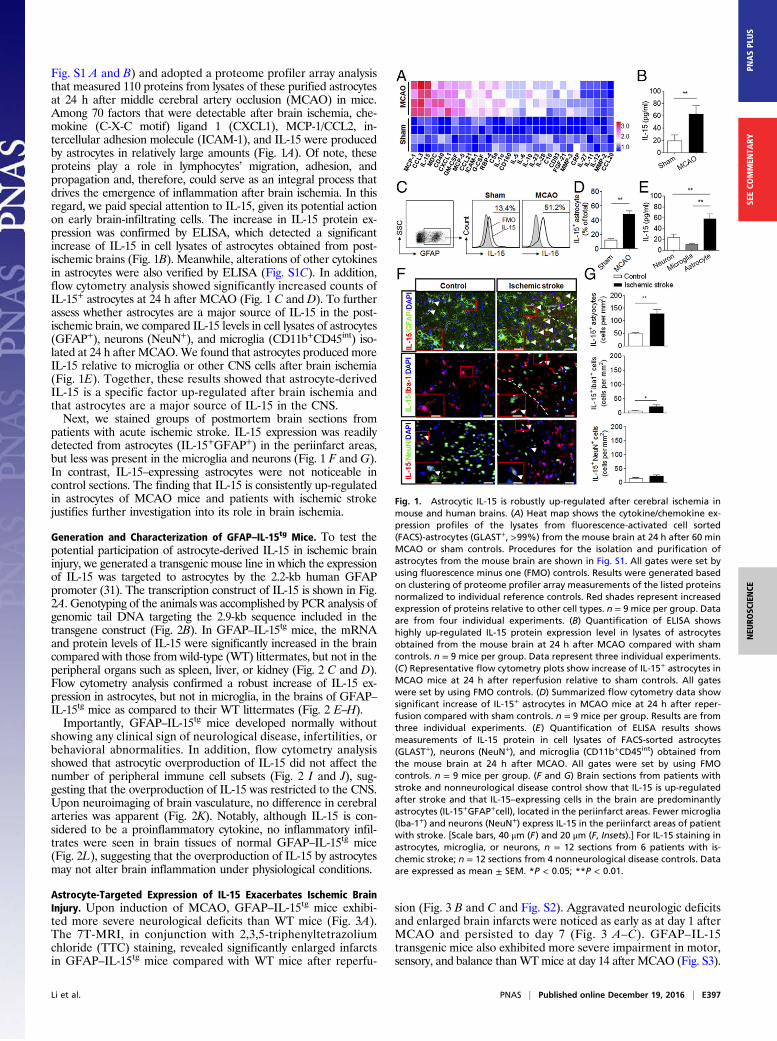
Fig. S1 A and B) and adopted a proteome profiler array analysisthat measured 110 proteins from lysates of these purified astrocytesat 24 h after middle cerebral artery occlusion (MCAO) in mice.Among 70 factors that were detectable after brain ischemia, che-mokine (C-X-C motif) ligand 1 (CXCL1), MCP-1/CCL2, in-tercellular adhesion molecule (ICAM-1), and IL-15 were producedby astrocytes in relatively large amounts (Fig. 1A). Of note, theseproteins play a role in lymphocytes’ migration, adhesion, andpropagation and, therefore, could serve as an integral process thatdrives the emergence of inflammation after brain ischemia. In thisregard, we paid special attention to IL-15, given its potential actionon early brain-infiltrating cells. The increase in IL-15 protein ex-pression was confirmed by ELISA, which detected a significantincrease of IL-15 in cell lysates of astrocytes obtained from post-ischemic brains (Fig. 1B). Meanwhile, alterations of other cytokinesin astrocytes were also verified by ELISA (Fig. S1C). In addition,flow cytometry analysis showed significantly increased counts ofIL-15+ astrocytes at 24 h after MCAO (Fig. 1 C and D). To furtherassess whether astrocytes are a major source of IL-15 in the post-ischemic brain, we compared IL-15 levels in cell lysates of astrocytes(GFAP+), neurons (NeuN+), and microglia (CD11b+CD45int) iso-lated at 24 h after MCAO.We found that astrocytes produced moreIL-15 relative to microglia or other CNS cells after brain ischemia(Fig. 1E). Together, these results showed that astrocyte-derivedIL-15 is a specific factor up-regulated after brain ischemia andthat astrocytes are a major source of IL-15 in the CNS.Next, we stained groups of postmortem brain sections from
patients with acute ischemic stroke. IL-15 expression was readilydetected from astrocytes (IL-15+GFAP+) in the periinfarct areas,but less was present in the microglia and neurons (Fig. 1 F and G).In contrast, IL-15–expressing astrocytes were not noticeable incontrol sections. The finding that IL-15 is consistently up-regulatedin astrocytes of MCAO mice and patients with ischemic strokejustifies further investigation into its role in brain ischemia.
Generation and Characterization of GFAP–IL-15tg Mice. To test thepotential participation of astrocyte-derived IL-15 in ischemic braininjury, we generated a transgenic mouse line in which the expressionof IL-15 was targeted to astrocytes by the 2.2-kb human GFAPpromoter (31). The transcription construct of IL-15 is shown in Fig.2A. Genotyping of the animals was accomplished by PCR analysis ofgenomic tail DNA targeting the 2.9-kb sequence included in thetransgene construct (Fig. 2B). In GFAP–IL-15tg mice, the mRNAand protein levels of IL-15 were significantly increased in the braincompared with those from wild-type (WT) littermates, but not in theperipheral organs such as spleen, liver, or kidney (Fig. 2 C and D).Flow cytometry analysis confirmed a robust increase of IL-15 ex-pression in astrocytes, but not in microglia, in the brains of GFAP–IL-15tg mice as compared to their WT littermates (Fig. 2 E–H).Importantly, GFAP–IL-15tg mice developed normally without
showing any clinical sign of neurological disease, infertilities, orbehavioral abnormalities. In addition, flow cytometry analysisshowed that astrocytic overproduction of IL-15 did not affect thenumber of peripheral immune cell subsets (Fig. 2 I and J), sug-gesting that the overproduction of IL-15 was restricted to the CNS.Upon neuroimaging of brain vasculature, no difference in cerebralarteries was apparent (Fig. 2K). Notably, although IL-15 is con-sidered to be a proinflammatory cytokine, no inflammatory infil-trates were seen in brain tissues of normal GFAP–IL-15tg mice(Fig. 2L), suggesting that the overproduction of IL-15 by astrocytesmay not alter brain inflammation under physiological conditions.
Astrocyte-Targeted Expression of IL-15 Exacerbates Ischemic BrainInjury. Upon induction of MCAO, GFAP–IL-15tg mice exhibi-ted more severe neurological deficits than WT mice (Fig. 3A).The 7T-MRI, in conjunction with 2,3,5-triphenyltetrazoliumchloride (TTC) staining, revealed significantly enlarged infarctsin GFAP–IL-15tg mice compared with WT mice after reperfu-
sion (Fig. 3 B and C and Fig. S2). Aggravated neurologic deficitsand enlarged brain infarcts were noticed as early as at day 1 afterMCAO and persisted to day 7 (Fig. 3 A–C). GFAP–IL-15transgenic mice also exhibited more severe impairment in motor,sensory, and balance thanWTmice at day 14 after MCAO (Fig. S3).
Fig. 1. Astrocytic IL-15 is robustly up-regulated after cerebral ischemia inmouse and human brains. (A) Heat map shows the cytokine/chemokine ex-pression profiles of the lysates from fluorescence-activated cell sorted(FACS)-astrocytes (GLAST+, >99%) from the mouse brain at 24 h after 60 minMCAO or sham controls. Procedures for the isolation and purification ofastrocytes from the mouse brain are shown in Fig. S1. All gates were set byusing fluorescence minus one (FMO) controls. Results were generated basedon clustering of proteome profiler array measurements of the listed proteinsnormalized to individual reference controls. Red shades represent increasedexpression of proteins relative to other cell types. n = 9 mice per group. Dataare from four individual experiments. (B) Quantification of ELISA showshighly up-regulated IL-15 protein expression level in lysates of astrocytesobtained from the mouse brain at 24 h after MCAO compared with shamcontrols. n = 9 mice per group. Data represent three individual experiments.(C) Representative flow cytometry plots show increase of IL-15+ astrocytes inMCAO mice at 24 h after reperfusion relative to sham controls. All gateswere set by using FMO controls. (D) Summarized flow cytometry data showsignificant increase of IL-15+ astrocytes in MCAO mice at 24 h after reper-fusion compared with sham controls. n = 9 mice per group. Results are fromthree individual experiments. (E) Quantification of ELISA results showsmeasurements of IL-15 protein in cell lysates of FACS-sorted astrocytes(GLAST+), neurons (NeuN+), and microglia (CD11b+CD45int) obtained fromthe mouse brain at 24 h after MCAO. All gates were set by using FMOcontrols. n = 9 mice per group. (F and G) Brain sections from patients withstroke and nonneurological disease control show that IL-15 is up-regulatedafter stroke and that IL-15–expressing cells in the brain are predominantlyastrocytes (IL-15+GFAP+cell), located in the periinfarct areas. Fewer microglia(Iba-1+) and neurons (NeuN+) express IL-15 in the periinfarct areas of patientwith stroke. [Scale bars, 40 μm (F) and 20 μm (F, Insets).] For IL-15 staining inastrocytes, microglia, or neurons, n = 12 sections from 6 patients with is-chemic stroke; n = 12 sections from 4 nonneurological disease controls. Dataare expressed as mean ± SEM. *P < 0.05; **P < 0.01.
Li et al. PNAS | Published online December 19, 2016 | E397
NEU
ROSC
IENCE
PNASPL
US
SEECO
MMEN
TARY
Reactive oxygen species (ROS) production was also significantlyincreased in GFAP–IL-15tg mice vs. WT controls at 24 h afterMCAO (Fig. S4). An additional comparison then verified thatastrocytes’ IL-15 expression was much greater in GFAP–IL-15tg
mice than in WT mice at 24 h after MCAO (Fig. 3 D–G). Ofnote, knockdown of astrocytic IL-15 by using shIL-15–expressinglentivirus reduced neurodeficits and infarct volume in WT micesubjected to MCAO and reperfusion (Fig. S5). These resultsdemonstrate that astrocyte-derived IL-15 exacerbates cerebralinfarction during the acute and delay phase after brain ischemia.
Augmented Accumulation of CD8+ T and NK Cells in GFAP–IL-15tg
Mice After Cerebral Ischemia. Reportedly, IL-15 is essential forthe maintenance of lymphocytes, such as NK and CD8+ T cells, inthe periphery. To determine the potential impact of IL-15 over-expression on the brain’s cellular infiltrates after ischemia, wemeasured the accumulation of infiltrating leukocytes and residentmicroglia after MCAO in brains of GFAP–IL-15tg and WTmice byusing flow cytometry (gating strategy in Fig. 4A). At 24 h afterreperfusion, we did not observe any significant difference in thenumbers of macrophages (CD11b+CD45highF4/80+), neutrophils
(CD11b+CD45highLy-6G+), microglia (CD11b+CD45int), or CD4+
T cells (CD3+CD4+) in the ischemic brains of WT compared withGFAP–IL-15tg mice (Fig. 4 B and C). In addition, the expression ofproinflammatory (CD86 and CD68) or antiinflammatory (CD206)markers in microglia was not significantly different in the ischemicbrains of WT and GFAP–IL-15tg mice (Fig. 4C). In contrast,GFAP–IL-15tg mice had significantly increased cerebral accumu-lations of CD8+ T (CD3+CD8+) and NK (CD3−NK1.1+) cells afterMCAO compared with their WT littermates (Fig. 4 D–G), andthese significant differences persisted at day 7 (Fig. 4 D and E). Ofinterest, significantly reduced numbers of CD8+ T and NK cells,but not other cell subsets, were also present in the spleen afterMCAO (Fig. 4 H–J).To examine whether astrocyte-targeted expression of IL-15 had
an impact on the functional status of CD8+ T and NK cells, wemeasured the expression of CD69, natural-killer group 2 memberD (NKG2D), interferon gamma (IFN-γ), and perforin in CD8+ Tand NK cells after MCAO (gating strategy in Fig. 5A). We foundsignificantly increased expression of CD69 and NKG2D in CD8+ Tcells in the ischemic brain of GFAP–IL-15tg mice (Fig. 5B). Simi-larly, increased expression of CD69, NKG2D, and IFN-γ was also
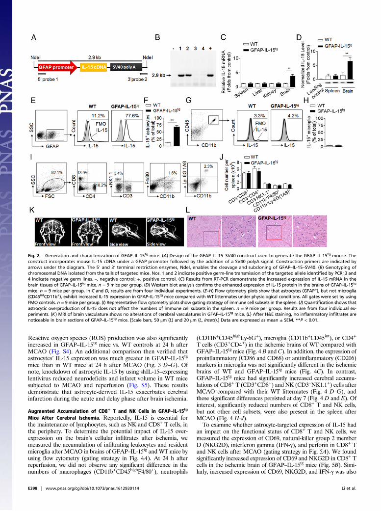
Fig. 2. Generation and characterization of GFAP–IL-15tg mice. (A) Design of the GFAP–IL-15–SV40 construct used to generate the GFAP–IL-15tg mouse. Theconstruct incorporates mouse IL-15 cDNA under a GFAP promoter followed by the addition of a SV40 polyA signal. Construction primers are indicated byarrows under the diagram. The 5′ and 3′ terminal restriction enzymes, NdeI, enables the cleavage and subcloning of GFAP–IL-15–SV40. (B) Genotyping ofchromosomal DNA isolated from the tails of targeted mice. Nos. 1 and 2 indicate positive germ-line transmission of the targeted allele identified by PCR; 3 and4 indicate negative germ lines. –, negative control; +, positive control. (C) Results from RT-PCR demonstrate the increased expression of IL-15 mRNA in thebrain tissues of GFAP–IL-15tg mice. n = 9 mice per group. (D) Western blot analysis confirms the enhanced expression of IL-15 protein in the brains of GFAP–IL-15tg
mice. n = 9 mice per group. In C and D, results are from four individual experiments. (E–H) Flow cytometry plots show that astrocytes (GFAP+), but not microglia(CD45intCD11b+), exhibit increased IL-15 expression in GFAP–IL-15tg mice compared with WT littermates under physiological conditions. All gates were set by usingFMO controls. n = 9 mice per group. (I) Representative flow cytometry plots show gating strategy of immune cell subsets in the spleen. (J) Quantification shows thatastrocytic overproduction of IL-15 does not affect the numbers of immune cell subsets in the spleen. n = 9 mice per group. Results are from four individual ex-periments. (K) MRI of brain vasculature shows no alterations of cerebral vasculatures in GFAP–IL-15tg mice. (L) After H&E staining, no inflammatory infiltrates arenoticeable in brain sections of GFAP–IL-15tg mice. [Scale bars, 50 μm (L) and 20 μm (L, Insets).] Data are expressed as mean ± SEM. **P < 0.01.
E398 | www.pnas.org/cgi/doi/10.1073/pnas.1612930114 Li et al.
seen in brain-infiltrating NK cells of GFAP–IL-15tg mice afterreperfusion (Fig. 5C). On the contrary, the expression of CD69,NKG2D, IFN-γ, and perforin was unaltered in splenic CD8+ Tand NK cells after MCAO (Fig. 5 D and E). These results suggestthat astrocyte-derived IL-15 may contribute to the activation andaccumulation of CD8+ T and NK cells in the ischemic brain.
Impact of CD8+ T and NK Cells on Ischemic Brain Injury in GFAP–IL-15tg
Mice. Recent studies have shown that CD8+ T and NK cells areboth prominent lymphocyte subsets in the ischemic brain andmay contribute to ischemic brain injury (32–35). To determinewhether the exacerbation of such injury in GFAP–IL-15tg mice ismediated by the impact of astrocytic IL-15 on CD8+ T and NKcells, we induced ischemia in WT and GFAP–IL-15tg mice givenanti-CD8 or -NK1.1 mAb 1 d before the induction of MCAO. Asreported (32, 36, 37), CD8+ T and NK1.1+ cells (NK and NKTcells) can be efficiently depleted with anti-CD8 and -NK1.1mAb, respectively (Fig. S6). Isotype IgG was used as a control. InMCAO mice treated with anti-CD8 mAb, anti-NK1.1 mAb, oranti-CD8 mAb + anti-NK1.1 mAb, infarct size and neurodeficitswere similar in WT and GFAP–IL-15tg mice (Fig. 6). In otherwords, the depletion of CD8+ T and/or NK cells reversed theaugmented brain injury in GFAP–IL-15tg mice. In contrast, inMCAOmice treated with the IgG control, the neurodeficits weremore severe and infarct sizes enlarged in GFAP–IL-15tg micecompared with their WT littermates (Fig. 6). Because NKT cellsreportedly do not significantly influence stroke (32, 33), theobserved effects of anti-NK1.1 mAb treatment, if any, could beattributed to NK cell depletion. Combined, these data demon-strate the necessity of CD8+ T and NK cells for astrocyte-derivedIL-15 to exacerbate ischemic brain injury.
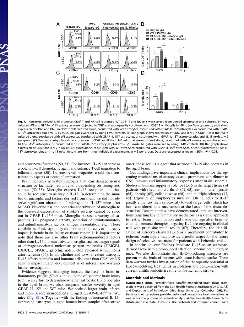
Astrocyte-Derived IL-15 Propagates CD8+ T and NK Cell Responses.To further investigate the impact of astrocyte-derived IL-15 on
CD8+ T and NK cells, we cocultured WT or GFAP–IL-15tg as-trocytes exposed to oxygen glucose deprivation (OGD) with WTCD8+ T or NK cells. CD8+ T and NK cells were isolated frompooled WT splenocytes and purified (purity > 98%; Fig. S7).Upon coculture with GFAP–IL-15tg astrocytes for 48 h, thepercentages of activated CD8+ T cells (CD69+CD8+) or NK cells(CD69+CD3−NK1.1+) and IFN-γ–expressing CD8+ T cells(IFN-γ+CD3+CD8+) or NK cells (IFN-γ+CD3−NK1.1+) weresignificantly increased compared with CD8+ T or NK cellscocultured with WT astrocytes (Fig. 7). Moreover, the effects ofGFAP–IL-15tg astrocytes on CD8+ T or NK cells were blockedby coincubation with an IL-15 mAb (Fig. 7), which selectivelyneutralized IL-15. These results suggest that astrocyte-derivedIL-15 can directly enhance CD8+ T and NK cell responses.
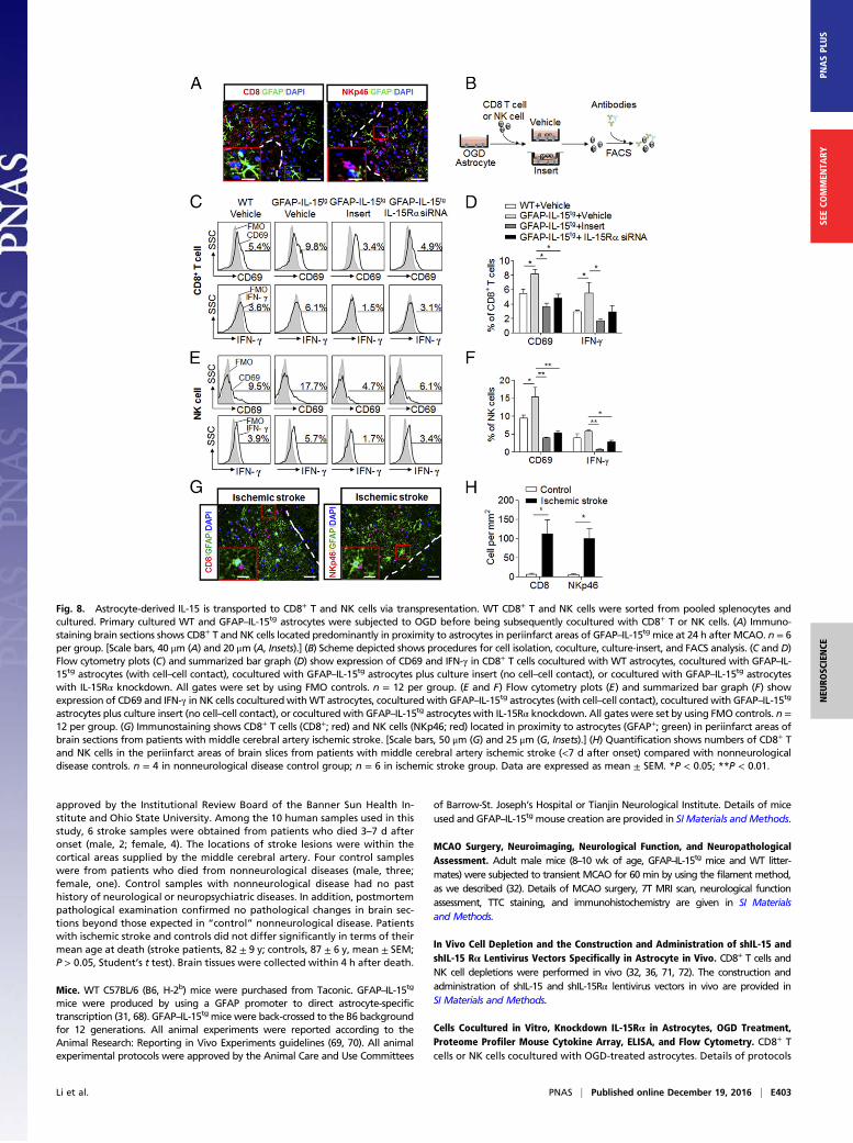
Astrocytes Deliver IL-15 in Trans to CD8+ T and NK Cells. Distinguishedfrom many other cytokines, intracellular IL-15 binds to the high-affinity IL-15Rα to form a complex, which is subsequently trans-ported to the cell surface for an efficient cross-presentation to targetcells. This process is termed “transpresentation,” and membrane-bound, rather than secreted, IL-15 is crucial in mediating its effectsin vivo (38–42). We showed that CD8+ T or NK cells are withinclose proximity to astrocytes in the periinfarct area of brain sectionsat 24 h after MCAO (Fig. 8A). We also performed ELISA to detectsecreted IL-15 in WT and GFAP–IL-15tg astrocyte-conditionedmedium, but secreted IL-15 levels were minimum and around thedetection threshold of the assay (∼10 pg/mL). This result suggeststhat IL-15 secreted by astrocytes may not have a major impact onthe target cells of interest (i.e., CD8+ T or NK cells).To determine whether astrocytes provide IL-15 in trans to
target cells, we cultured CD8+ T or NK cells in cell culturereservoirs with or without culture-insert separation from cocul-tured WT or GFAP–IL-15tg astrocytes. Consistent with previousstudies showing that surface-bound IL-15 has a major effect on
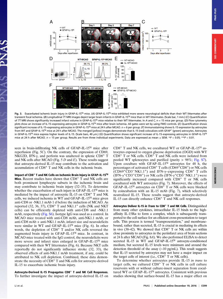
Fig. 3. Exacerbated ischemic brain injury in GFAP–IL-15tg mice. (A) GFAP–IL-15tg mice exhibited more severe neurological deficits than their WT littermates aftertransient focal ischemia. (B) Longitudinal 7T MRI images depict larger brain infarcts in GFAP–IL-15tg mice than inWT littermates. (Scale bar, 1 mm.) (C) Quantificationof 7T MRI shows significantly increased infarct volume in GFAP–IL-15tg mice relative to their WT littermates. In A and C, n = 15 mice per group. (D) Flow cytometryplots show an increase of IL-15–expressing astrocytes in GFAP–IL-15tg mice after brain ischemia. All gates were set by using FMO controls. (E) Quantification showssignificant increase of IL-15–expressing astrocytes in GFAP–IL-15tg mice at 24 h after MCAO. n = 6 per group. (F) Immunostaining shows IL-15 expression by astrocytesfromWT and GFAP–IL-15tg mice at 24 h after MCAO. The merged (yellow) images demonstrate that IL-15 (red) colocalizes with GFAP+ (green) astrocytes. Astrocytesin GFAP–IL-15tg mice express higher levels of IL-15. (Scale bars, 40 μm.) (G) Quantification shows significant increase of IL-15–expressing astrocytes in GFAP–IL-15tg
mice at 24 h after MCAO. n = 15 per group. Results are from three individual experiments. Data are expressed as mean ± SEM. *P < 0.05; **P < 0.01.
Li et al. PNAS | Published online December 19, 2016 | E399
NEU
ROSC
IENCE
PNASPL
US
SEECO
MMEN
TARY
target cells (38–40, 42, 43), we found that GFAP–IL-15tg astro-cyte-mediated enhancement of CD8+ T and NK cell responses,as manifested by their expression of CD69 or IFN-γ, was cell–cellcontact-dependent (Fig. 8 B–F). Furthermore, to investigate theinvolvement of IL-15Rα, we knocked down IL-15Rα expressionin GFAP–IL-15tg astrocytes using IL-15Rα siRNA, which effi-ciently reduces the expression of IL-15Rα in GFAP–IL-15tg as-trocytes (Fig. S8). After coculture, a significantly reducedexpression of CD69 or IFN-γ in CD8+ T cells and CD69 in NKcells was seen after silencing of IL-15Rα in GFAP–IL-15tg as-trocytes (Fig. 8 D and F). These findings demonstrate thatmembrane-bound IL-15 transpresented by IL-15Rα is essentialfor the activation and effector function of CD8+ T and NK cells.To determine whether the impact of astrocytic IL-15 on CD8+
T/NK cells requires transpresentation in vivo, we constructed alentivirus vector to specifically knock down IL-15Rα in astrocytes.At day 5 after vector injection, down-regulated IL-15Rα was seenin ∼90% of astrocytes in the injected hemisphere of GFAP–IL-15tg
mice (Fig. S9 A and B). We also found that the percentages of
CD8+ T and NK cells expressing CD69, NKG2D, or IFN-γ weredecreased after knockdown of IL-15Rα in GFAP–IL-15tg micecompared with GFAP–IL-15tg mice receiving a control vector at24 h after MCAO (Fig. S9 C–F). These results suggest that ex-pression of IL-15Rα is necessary for astrocytic IL-15 to boost CD8+
T and NK cell responses.Additionally, we noticed that human CD8+ T (CD8+) or NK
cells (NKp46+) resided in close proximity to astrocytes (GFAP+)in the periinfarct area (Fig. 8 G and H). These findings show anastrocytic up-regulation of IL-15 and physical closeness betweenIL-15–expressing astrocytes and CD8+ T or NK cells.
DiscussionThis study identifies IL-15 as a key factor produced by astrocytes incontrol of brain inflammation and neural injury after ischemia andreperfusion. As documented here, astrocyte-derived inflammatorymediators such as IL-15 are necessary and sufficient to promotecell-mediated immune responses to brain ischemia. These ampli-fied immune responses, as reflected by CD8+ T and NK cells, in
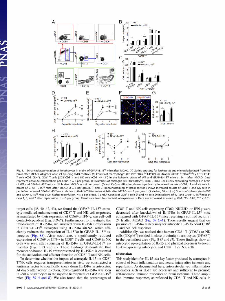
Fig. 4. Enhanced accumulation of lymphocytes in brains of GFAP–IL-15tg mice after MCAO. (A) Gating strategy for leukocytes and microglia isolated from thebrain after MCAO. All gates were set by using FMO controls. (B) Counts of macrophages (CD11b+CD45highF4/80+), neutrophils (CD11b+CD45highLy-6G+), CD4+
T cells (CD3+CD4+), CD8+ T cells (CD3+CD8+), and NK cells (CD3−NK1.1+) in the ischemic brains of WT and GFAP–IL-15tg mice at 24 h after MCAO. Datarepresent absolute cell numbers per brain. n = 8 per group. (C) Numbers of microglia (CD11b+CD45int), CD86-, CD68-, or CD206-expressing microglia in brainof WT and GFAP–IL-15tg mice at 24 h after MCAO. n = 8 per group. (D and E) Quantification shows significantly increased counts of CD8+ T and NK cells inbrains of GFAP–IL-15tg mice after MCAO. n = 8 per group. (F and G) Immunostaining of brain sections shows increased counts of CD8+ T and NK cells inperiinfarct areas of GFAP–IL-15tg mice relative to their WT littermates at 24 h after MCAO. n = 8 per group. (Scale bar, 50 μm.) (H) Counts of splenocytes in WTand GFAP–IL-15tg mice at 24 h after reperfusion. n = 8 per group. (I and J) Counts of CD8+ T cells (I) and NK cells (J) in spleens of WT and GFAP–IL-15tg mice atdays 1, 3, and 7 after reperfusion. n = 8 per group. Results are from four individual experiments. Data are expressed as mean ± SEM. *P < 0.05; **P < 0.01.
E400 | www.pnas.org/cgi/doi/10.1073/pnas.1612930114 Li et al.
turn, contribute to ischemic neural injury. Our findings provide abetter understanding of how astrocyte response to ischemia pro-motes brain injury.Brain-homing immune cells participate in the onset and pro-
gression of inflammatory responses in the ischemic brain (32–34),where they become receptive to CNS cells that they had not en-countered in the periphery. Among an array of factors derived byCNS cells, astrocytic IL-15 can impact the accumulation, activation,and effector function of brain-infiltrating immune cells, such asCD8+ T and NK cells, both of which are prominent lymphocytesubsets considered detrimental in ischemic brain injury (32, 34, 35).Consistent with these findings, we found that both CD8+ T and NKcells are preferential targets for astrocytic overproduction of IL-15to exacerbate ischemic brain damage. These findings are supportedby previous results showing that IL-15 promotes CD8+ T and NKcell responses in other diseases such as rheumatoid arthritis (44–46)and multiple sclerosis (47), suggesting that the enlarged infarct sizeseen in GFAP–IL-15tg mice may result from the enhanced CD8+ Tand NK responses. This notion is supported by the observation ofan astrocytic IL-15–induced increase of activated CD8+ T and NKcells, which suggests the involvement of direct cytotoxicity by CD8+
T and NK cells in postischemic lesion enlargement. In additionto direct cytotoxicity, the production of IFN-γ by CD8+ T andNK cells might activate other brain-homing immune cells, suchas proinflammatory macrophages, and favor CNS inflammation.Together, our results show that brain cells such as astrocytes canshape the phenotype and function of immune cells.
Different from many other cytokines, intracellular IL-15 binds tothe high-affinity IL-15Rα to form a complex, which is subsequentlytransported to the cell surface and delivered to target cells, aprocess called transpresentation. In the periphery, monocytes anddendritic cells are the main source of IL-15 (24, 25) and deliver IL-15 in trans to target cells. Despite a lack of direct evidence, recentstudies suggest that astrocytes may serve as a potential source todeliver IL-15 in trans to target cells in the inflamed CNS (47, 48).Here, we show that the impact of astrocyte-derived IL-15 on CD8+
and NK cells depends on both cell-to-cell contact and the expres-sion of IL-15Rα in astrocytes, similar to the process of IL-15transpresentation seen in peripheral organs (20, 21, 49). In addi-tion, we found that the amount of secreted IL-15 derived by as-trocytes is just above the detectable threshold and much lower thanthat seen in astrocyte lysates, suggesting that the impact of secretedIL-15 on target cells such as CD8+ T and NK cells is minimal.Overall, our data provide evidence that astrocytes are a majorsource of IL-15 in the ischemic brain. Furthermore, trans-presentation may serve as a crucial mechanism responsible for thedetrimental effect of astrocytic IL-15 on ischemic brain injury.Our data assign a role to astrocytes as stimuli of infiltrating
CD8+ T and NK cells in the ischemic brain. We noticed thatoverproduction of astrocytic IL-15 had no significant impact onthe numbers of other immune subsets, such as monocytes andneutrophils; however, we could not exclude the potential impactof these cells on astrocytic IL-15–induced exacerbation of ischemicbrain injury. Although not addressed in this study, IL-15 could par-ticipate in other aspects of neuroinflammation, given its chemotactic
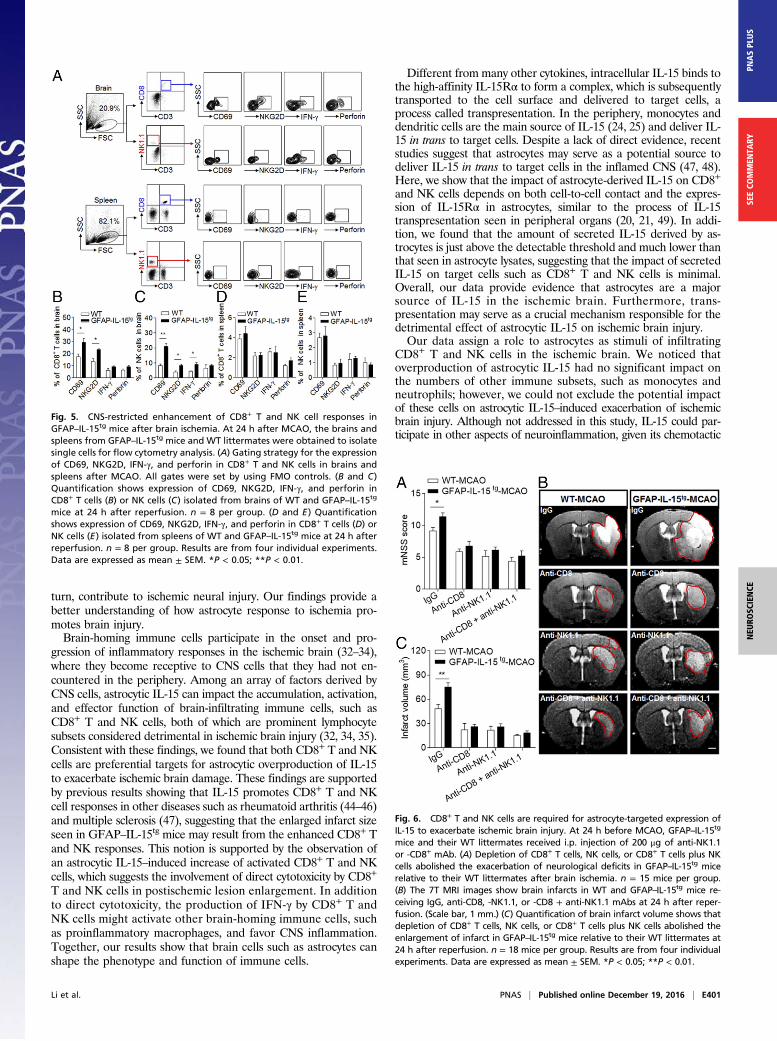
Fig. 5. CNS-restricted enhancement of CD8+ T and NK cell responses inGFAP–IL-15tg mice after brain ischemia. At 24 h after MCAO, the brains andspleens from GFAP–IL-15tg mice and WT littermates were obtained to isolatesingle cells for flow cytometry analysis. (A) Gating strategy for the expressionof CD69, NKG2D, IFN-γ, and perforin in CD8+ T and NK cells in brains andspleens after MCAO. All gates were set by using FMO controls. (B and C)Quantification shows expression of CD69, NKG2D, IFN-γ, and perforin inCD8+ T cells (B) or NK cells (C) isolated from brains of WT and GFAP–IL-15tg
mice at 24 h after reperfusion. n = 8 per group. (D and E) Quantificationshows expression of CD69, NKG2D, IFN-γ, and perforin in CD8+ T cells (D) orNK cells (E) isolated from spleens of WT and GFAP–IL-15tg mice at 24 h afterreperfusion. n = 8 per group. Results are from four individual experiments.Data are expressed as mean ± SEM. *P < 0.05; **P < 0.01.
Fig. 6. CD8+ T and NK cells are required for astrocyte-targeted expression ofIL-15 to exacerbate ischemic brain injury. At 24 h before MCAO, GFAP–IL-15tg
mice and their WT littermates received i.p. injection of 200 μg of anti-NK1.1or -CD8+ mAb. (A) Depletion of CD8+ T cells, NK cells, or CD8+ T cells plus NKcells abolished the exacerbation of neurological deficits in GFAP–IL-15tg micerelative to their WT littermates after brain ischemia. n = 15 mice per group.(B) The 7T MRI images show brain infarcts in WT and GFAP–IL-15tg mice re-ceiving IgG, anti-CD8, -NK1.1, or -CD8 + anti-NK1.1 mAbs at 24 h after reper-fusion. (Scale bar, 1 mm.) (C) Quantification of brain infarct volume shows thatdepletion of CD8+ T cells, NK cells, or CD8+ T cells plus NK cells abolished theenlargement of infarct in GFAP–IL-15tg mice relative to their WT littermates at24 h after reperfusion. n = 18 mice per group. Results are from four individualexperiments. Data are expressed as mean ± SEM. *P < 0.05; **P < 0.01.
Li et al. PNAS | Published online December 19, 2016 | E401
NEU
ROSC
IENCE
PNASPL
US
SEECO
MMEN
TARY
and prosurvival functions (50, 51). For instance, IL-15 can serve asa potent T-cell chemotactic agent and enhance T-cell migration toinflamed tissue (50). Its prosurvival properties could also con-tribute to aspects of neuroinflammation.Brain ischemia activates microglia that can damage neural
structure or facilitate neural repair, depending on timing andcontext (52–55). Microglia express IL-15 receptors and thuscould be receptive to astrocytic IL-15. In determining the num-ber of microglia and factors derived from them, we did not ob-serve significant alteration of microglia in IL-15tg mice afterMCAO. Nevertheless, the potential contribution of microglia tothe observed exacerbation of stroke’s severity cannot be ruledout in GFAP–IL-15tg mice. Microglia possess a variety of ca-pacities (i.e., phagocytic activity, secretion of proinflammatoryand antiinflammatory factors, antigen presentation, etc.). Thesecapabilities of microglia may enable them to directly or indirectlyimpact ischemic brain injury or tissue repair. It is important tonote that there are also other brain ischemia-induced factorsother than IL-15 that can activate microglia, such as danger signalsor damage-associated molecular pattern molecules (HMGB1,CX3CL1, SPARC, galectin, notch, etc.) released within hoursafter ischemia (56). In all, whether and to what extent astrocyticIL-15 affects microglia and immune cells other than CD8+ or NKcells to impact infarct development is of interest and warrantsfurther investigation.Evidence suggests that aging impacts the baseline brain in-
flammatory profile (57–60) and outcome of ischemic brain injury(61). In an effort to determine whether astrocytic IL-15 operatesin the aged brain, we also compared stroke severity in agedGFAP–IL-15tg and WT mice. We noticed larger brain infarctsand more severe neurodeficits in aged GFAP–IL-15tg vs. WTmice (Fig. S10). Together with the finding of increased IL-15–expressing astrocytes in aged human brain samples after stroke
onset, these results suggest that astrocytic IL-15 also operates inthe aged brain.Our findings have important clinical implications for the op-
erating mechanisms of astrocytes as a prominent contributor toCNS immune and inflammatory responses after brain ischemia.Studies in humans support a role for IL-15 in the target tissues ofpatients with rheumatoid arthritis (62, 63), autoimmune myositis(64), obesity (65), celiac disease (66), and multiple sclerosis (47,48). Exposure of lymphocytes such as CD8+ T cells to IL-15greatly enhances their cytotoxicity toward target cells, which hasbeen considered as a mechanism at the basis of the tissue de-struction. Recent studies have demonstrated immune interven-tions targeting key inflammatory mediators as a viable approachto restrict brain inflammation and tissue damage after brain is-chemia. Immune therapies targeting IL-1 are ongoing in clinicaltrial with promising initial results (67). Therefore, the identifi-cation of astrocyte-derived IL-15 as a prominent contributor toischemic brain injury may provide a useful target for the futuredesign of selective treatment for patients with ischemic stroke.In conclusion, our findings implicate IL-15 as an astrocyte-
derived factor with a pronounced effect on ischemic brain injury inmice. We also demonstrate that IL-15–producing astrocytes arepresent in the brain of patients with acute ischemic stroke. Thesedata warrant further investigation of the therapeutic potential ofIL-15–modifying treatments in isolation and combination withcurrent antithrombotic treatments for ischemic stroke.
Materials and MethodsHuman Brain Tissue. Formalin-fixed paraffin-embedded brain tissue cross-sections were obtained from the Sun Health Research Institute (Sun City, AZ)and Department of Pathology, Ohio State University (Columbus, OH). Pa-tients or their caregivers provided informed consent for brain donation, aswell as for the purpose of research analysis at the Sun Health Research In-stitute and Ohio State University. The protocols and informed consent were
Fig. 7. Astrocyte-derived IL-15 promotes CD8+ T and NK cell responses. WT CD8+ T and NK cells were sorted from pooled splenocytes and cultured. Primarycultured WT and GFAP–IL-15tg astrocytes were subjected to OGD and subsequently cocultured with CD8+ T or NK cells for 48 h. (A) Flow cytometry plots showexpression of CD69 and IFN-γ in CD8+ T cells cultured alone, cocultured with WT astrocytes, cocultured with GFAP–IL-15tg astrocytes, or cocultured with GFAP–IL-15tg astrocytes plus anti–IL-15 mAb. All gates were set by using FMO controls. (B) Bar graph shows expression of CD69 and IFN-γ in CD8+ T cells that werecultured alone, cocultured with WT astrocytes, cocultured with GFAP–IL-15tg astrocytes, or cocultured with GFAP–IL-15tg astrocytes plus anti–IL-15 mAb. n = 9per group. (C) Flow cytometry plots show expression of CD69 and IFN-γ in NK cells that were cultured alone, cocultured with WT astrocytes, cocultured withGFAP–IL-15tg astrocytes, or cocultured with GFAP–IL-15tg astrocytes plus anti–IL-15 mAb. All gates were set by using FMO controls. (D) Bar graph showsexpression of CD69 and IFN-γ in NK cells cultured alone, cocultured with WT astrocytes, cocultured with GFAP–IL-15tg astrocytes, or cocultured with GFAP–IL-15tg astrocytes plus anti–IL-15 mAb. Results are from three individual experiments, n = 9 per group. Data are expressed as mean ± SEM. *P < 0.05.
E402 | www.pnas.org/cgi/doi/10.1073/pnas.1612930114 Li et al.
approved by the Institutional Review Board of the Banner Sun Health In-stitute and Ohio State University. Among the 10 human samples used in thisstudy, 6 stroke samples were obtained from patients who died 3–7 d afteronset (male, 2; female, 4). The locations of stroke lesions were within thecortical areas supplied by the middle cerebral artery. Four control sampleswere from patients who died from nonneurological diseases (male, three;female, one). Control samples with nonneurological disease had no pasthistory of neurological or neuropsychiatric diseases. In addition, postmortempathological examination confirmed no pathological changes in brain sec-tions beyond those expected in “control” nonneurological disease. Patientswith ischemic stroke and controls did not differ significantly in terms of theirmean age at death (stroke patients, 82 ± 9 y; controls, 87 ± 6 y, mean ± SEM;P > 0.05, Student’s t test). Brain tissues were collected within 4 h after death.
Mice. WT C57BL/6 (B6, H-2b) mice were purchased from Taconic. GFAP–IL-15tg
mice were produced by using a GFAP promoter to direct astrocyte-specifictranscription (31, 68). GFAP–IL-15tg mice were back-crossed to the B6 backgroundfor 12 generations. All animal experiments were reported according to theAnimal Research: Reporting in Vivo Experiments guidelines (69, 70). All animalexperimental protocols were approved by the Animal Care and Use Committees
of Barrow-St. Joseph’s Hospital or Tianjin Neurological Institute. Details of miceused and GFAP–IL-15tg mouse creation are provided in SI Materials andMethods.
MCAO Surgery, Neuroimaging, Neurological Function, and NeuropathologicalAssessment. Adult male mice (8–10 wk of age, GFAP–IL-15tg mice and WT litter-mates) were subjected to transient MCAO for 60 min by using the filament method,as we described (32). Details of MCAO surgery, 7T MRI scan, neurological functionassessment, TTC staining, and immunohistochemistry are given in SI Materialsand Methods.
In Vivo Cell Depletion and the Construction and Administration of shIL-15 andshIL-15 Rα Lentivirus Vectors Specifically in Astrocyte in Vivo. CD8+ T cells andNK cell depletions were performed in vivo (32, 36, 71, 72). The construction andadministration of shIL-15 and shIL-15Rα lentivirus vectors in vivo are provided inSI Materials and Methods.
Cells Cocultured in Vitro, Knockdown IL-15Rα in Astrocytes, OGD Treatment,Proteome Profiler Mouse Cytokine Array, ELISA, and Flow Cytometry. CD8+ Tcells or NK cells cocultured with OGD-treated astrocytes. Details of protocols
Fig. 8. Astrocyte-derived IL-15 is transported to CD8+ T and NK cells via transpresentation. WT CD8+ T and NK cells were sorted from pooled splenocytes andcultured. Primary cultured WT and GFAP–IL-15tg astrocytes were subjected to OGD before being subsequently cocultured with CD8+ T or NK cells. (A) Immuno-staining brain sections shows CD8+ T and NK cells located predominantly in proximity to astrocytes in periinfarct areas of GFAP–IL-15tg mice at 24 h after MCAO. n = 6per group. [Scale bars, 40 μm (A) and 20 μm (A, Insets).] (B) Scheme depicted shows procedures for cell isolation, coculture, culture-insert, and FACS analysis. (C and D)Flow cytometry plots (C) and summarized bar graph (D) show expression of CD69 and IFN-γ in CD8+ T cells cocultured with WT astrocytes, cocultured with GFAP–IL-15tg astrocytes (with cell–cell contact), cocultured with GFAP–IL-15tg astrocytes plus culture insert (no cell–cell contact), or cocultured with GFAP–IL-15tg astrocyteswith IL-15Rα knockdown. All gates were set by using FMO controls. n = 12 per group. (E and F) Flow cytometry plots (E) and summarized bar graph (F) showexpression of CD69 and IFN-γ in NK cells coculturedwithWT astrocytes, cocultured with GFAP–IL-15tg astrocytes (with cell–cell contact), cocultured with GFAP–IL-15tg
astrocytes plus culture insert (no cell–cell contact), or cocultured with GFAP–IL-15tg astrocytes with IL-15Rα knockdown. All gates were set by using FMO controls. n =12 per group. (G) Immunostaining shows CD8+ T cells (CD8+; red) and NK cells (NKp46; red) located in proximity to astrocytes (GFAP+; green) in periinfarct areas ofbrain sections from patients with middle cerebral artery ischemic stroke. [Scale bars, 50 μm (G) and 25 μm (G, Insets).] (H) Quantification shows numbers of CD8+ Tand NK cells in the periinfarct areas of brain slices from patients with middle cerebral artery ischemic stroke (<7 d after onset) compared with nonneurologicaldisease controls. n = 4 in nonneurological disease control group; n = 6 in ischemic stroke group. Data are expressed as mean ± SEM. *P < 0.05; **P < 0.01.
Li et al. PNAS | Published online December 19, 2016 | E403
NEU
ROSC
IENCE
PNASPL
US
SEECO
MMEN
TARY
for isolation of CD8+ T cells, NK cells, and astrocytes (73), OGD treatment (32,74, 75), IL-15Rα siRNA-infected astrocyte, proteome profiler mouse cytokinearray, ELISA, and flow cytometry are provided in SI Materials and Methods.
Statistics. Details for statistical analyses are provided in SIMaterials andMethods.Data are expressed as mean ± SEM. P < 0.05 was considered significant.
ACKNOWLEDGMENTS. This study was supported in part by NationalBasic Research Program of China Grant 2013CB966900; National ScienceFoundation of China Grants 81230028, 81301044, 81471535, and 81322018;the Youth Top-Notch Talent Support Program; National Institutes ofHealth Grant R01NS092713; American Heart Association Grant16SDG27250236; and National Multiple Sclerosis Society Research Grant RG-1507-05318.
1. Kamel H, Iadecola C (2012) Brain-immune interactions and ischemic stroke: Clinical
implications. Arch Neurol 69(5):576–581.2. Iadecola C, Anrather J (2011) The immunology of stroke: From mechanisms to
translation. Nat Med 17(7):796–808.3. Eltzschig HK, Eckle T (2011) Ischemia and reperfusion—from mechanism to trans-
lation. Nat Med 17(11):1391–1401.4. Eltzschig HK, Carmeliet P (2011) Hypoxia and inflammation. N Engl J Med 364(7):
656–665.5. Chamorro Á, et al. (2012) The immunology of acute stroke. Nat Rev Neurol 8(7):
401–410.6. Chen F, et al. (2014) Non-pharmaceutical therapies for stroke: Mechanisms and clinical
implications. Prog Neurobiol 115:246–269.7. Shi FD, Ljunggren HG, La Cava A, Van Kaer L (2011) Organ-specific features of natural
killer cells. Nat Rev Immunol 11(10):658–671.8. Long EO, Kim HS, Liu D, Peterson ME, Rajagopalan S (2013) Controlling natural killer
cell responses: Integration of signals for activation and inhibition. Annu Rev Immunol
31:227–258.9. Sun JC, Lanier LL (2011) NK cell development, homeostasis and function: Parallels with
CD8+ T cells. Nat Rev Immunol 11(10):645–657.10. Quan N, Banks WA (2007) Brain-immune communication pathways. Brain Behav
Immun 21(6):727–735.11. Banks WA (2010) Mouse models of neurological disorders: A view from the blood-
brain barrier. Biochim Biophys Acta 1802(10):881–888.12. Banks WA, Erickson MA (2010) The blood-brain barrier and immune function and
dysfunction. Neurobiol Dis 37(1):26–32.13. SofroniewMV (2015) Astrocyte barriers to neurotoxic inflammation. Nat Rev Neurosci
16(5):249–263.14. Liu Z, Chopp M (2016) Astrocytes, therapeutic targets for neuroprotection and neu-
rorestoration in ischemic stroke. Prog Neurobiol 144:103–120.15. Wang W, et al. (2008) Rat focal cerebral ischemia induced astrocyte proliferation and
delayed neuronal death are attenuated by cyclin-dependent kinase inhibition. J Clin
Neurosci 15(3):278–285.16. Fang SH, et al. (2006) Increased expression of cysteinyl leukotriene receptor-1 in the
brain mediates neuronal damage and astrogliosis after focal cerebral ischemia in rats.
Neuroscience 140(3):969–979.17. Forslin Aronsson S, et al. (2006) Alpha-melanocyte-stimulating hormone is neuro-
protective in rat global cerebral ischemia. Neuropeptides 40(1):65–75.18. Ye YL, et al. (2007) Cilostazol, a phosphodiesterase 3 inhibitor, protects mice against
acute and late ischemic brain injuries. Eur J Pharmacol 557(1):23–31.19. Ma A, Koka R, Burkett P (2006) Diverse functions of IL-2, IL-15, and IL-7 in lymphoid
homeostasis. Annu Rev Immunol 24:657–679.20. Waldmann TA (2006) The biology of interleukin-2 and interleukin-15: Implications for
cancer therapy and vaccine design. Nat Rev Immunol 6(8):595–601.21. Budagian V, Bulanova E, Paus R, Bulfone-Paus S (2006) IL-15/IL-15 receptor biology: A
guided tour through an expanding universe. Cytokine Growth Factor Rev 17(4):
259–280.22. Dubois S, Mariner J, Waldmann TA, Tagaya Y (2002) IL-15Ralpha recycles and presents
IL-15 In trans to neighboring cells. Immunity 17(5):537–547.23. Stonier SW, Schluns KS (2010) Trans-presentation: A novel mechanism regulating IL-
15 delivery and responses. Immunol Lett 127(2):85–92.24. Neely GG, et al. (2001) Lipopolysaccharide-stimulated or granulocyte-macrophage
colony-stimulating factor-stimulated monocytes rapidly express biologically active IL-15 on their cell surface independent of new protein synthesis. J Immunol 167(9):
5011–5017.25. Ferlazzo G, et al. (2004) Distinct roles of IL-12 and IL-15 in human natural killer cell
activation by dendritic cells from secondary lymphoid organs. Proc Natl Acad Sci USA
101(47):16606–16611.26. McInnes IB, Leung BP, Sturrock RD, Field M, Liew FY (1997) Interleukin-15 mediates T
cell-dependent regulation of tumor necrosis factor-alpha production in rheumatoidarthritis. Nat Med 3(2):189–195.
27. Sakai T, et al. (1998) Interleukin 15 activity in the rectal mucosa of inflammatory
bowel disease. Gastroenterology 114(6):1237–1243.28. Lee YB, Satoh J, Walker DG, Kim SU (1996) Interleukin-15 gene expression in human
astrocytes and microglia in culture. Neuroreport 7(5):1062–1066.29. McInnes IB, Gracie JA (2004) Interleukin-15: A new cytokine target for the treatment
of inflammatory diseases. Curr Opin Pharmacol 4(4):392–397.30. Gómez-Nicola D, Valle-Argos B, Pita-Thomas DW, Nieto-Sampedro M (2008) In-
terleukin 15 expression in the CNS: Blockade of its activity prevents glial activation
after an inflammatory injury. Glia 56(5):494–505.31. Lee Y, Messing A, Su M, Brenner M (2008) GFAP promoter elements required for
region-specific and astrocyte-specific expression. Glia 56(5):481–493.32. Gan Y, et al. (2014) Ischemic neurons recruit natural killer cells that accelerate brain
infarction. Proc Natl Acad Sci USA 111(7):2704–2709.
33. Kleinschnitz C, et al. (2010) Early detrimental T-cell effects in experimental cerebralischemia are neither related to adaptive immunity nor thrombus formation. Blood115(18):3835–3842.
34. Yilmaz G, Arumugam TV, Stokes KY, Granger DN (2006) Role of T lymphocytes andinterferon-gamma in ischemic stroke. Circulation 113(17):2105–2112.
35. Zhang Y, et al. (2014) Accumulation of natural killer cells in ischemic brain tissues andthe chemotactic effect of IP-10. J Neuroinflammation 11(1):79.
36. Hao J, et al. (2010) Central nervous system (CNS)-resident natural killer cells suppressTh17 responses and CNS autoimmune pathology. J Exp Med 207(9):1907–1921.
37. Mracsko E, et al. (2014) Antigen dependently activated cluster of differentiation 8-positive T cells cause perforin-mediated neurotoxicity in experimental stroke.J Neurosci 34(50):16784–16795.
38. Lucas M, Schachterle W, Oberle K, Aichele P, Diefenbach A (2007) Dendritic cellsprime natural killer cells by trans-presenting interleukin 15. Immunity 26(4):503–517.
39. Huntington ND, et al. (2009) IL-15 trans-presentation promotes human NK cell de-velopment and differentiation in vivo. J Exp Med 206(1):25–34.
40. Lee GA, et al. (2011) Different NK cell developmental events require different levelsof IL-15 trans-presentation. J Immunol 187(3):1212–1221.
41. Huntington ND, et al. (2011) IL-15 transpresentation promotes both human T-cellreconstitution and T-cell-dependent antibody responses in vivo. Proc Natl Acad SciUSA 108(15):6217–6222.
42. Stonier SW, Ma LJ, Castillo EF, Schluns KS (2008) Dendritic cells drive memory CD8T-cell homeostasis via IL-15 transpresentation. Blood 112(12):4546–4554.
43. Kokaji AI, Hockley DL, Kane KP (2008) IL-15 transpresentation augments CD8+ T cellactivation and is required for optimal recall responses by central memory CD8+ Tcells. J Immunol 180(7):4391–4401.
44. Díaz-Torné C, et al. (2014) Rituximab-induced interleukin-15 reduction associatedwith clinical improvement in rheumatoid arthritis. Immunology 142(3):354–362.
45. González-Alvaro I, et al. (2006) Interleukin-15 and interferon-gamma participate inthe cross-talk between natural killer and monocytic cells required for tumour necrosisfactor production. Arthritis Res Ther 8(4):R88.
46. Dalbeth N, Callan MF (2002) A subset of natural killer cells is greatly expanded withininflamed joints. Arthritis Rheum 46(7):1763–1772.
47. Saikali P, Antel JP, Pittet CL, Newcombe J, Arbour N (2010) Contribution of astrocyte-derived IL-15 to CD8 T cell effector functions in multiple sclerosis. J Immunol 185(10):5693–5703.
48. Broux B, et al. (2015) IL-15 amplifies the pathogenic properties of CD4+CD28- T cells inmultiple sclerosis. J Immunol 194(5):2099–2109.
49. Huang D, et al. (2006) The neuronal chemokine CX3CL1/fractalkine selectively recruitsNK cells that modify experimental autoimmune encephalomyelitis within the centralnervous system. FASEB J 20(7):896–905.
50. Perera LP, Goldman CK, Waldmann TA (1999) IL-15 induces the expression of che-mokines and their receptors in T lymphocytes. J Immunol 162(5):2606–2612.
51. McInnes IB, et al. (1996) The role of interleukin-15 in T-cell migration and activation inrheumatoid arthritis. Nat Med 2(2):175–182.
52. Szalay G, et al. (2016) Microglia protect against brain injury and their selectiveelimination dysregulates neuronal network activity after stroke. Nat Commun 7:11499.
53. Li M, et al. (September 5, 2016) Colony stimulating factor 1 receptor inhibitioneliminates microglia and attenuates brain injury after intracerebral hemorrhage.J Cereb Blood Flow Metab, 10.1177/0271678X16666551.
54. Patel AR, Ritzel R, McCullough LD, Liu F (2013) Microglia and ischemic stroke: Adouble-edged sword. Int J Physiol Pathophysiol Pharmacol 5(2):73–90.
55. Ritzel RM, et al. (2015) Age- and location-related changes in microglial function.Neurobiol Aging 36(6):2153–2163.
56. Taylor RA, Sansing LH (2013) Microglial responses after ischemic stroke and in-tracerebral hemorrhage. Clin Dev Immunol 2013:746068.
57. Felzien LK, McDonald JT, Gleason SM, Berman NE, Klein RM (2001) Increased che-mokine gene expression during aging in the murine brain. Brain Res 890(1):137–146.
58. Gemechu JM, Bentivoglio M (2012) T cell recruitment in the brain during normalaging. Front Cell Neurosci 6:38.
59. Ritzel RM, et al. (2016) Age-associated resident memory CD8 T cells in the centralnervous system are primed to potentiate inflammation after ischemic brain injury.J Immunol 196(8):3318–3330.
60. Liu F, Yuan R, Benashski SE, McCullough LD (2009) Changes in experimental strokeoutcome across the life span. J Cereb Blood Flow Metab 29(4):792–802.
61. Sieber MW, Claus RA, Witte OW, Frahm C (2011) Attenuated inflammatory responsein aged mice brains following stroke. PLoS One 6(10):e26288.
62. Ferrari-Lacraz S, et al. (2004) Targeting IL-15 receptor-bearing cells with an antagonistmutant IL-15/Fc protein prevents disease development and progression in murinecollagen-induced arthritis. J Immunol 173(9):5818–5826.
63. Yoshida S, et al. (2014) Lack of association between IL-15 genetic variants and pro-gression of joint destruction in Japanese patients with rheumatoid arthritis. AnnRheum Dis 73(4):784–785.
E404 | www.pnas.org/cgi/doi/10.1073/pnas.1612930114 Li et al.
64. Huang PL, et al. (2015) Skeletal muscle interleukin 15 promotes CD8(+) T-cell function
and autoimmune myositis. Skelet Muscle 5:33.65. Quinn LAB, Conner J, Pistilli E, Wolden-Hanson T (2011) Overexpression of in-
terleukin-15 in mice promotes resistance to diet-induced obesity, increased insulin
sensitivity, and markers of oxidative skeletal muscle metabolism. Int J Infereron
Cytokine Mediator Res 3:29–42.66. van Bergen J, Mulder CJ, Mearin ML, Koning F (2015) Local communication among
mucosal immune cells in patients with celiac disease. Gastroenterology 148(6):
1187–1194.67. Emsley HC, et al.; Acute Stroke Investigators (2005) A randomised phase II study of
interleukin-1 receptor antagonist in acute stroke patients. J Neurol Neurosurg
Psychiatry 76(10):1366–1372.68. Brenner M, Kisseberth WC, Su Y, Besnard F, Messing A (1994) GFAP promoter directs
astrocyte-specific expression in transgenic mice. J Neurosci 14(3 Pt 1):1030–1037.69. Schulz KF, Altman DG, Moher D; CONSORT Group (2010) CONSORT 2010 statement:
Updated guidelines for reporting parallel group randomised trials. BMJ 340:c332.70. Kilkenny C, Browne WJ, Cuthill IC, Emerson M, Altman DG (2010) Improving bio-
science research reporting: The ARRIVE guidelines for reporting animal research. PLoS
Biol 8(6):e1000412.71. Shi FD, et al. (2000) Natural killer cells determine the outcome of B cell-mediated
autoimmunity. Nat Immunol 1(3):245–251.72. Allen CB, Schneider BK, White CW (2001) Limitations to oxygen diffusion and equil-
ibration in in vitro cell exposure systems in hyperoxia and hypoxia. Am J Physiol Lung
Cell Mol Physiol 281(4):L1021–L1027.73. Dugan LL, Bruno VM, Amagasu SM, Giffard RG (1995) Glia modulate the response of
murine cortical neurons to excitotoxicity: Glia exacerbate AMPA neurotoxicity.
J Neurosci 15(6):4545–4555.74. Newcomb-Fernandez JK, et al. (2001) Concurrent assessment of calpain and caspase-3
activation after oxygen-glucose deprivation in primary septo-hippocampal cultures.
J Cereb Blood Flow Metab 21(11):1281–1294.
75. Sen Santara S, et al. (2013) Globin-coupled heme containing oxygen sensor solubleadenylate cyclase in Leishmania prevents cell death during hypoxia. Proc Natl Acad SciUSA 110(42):16790–16795.
76. Li Y, et al. (2000) Intrastriatal transplantation of bone marrow nonhematopoietic cellsimproves functional recovery after stroke in adult mice. J Cereb Blood Flow Metab20(9):1311–1319.
77. Chen J, et al. (2001) Therapeutic benefit of intravenous administration of bonemarrow stromal cells after cerebral ischemia in rats. Stroke 32(4):1005–1011.
78. Zhang L, et al. (2002) A test for detecting long-term sensorimotor dysfunction in themouse after focal cerebral ischemia. J Neurosci Methods 117(2):207–214.
79. Bouët V, et al. (2007) Sensorimotor and cognitive deficits after transient middle ce-rebral artery occlusion in the mouse. Exp Neurol 203(2):555–567.
80. Clarkson AN, Huang BS, Macisaac SE, Mody I, Carmichael ST (2010) Reducing excessiveGABA-mediated tonic inhibition promotes functional recovery after stroke. Nature468(7321):305–309.
81. Schaar KL, Brenneman MM, Savitz SI (2010) Functional assessments in the rodentstroke model. Exp Transl Stroke Med 2(1):13.
82. Schallert T, Fleming SM, Leasure JL, Tillerson JL, Bland ST (2000) CNS plasticity andassessment of forelimb sensorimotor outcome in unilateral rat models of stroke,cortical ablation, parkinsonism and spinal cord injury. Neuropharmacology 39(5):777–787.
83. Tang Z, et al. (2014) CX3CR1 deficiency suppresses activation and neurotoxicity ofmicroglia/macrophage in experimental ischemic stroke. J Neuroinflammation 11:26.
84. Xing B, et al. (2008) Ischemic postconditioning inhibits apoptosis after focal cerebralischemia/reperfusion injury in the rat. Stroke 39(8):2362–2369.
85. Wu LJ, et al. (2012) The voltage-gated proton channel Hv1 enhances brain damagefrom ischemic stroke. Nat Neurosci 15(4):565–573.
86. Liu Q, et al. (2016) Neural stem cells sustain natural killer cells that dictate recoveryfrom brain inflammation. Nat Neurosci 19(2):243–252.
87. Lenart B, Kintner DB, Shull GE, Sun D (2004) Na-K-Cl cotransporter-mediated in-tracellular Na+ accumulation affects Ca2+ signaling in astrocytes in an in vitroischemic model. J Neurosci 24(43):9585–9597.
Li et al. PNAS | Published online December 19, 2016 | E405
NEU
ROSC
IENCE
PNASPL
US
SEECO
MMEN
TARY