Real-time Tissue Elastography for applications using Endoscopic
65
31-3-13 H H i i t t a a c c h h i i R R e e a a l l - - t t i i m m e e T T i i s s s s u u e e E E l l a a s s t t o o g g r r a a p p h h y y : : P P u u b b l l i i c c a a t t i i o o n n s s & & I I n n t t e e r r n n a a t t i i o o n n a a l l C C o o m m m m u u n n i i c c a a t t i i o o n n s s C C l l i i n n i i c c a a l l A A b b s s t t r r a a c c t t s s
Transcript of Real-time Tissue Elastography for applications using Endoscopic

31-3-13
HHiittaacchhii RReeaall--ttiimmee TTiissssuuee
EEllaassttooggrraapphhyy::
PPuubblliiccaattiioonnss && IInntteerrnnaattiioonnaall
CCoommmmuunniiccaattiioonnss
CClliinniiccaall AAbbssttrraaccttss

31-3-13
HHiittaacchhii RReeaall--ttiimmee TTiissssuuee EEllaassttooggrraapphhyy ffoorr aapppplliiccaattiioonnss uussiinngg
EEnnddoossccooppiicc UUllttrraassoouunndd

31-3-13
ENDOSCOPIC ULTRASOUND ELASTOGRAPHY FOR DIFFERENTIAL DIAGNOSIS OF PANCREATIC MASSES: A META-ANALYSIS
Duan-min Hu, Ting-ting Gong, Qi Zhu
Abstract
Background
Distinguishing malignant from benign pancreatic tumors is challenging with current imaging techniques. Endoscopic ultrasound (EUS) elastography has further improved the efficacy of EUS for characterizing pancreatic lesions.
Aims
To assess, by combining data from existing trials, the accuracy of EUS elastography in diagnosing malignant tumors for patients with pancreatic masses.
Methods
All relevant studies published were identified by systematic searching of databases. A meta-analysis was performed using a random-effects model to combine study results.
Results
Seven studies involving 752 patients were included. The sensitivity of EUS elastography for differential diagnosis of solid pancreatic masses was 97 % (95 % CI, 0.95–0.98), and the specificity was 76 % (95 % CI, 0.69–0.82). The area under the curve under summary receiver operating characteristic (SROC) was 0.9529. The combined positive likelihood ratio was 3.71 (95 % CI, 2.72–5.07), and the negative likelihood ratio was 0.05 (95 % CI, 0.02–0.13).
Conclusion
Our meta-analysis shows that EUS elastography is a useful tool for differential diagnosis of solid pancreatic neoplasms with very high sensitivity and relatively low specificity. The results indicate that EUS elastography not only provides information complementary to that from EUS but also potentially increases the yield of fine needle aspiration and reduces the number of unnecessary biopsies
Digestive Diseases and Sciences, January 10 2013 [Epub ahead of print]
________________
BRONCHIAL ENDOSCOPIC ULTRASOUND ELASTOGRAPHY: PRELIMINARY FEASIBILITY DATA.
Trosini-Désert V, Jeny F, Taillade L, Vignot S, Zribi H, Capron F, Similowski T. To the Editors: Medical elastography consists of biomechanically characterising a zone of tissue on the basis of its response to the application of mechanical stress. This stress can be quasistatic (local compression) or vibratory (propagation of shear waves). In the various medical applications of elastography, the response to the stress is described by mapping the tensile modulus, or Young‟s modulus. Young‟s modulus corresponds to the slope of the stress–strain relationship measured during a series of tensile tests [1]. The elasticity of a tissue depends on its nature, its state (fat infiltration or fibrosis) and its homogeneity. A tumour situated in a zone of healthy tissue can, therefore, be detected by its decreased elasticity. The tumour can also be described in space, based on the principle that within

31-3-13
anisotropic materials (typically represented by heterogeneous tissues), the value of Young‟s modulus varies as a function of the direction of the force applied to the material tested. Simple colour coding of the tensile response provides mapping of the elasticity of the zone examined. Very hard tissues are generally coded as blue, while soft tissues are coded as red and intermediate tissues are coded as green. Elastography is now used in various fields of medicine, often in combination with ultrasound (ultrasound elastography, sometimes called computer-assisted palpation). It has been applied to the diagnosis of breast [2], thyroid [3] and prostate tumours [4], in vascular disease [5], and in hepatology [6]. Elastography has also been combined with gastrointestinal endoscopic ultrasound to investigate pancreatic masses [7] and nodal invasion by rectal cancer [8]. A recent meta-analysis of the performances of gastrointestinal endoscopic ultrasound elastography to distinguish between benign and malignant lymph nodes concluded to a sensitivity of 88%, a specificity of 85% and an area under the receiver operating characteristics curve of 0.9456 [9]. These performances are superior to those of endoscopic ultrasound alone [10]. To our knowledge, elastography in combination with bronchial endoscopic ultrasound has not yet been evaluated. We report preliminary feasibility data and the first evaluation of this technique. This study was approved by the local ethics committee (Comité de Protection des Personnes Ile-de-France, 6 Pitié-Salpêtrière, Paris, France). All patients referred to our centre for assessment of mediastinal lymphadenopathy between February and May 2012 were studied by bronchial endoscopic ultrasound elastography under light general anaesthesia (10 patients, 13 lymph node areas measuring 10–30 mm). Real-time elastographic mapping was performed using an ultrasound elastography module incorporated into a ultrasound machine (Hi-vision Avius1; Hitachi Medical Systems, Kashiwa, Japan) coupled with a bronchial endoscopic ultrasound probe (EB1970 video bronchoscope; Pentax, Tokyo, Japan). Elasticity colour mapping was performed for each lymph node studied by superimposing the colour coding of tensile responses with the endoscopic B-mode ultrasound image and by defining the frequency histogram of the responses in the zone studied. Transbronchial needle aspiration (TBNA) was performed in each case using a 22-gauge needle (sono Tip1 EBUS; MediGlobe, Rosenheim, Germany) by targeting, as far as possible, the zone identified as being the least elastic. Colour mapping of the tissue studied and the corresponding elasticity histogram were obtained in every case (fig. 1). The elastography module proved easy to use and prolonged the examination time by only a few minutes. The five lymph nodes demonstrated to be malignant on histological examination of the TBNA material were characterised by decreased elasticity (dominant blue colour, elasticity ranging from 10 to 49 on the histogram and .80% of the tissue considered to be „„hard‟‟ in the target zone) (table 1). No malignant cell was identified in the other eight lymph nodes (elasticity ranging from 55 to 167 and 6–71% hard zones) regardless of the final diagnosis. Although discussing specificity and sensitivity is not reasonably possible with such a small sample, these preliminary results are consistent with the results published for gastrointestinal endoscopic ultrasound [9, 10]. Notably, mediastinoscopy was not performed in those of our patients where TBNA did not provide diagnostic proof because a therapeutic decision was taken based on other factors (table 1). In futures studies, the performances of endobronchial elastography will have to be assessed against the results of mediastinoscopy as the gold-standard approach. From this experience, we conclude that transbronchial biomechanical analysis can be performed during bronchial endoscopic ultrasound. Tracheobronchial cartilage does not appear to interfere with collection of this type of information. The preliminary results suggest that elastography could possibly improve the diagnostic yield of bronchial endoscopic ultrasound, as has already been demonstrated in gastrointestinal endoscopy. This is all the more important as endobronchial ultrasonography has a low negative predictive value regarding the neoplastic involvement of mediastinal lymph nodes (see [11] for an example). We propose that our preliminary results justify the setting-up of large-scale studies to precisely describe the operating characteristics of bronchial endoscopic ultrasound elastography. It would also be interesting to determine whether elastography could improve guidance of TBNA in heterogeneous lymph node masses or limit the number of TBNAs performed, by primarily targeting the lymph nodes most likely to be malignant.

31-3-13
FIGURE 1. Screenshots from the elastography module linked to an endoscopic ultrasound. The histograms show the elastography colour dispersion in the region of interest defined and also give the mean of elasticity (0: coded in blue; 255: coded in red). a–c) A benign lymph node is compressible (coded as green (a) and histogram centred around 150 (c)) whereas d–e) a malignant lymph node appears less compressible (coded as blue (d) and histogram is around 20 with a low colourimetric mean (f)).
REFERENCES 1 Ophir J, Cespedes I, Ponnekanti H, et al. Elastography: a quantitative method for imaging the elasticity of biological tissues. Ultrason Imaging 1991; 13: 111–134. 2 Barr RG, Destounis S, Lackey LB 2nd., et al. Evaluation of breast lesions using sonographic elasticity imaging: a multicenter trial. J Ultrasound Med 2012; 31: 281–287. 3 Shuzhen C. Comparison analysis between conventional ultrasonography and ultrasound elastography of thyroid nodules. Eur J Radiol 2012; 81: 1806–1811.

31-3-13
4 Brock M, von Bodman C, Palisaar RJ, et al. The impact of real-time elastography guiding a systematic prostate biopsy to improve cancer detection rate: a prospective study of 353 patients. J Urol 2012; 187: 2039–2043. 5 Baldewsing RA, Schaar JA, Mastik F, et al. Local elasticity imaging of vulnerable atherosclerotic coronary plaques. Adv Cardiol 2007; 44: 35–61. 6 Cardoso AC, Carvalho-Filho RJ, Stern C, et al. Direct comparison of diagnostic performance of transient elastography in patients with chronic hepatitis B and chronic hepatitis C. Liver Int 2012; 32: 612–621. 7 Iglesias-Garcia J, Larino-Noia J, Abdulkader I, et al. Quantitative endoscopic ultrasound elastography: an accurate method for the differentiation of solid pancreatic masses. Gastroenterology 2010; 139: 1172–1180. 8 Puli SR, Reddy JB, Bechtold ML, et al. Accuracy of endoscopic ultrasound to diagnose nodal invasion by rectal cancers: a metaanalysis and systematic review. Ann Surg Oncol 2009; 16: 1255–1265. 9 Xu W, Shi J, Zeng X, et al. EUS elastography for the differentiation of benign and malignant lymph nodes: a meta-analysis. Gastrointest Endosc 2011; 74: 1001–1009. 10 Giovannini M, Botelberge T, Bories E, et al. Endoscopic ultrasound elastography for evaluation of lymph nodes and pancreatic masses: a multicenter study. World J Gastroenterol 2009; 15: 1587–1593. 11 Bauwens O, Dusart M, Pierard P, et al. Endobronchial ultrasound and value of PET for prediction of pathological results of mediastinal hot spots in lung cancer patients. Lung Cancer 2008; 61: 356–361. Eur Respir J. 2013 Feb;41(2):477-9. doi: 10.1183/09031936.00124812
________________
CAN EUS ELASTOGRAPHY IMPROVE LYMPH NODE STAGING IN ESOPHAGEAL CANCER? Mate Knabe • Erwin Günter • Christian Ell • Oliver Pech Abstract Background Endoscopic ultrasound (EUS) elastography can assess the hardness of tissue by measuring its elasticity. Few data have been published on EUS elastography for lymph node (LN) staging in patients with esophageal cancer. This study analyzes the value of elastography as an additional diagnostic tool for LN staging. Methods Forty patients (mean age 68 years) with known esophageal cancer (34 Barrett‟s carcinoma, 6 squamous cell carcinoma) were included prospectively. On conventional EUS, suspicious LNs were assessed using sonomorphologic criteria, and EUS elastography was then used to assess their tissue hardness. The sonomorphologic criteria and elastographic images for the LN were later reviewed on recorded video clips by an endosonographer blinded to the histology results. The proportions of color pixels in LNs in selected patients were assessed using computer analysis of the elastography images. Fine-needle aspiration was performed in all of the LNs, and the histological/ cytological results were used as the gold standard. Results Twenty-one of the 40 LNs examined (52.5 %) were positive for neoplasia, confirmed by histology/cytology. The first assessment by the examiner during the procedure, based on sonomorphologic criteria, showed sensitivity of 91.3 % and specificity of 64.7 %. EUS elastography alone had sensitivity of 100 % and specificity of 64.1 %. When computer analysis of the elastographic images was added, the specificity improved significantly to 86.7 %, with a slight decrease in sensitivity to 88.9 %. Conclusions EUS elastography is easily included in clinical staging and, particularly with computer-aided pixel analysis, significantly improves the specificity of LN staging. Surg Endosc. 2013 Apr;27(4):1196-202.
________________________

31-3-13
ENDOSCOPIC ULTRASOUND IMAGE ENHANCEMENT ELASTOGRAPHY Julio Iglesias-Garcia, MD, PhD*, J. Enrique Domínguez-Muñoz, MD, PhD INTRODUCTION Endoscopic ultrasound (EUS) has evolved in recent years into a technique with a major clinical impact in digestive and mediastinal diseases. In fact, EUS has represented a major advance in the diagnosis and staging of several tumors, and can determine a change in diagnosis and management in 25% to 50% of cases. However, EUS is not only useful providing excellent images for detection and staging of several malignancies, it also provides guidance for fine-needle aspiration (FNA) and biopsies of almost all lesions detected during a standard procedure. Overall accuracy of EUS guided FNA can be considered excellent, with sensitivities between 80% and 85%, and specificities close to 100%. However, differential diagnosis of certain lesions, based only on B-mode image can be challenging and EUS-guided FNA and/or biopsy is technically demanding and multiple punctures of the lesions can be necessary to obtain sufficient tissue for cytohistologic assessment. EUS-guided FNA can also be associated with false negative results, mainly in patients with solid pancreatic masses with the underlying diagnosis of chronic pancreatitis. Another limitation is related to the evaluation of lymph nodes. When several lymph nodes appear suspicious, the choice of which one to puncture is not always clear. Finally, EUS and EUS-guided FNA are associated with a small, but not insignificant, morbidity. With this background, new methods allowing better characterization of lesions evaluated by EUS are essential to avoid the realization of unnecessary FNA and/or biopsies, to allow more accurate characterization of lesions before the puncture, and possibly to reduce complication rates. One of these new available methods is elastography. It is well known that certain diseases, such as cancer, may induce changes in tissue stiffness. Elastography is a method for the real-time evaluation of tissue stiffness. This technique has been previously used for the analysis of superficial organ lesions, such as those of the breast and prostate. Elastographic images are an index of tissue elasticity, which may be related to histopathologic features. It has been considered virtual biopsy. Now, elastographic evaluation can be performed by EUS. Several studies have demonstrated that EUS-elastography is a promising technique with a high accuracy for the differential diagnosis of solid pancreatic tumors and lymph nodes. This article analyzes the theoretical aspects and methodology of elastography, and reviews the actual indications and further development of this relatively novel method Gastrointest Endoscopy Clin N Am 22 (2012) 333–348
________________
DIAGNOSTIC ACCURACY OF QUANTITATIVE EUS ELASTOGRAPHY FOR DISCRIMINATING MALIGNANT FROM BENIGN SOLID PANCREATIC MASSES: A PROSPECTIVE, SINGLE-CENTER STUDY Muhammad F. Dawwas, MRCP, Hatim Taha, MRCP, John S. Leeds, MRCP, MD, Manu K. Nayar, MRCP, Kofi W. Oppong, FRCP Newcastle upon Tyne, United Kingdom Background: Recent data suggest that quantitative EUS elastography, a novel technique that allows real-time quantification of tissue stiffness, can accurately differentiate malignant from benign solid pancreatic masses. Objective: To externally validate the diagnostic utility of this technique in an independent cohort. Design and Setting: Prospective, single-center study. Patients, Interventions, and Methods: A total of 104 patients with evidence of a solid pancreatic mass on cross-sectional imaging and/or endosonography underwent 111 quantitative EUS elastography procedures. Multiple elastographic measurements of the mass lesion and soft-tissue reference areas were undertaken, and the corresponding strain ratios (SRs) were calculated. The final diagnosis was based on pancreatic cytology or histology.

31-3-13
Main Outcome Measurements: The area under the receiver-operating characteristic curve, sensitivity, specificity, positive predictive value, negative predictive value, and overall accuracy of quantitative EUS elastography for discriminating malignant from benign pancreatic masses. Results: The final diagnoses were primary pancreatic carcinoma (71.2%), neuroendocrine tumor (10.6%), metastatic cancer (1.9%), and pancreatitis (16.3%). Malignant masses had a higher SR (P _ .01) and lower mass elasticity (P _ .003) than inflammatory ones. The areas under the receiver-operating characteristic curve for the detection of pancreatic malignancy of both SR and mass elasticity (0.69 and 0.72, respectively) were less favorable than reported recently. At the cut points providing the highest accuracy in this cohort (4.65 for SR and 0.27% for mass elasticity), quantitative EUS elastography had a sensitivity of 100.0% and 95.7%, specificity of 16.7% and 22.2%, positive predictive value of 86.1% and 86.4%, negative predictive value of 100.0% and 50.0%, and overall accuracy of 86.5% and 83.8%, respectively. Limitations: Relatively small number of patients with benign disease. Conclusion: In the largest single-center study to date, the diagnostic utility of quantitative EUS elastography for discriminating pancreatic masses was modest, suggesting that it may only supplement rather than supplant the role of pancreatic tissue sampling in the future. Gastrointest Endosc 2012;76:953-61.
________________
UTILITY OF QUANTITATIVE ENDOSCOPIC ULTRASOUND ELASTOGRAPHY (QEUSE) FOR THE DIAGNOSIS OF PANCREATIC MALIGNANCY: A LARGE SINGLE-CENTRE EXPERIENCE
M F Dawwas, H Taha, J S Leeds, M N Nayar, K W Oppong Freeman Hospital, Newcastle Upon Tyne, UK
Abstract
Introduction Recent data suggest that QEUSE, a novel technique that allows real-time quantification of tissue stiffness, can accurately differentiate benign from malignant solid pancreatic masses (area under the receiver operating curve [AUROC]=0.98).
1 External validation of the diagnostic utility of this
technique has not been carried out.
Methods 101 patients with CT and/or EUS-proven solid pancreatic masses underwent 108 QEUSE procedures using the Hitachi EUB-7500 or Preirus ultrasound workstation and Pentax linear echoendoscopes. Multiple elastographic measurements of the mass lesion (A) and soft tissue references areas (B) were undertaken and the corresponding strain ratios (B/A) were calculated. Final diagnosis was based on EUS-fine needle aspiration (EUS-FNA) cytology, biliary brushings and/or resection specimen histology. The diagnostic accuracy of QEUSE for discriminating malignant from benign pancreatic masses was assessed.
Results The median lesion size was 3 cm. The final underlying diagnoses were primary pancreatic carcinoma (71.3%), neuroendocrine tumour (9.9%), metastatic cancer (2%) and pancreatitis (16.8%). Malignant pancreatic masses had a higher strain ratio (p=0.002) and lower mass elasticity (p=0.003) than inflammatory ones. However, the AUROC for the detection of pancreatic malignancy was only 0.74 for the strain ratio and only 0.73 for the mass elasticity. Similarly, the diagnostic accuracy of QEUSE for detecting pancreatic malignancy in our cohort was less favourable than those reported recently (see Abstract OC-109 table 1), with lower strain ratio (4.62 vs 6.04) and higher pancreatic mass elasticity cutoffs (0.27 vs 0.05) providing the highest accuracy.
Conclusion In the largest single-centre study of QEUSE of the pancreas reported to date, we found this technology to be less accurate and specific for differentiating pancreatic masses than recently reported, suggesting that it may only complement rather than substitute the role of pancreatic EUS-FNA in the future.
Reference 1. Iglesias-Garcia, et al. Gastroenterology 2010.
Gut 2012;61:A47-A48 doi:10.1136/gutjnl-2012-302514a.109

31-3-13
STRAIN ASSESSMENT IN SURGICALLY RESECTED INFLAMMATORY AND NEOPLASTIC BOWEL LESIONS.
Havre RF, Leh S, Gilja OH, Odegaard S, Waage JE, Baatrup G, Nesje LB.
Source
Institute of Medicine, University of Bergen.
Abstract
Purpose: To investigate whether ultrasound-based strain imaging can discriminate between colorectal adenocarcinomas and stenotic Crohn's lesions in newly resected surgical specimens.Materials and Methods: Resected surgical specimens from 27 patients electively operated for colorectal tumors or stenotic lesions from Crohn's disease were prospectively examined with ultrasonography using a Hitachi HV 900 US scanner with real-time elastography (RTE). Three different methods were applied to assess tissue strain: A four-level categorical visual classification, a continuous visual analog scale (VAS, 0 - 100) and a strain ratio (SR) measurement between the lesion and surrounding reference tissue. The imaged sections were marked and subsequently examined by a pathologist. Results from RTE were evaluated according to diagnosis, degree of fibrosis, inflammatory parameters, tumor stage and grade.Results: 16 sections from Crohn's lesions, 18 sections from adenocarcinomas and 4 sections from adenomas were examined. Both adenocarcinomas and Crohn's lesions were found to be harder than the surrounding tissue, but they could not be discriminated from each other by any of the strain imaging evaluation methods. All adenocarcinomas had significantly higher strain ratios than adenomas. The categorical classification differentiated poorly between Crohn's lesions, adenocarcinomas and adenomas. Categorical evaluation and VAS score showed fair interobserver agreement. SR measurements provided semi-quantitative strain data and added improved information about elasticity properties, despite substantial intra-observer variation.Conclusion: Sonoelastography with SR measurements and visual evaluation of strain differences could not differentiate stenotic Crohn's lesions from adenocarcinomas in resected bowel specimens. A small number of adenomas were found to be significantly softer than adenocarcinomas using the same evaluation methods. The tumor stage or grade did not have a significant impact on the elastography results
Ultraschall Med. 2012 Nov 15. [Epub ahead of print]
_________________ ENDOSCOPIC ULTRASOUND, ENDOSCOPIC SONOELASTOGRAPHY, AND STRAIN RATIO EVALUATION OF LYMPH NODES WITH HISTOLOGY AS GOLD STANDARD. Larsen MH, Fristrup C, Hansen TP, Hovendal CP, Mortensen MB.
Source
Department of Surgical Gastroenterology, Odense University Hospital, Odense, Denmark.
Abstract
Background and study aims: Accurate lymph node staging is essential for the selection of an optimal treatment in patients with upper gastrointestinal cancer. Endoscopic ultrasound (EUS) and fine-needle aspiration (FNA) are considered to be the most accurate method for locoregional staging. Endoscopic sonoelastography (ESE) assesses the elasticity of lymph nodes and has been used to differentiate lymph nodes with promising results. The aim of this study was to evaluate the use of EUS, EUS - FNA, ESE, and ESE-strain ratio using histology as the gold standard.

31-3-13
Patients and methods: Patients with upper gastrointestinal cancer who were referred for EUS examination were enrolled if surgical treatment was planned and the patient had a lymph node that was accessible for EUS - FNA and EUS-guided fine-needle marking (FNM). The lymph node was classified using EUS, ESE, and ESE-strain ratio. Finally, EUS - FNA and EUS - FNM were performed. The marked lymph node was isolated during surgery for histological examination.
Results: The marked lymph node was isolated for separate histological examination in 56 patients, of whom 22 (39 %) had malignant lymph nodes and 34 (61 %) had benign lymph nodes. There were no complications of EUS - FNM. The sensitivity of EUS for differentiation between malignant and benign lymph nodes was 86 % compared with 55 % - 59 % for the different ESE modalities. The specificity of EUS was 71 % compared with 82 % - 85 % using ESE modalities.
Conclusion: The use of the EUS - FNM technique enabled the identification of a specific lymph node and thereby the use of histology as gold standard. ESE and ESE-strain ratio were no better than standard EUS in differentiating between malignant and benign lymph nodes in patients with resectable upper gastrointestinal cancer
Endoscopy. 2012 Aug;44(8):759-66. Epub 2012 Jul 2.
_________________
ENDOSCOPIC ULTRASOUND-GUIDED ELASTOGRAPHY IN THE NODAL STAGING OF OESOPHAGEAL CANCER. Paterson S, Duthie F, Stanley AJ.
AIM:
To assess quantitative endoscopic ultrasound (EUS)-guided elastography in the nodal staging of oesophago-gastric cancers.
METHODS:
This was a single tertiary centre study assessing 50 patients with established oesophago-gastric cancer undergoing EUS-guided fine needle aspiration biopsy (FNAB) of lymph nodes between July 2007 and July 2009. EUS-guided elastography of lymph nodes was performed before EUS-FNAB. Standard EUS characteristics were also described. Cytological determination of whether a lymph node was malignant or benign was used as the gold standard for this study. Comparisons of elastography and standard EUS characteristics were made between the cytologically benign and malignant nodes. The main outcome measure was the accuracy of elastography in differentiating between benign and malignant lymph nodes in oesophageal cancers.
RESULTS:
EUS elastography and FNAB were performed on 53 lymph nodes. Cytological malignancy was found in 23 nodes, one was indeterminate, one was found to be a gastrointestinal stromal tumor and 25 of the nodes were negative for malignancy. On 3 occasions insufficient material was obtained for analysis. The area under the curve for the receiver operating characteristic curve for elastography strain ratio was 0.87 (P < 0.0001). Elastography strain ratio had a sensitivity 83%, specificity 96%, positive predictive value 95%, and negative predictive value 86% for distinguishing between malignant and benign nodes. The overall accuracy of elastography strain ratio was 90%. Elastography was more sensitive and specific in determining malignant nodal disease than standard EUS criteria.

31-3-13
CONCLUSION:
EUS elastography is a promising modality that may complement standard EUS and help guide EUS-FNAB during staging of upper gastrointestinal tract cancer.
World J Gastroenterol. 2012 Mar 7;18(9):889-95.
________________________
REAL-TIME ELASTOGRAPHY: STRAIN RATIO MEASUREMENTS ARE INFLUENCED BY THE POSITION OF THE REFERENCE AREA. Havre RF, Waage JR, Gilja OH, Odegaard S, Nesje LB.
PURPOSE:
Real-time elastography (RTE) is an ultrasound-based method for the visualization of relative strain distribution in soft tissues. Strain ratio is a semi-quantitative measurement of strain differences between two user-defined areas in an elastogram. The aim of this study was to evaluate the impact of the size and location of a reference area when measuring the strain ratio of focal lesions in a tissue-mimicking phantom and in normal liver tissue. We also investigated whether the strain ratio was affected by changing the scanner parameter: elasticity dynamic range (E-dyn).
MATERIALS AND METHODS:
Two investigators individually collected data by scanning 4 spherical inclusions with different elasticity in a phantom in which the elastic modulus was known in both the lesions and the background. Subsequently, a liver scan was performed in-vivo using the same scanning protocol. Five different setups with changes in reference area position or size were tested. All eight levels of the scanner setting E-dyn were recorded for each setup and the strain ratio was measured in 3 different representative elastograms for each recording situation.
RESULTS:
The four inclusions had significantly different mean strain ratio levels (p < 0.01) when compared to the surrounding material. Changing the position of the reference area to a deeper position influenced the strain ratio measurements significantly for all phantom lesions and in the liver. Changing the size of the reference area, while keeping the center depth unchanged, did not influence the mean strain ratio levels significantly. The strain ratio was independent of the E-dyn parameter setting. The intra- and interobserver reliability was high when measuring the strain ratio with a free-hand technique.
CONCLUSION:
Strain ratio provides reproducible measurements of inclusions representing different elastic contrasts using a free-hand technique in vitro. Changes in the distance of the reference areas to the ultrasound probe, representing the stress source, seem to have a significant impact on strain ratio measurements.
Ultraschall Med. 2011 Jun 10. [Epub ahead of print]
________________________

31-3-13
REAL-TIME TISSUE ELASTOGRAPHY FOR THE DIAGNOSIS OF LYMPH NODE METASTASIS IN ORAL SQUAMOUS CELL CARCINOMA.
Ishibashi N, Yamagata K, Sasaki H, Seto K, Shinya Y, Ito H, Shinozuka K, Yanagawa T, Onizawa K, Bukawa H.
Source
Oral and Maxillofacial Surgery, Clinical Sciences, Graduate School of Comprehensive Human Sciences, University of Tsukuba, Tsukuba, Ibaraki, Japan.
Abstract
We compared conventional ultrasound (US) B-mode, color Doppler and elastographic assessment of lymph node (LN) stiffness against pathological findings from surgical samples, to determine the most useful factors for identifying LN metastases. Seventy-one LNs in 19 patients with oral squamous cell carcinoma (OSCC) were examined. Using our new system, elastography images were scored from 1-5. The score 1-4 were correlated with the blue area of each LN, which indicated increased stiffness: (1) none; (2) <50%; (3) 50%; or (4) >50%. A score 5 indicated central necrosis and did not correlate with the blue area. We found significant differences in minimal diameter, shape index, margin, internal structure, hilus presence or absence, elastography score and percentage of blue area between metastatic and nonmetastatic LNs. Stepwise regression analysis identified elastography score 3-5 as an independent significant LN metastatic factor, suggesting that our scoring system may be useful for accurately diagnosing metastatic LNs
Ultrasound Med Biol. 2012 Mar;38(3):389-95. Epub 2012 Jan 21.
________________________
EFFICACY OF AN ARTIFICIAL NEURAL NETWORK-BASED APPROACH TO ENDOSCOPIC ULTRASOUND ELASTOGRAPHY IN DIAGNOSIS OF FOCAL PANCREATIC MASSES.
Săftoiu A, Vilmann P, Gorunescu F, Janssen J, Hocke M, Larsen M, Iglesias-Garcia J, Arcidiacono P, Will U, Giovannini M, Dietrich CF, Havre R, Gheorghe C, McKay C, Gheonea DI, Ciurea T; European EUS Elastography Multicentric Study Group.
Source
Gastroenterology Department, University of Medicine and Pharmacy, Craiova, Romania; Department of Surgical Gastroenterology, Gentofte & Herlev Hospital, University of Copenhagen, Denmark.
Abstract
BACKGROUND & AIMS:
By using strain assessment, real-time endoscopic ultrasound (EUS) elastography provides additional information about a lesion's characteristics in the pancreas. We assessed the accuracy of real-time EUS elastography in focal pancreatic lesions using computer-aided diagnosis by artificial neural network analysis.
METHODS:
We performed a prospective, blinded, multicentric study at of 258 patients (774 recordings from EUS elastography) who were diagnosed with chronic pancreatitis (n = 47) or pancreatic adenocarcinoma (n = 211) from 13 tertiary academic medical centers in Europe (the European EUS Elastography Multicentric Study Group). We used postprocessing software analysis to compute individual frames of

31-3-13
elastography movies recorded by retrieving hue histogram data from a dynamic sequence of EUS elastography into a numeric matrix. The data then were analyzed in an extended neural network analysis, to automatically differentiate benign from malignant patterns.
RESULTS:
The neural computing approach had 91.14% training accuracy (95% confidence interval [CI], 89.87%-92.42%) and 84.27% testing accuracy (95% CI, 83.09%-85.44%). These results were obtained using the 10-fold cross-validation technique. The statistical analysis of the classification process showed a sensitivity of 87.59%, a specificity of 82.94%, a positive predictive value of 96.25%, and a negative predictive value of 57.22%. Moreover, the corresponding area under the receiver operating characteristic curve was 0.94 (95% CI, 0.91%-0.97%), which was significantly higher than the values obtained by simple mean hue histogram analysis, for which the area under the receiver operating characteristic was 0.85.
CONCLUSIONS:
Use of the artificial intelligence methodology via artificial neural networks supports the medical decision process, providing fast and accurate diagnoses.
Clin Gastroenterol Hepatol. 2012 Jan;10(1):84-90.e1. Epub 2011 Oct 1.
________________________
EUS ELASTOGRAPHY FOR THE DIFFERENTIATION OF BENIGN AND MALIGNANT LYMPH NODES: A META-ANALYSIS Wei Xu, MD, Jian Shi, MD, Xin Zeng, MD, Xiang Li, MD, Wei-Fen Xie, MD, Jia Guo, MD, Yong Lin, MD, Shanghai, China Background: EUS elastography is a new technique for differentiating benign and malignant lymph nodes (LNs) by describing the mechanical property of the target tissue. Objective: To assess the accuracy of EUS elastography by pooling data of existing trials. Design: Seven studies involving 368 patients with 431 LNs were included. Meta-analysis was performed. Pooling was conducted in a fixed-effect model or a random-effect model. Patients: This study involved 368 patients. Intervention: EUS elastography. Main Outcome Measurements: Meta-analysis and meta-regression analysis. Results: The pooled sensitivity of EUS elastography for the differential diagnosis of benign and malignant LNs was 88% (95% confidence interval [CI] 0.83-0.92), and the specificity was 85% (95% CI, 0.79-0.89). The area under the curve under summary receiver operating characteristic (SROC) was 0.9456. The pooled positive likelihood ratio was 5.68 (95% CI, 2.86-11.28), and the negative likelihood ratio was 0.15 (95% CI, 0.10-0.21). The subgroup analysis by excluding the outliers provided a sensitivity of 85% (95% CI, 0.79-0.90) and a specificity of 91% (95% CI, 0.85-0.95) for the differential diagnosis of benign and malignant LNs. The area under the curve under SROC was 0.9421. Limitations: A small number of studies met inclusion criteria. Conclusion: EUS elastography is a promising, noninvasive method for differential diagnosis of malignant LNs and may prove to be a valuable supplemental method to EUS-guided FNA. Gastrointest Endosc 2011;74:1001-9.

31-3-13
SEMI-QUANTIFICATION OF SONOELASTOGRAPHY IN CROHN STENOSES AND COLORECTAL TUMOURS EX VIVO
R.F. Havre, S. Leh , O.H. Gilja, S. Ødegaard, G. Baatrup, L.B. Nesje
Purpose
To investigate if adenocarcinomas, adenomas and Crohn stenoses in resected bowel specimens could be separated by semi-quantification of tissue strain.
Material & Methods
We investigated surgical specimens from 26 patients, 9 patients with Crohn's disease with stenotic symptoms, 17 patients with colorectal tumours shortly after resection. 39 sections of tumours or stenotic lesions were examined by free-hand ultrasonography. We used Hitachi HV 900 US scanner with Real-Time Elastography and L54M (9-13 MHz) linear probe. Tissue strain in Crohn lesions and tumours was semi-quantified by: strain ratio (SR), visual analog scale (VAS, 0-100) and a four-point categorical visual scale (1-4). The pathological diagnosis was used as reference standard.
Results
15 sections of Crohn stenoses, 17 sections of adenocarcinomas and 4 sections of adenomas were examined. Adenocarcinomas and Crohn stenoses were harder than surrounding tissue, but the entities could not be separated by semi-quantification of strain images: mean strain ratio (2.34 vs. 2.55, p = 0.657), VAS (75,2 vs. 81.6, p = 0.092) and categorical visual scale (2.65 vs. 2.55, p = 0.504). Four adenomas had significantly lower strain compared to adenocarcinomas: mean strain ratio (1.29 vs. 2.34, p < 0.001), VAS (59.8 vs. 75.2, p < 0.001) and by categorical visual scale: (1.50 vs. 2.65, p < 0.001).
Conclusion
Free-hand sonoelastography cannot separate Crohn stenoses from adenocarcinomas in resected bowel specimens using strain ratio or visual evaluation. A small number of adenomas were significantly different from adenocarcinomas using the same evaluation methods.
Ultrasound in Medicine and Biology, Volume 37, Issue 8, Supplement , Page S92, August 2011
________________________
INTRAOPERATIVE REAL-TIME ELASTOGRAPHY OF THE PANCREAS: FIRST EXPERIENCE
P. Abitabile, C.A. Maurer
Purpose
First experience with intraoperative real-time elastography (IO-RTE) in pancreatic surgery is reported.
Material & Methods
Real-time elastography is based on the analysis of backscattered signals during compression of tissue along the longitudinal axis of the ultrasound transducer. Elastic tissue (green) can be visually distinguished from non-elastic (blue) tissue. The elasticity index (EI) determines the relative elasticity of the area of interest compared to the elasticity of the surrounding tissue area.
Results

31-3-13
In two patients with painless jaundice and double duct sign at ERCP, the presence of a periampullary cancer was suspected. Due nonconclusive additional investigations, we preferred to obtain a diagnosis by exploratory laparotomy. In patient 1, IO-RTE revealed homogeneous, mainly green-colored pancreatic tissue (EI = 0.38). The periampullary area was elastographically well blue-demarcated, 4.2 times more (EI = 0.09) than other pancreatic tissue. In patient 2, diffuse induration of pancreatic head was found. IO-RTE showed a homogeneously blue pancreatic head that was four times harder (EI = 0.07) compared to the pancreatic body and tail (EI = 0.28). Pancreatic head specimens revealed a small periampullary ductal adenocarcinoma (patient 1) and a diffuse ductal adenocarcinoma of the pancreatic head (patient 2), respectively.
Conclusion
IO-RTE is a promising new technology that permits visualization of tissue elasticity and quantification of relative tissue stiffness. Intraoperative IO-RTE might help to detect solid tumors of the pancreas and to make appropriate intraoperative decisions in pancreatic surgery.
Ultrasound in Medicine and Biology, Volume 37, Issue 8, Supplement , Page S70, August 2011
________________________ A PILOT STUDY OF TRANSRECTAL ENDOSCOPIC ULTRASOUND ELASTOGRAPHY IN INFLAMMATORY BOWEL DISEASE Nadan Rustemovic*, Silvija Cukovic-Cavka, Marko Brinar, Davor Radić, Milorad Opacic, Rajko Ostojic and Boris Vucelic Background: Using standard diagnostic algorithms it is not always possible to establish the correct phenotype of inflammatory bowel disease which is essential for therapeutical decisions. Endoscopic ultrasound elastography is a new endoscopic procedure which can differentiate the stiffness of normal and pathological tissue by ultrasound. Therefore, we aimed to investigate the role of transrectal ultrasound elastography in distiction between Crohn‟s disease and ulcerative colitis. Methods: A total 30 Crohn‟s disease, 25 ulcerative colitis, and 28 non-inflammatory bowel disease controls were included. Transrectal ultrasound elastography was performed in all patients and controls. In all ulcerative coltis patients and 80% of Crohn‟s disease patients endoscopy was performed to assess disease activity in the rectum. Results: Significant difference in rectal wall thickness and strain ratio was detected between patients with Crohn‟s disease and controls (p = 0.0001). CD patients with active disease had higher strain ratio than patients in remission (p = 0.02). In ulcerative colitis group a significant difference in rectal wall thickness was found between controls and patients with active disease (p = 0.03). A significant difference in rectal wall thickness (p = 0.02) and strain ratio (p = 0.0001) was detected between Crohn‟s disease and ulcerative colitis patient group. Crohn‟s disease patients with active disease had a significantly higher strain ratio compared to ulcerative colitis patients with active disease (p = 0.0001). Conclusion: Transrectal ultrasound elastography seems to be a promising new diagnostic tool in the field of inflammatory bowel disease. Further study on a larger cohort of patients is needed to definitely assess the role of transrectal ultrasound elastography in inflammatory bowel disease. BMC Gastroenterology 2011, 11:113
________________________

31-3-13
IMAGES OF COLONIC REAL-TIME TISSUE SONOELASTOGRAPHY CORRELATE WITH THOSE OF COLONOSCOPY AND MAY PREDICT RESPONSE TO THERAPY IN PATIENTS WITH ULCERATIVE COLITIS Daisuke Ishikawa, Takafumi Ando, Osamu Watanabe, Kazuhiro Ishiguro, Osamu Maeda, Nobuyuki Miyake, Masanao Nakamura, Ryoji Miyahara, Naoki Ohmiya, Yoshiki Hirooka, Emad M El-Omar and Hidemi Goto Abstract Background: Real-time tissue sonoelastography (EG) is a new non-invasive technique that visualizes differences in tissue strain. We evaluated the usefulness of EG in patients with ulcerative colitis (UC) by investigating the association between EG and colonoscopic findings and disease activity. Methods: Thirty-seven UC patients undergoing EG and colonoscopy were invited to enroll. EG findings were classified as normal, homogeneous, random, or hard, and colonoscopic findings as normal, mucosal edema and erosion, punched-out ulcer, and extensive mucosal abrasion. Clinical findings were evaluated using clinical activity index (CAI) scores for each patient at colonoscopy. Results: On EG, 10 cases were classified as normal, 11 as homogeneous, 6 as random, and 10 as hard. EG findings showed a significant correlation those of colonoscopy (p < 0.001). Seven of 10 (70%) normal-type patients were in the remission phase, while all 6 random-type patients were in the active phase. Among active-phase patients, 4 of 7 (57%) homogeneous-type patients responded to steroid or leukocytapheresis therapy, while 3 of 6 (50%) random-type patients required treatment with cyclosporine. Three of 10 (30%) hard-type patients required colectomy. Conclusions: In this small series, EG findings reflected colonoscopic findings and correlated with disease activity among patients with UC. BMC Gastroenterology 2011, 11:29
________________________ BEYOND CONVENTIONAL ENDOSCOPIC ULTRASOUND: ELASTOGRAPHY, CONTRAST ENHANCEMENT AND HYBRID TECHNIQUES. Gheonea DI, Săftoiu A. PURPOSE OF REVIEW: Endoscopic ultrasound (EUS) recently became a technique with a major clinical impact in digestive diseases. EUS determines a change in the diagnosis and management of more than half of examined patients. This review summarizes recent advances in the complementary EUS examination modalities such as elastography and microbubble contrast enhancement. RECENT FINDINGS: EUS elastography is a well documented method that allows characterization and differentiation of pancreatic cancer and chronic pancreatitis. Quantitative elastography methods, especially based on computer analyses, retrieve numeric values and possibly eliminate the human bias. The use of contrast-enhanced EUS also allows a better visualization and differentiation of focal pancreatic lesions. A hypoenhanced mass as compared with the surrounding pancreatic parenchyma is highly suggestive for pancreatic adenocarcinoma, whereas a hyperenhanced lesion indicates an inflammatory mass. Furthermore, hybrid EUS imaging techniques (in combination with computed tomography or magnetic resonance) might be useful for an increased diagnostic confidence. SUMMARY: Despite its advantages in assessing the organs situated near the gastrointestinal tract, EUS is still an operator-dependent technique. The new EUS examination modalities incorporated in modern ultrasound systems allow a highly accurate diagnosis Curr Opin Gastroenterol. 2011 Sep;27(5):423-9.
________________________

31-3-13
ENDORECTAL ELASTOGRAPHY IN THE EVALUATION OF RECTAL TUMOURS
Waage, J. E. R., Havre, R. F., Ødegaard, S., Leh, S., Eide, G. E. and Baatrup, G.
Aim Real-time elastography visualizes tissue compliance using an ultrasound platform. Elastography has been used, particularly in the breast, to characterize indeterminate lesions on B-mode imaging as either benign or malignant. The primary aim of this study was to assess the feasibility of routine endorectal elastography to evaluate rectal neoplasia. The secondary aim was to correlate elastography data with histopathological end-points.
Method Sixty-nine patients referred to the outpatient clinic of the Department of Colorectal Surgery at Haukeland University Hospital for the evaluation of rectal tumours were included in this prospective cohort study. All patients underwent digital rectal examination, rigid rectoscopy with biopsy, endorectal ultrasonography and endorectal elastography. In each case a strain ratio was calculated, comparing the tumour tissue with adjacent reference tissue that appeared normal on ultrasound scanning.
Results Histopathologically there were 23 adenomas and 45 adenocarcinomas. One patient died before surgical treatment. Adequate elastography images were obtained in 66/69 (96%) patients. Optimal discrimination of malignant and benign lesions was obtained using a strain ratio cut-off value of 1.25 (sensitivity, 0.93; specificity, 0.96; and accuracy, 0.94).
Conclusion Endorectal elastography can be performed as an integral part of the clinical evaluation of rectal tumours and has good patient compliance. The method is a promising modality for the discrimination between adenocarcinoma and adenoma of the rectum.
Colorectal Disease, Volume 13, Issue 10, pages 1130–1137, October 2011
________________________ EUS ELASTOGRAPHY COMBINED WITH THE STRAIN RATIO OF TISSUE ELASTICITY FOR DIAGNOSIS OF SOLID PANCREATIC MASSES. Itokawa F, Itoi T, Sofuni A, Kurihara T, Tsuchiya T, Ishii K, Tsuji S, Ikeuchi N, Umeda J, Tanaka R, Yokoyama N, Moriyasu F, Kasuya K, Nagao T, Kamisawa T, Tsuchida A. Department of Gastroenterology and Hepatology, Tokyo Medical University, 6-7-1 Nishishinjuku, Shinjuku-ku, Tokyo, 160-0023, Japan. Recently, the usefulness of endoscopic ultrasound (EUS) elastography has been reported for the diagnosis of pancreatic lesions. In the present study, we retrospectively assessed EUS elastography as a diagnostic tool by evaluating tissue elasticity distribution and elasticity semiquantification, using the strain ratio (SR) of tissue elasticity, in patients with pancreatic masses. One hundred and nine patients who underwent EUS elastography between September 2006 and May 2009 were retrospectively evaluated. The final diagnosis was chronic pancreatitis (CP) in 20 patients [6 with non-mass-forming pancreatitis, 7 with mass-forming pancreatitis (MFP), and 7 with autoimmune pancreatitis (AIP)], pancreatic cancer (PC) in 72, pancreatic neuroendocrine tumor (PNET) in 9, and normal pancreas in 8. The tissue elasticity distribution calculation was performed in real time, and the results were represented in color in fundamental B-mode imaging. In addition, we performed quantification using the SR (non-mass area/mass area). Elastography for all PC patients showed intense blue coloration, indicating malignant lesions. In contrast, MFP presented with a mixed coloration pattern of green, yellow, and low-intensity blue. Normal controls showed an even distribution of green to red. The mean SR was 23.66 ± 12.65 for MFP and 39.08 ± 20.54 for PC (P < 0.05). Endoscopic ultrasound elastography is a promising diagnostic tool for defining the tissue characteristics of pancreatic masses. In addition, semiquantitative analysis of elasticity using the SR may allow the differentiation of MFP from PC. J Gastroenterol. 2011 Jun;46(6):843-53. Epub 2011 Apr 20
31-3-13
. USEFULNESS OF QUANTITATIVE ENDOSCOPIC ULTRASOUND (EUS) ELASTOGRAPHY FOR DIAGNOSING CHRONIC PANCREATITIS (CP). J. Iglesias-Garcia (1, 2), M. Castiñeira-Alvariño (2), J. Larino-Noia (1, 3), M. Luaces (2), R. Ferreiro (1, 2), J. E. Dominguez-Munoz (1, 2) [1] Gastroenterology Department, [2] Foundation for Research in Digestive Diseases (FIENAD), University Hospital of Santiago de Compostela, Spain.
INTRODUCTION: Endoscopic-ultrasonography (EUS) has become the method of choice for the diagnosis of chronic pancreatitis (CP), however diagnostic criteria are under debate. Analysis of tissue stiffness by quantitative EUS-elastography (Q-EUS-E) may provide additional information. AIM: of the study was to evaluate the usefulness of Q-EUS-E for the diagnosis of CP. METHODS: 191 consecutive patients (mean age 52 years, [21–85], 103 male), who underwent EUS with the suspicion of CP and follow-up of CP were prospectively evaluated. EUS-elastography was performed with radial Pentax and Hitachi 900. EUS criteria of CP were evaluated, and patients were classified according to Rosemont Classification (RC). Two areas were selected for quantitative elastographic analysis: A, pancreatic parenchyma and B, soft peripancreatic area. B/A (strain-ratio) was considered the result of elastographic evaluation. Three determinations were performed in each patient (head, body and tail of the pancreas), and the mean was considered as final result. Data are compared by ANOVA test. Association between strain-ratio and number of EUS criteria was analyzed by linear regression. RESULTS: 99 (51.8%) patients were considered normal, 22 (11.5%) indeterminate for CP, 40 (20.9%) suggestive of CP and 30 (15.8%) consistent for CP. Strain-ratio was significantly different according to RC: 1.80 (95% CI: 1.73–1.87) in normal pancreas, 2.41 (95% CI: 2.23–2.60) in indeterminate CP, 2.89 (95% CI: 2.73–3.05) in suggestive for CP, and 3.69 (95% CI: 3.37–4.00) in consistent with CP (p<0.001). Correlation between EUS criteria and strain-ratio was 0.807 (p<0.0001). CONCLUSION: Q-EUS-E is a useful tool for the diagnosis of CP, supporting EUS findings. The three groups of the RC probably represent different stages of CP. EuroEUS 2011, Marseille, France, May 26-27
________________________ INTRAOPERATIVE ELASTOGRAPHY OF THE PANCREAS: FIRST EXPERIENCE. P. Abitabile Department of Surgery, Kantonsspital Liestal, CH-4410 Liestal, Switzerland. PURPOSE: First experience with intraoperative real time elastography (IO-RTE) in pancreatic surgery is reported. MATERIALS AND METHODS: Real-time elastography is based on the analysis of backscattered signals during compression of tissue along the longitudinal axis of the ultrasound transducer. Elastic tissue (green) can be visually distinguished from non-elastic (blue) tissue. The elasticity index (EI) determines the relative elasticity of the area of interest compared to the elasticity of the surrounding tissue area. RESULTS: In two patients with painless jaundice and double duct sign at ERCP, the presence of a periampullary cancer was for debate. Due non conclusive additional investigations, we preferred to force diagnosis by exploratory laparotomy. In patient 1, IO-RTE revealed homogeneous, mainly green colored pancreatic tissue (EI=0.38). The periampullary area was well elastographically blue-demarcated, 4.2 times harder (EI=0.09) than other pancreatic tissue. In patient 2, diffuse induration of pancreatic head was found. IO-RTE showed a homogeneously blue pancreatic head that was 4 times harder (EI=0.07) compared to the pancreatic body and tail (EI=0.28).

31-3-13
Pancreatic head specimens revealed a small periampullary ductal adenocarcinoma (patient 1) and a diffuse ductal adenocarcinoma of pancreatic head (patient 2), respectively. CONCLUSION: IO-RTE is a promising new technology that permits to visualize tissue elasticity and to quantify relative tissue stiffness. Intraoperative IO-RTE might help to detect solid tumors of the pancreas and to make appropriate intraoperative decisions in pancreatic surgery. EuroEUS 2011, Marseille, France, May 26-27
________________________ QUANTITATIVE ENDOSCOPIC ULTRASOUND (EUS) ELASTOGRAPHY: HUE-HISTOGRAM VS STRAIN RATIO FOR THE DIFFERENTIAL DIAGNOSIS OF SOLID PANCREATIC MASSES.
J. Lariño-Noia, J. Iglesias-Garcia, J. E. Dominguez-Munoz Gastroenterology Department and Foundation for Research in Digestive Diseases, University Hospital of Santiago de Compostela, Spain.
INTRODUCTION: Quantitative EUS-elastography allows analyzing tissue stiffness. Elastography may be analyzed either by strain ratio (SR) or hue histogram analysis (HHA). AIM OF THE STUDY: was to evaluate the accuracy of SR and HHA for the differential diagnosis of solid pancreatic masses. METHODS: 60 consecutive patients (mean age 61 years, range 17–86, 30 male), with a solid pancreatic mass at EUS were prospectively included. Elastography was performed with linear Pentax-EUS and Hitachi-Preirus. For HHA, the tumor area was selected and analyzed. The mass (area A) and a peripancreatic soft reference area (B) were selected for SR analysis (quotient B/A). Final diagnosis was based on surgery and/or EUS-biopsy. Data are shown as mean (95% CI) and analyzed by ANOVA. Diagnostic accuracy was calculated. RESULTS: Size of masses was 33.3 ±19.0 mm. Tumors were located in the head (n=35), body (n=19) and tail (n=6) of the pancreas. Final diagnosis was pancreatic adenocarcinoma (n=32), neuroendocrine tumor (NET) (n=6), pancreatic metastasis (n=7), and inflammatory masses (n=15). Results of HHA were 92.3 (79.2–105.4) in benign masses, 24.7 (21.5–27.4) in malignant tumors, and 15.5 (10.7–20.2) in NET (p<0.001). SR was 8.4 (2.7–14.0) in benign masses, 45.8 (34.3–51.3) in malignant tumors and 119.6 (44.9–194.3) in NET (p<0.001). Sensitivity and specificity of SR for diagnosing malignancy were 97.8% and 93.3% (cut-off 11.74) (AUC=0.959), and of HHA 100% and 93.3%, respectively (cut-off 63.3) (AUC=0.995). CONCLUSION: Elastography is a very useful tool for the differential diagnosis of solid pancreatic masses. SR and HHA are equivalent methods for stiffness quantification. EuroEUS 2011, Marseille, France, May 26-27
________________________

31-3-13
ULTRASOUND ELASTICITY IMAGING CAN DISTINGUISH INTESTINAL INFLAMMATION FROM FIBROSIS IN THE TNBS MODEL OF CROHN’S DISEASE.
R. W. Stidham 1,*
, J. Xu 2, L. A. Johnson
1, K. Kim
2, J. Rubin
3, D. Moons
4, B. McKenna
4, P. D.
Higgins 1
1Internal Medicine, UNIVERSITY OF MICHIGAN, Ann Arbor,
2Internal Medicine, University of
Pittsburgh, Pittsburgh, 3Radiology,
4Pathology, UNIVERSITY OF MICHIGAN, Ann Arbor, United
States
Background: Bowel wall thickening in Crohn‟s disease may result from both inflammatory or fibroitic changes in tissue architecture. In symptomatic patients with obstructive symptoms, deciding between medical intensification versus surgical management frequently depends on distinguishing whether intestinal stenosis is the result of predominately inflammatory or fibrotic features. Available biomarkers and imaging modalities lack sufficient performance characteristics to accurately distinguish intestinal inflammation from fibrosis. Ultrasound (US) elasticity imaging (UEI) uses standard abdominal US B-scan data for novel image analysis to determine the mechanical properties of tissues. Previous work from our group showed that UEI differentiates fibrotic from normal intestine in rodent models, and correlates with histopathology in resected intestine from Crohn‟s patients. Here we present the first studies demonstrating UEI can distinguish acutely inflamed from fibrotic intestinal wall changes in the TNBS model of colitis.
Methods: Female Lewis rats were exposed to a single treatment TNBS enema yielding a model of acute colitis (N=5), 6 weekly treatments generating a model of intestinal fibrosis (N=5), and a PBS enema control group (N=6). UEI scanning was performed immediately prior to sacrifice. UEI strain calculations in regions of interest were made using a novel speckle-tracking algorithm. Resected bowel segments were evaluated with histopathology, real-time PCR, and Western blot for evidence of inflammation and fibrosis.
Results: UEI demonstrated consistently lower strain (less tissue deformation, or more stiffness) in rats with intestinal fibrosis (-1.10, SD=0.17) compared to rats with acute inflammatory bowel changes (-2.07, SD=0.72), p=0.037 by two-tailed T-test with unequal variance. UEI strain showed PBS control rats to have the softest distal colon (-3.57 ± 0.35), illustrating progressively increasing stiffness from normal, to inflammatory, to fibrotic intestinal changes. Histopathologic comparison of normal, inflamed, and fibrotic tissue revealed consistent changes in fibrosis and inflammation, and increases in the transcription of TGFβ, fibronectin, IGF-1, collagen 1α, CTGF, IL1, and IL6, with TNBS, and in expression of α-smooth muscle actin protein in the chronic fibrosis model. Conclusions: UEI can distinguish acutely inflamed from predominately fibrotic bowel in a TNBS model of Crohn‟s disease. Continued development of UEI scanning techniques and refinement of speckle tracking algorithms may provide further discriminatory accuracy. We have previously demonstrated feasibility of UEI scanning of bowel in humans. If successful, UEI could be utilized to aid clinicians in medical and surgical decision-making, provide longitudinal information on natural history and phenotype, and as a non-invasive measure of therapeutic response. DDW 2011, May 7-10, Chicago
________________________
ENDOSCOPIC ULTRASOUND ELASTOGRAPHY Marc Giovannini Department of Oncology, Paoli-Calmettes Institute, Marseille, France
Sonoelastography is based on the knowledge that some diseases, such as cancer, lead to a change in tissue hardness. Elastography examines the elastic properties of tissues by applying a slight compression to the tissue and comparing the images obtained before and after this compression. Endoscopic ultrasonography (EUS) is today the best technique to diagnose a small pancreatic mass and to determine the histology of such lesions. However, the accuracy of EUS-FNA is around 85–

31-3-13
90%. In this study, elastography was used to differentiate benign from malignant pancreatic masses. The bright future of the second generation of elastography, the quantitative elastography or ratio elastography, is also discussed.
Pancreatology 2011;11 (Suppl. 2):34-39
________________________
ACCURACY OF ENDOSCOPIC ULTRASOUND ELASTOGRAPHY USED FOR DIFFERENTIAL DIAGNOSIS OF FOCAL PANCREATIC MASSES: A MULTICENTER STUDY.
Authors: Săftoiu A, Vilmann P, Gorunescu F, Janssen J, Hocke M, Larsen M, Iglesias-Garcia J, Arcidiacono P, Will U, Giovannini M, Dietrich C, Havre R, Gheorghe C, McKay C, Gheonea DI, Ciurea T
BACKGROUND AND STUDY AIMS: Endoscopic ultrasound (EUS) elastography represents a new imaging procedure that might characterize the differences of hardness and strain between diseased tissue and normal tissue. The aim of this study was to assess the efficiency of EUS elastography for the differentiation of focal masses in chronic pancreatitis and pancreatic cancer.
PATIENTS AND METHODS: The study group comprised 258 patients with focal pancreatic masses included prospectively at 13 participating centers. Qualitative analysis of the diagnoses made by two expert doctors using all recorded video clips was performed in order to test the interobserver variability. A post-processing software analysis was used to examine the EUS elastography videos by calculating average-hue histograms of individual elastography images. The quantitative information was used to calculate intra-observer variability and the accuracy of the method.
RESULTS: Qualitative analysis of the recorded videos revealed a kappa value of 0.72. Intra-observer variability analysis revealed that the single measure intraclass correlation ranged between 0.86 and 0.94. The average-hue histogram analysis of the data indicated a sensitivity of 93.4 %, a specificity of 66.0 %, a positive predictive value of 92.5 %, a negative predictive value of 68.9 %, and an overall accuracy of 85.4 %, based on a cut-off value of 175. Area under the receiver operating characteristic curve (AUROC) was 0.854 ( P < 0.0001) with a confidence interval of 0.804 – 0.894.
CONCLUSION: The value of quantitative analysis of EUS elastography recordings was proven by good reproducibility of the videos, as well as good parameters of the AUROC analysis.
Endoscopy. 2011 Mar 24; [Epub ahead of print]
________________________
THE UTILITY OF QUANTITATIVE ENDOSCOPIC ULTRASOUND ELASTOGRAPHY FOR THE DIAGNOSIS OF SOLID PANCREATIC MASSES
M F Dawwas *, H Taha, J Leeds, M Nayar, K Oppong Department of Gastroenterology, Freeman Hospital, Newcastle upon Tyne, UK
Abstract
Introduction Recent data suggests that endoscopic ultrasound elastography, a novel technique that allows real-time quantification of tissue stiffness, can accurately differentiate benign from malignant solid pancreatic masses.
1 External validation of the diagnostic utility of this technique has not been
reported.
Methods We carried out quantitative EUSE on 31 consecutive patients with EUS-proven solid pancreatic masses using the linear Hitachi EUB-7500. Multiple quantitative elastographic measurements of the mass lesion (A) and soft tissue references areas (B) were undertaken in each patient and the corresponding strain ratios (B/A) were calculated. Final diagnosis was based on EUS-fine needle aspiration cytology and/or resection specimen histology. The diagnostic accuracy of EUS elastography in detecting malignancy was calculated using receiver operating curve analysis.

31-3-13
Results The mean lesion size was 27.6 (SD 9.8) mm. The final diagnoses were pancreatic adenocarcinoma (n=24), inflammatory mass (n=5) and neuroendocrine tumour (n=2). Both strain ratio and pancreatic mass elasticity were significantly higher among patients with pancreatic malignant tumours compared with those with inflammatory masses. However, the sensitivity, specificity, accuracy and area under the receiver operating curve of EUSE for correctly diagnosing pancreatic malignancy in our cohort (table 1) were less favourable than those reported recently, with lower mean strain ratio (4.62 vs 6.04) and higher pancreatic mass elasticity cut-offs (0.28 vs 0.05) providing the highest accuracy.
Conclusion Quantitative EUS elastography is a promising tool for the differential diagnosis of solid pancreatic masses although its accuracy in our experience has been less favourable than recently reported. Further assessment of the utility of this technique in other cohorts is warranted.
Gut 2011;60: A78 doi:10.1136 ________________________
QUANTITATIVE ENDOSCOPIC ULTRASOUND ELASTOGRAPHY: AN ACCURATE METHOD FOR THE DIFFERENTIATION OF SOLID PANCREATIC MASSES.
Iglesias-Garcia J, Larino-Noia J, Abdulkader I, Forteza J, Dominguez-Munoz JE.
Department of Gastroenterology, University Hospital of Santiago de Compostela, Santiago de Compostela, Spain. [email protected]
BACKGROUND & AIMS: Qualitative endoscopic ultrasound (EUS) elastography is an accurate but subjective tool for the differential diagnosis of solid pancreatic masses. Second-generation EUS elastography allows quantitative analysis of tissue stiffness. We evaluated the accuracy of quantitative, second-generation EUS elastography in the differential diagnosis of solid pancreatic masses. METHODS: The study included 86 consecutive patients who underwent EUS for the evaluation of solid pancreatic masses. EUS elastography was performed with the linear Pentax EUS and the Hitachi EUB900. Representative areas from the mass (A) and soft reference areas (B) were analyzed. The result of the elastographic evaluation was defined by the quotient B/A (strain ratio). Final diagnosis was based on histology of surgical specimens and cytology of EUS-fine-needle aspiration samples. The diagnostic accuracy of EUS elastography in detecting malignancy was calculated using receiver operating curve analysis. RESULTS: The mean size of the pancreatic masses was 31.4 ± 12.3 mm. The final diagnoses were pancreatic adenocarcinoma (n = 49), inflammatory mass (n = 27), malignant neuroendocrine tumor (n = 6), metastatic oat-cell lung cancer (n = 2), pancreatic lymphoma (n = 1), and pancreatic solid pseudopapillary tumor (n = 1). The strain ratio was significantly higher among patients with pancreatic malignant tumors compared with those with inflammatory masses. The sensitivity and specificity of strain ratio for detecting pancreatic malignancies were 100% and 92.9%, respectively (area under the receiver operating curve, 0.983). CONCLUSIONS: Quantitative, second-generation EUS elastography is useful for differential diagnosis of solid pancreatic masses. It allows for a quantitative and objective evaluation of tissue stiffness, which indicates the malignant or benign nature of the pancreatic lesion. Gastroenterology. 2010 Oct;139(4):1172-80
________________________

31-3-13
SEMI-QUANTIFICATION OF ELASTIC CONTRASTS BY STRAIN RATIO IN ELASTOGRAPHY - POSSIBILITIES AND PITFALLS RF Havre (1,2), JER Waage (3), OH Gilja (1,2), S Ødegaard (1,2), LB Nesje (1,2) (1) Institute of Medicine, University of Bergen, Norway (2) National Centre for Ultrasound in Gastroenterology, Dept. of Medicine, Haukeland University Hospital, Bergen, Norway (3) Department of Surgery, Haukeland University Hospital, Bergen, Norway Background: Strain ratio (SR) allows comparison of relative mean strain between two user-defined areas in elastograms. The method may improve sonoelastography interpretation in separating malignant from other lesions in soft tissue. Material and method: We have used SR for scanning four inclusions with hardness differing from the background material with a free-hand technique. We evaluated the impact of changing size and depth of reference area when using SR for semi-quantification of elasticity contrasts in a tissue-mimicking phantom. We also interrogated whether SR was affected by changes in Elasticity dynamic range (E-dyn), a scanner setting that sets the thresholds for the colour-representation of different strains within the ROI. Results: The study yielded significantly different mean SR levels for each of the inclusions compared to the surrounding material. Changing the position of the reference sample to a deeper position influenced the measurements of SR significantly for all lesions. Changing the size of the reference area, keeping the centre depth unchanged, did not influence mean SR levels significantly. SRs were independent of the E-dyn parameter setting, which strongly influences colour distribution. Interobserver correlation was lowest when the reference area was positioned superficially to the lesion and best when the reference area had similar size and distance from the probe as the inclusion area. Increasing SR (in harder lesions) lead to lower interobserver agreement and correlation. Conclusion: SR provides reproducible measurements of elastic contrasts using a free-hand elastography technique in vitro, and may represent a useful tool for interpretation and documentation in soft-tissue characterisation. Euroson 2010, Copenhagen August 22 – 25
th
________________________
ENDORECTAL ELASTOGRAPHY FOR THE EVALUATION OF RECTAL TUMOURS AND PERIRECTAL LYMPH NODES Jo Waage, RF Havre S Odegaard, S Leh , GE Eide , CG Baatrup Haukeland University Hospital, Bergen, Norway Background: The treatment of rectal tumours has become tailor-made which increases the demand for accurate preoperative staging. Real-time elastography (RTE) images relative tissue hardness using ultrasound and tissue strain. The method has shown promising capability to identify malignancy in various organs including the pancreas, breast, thyroid and prostate gland. Our goals were to as-sess the feasibility of endorectal RTE in the evaluation of rectal tumours, and to evaluate a possible role of RTE in the classification and staging of rectal tumours. Material and method: The study was designed as a prospective cohort feasibility study. A total of 69 patients were included. Feasibility parameters were patient compliance and the ability to obtain adequate elastography images within our standard framework of the out-patient clinic. A ratio between strain in the tumor tissue and in the adjacent reference tissue was recorded in representative strain images. In an ongoing study perirectal lymph nodes have also been examined and strain ratios measured. Results: RTE was feasible within the standard framework of the outpatient clinical evaluation. A single examiner could perform the examination without assistance. No examination was aborted due to patient discomfort and in 96% of the examinations adequate elastography images were obtained. The best separation between malignant and benign primary lesions was obtained using a strain ratio cut-off value of 1.25, yielding a sensitivity, specificity and accuracy of 0.93,0.96 and 0.94. Conclusion: Endorectal RTE can be performed as an integral part of the clinical evaluation of rectal tumours with a good patient compliance. The method seems to be a promising modality for the classification and

31-3-13
staging of rectal tumours. Further studies are commenced to investigate the capability of RTE in the staging of rectal tumours and perirectal lymph nodes. Euroson 2010, Copenhagen August 22 – 25
th
________________________ PROSPECTIVE COHORT STUDY COMPARING TRANSIENT EUS GUIDED ELASTOGRAPHY TO EUS FNA FOR THE DIAGNOSIS OF SOLID PANCREATIC MASS LESIONS J. Mayerle1, P. Simon1, E. J. Dickson2, M. M. Lerch1, C. Carter2, C. J. McKay2 1Department of Medicine A, Ernst-Moritz-Arndt-University, Greifswald, Germany., 2Lister Departmen of Surgery, Glasgow Royal Infirmary, Glasgow, Scotland, UK Introduction: EUS-guided realtime-Elastography examines tissue stiffness. Calculation of the reconstructed strain field (stiffness) can be assessed quantitatively and is expressed as strain ratio (SR). Objectives: We evaluated whether EUS-guided transient elastography would increase the diagnostic accuracy compared to EUS-FNA for pancreatic mass lesions. Patients &methods: In a prospective cohort study (10/2008-10/2009) we recruited 89 consecutive patients with a solid pancreatic mass and performed EUS, EUS-guided elastography with a linear Pentax EUS-scope and the HITACHI-EUB-7500 as well as EUS-guided-FNA using a 22G Cook needle. Definite diagnosis by cytology or histology was regarded as the gold standard. Results: 71 patients had malignant lesions and 18 patients presented with benign lesions. Median SR of benign lesions was 16 (±13.86 95%CI) compared to 44.4 (±8.8 95%CI) for malignant lesions (p<0.001). Optimal Cut-Off as of ROC analysis was 24.8. Elastography detected malignant lesions with a sensitivity of 96%, a specificity of 42%. Overall accuracy was 84% with an AUC of 0.76. EUS-FNA detected malignant lesions with a sensitivity of 84%, a specificity of 100%. Accuracy was 88%. B-Mode EUS in the hands of an experienced endosonographer achieved a sensitivity of 93%, a specificity of 68% and overall accuracy here was 88%. Conclusion: At present, elastography of pancreatic mass lesions has a low specificity for discriminating benign from malignant pancreatic lesions and therefore cannot yet replace FNA. While elastography is a promising technique SR should not overrule the assessment by an experienced endosonographer. However, in less experienced hands SR might help in identifying malignant lesions. DGVS, September 15
th – 18
th, 2010, Stuttgart
________________________
COMBINED CONTRAST-ENHANCED POWER DOPPLER AND REAL-TIME SONOELASTOGRAPHY PERFORMED DURING EUS, USED IN THE DIFFERENTIAL DIAGNOSIS OF FOCAL PANCREATIC MASSES Adrian Săftoiu, Sevastiƫa Iordache, Dan Ionuƫ Gheonea, Carmen Popescu, et al. Background Contrast-enhanced power Doppler (CEPD) and real-time sonoelastography (RTSE) performed during EUS were previously described to be useful for the differential diagnosis between chronic pseudotumoral pancreatitis and pancreatic cancer. Objective: To prospectively assess the accuracy of the combination of CEPD and RTSE to differentiate pancreatic focal masses. Design: Cross-sectional feasibility study. Setting: A tertiary-care academic referral center. Patients: The study group included 54 patients with chronic pancreatitis (n = 21) and pancreatic adenocarcinoma (n = 33). Interventions: Both imaging methods (CEPD and RTSE) were performed sequentially during the same EUS examination. Power Doppler mode examination was performed after intravenous injection of a second-generation contrast agent (2.4 mL of SonoVue), and the data were digitally recorded,

31-3-13
comprising both the early arterial phase and venous/late phase. Three 10-second sonoelastographic videos were also digitally recorded that included the focal mass and the surrounding pancreatic parenchyma. Postprocessing analyses based on specially designed software were used to analyze the CEPD and RTSE videos. A power Doppler vascularity index was used to characterize CEPD videos, the values being averaged during a 10-second video in the venous phase. Hue histogram analysis was used to characterize RTSE videos, with the mean hue histogram values being also averaged during a 10-second video. Main Outcome Measurements: To differentiate chronic pancreatitis and pancreatic cancer. Results: The sensitivity, specificity, and accuracy of combined information provided by CEPD and RTSE to differentiate hypovascular hard masses suggestive of pancreatic carcinoma were 75.8%, 95.2%, and 83.3%, respectively, with a positive predictive value and negative predictive value of 96.2% and 71.4%, respectively. Limitation: A single-center, average size of study population. Conclusions: A combination of CEPD and RTSE performed during EUS seems to be a promising method that allows characterization and differentiation of focal pancreatic masses. Gastrointestinal Endoscopy Volume 72, Issue 4 , Pages 739-747, October 2010
________________________
ENDOSONOGRAPHIC ELASTOGRAPHY OF THE ANAL SPHINCTER IN PATIENTS WITH FECAL INCONTINENCE.
Allgayer H, Ignee A, Dietrich CF.
Department of Gastroenterology and Metabolism, Rehaklinik Ob der Tauber, Reha Zentren Baden-Württemberg, Germany.
OBJECTIVE:
In fecal incontinence the role of elastography has not yet been evaluated. We performed a trial to further characterize the internal and external anal sphincter in patients with fecal incontinence and compared a visual assessment scale with a computerized program for quantifying elastic properties of the anal sphincter.
MATERIAL AND METHODS:
Fifty consecutive patients with fecal incontinence were studied (n = 31 following lower anterior resection, n = 8 with Crohn's disease, n = 9 following colon surgery, n = 2 others). Elastogram color distribution within the sphincter representing elastic properties was quantified using a visual analog scale and an off-line computerized area calculation program.
RESULTS:
The main finding was that the inner anal sphincter (IAS) differed significantly from the external anal sphincter (EAS) with regard to elastogram color distribution. There were no significant correlations with clinical and functional parameters. There was, however, a non-significant increase in the percentage of blue (hard) areas in the IAS in patients neoadjuvantly irradiated for rectal or cervical cancer compared to non-irradiated patients, which was accompanied by a significant decrease in the resting sphincter pressure (p < 0.009).
CONCLUSIONS:
The IAS, a smooth muscle, and the EAS, a striated muscle, have different elastogram color distributions, probably reflecting their different elastic properties. The absence of significant correlations with the major clinical and functional parameters suggests that in routine clinical practice

31-3-13
ultrasound real-time elastography may not yield additional information in patients with fecal incontinence. There may be exceptions, particularly in irradiated patients
Scand J Gastroenterol. 2010;45(1):30-8.
________________________ CAN EUS ELASTOGRAPHY IMPROVE LYMPH NODE STAGING IN ESOPHAGEAL CANCER? RESULTS OF A PROSPECTIVE STUDY FROM A TERTIARY REFERRAL CENTER M. Borgulya', E. Gunter', C. Ell', O. Pech' 1 Gastroentero/ogy, DR. Horst-Schmidt-Kliniken, Wiesbaden, Germany INTRODUCTION/OBJECTIVES: Endosonographic (EUS) elastography can assess tissue hardness by measuring its elasticity. However, there is only scarce data on the role of EUS elastography for lymph node (LN) staging in esophageal cancer. The aim of this study was to analyze the value of elastography as an additional diagnostic tool for LN staging. AIMS & METHODS: 24 patients (mean age 67) with known esophageal cancer (23 Barrett's carcinomas, one squamous cell carcinoma) were included in this prospective, blinded study. Suspicious LN on conventional EUS were assessed in terms of their sonomorphologic criteria. A subsequent evaluation of the tissue hardness was performed by EUS elastography during the procedure by the endosonographer. After the procedure the sonomorphologic criteria of the LN and the elastographic pictures were reviewed on recorded video clips by an endosonographer blinded for the histologic results. In addition, a computer analysis by counting the number of color-pixels in the respective LN at 16 selected patients was performed. For all investigated LN fine needle aspiration was performed and histologic/cytologic results served as a golden standard. RESULTS: Out of 24 examined LN, 14 were found to be positive for neoplasia confirmed by histology/cytology. The first assessment by the examiner during the procedure following sonomorphologic criteria showed a sensitiVity of 100% and a specificity of 40%. EUS elastography alone had a sensitivity of 93% and a specificity of 60%. Sensitivity and specificity improved significantly to 100% and 90%, respectively, by adding the computer analysis of the elastographic pictures. CONCLUSION: EUS elastography can be easily integrated in the clinical staging. The use of elastography was able to improve specificity significantly, especially by adding computer-aided pixel analysis. Endoscopy 2010; 42 (Suppl I) A159
________________________ INTEROBSERVER AGREEMENT OF ENDOSCOPIC ULTRASONOGRAPHY AND ENDOSCOPIC SONOELASTOGRAPHY IN THE EVALUATION OF lYMPH NODES. M. H. Larsen, C. W. Fristrup, T. Pless, M. B. Mortensen Center for Surgical Ultrasound, Department of Surgery, Odense University Hospital, DK-SOOO, Odense C, Denmark. INTRODUCTION: There is a lack of studies on the interobserver variability of endoscopic ultrasonography (EUS) and especially endoscopic sonoelastography (ES). The aim ofthis study was to evaluate the interobserver agreement in the evaluation of a specific lymph node using EUS, ES, and ES strain ratio. The ES strain ratio was also used to differentiate between benign and malignant lymph nodes and the interobserver agreement was evaluated with the cytology as gold standard. PATIENTS AND METHODS: This study prospectively enrolled 52 patients with upper gastrointestinai malignancies. One lymph node was evaluated per patient by two observers in randomized order. The second observer was blinded to the patient history and to the resuits of the first observer. EUS, ES, and ES strain ratio were performed under standardized conditions. Only one lymph node was excluded due to inadequate

31-3-13
ES images. Thus, 51 lymph nodes were included in the anaiysis of the interobserver agreement. RESULTS: ES evaluation was possible in 98% of the patients and EUS in 100%. Using EUS, ES, and an ES scoring system the kappa values were 0.80, 0.58, and 0.35 respectively. An ES strain ratio of 3.81 was defined as the cut-off value between benign and malignant lymph nodes using the cytology as gold standard (n=55). Using this modality a kappa value of 0.59 was obtained. CONCLUSION: ES and ES strain ratio evaluationof lymph nodes were feasible and may be reproduced with a good interobserver agreement in a blinded clinical setup. A predefined ES scoring system provided only poor interobserver agreement. ES strain ratio seemed promising but larger studies are needed evaluating this new feature. EuroEUS 2010, Tel Aviv, Israel, May 30 – 31
st
________________________
PICTORIAL REVIEW ON THE ROLE OF REAL TIME TISSUE ELASTOGRAPHY IN ANORECTAL ABNORMALITIES.
Rajayogeswaran B, Shah A, Ryan S, King's College Hospital, London
The use of elastography has been evaluated in thyroid, prostrate and breast disease, whilst little data has been published on its use in anorectal abnormalities.
The commonest cause of a rectal mass seen on imaging is a primary rectal adenocarcinoma. However, a number of benign pathologies may mimic an anorectal malignancy and distinguishing between these pathologies can be technically challenging to perform and difficult to interpret. We present our experience in a tertiary referral centre using the Hitachi EUB-7500HV and an electronic radial rectal transducer with a 360 degree field of view on assessing these abnormalities.
Real time tissue elastography, using the CAM algorithm, can provide diagnostic information to differentiate between various 'tissue types. We will illustrate the background physics to this technique and how the echo signals produced from different tissues can be converted into a translucent colour scale to represent tissue hardness characteristics.
Our pictorial review will illustrate colorectal lesions correlated with histological diagnosis as well as anorectal fistulae. The role of endoanal elastography in faecal incontinence will also be discussed.
Proceedings of the 21st Euroson Congress, 2009, Edinburgh, 6
th – 8
th December
________________________ ACCURACY OF ENDOSCOPIC ULTRASOUND ELASTOGRAPHY USED FOR DIFFERENTIAL DIAGNOSIS OF CHRONIC PANCREATITIS AND PANCREATIC CANCER: A MULTICENTRIC STUDY Saftoiu1, P. Vilmann2, F. Gorunescu3, U. Will4, M. Giovannini5, J. Janssen6, J. Iglesias-Garcia7, P. Arcidiacono8, M. Hocke9, C. McKay10, D.I. Gheonea1
1Gastroenterology, University of Medicine and Pharmacy, Craiova, Romania 2 Surgical Gastroenterology, Gentofte University Hospital, Copenhagen, Denmark 3 Biostatistics and Computer Science, University of Medicine and Pharmacy, Craiova, Romania 4 Gastroenterology, SRH Wald-Klinikum, Gera, Germany 5 Endoscopic Unit, Paoli-Calmettes Institut, Marseilles, France 6 Helios Klinikum, University of Witten/Herdecke, Wuppertal, Germany 7 Gastroenterology, University Hospital, Santiago de Compostela, Spain

31-3-13
8 Gastroenterology & Gastrointestinal Endoscopy, University Vita-Salute San Raffaele, Milan, Italy 9 Internal Medicine II, Friedrich-Schiller University, Jena, Germany 10 Hepatobiliary Surgery, Glasgow Royal Infirmary, Glasgow, United Kingdom INTRODUCTION: Endoscopic ultrasound (EUS) elastography represents a new imaging procedure that might characterize the differences of hardness and strain between diseased tissue and normal tissue. The method has been used for the differential diagnosis of focal pancreatic masses with variable accuracy and contradictory results. AIMS AND METHODS: The aim of the study was to assess the efficiency of a neural computing approach, applied to EUS elastography in the differentiating of chronic pancreatitis and pancreatic cancer. The study group comprised 125 patients with focal pancreatic masses, which were included prospectively in 9 reference centers. For each patient, three separate individual movies of 10 seconds were recorded digitally and uploaded in an on-line database system. A previous post-processing software analysis (based on the ImageJ software, NIH, Bethesda, MD, USA) was used to examine the EUS elastography movies by calculating average hue histograms from individual elastography images. The data were further subjected to a neural network-based approach using a multi-layer perceptron (MLP) model with both two and three hidden layers in the architecture in order to differentiate chronic pancreatitis from malignant pancreatic masses. RESULTS: Initially, the effectiveness of the neural network-based automatic diagnosis was assessed in order to provide a real-time decision support. A complete statistical analysis of the multi-layer perceptron diagnosing accuracy was performed. Thus, the main descriptive statistics parameters, such as: mean, standard deviation and confidence interval, obtained in 110 computer runs, providing a statistical power of 95%, have been computed. Moreover, the corresponding ROC curve, together with area under the curve, was also displayed. The testing accuracy of both MLP models was higher than 80%, with an average area under the ROC curve equaling 0.837. CONCLUSION: EUS elastography is a promising method that allows characterization and differentiation of pancreatic cancer and chronic pancreatitis. A robust methodology based on artificial NNs processing of the digital EUS elastography movies, enabled an acceptable prediction of the type of pancreatic lesions OP257, Gut 2009; 58 (Suppl II) A
________________________ ENDOSCOPIC ULTRASONOGRAPHY (EUS) STRAIN RATIO (SR-EUS) VS. CONTRAST-ENHANCED EUS (CE-EUS) FOR THE DIAGNOSIS OF FOCAL PANCREATIC SOLID LESIONS M. Giovannini, F. Figuereida, G. Monges, E. Bories, C. Pesenti, F. Caillol Endoscopic Unit, Paoli-Calmettes Institute, Marseille, France INTRODUCTION: There are many difficulties in the differential diagnosis between pancreatic cancer and chronic pancreatitis despite recent progress. As a means of trying to overcome this in the endossonography field, new image processors include the ability to assess and measure tissue stiffness (elastography) and the use of contrast agent to enhance the color Doppler pattern has been introduced. Our aim is to compare prospectively the ability of the SR-EUS and CE-EUS to differentiate between benign and malignant focal pancreatic lesions. AIMS & METHODS: Thirty-eight patients with a focal pancreatic lesion were included to date. EUS procedures were performed with linear-array echoendoscopes (EG 3870 UTK or EG38UT, Pentax), an ultrasound platform (Hitachi 7500 or 8500) with an integrated elastography module. In order to obtain the SR-EUS, a circular area is adjusted to the focal lesion and a second one is adjusted to the surrounding tissue. The SR-EUS (focal lesion area strain% / surrounding tissue area strain%) is calculated. After elastography, SonoVue® 2.4 mL (Bracco) is injected intravenously at a rate of 1 ml/sec, following a flash of 10 mL saline solution. The pattern of enhancement (hypo or hypervascular) is defined using power Doppler mode. EUS-FNA is performed by using a 22-gauge FNA needle (Echotip, Cook Endoscopy). The final diagnosis is based on the histological assessment of the EUS-FNA samples and/or surgical specimens when available. A positive cytological diagnosis
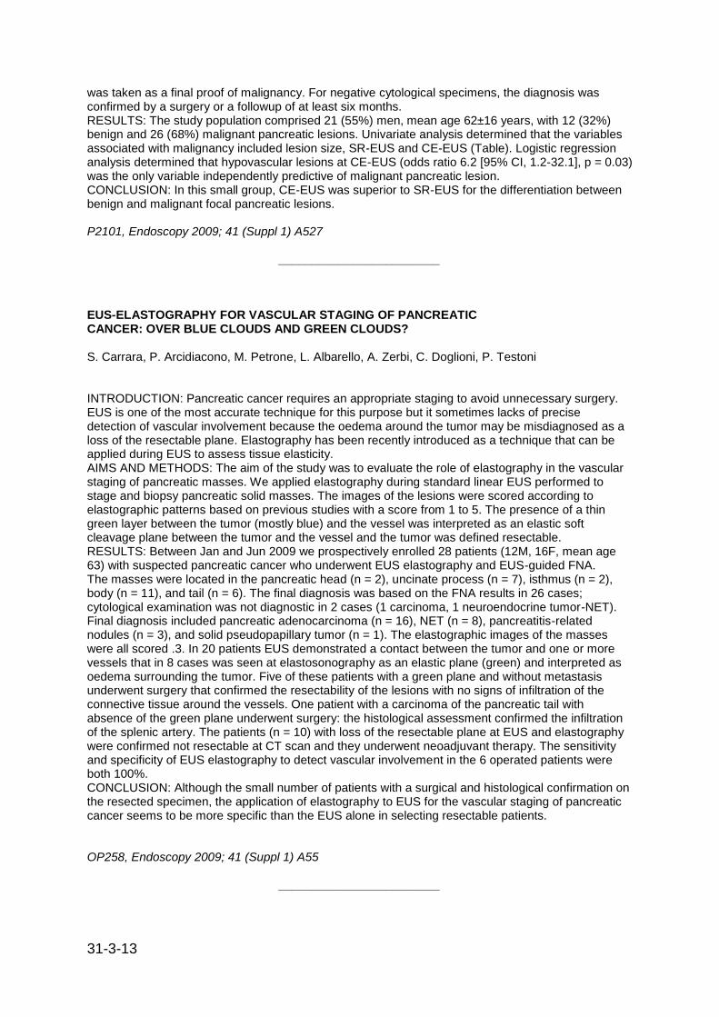
31-3-13
was taken as a final proof of malignancy. For negative cytological specimens, the diagnosis was confirmed by a surgery or a followup of at least six months. RESULTS: The study population comprised 21 (55%) men, mean age 62±16 years, with 12 (32%) benign and 26 (68%) malignant pancreatic lesions. Univariate analysis determined that the variables associated with malignancy included lesion size, SR-EUS and CE-EUS (Table). Logistic regression analysis determined that hypovascular lesions at CE-EUS (odds ratio 6.2 [95% CI, 1.2-32.1], p = 0.03) was the only variable independently predictive of malignant pancreatic lesion. CONCLUSION: In this small group, CE-EUS was superior to SR-EUS for the differentiation between benign and malignant focal pancreatic lesions. P2101, Endoscopy 2009; 41 (Suppl 1) A527
________________________ EUS-ELASTOGRAPHY FOR VASCULAR STAGING OF PANCREATIC CANCER: OVER BLUE CLOUDS AND GREEN CLOUDS? S. Carrara, P. Arcidiacono, M. Petrone, L. Albarello, A. Zerbi, C. Doglioni, P. Testoni INTRODUCTION: Pancreatic cancer requires an appropriate staging to avoid unnecessary surgery. EUS is one of the most accurate technique for this purpose but it sometimes lacks of precise detection of vascular involvement because the oedema around the tumor may be misdiagnosed as a loss of the resectable plane. Elastography has been recently introduced as a technique that can be applied during EUS to assess tissue elasticity. AIMS AND METHODS: The aim of the study was to evaluate the role of elastography in the vascular staging of pancreatic masses. We applied elastography during standard linear EUS performed to stage and biopsy pancreatic solid masses. The images of the lesions were scored according to elastographic patterns based on previous studies with a score from 1 to 5. The presence of a thin green layer between the tumor (mostly blue) and the vessel was interpreted as an elastic soft cleavage plane between the tumor and the vessel and the tumor was defined resectable. RESULTS: Between Jan and Jun 2009 we prospectively enrolled 28 patients (12M, 16F, mean age 63) with suspected pancreatic cancer who underwent EUS elastography and EUS-guided FNA. The masses were located in the pancreatic head (n = 2), uncinate process (n = 7), isthmus (n = 2), body (n = 11), and tail (n = 6). The final diagnosis was based on the FNA results in 26 cases; cytological examination was not diagnostic in 2 cases (1 carcinoma, 1 neuroendocrine tumor-NET). Final diagnosis included pancreatic adenocarcinoma (n = 16), NET (n = 8), pancreatitis-related nodules (n = 3), and solid pseudopapillary tumor (n = 1). The elastographic images of the masses were all scored .3. In 20 patients EUS demonstrated a contact between the tumor and one or more vessels that in 8 cases was seen at elastosonography as an elastic plane (green) and interpreted as oedema surrounding the tumor. Five of these patients with a green plane and without metastasis underwent surgery that confirmed the resectability of the lesions with no signs of infiltration of the connective tissue around the vessels. One patient with a carcinoma of the pancreatic tail with absence of the green plane underwent surgery: the histological assessment confirmed the infiltration of the splenic artery. The patients (n = 10) with loss of the resectable plane at EUS and elastography were confirmed not resectable at CT scan and they underwent neoadjuvant therapy. The sensitivity and specificity of EUS elastography to detect vascular involvement in the 6 operated patients were both 100%. CONCLUSION: Although the small number of patients with a surgical and histological confirmation on the resected specimen, the application of elastography to EUS for the vascular staging of pancreatic cancer seems to be more specific than the EUS alone in selecting resectable patients. OP258, Endoscopy 2009; 41 (Suppl 1) A55
________________________

31-3-13
REAL-TIME TISSUE ELASTOGRAPHY DETERMINES APPROPRIATE THERAPY AND PREDICTS THE PROGNOSIS OF ULCERATIVE COLITIS N. Miyake, T. Ando, O. Watanabe, K. Ishiguro, T. Kato, S. Hibi, S. Mimura, M. Nakamura, R. Miyahara, N. Ohmiya, Y. Hirooka, Y. Niwa, H. Goto Department of Gastroenterology, Nagoya University Graduate School of Medicine, Nagoya City, Aichi, Japan INTRODUCTION: Ulcerative colitis (UC) is a chronic inflammatory disease with frequent remissions and relapses. Although colonoscopic examination is important in determining treatment for patients with UC, it is an invasive procedure and can lead to complications. Non-invasive methods to facilitate evaluation of the large bowel have therefore been sought. Real-time tissue elastography is a new technique that visualizes the differences in tissue strain produced by freehand compression during routine ultrasonography. AIMS & METHODS: The aim of this study was to evaluate the usefulness of real-time tissue elastography in patients with UC. Real-time tissue elastography was performed before colonoscopy in 41 patients with UC. Findings were classified into four types (normal, homogeneous, random, hard) on the basis of color arrangement. Endoscopy findings were classified into four types as follows A: normal mucosa, B: mucosal edema and erosion, C: punched-out ulcer, D: extensive ulcer. We then compared the relationship between real-time tissue elastography and colonoscopy findings, and also investigated whether elastography could reflect clinical stage, therapeutic response, and prognosis of UC patients. RESULTS: On elastography, 13 cases were classified as normal, 15 as homogeneous, 6 as random, and 7 as hard; while on endoscopy, 13 were classified as type A, 18 as type B, 8 as type C, and 2 as type D. We found significant associations between normal type and type A, homogeneous type and type C, random type and type C, and hard type and type D (p < 0.001). Twelve patients (75%) with the normal type and six (66%) with the homogeneous type were in remission, while six (100%) with the random type and five (72%) with the hard type were in the active phase. All patients with the normal and homogeneous types responded to treatment with prednisolone and leukocytapheresis and were induced into remission. In contrast, no patients with the random or hard type responded to this treatment. Following the addition of tacrolimus, cyclosporine A and/or ganciclovir, five (83%) with the random type were induced into remission, whereas remission was achieved in only one (20%) with the hard type and three (50%) required colectomy. Of the 21 patients induced into remission and followed for more than a year, remission was maintained in 13 (76%) of 17 with the normal and homogeneous types and 1 (25%) of 4 with the random and hard types. CONCLUSION: Findings of real-time tissue elastography reflected colonoscopic findings. We consider that real-time tissue elastography may be a useful tool in determining the optimal treatment for and predicting the prognosis of UC patients. P0331, Gut 2009; 58 (Suppl II) A169
________________________
ACCURACY OF ENDOSCOPIC ULTRASOUND ELASTOGRAPHY USED FOR THE DIFFERENTIAL DIAGNOSIS OF CHRONIC PANCREATITIS AND PANCREATIC CANCER: A MULTICENTRIC STUDY. Adrian Săftoiu*, Peter Vilmann, Florin Gorunescu, Uwe Wild, Marc Giovannini, Jan Janssen, Julio Iglesias–Garcia, Paolo Arcidiacono, Michael Hocke, Collin McKay, Dan Ionuţ Gheonea. *Gastroenterology Department, University of Medicine and Pharmacy Craiova, Romania Background: Endoscopic ultrasound (EUS) elastography represents a new imaging procedure that might characterize the differences of hardness and strain between diseased tissue and normal tissue. The method has been used for the differential diagnosis of focal pancreatic masses (pancreatic cancer and chronic pancreatitis) with variable accuracy and contradictory results [1]. Aim: The aim of the study was to assess elastography during EUS examinations of focal pancreatic masses, and to consequently differentiate benign vs. malignant pancreatic masses in a prospective, blinded and multi–center design.
31-3-13
Method: A post–processing software analysis (based on the ImageJ software, NIH, Bethesda, MD, USA) was used to examine the EUS elastography movies by calculating average hue histograms from individual elastography images. The data was further subjected to an extended collaborative neural networks (NNs) computing analysis in order to differentiate benign versus malignant patterns. The study group comprised 125 patients with focal pancreatic masses which were included prospectively in 9 reference centers. For each patient, three separate individual movies of 10 seconds were recorded digitally and uploaded in an on–line database system (Figure 1). Final diagnosis was based on positive cytology results obtained through EUS–guided FNA, final pathology results obtained after surgery, as well as typical imaging findings associated with minimum 6 months of follow–up. Results: The effectiveness of a collaborative computing system, based on a NN approach was assessed in order to provide a real–time decision support for the medical diagnosis. A thorough statistical benchmarking process and a weighted voting system were employed to identify the best NN models as reliable classifiers and to obtain the overall automatic diagnosis. Multi–layer perceptron (MLP) neural networks with both one and two hidden layers of neurons (three–layer perceptron and four–layer perceptron) were trained to learn how to classify focal masses as benign or malignant and yielded an excellent testing performance, together with a high training performance. Consequently, the accuracy of both MLP models was higher than 90%, in accordance with previously published data. However, the NNs approach might provide a very fast and accurate diagnosis supporting and improving the human decision making, especially in difficult cases. Conclusions: EUS elastography is a promising method that allows characterization and differentiation of pancreatic cancer and chronic pancreatitis, especially if the standard methods of diagnosis fail to indicate precisely the benign or malignant nature. A robust methodology based on artificial NNs processing of the digital EUS elastography movies, enabled an optimal prediction of the type of pancreatic lesions. The final results of the study will be analyzed and confirmed as soon as the patient inclusion period ends. References: [1] Săftoiu A, et al., Neural network analysis of dynamic sequences of EUS elastography used for the differential diagnosis of chronic pancreatitis and pancreatic cancer, Gastrointest Endosc., 68:1086–1094, 2008.
Figure 1: Comparative EUS elastography aspects of (A) normal pancreas, (B) chronic pancreatitis and (C) pancreatic cancer Eighth International Conference on the Ultrasonic Measurement and Imaging of Tissue Elasticity, September 14 – 17, 2009, Vlissingen, The Netherlands ENDOSCOPIC ELASTOGRAPHY OF PANCREATIC DISEASE Dr Roald Havre, Dr Svein Ødegaard, Dr Odd Helge Gilja, Dr Lars Birger Nesje University of Bergen, Haukeland University Hospital, Norway Background: Differential diagnosis between neoplastic and inflammatory pancreatic disease is a medical challenge. Sonoelastography applied endoscopically form images of tissue-strain in soft tissues and may contribute to improved tissue characterisation. Material and Method: Twenty four patients admitted for evaluation of neoplastic and inflammatory pancreatic disease of unknown histology were examined with a linear echoendoscope (Pentax 3870 UTK) combined with ultrasound scanning with real-time elastography software (Hitachi EUB 8500/Hi vision 900). Inclusion period: 2006-08, 11 women and 13 men. Age: 16-81 years. Histology was obtained from 19 patients as endoscopic ultrasound guided Fine Needle Aspiration (EUS-FNA) or from surgical biopsy or specimen. In five cases we do not have follow up data (3) or the elastogram was inconclusive (2). Results: Eleven patients had an elastogram showing increased tissue hardness corresponding to a

31-3-13
hypoechoic pancreatic lesion on B-Mode scanning. Nine of these patients had malignant disease and 2 had benign disease. In 8 patients, no increased hardness was visualized corresponding to the pancreas and none of these patients have progressed with malignancy during follow up (4-18 months). Based on the 19 patients, EUS elastography has a sensitivity of visualizing malignancy as harder in 9/9 cases (100 %) and a specificity of 8/10 (80%). Conclusion: EUS Elastography may provide additional information in order to identify malignant lesions in the pancreas. However, distinction between benign and malignant focal lesions remains limited. The Strain-ratio between lesion and normal pancreatic tissue could not separate benign from malignant pancreatic lesions in this material. 12
th World Congress of the World Federation for Ultrasound in Medicine and Biology, 30
th August – 3
rd
September 2009, Sydney, Australia ________________________
ELASTOGRAPHY IN ENDOSCOPIC ULTRASOUND Prof Christoph Dietrich, Caritas-Krankenhaus Bad Mergentheim, University Frankfurt, Germany Endoscopic ultrasound (EUS) is widely regarded as the central discipline in endoscopy assessing a wide range of gastrointestinal diseases. However, differentiation of benign and malignant tissue has remained an unsolved problem and biopsy with cytological or histological confirmation is still the gold standard for tissue characterisation. EUS Real-time Tissue Elastography (Hitachi, Japan) is a recently developed imaging modality that differentiates tissues by their stiffness and has been evaluated in patients with histologically proven pancreatic tumours [Endoscopy 2008;40:910-917], lymph nodes [Endoscopy 2007;39:952-957], anorectal disorders [Endoscopic ultrasound, an introductory manual and atlas. Thieme 2006], and other applications. The technique involves the calculation of tissue elasticity within a sample area which is then displayed as a colour overlay of the real-time B-mode image. The complete colour spectrum from blue to red is used to encode the range of relative elasticity within the sample area for each elastogram. Elastographic and B-mode images are displayed simultaneously. It is of significance that EUS elastography can accurately determine how deeply a tumour has penetrated through the bowel wall. Detection of early cancer infiltration of lymph nodes by examination of their size, shape, and texture has been disappointing with all imaging methods. However, circumscribed and, therefore, early malignant lymph node infiltration, can be more reliably detected by analysing the elastography pattern of these lymph nodes. By combining fine-needle aspiration using curved linear-array instruments, with the use of (contrast-enhanced) ultrasound and elastography, EUS is finally becoming a state-of-the-art, minimally invasive alternative to exploratory surgery in many situations. 12
th World Congress of the World Federation for Ultrasound in Medicine and Biology, 30
th August – 3
rd
September 2009, Sydney, Australia
________________________
REAL-TIME TISSUE ELASTOGRAPHY IN THE DIAGNOSIS OF AUTOIMMUNE PANCREATITIS C. F. Dietrich, T. O. Hirche, M. Ott, A. Ignee Endoscopic ultrasound (EUS) elastography distinguishes tissues on the basis of their specific consistency. The preoperative diagnosis of autoimmune pancreatitis (AIP) is of the utmost importance in order to avoid surgery. The aim of this prospective evaluation of five patients was to investigate the role of this new technique in the characterization of mass lesions caused by AIP, with histology as the gold standard. All five patients with AIP presented with a characteristic stiff elastographic pattern not only of the mass lesion but also of the surrounding pancreatic parenchyma, which was not found in 17 patients with ductal adenocarcinoma and 10 healthy subjects. EUS elastography of the pancreas shows a typical and unique finding with homogenous stiffness of the whole organ, and this distinguishes AIP from the circumscribed mass lesion in ductal adenocarcinoma. Endoscopy 2009; 41: 718–720

31-3-13
DIAGNOSIS OF PANCREATIC DISORDERS USING CONTRAST-ENHANCED ENDOSCOPIC ULTRASONOGRAPHY AND ENDOSCOPIC ELASTOGRAPHY.
Hirooka Y, Itoh A, Kawashima H, Ohno E, Ishikawa T, Matsubara H, Itoh Y, Nakamura M, Miyahara R, Ohmiya N, Niwa Y, Ishigami M, Katano Y, Goto H.
Department of Endoscopy, Nagoya University Hospital, Nagoya City, Aichi Prefecture, Japan. [email protected]
Contrast-enhanced endoscopic ultrasonography (CE-EUS) and EUS-elastography are cutting-edge diagnostic modalities for pancreatic disorders. Each pancreatic disorder has characteristic hemodynamics. CE-EUS uses color Doppler flow imaging to classify pancreatic lesions into a spectrum of solid and cystic patterns. Although there is overlap in the patterns generated by specific types of tumors, some types of tumors tend to produce distinct flow images. EUS-elastography can assess tissue hardness by measuring its elasticity. This parameter appears to correlate with the malignant potential of the lesions. Tissue elasticity studies can provide information on both its pattern and distribution. The former is the conventional method of morphologic diagnosis, but it is restricted to observations made in a region of interest. The latter is an unbiased analysis that can be performed by image analysis software and is theoretically constant, regardless of regions of interest. The evolving modalities of CE-EUS and EUS-elastography might provide clinical utility in the diagnosis of pancreatic disorders.
Clin Gastroenterol Hepatol. 2009 Nov;7(11 Suppl):S63-7.
________________________ ACCURACY OF SECOND-GENERATION ENDOSCOPIC ULTRASOUND (EUS) ELASTOGRAPHY FOR THE DIFFERENTIAL DIAGNOSIS OF SOLID PANCREATIC MASSES: A QUANTITATIVE ANALYSIS OF TISSUE STIFFNESS Julio Iglesias-Garcia, Jose Larino, Enrique Dominguez-Munoz Background: EUS-elastography (EE) allows analyzing tissue stiffness during a standard endoscopic ultrasound examination. Contrarily to the subjective qualitative analysis associated to the first-generation elastography equipments, second-generationelastography allows a quantitative analysis of the tissue stiffness. Aim of the study was to evaluate the accuracy of second generation EUS-elastography for the differential diagnosis of solid pancreatic masses. Methods: 57 consecutive patients(mean age 60 years, 32-84 years, 41 male), who underwent EUS for the evaluation of solid pancreatic masses were prospectively included in the study. EUS-elastography was performed under conscious sedation with the linear Pentax EUS (EG 3830 UT) and the Hitachi EUB 900. Two different areas (A and B) from the region of interest were selected for the quantitative elastographic analysis: Area A is a representative area of the mass and B refers to a soft reference area. The quotient B/A (strain ratio) is considered as the result of the elastographic evaluation. EUS-FNA was performed in all cases for cytological diagnosis. Data are shown as median and 95%CI, and compared by the Student-t test. Diagnostic accuracy of EUS-elastography for detecting malignancy was calculated after drawing the corresponding ROC curves. Results: Size of solid pancreatic masses was 29.7±10.1 mm (mean±SD). Tumors were located in the head of the pancreas in 44 patients, in the body in 10 patients and in the tail in 3 patients. Final diagnosis was pancreatic cancer (PC) in 32 patients and inflammatory mass (IM) in 25 patients. Strain ratio was 16.62 (95%CI 7.26-30.64) in PC and 3.46 (95%CI 1.3-12.6) in IM (p<0.001). Sensitivity and specificity of EUS-elastography (strain ratio) for detecting pancreatic malignancy was 100% and 92%, respectively (cut-off point 5.85). The elastographic value of the mass (area A) was 0.02 (95%CI 0.01- 0.05) in PC and 0.22 (95%CI 0.03-0.49) in IM (p<0.001). The area under the ROC curve for detecting malignancy by the analysis of the elastographic value of A (AUC=0.944) was slightly higher to that obtained by the analysis of the strain ratio (quotient B/A) (AUC=0.910). Conclusion: Second-generation EUS-elastography is a very useful tool for the differential diagnosis of solid pancreatic masses. It allows a quantitative and objective evaluation of tissue stiffness, and thus provides additional information for the detection of malignancy.

31-3-13
Digestive Disease Week, May 30th – June 4
th, 2009, Chicago, USA, 853
________________________
QUANTITATIVE ANALYSIS OF EUS-ELASTOGRAPHY IN PANCREATIC CANCER AND HEALTHY CONTROLS Henning Schrader, Malte Wiese, Mark Ellrichmann, Bjoern A. Menge, Waldemar Uhl, Wolfgang E. Schmidt, Juris J.Meier Introduction: Endoscopic ultrasound (EUS) elastography is a promising new technology for the visualization of tissue elasticity in EUS examinations. Recent data show a high sensitivity in detecting malignant pancreatic lesions, but there are limitations in the technical procedure and evaluation is still subjective. The aim of our study was to develop a quantitative analysis of EUS-elastography in patients with malignant pancreatic masses and controls. Patients and methods: 86 patients (42m, 44w) with pancreatic cancer and 28 healthy controls (11m, 17w) were investigated by EUS-elastography. In all patients diagnosis of malignancy was confirmed by histology or cytology. Over 80% in the patient group suffered from ductal adenocarcinoma. Quantification of elastography data was performed by analysis of EUS videos evaluating histogram values for the colours blue, green and red in a 10s video. A region of interest (tumour or normal pancreas) was defined in each video, and the mean-values for the different colours were measured three times during the video. Data from patients and controls were compared by using an unpaired t-test. Results: Figure 1 shows the video histogram analysis for the colours blue, green and red comparing the cancer and the control group. In pancreatic cancer the colour blue was dominant with a sensitivity of 100%. For normal pancreas the green colour was dominant with a similarly high sensitivity. The analysis of the red colour showed a significant difference for cancer and controls with a higher red colour in normal pancreas, but no absolute separation between the groups. Conclusions: Quantitative analysis of EUS-Elastography allows a highly sensitive discrimination between pancreatic cancer tissue and normal pancreas, especially by evaluating the blue colour. However, since this quantitative analysis is based on the subjective definition of ROI's, investigator with sufficient experience are still required. Thus, quantitative elastography may provide a potent new tool to identify malignant lesions in patients with pancreatic masses. Figure 1
Digestive Disease Week, May 30
th – June 4
th, 2009, Chicago, USA, M1430
________________________

31-3-13
REAL-TIME TISSUE ELASTOGRAPHY DETERMINES OPTIMAL TREATMENT AND PREDICTS THE PROGNOSIS OF ULCERATIVE COLITIS Osamu Watanabe, Takafumi Ando, Kazuhiro Ishiguro, Nobuyuki Miyake, Motofusa Hasegawa, Shinya Kondo, Tsuyoshi Kato, Ryoji Miyahara, Naoki Ohmiya, Yoshiki Hirooka, Yasumasa Niwa, Hidemi Goto Background/Aims: Ulcerative colitis (UC) is a chronic inflammatory disease with frequent remissions and relapses. Although colonoscopic examination is important in determining treatment for patients with UC, it is an invasive procedure and can lead to complications. Non-invasive methods to facilitate evaluation of the large bowel have therefore been sought. Realtime tissue elastography is a new technique that visualizes the differences in tissue strain produced by freehand compression during routine ultrasonography. The aim of this study was to evaluate the usefulness of real-time tissue elastography in patients with UC. Methods: Real-time tissue elastography was performed before colonoscopy in 41 patients with UC. Findings were classified into four types (normal, homogeneous, random, hard) on the basis of color arrangement. Endoscopy findings were classified into four types as follows A: normal mucosa, B: mucosal edema and erosion, C: punched-out ulcer, D: extensive ulcer. We then compared the relationship between real-time tissue elastography and colonoscopy findings, and also investigated whether elastography could reflect clinical stage, therapeutic response, and prognosis of UC patients. Results: On elastography, 13 cases were classified as normal, 15 as homogeneous, 6 as random, and 7 as hard; while on endoscopy, 13 were classified as type A, 18 as type B, 8 as type C, and 2 as type D. We found significant associations between normal type and type A, homogeneous type and type C, random type and type C, and hard type and type D (p<0.001). Twelve patients (75%) with the normal type and six (66%) with the homogeneous type were in remission, while six (100%) with the random type and five (72%) with the hard type were in the active phase. All patients with the normal and homogeneous types responded to treatment with prednisolone and leukocytapheresis and were induced into remission. In contrast, no patients with the random or hard type responded to this treatment. Following the addition of tacrolimus, cyclosporine A and/or ganciclovir, five (38%) with the random type were induced into remission, whereas remission was achieved in only one (20%) with the hard type and three (50%) required colectomy. Of the 21 patients induced into remission and followed for more than a year, remission was maintained in 13 (76%) of 17 with the normal and homogeneous types and 1 (25%) of 4 with the random and hard types. Conclusion: Findings of real-time tissue elastography reflected colonoscopic findings. We consider that real-time tissue elastography may be a useful tool in determining the optimal treatment for and predicting the prognosis of UC patients. Digestive Disease Week, May 30
th – June 4
th, 2009, Chicago, USA, T1430
________________________
ANALYSIS OF EUS ELASTOGRAPHY TO DIFFERENTIATE MASS FORMING PANCREATITIS AND PANCREATIC CANCER BY USING STRAIN RATIO Fumihide Itokawa, Takao Itoi, Atsushi Sofuni, Takayoshi Tsuchiya, Toshio Kurihara, Fuminori Moriyasu INTRODUCTION:Recently, EUS elastography has been reported to supplemental information which can be applied for the diagnosis of pancreatic diseases. However, there is limit in evaluation only for color, and quantification by numerical value is required. AIM:The aim of our study was to evaluate the ability of EUS elastography and quantification by using strain ratio(non mass area/mass area:SR) in order to distinguish mass forming pancreatitis(MFP) from pancreatic cancer(PC). PATIENTS AND METHODS: The subjects were 105 patients performed an endoscopic ultrasound(EUS) for pancreas in our hospital till September 2006 to November 2008.The disease were 6 with mass forming pancreatitis(MFP), 5 with chronic pancreatitis(CP), 61 with pancreatic cancer (PC), 6 with neuroendocrine carcinoma (PNET), 4 with auto immune pancreatitis (AIP), 5 with SCN, 2 with SPN, 1 with Schwanoma, 1 with GIST, 1 with renal cell carcinoma pancreatic metastasis, 7 with IPMN,1 with malignant lymphoma and 5 with normal control. A histological diagnosis by

31-3-13
surgery or endoscopic ultrasonography fine needle aspiration (EUS-FNA) was performed except normal control. The ultrasound was used the HITACHI HI VISION900, and EUS scope was PENTAX EG-3630UR, EG-3670URK and EG-3870UTK. Strain ratio was subsequently performed to choose a mass area and a non-mass area, and the ratio was measured by calculating in real time. RESULTS: Elastography for all PC showed intense blue coloration,which indicated that the mass lesions had malignant aspects.While MFP presented the coloration pattern of mixed green, yellow and low intensity of blue. Normal control was an even application of green to red. The mean SR of MFP and PC were each 23.08±12.65 and 39.08±20.54,respectively, which was significant difference (p<0.05) CONCLUSION: EUS elastography is potentially capable of further defining the tissue characteristics of benign and malignant lesions. This study suggested that it was useful for the quantification by using strain ratio to characterize the tissue hardness of pancreatic disease and distinguish MFP from PC. Digestive Disease Week, May 30
th – June 4
th, 2009, Chicago, USA, W1439
________________________
USEFULNESS OF ENDOSCOPIC ULTRASOUND (EUS) ELASTOGRAPHY FOR THE DETECTION OF MALIGNANT INFILTRATION OF MEDIASTINAL AND ABDOMINAL LYMPH NODES Jose Larino, Julio Iglesias-Garcia, Ana Alvarez-Castro, Jose Mera, Marta Iglesias-Rivas, Ihab Abdulkader, Jeronimo Forteza, Enrique Dominguez-Munoz Background: EUS-elastography allows analyzing tissue stiffness during a standard EUS examination, which may be of help in the differential diagnosis of solid lesions. Detection of malignant infiltration of LN is highly relevant to define the optimal therapeutic strategy for different tumors. We hypothesized that EUS-elastography may provide with additional information to the conventional EUS B-mode for the detection of malignancy in mediastinal and abdominal lymph nodes (LN). The aim of the study was to evaluate the usefulness of EUS-elastography in this setting. Methods: 57 consecutive patients (mean age 64 years, 19-82, 43 males) who underwent EUS for the evaluation of LN were prospectively included in the study. EUS and elastography were performed under conscious sedation by using the linear Pentax EUS (EG 3830 UT) together with the Hitachi EUB 8500 and 900. EUS-guided fine needle aspiration was performed in all cases. Histology of surgical specimens was considered as the reference method in operated cases. Positive cytology for malignancy together with compatible EUS, PET and CT imaging were considered as the reference method to define malignancy in non-operated cases. EUS, PET, CT imaging, clinical presentation and a minimum follow-up of six months were required for final diagnosis of benign disease in cases of benign cytology. Elastographic pattern of the different LN are described. Probability of malignancy according to the EUS-elastographic pattern was calculated. Results: A total of 63 LN were evaluated. Size of LN was 17.4mm as a mean (range 4-59mm). 54 LN were located in the mediastinum, and 9 in the abdomen. Malignancy was confirmed by reference methods in 31 cases, whereas the remaining 32 LN were finally considered as benign. Three different elastographic patterns were identified: 1) a heterogeneous blue-predominant pattern (n=26 LN), 2) a heterogeneous green-predominant pattern (n=23 LN), and 3) a heterogeneous mixed green-blue pattern with geographical appearance and no color predominance (n=14 LN). Malignant LN showed either a blue-predominant pattern (n=24 LN) or a mixed green-blue pattern (n=7 LN). On the contrary, most benign LN (72%) showed a green-predominant pattern. The probability of malignancy in a LN showing a green-predominant pattern was of 0%, and of 92% in case of a blue-predominant pattern. Finally, the probability of malignancy in a LN showing a mixed green-blue pattern was of 50%. Conclusions: EUSelastography is a very useful tool for the differential diagnosis of mediastinal and abdominal LN. It provides with specific colour patterns supporting the malignant or benign nature of the LN. Digestive Disease Week, May 30
th – June 4
th, 2009, Chicago, USA, 231
________________________

31-3-13
TRANSRECTAL ENDOSCOPIC ULTRASOUND (TRUS) ELASTOGRAPHY IN INFLAMMATORY BOWEL DISEASE (IBD) Nadan Rustemovic, Silvija Cukovic-Cavka, Davor Radic, Milorad Opacic, Zeljko Krznaric, Irena Hrstic, Roland Pulanic, Boris Vucelic BACKGROUND AND AIMS: Establishing the diagnosis of Crohn's disease (MC) or ulcerative colitis (UC) sometimes is very difficult. When IBD is confined to the colon, there is a lack of diagnostic tools for distinction between Crohn's colitis and ulcerative colitis, which is especially important in the definitive phenotyping before surgical decision. The aim of this study was to assess the potential role of the TRUS elastography in distinction between MC and UC. The idea is based upon the fact that MC is transmural disease, and UC is limited to the mucosa and submucosa. These tissue characteristics are reflected in differences of the elasticity in rectal and perirectal tissue. Changes in the tissue elasticity can be obtained qualitatively by elastography with different coloures (from red-soft tissue to blue-hard tissue) or quantitatively using strain ratio score. METHODS AND RESULTS: Rectal wall thickness and elastomode of patients were measured by TRUS elastography. Endoscopist was blind for patient diagnosis. SPSS ver. 17 was used for statistical analysis. In pilot study we included 31 patients; 16 patients (52%) with MC and 15 patients (48%) with UC. Average thickness of rectal wall in all study patients was 6.43 mm (± 0.47 SE). In MC group mean rectal thickness was 7.28 mm (± 0.76 SE) compared to 5.52 mm (± 0.44 SE) in UC group. There was no statistical significant difference between MC and UC groups in perirectal thickness (t=1.97, df=29, p=0.058). Perirectal elastomode showed statistically significant difference between this two groups (.2=18.6, df=2, P<0,001). Twelve (75%) patients in MC group had hard elastomode compared to none of patients in UC group, meaning that hard elastogram had positive predictive value of 100% for patients with MC. We also evaluated strain ratio (SR) of rectal tissue. Strain ratio is ratio of strain between two regions of interest (ROI) in the same image. Mucosal tissue was used as first ROI and perirectal tissue as second. SR was measured 3 times and middle value was used in statistical analysis. Sixteen patients were included in our study; 7 (44%) with MC and 9 (56%) with UC. Mean value of SR was higher in MC group (1.07 ± 0.26) then in UC group (0.26 ±0.21). This difference was statistically significant (t=6.85, df=14, p<0.001). CONCLUSION: TRUS elastography provides a valuable information regarding the stiffness of the rectal and perirectal tissue, and can help to differentiate MC from UC. This is a promising new diagnostic tool in the field of IBD. Our study is ongoing and we expect improvement of the method, with increasing number of patients. Digestive Disease Week, May 30
th – June 4
th, 2009, Chicago, USA, W1258
________________________
ENDOSCOPIC ULTRASONOGRAPHY (EUS) STRAIN RATIO (SR-EUS) VS. CONTRAST-ENHANCED EUS (CE-EUS) FOR THE DIAGNOSIS OF FOCAL PANCREATIC SOLID LESIONS Fatima A. Figueiredo, Marc Giovannini, Erwan Bories, Christian Pesenti, Fabrice Caillol, Genevieve Monges, Jean Robert Delpero BACKGROUND: There are many difficulties in the differential diagnosis between pancreatic cancer and chronic pancreatitis despite recent progress. As a means of trying to overcome this in the endossonography field, the measurement of the tissue stiffness (elastography) by new image processors and the use of contrast agent to enhance the color Doppler pattern have been introduced. AIMS: Our aim is to compare prospectively the ability of the SR-EUS and CE-EUS to differentiate between benign and malignant focal pancreatic lesions. METHODS: Thirty-eight patients with a focal pancreatic lesion were included to date. EUS procedures were performed with linear-array echoendoscopes (FG36X or EG38UT, Pentax), an ultrasound platform (Hitachi 7500 or 8500) with an integrated elastography module. In order to obtain the SR-EUS, a circular area is adjusted to the focal lesion and a second one is adjusted to the surrounding tissue. The SR-EUS (focal lesion area strain % / surrounding tissue area strain %) is calculated. After elastography, SonoVue® 2.4 mL (Bracco) was injected intravenously at a rate of 1 ml/sec, following a flash of 10mL saline solution. The pattern of enhancement (hypo or hypervascular) was defined using power Doppler mode. EUS-FNA was performed by using a 22-gauge FNA needle (Echotip, Cook

31-3-13
Endoscopy,). The final diagnosis was based on the histological assessment of the EUS-FNA samples and/or surgical specimens when available. A positive cytological diagnosis was taken as a final proof of malignancy. For negative cytological specimens, the diagnosis was confirmed by surgery or follow-up of at least six months. RESULTS: The study population comprised 21 (55%) men, mean age 62±16 years, with 12 (32%) benign and 26 (68%) malignant pancreatic lesions. Univariate analysis determined that the variables associated with malignancy included lesion size, SR-EUS and CE-EUS (Table). Logistic regression analysis determined that hypovascular lesions at CE-EUS (odds ratio 6.2 [95% CI, 1.2-32.1], p=0.03) was the only variable independently predictive of malignant pancreatic lesion. CONCLUSION: In this small group, CE-EUS was superior to SR-EUS for the differentiation between benign and malignant focal pancreatic lesions.
Univariate analysis: SR = strain ratio CE = contrast enhanced
Digestive Disease Week, May 30th – June 4
th, 2009, Chicago, USA, 922
________________________
DIAGNOSIS OF INVOLVEMENT OF HEPATODUODENAL LIGAMENT AND LYMPHNODES USING EUS-ELASTOGRAPHY IN THE CASES OF BILIARY MALIGNANCIES Yoshiki Hirooka, Akihiro Itoh, Hiroki Kawashima, Toshifumi Kasugai, Eizaburo Ohno, Takuya Ishikawa, Hiroshi Matsubara, Ryoji Miyahara, Yoshiaki Katano, Naoki Ohmiya, Yasumasa Niwa, Hidemi Goto (Background) Real-time tissue elastography™(EG) (Hitachi, Japan) provides a new tissue characterization of degree of hardness in real time. We have performed this procedure over 600 cases for GI diseases using EUS with the function of EG (EUS-EG), and here we will report the diagnostic capability of the involvement of hepatoduodenal ligament (HDL) and lymphnodes (LN) that substantially influence the therapeutic strategy of biliary malignancies. (Patients and Methods) Seventy-five patients with biliary malignancies were enrolled in this study (40 with bile duct cancer and 35 with gallbladder cancer). There were 55 cases with enlarged LN (more than 5mm in diameter) and 20 cases who were estimated whether to have HDL involvement using EUS-EG. As a preliminary study, normal HDL images on EUS-EG were estimated for 10 patients. Diagnostic criteria of the HDL involvement and LN involvement were as follows; Involved HDL images were defined as inhomogeneous hardness based on the normal HDL images which were determined as homogeneous hardness in a preliminary study. Involved LN images were defined as the harder mass than the surrounding connective tissue or as inhomogeneous hardness mass. In this system, both B-mode and EUS-EG images are represented on dual screen at the same time, we can compare both images of same scan plane precisely. EUB-8500 and HV-900 (Hitachi, Japan) as an ultrasound diagnostic machine and EG-3630UR, EG-3670URK and EG-3780UTK (Pentax, Japan) as endosonoscopes were used in this study. (Results) The sensitivity and specificity diagnosing HDL involvement were 89%, 75%, respectively. The extension of inflammation was erroneously depicted as cancer involvement in some cases. The sensitivity and specificity diagnosing LN involvement were 96%, 89%, respectively. The granulomatous change in the enlarged LN was misdiagnosed in some cases. (Conclusion) EUS-EG may add the new information and be a useful method in the diagnosis of involvement of hepatoduodenal ligament and lymphnodes in biliary malignancies. Digestive Disease Week, May 30
th – June 4
th, 2009, Chicago, USA, M1432
________________________

31-3-13
EVALUATION OF LIVER FIBROSIS IN DIFFUSE LIVER DISEASE USING REAL-TIME TISSUE ELASTOGRAPHY Kenji Fujimoto, Chie Tatsumi, Kazuomi Ueshima, Tsuyoshi Shiina, Akiko Tonomura, Tsuyoshi Mitake, Keiji Yamamoto, Masatoshi Kudo, Michio Kato [Objective] Real-time Tissue Elastography (RTE) is developed for visualizing the tissue hardness/softness by using ultrasound. We have been investigating its ability of evaluating fibrosis in diffuse liver disease. Recently, newly developed low frequency probe (EUP-L52) has been applied to RTE, and a patient who had difficulty of visualizing RTE image for the reason of low penetration such as obesity has been improved. In this study, multiple linear regression analysis was performed using several features of RTE image to estimate the RTE fibrosis value, and compared with the fibrosis stage to evaluate the clinical usefulness of RTE. [Material and Method] 26 patients with chronic hepatitis C or liver cirrhosis diagnosed by liver biopsy, and 6 healthy volunteers were examined in this study. The indicated stages of fibrosis wereF0 in 2 patients, F1 in 6 patients, F2 in 8 patients, F3 in 6 patients, and F4 in 4 patients. RTE were performed with HITACHI HI VISION 900 and EUP-L52 linear probe(3-7MHz). Scan was performed through the right intercostal space to observe right lobe. Probe was slightly held to detect the strain by heartbeat. All RTE images were transferred to an external PC, and analyzed with prototype image analysis software. Color data inside the ROI were converted to relative strain value, and features of RTE image such as mean of relative strain value (MEAN), standard deviation of relative strain value (STD), area of blue region (AREA), and complexity of blue region (COM) were calculated. Then, multiple regression analysis was performed with features of RTE image and fibrosis stage. [Results] Features of RTE image were highly correlated with fibrosis stage. Correlation coefficient of MEAN, STD, AREA, and COM were r=-0.604, 0.593, 0.592, and 0.578. With these 4 parameters, multiple regression analysis was performed and derived the regression equation, which significantly fit with the data. RTE fibrosis value was calculated from this equation and had high correlation with fibrosis stage(r=0.729). [Conclusion] As a result of having analyzed RTE quantitatively, the quantity of characteristic reflected staging well. RTE is particularly useful as the modality that can grasp improvement of the fibrosis by a hepatitis diagnosis and the treatment non-invasively. “HI VISION” is a registered trademark of Hitachi Medical Corporation in U.S.A. Digestive Disease Week, May 30
th – June 4
th, 2009, Chicago, USA, M1774
________________________
TISSUE REAL-TIME ELASTOGRAPHY IN AUTOIMMUNE PANCREATITIS Andre I.
1 Michaela 0.
2, Dietrich C.F.
1
1 Department of Internal Medicine II, Caritas Hospital, Bad Mergentheim, Germany
2 Department of
Pathology, Caritas Hospital, Bad Mergentheim; Germany
Introduction: Endoscopic ultrasound (EUS) elastography is a recent imaging procedure that distinguishes tissues due to their specific consistency and has been recently evaluated in patients with histologically proven pancreatic tumours. The aim of this prospective evaluation was to investigate the role of this new technique for characterisation of mass lesions caused by autoimmune pancreatitis.
Summary: Prospective single centre evaluation enrolling 5 patients with mass lesions finally diagnosed as autoimmune pancreatitis. Elastographic recordings were compared to histology as gold standard in all 5 patients. Adequate and reproducible elastographic recordings of the pancreatic lesions could be obtained in all 5 patients with the final diagnosis of autoimmune pancreatitis. Patients with autoimmune pancreatitis presented with a characteristic stiff elastographic pattern not only of the mass lesion but also of the complementary pancreatic tissue which is unique and could not been found in patients with ductal adenocarcinoma and healthy subjects. Final diagnosis was achieved by transcutaneous biopsy in all patients. None of the patients with mass lesions and autoimmune pancreatitis have been operated.

31-3-13
Conclusions: EUS elastography of the pancreas shows a typical and unique finding with homogenous stiffness of the whole organ discriminating this disease from the circumscribed mass lesions in ductal adenocarcinoma and other neoplasia. Knowledge of this finding helps to avoid unnecessary operations.
EuroEUS, April 30 – May 2nd
, 2009, Berlin, Germany
________________________ REAL-TIME SONOELASTOGRAPHY - A NEW APPLICATION IN THE FIELD OF LIVER DISEASE Liana Gheorghe, Speranta Iacob, Cristian Gheorghe Hepatology Department, Center of Gastroenterology and Hepatology, Fundeni Clinical Institute, “Carol Davila” University of Medicine and Pharmacy, Bucharest, Romania Ultrasound elastography is a new imaging technique that allows a noninvasive estimation and imaging of tissue elasticity distribution within biological tissues using conventional real-time ultrasound equipment with modified software. Elastography has been reported to be useful for differentiation and characterization of various malignant tumors, such as breast, prostate, thyroid, pancreas, lymph nodes, gastrointestinal stromal tumors, hepatocellular carcinoma and liver metastasis. Transient and, more recently, real-time elastography has been proved to be useful for noninvasive assessment of liver fibrosis in patients with diffuse liver diseases. Elasticity imaging promises to make an important contribution to ultrasound practice. J Gastrointestin Liver Dis, December 2008 Vol.17 No 4, 469-474
________________________ ENDOSCOPIC ULTRASOUND ELASTOGRAPHY FOR EVALUATION OF LYMPH NODES AND PANCREATIC MASSES: A MULTICENTER STUDY Giovannini Marc, Botelberge Thomas, Bories Erwan, Pesenti Christian, Caillol Fabrice, Esterni Benjamin, Monges Geneviève, Arcidiacono Paolo, Deprez Pierre, Yeung Robert, Schimdt Walter, Schrader Hanz, Szymanski Carl, Dietrich Christoph, Eisendrath Pierre, Van Laethem Jean-Luc, Devière Jacques, Vilmann Peter, Saftoiu Andrian AIM: To evaluate the ability of endoscopic ultrasound (EUS) elastography to distinguish benign from malignant pancreatic masses and lymph nodes. METHODS: A multicenter study was conducted and included 222 patients who underwent EUS examination with assessment of a pancreatic mass (n = 121) or lymph node (n = 101). The classification as benign or malignant, based on the real time elastography pattern, was compared with the classification based on the B-mode EUS images and with the final diagnosis obtained by EUS-guided fine needle aspiration (EUSFNA) and/or by surgical pathology. An interobserver study was performed. RESULTS: The sensitivity and specificity of EUS elastography to differentiate benign from malignant pancreatic lesions are 92.3% and 80.0%, respectively, compared to 92.3% and 68.9%, respectively, for the conventional B-mode images. The sensitivity and specificity of EUS elastography to differentiate benign from malignant lymph nodes was 91.8% and 82.5%, respectively, compared to 78.6% and 50.0%, respectively, for the B-mode images. The kappa coefficient was 0.785 for the pancreatic masses and 0.657 for the lymph nodes. CONCLUSION: EUS elastography is superior compared to conventional B-mode imaging and appears to be able to distinguish benign from malignant pancreatic masses and lymph nodes with a high sensitivity, specificity and accuracy. It might be reserved as a second line examination to help characterise pancreatic masses after negative EUS-FNA and might increase the yield of EUS-FNA for lymph nodes. World J Gastroenterol 2009 April 7; 15(13): 1587-1593
________________________

31-3-13
FEASIBILITY OF TISSUE ELASTOGRAPHY USING TRANSCUTANEOUS ULTRASONOGRAPHY FOR THE DIAGNOSIS OF PANCREATIC DISEASES Uchida H., Hirooka Y., Itoh A., et al. Objectives: We invesigated the feasibility of using real-time tissue elastography (EG) with transcutaneous ultrasonography (EG-US) for pancreatic diseases. Methods: A preliminary study (phase I) and a prospective (phase II) study were conducted. Phase I: subjects were 10 volunteers, 5 with cancer, 2 with endocrine tumor, 5 with chronic pancreatitis, 14 with intraductal papillary-mucinous neoplasm. To determine the charactcistic EG images (diagnostic criteria for phase II), B-mode images were compared with EG images and histopathologic findings. Phase II: 53 consecutive patients were enrolled. The visualization rate by EG-US in lesions visualized by B mode was assessed and the correct diagnosis rate by B-mode alone (B-diagnosis) or in combination with EG-US was evaluated. Results: Phase I: normal parenchyma was a homogeneous color. In cancer, EG-US showed a markedly hard area wih soft spots inside. Endocrine tumor was uniform and soft comparable to parenchyma. Chronic pancreatitis showed a mixture of various colors. Phase II: we identified 77.4% (41/53) of the Iesions and observed 60.0% (15/25) of thc cancers, 100% (3/3) of the endocine tumor, 92.0% (23/25) of the cases of chronic pancreatitis cases on EG- US. The B-diagnosis rates ranged from about 70% - 80%. The diagnosis rates of the combination were more than 90% of lesions of each type. Conclusions: The EG-US is feasible in the diagnosis of pancreatic diseases. Pancreas 2009;38(1):17-22
________________________
INDICATIONS AND LIMITATIONS OF ENDOSCOPIC ULTRASOUND ELASTOGRAPHY FOR EVALUATION OF FOCAL PANCREATIC LESIONS T. O. Hirche, A. Ignee, A. P. Barreiros, D. Schreiber−Dietrich, S. Jungblut, M. Ott, H. Hirche, C. F. Dietrich Background and study aim: Endoscopic−ultrasound−guided elastography (EUS−elastography) is a recently introduced imaging procedure that distinguishes tissues on the basis of their specific consistency. The aim of this prospective study was to investigate the role of this new technique in the characterization and differentiation of focal pancreatic lesions. Patients and methods: This prospective study enrolled 70 patients with unclassified solid lesions of the pancreas and 10 controls with a healthy pancreas. In all patients elastography recordings were compared with cytology/histology findings as the gold standard. Results: Adequate EUS−elastography of the pancreas was performed in all healthy controls but in only 56% of patients with solid pancreatic lesions. The main limitation of elastographic image acquisition was incomplete delineation of the border of lesions greater than 35mm in diameter (39%) or of lesions at some distance from the transducer (10%). Elastographic recordings were also hampered by the fact that the surrounding tissue, which is used as an internal reference standard for strain calculation, was insufficiently displayed in the case of larger lesions. The reduced ratio of target to surrounding tissue resulted in the formation of color artifacts and in impaired reproducibility. In contrast, the majority of lesions smaller than 35mm in diameter were adequately and reproducibly evaluated by EUS−elastography (91%). The clinical use for differential diagnosis, however, seems limited, since strain images from all kinds of pancreatic masses were found to be harder than the surrounding tissues, irrespective of the underlying nature of the lesion (i. e., malignant vs. benign). EUS−elastography predicted the nature of pancreatic lesions with poor diagnostic sensitivity (41%), specificity (53%), and accuracy (45%). Conclusion: EUS−elastography of the pancreas has the potential to obtain some complementary information that would improve tissue characterization. Its clinical utility, however, remains questionable, and it seems unlikely that the information provided will obviate the necessity of obtaining tissue samples for confirmation of a final pathologic diagnosis. Endoscopy 2008; 40: 910-917
________________________

31-3-13
US ELASTOGRAPHY: CURRENT STATUS AND PERSPECTIVES [Article in German] Janssen J. Medizinische Klinik 2, Helios Klinikum Wuppertal. [email protected] (Endo)sonographic real-time elastography is a new method to describe the mechanical properties of tissue. Similar to colour flow Doppler ultrasonography, a region of interest is defined. The relative stiffness of the tissues within this area is described by colours superimposing on the B-mode image. Real-time elastography can be performed with linear scanners for transcutaneous use, rigid endocavitary probes and with flexible echoendoscopes. The probes can be used to compress the tissue. The elasticity modulus is calculated from the resulting deformation of the tissue. In endoscopic ultrasound, arterial and cardiac pulsations or respiratory movements cause the deformation of the tissue that is used for the calculation. Several studies have demonstrated that real-time elastography is feasible and improves the diagnostic accuracy for tumours of the breast, the prostate, the cervix, and the thyroid gland. Endosonographic elastography has been employed in the examination of lymph nodes and the pancreas. For the differentation between benign and malignant lymph nodes, the accuracy is reported to be 85 % to 90 %. Therefore, the method seems to be useful to select lymph nodes suitable for biopsy. The elastographic pattern of malignant tumours of the pancreas is different from that of the normal pancreas, but similar to that of chronic pancreatitis due to the same biomechanical architecture. Therefore, the early diagnosis of cancer within chronic pancreatitis will probably not be improved by elastography. In summary, (endo)sonographic real-time elastography is a promising new method. Nevertheless, prospective studies are needed to define useful applications and the clinical significance of the method. Gastroenterol. 2008 Jun;46(6):572-9.
________________________ NEURAL NETWORK ANALYSIS OF DYNAMIC SEQUENCES OF EUS ELASTOGRAPHY USED FOR THE DIFFERENTIAL DIAGNOSIS OF CHRONIC PANCREATITIS AND PANCREATIC CANCER Adrian Saftoiu, MD, PhD, Peter Vilmann, MD, PhD, Florin Gorunescu, PhD, Dan Ionut¸ Gheonea, MD, Marina Gorunescu, PhD, Tudorel Ciurea, MD, PhD, Gabriel Lucian Popescu, Eng, MSc, Alexandru Iordache, Eng, Hazem Hassan, MD, Sevastit¸a Iordache, MD Craiova, Romania, Copenhagen, Denmark Background: EUS elastography is a newly developed imaging procedure that characterizes the differences of hardness and strain between diseased and normal tissue. Objective: To assess the accuracy of real-time EUS elastography in pancreatic lesions. Design: Cross-sectional feasibility study. Patients: The study group included, in total, 68 patients with normal pancreas (N Z 22), chronic pancreatitis (N = 11), pancreatic adenocarcinoma (N = 32), and pancreatic neuroendocrine tumors (N = 3). A subgroup analysis of 43 cases with focal pancreatic masses was also performed. Interventions: A postprocessing software analysis was used to examine the EUS elastography movies by calculating hue histograms of each individual image, data that were further subjected to an extended neural network analysis to differentiate benign from malignant patterns. Main Outcome Measurements: To differentiate normal pancreas, chronic pancreatitis, pancreatic cancer, and neuroendocrine tumors. Results: Based on a cutoff of 175 for the mean hue histogram values recorded on the region of interest, the sensitivity, specificity, and accuracy of differentiation of benign and malignant masses were 91.4%, 87.9%, and 89.7%, respectively. The positive and negative predictive values were 88.9% and 90.6%, respectively. Multilayer perceptron neural networks with both one and two hidden layers of neurons (3-layer perceptron and 4-layer perceptron) were trained to learn how to classify cases as benign or malignant, and yielded an excellent testing performance of 95% on average, together with a high training performance that equaled 97% on average. Limitation: A lack of the surgical standard in all cases.

31-3-13
Conclusions: EUS elastography is a promising method that allows characterization and differentiation of normal pancreas, chronic pancreatitis, and pancreatic cancer. The currently developed methodology, based on artificial neural network processing of EUS elastography digitalized movies, enabled an optimal prediction of the types of pancreatic lesions. Future multicentric, randomized studies with adequate power will have to establish the clinical impact of this procedure for the differential diagnosis of focal pancreatic masses. Gastrointest Endosc 2008;68:1086-94.
_______________________
EUS ELASTOGRAPHY FOR PANCREATIC MASS LESIONS: BETWEEN IMAGE AND FNA? Chronic pancreatitis and pancreatic cancer are major causes of pancreatic disease–related morbidity and mortality. Because these diseases often occur concurrently, detection of focal cancer in a background of inflammation can be especially difficult. The diagnosis and management of both chronic pancreatitis and pancreatic cancer rely heavily upon endoscopic and radiologic imaging. The main techniques include CT; magnetic resonance imaging (MRI); ERCP; and EUS, especially EUS-guided sampling methods (EUS-guided FNA [EUS-FNA] or EUS-guided Trucut biopsy [EUS-TCB]). Continued improvements in the technology and use of uniform diagnostic criteria have increased our ability to obtain accurate staging, diagnosis, and prognosis. Among these techniques, EUS and EUS-FNA and/or EUS-TCB have proven to be very powerful techniques to accurately diagnose chronic pancreatitis and pancreatic cancer. For EUS evaluation of pancreatic cancer, EUS-FNA is considered a safe, accurate, and efficient procedure; however, multiple biopsies (on average 5-7) are usually required, because of the inability to specifically target tumor cells that are admixed with inflammation and/or fibrosis.1 When compared with other imaging modalities, the results of EUS-FNA of pancreatic masses are excellent and achieve a sensitivity of 85% to 90% and a specificity of virtually 100%.2-4 However, lower (<75%) sensitivity is reported when there is a coexistent presence of chronic pancreatitis or „„pseudotumoral‟‟ pancreatitis. 5,6 This is an important issue, because approximately 20% to 35% of patients who undergo EUS-FNA of pancreatic lesions have features of underlying chronic pancreatitis. 7,8 Given these limitations, new technologies, eg, EUS elastography, which measures tissue stiffness, are being investigated. Elastography uses sonic and US waves to ompress tissue. Tissue that is fibrotic and stiff will compress less than softer, healthy tissue.9 Because the malignant and chronic inflammatory tissue is usually harder than the adjacent normal tissue, by measuring the tissue strain induced by compression, we can estimate tissue hardness, which may be useful to distinguish pancreatic cancer and chronic pancreatitis compared with normal tissue.10 In this month‟s issue of Gastrointestinal Endoscopy, Saftoiu et al11 describe the accuracy of real-time EUS elastography in a group of 68 patients, hoping to differentiate between normal pancreas (n = 22), chronic pancreatitis (n=11), pancreatic cancer (n=32), and neuroendocrine tumors (n=3). In their study, they used clinical history and imaging criteria to define „„normal pancreas,‟‟ and cytologic diagnosis, surgery, or 6-month clinical follow-up to distinguish malignancy from „„pseudotumoral‟‟ pancreatitis. The investigators then performed a postprocessing analysis, based on neural networks, to analyze the EUS elastography video sequences. The degree of tissue stiffness was indicated by the elastography and was graphically represented by a color-hue scale. By using a cutoff of 175 for the mean hue histogram values recorded on the region of interest, differentiation of benign and malignant masses was achieved, with a high sensitivity (91.4%), specificity (88.9%), and accuracy (90.6%). Furthermore, in a subgroup analysis, good sensitivity (93.8%) and overall accuracy (86%) was reported to differentiate pancreatic cancer from „„pseudotumoral‟‟ pancreatitis. However, the specificity was low (63.6%). When using a more stringent cutoff, of 190, specificity increased to 90.9%, and a positive predictive value of 94.7% was demonstrated but at the sacrifice of sensitivity. These results suggest that this technique can be complementary to EUS in its ability to improve the diagnostic accuracy of pancreatic mass lesions of chronic pancreatitis versus pancreatic cancer, which is a major limitation of EUS-FNA. EUS elastography has previously been shown to distinguish benign and malignant pancreatic lesions and lymph nodes.12,13 However, a study by Janssen et al10 reported that EUS elastography has limited accuracy (60%) for diagnosing chronic pancreatitis. This is likely because these studies used qualitative electrographic analyses (predominant color, distribution of colors, and constancy of pattern), which are subject to interobserver variation. In addition, chronic pancreatitis may also have some degree of tissue stiffness similar to pancreatic cancer, which can make the differentiation difficult.13 Saftoiu et al11 were able to overcome this
31-3-13
limitation by developing a quantitative, thus objective, image interpretation algorithm by using a sophisticated statistical method called artificial neural network analysis. Artificial neural network analyses are computational analytical tools that are inspired by the biological nervous system. These consist of networks of highly interconnected computer processors called „„neurons,‟‟ which are capable of performing parallel computations for data processing and knowledge representation.14 Just the way the human brain processes information, a neural network learns through repeated adjustments of the weighted interconnections and can infer complex data. There are several issues that should be considered about this study. Perhaps the most significant issue is of placing this technology in the context of our current clinical algorithm. Most patients with pancreatic cancer are treated with chemotherapy or surgical resection. For chemotherapy, a tissue diagnosis is a nearly absolute requirement. It is unlikely that any imaging methods, particularly one with a specificity of <95%, will replace biopsy. For potentially surgical patients, the main issue is to exclude „„nonsurgical‟‟ diseases, such as autoimmune pancreatitis, metastatic cancers to the pancreas, or chronic pancreatitis. In this regard, the current study is unlikely to change current clinical practice. The main value may be to guide biopsy in cases where there is diffuse inflammatory change, to possibly reduce the number of biopsies needed, and to further exclude disease in patients with a low probability of cancer, such as those with an equivocal CTor MRI (eg, „„prominent head of pancreas‟‟ in the absence of a defined mass). Another value, although not specifically evaluated in this study, may be to distinguish normal pancreas from chronic pancreatitis. Although EUS has generally been shown to be accurate,15-17 there is only modest interobserver agreement among expert endosonographers (k=0.45).18 An objective measure, such as elastography, may provide some needed standardization. Other efforts to standardize EUS criteria for chronic pancreatitis include the Rosemont classification, which gives different weights to each EUS criteria in predicting the diagnosis of chronic pancreatitis.19 Future studies should compare traditional methods, weighted qualitative methods, and new quantitative methods, such as EUS elastography. Other key technical issues to resolve with elastography are how to integrate it into real-time EUS. In the current study, all images were analyzed off-line under more ideal circumstances. As with other image interpretation methods, the technology must provide the information at the time a decision must be rendered, namely, at the time of the procedure. Also, it would be very helpful to consider applying elastography to other methods of imaging the pancreas, such as MRI. Recent advances in MRI-elastography, particularly for detection of fibrosis and/or cirrhosis of the liver, are highly promising and have the potential to offer an even less-invasive diagnosis of chronic pancreatitis.20 In summary, the use of EUS with different complementary invasive and noninvasive techniques is evolving and aims to provide accurate differentiation of pancreatic masses. The use of neural network analysis of dynamic sequences of EUS-elastography is a promising new technique that may provide important additional information to distinguish benign and malignant pancreatic lesions. The optimal role of this technique is likely to guide EUS-FNA to regions most likely to harbor cancer and to exclude cancer in patients with a low probability. Further investigation will be required to determine these outcomes. Kanwar R. S. Gill, MD Michael B. Wallace, MD, MPH Department of Gastroenterology and Hepatology Mayo Clinic Jacksonville, Florida, USA Abbreviations: EUS-FNA, EUS-guided FNA; EUS-TCB, EUS-guided trucut biopsy; MRI, magnetic resonance imaging. REFERENCES 1. Chang KJ, Albers CG, Erickson RA, et al. Endoscopic ultrasound-guided fine needle aspiration of pancreatic carcinoma. Am J Gastroenterol 1994;89:263-6. 2. Eloubeidi MA, Jhala D, Chhieng DC, et al. Yield of endoscopic ultrasound-guided fine-needle aspiration biopsy in patients with suspected pancreatic carcinoma. Cancer 2003;99:285-92. 3. Gress F, Gottlieb K, Sherman S, et al. Endoscopic ultrasonographyguided fine-needle aspiration biopsy of suspected pancreatic cancer. Ann Intern Med 2001;134:459-64. 4. Harewood GC, Wiersema MJ. Endosonography-guided fine needle aspiration biopsy in the evaluation of pancreatic masses. Am J Gastroenterol 2002;97:1386-91. 5. Varadarajulu S, Tamhane A, Eloubeidi MA. Yield of EUS-guided FNA of pancreatic masses in the presence or the absence of chronic pancreatitis. Gastrointest Endosc 2005;62:728-36; quiz 751, 753.
31-3-13
6. Ardengh JC, Lopes CV, Campos AD, et al. Endoscopic ultrasound and fine needle aspiration in chronic pancreatitis: differential diagnosis between pseudotumoral masses and pancreatic cancer. JOP 2007;8:413-21. 7. Brand B, Pfaff T, Binmoeller KF, et al. Endoscopic ultrasound for differential diagnosis of focal pancreatic lesions, confirmed by surgery. Scand J Gastroenterol 2000;35:1221-8. 8. Fritscher-Ravens A, Brand L, Knofel WT, et al. Comparison of endoscopic ultrasound-guided fine needle aspiration for focal pancreatic lesions in patients with normal parenchyma and chronic pancreatitis. Am J Gastroenterol 2002;97:2768-75. 9. Itoh A, Ueno E, Tohno E, et al. Breast disease: clinical application of US elastography for diagnosis. Radiology 2006;239:341-50. 10. Janssen J, Schlorer E, Greiner L. EUS elastography of the pancreas: feasibility and pattern description of the normal pancreas, chronic pancreatitis, and focal pancreatic lesions. Gastrointest Endosc 2007;65:971-8. 11. Saftoiu A, Vilmann P, Gorunescu F, et al. Neural network analysis of dynamic sequences of EUS elastography used for the differential diagnosis of chronic pancreatitis and pancreatic cancer. Gastrointest Endosc 2008;68:1086-94. 12. Giovannini M, Hookey LC, Bories E, et al. Endoscopic ultrasound elastography: the first step towards virtual biopsy? Preliminary results in 49 patients. Endoscopy 2006;38:344-8. 13. Fritscher-Ravens A. Blue clouds and green clouds: virtual biopsy via EUS elastography? Endoscopy 2006;38:416-7. 14. Ramesh AN, Kambhampati C, Monson JR, et al. Artificial intelligence in medicine. Ann R Coll Surg Engl 2004;86:334-8. 15. Sahai AV, Zimmerman M, Aabakken L, et al. Prospective assessment of the ability of endoscopic ultrasound to diagnose, exclude, or establish the severity of chronic pancreatitis found by endoscopic retrograde cholangiopancreatography. Gastrointest Endosc 1998;48:18-25. 16. Wiersema MJ, Hawes RH, Lehman GA, et al. Prospective evaluation of endoscopic ultrasonography and endoscopic retrograde cholangiopancreatography in patients with chronic abdominal pain of suspected pancreatic origin. Endoscopy 1993;25:555-64. 17. Buscail L, Escourrou J, Moreau J, et al. Endoscopic ultrasonography in chronic pancreatitis: a comparative prospective study with conventional ultrasonography, computed tomography, and ERCP. Pancreas 1995;10:251-7. 18. Wallace MB, Hawes RH, Durkalski V, et al. The reliability of EUS for the diagnosis of chronic pancreatitis: interobserver agreement among experienced endosonographers. Gastrointest Endosc 2001;53:294-9. 19. Hernandez LV, Sahai A, Brugge WR, et al. Standardized weighted criteria for EUS features of chronic pancreatitis: the Rosemont classification [abstract]. Gastrointest Endosc 2008;67:AB96-7. 20. Talwalkar JA, Yin M, Fidler JL, et al. Magnetic resonance imaging of hepatic fibrosis: emerging clinical applications. Hepatology 2008;47:332-42. Editorial, Volume 68, No. 6 : 2008 GASTROINTESTINAL ENDOSCOPY 1097
________________________
INDICATIONS AND LIMITATIONS FOR ENDOSONOGRAPHY WITH ELASTOGRAPHY IN PATIENTS WITH PANCREATIC DISEASE Ignee A, Hirche TO, Barreiros AP, Schreiber-Dietrich D, Ott M, Dietrich CF Internal Medicine 2, Caritas Hospital, Bad Mergentheim, Germany; Medical Clinic 1, Johann Wolfgang Goethe University Clinic, Frankfurt, Germany;Paediatrics, Caritas Hospital, Bad Mergentheim, Germany;Pathology, Caritas Hospital, Bad Mergentheim, Germany Objectives: Recently, endoscopic ultrasound has been using elastography to provide an image-guided method for distinguishing tissue with regard to its specific consistency. The objective of this prospective study was to examine to what extent this new technology could be used to differentiate and characterise normal pancreatic tissue and focal pancreatic diseases. Patients and methods: Prospective study, 108 patients with unclassified pancreatic lesions and 10 patients with a healthy pancreas. The elastographic results were compared with histology as the gold standard. Results: It was only possible to document adequate elastographic examinations for 30% of the study population. The fundamental limitation was the restricted penetration depth, which did not allow for

31-3-13
reproducible elastographic depiction for lesions with diameters above 30 mm (58% of the study population). The second most common reason was artefacts (15%), primarily in patients with calcified chronic pancreatitis and cystic lesions. By contrast, it was possible to carry out adequate and reproducible elastograms on all healthy organs (soft to intermediate specimens) and on the majority of small pancreatic masses < 30 mm (hard specimens). The clinical benefit of differential diagnosis is slight as all pancreatic masses, which were harder than the surrounding pancreatic tissue, were represented irrespective of their nature. The only exception was patients with autoimmune pancreatitis for whom a consistently harder specimen was detected, which enabled these patients to be distinguished from patients with other entities. Conclusion: Endosonography with elastography provides additional and complementary information for improved tissue characterisation in patients with pancreatic disease. The clinical benefit remains unclear.
Ultraschall in Med 2008;29:S134 - S135. (translated from German)
________________________ FREEHAND REAL-TIME ELASTOGRAPHY: IMPACT OF SCANNING PARAMETERS ON IMAGE QUALITY AND IN VITRO INTRA- AND INTEROBSERVER VALIDATIONS. Havre RF, Elde E, Gilja OH, Odegaard S, Eide GE, Matre K, Nesje LB.
National Centre for Ultrasound in Gastroenterology, Department of Medicine, Haukeland University Hospital, Bergen, Norway; Institute of Medicine, University of Bergen, Bergen, Norway.
Real-time elastography is a method for visualization of the elastic properties of soft tissue and may potentially enable differentiation between malignant and benign pathologic lesions. Our aim was to validate the method on a tissue-mimicking (TM) phantom and to evaluate the influence of different scanning parameters and investigator variability. A TM-phantom containing eight spherical inclusions with known storage modulus was examined using two different transducers on an ultrasound (US) scanner equipped with software for real-time elasticity imaging. The ultrasound transducers were moved vertically in a repetitive manner to induce strain. Two investigators performed series of standardized elastography scans applying a 0-4 categorical quality scale to evaluate the influence of seven parameters: dynamic range of elasticity, region-of-interest, frequency of transducer movement, rejection of elastogram noise, frame rate, persistence and smoothing. Subsequently, repeated examinations of four selected inclusions were performed using a visual analog scale (VAS) where investigators marked a 100 mm horizontal line representing the span in image quality based on experience from the first examination. The hardest and softest inclusions were imaged more clearly than the inclusions with elasticity more similar to the background material. Intraobserver agreement on elastogram quality was good (kappa: 0.67 - 0.75) and interobserver agreement average (kappa: 0.55 - 0.56) when using the categorical scale. The subsequent VAS evaluation gave intraclass-correlation coefficients for the two observers of 0.98 and 0.93, respectively, and an interclass-correlation coefficient of 0.93. Real-time elastography adequately visualized isoechoic inclusions with different elastic properties in a TM-phantom with acceptable intra- and interobserver agreement. Dynamic range of elasticity was the parameter with most impact on the elastographic visualization of inclusions.
Ultrasound Med Biol. 2008 Oct;34(10):1638-50 .
________________________ ENDOSCOPIC ULTRASOUND ELASTOGRAPHY IN THE DIFFERENTIAL DIAGNOSIS OF PANCREATIC SOLID MASSES: TOWARDS THE VIRTUAL BIOPSY Julio Iglesias-Garcia, Jose Larino-Noia, Enrique Dominguez-Munoz Endoscopic ultrasound elastography (EUS-E) allows analyzing tissue stiffness during a standard endoscopic ultrasound(EUS) examination, thus providing with additional information about the features of solid lesions. The aim of the study was to evaluate the elastographic patterns of solid pancreatic masses and the diagnostic accuracy of EUS-E in this setting.
31-3-13
METHODS: 80 consecutive patients (mean age 62 years, 24-84 y., 54 males) who underwent EUS-E for the evaluation of solid pancreatic masses, and 10 controls with normal pancreas (mean age 59 years, 20- 83 y., 5 males), were prospectively included in the study. EUS-E was performed under conscious sedation by the linear Pentax EG 3830 UT and Hitachi EUB 8500. EUS-FNA was performed for cytological diagnosis after elastographic examination. The different elastographic patterns of solid pancreatic masses are described. Diagnostic sensitivity, specificity, PPV, NPV and overall accuracy of EUS-E for malignancy were calculated compared to final diagnosis based on cytology, histology of surgical specimens and clinical follow-up. RESULTS: Mean size of pancreatic masses was 31.7 ± 13.6 mm. Tumors were located in the head of the pancreas in 61 patients, in the body in 16 and in the tail in 3. Final diagnosis was pancreatic cancer (PC) in 48 cases, inflammatory mass (IM) in 23 patients, neuroendocrine tumor (NT) in 8 cases and metastatic tumor in 1 patient. 4 different patterns could be defined: 1) Homogeneous green, 2) heterogeneous green-predominant with yellow and red lines, 3) heterogeneous blue-predominant with a geographic appearance and green and red lines, and 4) homogeneous blue. A homogeneous green pattern was only observed in normal pancreas. 18 masses showed a heterogeneous green-predominant pattern, and all of them were IM. Thus, the probability of IM is 100% in the presence of this pattern. A heterogeneous, geographically distributed, blue-predominant pattern was found in 54 cases. This pattern was observed in all cases of malignant tumor (either PC or metastasis) and in 5 cases of IM. Thus, the probability of malignancy in the presence of this pattern is of 90.1%. A homogeneous blue pattern was exclusively seen in the 8 patients with NT. Thus, the probability of NT is 100% in the presence of this pattern. Sensitivity, specificity, PPV, NPV and overall accuracy for malignancy were 100%, 78.3%, 91.9%, 100% and 93.7% respectively. CONCLUSION: EUS-E is a very useful tool for the differential diagnosis of solid pancreatic masses. This technique adds important information to EUS evaluation by providing with highly specific atterns, which strongly support the benign or malignant nature of the disease. Digestive Disease Week, May 17
th – 22
nd, 2008, San Diego, USA (366)
________________________
NEURAL NETWORK ANALYSIS OF DYNAMIC SEQUENCES OF EUS ELASTOGRAPHY USED FOR THE DIFFERENTIAL DIAGNOSIS OF CHRONIC PANCREATITIS AND PANCREATIC CANCER Adrian Saftoiu, Peter Vilmann, Florin Gorunescu, Dan Ionut Gheonea, Marina Gorunescu, Tudorel Ciurea, Gabriel Lucian Popescu, Alexandru Iordache, Sevastita Iordache BACKGROUND Endoscopic ultrasound (EUS) elastography is a newly developed imaging procedure that characterizes the differences of hardness and strain between diseased tissue and normal tissue. AIM The aim of our study was to prospectively assess the accuracy of EUS elastography to differentiate between normal pancreas (N=22), chronic pancreatitis (N=11), pancreatic adenocarcinoma (N=32) and pancreatic neuroendocrine tumors (N=3). A post-processing analysis based on specially designed software was used to analyze the EUS elastography movies by calculating hue histograms of each individual image. Furthermore, a neural network analysis based on average hue histograms of the EUS elastography movies was tested in order to differentiate benign versus malignant EUS elastography patterns. RESULTS Based on the study group of 68 patients, the sensitivity, specificity and accuracy of differentiation of benign and malignant cases were 91.4%, 87.9% and 89.7%, based on a cut-off of 175 for the mean hue histogram values recorded inside the region of interest. The positive predictive value and negative predictive value were 88.9% and 90.6%, respectively. A multi-layer perceptron (MLP) neural network was trained to learn how to classify cases as benign or malignant, and yielded a very good testing performance of 95% in average, together with a high training performance equaling 97% in average. CONCLUSIONS EUS elastography is a promising method that allows characterization and differentiation of normal pancreas, chronic pancreatitis and pancreatic cancer. The method might prove useful for the differentiation of pancreatic tumors, as well as to target the EUS-FNA biopsy into harder regions suspicious of malignancy. The currently developed methodology based on artificial neural network processing of the EUS elastography digitalized movies, enabled an optimal prediction of the pancreatic lesions type. Future multicentric, randomized studies with adequate power will have

31-3-13
to establish the clinical impact of this procedure for the differential diagnosis of focal pancreatic masses. Digestive Disease Week, May 17
th – 22
nd, 2008, San Diego, USA (671)
________________________
ENDOSCOPIC ELASTOSONOGRAPHY IS HIGHLY PREDICTIVE OF DEFINITE PATHOLOGY Jan-Werner Poley, E. J. Kuipers BACKGROUND: Elastosonography is a technique that allows for real-time assessment of elasticity or stiffness of tissue with the aid of conventional ultrasound instruments. In recent years this technique has been developed for endoscopic ultrasound (EUS). Preliminary data show that it might be a useful adjunct to standard morphological criteria to define a lesion without FNA sampling, or to select the optimal location for FNA to increase the yield of tissue sampling. To further elucidate this hypothesis we evaluated the performance of elastosonography in series of patients referred for EUS. METHODS: patients were examined by an experienced endosonographer with a linear echoendoscope (Pentax 3830-UT) combined with a Hitachi EUB-8500 system to allow for realtime processing of elastonography images. Based on these images lesions were classified as either benign, malignant or indeterminate based on previously published data. A definite diagnosis was made on results of either EUS-FNA, surgical pathology or clinical follow-up (at least six months). RESULTS: 41 procedures were performed in the same number of patients (M/F 17/24, age range 26 to 74 yrs): 25 procedures were done for mediastinal pathology, 13 for pancreatic masses, 1 each for rectal, adrenal and submucosal lesions. Elastosonograpy images were interpreted as benign in 10 cases (24%), indeterminate in 7 cases (17%), and malignant in 24 cases (59%). All patients with a benign result on elastosonography had benign disease during follow-up and EUS-FNA. Of the patients with lesions that were judged to be indeterminate on elastosonography (n=7; 17%) 5 had benign disease based on clinical follow-up and EUS-FNA whereas 2 had malignant disease based on EUS-FNA. In the group with malignant elastosonography (n= 24) one patient had benign disease. A comparison of the elastography classes benign plus indeterminate versus malignant yielded a sensitivity of elastonosonography of 92%, a specificity of 94%, and negative and positive predictive values of respectively 96% and 88%. CONCLUSION: Elastosonography is highly sensitive and specific in determining the nature of both mediastinal and pancreatic lesions with excellent positive and negative predictive values and can therefore be helpful in guiding EUS-FNA and patient management. Digestive Disease Week, May 17
th – 22
nd, 2008, San Diego, USA, (760)
________________________
ENDOSCOPIC ULTRASOUND ELASTOGRAPHY FOR THE UPPER GASTROINTESTINAL TRACT DISEASE Ryoji Miyahara, Yasumasa Niwa, Masanao Nakamura, Yoichi Iguchi, Yoshiko Kodama, Kakunori Banno, Osamu Maeda, Takafumi Ando, Akihiro Itoh, Naoki Ohmiya, Yoshiki Hirooka, Hidemi Goto BACKGROUND & AIM: It is known that some diseases, such as cancer, lead to changes in the hardness of tissue. Endoscopic Ultrasound Elastography (EUE) is a new technique to assess the elasticity of tissue. It is expected to improve the ability for diagnosis. The aim of this study was to evaluate EUE to differentiate between benign and malignant gastrointestinal masses and lymph nodes. PATIENT & METHOD: From July 2006 to September 2007, 35 patients underwent EUE examinations. Nine patients underwent evaluation of esophageal cancer, 7 of gastric cancer, 14 of submucosal tumor, 2 of mediastinal lymph nodes, which all cases were diagnosed histopahologically after examinations. RESULT: All cases could be drawn images of EUE, which can be classified into three patterns. Type 1 is a common finding among the normal walls of digestive tract, which showed about the same hardness as nearby fatty tissues and was demonstrated from red to green on the images. Type 2 and 3 were image findings of tumors or inflammatory diseases. Both were demonstrated to be harder than

31-3-13
nearby adipose tissues. We classified the cases demonstrated mosaic-like into type 2 and ones demonstrated uniformly into type 3. In the stomach or esophageal cancer cases, a neoplasm part was drawn as type 2 or type 3 and change of hardness was closely similar to the loupe image of pathology. About the lymph node including stomach or esophageal cancer cases, the malignant lymph node was drawn as type3, but the inflammatory lmph node the view of type 2. In the submucosal tumor, GIST was shown as type 3 and aberrant pancreas was shown as type1. No complication occurred with EUE examinations. CONCLUSION: EUE could make it much easier to diagnose benign or malignancy of extrinsic lymph nodes. In the field of diagnosis of invasion depth of stomach or esophageal tumor, we have impression that the border in deep parts becomes clearer. Digestive Disease Week, May 17
th – 22
nd, 2008, San Diego, USA (M1402)
________________________
USEFULNESS OF THE CHARACTERIZATION OF TISSUE HARDNESS OF PANCREATIC MASS USING ELASTOGRAPHY ENDOSCOPIC ULTRASOUND. -FIRST TRIAL OF THE QUANTIFICATION USING STRAIN RATIO- Itokawa Fumihide, Takao Itoi, Fuminori Moriyasu, Atsushi Sofuni, Toshio Kurihara, Kentarou Ishii, Syuujirou Tsuji, Nobuhito Ikeuchi, Takashi Kawai INTRODUCTION: Cancer is starting the change of tissue hardness by its own fibrosis from early stage. The reconstruction of tissue elasticity provides the sonographer with important additional information which can be applied for the diagnosis of these diseases. AIM: The aim of our study was to evaluate the ability of endoscopic ultrasound elastography to differentiate between benign and malignant pancreatic masses. PATIENTS AND METHODS: The subjects were 53 patients performed an endoscopic ultrasound(EUS) for pancreas in our hospital till September 2006 to October 2007. The disease were 5 with tumor forming pancreatitis(TFP) ,32 with pancreatic cancer(PC),1 with GIST,1 with renal cell carcinoma pancreatic metastas, 1 with IPMA and 13 with normal control. A histological diagnosis by surgery or endoscopic ultrasonography fine needle aspiration (EUS-FNA) was performed for all subjects. The ultrasound was used the HITACHI HI VISION900, and EUS scope was PENTAX EG-3630UR and EG-3670URK.The calculation of tissue elasticity distribution is performed in realtime and the results are represented in color over the radial B-mode image. Malignant tissue appeared in blue color, fibrosis in blue to green, and normal tissue in green to red. In addition, we performed the quantification by using strain ratio(non mass area/mass area:SR) in order to evaluate the objective hardness as numerical value between mass to non mass area especially to distinguish TFP from PC. RESULTS: Elastography for all PCs showed intense blue coloration,which indicated that the mass lesions had malignant aspects.While TFP presented the coloration pattern of mixed green, yellow and low intensity of blue. GIST and renal cell carcinoma pancreatic metastasis presented green and yellow patern. Normal control was an even application of green to red. The mean SR of TFP and PC were each 24.99±12.54 and 42.08±21.57,respectively,which was significant difference(p<0.05) CONCLUSION: EUS elastography is potentially capable of further defining the tissue characteristics of benign and malignant lesions. This study suggested that it was useful for the quantification by using strain ratio to characterize the tissue hardness of pancreatic disease and distinguish TFP from PC. Digestive Disease Week, May 17
th – 22
nd, 2008, San Diego, USA (M1443)
________________________

31-3-13
INDICATIONS AND LIMITATIONS OF TISSUE REAL-TIME ELASTOGRAPHY USING ENDOSCOPIC ULTRASOUND IN PANCREATIC DISEASE IgneeA
I, Hirche T
2, Barreiros AP
I, Schreiber-Dietrich D
1, Dietrich CF
I,
I Medical Department Caritas Hospital Bad Mergentheim, Germany;
2 Medical Department, Goethe
Universtity, Frankfurt, Germany Aim: Sonoelastography is an imaging procedure that distinguishes tissues due to their specific consistency. The aim of this prospective study was to investigate the role of this new technique for characterisation and differentiation of normal pancreatic parenchyma and pancreatic tissues affected by various focal diseases. Patients and methods: Prospective single center study, enrolling 108 patients with unclassified focal lesions of the pancreas and ten controls. EIastographic recordings were compared to histology as gold standard in all patients. Results: Adequate elastographic recordings of the pancreatic lesions could be obtained in 30% of our study population. This was mainly due to limited depth penetration (58%), artifacts (15%), that were particularly prominent in patients with calcified chronic pancreatitis and cystic lesions. In contrast, adequately and reproducible elastographic recordings were obtained from all healthy pancreatic organs (soft/intermediate pattern) and the majority of lesions smaller than 30 mm independent of their dignity (hard pattern). Patients with autoimmune pancreatitis presented with a unique stiff elastographic pattern that allowed for discrimination from all other disease entities. Conclusion: EUS elastography of the pancreas has the potential to achieve complementary information for improved tissue characterization especially in autoimmune pancreatitis. Ultraschall in Med, 2008, suppl 1, OP1.30 XXth Congress of European Federation of Societies for Ultrasound in Medicine and Biology/XIth
Romanian Conference of Ultrasound in medicine and Biology, May 31 – June 3 2008, Timisoara, Romania.
________________________
ENDOSONOGRAPHIC ELASTOGRAPHY IN THE DIAGNOSIS OF MEDIASTINAL LYMPH NODES J. Janssen, C. F. Dietrich, U. Will, L. Greiner Background and study aims: Ultrasonographic elastography is a new technique for describing the mechanical properties of tissue during real−time ultrasonography. The aim of this study was to test the feasibility of this method in endosonography (EUS) of the dorsal mediastinum, and to compare the elastographic patterns of lymph nodes with results from EUS−guided fine−needle aspiration biopsy (FNAB). Patients and methods: 50 consecutive patients undergoing EUS−guided FNAB of at least one paraesophageal lymph node were included. Each of these targeted lymph nodes was examined also elastographically. The elastographic patterns were described and compared with the histologic results by a first examiner. The elastographic classification was subsequently further tested by two blinded reviewers. Results: In total, 66 lymph nodes were examined; 37 lymph nodes revealed benign and 29 malignant tissue at the histologic evaluation. Good elastographic records were obtained for all lymph nodes. Of the 37 benign lymph nodes, 31 showed a homogeneous pattern of intermediate elasticity, whereas a dominance of hard tissue with variable patterns was found in 23 of 29 malignant lymph nodes. Applying these criteria, the accuracy range among the three examiners was between 81.8% and 87.9% for benign lymph nodes and between 84.6% and 86.4% for malignant ones. The interobserver agreement was excellent (kappa = 0.84). Conclusion: EUS elastography of mediastinal lymph nodes can be performed reliably. The results are good for a noninvasive technique, but they remain inferior to the success rate of EUS− guided FNAB. The method might occasionally be useful for targeting the most suitable lymph nodes for FNAB. Endoscopy 2007; 39: 952-957
________________________

31-3-13
DYNAMIC ANALYSIS OF ENDOSCOPIC ULTRASOUND ELASTOGRAPHY USED FOR THE DIFFERENTIATION OF CHRONIC PANCREATITIS AND PANCREATIC CANCER
A. Saftoiu 1, P. Vilmann
2, T. Ciurea
1, G. Popescu
3, A. Iordache
3, D. Gheonea
1, H. Hassan
2, F.
Gorunescu4.
1Gastroenterology Department, Uniuersity of Medicine
and Pharmacy, Craiova, Romania; 2
Department of Surgical Gastroenterology, Gentofte Uniuersity Hospital. Hellerup, Denmark;
3 IT Center,
4 Biostatistics Department. Uniuersity of Medicine and
Pharmacy, Craioua, Romania
INTRODUCTION: Endoscopic ultrasound elastography is a new imaging procedure that allows the reconstruction of elasticity distribution by characterizing the difference of hardness between diseased tissue and normal tissue. AIMS & METHODS: The aim of our study was to describe EUS characteristics encountered in 28 patients prospectively included with normal pancreas (N = 3), chronic pancreatitis (N ~ 5), pancreatic adenocarcinoma (N = 17) and pancreatic neuroendocrine tumors (N = 3). The pattern of real-time EUS elastography images was compared with the conventional EUS aspects, while the final diagnosis was obtained by EUS-FNA cytology analysis, by surgical pathology or minimum 6 months followup. RESULTS: EUS elastography patterns were compared in patients with pancreatic cancer, neuroendocrine tumors, chronic pancreatitis and normal pancreas. Post-processing analysis based on hue histograms of the EUS elastography average images was also done, yielding a semi-quantitative method to quantify the elasticity of the pancreatic tissue. The movies were analysed and filtered by computer-enhanced dynamic analysis using a public domain Java-based image processing tool (ImageJ) developed at the National Institutes of Health. The sensitivity, specificity and accuracy of differentiation of pancreatic adenocarcinoma and neuroendocrine tumors were 80.0%, 91.7% and 84.4%, based on a cut-off of 175 for the mean hue histogram values recorded on the region of interest. The positive predictive value and negative predictive value were 94.1 % and 73.3%. CONCLUSION: EUS elastography is a promising method that allows characterization and differentiation of normal pancreas, chronic pancreatitis and pancreatic cancer. The method might prove useful for the differentiation of pancreatic tumors, as well as to target the EUS-FNA biopsy into harder regions suspicious of malignancy. However, a post-processing analysis through the use of special software is required to correctly characterize the patients, while future studies on an increased number of patients are awaited.
Endoscopy 2007; 39 (Suppl I) A32. 15th United European Gastroenterology Week, October 29
th – 31
st , 2007, Paris, France
________________________
USEFULNESS OF THE CHARACTERIZATION OF TISSUE HARDNESS OF PANCREATIC MASS USING ELASTOGRAPHY ENDOSCOPIC ULTRASOUND
F. Itokawa, T. Itoi, F. Moriyasu, A. Sofuni.
Tokyo Medical University Hospital, Gastroenterology and hepatology, Tokyo, Japan
INTRODUCTION: Cancer is starting the change of tissue hardness by its own fibrosis from early stage. The reconstruction of tissue elasticity provides the sonographer with important additional information which can be applied for the diagnosis of these diseases.
AIMS & METHODS: The aim of our study was to evaluate the ability of endoscopic ultrasound elastography to differentiate between benign and malignant pancreatic masses. The subjects were 32 patients performed an endoscopic ultrasound (EUS) for pancreas in our hospital till September 2006 to April 2007. The disease were 4 with tumor forming pancreatitis (TFP), 20 with pancreatic cancer(PC), I with GIST and 7 with normal control. A histological diagnosis by surgery or endoscopic ultrasonography fine needle aspiration (EUS-FNA) was performed for all subjects. The ultrasound was used the HITACHI HI VISION900, and EUS scope was PENTAX EG-3630UR and EG-3670URK. The calculation of tissue elasticity distribution is performed in realtime and the results are represented in color over the radial B-mode image. Malignant tissue appeared in blue color, fibrosis in blue to green, and normal tissue in green to red. In addition, we performed the quantification by using strain ratio (non mass area / mass area: SR) in order to evaluate the objective hardness as numerical value between mass to non mass area especially to distinguish TFP from Pc.
RESULTS: Elastography for all PCs showed intense blue coloration, which indicated that the mass

31-3-13
lesions had malignant aspects. While TFP presented the coloration pattern of mixed green, yellow and low intensity of blue. GIST presented green and yellow pattern.
Normal control was an even application of green to red. The mean SR of TFP and PC were each 25.23±11.06,and 46.53±19.48, respectively, which was significant difference(p ~ 0.0026) CONCLUSION: EUS elastography is potentially capable of further defining the tissue characteristics of benign and malignant lesions. This study suggested that it was useful for the quantification by using strain ratio to characterize the tissue hardness of pancreatic disease and distinguish TFP from PC. Gut 2007; 56 (Suppl /II) AI81 15th United European Gastroenterology Week, October 29
th – 31
st , 2007, Paris, France
________________________
ENDOSONOGRAPHIC ELASTOGRAPHY OF THE ANAL SPHINCTER – A PROSPECTIVE STUDY H Allgayer
1, CF Dietrich
2
1 Rehaklinik Ob der Tauber, Digestive diseases, Bad Mergentheim, Germany
2 Caritas-Krankenhaus, Internal Medicine, Bad Mergentheim, Germany
(Translated from German)
Background: Inflammatory conditions and tumours can lead to an alteration of the tissue elasticity of organs, which can be confirmed and displayed with Elastography (so-called SELA) as a colour overlay of the B-mode. For certain tumours and inflammatory diseases it has been proven that SELA can provide the physician with potentially important information. We examined patients suffering from rectal incontinence to determine if the Endosonographic Elastography (so-called e-SELA) appearances of the anal sphincter correlate to the functional parameters as well as if it is in relation to traditional incontinence treatment.
Method: 30 Patients suffering from rectal incontinence (n=19 Rectum/Sigmoid resection, n=5 Crohn‟s disease, n=6 other). After evaluation of the rectal incontinence by using incontinence scores (CACP) and measuring manometer functional parameters, an anal 2D-Endosonography (EUS) examination of the internal/external sphincter (IS/ES) was performed with SELA (SonoElastography) (Hitachi EUB-8500). A semi-quantitative evaluation was made with a colour scale from 0-3: red (soft) through to blue (hard). After 2 weeks bio feedback / pelvic floor training, a second evaluation was carried out.
Results: The elasticity of the internal sphincter was significantly higher than the elasticity of the external sphincter, as a result, the hardness of the external sphincter was significantly higher that the hardness of the internal sphincter. There was a negative correlation by stimulated pressure on the anal sphincter but not by non- stimulated pressure.
The hardness (blue colouring) of the external sphincter was clearly higher for men that for women (2.0±0.4 vs. 1.0±0.4, p<0.031) and decreases with age, with significantly high values in the age category from 40-49 years (2.0±0.44) in comparison with the age range from 70-79 years. (1.0±0.83, p<0.02).
Conclusion: This examination shows that the E-SELA provides important additional information in relation to clinical significant sphincter characteristics such as elasticity/ hardness. Further clinical studies (to evaluate the effect of treatment and treatment decision) should be determined in prospective controlled studies.
XIXth Congress of European Federation of Societies for Ultrasound in Medicine and Biology & Dreiländertreffen, 24
th – 27
th October 2007, Leipzig, Germany
________________________

31-3-13
ELASTOSONOGRAPHY IN MALIGNANT RECTAL DISEASE: PRELIMINARY DATA G. Mezzi, P. G. Arcidiacono, S. Carrara, C. Boemo, P. A. Testoni: Elastography is an ultrasound technique that allowed to obtain images from the mechanical properties of soft tissue. It is well known that some diseases, such as cancer, lead to changes in tissue hardness. Elasticity imaging is a technique that reveals the physical properties of tissue and can determine changes in tissue hardness caused by disease [1±5]. We evaluated 20 consecutive patients (10 males, 10 females; mean age 61.15 years, range 29±84 years) with histologically confirmed distal rectal cancer for disease staging. Informed consent was given by all of subjects. All patients underwent rectal endoscopic ultrasound, which was performed using a flexible echo endoscope (an oblique−viewing instrument [fiber obtic or electronic video image] [Pentax, Hamburg, Germany]). The echondoscope was inserted and advanced beyond the lesion, under direct vision, to the rectosigmoid junction. Tumors were targeted to determine the depth of infiltration into or through the rectal wall. Frequencies commonly used for T−staging range from 7.5 MHz to 9 MHz. The endoscopic ultrasound elastography score was subdivided into five types on the basis of the echopattern distortion of the examined area, from 1 (normal) to 5 (advanced malignant neoplasia) (l" Figure 1). Rectal staging was performed according to Tumor−Node−Metastasis staging system (TNM). It was our experience that the elastography images showed a discreet correlation in the staging of advanced lesions (T3, 65%); and post−radiotherapy disease persistence has been correctly confirmed by elastography score (T3, 100%). These preliminary data have shown that elastogra− phy, performed during endoscopic ultrasound, is a method that can differentiate between benign and malignant rectal lesions, although further studies, in particular regarding the follow−up of neoadjuvant chemoradiation therapy and in benign lesions, are necessary. References 1 Frey H. Realtime elastography: a new ultrasound procedure for the reconstruction of tissue elasticity (in German, English abstract). Radiologe 2003; 43: 850±855 2 Lorenzen J, Sinkus R, Adam G. Elastography: quantitative imaging modality of the elastic tissue properties [in German, English abstract]. Rofo 2003; 175: 623±630 3 Cochlin DL, Ganatra RH, Griffiths DF. Elastography in the detection of prostatic cancer. Clin Radiol 2002; 57: 1014±1020 4 Saftoiu A, Vilman P. Endoscopic ultrasound elastography: a new imaging technique for the visualization of tissue elasticity distribution. J Gastrointest Liver Dis 2006; 15: 161±165 5 Giovannini M, Hookey LC, Bories E et al. Endoscopic ultrasound elastography: the first step towards virtual biopsy? Preliminary results in 49 patients. Endoscopy 2006; 38: 344±348 G. Mezzi Department of Gastroenterology and Gastrointestinal Endoscopy IRCCS Vita−Salute University, San Raffaele Scientific Institute of Milan Via Olgettina 60, 20132 Milan, Italy

31-3-13
Endoscopy 2007; 39: 375
________________________
EUS ELASTOGRAPHY OF THE PANCREAS: FEASIBILITY AND PATTERN DESCRIPTION OF THE NORMAL PANCREAS, CHRONIC PANCREATITIS, AND FOCAL PANCREATIC LESIONS Jan Janssen, MD, Eva Schlörer, MD, Lucas Greiner, MD Wuppertal, Germany Background: Initial clinical applications have shown that US elastography might be able to distinguish tissues because of their specific consistency. Objective: (1) To investigate the feasibility of EUS elastography of the pancreas and (2) to describe elastographic patterns of the normal pancreas and the pancreas affected by inflammatory or focal disease. Design: Prospective single-center study. Setting: Academic center of the University of Witten/Herdecke. Patients: Twenty patients with normal pancreas, 20 patients with chronic pancreatitis, and 33 patients with focal pancreatic lesion, histologically later proven in 32 of these 33 cases. Interventions: Commercially available US equipment was used. The elasticity of tissue was reconstructed in real time within a sample area and was translated into a color scale imaging relative tissue elasticity within this area. Representative loops of at least 20 seconds were recorded regarding each region of interest. Results: Adequate elastographic recordings could be obtained in all 73 patients. Patients with hypoechoic and intermediately echogenic normal pancreas revealed a relatively homogeneous elastographic pattern. Thirty-one focal lesions, including 30 neoplasms and most of the chronically inflamed pancreata had a honeycomb pattern dominated by hard strands. This pattern showed analogies to the histologic structure of 10 resected tumors. Other patients with chronic pancreatitis and those with hyperechoic healthy pancreas had miscellaneous elastographic appearances. Conclusions: EUS elastography of the pancreas is feasible and produces plausible results. The examination of homogeneous tissue is impaired by the relative scale used. Chronic pancreatitis and hard tumors cannot be distinguished by elastography, probably because of their similar fibrous structure. Gastrointest Endosc 2007;65:971-8.
________________________

31-3-13
DYNAMIC ANALYSIS OF EUS USED FOR THE DIFFERENTIATION OF BENIGN AND MALIGNANT LYMPH NODES Adrian Saftoiu, Peter Vilmann, Tudorel Ciurea, Gabriel Lucian Popescu, Alexandru Iordache, Hazem Hassan, Florin Gorunescu, Sevastita Iordache, (Hellerup, Denmark, Craiova, Romania) Background: EUS elastography was reported to offer supplemental information that allows a better characterization of tissue characteristics and that might enhance conventional EUS imaging. Objective: Our purpose was to apply real-time elastography during EUS examinations and to assess the accuracy of the differentiation of benign versus malignant lymph nodes. Design: Prospective cross-sectional feasibility study. Setting: Department of Surgical Gastroenterology, Gentofte University Hospital, Hellerup, Denmark. Patients: Patients diagnosed by EUS with cervical, mediastinal, or abdominal lymph nodes were included, with a total number of 78 lymph nodes examined. The final diagnosis of the type of lymph node was obtained by EUS FNA cytologic analysis or by surgical pathologic examination and by a minimum 6 months of follow-up. Interventions: Hue histogram analysis of the average images computed from EUS elastography movies was used to assess the color information inside the region of interest and to consequently differentiate benign and malignant lymph nodes. Main Outcome Measurements: Differentiate between malignant and benign lymph nodes. Results: By using mean hue histogram values, the sensitivity, specificity and accuracy for the differential diagnosis were 85.4%, 91.9%, and 88.5%, respectively, on the basis of a cutoff level of 166 (middle of green-blue rainbow scale). The proposed method might be useful to avoid color perception errors, moving artifacts, or possible selection bias induced by analysis of still images. Limitations: Lack of the surgical standard in all cases. Conclusions: Computer-enhanced dynamic analysis based on hue histograms of the EUS elastography movies represents a promising method that allows the differential diagnosis of benign and malignant lymph nodes, offering complementary information added to conventional EUS imaging. Gastrointestinal Endoscopy, Volume 66, No. 2 : 2007, 291 – 300
________________________
PRESSED FOR AN ANSWER: HAS ELASTOGRAPHY FINALLY COME TO EUS? Brian C. Jacobson, MD, MPH, Section of Gastroenterology, Boston University Medical Center Boston, Massachusetts, USA In this issue of Gastrointestinal Endoscopy, Saftoiu et al present their findings from a prospective study of elastography used during EUS evaluation of benign and malignant lymph nodes. The study represents a step forward in the transition of this fascinating technology from an investigational tool to a clinically meaningful test. In 1991, Ophir et al coined the term „„elastography‟‟ to describe an imaging technique that conveyed information about a tissue‟s relative firmness in response to compression, meaning it is more akin to palpation than inspection. Various forms of elastography exist, being used within the fields of optics, magnetic resonance imaging, and US. In the area of US, elastography can be based upon a tissue‟s response to compression to measure tissue deformation (compression elastography), a tissue‟s displacement in response to short bursts of vibration (transient elastography), or a tissue‟s effects on vibration amplitudes in response to continuous vibration (vibration elastography). Gastroenterologists may already be familiar with transient elastography in the form of the FibroScan (Echosens, Paris, France), a device that uses US to measure the effects of transient vibrations through the liver to measure the organ‟s stiffness or degree of fibrosis. But to better understand compression elastography, it is prudent to first review how US images themselves are created. Recall that a US transducer contains specialized (piezoelectrical) crystals that vibrate at specific frequencies when exposed to an alternating electrical field. These vibrations emit a pulse of sound waves that travel into target tissues, some of which ultimately „„bounce‟‟ back off the tissues. Immediately after launching this burst of waves, the crystals become „„ears,‟‟ awaiting the return of reflected sound waves (echoes) bouncing back from the tissues. Upon arrival, returning waves set the crystals vibrating once again, and these vibrations are converted to electrical

31-3-13
impulses used to construct an image. The returning echoes, which permit creation of an image, carry key information about the tissues. First, the time it takes for the echo to return conveys how far away, or deep, that structure is from the transducer. Second, the echo‟s intensity reflects the difference in densities (impedance) at the interface between adjacent structures off of which the sound wave „„bounced.‟‟ The traditional B („„brightness‟‟) mode images we are used to seeing in EUS convey each echo‟s intensity on a black and white (mostly gray) scale. Many single beams of echoes are then aligned to construct the familiar 2-dimensional image. In „„classic‟‟ compression elastography, stress is applied to the tissue by pressing the transducer slightly harder against the body. In the study by Saftoiu et al, however, the stress applied was simply the mild compression that tissues experience during respiration and vascular pulsations. Stress applied to a tissue results in strain or deformation within the tissue. Harder tissues deform less than softer tissues, and, as discussed, US echoes carry information about tissue location and density, both of which change slightly with deformation. Thus, by comparing echoes made with and without compression, or simply over several seconds of normal breathing and blood circulation, one can obtain information about how hard or soft the tissues are relative to their surroundings. Calculations of the relative elasticity of all tissues within a viewing trapezoid-shaped window (similar to Doppler imaging) are made in real time by the EUS processor, and a false-color overlay is superimposed on the B-mode images. In the current study, the software expressed elasticity in numerical values that ranged from 0 to 255 and used a rainbow spectrum of color in the overlay to provide visual cues about the tissues‟ hardness. Firm tissues appeared blue, soft tissues appeared red, and those in the intermediate range fell somewhere in the blue-green spectrum. How can we exploit this information? We know that cancer tends to be firmer than normal tissue. Therefore, Saftoiu et al, in addition to other endosonographers, attempted to use elastography to differentiate benign from malignant tissue based on EUS imaging. In the current study, the investigators focused on lymph nodes and compared EUS imaging criteria (ie, node size, shape, border, and echotexture characteristics), EUS-guided FNA results, and elastography findings. Their criterion standard was a composite of FNA results, surgical specimen findings, and clinical course. By using receiver operating characteristic curves, they determined that an elasticity cutoff of 166 yielded the greatest accuracy in distinguishing benign from malignant nodes. Not surprisingly, EUS criteria performed poorly in differentiating benign from malignant nodes, as others have demonstrated. FNA performed quite well, although this is in the context of being used as part of its own criterion standard. But, the exciting finding in the study was a reasonable sensitivity (85%) and good specificity (92%) for elastography when using the elasticity value of 166 to diagnose malignancy. The overall prevalence of malignant denopathy in the study was 53%, so the positive and negative predictive values of elastography were 92% and 85%, respectively. This means elastography in this study had a positive likelihood ratio (LR) of 10.5 and a negative LR ratio of 0.16. What does this mean clinically? A positive LR of 10.5 means that a positive test changes the disease probability by approximately 45%. So, if you think a node has a 50% likelihood of being malignant based on EUS criteria or just the clinical context (eg, a 2-cm mediastinal node adjacent to a T3 esophageal cancer) and elastography suggests it is malignant, then the node is 95% likely to be malignant. Therefore, besides helping you choose which nodes to preferentially aspirate, elastography may prove useful in staging nodes that are deemed inaccessible, perhaps because of intervening tumor or vessels. A negative LR of 0.16 changes the post test probability of disease approximately –35%. So, a node with a 50% pretest probability of harboring cancer is only about 15% likely to be positive if elastography suggests benignity. Is this cutoff value of 166 reliable? Not yet. The investigators are the first to acknowledge that this was based upon a single, relatively small data set and must be validated prospectively among new patient populations. Furthermore, there may be different optimal cutoffs based upon a node‟s location. For example, elastography must compare a node‟s hardness with the surrounding tissues to determine its response to compression. It is possible that the supporting structures in the mediastinum are different than those in the abdomen, and, therefore, future studies must determine if computer algorithms should consider a node‟s location when choosing the cutoff value for diagnosing malignancy. A potential major limitation to elastography is the degree of fibrosis that may be present in benign tissue, making the tissue firm and decreasing the accuracy of the test for diagnosing malignancy. For example, do benign reactive mediastinal nodes in a chronic smoker contain sufficient fibrosis to cause false-positive results for cancer? Furthermore, just how much cancer must be present to make a lymph node harder than usual? We are always concerned about false negative FNA results when only very small amounts of cancer are present within a lymph node. It is unclear whether elastography would prove to be any more sensitive than FNA. In fact, there were 2 false-negative FNAs in the

31-3-13
current study, but the investigators did not discuss the elastography findings from those cases. It would be interesting to know whether elastography classified those cases as positive for malignancy. The investigators should be commended for tackling another important shortcoming of elastography, namely the difficulty in using a single image for calculating degrees of elasticity. This is problematic, because an arbitrary choice of image could introduce bias (the endosonographer may preferentially choose the „„bluer‟‟ frames when most other frames depict a red hue). This would also create problems with inter- and intraobserver variation. To overcome this problem, the investigators integrated several seconds‟ worth of images into a computerized summation value to calculate the elasticity within the region of interest. They also developed software to drop images with hue distributions outside a reliable range, thereby excluding images most likely to represent artifact. These new protocols or some variation upon them will probably be incorporated into newer generations of the software used for elastography. Are there other foreseeable EUS uses for this technology? One can imagine that the elastography images of a gastric GI stromal tumor might be different from that of a pancreatic rest but probably not too different from a leiomyoma, limiting its utility in subepithelial lesions. Tiny intraductal probe-based systems are unlikely to reliably distinguish benign from malignant biliary or pancreatic strictures, because these are always firm, regardless of malignancy or benignity. Perhaps serous cystadenomas of the pancreas can be distinguished from mucinous cystic neoplasms based upon the viscosity of the cyst contents, a bit of an EUS „„Holy Grail‟‟ if you will. Avoiding any FNA that carries a risk of pancreatitis is a laudable goal. In the end, this technology is likely suited for exactly what these investigators are doing: trying to improve our ability to stage malignancy by either providing a „„virtual biopsy‟‟ or, more likely, guiding our needles to improve FNA accuracy and decreasing the number of FNA passes. The relative simplicity of applying elastography during EUS suggests that, if the „„pesky details,‟‟ eg, reliability, can be worked out, we are likely to see this technology develop into a standard adjunct to EUS. Just how pressed we will be to adopt elastography will undoubtedly depend upon the firmness of future data. Gastrointestinal Endoscopy Volume 66, No. 2 : 2007, 301-2
________________________
QUALITY ASSESSMENT OF FREE-HAND SONOELASTOGRAPHY IN VITRO
Roald Flesland Havre, Erlend Elde, Odd Helge Gilja Svein Ødegaard, and Lars B. Nesje National Centre for Ultrasound in Gastroenterology, Dept. of Medicine, Haukeland University Hospital, and Institute of Medicine, University of Bergen, Bergen, Norway.
Introduction: Sonoelastography is used to visualise elastic properties in soft tissue, and the method seems to have a promising ability to differentiate between malignant and benign pathologic lesions. We have evaluated the influence of different scanning parameters and the investigator performance on the quality of free-hand sonoelastography in vitro. Material and method: A tissue mimicking phantom containing eight spherical inclusions of known elastic moduli was examined with 7.5 Hz and 9 MHz linear transducers connected to an ultrasound scanner with sonoelastography programme (Hitachi EUB-8500). The probes were moved in a sinusoidal, repetitive manner to induce strain. Two investigators made separate scan series applying first a 0-4 cathegoric scale and a subsequently a Visual Analogue Scale (VAS) to score 7 parameters: dynamic range of elasticity (E-dyn), region of interest (ROI), frequency of transducer movement, rejection of elastogram noise, frame rate, persistence and smoothing. Summary: The hardest and softest inclusions were imaged more clearly than those with elasticity close to the background material. Intraobserver agreement in image quality assessment was good (Kappa: 0.67-0.75) and interobserver agreement average when using the categoric scale (Kappa: 0.55-0,56). The subsequent VAS evaluation showed better intra- and interobserver agreement with Intra-class correlation coefficients (Intra-CC) 0.983 and 0.931 (95%CI) and Interclass correlation coefficient (Inter-CC): 0.932 (95%CI). Conclusion: Sonoelastography adequately visualised isoechoic inclusion bodies in a TM-phantom with good intra- and interobserver agreement. The quality of the elastograms were mostly influenced by dynamic range of elasticity and this variable must be taken into account when scanning humans. EuroEUS, 4
th- 5
th May, 2007, Seville, Spain

31-3-13
ENDOSCOPIC SONOELASTOGRAPHY – INITIAL EXPERIENCE.
Roald Flesland Havre, Svein Ødegaard, Odd Helge Gilja, Eva Fosse, and Lars Birger Nesje National Centre for Ultrasound in Gastroenterology, Dept. of Medicine, Haukeland University Hospital, and Institute of Medicine, University of Bergen, Bergen, Norway. Background: Sonoelastography is an ultrasound method to visualise elastic properties of soft tissue based on the distribution of strain in response to stress. Tissue patholgy may alter the elastic properties and malignancy seems to generate harder tissue in most cases. Hence, sonoelastography may be a valuable tool in differentiating malignancy from other pathologic changes. Material & Method: We performed sonoelastography in 23 patients using a linear echoendoscope (Pentax GF3830-UT) and a Hitachi EUB-8500 ultrasound scanner. On admission, 18 of the patients had suspected malignant tumour/metastasis in the mediastinum (9), pancreas (4), oesophagus (2) or other locations(3). Five patients without suspected malignancy had chronic pancreatitis (2) or a myogenic tumour in upper GI-tract (3). EUS-FNA was performed in 8 patients and EUS-guided tru-cut biopsy in 4 patients. Summary: Seven tumours with suspected malignancy appeared harder than the surrounding tissue, all of which proved to be carcinomas. Two large GISTs with malignant changes presented with mixed elastograms. Six suspected tumours failed to present areas with harder tissue, 3 of these were sampled with no malignancy and none showed indications of malignancy during follow-up. One malignant mesotelioma of the pleura appeared softer than the reference tissue. One pancreatic cancer and one case of pancreatitis generated inconclusive sonoelastograms. The five patients without initial suspicion of malignancy all had lesions with negative elastograms. Conclusion: Endoscopic sonoelastography is a promising method to record tumour elasticity and may be helpful in detecting malignancy. Increased hardness as compared with surrounding tissue seems to indicate malignancy in most cases. EuroEUS, 4
th- 5
th May, 2007, Seville, Spain
________________________
DYNAMIC ANALYSIS OF ENDOSCOPIC ULTRASOUND (EUS) ELASTOGRAPHY USED FOR THE DIFFERENTIATION OF BENIGN AND MALIGNANT LYMPH NODES Adrian Saftoiu1,2, Peter Vilmann
l, Tudorel Ciurea
2, Gabriel Lucian Popescu
3,
Alexandru Iordache3, Hazem Hassan
l, Florin Gorunescu
4, Sevastifa Iordache
2
1 Department of Surgical Gastroenterology, Gentofte University Hospital, Hellerup, Denmark 2 Department of Gastroenterology, University of Medicine and Pharmacy Craiova, Romania 3 IT Center, University of Medicine and Pharmacy Craiova, Romania 4 Department of Biostatistics, University of Medicine and Pharmacy Craiova, Romania
INTRODUCTION: Endoscopic ultrasound (EUS) elastography is a recently developed imaging technique that enhances conventional EUS imaging by visualizing the tissue elasticity distribution, because it shows differences in hardness between various structures.
AIM: The aim of our study was to apply real-time elastography during EUS examinations and to assess the accuracy for the differentiation of benign versus malignant lymph nodes. A postprocessing computer-enhanced dynamic analysis based on hue histograms of the EUS elastography movies was tested. The final diagnosis of the type of lymph node was obtained by EUS-FNA cytology analysis and/or by surgical pathology, as well as by a minimum six months follow-up of the patients.
RESUL TS: Patients diagnosed by EUS with cervical, mediastinal or abdominal lymph nodes were prospectively included, with a total number of 78 lymph nodes examined by EUS elastography. Hue histogram analysis of the average images computed from EUS elastography movies was used to assess the color information inside the region of interest and to consequently differentiate benign and malignant lymph nodes. Based on mean hue histogram values the sensitivity, specificity and accuracy for the differential diagnosis were 85.4%, 91.9% and 88.5%, respectively, based on a cut-off level of 166 (middle of green-blue rainbow scale).

31-3-13
CONCLUSIONS: Computer-enhanced analysis based on hue histograms of the EUS elastography movies represents a promising method that allows the differential diagnosis of benign and malignant lymph nodes with a high sensitivity, specificity and accuracy, offering complementary information added to conventional EUS imaging.
EuroEUS, 4
th- 5
th May, 2007, Seville, Spain
________________________
ANALYSIS OF ENDOSCOPIC ULTRASOUND ELASTOGRAPHY USED FOR CHARACTERISATION AND DIFFERENTIATION OF BENIGN AND MALIGNANT LYMPH NODES Authors: A. Saftoiu,
1,2 P. Vilmann
1. H. Hassan
1. F. Gorunescu
3
1 Department of Surgical Gastroenterology. Gentofte University Hospital. Hellerup. Denmark
2Department of Gastroenterology. University of Medicine and Pharmacy Craiova. Romania
3Department of Biostatistics. University of Medicine and Pharmacy Craiova. Romania
Abstract: Purpose: Ultrasound elastography is a new imaging procedure which allows the reconstruction of elasticity distribution by characterising the difference of hardness between pathological and normal tissue. Materials and Methods: The aim of our study was to apply real-time elastography during endo-scopic ultrasound (EUS) examinations and to consequently characterise benign versus malignant lymph nodes. The pattern of real-time EUS elastography images was compared with the conventional EUS aspects of lymph nodes and with the final diagnosis obtained by EUS-FNA cytology analysis and/or by surgical pathology. Results: Patients diagnosed by EUS with cervical, mediastinal or abdominal lymph nodes were prospectively included, with a total number of 42 lymph nodes examined by EUS elastography. By using a qualitative pattern analysis, we were able to differentiate between benign and malignant lymph nodes with a high sensitivity, specificity and accuracy (91.7%, 94.4% and 92.86%, re-spectively), based on five pre-defined patterns obtained on EUS elastography. A quantitative analysis based on histograms of the EUS elastography images also allowed an excellent discri-mination between benign and malignant lymph nodes. Based on separate RGB channel histogram values, an "elasticity ratio" was further defined and yielded a sensitivity, specificity and accuracy for the differential diagnosis of 95.8 %, 94.4 % and 95.2% respectively, based on a cut-off level of 0.84. Conclusion: EUS elastography is a promising method which allows characterisation and differ-entiation of benign and malignant lymph nodes with a high sensitivity, specificity and accuracy, offering complementary information added to conventional EUS imaging.
Ultraschall in Med 2006; 27: 535 – 542
________________________ ELASTOGRAPHY AND CONTRAST-ENHACED (SONOVUE) COLOR DOPPLER PANCREATIC ENDOSCOPIC ULTRASOUND TO CHARACTERIZE PANCREATIC MASS. RESULTS IN 25 PATIENTS
M. Giovannini1, E. Bories1, C. Pesenti1, G. Monges1, V. Moutardier2, J. Delpero2 1 ENDOSCOPIC UNIT, 2 Surgery Unit, PAOLI-CALMETTES INSTITUTE, MARSEILLE, France INTRODUCTION: US contrast agent is widely use for abdominal US examination to differentiate benign than malignant liver tumors. It is well known that some diseases, such as cancer, lead to a change of tissue hardness. The reconstruction of tissue elasticity provides the sonographer with important additional information which can be applied for the diagnosis of these diseases. Elasticity imaging has recently attracted attention as a technique which directly reveals the physical property of tissue and enables us to determine the change of tissue hardness caused by diseases. The aim of

31-3-13
this study was to applied US contrast agent injection (SONOVUE) and elastography guided by endoscopic ultrasound to characterize pancreatic mass. AIMS & METHODS: Between May 2004 and April 2005, 25 patients (17 M and 8 F), mean age 62.5 years have had an endoscopic ultrasound (EUS) for a pancreatic mass. The lesion was located in the head of the pancreas (12 cases), in the body (5 cases) and in the tail (8 cases). The mean size of the lesion was 25 mm (range: 12-40 mm). An EUS-FNA was performed in all cases using a 22 gauges needle (Wilson-Cook). SONOVUE (8 ml) was injected intravenously and the lesion was studied in color-doppler before and after the injection. The US machine used was the platform HITACHI 6500 with harmonic system and the EUS scope was the PENTAX EG 38-UT. Pancreatic adenocarcinomas were described as without enhancement after SONOVUE injection. In another hand color Doppler enhancement after SONOVUE injection was showed in benign pancreatic nodule and endocrine tumor. Like colour Doppler examinations, tissue elasticity imaging was performed with EUS-scope EG 38-UT (PENTAX) and does not require additional instruments. The calculation of tissue elasticity distribution is performed in realtime and the examination results are represented in colour over the conventional B-mode image. Malignant tissue appeared in blue colour, fibrosis in green, normal tissue in yellow and fat in red. RESULTS: No complication occurred during the study. The final diagnosis of the pancreatic lesion was obtained by EUS-FNA (22 cases) and by Surgery (3 cases). Final histology was pancreatic adenocarcinomas (15 cases), endocrine tumors (2 cases), nodule of chronic pancreatitis (4 cases), pancreatic sarcoma (1 case), and pancreatic metastasis (3 cases). Concerning pancreatic adenocarcinoma, 14/15 lesions were not enhanced after SONOVUE injection only 1 lesion was enhanced it was a malignant IPMT. Regarding the endocrine tumor, 2/2 lesions were enhanced after us contrast injection, Pancreatic sarcoma and pancreatic metastasis were also enhanced after SONOVUE as the nodules of chronic pancreatitis. Elastography showed malignant aspects (intense blue coloration) for all pancreatic adenocarcinomas, endocrine tumor, pancreatic metastasis and pancreatic sarcoma but all nodules of chronic pancreatitis presented benign aspects (mixed green and low intensity of blue). CONCLUSION: the association of a non enhanced pancreatic mass + intense blue lesion in elastography is highly in favour of a pancreatic adenocarcinoma (accuracy 93.3%). the association of a enhanced pancreatic mass + intense blue lesion in elastography is highly in favour of a pancreatic carcinoma but non adenocarcinoma (endocrine, metastasis) and the association of a enhanced pancreatic mass + mixed green and low intense blue lesion in elastography is highly in favour of a benign pancreatic nodule as nodule of chronic pancreatitis. 13th United European Gastroenterology Week, October 15
th – 19
th, 2005, Copenhagen, Denmark
________________________
IS ELASTOGRAPHY GUIDED BY ENDOSCOPIC ULTRASOUND CAN DIFFERENTIATE BENIGN THAN MALIGNANT LYMPH NODES?
M. Giovannini1, L. Hookey1, E. Bories1, C. Pesenti1, G. Monges1, V. Moutardier2, J. Delpero2 1 Endoscopic Unit, 2 Surgery Unit, Paoli-Calmettes Institute, Marseille, France INTRODUCTION: It is well known that some diseases, such as cancer, lead to a change of tissue hardness. The reconstruction of tissue elasticity provides the sonographer with important additional information which can be applied for the diagnosis of these diseases. Elasticity imaging has recently attracted attention as a technique which directly reveals the physical property of tissue and enables us to determine the change of tissue hardness caused by diseases. The aim of this study was to applied elastosonography guided by endoscopic ultrasound to differentiate benign than malignant lymph nodes. AIMS & METHODS: Between March 2004 and April 2005, 25 patients (16M and 9F), mean age 64.6 years have had an EUS-FNA of lymph nodes during the staging of lung cancer (6 cases), esophageal carcinoma (5 cases), gastric cancer (3 cases), pancreatic cancer (2 cases), for a suspicion of LN relapse of a kidney cancer (2 cases), of a breast cancer (2 cases). An EUS-FNA was also performed in 5 cases for isolated LN.35 LN were biopsied and studied in sonoelastography in the same session. LN were located in the mediastinum (17 cases), in the cervical area (3 cases), in the celiac area (5

31-3-13
cases) and in the aorto-caval area (6 cases). The mean size of the lesion was 19.3 mm (range: 8-35 mm). An EUS-FNA was performed in all cases using a 22 gauges needle (Wilson-Cook). The realtime elasticity imaging described in this study, was performed with the SonoElastography module that was integrated into the platform of the HITACHI EUB-8500 system. Like colour Doppler examinations, tissue elasticity imaging was performed with EUS-scope EG 38-UT (PENTAX) and does not require additional instruments. The calculation of tissue elasticity distribution is performed in realtime and the examination results are represented in colour over the conventional B-mode image. Malignant tissue appeared in blue colour, fibrosis in green, normal tissue in yellow and fat in red. RESULTS: No complication occurred during the study. Final histology was malignant LN (24 cases) and inflammatory LN (11 cases). Sonoelastography of the LN concluded to a malignant LN in 24/24 cases. In the other hand, elastosonography concluded to a benign LN in 6/11 cases, 5 false positives were reported in this study. Sensibility and Specificity of Sonoelastography guided by EUS to differentiate benign tan malignant LN were respectively of 100% and 61.5%, PPV and NPV were respectively 77% and 100%. CONCLUSION: Sonoelastography will be in the future an adjunct to EUS and EUS-FNA to characterize the LN and to offer a better target in patient with multiple lymph nodes.
13th United European Gastroenterology Week, October 15
th – 19
th, 2005, Copenhagen, Denmark
________________________
ENDOSCOPIC ULTRASOUND ELASTOGRAPHY: THE FIRST STEP TOWARDS VIRTUAL BIOPSY? PRELIMINARY RESULTS IN 49 PATIENTS M.Giovannini, L.C .Hookey, E.Bories, C.Pesenti, G.Monges, J.R.Delpero Background and Study Aims: It is well known that some diseases, such as cancer, lead to changes in the hardness of tissue. Sonoelastography, a technique that allows the elasticity of tissue to be assessed during ultrasound examination, provides the ultrasonographer with important additional information that can be used for diagnosis. The aim of this study was to evaluate the ability of endoscopic ultrasound elastography to differentiate between benign and malignant pancreatic masses and lymph nodes. Patients and Methods: During a 12-month period, 49 patients underwent endoscopic ultrasound (EUS) examinations with elastography, conducted by a single endoscopist. Twenty-four patients underwent evaluation of a pancreatic mass (mean diameter 24.7 I 11.1mm) and 25 underwent evaluation of 31 lymph nodes. The mean diameter of the lymph nodes was 19.7 I 8.6 mm, and they were found in the cervical area (n = 3), mediastinum (n = 17), celiac arterial trunk region (n = 5), and aortocaval region (n = 6). Results: The sonoelastography images of pancreatic masses were interpreted as benign in four cases and malignant in 20. The sensitivity and specificity of sonoelastography in the diagnosis of malignant lesions were 100% and 67%, respectively. The sonoelastography images of the lymph nodes were interpreted as showing malignancy in 22 cases, benign conditions in seven, and indeterminate status in two. The sensitivity and specificity of sonoelastography for evaluating malignant lymph-node invasion were 100% and 50%, respectively. Conclusions: EUS elastography is potentially capable of further defining the tissue characteristics of benign and malignant lesions and can be used to guide biopsy sampling for diagnosis. Endoscopy 2006; 38: 1-5
________________________

31-3-13
EUS SONO-ELASTOGRAPHY FOR EVALUATION OF PANCREATIC MASSES. RESULTS OF A PROSPECTIVE MULTICENTRIC STUDY.
M.Giovannini, E.Bories, P.Arcidiacono P. Deprez, A. Yeung, W.Schimdt, D Schrader, D.Symanski, Ch Dietrich, P.Eisendrath, JL Van Laethem, J.Devière, P.Vilmann, A.Saftoiu, US contrast agent is widely use for abdominal US examination to differentiate benign than malignant liver tumors. It is well known that some diseases, such as cancer, lead to a change of tissue hardness. The reconstruction of tissue elasticity provides the sonographer with important additional information which can be applied for the diagnosis of these diseases. Elasticity imaging has recently attracted attention as a technique which directly reveals the physical property of tissue and enables us to determine the change of tissue hardness caused by diseases.The aim of this study was to applied US contrast agent injection (SONOVUE) and elastography guided by endoscopic ultrasound to characterize pancreatic mass. Patients and methods: Between October 2005 and February 2006, 121 patients (77 M and 44 F), mean age 63.6 years have had an endoscopic ultrasound (EUS) for a pancreatic mass. The lesion was located inthe unicnate process (14 cases) in the head of the pancreas (48 cases),in the isthmus (17 cases) in the body (29 cases) and in the tail (13 cases). The mean size of the lesion was 29.5 mm ( range : 7-80 mm). An EUS-FNA was performed in all cases using a 19 or 22 gauges needle (Wilson-Cook). The US machine used was the platform HITACHI 6500 with harmonic system and the EUS scope was the PENTAX EG 38-UT. Like colour Doppler examinations, tissue elasticity imaging was performed with EUS-scope EG 38-UT (PENTAX) and does not require additional instruments. The calculation of tissue elasticity distribution is performed in realtime and the examination results are represented in colour over the conventional B-mode image. Malignant tissue appeared in blue colour, fibrosis in green, normal tissue in yellow and fat in red. Results : No complication occurred during the study. The final diagnosis of the pancreatic lesion was obtained by EUS-FNA (82 cases) and by Surgery (39 cases). Final histology was pancreatic adenocarcinomas (72 cases), endocrine tumors (16 cases), benign nodule of chronic pancreatitis (30 cases), and pancreatic metastasis (3 cases). Elastography showed malignant aspects (intense blue coloration) for all pancreatic adenocarcinomas, endocrine tumor, pancreatic metastasis and pancreatic sarcoma but all nodules of chronic pancreatitis presented benign aspects (mixed green and low intensity of blue). The LN sonoelastography classification was done in 5 scores 1 to 5. If we consider score 1,2 as benign and score 3, 4 and 5 as malignant. The sensibility, specificity, Predictive positive value and predictive negative value of EUS Sonoelastography to differentiate benign than malignant pancreatic mass were respectively 80.6%,92.3%,93.3% and 78.1%. The global accuracy of this new technology was 89.2%. The Predictive negative value for malignancy of score 1-2 was 77.4% and the predictive positive value for malignancy of score 3,4 and 5 was 92.8% To conclude : EUS pancreatic sonoelastography is a very useful tool to differentiate benign than malignant pancreatic mass. EUS sonoelastography doesn‟t concurrence EUS-FNA but gives more datas in case of non contributive EUS biopsy.
EuroEUS, 28
th – 29
th April, 2006, Hamburg, Germany
________________________
EUS SONO-ELASTOGRAPHY FOR LYMPH NODES STAGING. RESULTS OF A PROSPECTIVE MULTICENTRIC STUDY.
M.Giovannini, E.Bories, P.Vilmann, A.Saftoiu, P. Deprez, A. Yeung, W.Schimdt, D Schrader, D.Symanski, Ch Dietrich, P.Eisendrath, JL Van Laethem, J.Devière, P.Arcidiacono. It is well known that some diseases, such as cancer, lead to a change of tissue hardness. The reconstruction of tissue elasticity provides the sonographer with important additional information which can be applied for the diagnosis of these diseases. Elasticity imaging has recently attracted attention as a technique which directly reveals the physical property of tissue and enables us to determine the change of tissue hardness caused by diseases. The aim of this study was to applied

31-3-13
elastosonography guided by endoscopic ultrasound to differentiate benign than malignant lymph nodes. Patients and methods: Between October 2005 and February 2006, 101 patients (56M and 45F), mean age 61.1 years have had an EUS-FNA of lymph nodes during the staging of lung cancer (26 cases), esophageal carcinoma (25 cases), gastric cancer (13 cases), pancreatic cancer (12 cases), for a suspicion of LN relapse of a kidney cancer (2 cases), of a breast cancer (8 cases) . An EUS-FNA was also performed in 15 cases for isolated LN . LN were located in the mediastinum (51 cases), in the cervical area (4 cases), in the celiac or mesenteric area (44 cases) and in perirectal space (2 cases). The mean size of the lesion was 20.1 mm ( range : 7-50 mm). An EUS-FNA was performed in all cases using a 22 gauges needle (Wilson-Cook). The realtime elasticity imaging described in this study, was performed with the SonoElastography module that was integrated into the platform of the HITACHI EUB-8500 system. Like colour Doppler examinations, tissue elasticity imaging was performed with EUS-scope EG 38-UT (PENTAX) and does not require additional instruments. The calculation of tissue elasticity distribution is performed in realtime and the examination results are represented in colour over the conventional B-mode image. Malignant tissue appeared in blue colour, fibrosis in green, normal tissue in yellow and fat in red. Results : No complication occurred during the study. Final histology was malignant LN (55 cases including 35 metastasis by an adenocarcinoma, 13 by a squamous cell carcinoma, 3 by an endocrine tumor, 1 melanoma and 5 lymphomas) and inflammatory LN (44 cases including 3 cases of sarcoidosis). The LN sonoelastography classification was done in 5 scores 1 to 5. If we consider score 1,2 and 3 as benign and score 4 and 5 as malignant. The sensibility, specificity, Predictive positive value and predictive negative value of EUS Sonoelastography to differentiate benign than malignant LN were respectively 100%,83.3%,100% and 75%. But, If we consider score 1,2 as benign and score 3, 4 and 5 as malignant. The sensibility, specificity, Predictive positive value and predictive negative value of EUS Sonoelastography to differentiate benign than malignant LN were respectively 88.10%,88.13%,91.22% and 84.10%. The accuracy of this new technique was between 88 and 89.10%.
Conclusion: Sonoelastography will be in the future an adjunct to EUS and EUS-FNA to characterize the LN and to offer a better target in patient with multiple lymph nodes .
EuroEUS, 28
th – 29
th April, 2006, Hamburg, Germany
________________________
ENDOSCOPIC ULTRASOUND ELASTOGRAPHY - A NEW IMAGING TECHNIQUE FOR THE VISUALIZATION OF TISSUE ELASTICITY DISTRIBUTION
Adtian Saftoiu1,2
, Peter Vilman1,
1 Department of Surgical Gastroenterology, Gentofte University Hospital. HelIernp, Denmark.
2 Department of Gastroenterology, University of Medicine and Pharmacy Craiova, Romania
Abstract Endoscopic ultrasound (EUS) elastography is an imaging procedure used for the visualization of tissue elasticity during usual EUS examinations. EUS elastography can be accomplished real-time with state-of-the-art ultrasound systems, with the images being represented in transparent color superimposed on the conventional gray-scale B-mode scans. The aim of this review was to introduce the potential range of applications of EUS elastography. EUS elastography might be useful for the differentiation of benign and malignant lymph nodes, with a qualitative pattern analysis and a quantitative histogram analysis of the color images being used to adequately classify the lesions. Mapping of the tissue elasticity distribution might be useful for the differential diagnosis of focal pancreatic masses, especially in the setting of chronic pancreatitis where the accuracy of EUS-guided fine needle aspiration is also low. EUS elastography might also enhance the defection and differentiation of various solid tumors (adrenal tumors, submucosal
31-3-13
tumors, etc.) situated nearby the gastrointestinal tract, Routine use of EUS elastography thus offers supplemental information that enhances conventional EUS imaging, with a possible decrease in the number of unnecessary EUS-FNA procedures used for tissue confirmation. However, future enhancements of the EUS elastography technology, as well as prospective, randomized studies will probably establish the clinical impact of dynamic elasticity imaging. J Gastrointestinal Liver Disease, June 2006 Vol.15 No.2, 161-165
________________________ ACCURACY OF ENDOSCOPIC ULTRASOUND ELASTOGRAPHY USED FOR THE DIAGNOSIS OF MALIGNANT LYMPH NODES
A. Saftoiu1, P. Vilmann1, H. Hassan1, F. Gorunescu2 1 Department of Surgical Gastroenterology, Gentofte University Hospital, Hellerup, Denmark 2 Department of Biostatistics, University of Medicine and Pharmacy Craiova, Craiova, Romania INTRODUCTION: Endoscopic ultrasound elastography is a recently developed imaging procedure that allows the reconstruction of elasticity distribution by characterizing the difference of hardness between diseased tissue and normal tissue. AIMS & METHODS: The aim of our study was to use real-time EUS elastography for the differential diagnosis of benign and malignant lymph nodes. Due to the results of a previously published pilot study the pattern of real-time EUS elastography images combined with post processing histogram analysis were used to differentiate benign and malignant lymph nodes. Final diagnosis was based on a combination of EUS-FNA cytology analysis (used for confirmation of malignancy), surgical pathology or 6 months follow-up. RESULTS: Patients diagnosed by EUS with cervical, mediastinal or abdominal lymph nodes were prospectively included, with a total number of 78 lymph nodes examined by EUS elastography. By using histogram analysis we were able to differentiate benign and malignant lymph nodes with a high sensitivity, specificity and accuracy (83.33%, 88.88% and 85.89%, respectively). Based on separate RGB channel histogram values, an ``elasticity ratio'' was further defined and yielded a higher sensitivity, specificity and accuracy, based on a cut-off level of 0.85. CONCLUSION: EUS elastography is a promising method that allows the characterization and differentiation of benign and malignant lymph nodes with a high sensitivity, specificity and accuracy, offering complementary information added to conventional EUS imaging.
Citation: Endoscopy 2006; 38 (Suppl II) A238 14th United European Gastroenterology Week, October 21st – 25
th, 2006, Berlin, Germany
________________________
EUS SONO-ELASTOGRAPHY FOR LYMPH NODES AND PANCREATIC MASSES STAGING - RESULTS OF A PROSPECTIVE MULTICENTRIC STUDY ON 222 PATIENTS
M. Giovannini1, E. Bories1, P. Arcidiacono2, P. Vilmann3, P. Deprez4, C. Dietrich5, W. Schmidt6, P. Eisendrat7, J. Deviere7 1
Endoscopic Unit, Paoli-Calmettes Institute, Marseille, France 2 Endoscopic Unit, San-Raffaele Hospital, Milano, Italy 3 Endoscopic Unit, Gentolfe Hospital, Copenhagen, Denmark 4 Endoscopic Unit, UCL, Brussels, Belgium 5 Endoscopic Unit, Caritas Krankenhaus, Bad Mergentheim, 6 Endoscopic Unit, Bochum, Germany 7
Endoscopic Unit, Erasme Hospital, Brussels, Belgium INTRODUCTION: It is well known that some diseases, such as cancer, lead to a change of tissue hardness. Elasticity imaging has recently attracted attention as a technique which directly reveals the physical property of tissue and enables us to determine the change of tissue hardness. The aim of

31-3-13
this study was to apply elastosonography guided by endoscopic ultrasound to differentiate benign
than malignant lymph nodes and pancreatic masses. AIMS & METHODS: Between October 2005 and February 2006, 101 patients (56 M and 45 F),underwent EUS-FNA of lymph nodes for staging of lung cancer (26 cases), esophageal carcinoma (25 cases), gastric cancer (13 cases), pancreatic cancer (12 cases), for a suspicion of LN relapse of a kidney cancer (2 cases), of a breast cancer (8 cases). The mean size of the lesion was 20.1 mm (range: 7-50 mm). During the same period, 121 patients (77 M and 44 F), underwent endoscopic ultrasound (EUS) for a pancreatic mass. The lesion was located in the uncinate process (14 cases), in the head of the pancreas (48 cases), in the isthmus (17 cases), in the body (29 cases) and in the tail (13 cases). The mean size of the lesion was 29.5 mm. An EUS-FNA was performed in all cases using 22 gauge needle. The realtime elasticity imaging described in this study was performed with the SonoElastography module integrated in the platform of the HITACHI EUB-8500 system. RESULTS: No complication occurred during the study.1. Concerning the LN, final histology was malignant LN (55 cases including 35 metastasis by an adenocarcinoma, 13 by a squamous cell carcinoma, 3 by an endocrine tumor, 1 melanoma and 5 lymphomas) and inflammatory LN (44 cases including 3 cases of sarcoidosis). The LN sonoelastography classification was done in 5 scores 1 to 5. Score 1, 2 was considered as benign and score 3, 4 and 5 as malignant, the sensibility, specificity, positive and negative predictive values of EUS sonoelastography to differentiate benign from malignant LN were respectively 88.10%, 88.13%, 91.22% and 84.10% with an accuracy of this new technique between 88 and 89.10%. 2. The final diagnosis of pancreatic lesions was pancreatic adenocarcinomas (72 cases), endocrine tumors (16 cases), benign nodule of chronic pancreatitis (30 cases), and pancreatic metastasis (3 cases). Elastography showed malignant aspects for all pancreatic adenocarcinomas, endocrine tumor, pancreatic metastasis and pancreatic sarcoma. All nodules of chronic pancreatitis presented benign aspects. The LN sonoelastography classification was done in 5 scores (1-5). If we considered the scores 1, 2 as benign and 3, 4 and 5 as malignant, the sensibility, specificity, positive and negative predictive values of EUS sonoelastography to differentiate benign from malignant pancreatic masses were respectively 80.6%, 92.3%, 93.3% and 78.1% with a global accuracy of this new technology of 89.2%. CONCLUSION: Sonoelastography will be in the future an interesting adjunct to EUS and EUS-FNA to characterize the LN and pancreatic masses. Citation: Endoscopy 2006; 38 (Suppl II) A52 14th United European Gastroenterology Week, October 21st – 25
th, 2006, Berlin, Germany