Ranula: Current Concept of Pathophysiologic Basis and ... · PDF fileORIGINAL SCIENTIFIC...
6
ORIGINAL SCIENTIFIC REPORT Ranula: Current Concept of Pathophysiologic Basis and Surgical Management Options Daniel Kokong 1,2 • Augustine Iduh 3 • Ikechukwu Chukwu 3 • Joyce Mugu 4 • Samuel Nuhu 5 • Sule Augustine 6 Published online: 13 February 2017 Ó The Author(s) 2017. This article is published with open access at Springerlink.com Abstract Background There is no consensus opinion on a definitive surgical management option for ranulas to curtail recurrence, largely from the existing gap in knowledge on the pathophysiologic basis. Aim To highlight the current scientific basis of ranula development that informed the preferred surgical approach. Design Retrospective cohort study. Setting Public Tertiary Academic Health Institution. Method A 7-year 7-month study of ranulas surgically managed at our tertiary health institution was undertaken—June 1, 2008– December 31, 2015—from case files retrieved utilising the ICD-10 version 10 standard codes. Results Twelve cases, representing 0.4 and 1.2% of all institutional and ENT operations, respectively, were managed for ranulas with a M:F = 1:1. The ages ranged from 5/12 to 39 years, mean = 18.5 years, and the disease was prevalent in the third decade of life. Main presentation in the under-fives was related to airway and feeding compromise, while in adults, cosmetic facial appearance. Ranulas in adults were plunging (n = 8, 58.3%), left-sided save one with M:F = 2:1. All were unilateral with R:L = 1:2. Treatment included aspiration (n = 2, 16.7%) with 100% recurrence, intra-/extraoral excision of ranula only (n = 4, 33.3%) with recurrence rate of 50% (n = 2, 16.7%), while marsupialisation in children (n = 1, 8.3%) had no recurrence. Similarly, transcervical approach (n = 5, 41.7%) with excision of both the ran- ula/sublingual salivary gland recorded zero recurrence. Recurrence was the main complication (n = 4, 33.3%). Conclusion With the current knowledge on the pathophysiologic basis, extirpation of both the sublingual salivary gland and the ranula by a specialist surgeon is key for a successful outcome. Introduction The term ranula was derived from the Latin word rana, meaning frog and ranula describing a little frog, denoting its resemblance to a bulging frog’s underbelly [1]. Hip- pocrates described ranula as due to chronic inflammation, while Pare ` thought ranula represents descent of the brain and the pituitary matter; and W. Boyd described ranula as a dilatation of the duct of the submandibular gland [2]. Ranulas are rare mucoceles that occur in the floor of the mouth through the mylohyoid muscle dehiscence located at the anterior 2/3 as observed in 45% of cadavers in a study and usually involve the major salivary glands [3, 4]. Specifically, the ranula originates in the body of the & Daniel Kokong [email protected] 1 Oto-Rhinolaryngology, University of Jos, Jos, Nigeria 2 ORL-Head and Neck Surgery, Jos University Teaching Hospital, Jos, Nigeria 3 Department of ORL-Head and Neck Surgery, Jos University Teaching Hospital, PMB 2076 Jos, Plateau, Nigeria 4 Jos University Teaching Hospital, PMB 2076 Jos, Nigeria 5 Department of Anaesthesia, College of Medicine, University of Jos & Jos University Teaching Hospital, Jos, Nigeria 6 Department of General Surgery, College of Medicine, University of Jos & Jos University Teaching Hospital, Jos, Nigeria 123 World J Surg (2017) 41:1476–1481 DOI 10.1007/s00268-017-3901-2
Transcript of Ranula: Current Concept of Pathophysiologic Basis and ... · PDF fileORIGINAL SCIENTIFIC...
ORIGINAL SCIENTIFIC REPORT
Ranula: Current Concept of Pathophysiologic Basis and SurgicalManagement Options
Daniel Kokong1,2 • Augustine Iduh3 • Ikechukwu Chukwu3 • Joyce Mugu4 •
Samuel Nuhu5 • Sule Augustine6
Published online: 13 February 2017
� The Author(s) 2017. This article is published with open access at Springerlink.com
Abstract
Background There is no consensus opinion on a definitive surgical management option for ranulas to curtail
recurrence, largely from the existing gap in knowledge on the pathophysiologic basis.
Aim To highlight the current scientific basis of ranula development that informed the preferred surgical approach.
Design Retrospective cohort study.
Setting Public Tertiary Academic Health Institution.
Method A7-year7-month studyof ranulas surgicallymanagedat our tertiaryhealth institutionwasundertaken—June1, 2008–
December 31, 2015—from case files retrieved utilising the ICD-10 version 10 standard codes.
Results Twelve cases, representing 0.4 and 1.2% of all institutional and ENT operations, respectively, were managed for
ranulas with a M:F = 1:1. The ages ranged from 5/12 to 39 years, mean = 18.5 years, and the disease was prevalent in
the third decade of life. Main presentation in the under-fives was related to airway and feeding compromise, while in
adults, cosmetic facial appearance. Ranulas in adults were plunging (n = 8, 58.3%), left-sided save one with M:F = 2:1.
All were unilateral with R:L = 1:2. Treatment included aspiration (n = 2, 16.7%) with 100% recurrence, intra-/extraoral
excision of ranula only (n = 4, 33.3%) with recurrence rate of 50% (n = 2, 16.7%), while marsupialisation in children
(n = 1, 8.3%) had no recurrence. Similarly, transcervical approach (n = 5, 41.7%) with excision of both the ran-
ula/sublingual salivary gland recorded zero recurrence. Recurrence was the main complication (n = 4, 33.3%).
Conclusion With the current knowledge on the pathophysiologic basis, extirpation of both the sublingual salivary
gland and the ranula by a specialist surgeon is key for a successful outcome.
Introduction
The term ranula was derived from the Latin word rana,
meaning frog and ranula describing a little frog, denoting
its resemblance to a bulging frog’s underbelly [1]. Hip-
pocrates described ranula as due to chronic inflammation,
while Pare thought ranula represents descent of the brain
and the pituitary matter; and W. Boyd described ranula as a
dilatation of the duct of the submandibular gland [2].
Ranulas are rare mucoceles that occur in the floor of the
mouth through the mylohyoid muscle dehiscence located at
the anterior 2/3 as observed in 45% of cadavers in a study
and usually involve the major salivary glands [3, 4].
Specifically, the ranula originates in the body of the
& Daniel Kokong
1 Oto-Rhinolaryngology, University of Jos, Jos, Nigeria
2 ORL-Head and Neck Surgery, Jos University Teaching
Hospital, Jos, Nigeria
3 Department of ORL-Head and Neck Surgery, Jos University
Teaching Hospital, PMB 2076 Jos, Plateau, Nigeria
4 Jos University Teaching Hospital, PMB 2076 Jos, Nigeria
5 Department of Anaesthesia, College of Medicine, University
of Jos & Jos University Teaching Hospital, Jos, Nigeria
6 Department of General Surgery, College of Medicine,
University of Jos & Jos University Teaching Hospital, Jos,
Nigeria
123
World J Surg (2017) 41:1476–1481
DOI 10.1007/s00268-017-3901-2

sublingual gland, in the ducts of Rivinus of the sublingual
gland, and infrequently from the minor salivary glands at
this location [5].
The major salivary glands have a unique predilection for
developing specific disease patterns: while the parotid
gland is the seat for pleomorphic adenomas, the sub-
mandibular is for sialolithiasis and the sublingual ranulas
[6].
Management of ranulas is a polarising topic, with con-
flicting evidence as to which treatment modality is best due
to the existing gap in knowledge on the current concept of
its aetiopathogenesis. A variety of surgical procedures have
been quoted in the literature ranging from simple aspiration
to complete or partial excision of the ranula and/or the
sublingual salivary gland, at times involving the sub-
mandibular salivary gland. They include: marsupialisation,
dissection, cryotherapy, sclerotherapy, hydro-dissection
and LASER ablation. The recurrence rate varies according
to the procedure performed [7].
This study was therefore designed to highlight the cur-
rent concept on the pathophysiologic basis of ranulas/mu-
cocoeles which invariably would influence the choice of an
appropriate surgical technique.
Materials and methods
A 7-year 7-month study of all diagnosed and surgically
managed cases of ranulas at the Jos University Teaching
Hospital, Nigeria, was undertaken between June 1, 2008,
and December 31, 2015. Retrieval of case files utilised the
standard codes as contained in the ICD-10 version 10 from
the health record’s databank. The medical records were
evaluated for the principal demographic, clinical, diag-
nostic and therapeutic data. We excluded minor operations
while generating data. A few pictures taken during an
operation depicting the typical ‘frog underbelly’ appear-
ance of ranulas and a typical plunging ranula specimen
following surgery were displayed. Diagnosis of ranula was
based on clinical presentation, ultrasonographic (USS)
findings as confirmed by cytochemical evaluation of the
viscous fluid content which yielded mucus and numerous
inflammatory cells, the chemical analysis of which showed
increased amylase and protein content, suggestive of sali-
vary secretion. However, final confirmation was based on
the histopathologic report of a cystic lesion lined by non-
keratinising stratified squamous epithelium with a fibrous
capsule having central pooling of mucin along with
mucinophages following H&E staining. Recurrence was
established following the appearance of a cervical swelling
at same operative site after a period of at least 6 months
which was confirmed as ranula. We followed up our cohort
for up to 4 years.
In the data analysis, the frequencies of the variables
were generated; simple measures of central tendency and
standard deviation were computed, while results were
presented in simple descriptive, pictorial forms and
figure legends.
The study observed the Declaration of Helsinki.
Results
Twelve cases were surgically operated on for ranula out of
the 2899 institutional operations constituting 0.4% of all
operations. Half were performed in ORL—Head & Neck
Surgery Department—that operated on 519 patients
accounting for 1.2% of all ENT operations. The male-to-
female ratio (M:F) was 1:1 with an age range of 5 months–
39 years, mean = 18.5 years, median = 25.5 yrs and a
mode of 25 years. The disease was prevalent in the third
decade of life in the age bracket 25–29 yrs (n = 4, 33.3%),
closely followed by 0–4 yrs (n = 3, 25.0%) (Fig. 1).
Main presentation in the under-five population included:
lingual swelling, snoring, obstructive sleep apnoea (OSA),
dysphagia, failure to thrive (FTT), and upper airway
obstruction (UAO), while in adults, cosmetic facial
appearance majorly except for a 25-year-old that had a
recurrence 10 years post-excision by a non-specialist sur-
geon at a different facility who presented with dysphagia,
noisy breathing, hot potato speech and impending upper
airway obstruction, in addition.
Duration of symptoms varies depending on age and size.
In early life, it can be as short as at birth to less than
6 months, while in adults, as long as 10 years. Ranulas in
adults were plunging with a male-to-female ratio
(M:F) = 2:1 and were unilateral save for one with a right-
to-left ratio (R:L ratio) = 1:2.
Treatment included aspiration (n = 2, 16.7%), with
100% recurrence, intra-/extraoral excision of ranula only
(n = 4, 33.3%) with half having recurrence (n = 2,
Age- group (years)/Gender
0
0.5
1
1.5
2
2.5
3
3.5
0 - 4 5 - 9 10 - 14 15 - 19 20 - 24 25 - 29 30 - 34 35 - 39
Key: Male - Female -
Freq
uenc
y
Fig. 1 Age–gender distribution of ranulas
World J Surg (2017) 41:1476–1481 1477
123
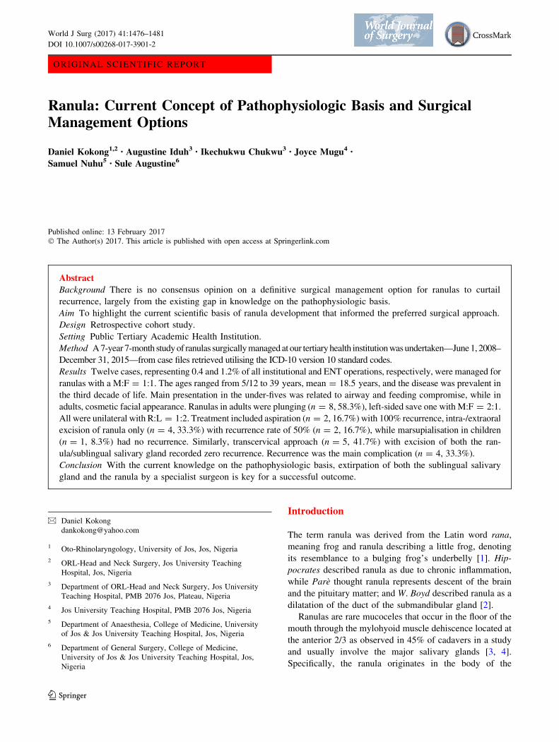
16.7%), while marsupialisation in children (n = 1, 8.3%)
having no recurrence (Fig. 2). Similarly, transcervical
approach (n = 5, 41.7%) with blunt dissection for excision
of both the ranula and sublingual salivary gland which
were plunging ranulas recorded zero recurrence after the
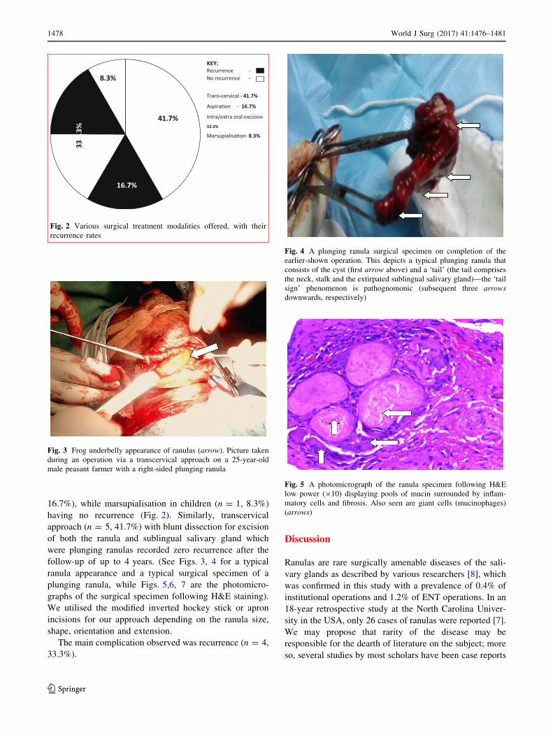
follow-up of up to 4 years. (See Figs. 3, 4 for a typical
ranula appearance and a typical surgical specimen of a
plunging ranula, while Figs. 5,6, 7 are the photomicro-
graphs of the surgical specimen following H&E staining).
We utilised the modified inverted hockey stick or apron
incisions for our approach depending on the ranula size,
shape, orientation and extension.
The main complication observed was recurrence (n = 4,
33.3%).
Discussion
Ranulas are rare surgically amenable diseases of the sali-
vary glands as described by various researchers [8], which
was confirmed in this study with a prevalence of 0.4% of
institutional operations and 1.2% of ENT operations. In an
18-year retrospective study at the North Carolina Univer-
sity in the USA, only 26 cases of ranulas were reported [7].
We may propose that rarity of the disease may be
responsible for the dearth of literature on the subject; more
so, several studies by most scholars have been case reports
KEY: Recurrence - No recurrence -
Trans-cervical - 41.7%
Aspira�on - 16.7%
Intra/extra oral excision-
33.3%
Marsupialisa�on- 8.3%.3%
33
41.7%
16.7%
8.3%
Fig. 2 Various surgical treatment modalities offered, with their
recurrence rates
Fig. 3 Frog underbelly appearance of ranulas (arrow). Picture taken
during an operation via a transcervical approach on a 25-year-old
male peasant farmer with a right-sided plunging ranula
Fig. 4 A plunging ranula surgical specimen on completion of the
earlier-shown operation. This depicts a typical plunging ranula that
consists of the cyst (first arrow above) and a ‘tail’ (the tail comprises
the neck, stalk and the extirpated sublingual salivary gland)—the ‘tail
sign’ phenomenon is pathognomonic (subsequent three arrows
downwards, respectively)
Fig. 5 A photomicrograph of the ranula specimen following H&E
low power (910) displaying pools of mucin surrounded by inflam-
matory cells and fibrosis. Also seen are giant cells (mucinophages)
(arrows)
1478 World J Surg (2017) 41:1476–1481
123
[9]. Studies reported female gender predilection with no
clear scientific basis including those of Chidzonga et al.
[10] and Zhao et al. [11] though with a distinct predilection
for males in cases of plunging ranulas in the latter; this was
reversed in this study in a ratio 2:1, but was in agreement
with the diving/plunging ranulas.
No age is spared for ranulas. Our series recorded an age
range of 5 months–39 years which is at variance with a
study which recorded 3–61 years [2].
Ranulas diagnosed on routine antenatal large enough to
warrant intrauterine decompression for a safe labour and
delivery have been reported [12]. This has suggested a
genetic basis for ranulas of early life including plunging
ranulas which were found to be more frequent in the ethnic
groups of the Maori and the Pacific Island Polynesians
[13, 14]. Intra-oral ranulas have been found frequently in
early life and young adults [11] which was observed in this
study where all the ranulas were seen in the younger age
group (n = 5, 41.7%). Ranulas develop slowly and typi-
cally present in the second and third decades of life [15] or
even later in life and are commonly plunging as observed
in our series with a male-to-female ratio of 2:1. For
unknown reason, the plunging ranulas were reported to
have a predilection for the right side [10, 11] which was at
variance with our study in which all were left-sided save
one. The duration of symptoms tends to be shorter in
children and young adults than in the adults. We may
speculate that the oral cavity which subserves vital func-
tions of deglutition and respiration can easily be compro-
mised by a space occupying lesion as it is situated in a
small space with rigid boundaries which make early pre-
sentation the norm. This was observed in our series as all
the children presented with failure to thrive (FTT) from
compromised feeding, while in compromised airway, pre-
sentations were majorly noisy breathing, snoring, obstruc-
tive sleep apnoea (OSA) and impending upper airway
obstruction with duration of symptoms less than 6 months.
In adults, however, the duration of symptoms tends to be
longer; this is because the oral cavity is wider as ranulas
tend to expand gradually and herniate through the mylo-
hyoid dehiscence, and extend into deep neck spaces to
appear in the neck and distant locations, hence the name
‘diving/plunging’. Intra-thoracic extensions of plunging
ranulas have been documented in adults [15–17].
There is no consensus opinion on the definitive man-
agement of these lesions, and there is often great variation
in practice. Multiple options exist, including surveillance,
needle aspiration, surgical excision of the cyst, sublingual
gland excision along with the cyst, marsupialisation, scle-
rotherapy, laser excision or cryosurgery [17]. The scle-
rotherapy employs bleomycin—antineoplastic antibiotic of
Streptomyces verticillus—OK-432 (Picibanil), a lyophi-
lised mixture of low virulent strain of Streptococcus pyo-
genes incubated with benzyl penicillin, that have been
found to produce good effect [18, 19]. In lesions diagnosed
antenatally where the oral mass can be life-threatening,
Kolker et al. [20] described the ex utero intrapartum
treatment (the EXIT technique).
However, different outcomes have been reported with
each approach having varying complications. Recurrence
has been reported the main culprit with different scholars
advocating excision of either ranula alone, ranula with the
sublingual gland or ranula with the submandibular gland.
This goes to demonstrate that the aetiopathogenesis of
ranulas was yet to be fully understood.
The current scientific knowledge reveals that ranulas
originate primarily from the sublingual salivary gland
Fig. 6 A photomicrograph of the ranula specimen following H&E
low power (910) showing extracellular pools of salivary mucin
(arrow) surrounded by inflammatory cells and fibrosis
Fig. 7 A photomicrograph of the ranula specimen following H&E
low power (94) showing extracellular pools of salivary mucin
(arrow) surrounded by inflammatory cells and fibrosis. Normal
salivary gland tissue seen below confirming our approach of en bloc
removal of both ranula and the offending sublingual salivary gland
World J Surg (2017) 41:1476–1481 1479
123

which is a spontaneous secretor of saliva, that is, produces
saliva without parasympathetic stimulation that occurs
during feeding, which is drained by 6–20 ducts scattered in
the floor of the oral cavity called ducts of Rivinus. They are
located majorly at the posterior and superior aspects, while
at the anterior part, they coalesce into a single duct termed
the Bartholin’s duct which empties into the Wharton’s duct
of the submandibular salivary gland. The sublingual sali-
vary gland is almond shaped, weighs 2–4 g and produces
mainly mucus secretions. It lacks a true capsule but rather
mucosal fold of the floor of the mouth which envelopes it
[21]. The gland is resistant to obstruction because of this
unique anatomical arrangement.
Congenitally, ranula occurs following imperforate sali-
vary gland duct and ostial stenosis leading to cyst forma-
tion. Trauma to the sublingual gland duct leads to mucus
extravasation into the submucosa via hydrostatic pressure
and formation of pseudocyst from mucus escape reaction
(MER). Trauma directly damages the acini with conse-
quent ductal obstruction, and back-pressure of secretion
builds up with subsequent acini rupture. Subsequently,
there is increased hydrostatic pressure, extravasation of
mucus, and then pseudocyst formation. Congenital nar-
rowing of the duct, dehiscence of the mylohyoid muscle
and sialolithiasis have also been implicated in ranula for-
mation. This was confirmed in a study where experimental
ligation of sublingual gland duct resulted in ranula for-
mation, while ligation of submandibular gland did not and
that of parotid gland led to atrophy [2].
Regarding superficial mucoceles, however, trauma does
not always appear to play an important role in the patho-
genesis. In many cases, mucosal inflammation that
involves the minor gland duct results in blockage, dilata-
tion and rupture of the duct with subepithelial spillage of
fluid. Changes in minor salivary gland function and com-
position of the saliva may contribute to their development.
In some cases, an immunological reaction may be the
cause. Studies have revealed increased levels of matrix
metalloproteinase, tumour necrosis factor-a, type IV col-
lagenase and plasminogen activators in mucoceles com-
pared with that of whole saliva. These factors are further
hypothesised to enhance the accumulation of proteolytic
enzymes that are responsible for the invasive character of
extravasated mucus [22, 23].
In a study, Sigismund et al. [4] in a retrospective anal-
ysis of 65 patients reported a recurrence prevalence of
3.6% following complete excision of the sublingual gland
alone compared with 36.7% prevalence with ranula exci-
sion alone; by implication, the former is 910 better than
the latter. He did not perform combined excision of the
ranula with the sublingual salivary gland.
In our series, recurrence following aspiration was 100%,
while that by intra-/extraoral ranula excision alone was
50%. These were done mainly by the non-specialist sur-
geons. However, combined ranula with the sublingual
gland excision yielded zero recurrence so was the only case
in an infant that had marsupialisation. We utilised the
transcervical approach with blunt dissection to approach
the ranula and remove the sublingual salivary gland for
plunging ranulas rather than combined transcervical with
transoral approaches. We employed any of the various neck
incision types suitable for a particular case depending on
the size, shape, extent and orientation of the ranula. The
modified inverted hockey stick or apron incisions would
suffice for most presentations.
Ranula is a clinical diagnosis, and imaging studies are
done mainly to know the extension of swelling prior to
surgery or when the diagnosis is unclear. Computed
tomography and specifically the presence of ‘tail sign’ is
pathognomonic for the plunging ranula [24–26]. This ‘tail’
is due to extension behind the mylohyoid muscle and
confirms the ranula to arise from the sublingual gland [27].
This may explain the zero recurrence in our combined
ranula with sublingual salivary gland excision approach
which was also observed in a study [7]. We may not
advance explanations for the resolution observed in 50% of
the cases that had only ranula excision, but the success rate
in marsupialisation and micro-marsupialisation in children
has been documented [28].
In addition, confirmation would be required via cyto-
chemical analysis to demonstrate the characteristic vis-
cous fluid content laden with mucin, inflammatory cells,
protein, salivary amylase indicating salivary gland origin.
The cystic lesion is, however, confirmed histopathologi-
cally as ranula by the presence of peripheral fibrosis, lined
by non-keratinising stratified squamous epithelial layer
with central pool of mucin, inflammatory cells and
mucinophages following H&E staining [29] as demon-
strated in our series.
Conclusion
With the current knowledge of the pathophysiologic basis,
extirpation of both the sublingual salivary gland and the
ranula by a specialist surgeon is key for a successful
outcome.
Acknowledgements The authors wish to say a big thank you to staff
of the health record’s department for making this study possible. The
skills of a Secretarial Staff of the Oral and Maxillofacial Department,
Mr Innocent Amanum, cannot go unnoticed for the painstaking well-
designed diagrams/pictures.
Compliance with ethical standards
Conflict of interest None.
1480 World J Surg (2017) 41:1476–1481
123

Open Access This article is distributed under the terms of the
Creative Commons Attribution 4.0 International License (http://crea
tivecommons.org/licenses/by/4.0/), which permits unrestricted use,
distribution, and reproduction in any medium, provided you give
appropriate credit to the original author(s) and the source, provide a
link to the Creative Commons license, and indicate if changes were
made.
References
1. Crysdale WS, Mendelsohn JD, Conley S (1988) Ranulas–muco-
celes of the oral cavity: experience in 26 children. Laryngoscope
98(3):296–298
2. Golden B, Drake AF, Talavera F et al (2016) Ranulas and
plunging ranulas. Medscape Last updated March 28, 2014
3. Engel JD, Ham SD, Cohen DM (1987) Mylohyoid herniation:
gross and histologic evaluation with clinical correlation. Oral
Surg 63:55–59
4. Sigismund PE, Bozzato A, Schumann M et al (2013) Manage-
ment of ranula: 9 years’ clinical experience in pediatric and adult
patients. J Oral Maxillofac Surg 71(3):538–544
5. Flaitz CM, Hicks MJ, Butler DF et al (2016) Ranulas and
mucocoeles. Medscape Last updated May 19, 2015
6. Curtin HD (2007) Imaging of the Salivary gland. In: Myers
Eugene N, Ferris Robert L (eds) Myers’ salivary gland disorders.
Springer, Berlin, pp 17–31
7. Patel MR, Deal AM, Shockley WW (2009) Oral and plunging
ranulas: what is the most effective treatment? Laryngoscope
119(8):1501–1509
8. Rho MH, Kim DW, Kwon SS et al (2006) OK-432 Sclerotherapy
of plunging ranula in 21 patients. It can be a substitute for sur-
gery. AJNR Am J Neuroradiol 27(5):1090–1095
9. Dayton K, Ryan MF (2014) Symptomatic floor of mouth swelling
with neck extension in a 14 year-old girl. Case Rep Pediatr
2014:8319–8323
10. Chidzonga MM, Rusakaniko S (2004) Ranula: another HIV/
AIDS associated oral lesion in Zimbabwe. Oral Dis 10:229–232
11. Zhao YF, Jia Y, Chen XM et al (2004) Clinical Review of 580
Ranulas. Oral surg Oral Med Oral Pathol Oral Radiol Endod
98:281–287
12. George MM, Mirza O, Solanki K et al (2015) Serious neonatal
airway obstruction with massive congenital sublingual ranula and
contralateral occurrence. Ann Med Surg (Lond) 4(2):136–139
13. Morton RP, Ahmad Z, Jain P (2010) Plunging ranula: congenital
or acquired? Otolaryngol Head Neck Surg 142:104–107
14. Davison MJ, Morton RP, McIvor NP (1998) Plunging ranula:
clinical observations. Head Neck 20:63–68
15. Batsakis JG, McClatchey KD (1988) Cervical ranulas. Ann Otol
Rhinol Laryngol 97:561–562
16. Pang CE, Lee TS, Pang KP et al (2005) Thoracic ranula: an
extremely rare case. J Laryngol Otol 119(3):233–234
17. Zhi K, Gao L, Ren W (2014) What is new in management of
pediatricranula? Curr Opin Otolaryngol Head Neck Surg
22(6):525–529
18. Fukase S, Ohta N, Inamura K et al (2003) Treatment of ranula
with intracystic injection of the streptococcal preparation OK-
432. Ann Otol Rhinol Laryngol 112:214–220
19. Kim KH, Sung MW, Roh JL et al (2004) Sclerotherapy for
congenital lesions in the head and neck. Otolaryngol Head Neck
Surg 131:307–316
20. Kolker MT, Batti JS, Schoem SR (2004) The ex utero treatment
procedure for congenital ranula in a Jehovah Witness. Oto-
laryngol Head Neck Surg 130:508–510
21. Windisch G, Weiglein AH, Kiesler K (2004) Herniation of the
mylohyoid muscle. J Craniofac Surg 15:566–569
22. Azuma M, Tamatani T, Fukui K et al (1995) Proteolytic enzymes
in salivary extravasation mucocoeles. J Oral Pathol Med
24(7):299–302
23. Hoque MO, Azuma M, Sato M (1998) Significant correlation
between matrix metalloproteinase and TNF-a in salivary
extravasation mucocoele. J Oral Pathol Med 27(1):30–33
24. Anastassov GE, HaiavyJ Solodnik P et al (2000) Submandibular
gland mucocele: diagnosis and management. Oral Surg Oral Med
Oral Pathol Oral Radiol Endod 89:59–63
25. Coit WE, Hamsberger HR, Osborn AG et al (1987) Ranula and
their mimics: CT evaluation. Radiology 163:211–216
26. Charnoff SK, Carter BL (1986) Plunging ranula: CT diagnosis.
Radiology 158:467–468
27. Arunachalam P, Priyadharshini N (2010) Recurrent plunging
ranula. J Indian Assoc Paediatr Surg 15(1):36–38
28. Yuca K, Bayram I, Cankaya H et al (2005) Paediatric intraoral
ranulas: an analysis of nine cases. Tohoku J Exp Med
205:151–155
29. Nilesh K, Malik NA, Patil P, Chapi DM (2015) Large plunging
ranula presenting as isolated neck swelling: steps in diagnosis and
surgical steps in management. J Clin Diagn Res 9(6):01–03
World J Surg (2017) 41:1476–1481 1481
123