RADIOGRAPHY Veterinary dental nursing procedures RADIOGRAPHY.
57
RADIOGRAPHY Veterinary dental nursing procedures RADIOGRAPHY
-
Upload
allen-carson -
Category
Documents
-
view
256 -
download
1
Transcript of RADIOGRAPHY Veterinary dental nursing procedures RADIOGRAPHY.

RADIOGRAPHY
Veterinary dental nursing procedures
RADIOGRAPHY

5888H - Veterinary Dental Nursing
RADIOGRAPHY
Radiology vs Radiography
• Radiography– taking and processing pictures
• Radiology– interpreting pictures

5888H - Veterinary Dental Nursing
RADIOGRAPHY
Purpose
• Diagnosis
• Planning
• Post-treatment
• Prognosis

5888H - Veterinary Dental Nursing
RADIOGRAPHY
Radiographs to diagnose
• Congenital e.g:– Malformations
• Acquired e.g:– Periodontal disease– Fractured teeth– Discoloured teeth– “Neck lesions”– Jaw lesions

5888H - Veterinary Dental Nursing
RADIOGRAPHY
Planning
• Extractions– Root infections– Impacted teeth– Root fusion (ankylosis)– Feline “neck lesions”
• Endodontics– Root canal treatment
• XR v impt for measuring length of drills

5888H - Veterinary Dental Nursing
RADIOGRAPHY
Monitoring
• Lesion progression
• Post op– Assess treatment success

5888H - Veterinary Dental Nursing
RADIOGRAPHY
Film Types
• Screen Film– Intensifying screens within cassettes permit
shorter exposure times– But cassettes don’t fit into mouth
• Non-Screen Film– Normal
• Slow & longer exposure but good detail• Small & flexible
– Dental• Small pre-cut sizes• Has 2 sides:
– One incorporates foil/lead backing – Other with a raised dot – point to the beam

5888H - Veterinary Dental Nursing
RADIOGRAPHY
Dental Film

5888H - Veterinary Dental Nursing
RADIOGRAPHY
Film positioning
• Extraoral
• Intraoral

5888H - Veterinary Dental Nursing
RADIOGRAPHY
Film positioning
• Intraoral– Less superimposition of teeth– Better detail
• Film closer to tooth• Less scattered radiation
– Usually need dental film

5888H - Veterinary Dental Nursing
RADIOGRAPHY
Patient positioning
• Mandible- animal in dorsal recumbency
• Maxilla- animal in ventral recumbency

5888H - Veterinary Dental Nursing
RADIOGRAPHY
Full mouth views
• Would require 6-8 views to evaluate all 4 quadrants

5888H - Veterinary Dental Nursing
RADIOGRAPHY
Patient positioning
• Motionless– Heavy sedation– General anaesthesia
• Positioning aids– Sandbags, lead sleeves– Troughs, Foam wedges, Syringe cases– Tape– Cardboard or spring loaded devices

5888H - Veterinary Dental Nursing
RADIOGRAPHY
X-Ray Machines
• Normal
• Dental – Smaller – More manoeuvrable– Safer – Better images

5888H - Veterinary Dental Nursing
RADIOGRAPHY
X-Ray Machines
• General Purpose– Large and difficult to manipulate
• Ideally would want to get to within 15 cm
• Dental– Easy to manipulate close in– Safer (less scatter)– Wall or castor mounted– Simple to operate
• fixed kV and mA • Adjust time
5888H - Veterinary Dental Nursing
RADIOGRAPHY
Dental X-Ray Machines

5888H - Veterinary Dental Nursing
RADIOGRAPHY
Developing
• Manual– Mini dark room boxes available
• Automatic– Needs to be able to handle small dental
film
• Within film envelope– Special dental films
5888H - Veterinary Dental Nursing
RADIOGRAPHY
Accessories
• Film positioners
• Bite blocks (props)
• Film viewer with magnifier
• Film marker
• Film storage envelopes

5888H - Veterinary Dental Nursing
RADIOGRAPHY
Accessories
Film Viewer
Film Positioners
5888H - Veterinary Dental Nursing
RADIOGRAPHY
How many views?
• At least 2 views at right angles– Plus extra obliques to highlight specific
problems
5888H - Veterinary Dental Nursing
RADIOGRAPHY
Tooth image distortion
• Size accuracy & fine detail more important than for normal radiographs– Require exact measurements in
endodontics- measuring exact length of root canal
– Small & subtle lesions may need to be detected

5888H - Veterinary Dental Nursing
RADIOGRAPHY
Types of distortion
• Anatomic– When film cannot be positioned parallel to
object being measured (tooth)
• Beam magnification– Point source effect
• Blurring– Movement – Aperture effect– Scatter

5888H - Veterinary Dental Nursing
RADIOGRAPHY
Anatomic distortion
• Shortening
• Lengthening

5888H - Veterinary Dental Nursing
RADIOGRAPHY
Beam magnification
• Point source effect

5888H - Veterinary Dental Nursing
RADIOGRAPHY
Blurring distortion
• Movement
• Aperture effect (depth of field)
• Scatter (thickness of tissue)

5888H - Veterinary Dental Nursing
RADIOGRAPHY
Reducing distortion
• Centering • Coning down (collimation)• Longer anode-film distance
– Reduces point source effect
• If possible– Film parallel to length of tooth
• Otherwise– Bisecting angle technique

5888H - Veterinary Dental Nursing
RADIOGRAPHY
Radiographic Beam Direction
• 90° Angle (Parallel) Beam technique– Film & Tooth parallel (< 15° separation)
• Possible for lower PM2-3 M1-3 (film inside mouth)• Possible for lower PM1 and all Ms (film outside)
– Beam perpendicular
• Bisecting Angle Beam technique– Film & Tooth not parallel (>15° separation)
• Required for all Incisors and Cuspids (Canines)
– Beam angle is simply averaged to compensate

5888H - Veterinary Dental Nursing
RADIOGRAPHY
Parallel technique

5888H - Veterinary Dental Nursing
RADIOGRAPHY
Parallel technique

5888H - Veterinary Dental Nursing
RADIOGRAPHY
Mandibular molars
• Can position film parallel
• Place lingual side
• Perpendicular beam

5888H - Veterinary Dental Nursing
RADIOGRAPHY
Parallel technique

5888H - Veterinary Dental Nursing
RADIOGRAPHY
Bisecting Angle
5888H - Veterinary Dental Nursing
RADIOGRAPHY
Bisecting angle technique
1. Get Long Axis of tooth
2. Get Long Axis of film
3. Bisecting Axis = half way between 1 and 2
4. Aim beam perpendicular to Bisecting Axis

5888H - Veterinary Dental Nursing
RADIOGRAPHY
Bisecting angle technique
• Use a straight object (e.g. tongue depressor) to help imagine bisecting angle

5888H - Veterinary Dental Nursing
RADIOGRAPHY
Mandibular Canine
1. Film2. Tooth
3. Bisecting Angle
X-RA
Y BE
AM

5888H - Veterinary Dental Nursing
RADIOGRAPHY
The imaginary bisecting line
• Is simply ‘eyeballed’

5888H - Veterinary Dental Nursing
RADIOGRAPHY
Mandibular Canine

5888H - Veterinary Dental Nursing
RADIOGRAPHY
Rostral mandibular PMs
• Symphysis prevents placing film parallel
• Bisecting angle
5888H - Veterinary Dental Nursing
RADIOGRAPHY
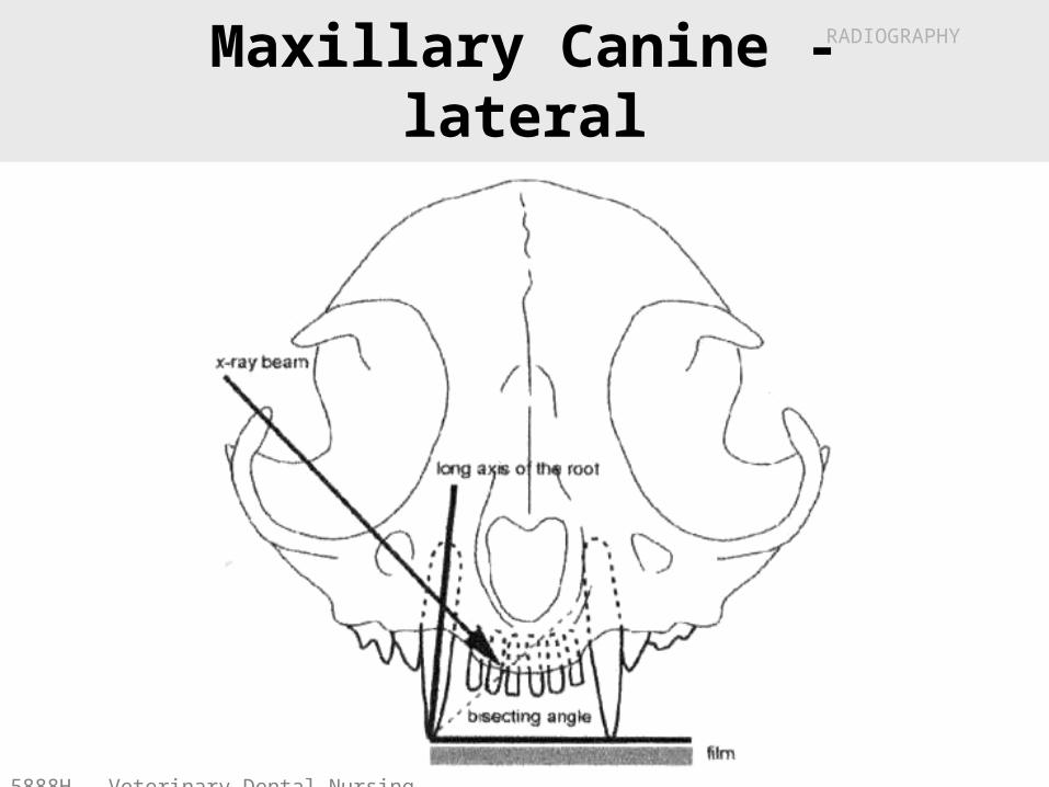
Maxillary Canine - lateral

5888H - Veterinary Dental Nursing
RADIOGRAPHY
Maxillary Canine – lat. oblique

5888H - Veterinary Dental Nursing
RADIOGRAPHY
Maxillary Canine – lat. oblique

5888H - Veterinary Dental Nursing
RADIOGRAPHY
Carnassial

5888H - Veterinary Dental Nursing
RADIOGRAPHY
A Near-Parallel technique

5888H - Veterinary Dental Nursing
RADIOGRAPHY
A Near-Parallel technique
5888H - Veterinary Dental Nursing
RADIOGRAPHY
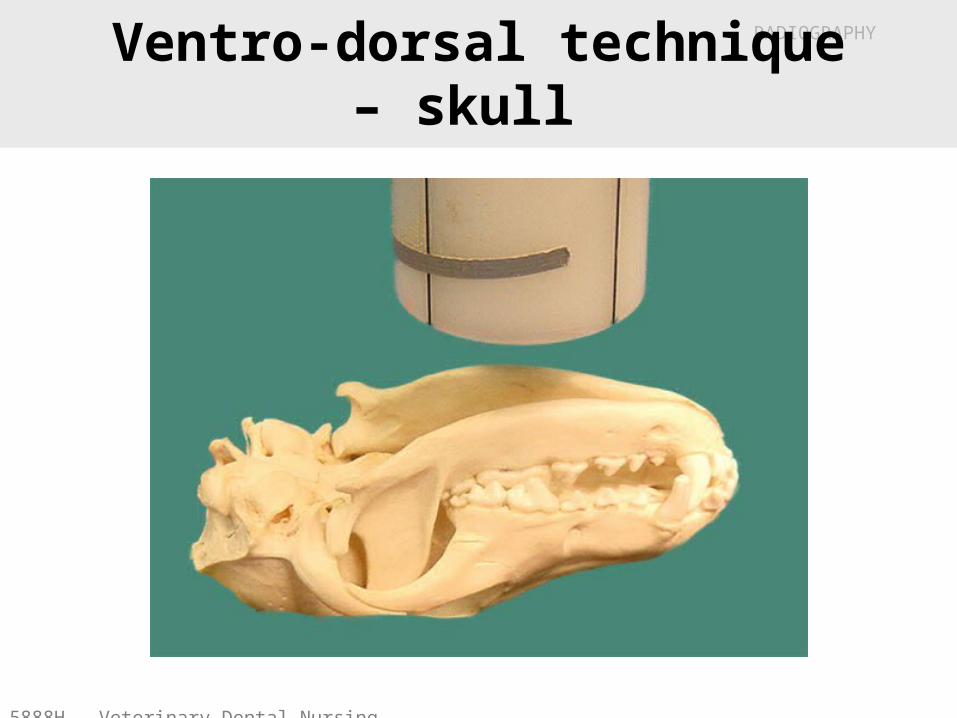
Ventro-dorsal technique – skull

5888H - Veterinary Dental Nursing
RADIOGRAPHY
Dorso-ventral technique – skull

5888H - Veterinary Dental Nursing
RADIOGRAPHY
45° Lateral Oblique technique
Mandible
5888H - Veterinary Dental Nursing
RADIOGRAPHY
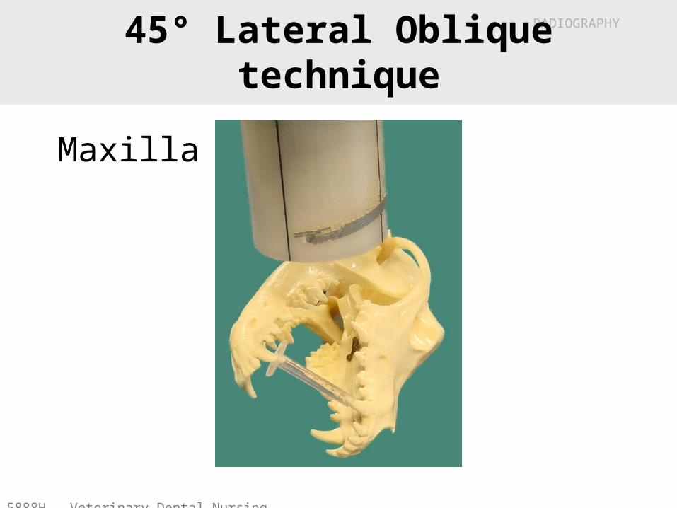
45° Lateral Oblique technique
Maxilla

5888H - Veterinary Dental Nursing
RADIOGRAPHY
Mandibular incisors/canines
• Dorsal recumbency
• Bisecting angle
• Center on1st Incisors

5888H - Veterinary Dental Nursing
RADIOGRAPHY
Any problems?
• No - normal

5888H - Veterinary Dental Nursing
RADIOGRAPHY
Any problems?
• Periodontal disease

5888H - Veterinary Dental Nursing
RADIOGRAPHY
Any problems?
• Apical lysis (probably apical abscess)

5888H - Veterinary Dental Nursing
RADIOGRAPHY
Any problems?
• Tooth hardening (sclerosis) after pulpitis

5888H - Veterinary Dental Nursing
RADIOGRAPHY
Any problems?
• ‘Root canal’ has been performed

5888H - Veterinary Dental Nursing
RADIOGRAPHY
X-Ray Safety
• Usual precautions
• Remember: – 1 brick = 0.5 mm lead

5888H - Veterinary Dental Nursing
RADIOGRAPHY
Digital Imaging
• Still uses X-Rays
• Uses a small X-Ray sensor
• Immediate image obtained– Can easily manipulate
image• Enlarge• Contrast • Store & Share

5888H - Veterinary Dental Nursing
RADIOGRAPHY
The End