QUANTITATIVE HYPERSPECTRAL IMAGING OF ... HYPERSPECTRAL IMAGING OF HISTORICAL DOCUMENTS: TECHNIQUE...
10
QUANTITATIVE HYPERSPECTRAL IMAGING OF HISTORICAL DOCUMENTS: TECHNIQUE AND APPLICATIONS R. Padoan 1 , Th.A.G. Steemers 1 , M.E. Klein 2 , B.J. Aalderink 2 , G. de Bruin 1 1 National Archive of The Netherlands Postbus 90520, 2509 LM The Hague, The Netherlands T: +3170331549597, F: +31703315540, E-mail: [email protected] 2 Art Innovation bv, Zutphenstraat 25, 7575 EJ Oldenzaal, The Netherlands E-mail: [email protected] ABSTRACT Hyperspectral imaging describes the technique of photographing an object using several well-defined optical bands in a broad spectral range. Using satellite- and aircraft-based instruments, macro-scale hyperspectral imaging is well established for geo-observation with applications in many different fields such as geology, archaeology and defence. Microscope-based hyperspectral instruments and analysis techniques are successfully employed in bio-medical research. In order to transfer this technique to a non-destructive analysis method of historical documents kept in archives and libraries, a dedicated instrument has been specifically designed by request of the Nationaal Archief (National Archive of the Netherlands). This novel tool provides fully calibrated spectral reflectance data at 70 wavelengths ranging from the near-UV (365 nm) via the visible into the near- infrared spectral region (1100 nm). Recordings are made over an area of 125 mm x 125 mm, using low-intensity wavelength-tunable light sources, while the temperature and relative humidity in the instrument measurement cabinet are constantly monitored. Even when recording all wavelength bands, a document is exposed for less than 15 minutes and analysis can be subsequently carried out using the digital dataset at any time for various purposes. This guarantees that the instrument is a completely non-invasive tool that can be applied safely even to objects of great value. Quantitative analysis of the extracted calibrated data can be performed, using chemometric methods, to identify writing materials such as inks and pigments to map their concentrations and to monitor the aging processes over various substrates. Excellent results have been obtained for distinguishing various inks, enhancing the contrast of watermarks and improve legibility of faded, or hidden text, even on carbonised paper. Current studies concentrate on the identification, quantification and monitoring of damages present on archival material before and after exposition and during storage conditions, such as biological damages, metal gall inks corrosion, paper oxidation, and pigments fading. INTRODUCTION During the last three decades, multi-spectral imaging technology, implemented on satellite and aeroplane-based platforms, has found an enormous range of applications in many subjects, such as mineralogical mapping of the earth’s surface, agricultural and environment control, and military defence 21 . Spectral imaging techniques have also found a wide spread use in microscopic analysis of biological targets for the identification and mapping of skin tumours, and the study of plant, animal and human tissues for various purposes. In the field of conservation of artistic and historic objects, such as paintings and documents, multi-spectral imaging has a long tradition as a non-destructive tool for the surveying of underdrawings, additional touches and, to a certain degree, pigments identification 4 . More recently, multi-spectral imaging has been used in the detection of iron gall inks and to produce qualitative analysis of ink corrosion 8,9,12 . In recent years, new spectral imaging systems that feature significantly more channels than conventional multi-spectral imagers, have been developed and applied to historic documents 12 . Due to their increased spectral resolution these “hyperspectral” imagers proved to be very useful to detect differences in writings and substrates and to enhance visibility of faint features such as palimpsests. However, conventional spectral imagers normally lack a 1 9th International Conference on NDT of Art, Jerusalem Israel, 25-30 May 2008 For more papers of this publication click: www.ndt.net/search/docs.php3?MainSource=65
Transcript of QUANTITATIVE HYPERSPECTRAL IMAGING OF ... HYPERSPECTRAL IMAGING OF HISTORICAL DOCUMENTS: TECHNIQUE...
QUANTITATIVE HYPERSPECTRAL IMAGING OF HISTORICAL DOCUMENTS:
TECHNIQUE AND APPLICATIONS
R. Padoan1, Th.A.G. Steemers1, M.E. Klein2, B.J. Aalderink2, G. de Bruin1 1National Archive of The Netherlands
Postbus 90520, 2509 LM The Hague, The Netherlands T: +3170331549597, F: +31703315540, E-mail: [email protected]
2Art Innovation bv, Zutphenstraat 25, 7575 EJ Oldenzaal, The Netherlands E-mail: [email protected]
ABSTRACT Hyperspectral imaging describes the technique of photographing an object using several well-defined optical bands in a broad spectral range. Using satellite- and aircraft-based instruments, macro-scale hyperspectral imaging is well established for geo-observation with applications in many different fields such as geology, archaeology and defence. Microscope-based hyperspectral instruments and analysis techniques are successfully employed in bio-medical research. In order to transfer this technique to a non-destructive analysis method of historical documents kept in archives and libraries, a dedicated instrument has been specifically designed by request of the Nationaal Archief (National Archive of the Netherlands). This novel tool provides fully calibrated spectral reflectance data at 70 wavelengths ranging from the near-UV (365 nm) via the visible into the near-infrared spectral region (1100 nm). Recordings are made over an area of 125 mm x 125 mm, using low-intensity wavelength-tunable light sources, while the temperature and relative humidity in the instrument measurement cabinet are constantly monitored. Even when recording all wavelength bands, a document is exposed for less than 15 minutes and analysis can be subsequently carried out using the digital dataset at any time for various purposes. This guarantees that the instrument is a completely non-invasive tool that can be applied safely even to objects of great value. Quantitative analysis of the extracted calibrated data can be performed, using chemometric methods, to identify writing materials such as inks and pigments to map their concentrations and to monitor the aging processes over various substrates. Excellent results have been obtained for distinguishing various inks, enhancing the contrast of watermarks and improve legibility of faded, or hidden text, even on carbonised paper. Current studies concentrate on the identification, quantification and monitoring of damages present on archival material before and after exposition and during storage conditions, such as biological damages, metal gall inks corrosion, paper oxidation, and pigments fading. INTRODUCTION During the last three decades, multi-spectral imaging technology, implemented on satellite and aeroplane-based platforms, has found an enormous range of applications in many subjects, such as mineralogical mapping of the earth’s surface, agricultural and environment control, and military defence21. Spectral imaging techniques have also found a wide spread use in microscopic analysis of biological targets for the identification and mapping of skin tumours, and the study of plant, animal and human tissues for various purposes. In the field of conservation of artistic and historic objects, such as paintings and documents, multi-spectral imaging has a long tradition as a non-destructive tool for the surveying of underdrawings, additional touches and, to a certain degree, pigments identification4. More recently, multi-spectral imaging has been used in the detection of iron gall inks and to produce qualitative analysis of ink corrosion8,9,12. In recent years, new spectral imaging systems that feature significantly more channels than conventional multi-spectral imagers, have been developed and applied to historic documents12. Due to their increased spectral resolution these “hyperspectral” imagers proved to be very useful to detect differences in writings and substrates and to enhance visibility of faint features such as palimpsests. However, conventional spectral imagers normally lack a
1
9th International Conference on NDT of Art, Jerusalem Israel, 25-30 May 2008For more papers of this publication click: www.ndt.net/search/docs.php3?MainSource=65

rigorous calibration of measurement data, which is a prerogative in obtaining a quantitative hyperspectral analysis of recorded objects and opens the possibility to compare images obtained from different targets at different times. Furthermore, uncalibrated hyperspectral imagers can not make optimum use of the wealth of mathematical tools mainly developed for analyzing spectral images of the earth’s surface13. In 2004, the Nationaal Archief, in collaboration with the company Art Innovation B.V., started a study on the use of Quantitative Hyperspectral Imaging (QHSI) technique for monitoring and quantifying aging processes of archival documents during storage and exposition. One important goal was the characterization and quantification of paper yellowing, which is an indirect indicator for a characteristic aging process of this common substrate material. In order to study local changes in paper yellowness of documents - or in general of their spectral reflectance – much attention was given to the reproducibility of measurements of the same object area at different times and the possibility to compare spectral properties and their possible variances for different documents. Therefore, an instrument was required to record spectral images with high spectral and spatial resolution and produce calibrated datasets allowing a quantitative comparison of spectral characteristics of documents. In order to fulfill these requirements, the “SEPIA” quantitative hyperspectral imaging system [1] has been developed as a nondestructive multifunctional analysis tool that provides both spatial and spectral data of archival documents, such as paper, parchment, leather, textiles, inks, pigments et cetera. THE “SEPIA” QUANTITATIVE HYPERSPECTRAL IMAGER (QHSI) Concept of Quantitative Hyperspectral Imaging The term “hyperspectral imaging” (HSI) refers to the acquisition of a series of digital images at a large number of different, well-defined, optical wavelengths in the ultra-violet, visible and near-infrared spectral regions. By applying proper calibration procedures, the value of any pixel of a digital image of the series represents a precise measurement of the portion of light reflected from the corresponding location on the target at a particular wavelength. The result of a hyperspectral imaging measurement is the so-called hyperspectral data cube, which contains one spectral reflectance image for each wavelength band (see Fig. 1). The pixel values at the same pixel coordinate in all images of the data cube correspond to an entire reflectance spectrum of the document at the corresponding location.
r efle
ctan
c e
wavele
ngth
500 nm
700 nm
900 nm
1100 nm
2048
pix
els
/ 125
mm
2048 pixels / 125 mm70
wav
elengths
Fig. 1: The hyperspectral data cube is the result of a hyperspectral measurement.
It contains for each wavelength a calibrated spectral reflectance image, so that for any image pixel an entire spectral reflectance curve is provided.
2

Calibrated spectral reflectance values contained in the hyperspectral datacube are independent from the specific instrument settings during measurement (camera exposure, light intensity distribution etc.). As opposed to a mere qualitative comparison of different spectral images, numeric data contained in hyperspectral datacubes can be used to compare optical document properties in a quantitative way. For example, calculations can be performed with data derived from multiple recordings of the same document area, at different times, or even comparing measurements of different documents. A great advantage of calibrated quantitative hyperspectral datacubes is the direct recording, in a digital format, of the complete information set collected by the instrument. This data can be easily reused during future research with different regions-of-interests or even on entirely different subjects without the necessity to put up again the original artifact to the stress of a new measurement. Working Principle and Properties of the Instrument The SEPIA instrument is based on two identical wavelength tunable light sources, called “TULIPS” and a monochromatic CCD camera, as shown schematically in figure 2. The object under investigation (e.g. a single-sheet document, a bound volume, a map etc.) is placed under the camera and illuminated from two sides by the “TULIPS” under an angle of 45º, which ensures an homogeneous light intensity and a non-specular imaging by the camera. The entire setup is enclosed in a light-proof cabinet in order to avoid any stray light from external sources that would disturb the measurement. Compared with the conventional approach of using a strong white light source and spectral filtering in the camera itself, the tunable light source approach has a decisive advantage in terms of safety; light energy absorbed by the object during a measurement is in fact kept at an absolute minimum, so that even for very fragile objects there is virtually no risk of any light-induced damage.
hi-res digitalcamera
object support
TULIPS B
wavele
ngth
tun
able
light
sourc
e
TULIPS A
wavelength
tunable
light source
computer control
documentligh-proofcabinet
Fig. 2: Setup scheme of the quantitative hyperspectral imager.
The computer-controlled “TULIPS” are tuned synchronously through up to 70 different wavelength bands, ranging from the near-UV (365 nm), via the entire visible into the near-infrared (1100 nm) of the spectral range. The wavelength emitted by the light sources can be tuned in small wavelength steps of typically 10 nm, while the spectral bandwidth ranges from 10 nm to 16 nm. The combination of small tuning steps and high spectral resolution is of particular importance in the visible range, where the resulting reflectance curves can be used to calculate the standard CIE colour indices10.
At each wavelength band, the document is imaged by the 4 mega-pixel monochrome digital camera, which has a field-of-view of 125 mm × 125 mm. Due to the very high resolution of
3

60 µm × 60 µm per pixel on the document (i.e. >400 dpi), the instrument provides reliable measurement data even from areas of thin lines of handwritings or prints. This is a basic feature for the “pure” spectral recording of inks and pigments areas in order to distinguish different types of writing materials and map their distribution on the document. The 70 recorded spectral images are calibrated by using data from a reference measurement of a diffuse white spectral standard target (Spectralon©, provided by Labsphere inc.). The resulting hyperspectral datacube is stored as a set of 16 bit grayscale images that can be easily imported by most conventional image processing software such as Adobe Photoshop©. Hyperspectral Data Analysis Methods There are numerous ways in which information organized in hyperspectral datacubes can be analyzed and in which analysis results can be visualized in order to answer the research questions at hand. Usefulness of various analysis methods depends obviously on the type of application, various of which will be discussed in the following paragraphs. In general, approaches for the analysis of hyperspectral data can be distinguished as follows: • Processing of individual spectral images; typically for contrast enhancement (e.g.
watermarking and underdrawings). • Combining multiple spectral images using standard formulas; e.g. to calculate CIE color
indices or generate true-color (sRGB-space) images. • Combining multiple spectral images using ad hoc formulas to calculate pixel ratio images,
pixel difference images etc; this approach can be very successful for enhancing the legibility of faint text or other features and to improve the visualization of results.
• Performing feature extraction and classification calculations (e.g. principle component analysis) on spectral curves of each pixel; for distinguishing ink or pigment types and mapping their concentrations. Mostly for the analysis of hyperspectral data of the earth’s surface. A wide range of powerful mathematical tools have been developed in the past, and can now be applied to quantitative hyperspectral data of historic documents.
• Extraction of pixel value statistics for regions-of-interest (ROIs) in spectral images, including mean spectral values and their variances. Comparison of individual mean spectral curves of ROI’s can be very useful for distinguishing materials and study their conditions and to compare them with reference data obtained with conventional photo-spectrometers.
All these methods can be applied to analyze individual document measurements. Each method requires only a single hyperspectral data cube. They can also be applied to analyze several measurements of the same document at different times (treatment / degradation studies) or to compare several different objects with each other or with a database, in which case information from several data cubes has to be combined. APPLICATIONS IN AGING MONITORING OF DOCUMENTS Paper Yellowing Oxidative reactions of paper components, such as cellulose, hemicellulose, lignin, wood impurities, dyes, additives, and sizing agents, result in the formation of colored degradation products containing carbonyl groups (C=O) and carbon-carbon double bonds (C=C) 1,2,15,19. These reactions occur in uncontrolled environmental conditions (elevated temperature and relative humidity parameters), in the presence of pollutants such as SO2 and NO2, or due to light absorption by the chromophores of paper components. Both oxidative and photo-oxidative reactions contribute to the formation of new functional groups that behave as new acceptors and therefore accelerate the yellowing process of paper. Oxidation reactions are
4

typically more intense in modern industrially-produced paper, if compared to older hand-made paper, due to the adoption of new machines and the use of wood instead of cotton as the main ingredient for the production of paper pulp. This resulted in paper with shorter fibres and a high content of lignin, hemicellulose and various fastening chemical additives. It is common practice that conservators and curators try to determine and compare the yellowing of paper simply by using the naked eye. However, human colour vision performs poorly when it has to compare colours of objects that cannot be viewed simultaneously. Therefore, a visual characterization of discolouration is entirely inadequate and subjective for detecting and quantifying this kind of damage and relating it to possible causes such as storage and exposition conditions, or conservation treatments. As opposed to this, quantitative hyperspectral imaging (QHSI) proves to be very well-suited for addressing this type of research questions. QHSI allows the measuring of colour variations within an archival unit repeatedly, so that any colour changes can be documented in an accurate and objective manner. Fig. 3 shows results of a hyperspectral measurement of the royal personal agenda of Queen Juliana of the Netherlands and her mother Queen Wilhelmina. In concordance with legal archive time prescription, the agenda has been separated in two sections twenty years ago. The first section relating to Queen Wilhelmina has been made available to the public by the Nationaal Archief, whereas the second part has been kept for several more years in the royal cabinet. This difference in conservation environments resulted in two different series of ageing processes for the same material. The effect of this is barely visible with the naked eye but it can be accurately quantified and visualized by hyperspectral imaging. Figure 3A shows the double-page, where the agenda had been separated, as a real-colour image calculated from the measured visible reflectance images (380 nm to 780 nm). The right page, which had been kept in the royal cabinet for several years longer, exhibits small variations in the substrate colour as compared to the left page that looks more homogenous.
A B Figure 3A: Real-colour image of the separation page of the royal agenda calculated from hyperspectral
measurements. 3B: Colour-coded image of the yellow-index (YI). The colour shades from blue via green towards red encode yellow indices between 23 and 29.
Fig. 3B is a colour-coded image of the yellow index of the same document area, which is calculated for each pixel from its spectral reflectance curve3,10. Local yellow index variations between 23 to 29 are encoded as colour changes from blue via green to red. The right page is clearly less homogeneous and in particular, it shows rectangular sections of increased yellow
5

indices (red colour), which were probably more exposed to the environment in the royal cabinet. This analysis method is thus not only useful to detect and visualize differences in aging processes on a document. The numeric result of the hyperspectral measurement lends itself also for statistical approaches where, for example, the object condition at the time of measurement is represented by a histogram of the yellow index over the object area. Future aging could than be detected and quantified by comparing histograms calculated from measurements taken for example before and after an exhibition. Aging of Ink A huge range of natural and artificial substances may occur on historic documents. However, in the field of conservation of archival material priority is normally given to studying and monitoring of inks12,16,17. Ink used on documents throughout history actually comprehends an enormous group of different substances, each of which having its own chemical and physical characteristics and different ways of reacting with other substrates and to environmental conditions. In the last decades, an impressive number of useful techniques such as FTIR, XRD, UV fluorescence, XRF and PIXE5,6, have been developed for detecting and characterizing inks. Many of these techniques concentrate on metal gall ink, due to its centennial use in the production of handwritings and its well-known instability. Despite of its incapacity to provide chemical information, QHSI can be an important tool to determine conditions and distribution of writing substances over a wide range of support materials. One important characteristic of this technique is that it does not require an a priori – and therefore uninformed – selection of the measured regions-of-interest (ROI’s). These ROI’s can in fact be repeatedly redefined for further analysis. Therefore, the risk of missing interesting effects in recordings, due to unsuitable choices of the ROI’s, is minimized.
A B Fig. 4A: Real-colour image of deteriorated ink letter on paper substrate. 4B: Colour-coded image of same area
showing ink areas with similar spectral responses in the same colour (spectral mapping). Another important characteristic of QHSI is the possibility to apply mathematical algorithms to spectral data in order to map spatial distribution of ink deterioration and monitor its development in time due to aging processes. As an example, figure 4 shows a colour image (Fig.4A) of a letter on a circa 110 years old document (iron-gall ink on paper) featuring “bleeding” and corrosion. A mathematical algorithm, called Modified Spectral Angle Similarity (MSAS)9, has been applied on spectral data of this area resulting in the false-colour image shown in figure 4B. Here, pixels with similar spectral response, which presumably represent areas of comparable condition, are shown in the same colour. A large range of such mathematical methods, for the classification of areas with similar properties, has been developed especially for hyperspectral imaging applications in geo-sciences. They can be applied to visualize inhomogeneities of inks and surrounding substrate areas affected by chemical deterioration. In particular, paper alteration provoked by the migration of iron-II
6

ions, in combination with many other factors, can be identified and quantified on both recto and verso of the page12,18. Biological and Physical Damages Moulds and fungi are a major threat to any biological material kept in a conservation institution due to their extremely rapid and persistent way of affecting substrates with often devastating results20. It is known that their visual chromatic characteristics depend on their origin and state of development, which provides the possibility to study these features by using optical-spectral analysis. In order to demonstrate the possibility of applying QHSI to the study of biological damages, an induced cultivation of mould, on four different substrates (parchment, Japanese paper, leather and a XVIII-century paper), have been prepared. Figure 5 shows three different spectral images of the samples, recorded five days after inducing a spore contamination. Depending on the growing stage of moulds, which are in this case more advanced on parchment and ancient paper than on Japanese paper and leather, the affected areas exhibit significant differences in their spectral response. These differences can in principle be exploited to identify the type of mould and quantify its progress.
A B C Fig.5 Spectral images of mould samples at (A) 365 nm, (B) 600 nm, and (C)1100 nm wavelength.
Substrate materials are parchment (top left), Japanese paper (top right), leather (bottom left) and an XVIII-century paper (bottom right).
True extension of physical damages of substrates is also sometimes difficult to detect by conventional visual inspections, especially when surrounded by heavily corroded ink areas. Figure 6 shows spectral images of a document recorded with the QHSI before its display in the exposition room of the Nationaal Archief. Only the infra-red spectral image, at 1100 nm, showed the real dimensions of the damaged area, which is important in order to provide an adequate conservation treatment to avoid further damages.
A B C D Fig.6: Paper damage on an iron gall ink drawing shown in a real-colour image (6A)
and in spectral images at 365nm (6B), 600nm (6C) and 1100nm (6D). The extension of the damage is only visible in the near-infrared region.
7
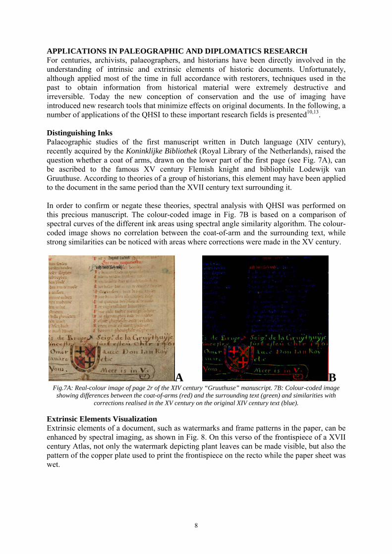
APPLICATIONS IN PALEOGRAPHIC AND DIPLOMATICS RESEARCH For centuries, archivists, palaeographers, and historians have been directly involved in the understanding of intrinsic and extrinsic elements of historic documents. Unfortunately, although applied most of the time in full accordance with restorers, techniques used in the past to obtain information from historical material were extremely destructive and irreversible. Today the new conception of conservation and the use of imaging have introduced new research tools that minimize effects on original documents. In the following, a number of applications of the QHSI to these important research fields is presented10,13. Distinguishing Inks Palaeographic studies of the first manuscript written in Dutch language (XIV century), recently acquired by the Koninklijke Bibliothek (Royal Library of the Netherlands), raised the question whether a coat of arms, drawn on the lower part of the first page (see Fig. 7A), can be ascribed to the famous XV century Flemish knight and bibliophile Lodewijk van Gruuthuse. According to theories of a group of historians, this element may have been applied to the document in the same period than the XVII century text surrounding it. In order to confirm or negate these theories, spectral analysis with QHSI was performed on this precious manuscript. The colour-coded image in Fig. 7B is based on a comparison of spectral curves of the different ink areas using spectral angle similarity algorithm. The colour-coded image shows no correlation between the coat-of-arm and the surrounding text, while strong similarities can be noticed with areas where corrections were made in the XV century.
A B Fig.7A: Real-colour image of page 2r of the XIV century “Gruuthuse” manuscript. 7B: Colour-coded image
showing differences between the coat-of-arms (red) and the surrounding text (green) and similarities with corrections realised in the XV century on the original XIV century text (blue).
Extrinsic Elements Visualization Extrinsic elements of a document, such as watermarks and frame patterns in the paper, can be enhanced by spectral imaging, as shown in Fig. 8. On this verso of the frontispiece of a XVII century Atlas, not only the watermark depicting plant leaves can be made visible, but also the pattern of the copper plate used to print the frontispiece on the recto while the paper sheet was wet.
8

A B C Fig. 8: Real-colour image (8A) and false-colour image (8B) enhancing the watermark
(plant leaves on the left), and laid lines, and the effect of the printing copper plate on the sheet structure (8C, prints where usually made on wet paper).
Legibility Enhancement of Deteriorated Documents In former times, chemical solutions were often applied on documents in order to improve readability of extremely deteriorated texts. Famous examples include treatments of parchments to read palimpsests, and the application of reagents to carbonised papyrus to enhance contrast between the substrate and inks applied on it. Unfortunately, long and short term side-effects of these treatments were not taken in consideration at these times. Nowadays, spectral imaging represents a valuable alternative to obtain readability of texts without a direct invasive or even destructive action on the objects themselves. Fig. 9 demonstrates the possibility of using the QHSI measurement to enhance contrast of written text on a fragment of a carbonised book page.
A B Fig. 9A Real-colour image and 9B false-colour image with enhanced writing
on a fragment of a carbonised book page. CONCLUSION In this contribution, it has been demonstrated that quantitative hyperspectral imaging is a very versatile analytical tool, with numerous applications in conservation, exposition management, historical forensic research, biological and environmental monitoring, and others subjects. The SEPIA quantitative hyperspectral imaging system provides fully spatially and spectrally calibrated measurements, enabling an objective analysis of documents. Obviously this technique could be very useful also for many other fields of research, including the study of works of art. Application examples discussed here are the result of an initial explorative use of this new technique and a more detailed and deeper research is currently in progress at the Nationaal Archief and mainly focuses on the study of archival documents. Future research activities will be centred on a systematic investigation of the potential of the quantitative hyperspectral imaging technique, taking into account the complexity of materials and aging process of archival units and their role as carriers of valuable, and sometimes hidden, information of our history.
9

ENDNOTES 1. The “SEPIA” Quantitative Hyperspectral Imager is developed by the company Art
Innovation B.V. (Oldenzaal, The Netherlands) on request of the Nationaal Archief and is under this name commercially available in different configurations.
REFERENCES 1. V. Bukovsky, M trnkova, The influence of secondary chromophores on the light induced
oxidation of paper, in Restaurator Vol 24, 2003, pp.18-35 2. H.A.Carter, The chemistry of paper preservation, in Journal of chemical education Vol 73
N.11, November 1996 3. Deutsche Industrienorm DIN6167, “Beschreibung der Vergilbung von nahezu weissen
oder farblosen Materialien”, January 1980 4. R. J. Feller, Color science in the examination of museum objects, the Getty Conservation
Institute, Los Angeles, 2001 5. N.Ferrer, M.C.Sistach, Characterisation by FTIR Spectroscopy of Ink Components in
Ancient Manuscripts, in Restaurator Vol 26, 2005 pp 105-117 6. O. Hahn, X-ray fluorescence analysis of iron gall inks , pencils and coloured crayons, in
Studies in conservation vol. 50, 2005 pp.23-32 7. J.B.G.A. Havermans, Environmental influences on the deterioration of paper, Rotterdam,
1995 8. J.B.G.A. Havermans, H.A.Aziz, J.H. Scholten, Non destructive detection of iron gall inks
by means of multispectral imaging. Part 2: Application on original objects affected with iron gall ink corrosion, 24 (2), 88-94, 2003.
9. S. Homayouni and M.Roux, Hyperspectral image analysis for material mapping using spectral matching, Proceedings of the XX ISPRS Congress, 12-23 July 2004 Istanbul, in Proceedings volume IAPRS, Vol.XXXV
10. R.W.G. Hunt, “Measuring Colour”, Fountain Press Kingston-upon-Thames England, Third Ed. 1998, p. 344
11. J. Kolar, M. Strlič Iron Gall Inks: on manufacture characterization degradation and stabilization, Ljubljana, 2006
12. M.Kubik, Hyperspectral Imaging: A new technique for the non invasive study of artworks, in Physical techniques in the study of art, archeology and cultural heritage, Vol2, Amsterdam 2007
13. C. Lee and D. A. Landgrebe, "Analyzing High Dimensional Multispectral Data”, IEEE Transactions on Geoscience and Remote Sensing, Vol. 31, No. 4, pp 792-800, July, 1993
14. Public Record Office, An introduction to writing inks, Richmond 1998 15. J.L.Pedersoli, The assessment of chemiluminescence to investigate the oxidation of paper,
in Contribution to conservation, London, 2002 16. Public Record Office, An introduction to printing inks, Richmond 1999 17. B. Reissland, M.W.Cowan, The light sensitivity of iron gall inks, in Contribution to the
Baltimore Congress IIC, 2-6 September 2002 18. M. Strlič, J. Kolar, Ageing and stabilisation of paper, Ljubljana, 2005 19. A.P. Verhoeff, J.van Wijnen, Enumeration and identification of airborne viable mould
propagules, Amsterdam, 1988 20. Yuhas R.H., Goetz A.F.H. and Boardman J.W., 1992, “Discrimination among semi-arid
landscape endmembers using the Spectral Angle Mapper (SAM) algorithm”, In Summaries of the Third Annual JPL Airborne Geoscience Workshop, JPL Publication 92-hyh14. Vol. I. pp. 147-149.
Back to Top
10