
PROBLEMS of THE NEONATAL PERIOD Problems... · 2019-03-11 · CBC, LP, & culture results....
82
PROBLEMS of the NEONATAL PERIOD Susan Fisher-Owens, MD, MPH, FAAP Clinical Professor of Clinical Pediatrics Clinical Professor of Preventive and Restorative Dental Sciences University of California, San Francisco Zuckerberg San Francisco General Hospital UCSF Family Medicine Board Review: Improving Clinical Care Across the Lifespan San Francisco March 11, 2019
Transcript of PROBLEMS of THE NEONATAL PERIOD Problems... · 2019-03-11 · CBC, LP, & culture results....
PROBLEMS of the NEONATAL PERIOD
Susan Fisher-Owens, MD, MPH, FAAPClinical Professor of Clinical Pediatrics
Clinical Professor of Preventive and Restorative Dental SciencesUniversity of California, San Francisco
Zuckerberg San Francisco General Hospital
UCSF Family Medicine Board Review: Improving Clinical Care Across the Lifespan
San FranciscoMarch 11, 2019
∗ I have nothing to disclose
Disclosures
2
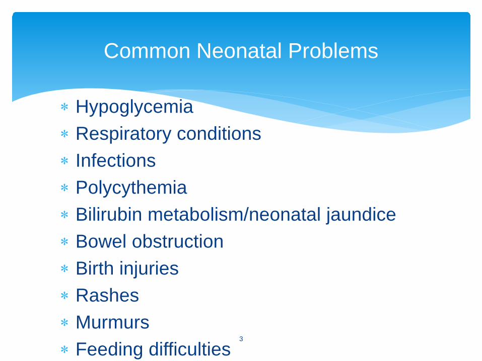
∗ Hypoglycemia∗ Respiratory conditions∗ Infections∗ Polycythemia∗ Bilirubin metabolism/neonatal jaundice∗ Bowel obstruction∗ Birth injuries∗ Rashes∗ Murmurs∗ Feeding difficulties
Common Neonatal Problems
3
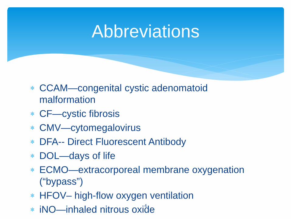
∗ CCAM—congenital cystic adenomatoid malformation
∗ CF—cystic fibrosis∗ CMV—cytomegalovirus∗ DFA-- Direct Fluorescent Antibody∗ DOL—days of life∗ ECMO—extracorporeal membrane oxygenation
(“bypass”)∗ HFOV– high-flow oxygen ventilation∗ iNO—inhaled nitrous oxide
Abbreviations
4
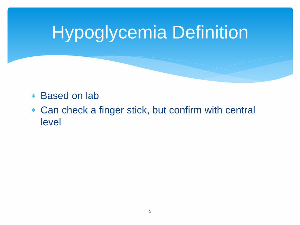
∗ Based on lab∗ Can check a finger stick, but confirm with central
level
Hypoglycemia Definition
5
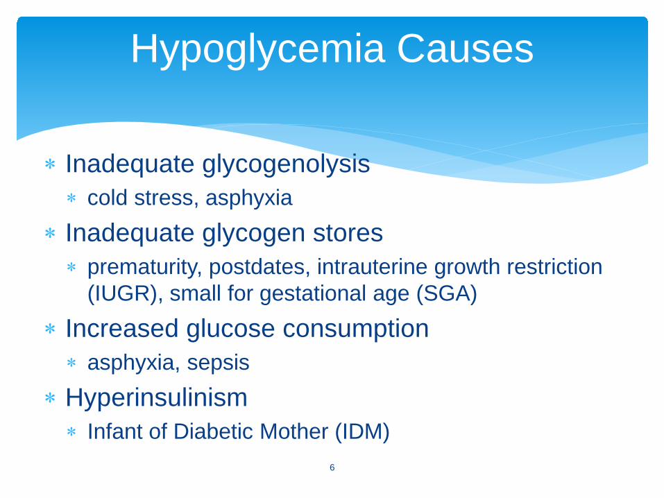
∗ Inadequate glycogenolysis ∗ cold stress, asphyxia
∗ Inadequate glycogen stores ∗ prematurity, postdates, intrauterine growth restriction
(IUGR), small for gestational age (SGA)∗ Increased glucose consumption ∗ asphyxia, sepsis
∗ Hyperinsulinism ∗ Infant of Diabetic Mother (IDM)
Hypoglycemia Causes
6
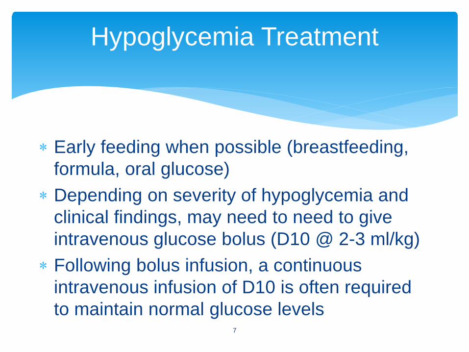
∗ Early feeding when possible (breastfeeding, formula, oral glucose)
∗ Depending on severity of hypoglycemia and clinical findings, may need to need to give intravenous glucose bolus (D10 @ 2-3 ml/kg)
∗ Following bolus infusion, a continuous intravenous infusion of D10 is often required to maintain normal glucose levels
Hypoglycemia Treatment
7
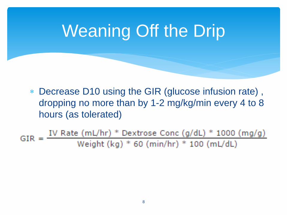
Weaning Off the Drip
8
∗ Decrease D10 using the GIR (glucose infusion rate) , dropping no more than by 1-2 mg/kg/min every 4 to 8 hours (as tolerated)
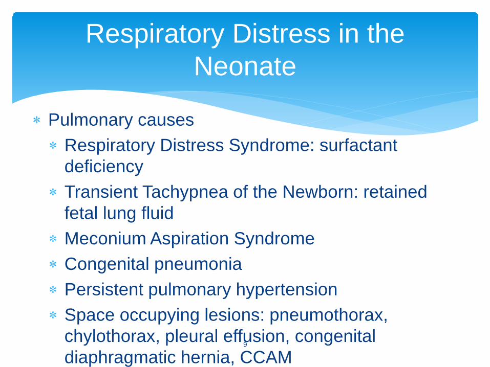
∗ Pulmonary causes∗ Respiratory Distress Syndrome: surfactant
deficiency∗ Transient Tachypnea of the Newborn: retained
fetal lung fluid∗ Meconium Aspiration Syndrome∗ Congenital pneumonia∗ Persistent pulmonary hypertension∗ Space occupying lesions: pneumothorax,
chylothorax, pleural effusion, congenital diaphragmatic hernia, CCAM
Respiratory Distress in the Neonate
9
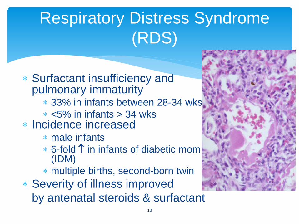
∗ Surfactant insufficiency and pulmonary immaturity
∗ 33% in infants between 28-34 wks∗ <5% in infants > 34 wks
∗ Incidence increased∗ male infants∗ 6-fold ↑ in infants of diabetic mom
(IDM)∗ multiple births, second-born twin
∗ Severity of illness improvedby antenatal steroids & surfactant
Respiratory Distress Syndrome (RDS)
10
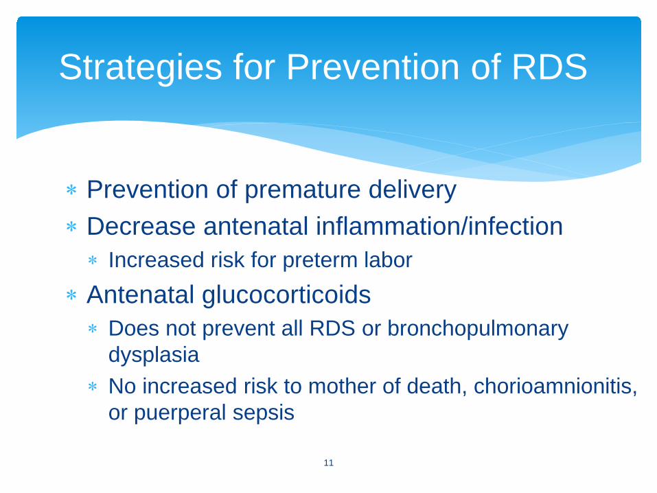
∗ Prevention of premature delivery∗ Decrease antenatal inflammation/infection∗ Increased risk for preterm labor
∗ Antenatal glucocorticoids∗ Does not prevent all RDS or bronchopulmonary
dysplasia∗ No increased risk to mother of death, chorioamnionitis,
or puerperal sepsis
Strategies for Prevention of RDS
11
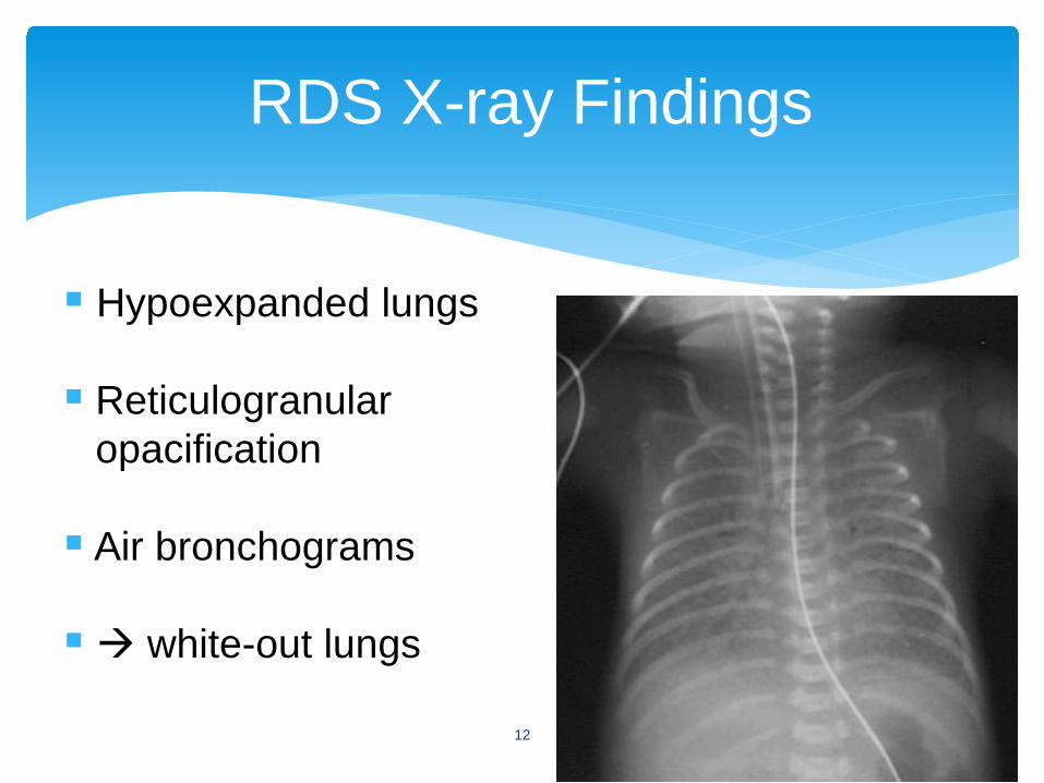
RDS X-ray Findings
Hypoexpanded lungs
Reticulogranularopacification
Air bronchograms
white-out lungs
12
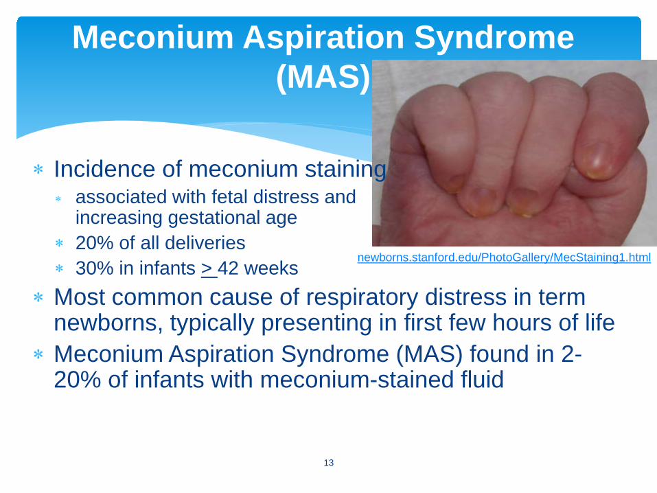
∗ Incidence of meconium staining∗ associated with fetal distress and
increasing gestational age ∗ 20% of all deliveries ∗ 30% in infants > 42 weeks
∗ Most common cause of respiratory distress in term newborns, typically presenting in first few hours of life
∗ Meconium Aspiration Syndrome (MAS) found in 2-20% of infants with meconium-stained fluid
Meconium Aspiration Syndrome (MAS)
newborns.stanford.edu/PhotoGallery/MecStaining1.html
13
∗ Hypoxia, acidosis lead to fetal gasping ( aspiration)
∗ Disease range: mild to severe disease with air leaks, pulmonary hypertension, respiratory failure, and death (iNO, HFOV, and ECMO improve survival)
MAS, cont’d
14
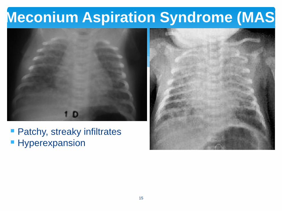
Meconium Aspiration Syndrome (MAS)
Patchy, streaky infiltrates Hyperexpansion
15
∗ Delayed clearance of fetal lung fluid∗ Term or near-term infants∗ Delivered via c-section and/or no/little labor∗ Chest Xrays: lung hyperaeration, prominent
pulmonary vascular markings, interstitial fluid, pleural effusion
∗ Transient respiratory symptoms (tachypnea, occasional hypoxia, rare dyspnea) resolve within 2-5 days
Transient Tachypnea of Newborn (TTN)
16
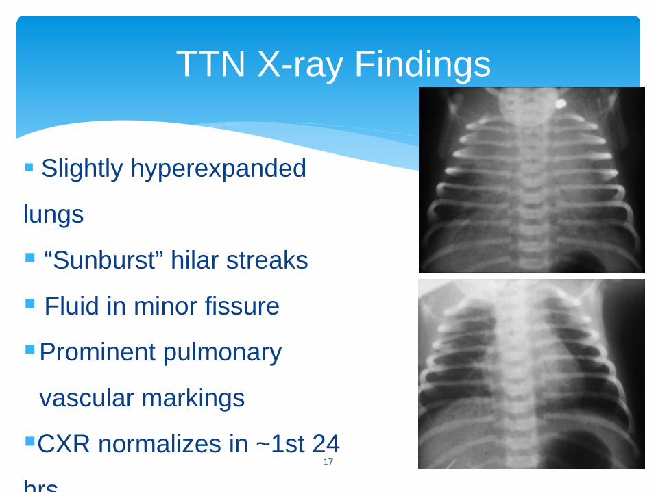
TTN X-ray Findings
Slightly hyperexpanded
lungs
“Sunburst” hilar streaks
Fluid in minor fissure
Prominent pulmonary
vascular markings
CXR normalizes in ~1st 24
hrs17
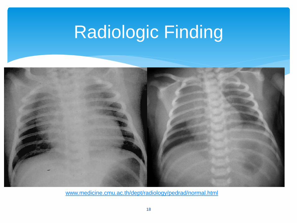
Radiologic Finding
www.medicine.cmu.ac.th/dept/radiology/pedrad/normal.html
18
∗ Hyperthermia, hypothermia∗ Hypovolemia, shock, metabolic acidosis∗ Cardiac disease∗ Cyanotic congenital heart disease∗ Left-sided obstructive lesions (coarctation)∗ Congestive heart failure∗ Myocardopathy∗ Myocarditis
∗ Polycythemia∗ Sepsis
Extra-Pulmonary Causes of Respiratory Distress in the Neonate
19
∗ Bacterial infections∗ Group B Streptococcus ∗ E. coli ∗ Listeria monocytogenes
∗ Viral infections∗ Herpes simplex ∗ Hepatitis B and C
Perinatal Infections
∗ TORCH infections: Incidence is 0.5-2.5%; many infants are asymptomatic at delivery∗ Toxoplasma gondii,
Treponema pallidum∗ “Other”: syphilis ∗ Rubella∗ Cytomegalovirus (most
common)∗ Herpes20
∗ Prematurity < 37 weeks gestation∗ Chorioamnionitis∗ Prolonged ruptured membranes > 24 hours∗ GBS-positive mother∗ Male infant
Risk Factors for Early-Onset Sepsis
21
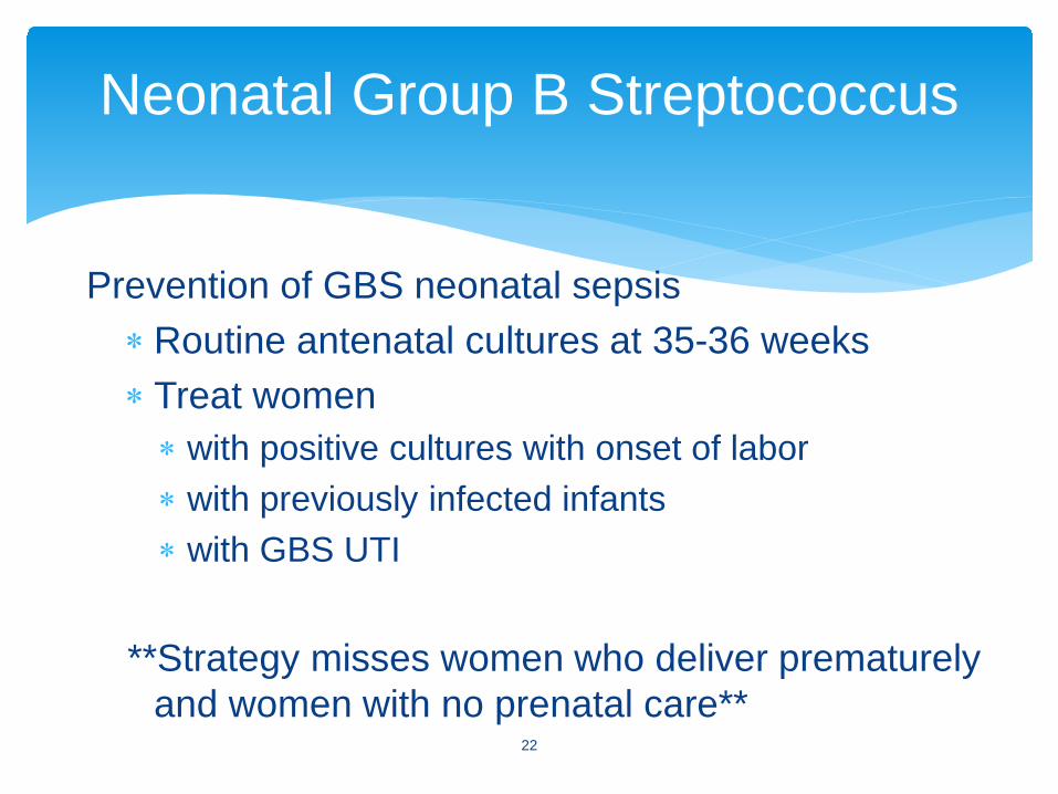
Prevention of GBS neonatal sepsis∗Routine antenatal cultures at 35-36 weeks∗ Treat women∗ with positive cultures with onset of labor∗ with previously infected infants ∗ with GBS UTI
**Strategy misses women who deliver prematurely and women with no prenatal care**
Neonatal Group B Streptococcus
22
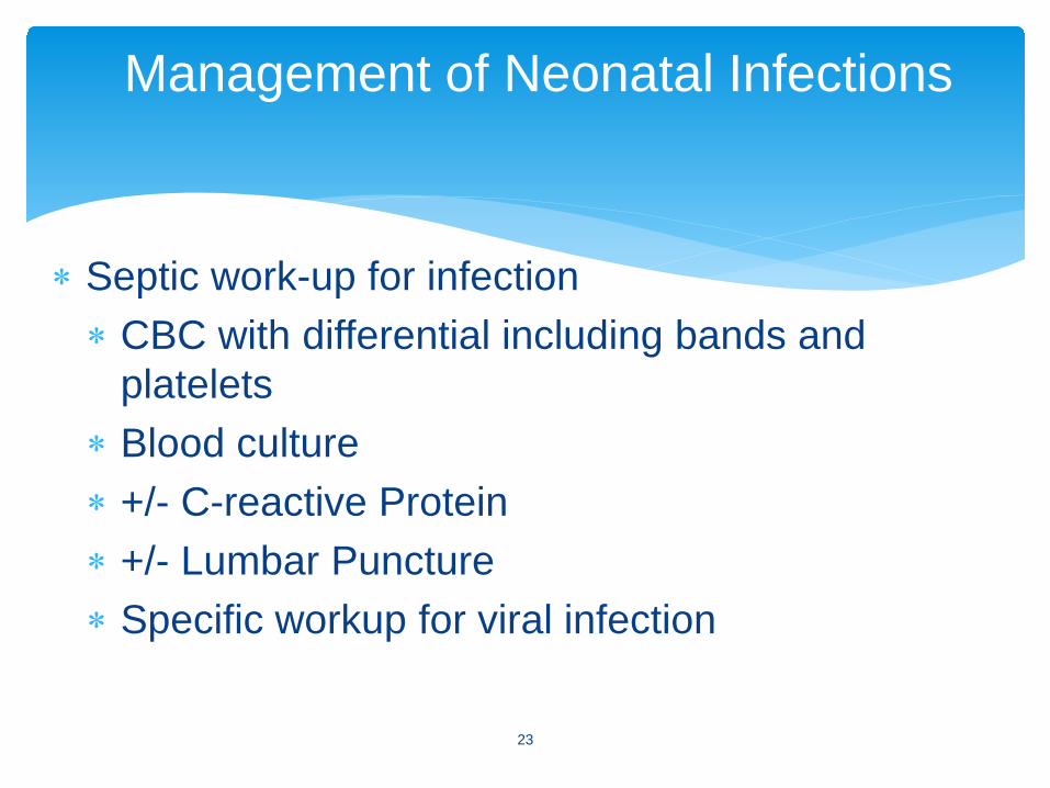
∗ Septic work-up for infection∗ CBC with differential including bands and
platelets ∗ Blood culture∗ +/- C-reactive Protein ∗ +/- Lumbar Puncture ∗ Specific workup for viral infection
Management of Neonatal Infections
23
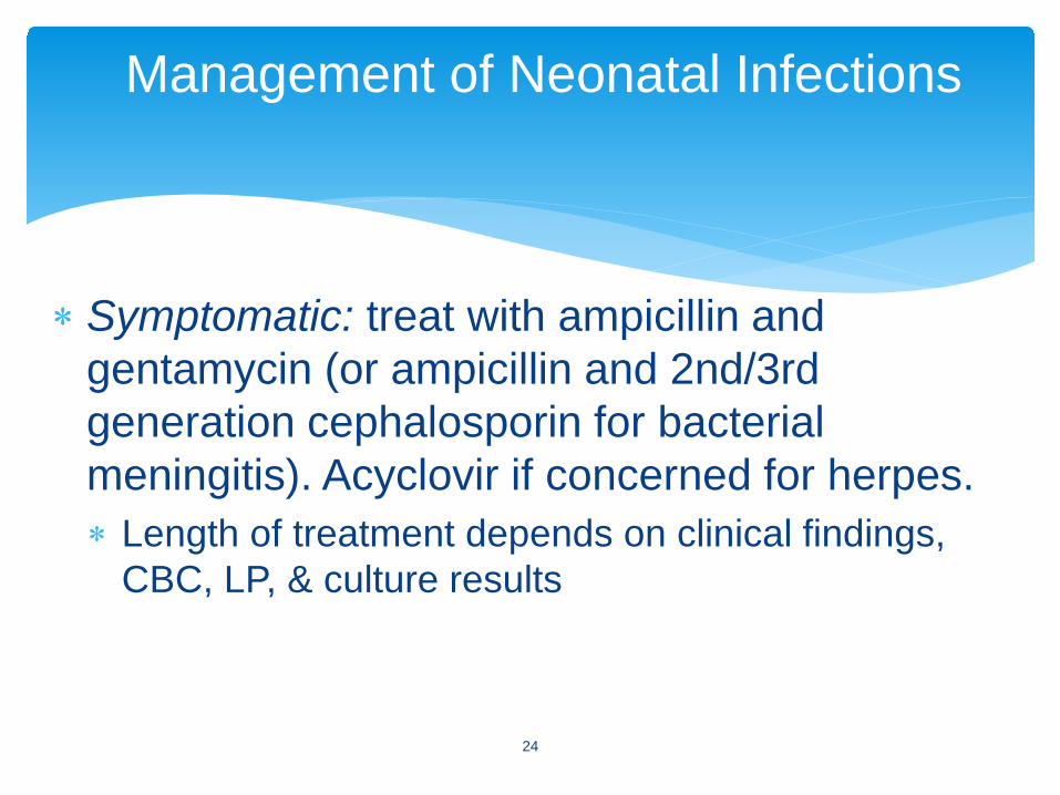
∗ Symptomatic: treat with ampicillin and gentamycin (or ampicillin and 2nd/3rd generation cephalosporin for bacterial meningitis). Acyclovir if concerned for herpes.∗ Length of treatment depends on clinical findings,
CBC, LP, & culture results
Management of Neonatal Infections
24
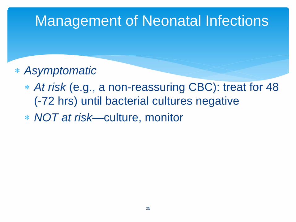
∗ Asymptomatic∗ At risk (e.g., a non-reassuring CBC): treat for 48
(-72 hrs) until bacterial cultures negative∗ NOT at risk—culture, monitor
Management of Neonatal Infections
25

∗ Hepatitis B vaccine prior to hospital discharge for all infants (<12 hr if Mom HBsAg positive)
∗ HBIG (hepatitis B immunoglobulin) plus vaccine for infants born to HBsAg positive mother <12 hours of life
∗ All infants should receive routine Hepatitis B vaccine during infancy (1-2 month and 6 months)
∗ Breastfeeding safe with HBsAg positive mother with vaccine plus HBIG treatment for the infant
Prevention of Transmission of Perinatal Hepatitis B
26
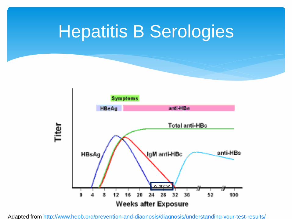
Hepatitis B Serologies
27
WINDOW
Adapted from http://www.hepb.org/prevention-and-diagnosis/diagnosis/understanding-your-test-results/
High-risk mothers screened during pregnancy∗ Vertical transmission rate is 5-10% ∗ Hepatitis C antibody titers obtained on infant at 6 and
12 months (even 18 months), or Hepatitis C PCR at 4 mos
What about breastfeeding with Hepatitis C+ mother?∗ Variable amounts of virus in milk∗ Studies have not shown increase risk of transmission
of Hepatitis C with breastfeeding∗ Recommend pump/dump if cracked/bleeding nipples
Perinatal Hepatitis C
28
∗ SGA, IUGR, postnatal growth failure∗ Microcephaly, hydrocephalus, intracranial
calcifications∗ Hepatosplenomegaly, hepatitis, jaundice (elevated
direct component)∗ Anemia (hemolytic), thrombocytopenia∗ Skin rashes, petechiae∗ Abnormalities of long bones ∗ Chorioretinitis, cataracts, glaucoma∗ Nonimmune hydrops∗ Developmental and learning disabilities
Perinatal TORCH Infections—Non-Specific Findings
29
∗ Toxoplasmosis: hydrocephalus, chorioretinitis, generalized intracranial calcifications (random distribution)
∗ Syphilis: osteochondritis, periosteal new bone formation, rash, snuffles
∗ Rubella: cataracts, “blueberry muffin” rash, patent ductus arteriosus, pulmonary stenosis, deafness
∗ Cytomegalovirus: microcephaly, periventricular calcifications, hydrocephalus, chorioretinitis, petechiae, thrombocytopenia, hearing loss (progressive)
Perinatal TORCH Infections—Specific Findings
30
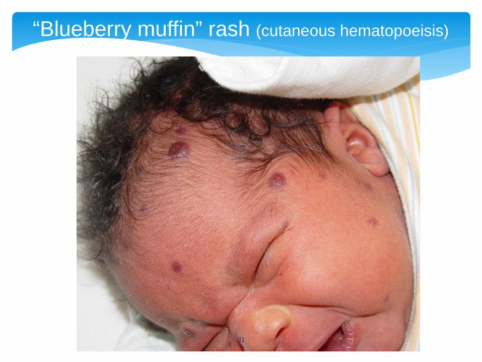
“Blueberry muffin” rash (cutaneous hematopoeisis)
31
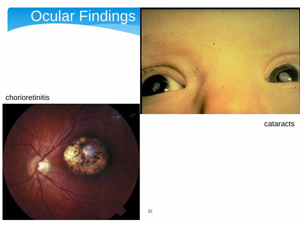
Ocular Findings
chorioretinitis
cataracts
32
∗ HSV-1 (15 to 20%) and HSV-2 (80 to 85%) ∗ Neonatal infections with primary HSV is 35-50%∗ Neonatal infections with recurrent HSV is 0-5%∗ Increased risk of transmission with prolonged
rupture of membranes, forceps or vacuum delivery, fetal scalp monitoring, preterm infants
∗ 75% of cases have no history of maternal infection, nor evidence of skin lesions∗ One may need to start treatment based on clinical
presentation and suspicion of infection
Neonatal Herpes Simplex
33
∗ Disseminated (systemic) disease: ∗ Early onset (1st week of life), 25% of cases∗ Sepsis syndrome, liver dysfunction, pneumonia
∗ CNS disease: meningoencephalitis∗ 2nd-3rd week of life, 35% of cases∗ Fever, irritability, abnormal CSF, seizures∗ Early treatment improves outcome, but 40-50% infants
have residual neurodevelopmental disability∗ Localized disease: skin, eyes, mouth, 40% of
cases
Herpes Simplex: Clinical Presentations
34
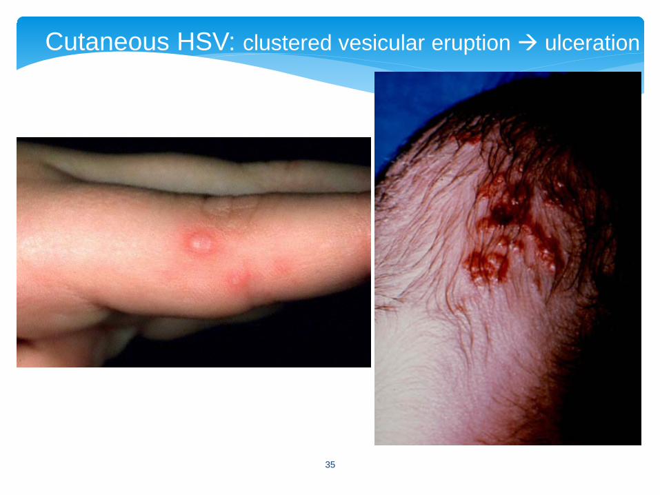
Cutaneous HSV: clustered vesicular eruption ulceration
35

∗ Toxoplasmosis∗ maternal antibody titer and neonatal IGM antibody
∗ Syphilis∗ RPR or VDRL positive, obtain titers, order treponemal-
specific test (FTA or MHA-TP)∗ CMV∗ urine culture
Diagnosis of TORCH Infections
36
∗ Herpes simplex∗ Surveillance: conjunctival, nasopharyngeal, and rectal swabs for
Direct Fluorescent Antibody (DFA) 24-48 hours after birth if suspect exposure
∗ Culture of vesicle scrapings when lesions are present∗ DFA of vesicle scrapings ∗ PCR: detect HSV-DNA in CSF
Diagnosis of TORCH Infections
37
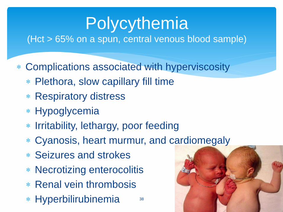
∗ Complications associated with hyperviscosity∗ Plethora, slow capillary fill time∗ Respiratory distress∗ Hypoglycemia∗ Irritability, lethargy, poor feeding∗ Cyanosis, heart murmur, and cardiomegaly∗ Seizures and strokes∗ Necrotizing enterocolitis∗ Renal vein thrombosis∗ Hyperbilirubinemia
Polycythemia(Hct > 65% on a spun, central venous blood sample)
38
∗ If symptomatic neonate with polycythemia, or an infant with excessively high hematocrit (> 70%)--by dilutional exchange, correcting Hct to approx 55%
Volume of blood = Wt (kg) X 80 cc/kg X (Hctobs – Hct desired)
Hctobs
∗ Blood is removed through umbilical artery or umbilical venous catheter and normal saline is infused for blood volume replacement
Polycythemia--Treatment
39
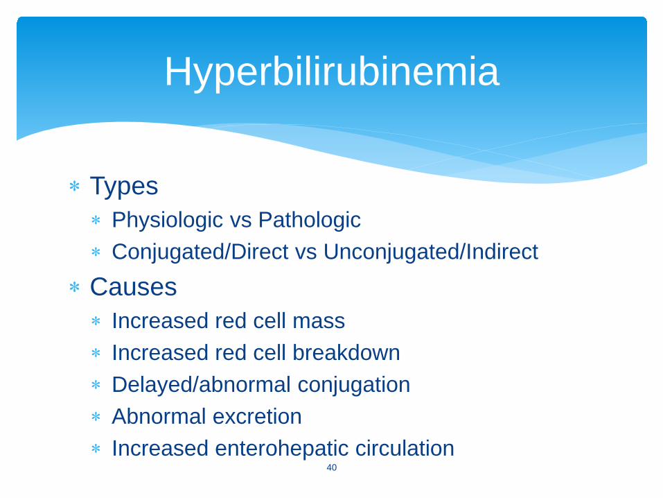
∗ Types∗ Physiologic vs Pathologic∗ Conjugated/Direct vs Unconjugated/Indirect
∗ Causes∗ Increased red cell mass∗ Increased red cell breakdown∗ Delayed/abnormal conjugation∗ Abnormal excretion∗ Increased enterohepatic circulation
Hyperbilirubinemia
40
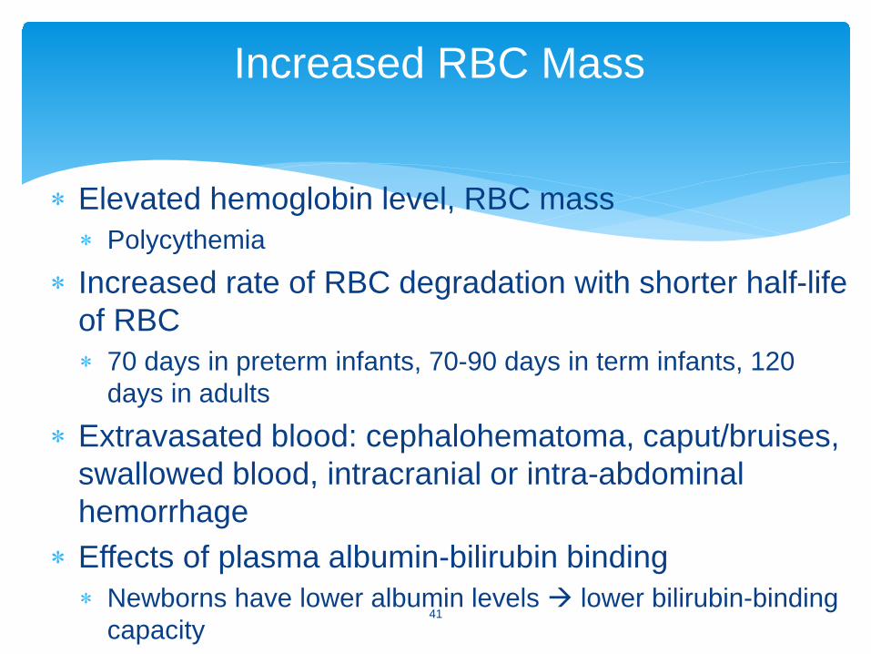
∗ Elevated hemoglobin level, RBC mass∗ Polycythemia
∗ Increased rate of RBC degradation with shorter half-life of RBC ∗ 70 days in preterm infants, 70-90 days in term infants, 120
days in adults
∗ Extravasated blood: cephalohematoma, caput/bruises, swallowed blood, intracranial or intra-abdominal hemorrhage
∗ Effects of plasma albumin-bilirubin binding∗ Newborns have lower albumin levels lower bilirubin-binding
capacity
Increased RBC Mass
41
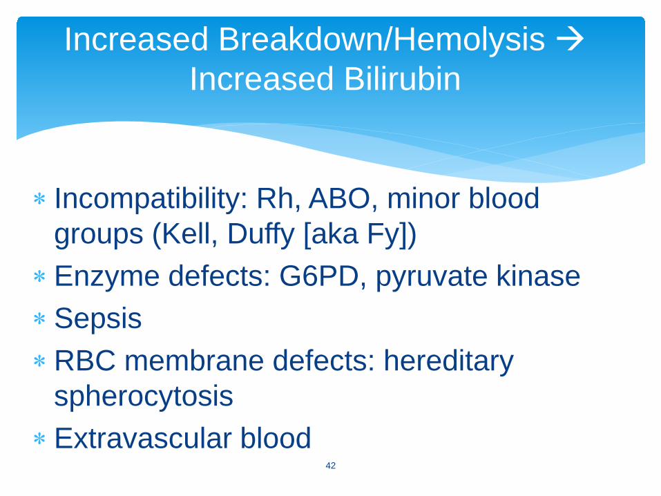
∗ Incompatibility: Rh, ABO, minor blood groups (Kell, Duffy [aka Fy])
∗Enzyme defects: G6PD, pyruvate kinase∗Sepsis∗RBC membrane defects: hereditary
spherocytosis∗Extravascular blood
Increased Breakdown/Hemolysis Increased Bilirubin
42

∗ Neonatal hepatitis ∗ Sepsis∗ Prematurity∗ Breast milk jaundice∗ Hypothyroidism∗ Sepsis∗ Congenital enzyme deficiency eg Crigler-Najjar∗ Metabolic diseases, e.g., galactosemia
Impaired Conjugation
43
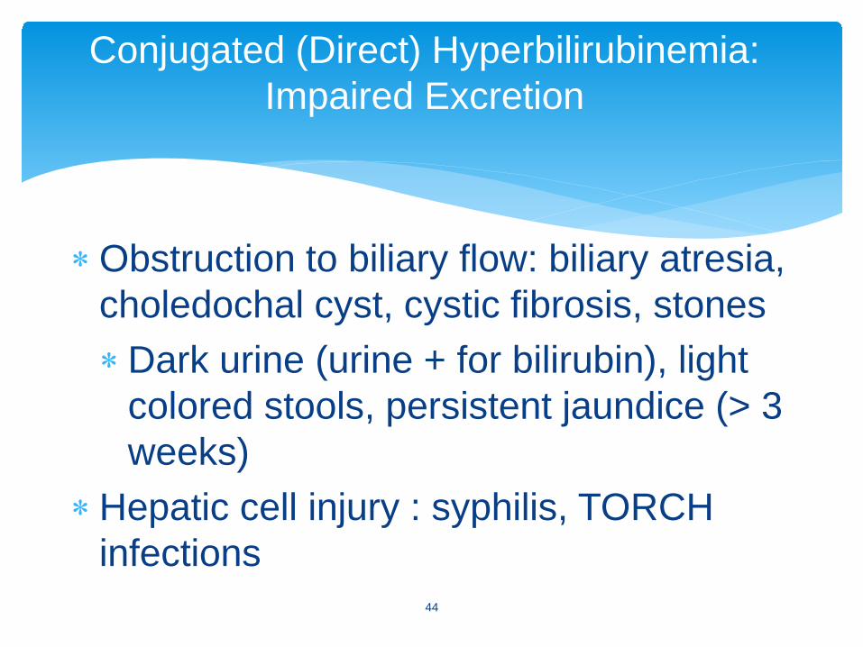
∗Obstruction to biliary flow: biliary atresia, choledochal cyst, cystic fibrosis, stones ∗Dark urine (urine + for bilirubin), light
colored stools, persistent jaundice (> 3 weeks)
∗Hepatic cell injury : syphilis, TORCH infections
Conjugated (Direct) Hyperbilirubinemia: Impaired Excretion
44
∗Hepatic dysfunction: E. coli (UTI)∗Toxic effects: hyperalimentation
cholestasis∗Metabolic errors: galactosemia∗Chronic “overload”: erythroblastosis fetalis,
G6PD, spherocytosis
Conjugated (Direct) Hyperbilirubinemia: Impaired Excretion, cont’d
45
∗ Conjugated bilirubin—unconjugated, reabsorbed
∗ Enterohepatic circulation and reabsorption is enhanced by:∗ Gut sterility (urobilinogen and stercobilinogen)∗ Bowel dysmotility (preterm infants, effects of
magnesium or morphine)∗ Ileus∗ Obstruction: atresia, pyloric stenosis, meconium
plugs, cystic fibrosis∗ Delayed feeding (“breast-feeding jaundice”)
Enterohepatic Circulation
46
∗ Hemolysis∗ Onset of jaundice in 1st 24 hours∗ Rapid rate of rise of bili (>0.5mg/dL per hour)∗ Hepatosplenomegaly, pallor∗ Family history (G6PD, spherocytosis)∗ “Set-up” with incompatibility, Coombs (+DAT), elevated
reticulocytes, abnormal hemolytic smear∗ Sepsis or inborn error∗ Emesis, lethargy, poor feeding∗ Hepatosplenomegaly, tachypnea, temperature
instability
Causes Suggested by Clinical Findings
47
∗ Increased susceptibility to neurotoxicity seen with asphyxia, sepsis, acidosis, prematurity, and hemolysis∗ Consider treatment at lower levels of unconjugated
bilirubin in these cases∗ When to worry∗ Visible jaundice in the first 24 hours of life∗ Serum bilirubin rising rapidly > 5 mg/dl/24 hrs∗ Prolonged hyperbilirubinemia > 1 week term infant
and > 2 weeks in the preterm∗ Direct bilirubin > 2mg/dl
Management of Indirect Hyperbilirubinemia
48
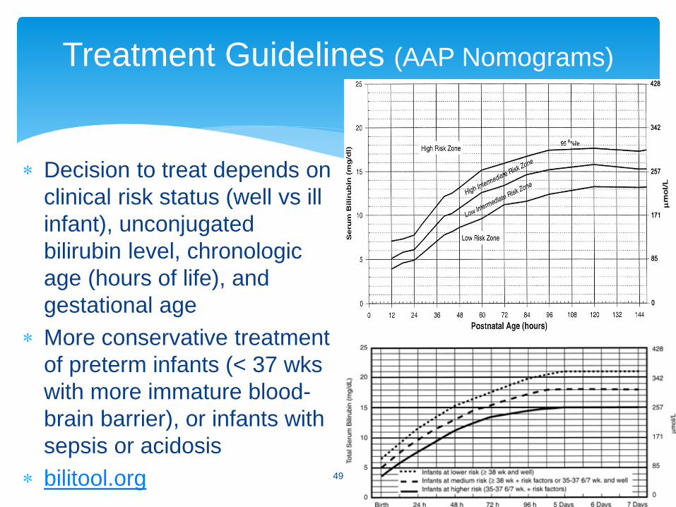
∗ Decision to treat depends on clinical risk status (well vs ill infant), unconjugated bilirubin level, chronologic age (hours of life), and gestational age
∗ More conservative treatment of preterm infants (< 37 wks with more immature blood-brain barrier), or infants with sepsis or acidosis
∗ bilitool.org
Treatment Guidelines (AAP Nomograms)
49
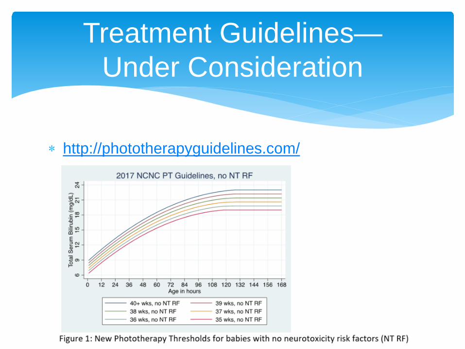
∗ http://phototherapyguidelines.com/
Treatment Guidelines—Under Consideration
50
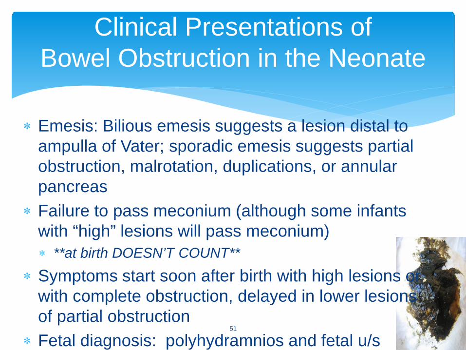
∗ Emesis: Bilious emesis suggests a lesion distal to ampulla of Vater; sporadic emesis suggests partial obstruction, malrotation, duplications, or annular pancreas
∗ Failure to pass meconium (although some infants with “high” lesions will pass meconium) ∗ **at birth DOESN’T COUNT**
∗ Symptoms start soon after birth with high lesions or with complete obstruction, delayed in lower lesions of partial obstruction
∗ Fetal diagnosis: polyhydramnios and fetal u/s
Clinical Presentations ofBowel Obstruction in the Neonate
51
∗ Atresia: complete obstruction of the lumen∗ 30% occur in duodenum (distal to ampulla)
∗ Stenosis: narrowing of the lumen∗ intrinsic cause or compression by extrinsic lesions
(annular pancreas, peritoneal bands)∗ plain films not diagnostic∗ emesis (amount and onset) depends on degree of
obstruction
Obstruction in the Newborn
52
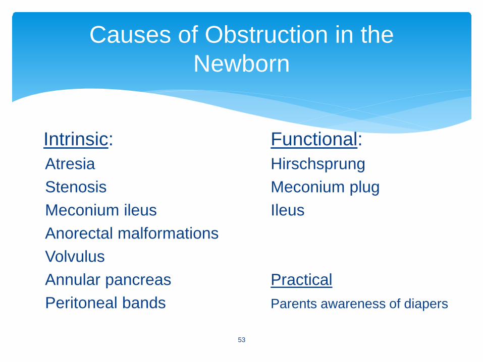
Intrinsic: Functional: Atresia HirschsprungStenosis Meconium plugMeconium ileus IleusAnorectal malformationsVolvulusAnnular pancreas PracticalPeritoneal bands Parents awareness of diapers
Causes of Obstruction in the Newborn
53
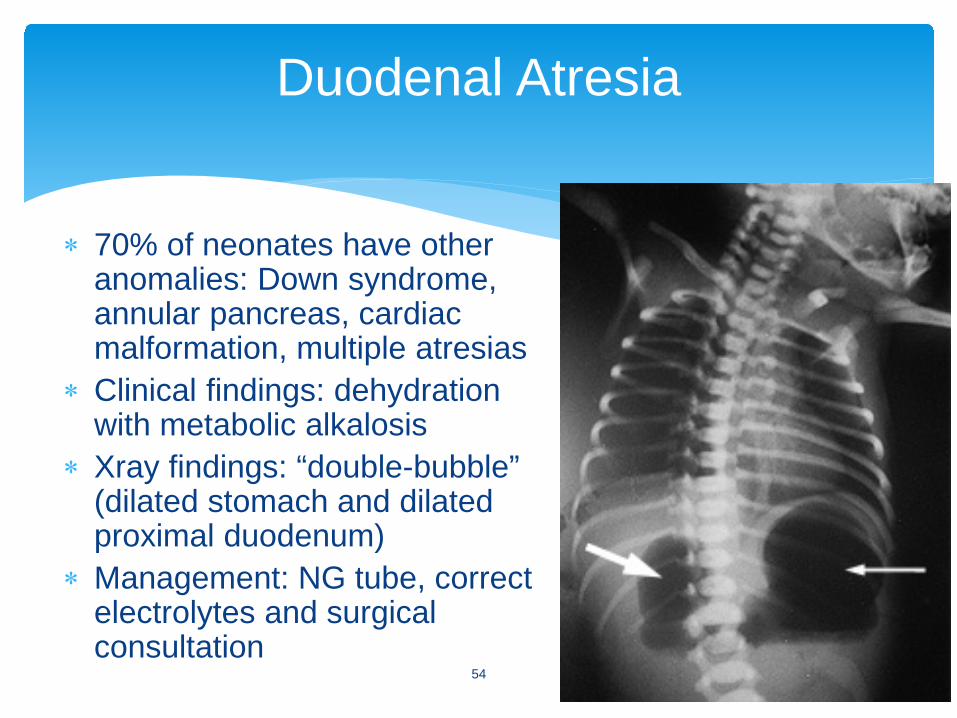
∗ 70% of neonates have other anomalies: Down syndrome, annular pancreas, cardiac malformation, multiple atresias
∗ Clinical findings: dehydration with metabolic alkalosis
∗ Xray findings: “double-bubble” (dilated stomach and dilated proximal duodenum)
∗ Management: NG tube, correct electrolytes and surgical consultation
Duodenal Atresia
54
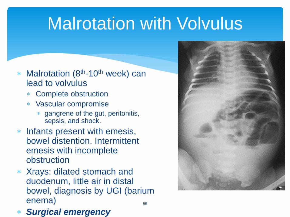
∗ Malrotation (8th-10th week) can lead to volvulus ∗ Complete obstruction ∗ Vascular compromise
∗ gangrene of the gut, peritonitis, sepsis, and shock.
∗ Infants present with emesis, bowel distention. Intermittent emesis with incomplete obstruction
∗ Xrays: dilated stomach and duodenum, little air in distal bowel, diagnosis by UGI (barium enema)
∗ Surgical emergency
Malrotation with Volvulus
55
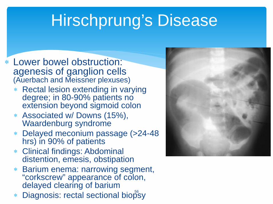
∗ Lower bowel obstruction: agenesis of ganglion cells (Auerbach and Meissner plexuses)∗ Rectal lesion extending in varying
degree; in 80-90% patients no extension beyond sigmoid colon
∗ Associated w/ Downs (15%), Waardenburg syndrome
∗ Delayed meconium passage (>24-48 hrs) in 90% of patients
∗ Clinical findings: Abdominal distention, emesis, obstipation
∗ Barium enema: narrowing segment, “corkscrew” appearance of colon, delayed clearing of barium
∗ Diagnosis: rectal sectional biopsy
Hirschprung’s Disease
56
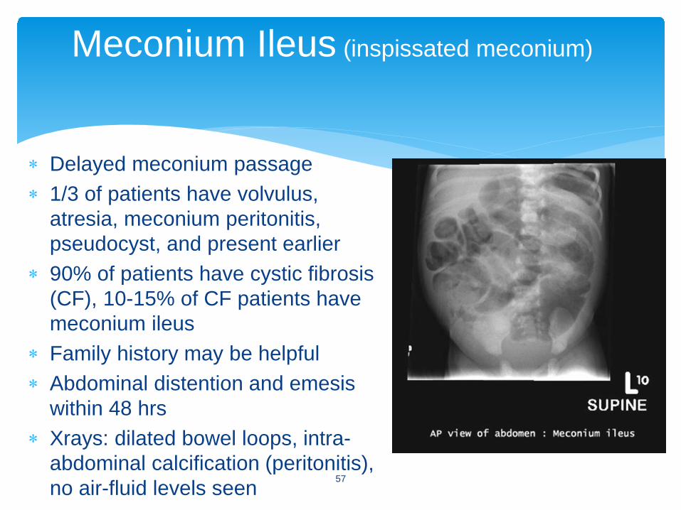
∗ Delayed meconium passage∗ 1/3 of patients have volvulus,
atresia, meconium peritonitis, pseudocyst, and present earlier
∗ 90% of patients have cystic fibrosis (CF), 10-15% of CF patients have meconium ileus
∗ Family history may be helpful∗ Abdominal distention and emesis
within 48 hrs∗ Xrays: dilated bowel loops, intra-
abdominal calcification (peritonitis), no air-fluid levels seen
Meconium Ileus (inspissated meconium)
57
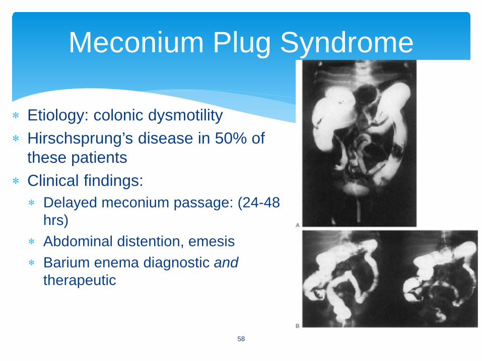
∗ Etiology: colonic dysmotility ∗ Hirschsprung’s disease in 50% of
these patients∗ Clinical findings:∗ Delayed meconium passage: (24-48
hrs)∗ Abdominal distention, emesis∗ Barium enema diagnostic and
therapeutic
Meconium Plug Syndrome
58
∗ Cephalhematoma∗ Caput succedaneum∗ Subgaleal hematoma∗ Erb’s palsy∗ Klumpke’s palsy∗ Clavicular fracture∗ Phrenic nerve injury with diaphragmatic
paralysis
Birth Injuries
59
http://nursingcrib.com/wp-content/uploads/caput-and-cephal.jpg?9d7bd460
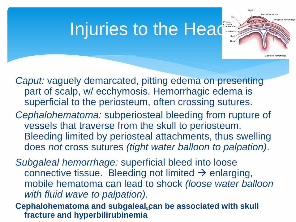
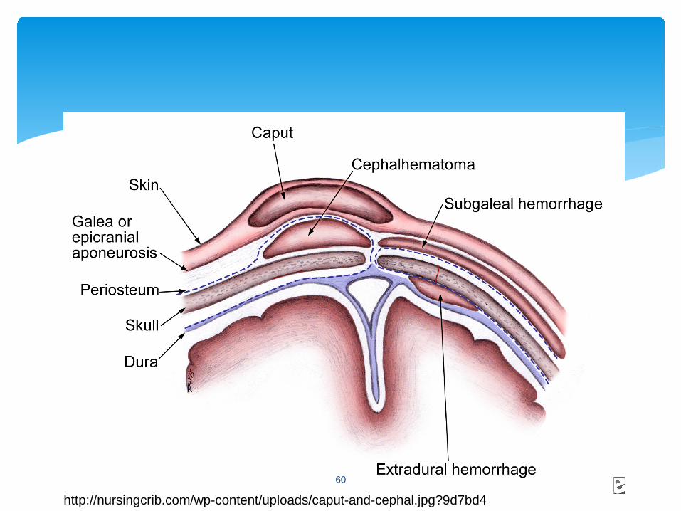
Caput: vaguely demarcated, pitting edema on presenting part of scalp, w/ ecchymosis. Hemorrhagic edema is superficial to the periosteum, often crossing sutures.
Cephalohematoma: subperiosteal bleeding from rupture of vessels that traverse from the skull to periosteum. Bleeding limited by periosteal attachments, thus swelling does not cross sutures (tight water balloon to palpation).
Subgaleal hemorrhage: superficial bleed into loose connective tissue. Bleeding not limited enlarging, mobile hematoma can lead to shock (loose water balloon with fluid wave to palpation).
Cephalohematoma and subgaleal can be associated with skull fracture and hyperbilirubinemia
Injuries to the Head
61

Subgaleal
http://www.pediatriconcall.com/fordoctor/casereports/subgaleal_hematoma.asp62
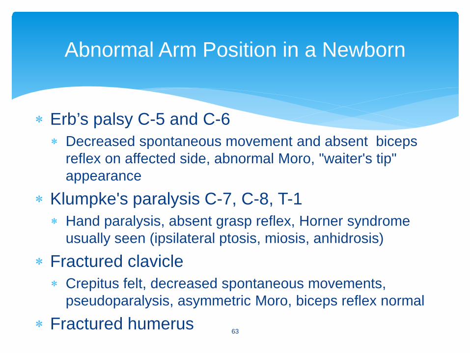
∗ Erb’s palsy C-5 and C-6∗ Decreased spontaneous movement and absent biceps
reflex on affected side, abnormal Moro, "waiter's tip" appearance
∗ Klumpke's paralysis C-7, C-8, T-1∗ Hand paralysis, absent grasp reflex, Horner syndrome
usually seen (ipsilateral ptosis, miosis, anhidrosis)
∗ Fractured clavicle ∗ Crepitus felt, decreased spontaneous movements,
pseudoparalysis, asymmetric Moro, biceps reflex normal
∗ Fractured humerus
Abnormal Arm Position in a Newborn
63
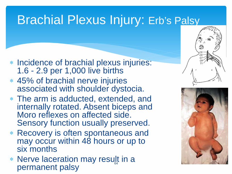
∗ Incidence of brachial plexus injuries: 1.6 - 2.9 per 1,000 live births
∗ 45% of brachial nerve injuries associated with shoulder dystocia.
∗ The arm is adducted, extended, and internally rotated. Absent biceps and Moro reflexes on affected side. Sensory function usually preserved.
∗ Recovery is often spontaneous and may occur within 48 hours or up to six months
∗ Nerve laceration may result in a permanent palsy
Brachial Plexus Injury: Erb’s Palsy
64
∗ Erythema toxicum neonatorum (“E tox”)∗ Benign pustular melanosis (“BPN”)∗ Hemangiomata∗ Nevus flammeus∗ Capillary∗ Cavernous∗ Mixed∗ Port wine stain
Common Neonatal Skin Conditions
65
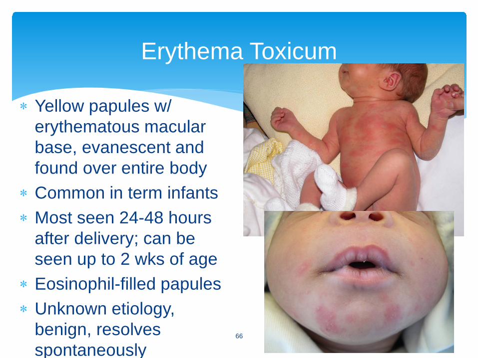
∗ Yellow papules w/ erythematous macular base, evanescent and found over entire body
∗ Common in term infants∗ Most seen 24-48 hours
after delivery; can be seen up to 2 wks of age
∗ Eosinophil-filled papules∗ Unknown etiology,
benign, resolves spontaneously
Erythema Toxicum
66

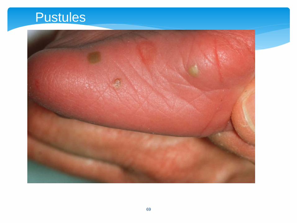
∗ Epidemiology: seen in 4.4% of African-American infants, 0.2% in white infants
∗ Lesion∗ Superficial pustular lesions that easily rupture then leave
a scaley “collar” around hyperpigmented macules∗ Fade within weeks to months
∗ Location: most in clusters under chin, nape of neck, forehead, and may be on trunk and extremities
∗ Sterile, transient, and not associated with systemic disease
Benign Pustular Melanosis
67

Pustules with scaling “collar”
68
Pustules
69
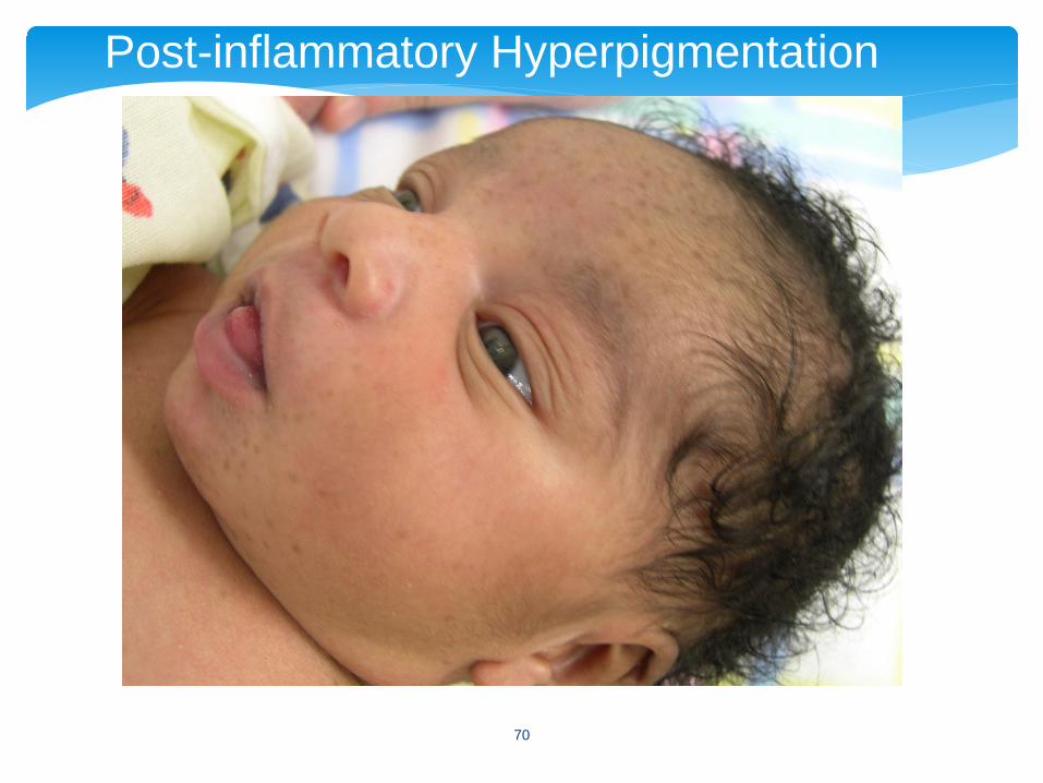
Post-inflammatory Hyperpigmentation
70
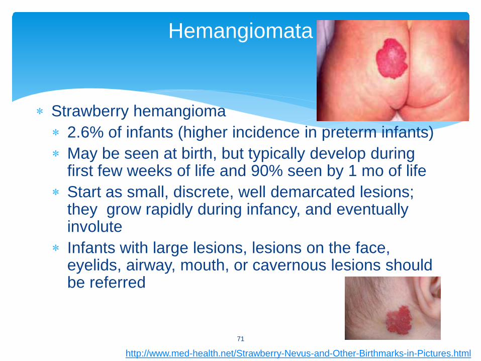
∗ Strawberry hemangioma∗ 2.6% of infants (higher incidence in preterm infants)∗ May be seen at birth, but typically develop during
first few weeks of life and 90% seen by 1 mo of life∗ Start as small, discrete, well demarcated lesions;
they grow rapidly during infancy, and eventually involute
∗ Infants with large lesions, lesions on the face, eyelids, airway, mouth, or cavernous lesions should be referred
Hemangiomata
71
http://www.med-health.net/Strawberry-Nevus-and-Other-Birthmarks-in-Pictures.html
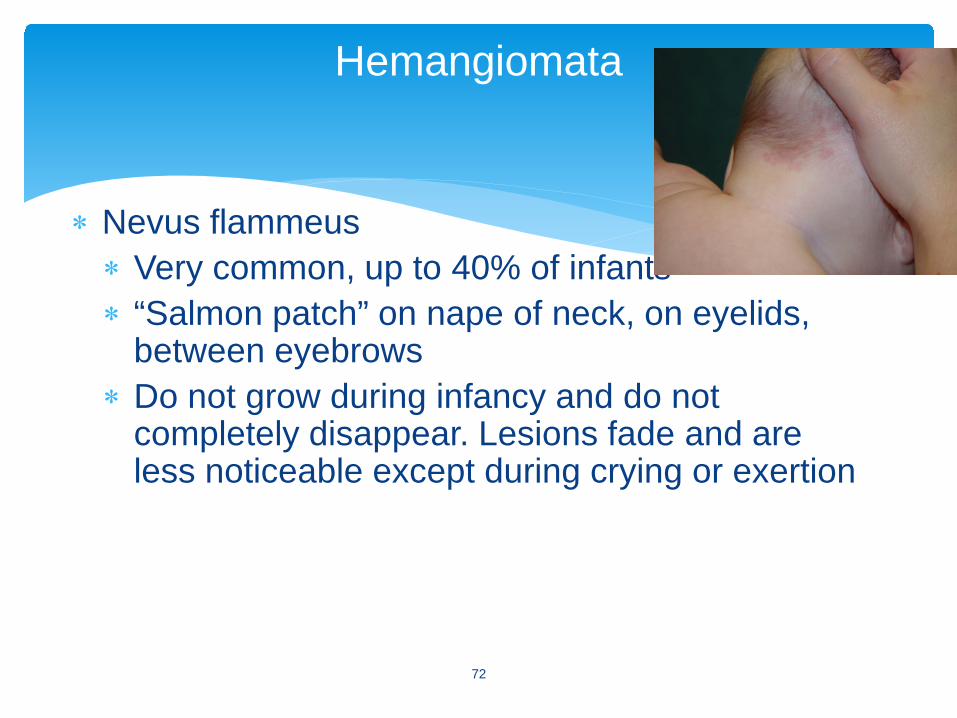
∗ Nevus flammeus∗ Very common, up to 40% of infants∗ “Salmon patch” on nape of neck, on eyelids,
between eyebrows∗ Do not grow during infancy and do not
completely disappear. Lesions fade and are less noticeable except during crying or exertion
Hemangiomata
72
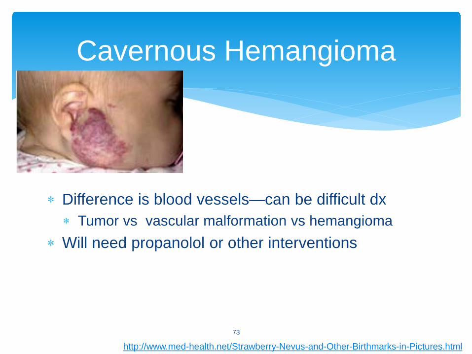
∗ Difference is blood vessels—can be difficult dx∗ Tumor vs vascular malformation vs hemangioma
∗ Will need propanolol or other interventions
Cavernous Hemangioma
73
http://www.med-health.net/Strawberry-Nevus-and-Other-Birthmarks-in-Pictures.html
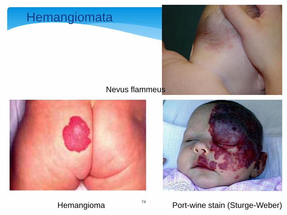
Hemangioma Port-wine stain (Sturge-Weber)
Hemangiomata
Nevus flammeus
74
Hemangiomas
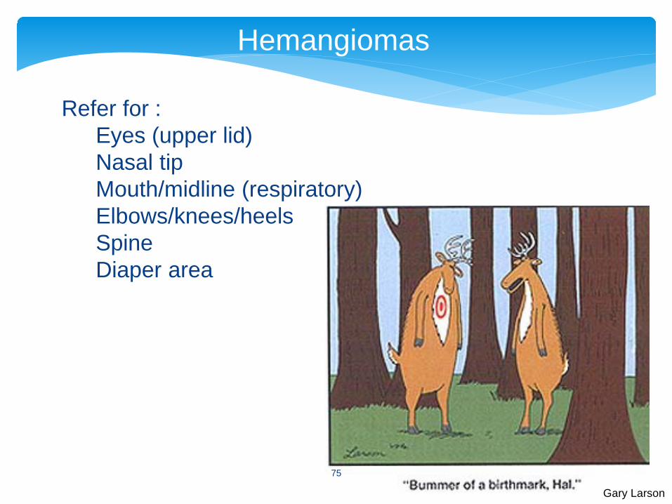
Refer for :Eyes (upper lid)Nasal tipMouth/midline (respiratory)Elbows/knees/heelsSpineDiaper area
Gary Larson
75
∗ What you hear when∗ Day of birth/1st DOL—outflow stenoses∗ After 1st day—coarctation∗ 1st week—left-to-right shunts (PDA, VSD, etc)∗ “Tachycardia”/bradycardia of newborn (DOL ~3)
∗ How? Training ear to VSD vs patent ductus∗ Stanford’s newborn nursery site:
http://newborns.stanford.edu/PhotoGallery/Heart.html
Cardiac/Murmurs
76

∗ What else to see?∗ Congestive heart failure—sweating, poor
feeding, failure to grow, HSM∗ What else to do? Check pre- and post-
ductal saturation after 24 HOL∗ Post-ductal <95%, or gradient >3%
Cardiac/Murmurs, cont’d
77
Breastfeeding
∗ Benefits∗ Challenges∗ Who is your patient?
∗ Resources∗ Lactation∗ Public health nurses∗ Local groups/stores/insurance
78
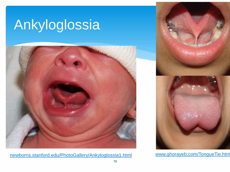
Ankyloglossia
newborns.stanford.edu/PhotoGallery/Ankyloglossia1.html www.ghorayeb.com/TongueTie.htm79
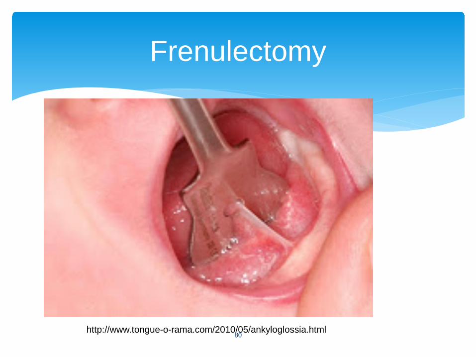
Frenulectomy
http://www.tongue-o-rama.com/2010/05/ankyloglossia.html80
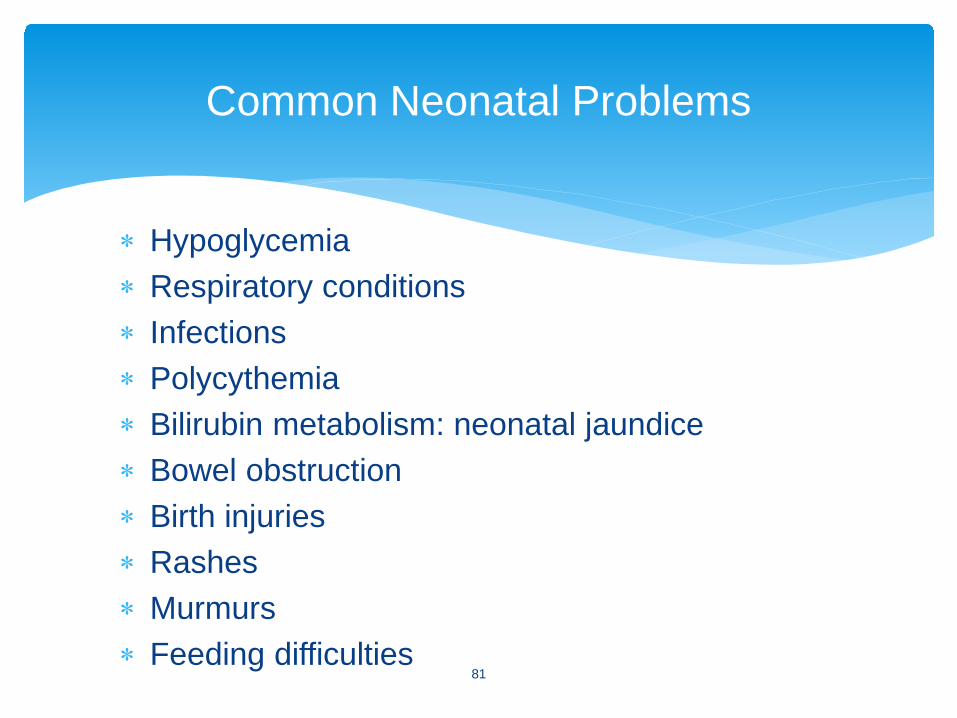
∗ Hypoglycemia∗ Respiratory conditions∗ Infections∗ Polycythemia∗ Bilirubin metabolism: neonatal jaundice∗ Bowel obstruction∗ Birth injuries∗ Rashes∗ Murmurs∗ Feeding difficulties
Common Neonatal Problems
81