Prezentacja programu PowerPoint - wl.cm.umk.pl · ANAMNESIS Pain (severe ... must be converted...
25
CHEST INJURIES Jacek Piątkowski M.D., Ph. D.
Transcript of Prezentacja programu PowerPoint - wl.cm.umk.pl · ANAMNESIS Pain (severe ... must be converted...

CHEST INJURIES
Jacek Piątkowski M.D., Ph. D.

CHEST INJURIES
3-4% of all injuries
8% of patients hospitalized due to injuries
65% of patients who died at the accident place

CLASSIFICATION OF THE CHEST INJURIES
(American Association for Surgery of Trauma)
Injuries of the chest wall
Injuries leading to respiratory failure
Injuries leading to hypovolemic shock
Injuries leading to infection/sepsis

CLASSIFICATION OF THE CHEST INJURIES
INJURIES OF THE CHEST WALL
Contusions
Hematomas
Rib fractures
Skin scapes
Superficial wounds
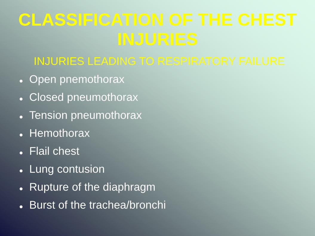
CLASSIFICATION OF THE CHEST INJURIES
INJURIES LEADING TO RESPIRATORY FAILURE
Open pnemothorax
Closed pneumothorax
Tension pneumothorax
Hemothorax
Flail chest
Lung contusion
Rupture of the diaphragm
Burst of the trachea/bronchi

CLASSIFICATION OF THE CHEST INJURIES
INJURIES LEADING TO HYPOVOLEMIC SHOCK
Burst of the lung
Wounds of the heart
Wounds of the big vessels
INJURIES LEADING TO INFECTION/SEPSIS
Esophageal perforation

DIAGNOSTICS OF THE CHEST INJURIES
ANAMNESIS
Pain (severe, light, superficial), dyspnoea, hemoptysis
Time, action and force of trauma
Another diseases (especially of respiratory and circulatorysystem)
PHYSICAL EXAMINATION
heart rate, blood pressure, respiration rate
color, temperature and moisture of the skin
state of consciousness
other injuries

DIAGNOSTICS OF THE CHEST INJURIES
LOCAL EXAMINATION
external signs of the injury
respiratory movements
deformation of the chest
subcutaneous emphysema
displacement of the trachea
local and transferred pain
breath sounds
cardiac action
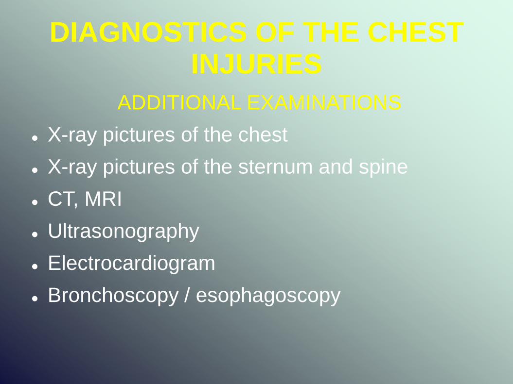
DIAGNOSTICS OF THE CHEST INJURIES
ADDITIONAL EXAMINATIONS
X-ray pictures of the chest
X-ray pictures of the sternum and spine
CT, MRI
Ultrasonography
Electrocardiogram
Bronchoscopy / esophagoscopy

DIAGNOSTICS OF THE CHEST INJURIES
LABORATORY TESTS
Blood tests (blood type, morphology, haemostasis)
Blood gasometry (partial pressure of oxygenand carbon dioxide) – PO
2<50mmHg and
PCO2>50mmHg – indication for mechanical
ventilation (respirator)

Thoracocentesis (punction of the pleural cavity)
The procedure fast and simple
Leads to diagnosis (air or blood)
Emergency treatment of tension pneumothorax
Therapeutic procedure in the case of small pnemothorax or hemothorax

INJURIES OF THE CHEST WALL
Contusions
Hematomas
Rib fractures
Skin scapes
Superficial wounds
Analgesics drugs
Intercostal nerve block
Adhesive strapping
Respiratory rehabilitation
Mucolytics

PNEUMOTHORAX
OPEN
becouse of injuries penetrating into pleural cavity
must be converted promptly to closed pneumothorax
CLOSED
becouse of leasion of the lung or its spontaneous rupture (emphysemotous bullae)
TENSION
large amount of air under pressure enters the pleural cavity
often subcutaneous emphysema
emergency state requiring fast reaction

PNEUMOTHORAX
TREATMENT
Observation (no symptoms of respiratory disfunction)
Punction of the pleural cavity (thoracocentesis)
Drainage of the pleural cavity (insertion of a chest tube and suction till full expansion of the lung)

FLAIL CHEST
double fractures of a few (at least 4) neighbouring ribs paradoxical motion of the chest (on inspiration the flexible area is pulled inward and pushed outward on expiration – ventilatory efficiency is obviously decreased) little disfunction of ventilation – immobilization with adhesive strapping (no circumferential) significant pulmonary disfunction – mechanical ventilation with respirator – eliminates paradoxical motion (as long as necessary even many weeks)

LUNG CONTUSION
at 50% of patients with severe blunt toracic injuries
it may lead to adult respiratory distress syndrome
(ARDS)
it is necessary to controll blood gasometry every 6-8
hours
in the presence of ventilation abnormalities early
mechanical ventilation with positive end-expiratory
presure (PEEP)

HEMOTHORAX
Becouse of bleeding from the lung or chest wall
PUNCTION OF THE PLEURAL CAVITY
<300ml – END 300-1000ml – DREINAGE
<300ml/h – efficient circulatory >300ml/h – circulatory insufficient
Punction >1000ml THORACOTOMY

DREINAGE PLEURAL CAVITY
VI-VII intercostal space in the midaxillary line
Suction negative pressure – 10-15cm of water
X-ray to controll of lung expansion
Closure dreinage after 48-72 hours
After next 24 hours controll X-ray – if the lung is expanded we can remove the dreinage

DREINAGE PLEURAL CAVITY

TRAUMATIC RUPTURE OF THE DIAPHRAGM
Becouse of blunt or sharp trauma
Over 90% on the left side
Usually related to other multiple injuries
Provides to respiratory failure
Diagnosis is based on the presence of abdominal organs in the chest (X-ray, CT)
Indication for operative treatment ( prefered abdominal incision for better exploration for visceral injuries)
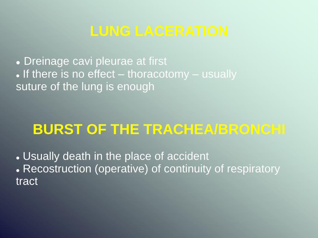
LUNG LACERATION
Dreinage cavi pleurae at first If there is no effect – thoracotomy – usually suture of the lung is enough
BURST OF THE TRACHEA/BRONCHI
Usually death in the place of accident Recostruction (operative) of continuity of respiratory tract

HEART INJURIES
HEART CONTUSIONS
becouse of blunt injuries
ECG signs and treatment are similar to myocardial infarction
in severe cases mechanical assistance with the intra-aortic baloon pump may be reqiured
PENETRATING WOUNDS
exsanguination is the most cause of death
sometimes cardiac tamponade (compression of the heart by blood in the pericardial sac) helps to save the patient
indication to operative therapy – V-th inercostal space on the left side

WOUNDS OF THE BIG VESSELS
Mortality at the accident site – 95%
Symptoms of hypovolemic shock
For diagnosis the best is angioCT
Indication for rapid surgery
The median sternotomy is the incision of choice
Usually required extracorporeal circulation
The best results - endovascular surgery (stent-graft)

INDICATIONS FOR THORACOTOMY
RAPID
Penetrating wounds with injury of the heart or big vesselsand symptoms of hypovolemic shock
Cardiac tamponade
URGENT
Injuries of the esophagus and diaphragm
Persistent heamorrage into pleural cavity
(dreinage >300ml during 3-4 hours with symptoms of hypovolemic shock)
Persistent air leak without lung expansion with symtpomsof respiratory respiratory insufficiency

INDICATIONS FOR THORACOTOMY
DELAYED
Hemothorax which can't be evacuated with dreinage (blood clots)
Purulent complications (pleural empyema, phlegmon of the mediastinum)
Pleural adhesions making impossible lungexpanding (decortication – removing fibrinemembrane off the lung)