Pressure Ulcer Assessment, Prevention & Management Print ...
38
Pressure Ulcer Assessment, Prevention & Management 2.0 Contact Hours First Published: May 17, 2012 Course Expires: March 17, 2016 Copyright © 2012 by RN.com All Rights Reserved Reproduction and distribution of these materials is prohibited without the express written authorization of RN.com
Transcript of Pressure Ulcer Assessment, Prevention & Management Print ...
Pressure Ulcer Assessment, Prevention & Management
2.0 Contact Hours
First Published: May 17, 2012
Course Expires: March 17, 2016
Copyright © 2012 by RN.com
All Rights Reserved
Reproduction and distribution of these
materials is prohibited without the express
written authorization of RN.com
nicollel
RN.com
Acknowledgements
RN.com acknowledges the valuable contributions of…
Shelley Lynch, MSN, RN, CCRN, co-author of Pressure Ulcer Assessment, Prevention, &
Management. Shelley has over 10 years of critical care nursing experience. She completed her
Bachelors of Science in Nursing from Hartwick College and her Master’s of Science in Nursing
with a concentration in education from Grand Canyon University. Shelley worked in a variety of
intensive care units in some of the top hospitals in the United States including: Johns Hopkins
Medical Center, Massachusetts General Hospital, New York University Medical Center, Tulane
Medical Center, and Beth Israel Deaconess Medical Center. She is the author of RN.com's:
Diabetes Overview, Thrombolytic Therapy for Acute Ischemic Stroke: t- PA/Activase, ICP
Monitoring, Abdominal Compartment Syndrome, Chest Tube Management, Acute Coronary
Syndrome: A Spectrum of Conditions and Emerging Therapies, and Understanding Intra-
Abdominal Pressures.
Kristen Lavoie, BSN, RN, CDE, CWOCN, co-author of Pressure Ulcer Assessment, Prevention, &
Management. Kristen has over 10 years experience as a certified Wound Ostomy and
Continence nurse both in the home care setting and in the acute care arena. Recently, she has
also worked towards a certification in Diabetes Education and has been the lead project
manager on a hospital wide performance improvement initative on caring for the hospitalized
diabetic patient.
Disclaimer
RN.com strives to keep its content fair and unbiased.
The authors, planning committee, and reviewers have no conflicts of interest in relation to this
course. Conflict of Interest is defined as circumstances a conflict of interest that an individual
may have, which could possibly affect Education content about products or services of a
commercial interest with which he/she has a financial relationship.
There is no commercial support being used for this course. Participants are advised that the
accredited status of RN.com does not imply endorsement by the provider or ANCC of any
commercial products mentioned in this course.
There is no "off label" usage of drugs or products discussed in this course.
You may find that both generic and trade names are used in courses produced by RN.com. The
use of trade names does not indicate any preference of one trade named agent or company
over another. Trade names are provided to enhance recognition of agents described in the
course.
Note: All dosages given are for adults unless otherwise stated. The information on medications
contained in this course is not meant to be prescriptive or all-encompassing. You are
encouraged to consult with physicians and pharmacists about all medication issues for your
patients.
Purpose
The purpose of Pressure Ulcer Assessment, Preventio, & Management is to:
• Provide a brief review of skin anatomy and physiology.
• Educate the RN on measures to accurately assess and stage pressure ulcers in order to
drive treatment options, affect reimbursement, and provide benchmark data.
• Provide guidelines of prevention and tools for early detection in order to optimize and
maintain skin integrity.
Learning Objectives
After successful completion of this course, you will be able to:
1. Define basic pathophysiology of skin and pressure ulcers.
2. State the regulatory guidelines and reimbursement.
3. Describe all the stages of pressure ulcers.
4. Assess for the risks of skin breakdown.
5. State five ways to prevent pressure ulcers.
6. Describe proper documentation on pressure ulcers.
Introduction
The skin is a complex organ system that has many important functions. The skin functions as a
protective barrier against external organisms, maintains temperature control, senses our
surroundings, eliminates wastes, and synthesizes Vitamin D.
This course will review the anatomy and physiology of skin, skin assessment, regulatory
guidelines, stage of pressure ulcers, risk of skin breakdown, prevention strategies, and proper
documentation.
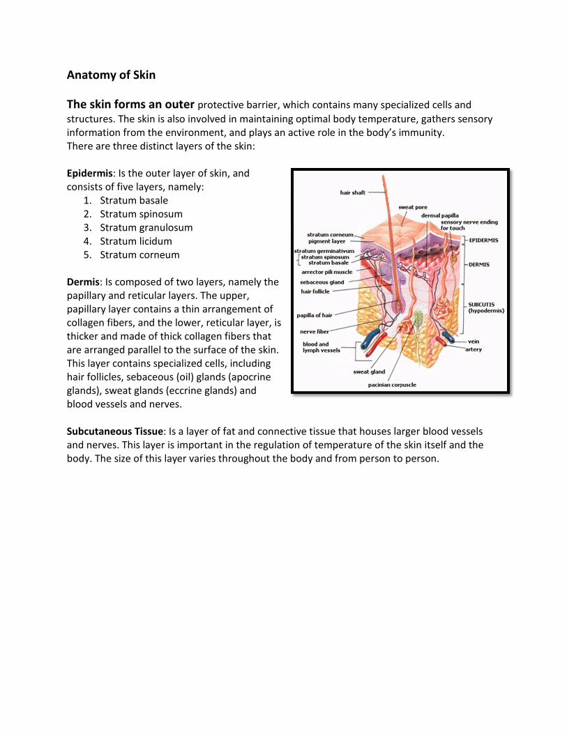
Anatomy of Skin
The skin forms an outer protective barrier, which contains many specialized cells and
structures. The skin is also involved in maintaining optimal body temperature, gathers sensory
information from the environment, and plays an active role in the body’s immunity.
There are three distinct layers of the skin:
Epidermis: Is the outer layer of skin, and
consists of five layers, namely:
1. Stratum basale
2. Stratum spinosum
3. Stratum granulosum
4. Stratum licidum
5. Stratum corneum
Dermis: Is composed of two layers, namely the
papillary and reticular layers. The upper,
papillary layer contains a thin arrangement of
collagen fibers, and the lower, reticular layer, is
thicker and made of thick collagen fibers that
are arranged parallel to the surface of the skin.
This layer contains specialized cells, including
hair follicles, sebaceous (oil) glands (apocrine
glands), sweat glands (eccrine glands) and
blood vessels and nerves.
Subcutaneous Tissue: Is a layer of fat and connective tissue that houses larger blood vessels
and nerves. This layer is important in the regulation of temperature of the skin itself and the
body. The size of this layer varies throughout the body and from person to person.
Understanding Pressure Ulcers
Why is it important to understand prevention, assessment, and documentation of pressure
ulcers?
1. Reducing pressure ulcers is a national goal.
2. Pressure ulcers are both a high-cost and high-volume adverse event.
3. Due to the negative health and economic effects of pressure ulcers, prevention is a
priority.
Healthy People 2010 & 2020
Reducing the prevalence of pressure ulcers is a national goal in the United States. It is a part of
the Healthy People 2010 initiative for the Nation’s health.
Reducing the rate of pressure ulcer-related hospitalizations among older adults is also a
Healthy People 2020 initiative (U.S. Department of Health and Human Services, 2012).
Regulatory Guidelines
Pressure ulcers are now reportable to state and federal agencies. The information is placed in
databases that can be accessed by the public (Centers for Medicare & Medicaid Services [CMS],
2012).
The Center for Medicare and Medicaid (CMS) is working together with the National Quality
Forum to improve hospital care and safety for the hospitalized patient (CSM, 2012).

Never Events
Ten “never events” are targeted to be eliminated because they have been deemed high cost or
high volume or both. These events result in the assignment of a case to an MS-DRG* that has a
higher payment when present as a secondary diagnosis, and could reasonably have been
prevented through the application of evidence-based guidelines. One of these events is
hospital-acquired pressure ulcers (CMS, 2012).
Hospital acquired conditions such as Stage III and Stage IV pressure ulcers are not reimbursable
at the MS-DRG rate (Miller, 2009).
*The Medicare Severity - Diagnosis Related Groups (MS-DRGs) are payment groups designed for the
Medicare population. Patients who have similar clinical characteristics and similar costs are assigned to a
MS-DRG. The MS-DRG will be linked to a fixed payment amount based on the average cost of patients in
the group.
Federal Implications
US Centers for Medicare and Medicaid services challenge acute care hospitals to link quality
and financial performance.
This challenge has opened up opportunities for acute care hospitals to reduce or even eliminate
hospital-acquired pressure ulcers (CMS, 2012).
POA Indicator Requirement
A pressure ulcer that is present at the time of admission is classified as POA (Present On
Admission), and is defined as being present at the time the order for inpatient admission
occurs.
At this time, the following hospitals are EXEMPT from the POA indicator requirement:
• Critical Access Hospitals (CAHs) and Long-Term Care Hospitals (LTCHs).
• Maryland Waiver Hospitals, Cancer Hospitals, and Children’s Inpatient Facilities.
• Rural Health Clinics and Federally Qualified Health Centers (FQHCs).
• Religious Non-Medical Health Care Institutions.
• Inpatient Psychiatric Hospitals, Inpatient Rehabilitation Facilities (IRFs), and
Veterans Administration/Department of Defense Hospitals.
(CMS, 2011)
Did You Know?
According to the Agency for Healthcare Research and Quality (AHRQ), in 2009, 6,428 never
events were reported. In Minnesota alone, 39% of never events were from pressure
ulcers/bedsores (AHRQ, 2009). You can visit the www.ahrq.gov website to find out how your
individual state’s statistics on pressure ulcers compare.
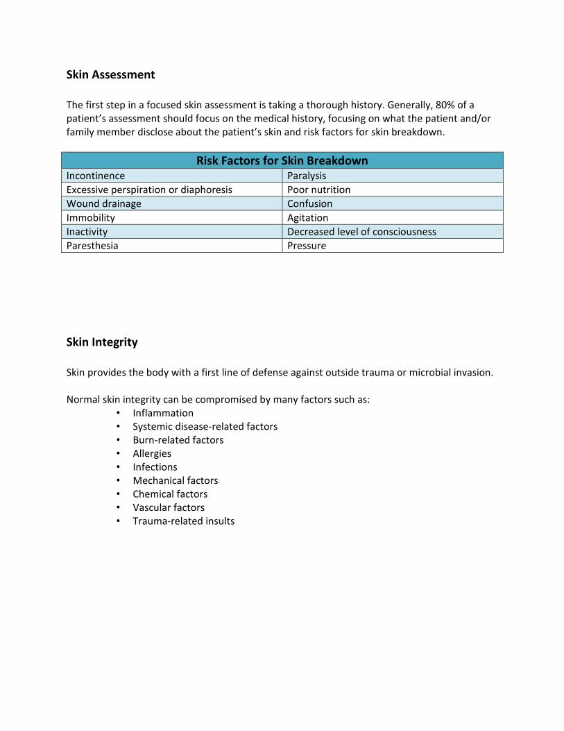
Skin Assessment
The first step in a focused skin assessment is taking a thorough history. Generally, 80% of a
patient’s assessment should focus on the medical history, focusing on what the patient and/or
family member disclose about the patient’s skin and risk factors for skin breakdown.
Risk Factors for Skin Breakdown Incontinence Paralysis
Excessive perspiration or diaphoresis Poor nutrition
Wound drainage Confusion
Immobility Agitation
Inactivity Decreased level of consciousness
Paresthesia Pressure
Skin Integrity
Skin provides the body with a first line of defense against outside trauma or microbial invasion.
Normal skin integrity can be compromised by many factors such as:
• Inflammation
• Systemic disease-related factors
• Burn-related factors
• Allergies
• Infections
• Mechanical factors
• Chemical factors
• Vascular factors
• Trauma-related insults

Focused History Questions
Focused history questions that will guide an age-specific skin assessment are summarized in the
following table.
Adults Geriatric Pediatric
• Past skin diseases
• Sun exposure
• Recent change in wart
or mole
• Sore that has not
healed
• Signs of abuse
• Dryness and itching
• Bruising tendency
• Longer healing time
• Nail texture changes
• Signs of abuse
• Use/type of diaper
cream or bathing
products
• Rashes or lesions
• Bruising
• Allergy
• Signs of abuse
• Injury history
• Sun exposure
Physical Exam
The skin is practically the only organ you can see. For that
reason, you should closely examine the skin – ALL OF IT. A
visual exam will enlighten you to many potential or evident
skin problems that the patient may not be aware of. When
you are visualizing the skin and see an actual or potential
issue, compare this area to adjacent tissue.
Look for localized areas of heat
or coolness. Heat may indicate
underlying inflammation, but
coolness may indicate
underlying tissue damage. Any
skin changes should be
documented and reported
immediately.
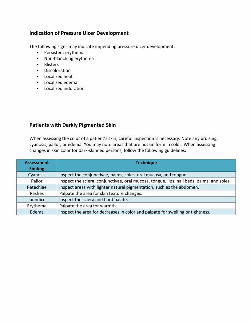
Indication of Pressure Ulcer Development
The following signs may indicate impending pressure ulcer development:
• Persistent erythema
• Non-blanching erythema
• Blisters
• Discoloration
• Localized heat
• Localized edema
• Localized induration
Patients with Darkly Pigmented Skin
When assessing the color of a patient’s skin, careful inspection is necessary. Note any bruising,
cyanosis, pallor, or edema. You may note areas that are not uniform in color. When assessing
changes in skin color for dark-skinned persons, follow the following guidelines:
Assessment
Finding
Technique
Cyanosis Inspect the conjunctivae, palms, soles, oral mucosa, and tongue.
Pallor Inspect the sclera, conjunctivae, oral mucosa, tongue, lips, nail beds, palms, and soles.
Petechiae Inspect areas with lighter natural pigmentation, such as the abdomen.
Rashes Palpate the area for skin texture changes.
Jaundice Inspect the sclera and hard palate.
Erythema Palpate the area for warmth.
Edema Inspect the area for decreases in color and palpate for swelling or tightness.

Temperature, Moisture & Texture
Skin temperature can range from cool to warm. Warm is always normal. Note if the overall
skin’s temperature is cool or warm, or if it is localized.
Normally, your patient’s skin should be dry with only a slight amount of moisture. Overly moist
skin may be due to environmental conditions, anxiety, obesity, hyperthyroidism, fever, or
diaphoresis. Dry skin affects approximately 59% to 85% of person’s older than 64 years of age
(Hess, 2008). Many factors contribute to dry skin, including a low-humidity environment, the
patient’s personal habits (smoking, alcohol intake, and poor nutrition), seasonal changes,
chronic diseases, medications, and skin cleaners (Hess, 2010).
Inspect the skin for a normally smooth, mobile texture. You can check skin turgor by grasping
the skin on the top of the hand and gently pulling up. After letting go of the skin, the skin
should “snap” back into place within three seconds. Skin that remains elevated or “tented” may
be due to age related changes, dehydration, or a combination of both.
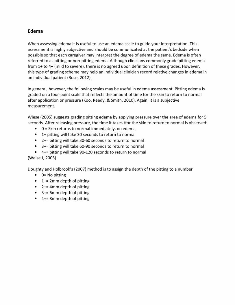
Edema
When assessing edema it is useful to use an edema scale to guide your interpretation. This
assessment is highly subjective and should be communicated at the patient’s bedside when
possible so that each caregiver may interpret the degree of edema the same. Edema is often
referred to as pitting or non-pitting edema. Although clinicians commonly grade pitting edema
from 1+ to 4+ (mild to severe), there is no agreed upon definition of these grades. However,
this type of grading scheme may help an individual clinician record relative changes in edema in
an individual patient (Rose, 2012).
In general, however, the following scales may be useful in edema assessment. Pitting edema is
graded on a four-point scale that reflects the amount of time for the skin to return to normal
after application or pressure (Koo, Reedy, & Smith, 2010). Again, it is a subjective
measurement.
Wiese (2005) suggests grading pitting edema by applying pressure over the area of edema for 5
seconds. After releasing pressure, the time it takes tfor the skin to return to normal is observed:
• 0 = Skin returns to normal immediately, no edema
• 1+ pitting will take 30 seconds to return to normal
• 2+= pitting will take 30-60 seconds to return to normal
• 3+= pitting will take 60-90 seconds to return to normal
• 4+= pitting will take 90-120 seconds to return to normal
(Weise J, 2005)
Doughty and Holbrook’s (2007) method is to assign the depth of the pitting to a number
• 0= No pitting
• 1+= 2mm depth of pitting
• 2+= 4mm depth of pitting
• 3+= 6mm depth of pitting
• 4+= 8mm depth of pitting
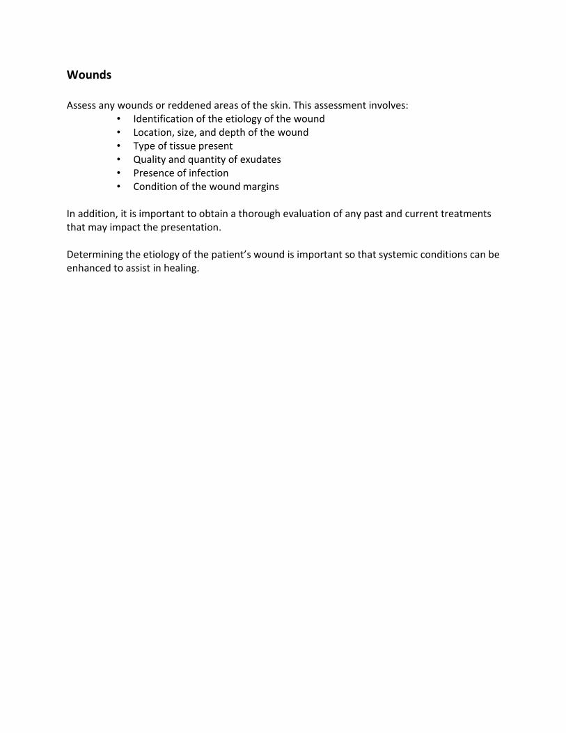
Wounds
Assess any wounds or reddened areas of the skin. This assessment involves:
• Identification of the etiology of the wound
• Location, size, and depth of the wound
• Type of tissue present
• Quality and quantity of exudates
• Presence of infection
• Condition of the wound margins
In addition, it is important to obtain a thorough evaluation of any past and current treatments
that may impact the presentation.
Determining the etiology of the patient’s wound is important so that systemic conditions can be
enhanced to assist in healing.
Wound Etiology
Acute wounds are usually obvious in the etiology (i.e., surgery, trauma) but the causative
factors in chronic wounds can be less apparent. Often location is an indication of its cause.
Chronic wounds are important to differentiate since their pathophysiology, and thus
management pathways differ. Characteristic clinical location and appearance usually allows for
clear distinction between ischemic, venous, and neuropathic ulcers (Armstrong & Meyr, 2012).
• Ischemic Ulcers: Result of inadequate perfusion due to arterial obstruction.
• Venous Ulcer: Commonly located between the knee and ankle. Commonly from
deep vein thrombosis and venous valvular incompetence.
• Pressure Ulcers: Usually located over bony prominences. Areas of necrosis and
ulceration where soft tissue structures are compressed between osseous
prominences or hard external surfaces.
• Diabetic/Neuropathic Ulcers- Chronic ulceration in patients with diabetes is
multifactorial, due to a combination of diabetic neuropathy, autonomic
dysfunction, and vascular insufficiency. They happen at locations of the body
with repeated trauma such as plantar metatarsal heads. Areas of the foot are
often exposed to repetitive trauma (toes and sides of feet)
• Malignant Ulcers: Tumors can present similar to chronic wounds.
• Hypertension Ulcers: These are uncommon. Often associated with arterial
hypertension in patients with palpable pulses.
(Armstrong & Meyr, 2012).
**This course will not focus on diabetic, arterial, or venous ulcers. It will also not focus on other
skin issues such as: moles, lesions, or burns. It will focus on the pressure induced ulcer.
The Pressure Ulcer
Pressure ulcers are lesions caused by unrelieved pressure that results in damage to the
underlying tissue. Generally, these are the result of soft tissue compression between a body
prominence and an external surface for a prolonged period of time (Berlowitz, 2012).
Pressure Ulcer Staging
Recently in 2008, the National Pressure Ulcer Advisory Panel updated the definitions of the
pressure ulcer staging. In order to prevent and treat pressure ulcers, it is important to
understand the definitions of the following stages:
• Stage 1
• Stage 2
• Stage 3
• Stage 4
• Unstageable
• Suspected deep tissue injury
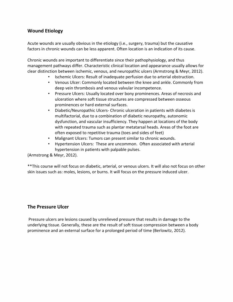
Stage 1
• Non-blanchable erythema
• Intact skin with non-blanchable redness of a localized
area usually over a bony prominence.
• Darkly pigmented skin may not have visible blanching;
it’a color may differ from the surrounding area.
• The area may be painful, firm, soft, warmer or cooler
than adjacent tissue.
(National Pressure Ulcer Advisory Panel [NPUAP], 2010)
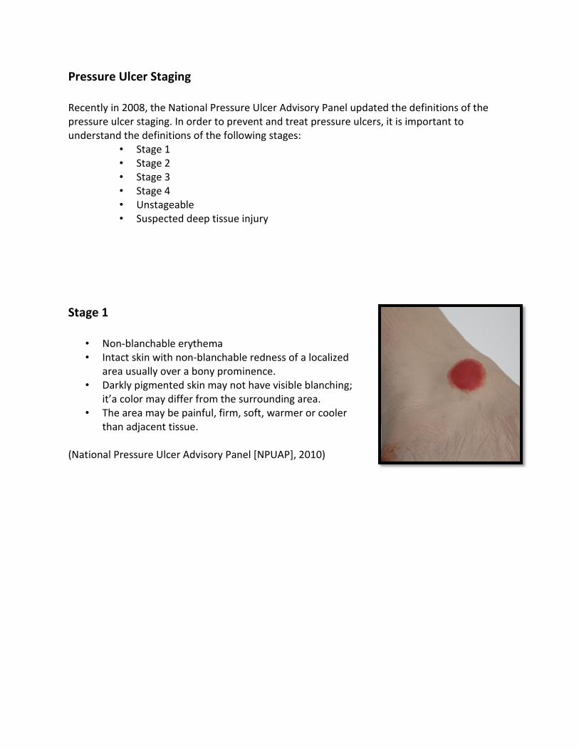
Stage 2
• Partial thickness skin loss
• Loss of dermis presenting as a shallow open ulcer
with a red pink wound bed, without slough
• Intact or open/ruptured serum-filled blister
(NPUAP, 2010)
Stage 3
• Full thickness skin loss
• Subcutaneous fat may be visible but bone,
tendon or muscle are not exposed
• Slough may be present but does not obscure
the depth of the tissue loss
• Undermining and tunneling may be present
• Depth of Stage 3 varies by anatomical location
• Shallow Stage 3 pressure ulcers
– Occiput, malleolus, bridge of nose, ear
(NPUAP, 2010)

Stage 4
• Full thickness tissue Loss
• Exposed bone, tendon, or muscle
• Slough or eschar may be present on some parts of
the wound bed
• Tunneling and undermining are often present.
• May be extending into muscle and/or supporting
structures making osteomyelitis possible
(NPUAP, 2010)
Unstageable Pressure Ulcer
• Depth unknown
• Full thickness tissue loss in which the base of the
ulcer is covered by slough (yellow, tan, gray, green,
or brown) and/or eschar (tan, brown, or black) in
the wound bed
• Until enough slough and/or eschar is removed to
expose the base of the wound, the true depth
cannot be determined
(NPUAP, 2010)
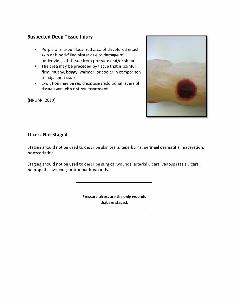
Suspected Deep Tissue Injury
• Purple or maroon localized area of discolored intact
skin or blood-filled blister due to damage of
underlying soft tissue from pressure and/or shear
• The area may be preceded by tissue that is painful,
firm, mushy, boggy, warmer, or cooler in comparison
to adjacent tissue
• Evolution may be rapid exposing additional layers of
tissue even with optimal treatment
(NPUAP, 2010)
Ulcers Not Staged
Staging should not be used to describe skin tears, tape burns, perineal dermatitis, maceration,
or excoriation.
Staging should not be used to describe surgical wounds, arterial ulcers, venous stasis ulcers,
neuropathic wounds, or traumatic wounds.
Pressure ulcers are the only wounds
that are staged.
Risk Assessment
Why is it important to do a risk assessment for all patients?
• Used to identify patient’s potential for or actual risk for developing pressure
ulcers.
• Assists with targeting appropriate interventions for prevention of pressure
ulcers.
• Risk assessment tools should be conducted on admission and repeated regularly
and as frequently as required by patient acuity. Reassessment must be
completed if there is any change to the patient’s condition.
(NPUAP, 2010)
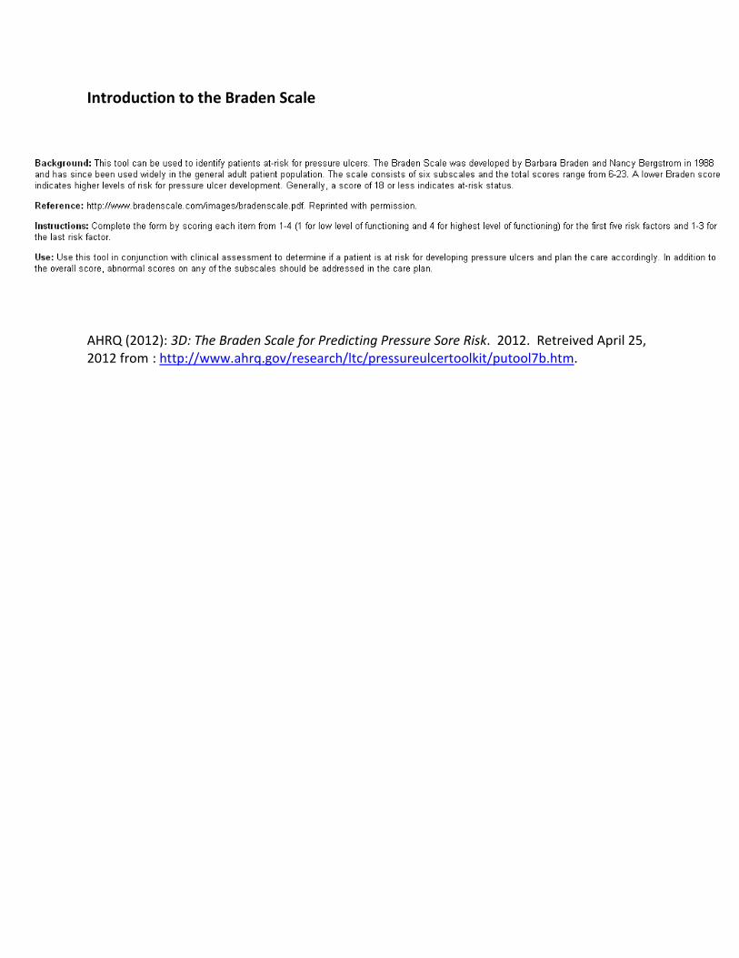
Introduction to the Braden Scale
AHRQ (2012): 3D: The Braden Scale for Predicting Pressure Sore Risk. 2012. Retreived April 25,
2012 from : http://www.ahrq.gov/research/ltc/pressureulcertoolkit/putool7b.htm.
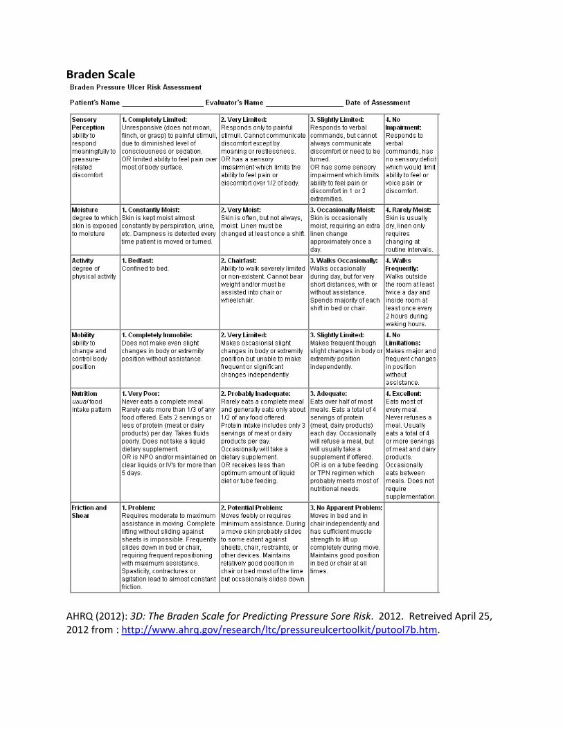
Braden Scale
AHRQ (2012): 3D: The Braden Scale for Predicting Pressure Sore Risk. 2012. Retreived April 25,
2012 from : http://www.ahrq.gov/research/ltc/pressureulcertoolkit/putool7b.htm.
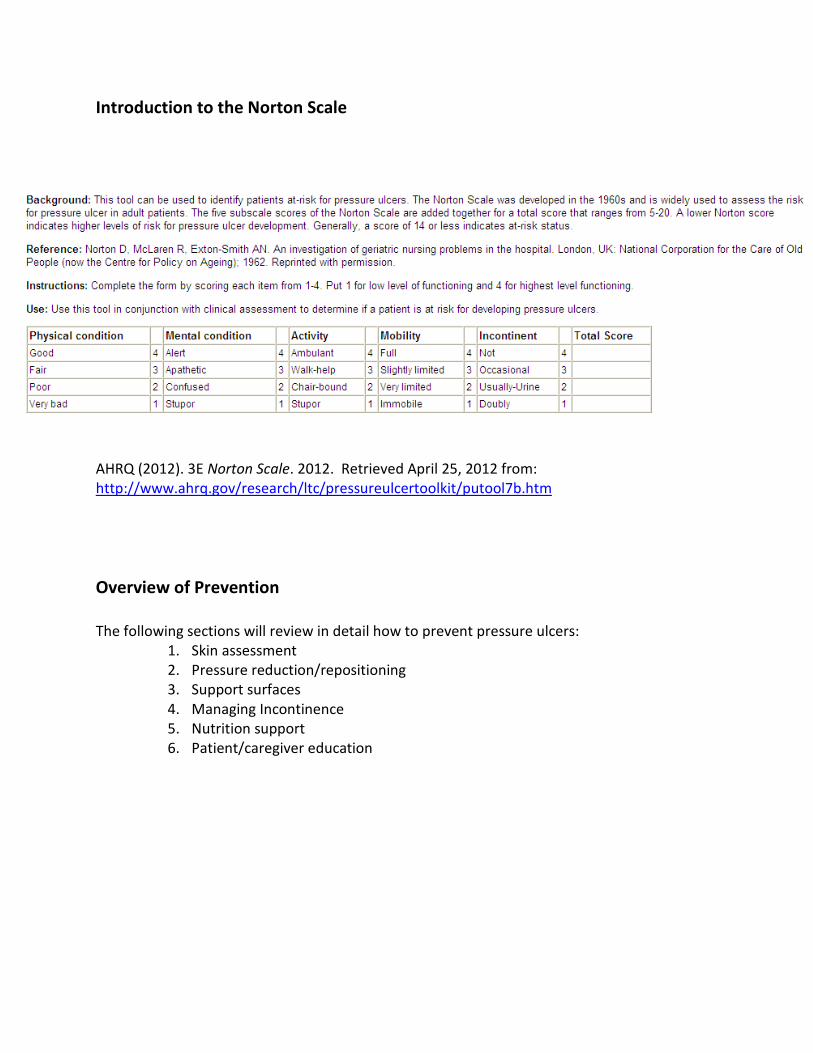
Introduction to the Norton Scale
AHRQ (2012). 3E Norton Scale. 2012. Retrieved April 25, 2012 from:
http://www.ahrq.gov/research/ltc/pressureulcertoolkit/putool7b.htm
Overview of Prevention
The following sections will review in detail how to prevent pressure ulcers:
1. Skin assessment
2. Pressure reduction/repositioning
3. Support surfaces
4. Managing Incontinence
5. Nutrition support
6. Patient/caregiver education
Skin Assessment
A head-to-toe skin inspection should occur on admission to an institution, and every 24 hours
thereafter (or per institution policy).
As a reminder, the skin assessment should include but not be limited to skin temperature, skin
color, skin texture/turgor, skin integrity, and moisture status (NPUAP, 2010).
Pressure Redistribution/Repositioning
Immobility is the most significant risk factor for pressure ulcer development. Patients with any
degree of immobility should be closely monitored for pressure ulcer development.
Repositioning involves moving a patient (i.e. changing their position from sitting or lying in bed),
in order to redistribute pressure and make the patient more comfortable. This should be done
at regular intervals.
• Failure to reposition will result in tissue ischemia and probable tissue damage.
• Frequency of repositioning will depend on the patient’s activity/mobility level,
the patient’s tissue tolerance to pressure, and the patient’s overall skin and
medical condition.
• Avoid positioning directly onto medical devices such as tubes or drains.
• Avoid positioning on bony prominences with existing pressure ulcers.
• Repositioning should be at a 30-degree tilted side-lying position. Avoidance of
increased pressure positions such as 90-degree side-lying position or semi-
recumbent position.
• Use transfer aides to reduce friction and shear.
― LIFT, don’t drag while repositioning.
(NPUAP, 2010)
Support Services • A supportive surface is defined by National Pressure Ulcer Advisory Panel (NPUAP) as “a
specialized device for pressure redistribution designed for management of tissue loads,
microclimate, and/or other therapeutic functions.”
• Examples of support surfaces include: mattress, integrated
bed system, mattress replacement or overlay, or seat cushion
or seat cushion overlay.
• Choose a support surface compatible with the care setting.
• All individuals at risk for pressure ulcers should continue to
be turned and repositioned on a regular basis when a
support surface is in place.
• Limit the amount of linen between the individual and the support surface.
• Ideally heels should be “floated” off the bed surface due to the small surface area of the
heel making it a challenge to try to redistribute the load from the heel through the use
of pressure-redistribution device (Lyman, 2009).
• Prolonged sitting results in a higher risk of pressure ulcer development.
― Using a pressure-redistribution seat cushion for individuals whose mobility is
reduced and are at risk for pressure ulcer development.
― Limiting time an individual spends in a chair without pressure relief.
Managing Incontinence
Moisture from incontinence can contribute to pressure ulcer development by macerating the
skin and increasing friction injuries. Pressure ulcers are four times more likely in incontinent
patients than those who are continent (Wound Ostomy and Continence Nurses Society
[WOCN], 2010).
• Patients who are incontinent should be cleaned as soon as possible after soiling.
• Specialized incontinence cleansers or soaps that are neutral in PH and contain
moisturizer are recommended.
• Do not leave patient on a bedpan longer than necessary.
• Use of protective skin barriers for incontinent patients protects skin the from
excessive moisture related to incontinence.
• Pat skin when cleaning and drying; do not rub.
• When documenting on skin breakdown with an incontinent individual it is
imperative to determine if skin breakdown is due to irritant dermatitis vs.
pressure or possibly both.
Donut-type devices or rings
have been shown to cause
ischemia over the pressure
area causing more damage
than good.
Nutritional Support
• All individuals with a pressure ulcer or risk for pressure ulcer should be referred to a
dietitian for assessment and intervention of nutritional problems.
• Individuals should be assessed for their ability to eat independently.
• Enhanced foods and/or oral supplements should be offered between meals if needed.
• Consider enteral or parenteral nutrition support when oral intake is inadequate if
appropriate.
• Maintain hydration (offer fluids on regular basis when not contraindicated).
• Provide adequate protein intake.
– 1.25-1.5grams protein/kg body weight daily for patient with an existing pressure
ulcer.
(NPUAP, 2010)
Patient/Caregiver Education
Training in the correct methods of repositioning and use of equipment should be offered to all
involved in the care of individuals at risk for pressure ulcers (NPUAP, 2010).
Educate the patient and caregiver that Immobility is the most significant risk factor for pressure
ulcer development (NPUAP, 2010).
Teach the family/patient about the normal healing process and the signs and symptoms that
should be brought to a professional’s attention (NPUAP, 2010).
Pressure ulcer prevention is enhanced when both patients and their caregivers are included
(Akkuzu, Aeslantas, Kosker, & Sen, 2009).
Overview of Documentation
The following slides will review how to document for pressure ulcers.
1. Pressure ulcer stage
2. Anatomical location
3. Wound measurements
4. Appearance of wound bed
5. Assessment of drainage
6. Condition of periwound skin
7. Wound care performed
8. Patients tolerance to wound care
9. Wound progress towards goal
10. Pain management
Pressure Ulcer Stage
All assessments and skin inspection findings should be documented within 24 hours of
admission or according to institution policy.
Pressure ulcer assessment must include the stage of pressure ulcer: Stage 1, 2, 3, 4,
unstageable, or suspected deep tissue injury.
Pressure ulcers should never be reversed staged. Once layers of tissue and supporting
structures are gone they are not replaced. Instead, the wound is filled with granulation tissue.
• Example: Once a pressure ulcer is a Stage 4 it will become a healing Stage 4 once
it begins to granulate in.
(Bryant & Nix, 2007)
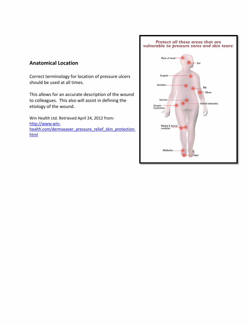
Anatomical Location
Correct terminology for location of pressure ulcers
should be used at all times.
This allows for an accurate description of the wound
to colleagues. This also will assist in defining the
etiology of the wound.
Win Health Ltd. Retrieved April 24, 2012 from:
http://www.win-
health.com/dermasaver_pressure_relief_skin_protection.
html
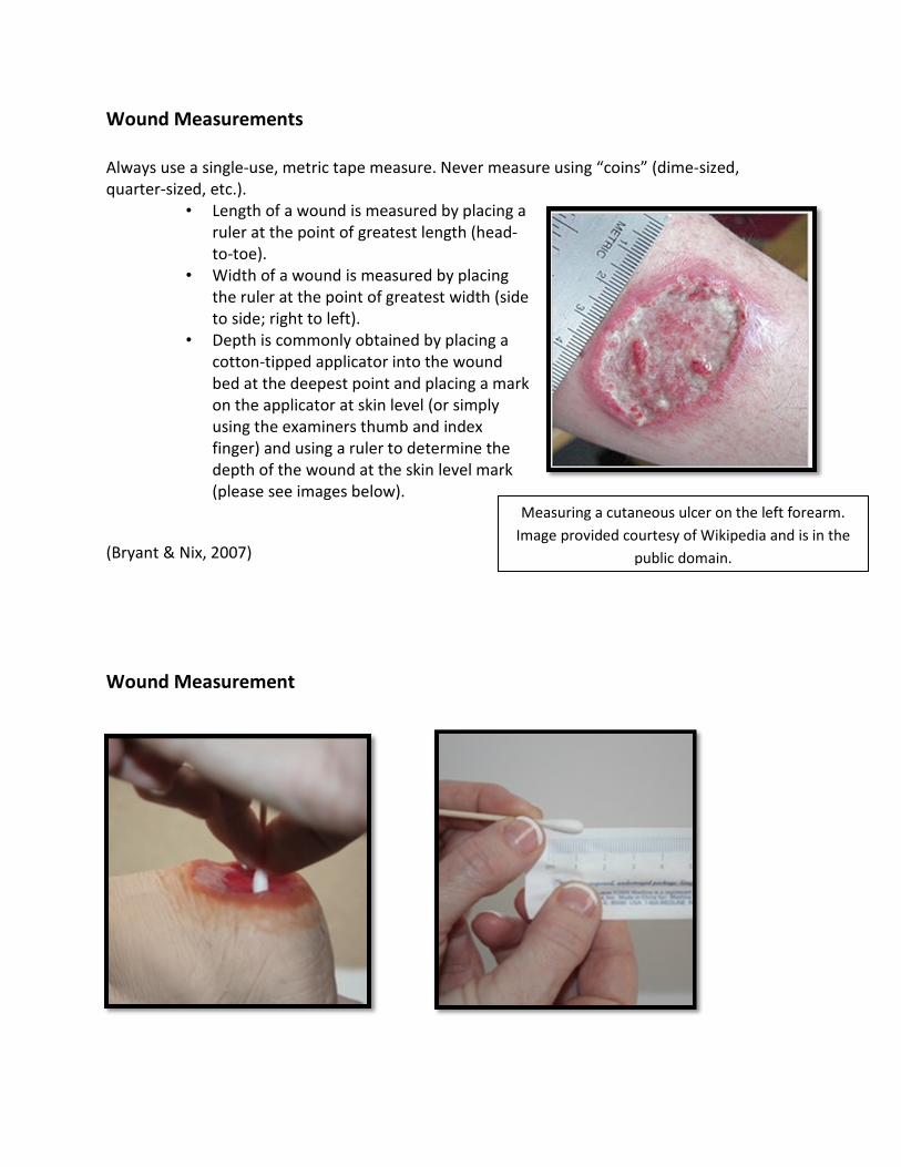
Wound Measurements
Always use a single-use, metric tape measure. Never measure using “coins” (dime-sized,
quarter-sized, etc.).
• Length of a wound is measured by placing a
ruler at the point of greatest length (head-
to-toe).
• Width of a wound is measured by placing
the ruler at the point of greatest width (side
to side; right to left).
• Depth is commonly obtained by placing a
cotton-tipped applicator into the wound
bed at the deepest point and placing a mark
on the applicator at skin level (or simply
using the examiners thumb and index
finger) and using a ruler to determine the
depth of the wound at the skin level mark
(please see images below).
(Bryant & Nix, 2007)
Wound Measurement
Measuring a cutaneous ulcer on the left forearm.
Image provided courtesy of Wikipedia and is in the
public domain.
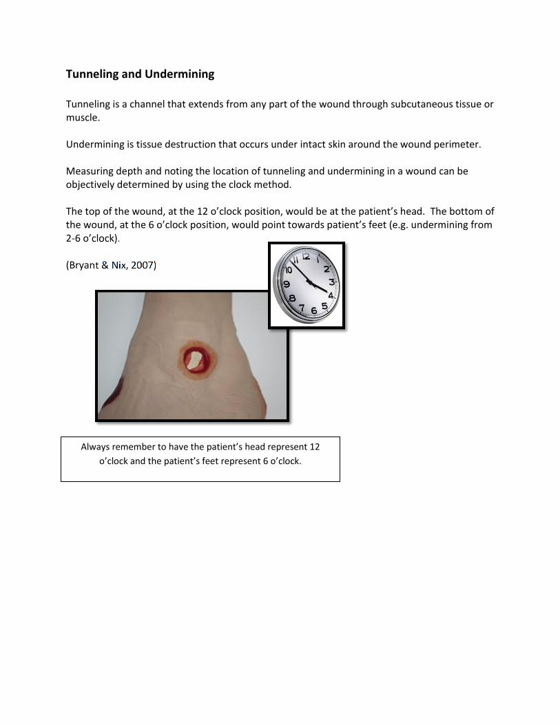
Tunneling and Undermining
Tunneling is a channel that extends from any part of the wound through subcutaneous tissue or
muscle.
Undermining is tissue destruction that occurs under intact skin around the wound perimeter.
Measuring depth and noting the location of tunneling and undermining in a wound can be
objectively determined by using the clock method.
The top of the wound, at the 12 o’clock position, would be at the patient’s head. The bottom of
the wound, at the 6 o’clock position, would point towards patient’s feet (e.g. undermining from
2-6 o’clock).
(Bryant & Nix, 2007)
Always remember to have the patient’s head represent 12
o’clock and the patient’s feet represent 6 o’clock.

Appearance of Wound Bed
Types and amount of tissues in the wound bed should always be assessed and documented.
Many pressure ulcers have a combination of different types of tissues in the wound bed. These
combinations should be documented in percentages.
Type and amount of granulation tissue in a wound bed
will be indicative of where that pressure ulcer is in the
healing phase.
(Bryant & Nix, 2007)
Tissue Type & Thickness
The wound bed tissue reveals the phase and progress of wound healing.
There are tissue colors that can be seen in pressure ulcers such as pink, red, black, and
yellow/beige:
• Epithelial tissue is "pearly pink" in color.
• Granulation tissue is beefy red.
• Necrotic tissue is usually black, brown, or tan and known as eschar.
• Yellow necrotic tissue is known as slough (it can also be tan, gray, green, or
brown).
(NPUAP, 2010)
Documentation Example:
80% of the wound bed contains
yellow slough and 20% contains
granulation tissue.
Assessment of Drainage
The amount, type, and odor of wound drainage should always be assessed and documented.
• Amount: Assessed as none, light, moderate, or heavy.
• Type: Assessed as being clear, serous sanguineous, sanguineous, purulent, tan,
or bloody.
• Odor: Assessed as being absent, faint, moderate, or strong.
– It is important to know that most wounds do have an odor and the type
of dressing can affect the wound odor, as well as the presence of
devitalized tissue.
(Bryant & Nix, 2007)
Condition of Periwound Skin
The integrity of periwound skin will ensure clues to the effectiveness of the treatment choice or
dressing application.
For example, maceration of the periwound skin could indicate poor application of dressing. If
the dressing overlaps on the periwound skin or when exudate is allowed to pool on periwound
skin, these indicate an inappropriate dressing application.
Periwound skin should be assessed for:
• Color (erythema or white)
• Temperature (cool or warm)
• Texture (moist, indurated, boggy, or dry)
• Integrity (candidiasis, epidermal stripping, pustules)
(Bryant & Nix, 2007)
Wound Margins
• Pay close attention to wound margins, looking specifically for undermining or dead
spaces.
• Dead spaces are areas where the wound edges have come away from the wound base.
• These areas may show signs of poor circulation such as grey or purple coloration.
• Dead spaces should carefully be investigated to determine the extent of the
undermining.
(Hess, 2005)
Wound Care Performed
Documentation of the wound care should be clear and compatible with the wound care orders
which are prescribed for the patient.
The type of cleansing solution, type of dressing used, and the application of any secondary
dressing should always be clearly documented each time the wound care is performed.
Patients’ Tolerance to Wound Care
PAIN:
• Assess for pressure-ulcer-related pain in adults using a validated scale.
• Assessment of pain should include an assessment of body language and
nonverbal cues.
• Wound pain can indicate deterioration, infection, or even inappropriate wound
treatments.
• Optimize pressure ulcer care to ensure that it is coordinated with pain
medication administration.
• Pain should be measured and rated prior to each dressing change and post
dressing change to determine if the appropriate interventions for pain
management were initiated during wound care.
(NPUAP, 2010)
Wound Process Towards Goal
• Pressure ulcers should be assessed at each dressing change for progress towards
healing.
• Pressure ulcer scale for healing (PUSH):
– Validated tool developed by the NPUAP to monitor pressure ulcer healing over
time (NPUAP, 2010)
– Monitors 3 parameters considered most indicative of healing:
1. Size (length & width), exudate amount, and tissue type
2. Record a sub score 0-5 (size) & 0-4 (exudate & tissue type); total score
calculated ranging from 0-17 (0 = healed)
3. Comparison of total scores over time provides an indication of the
improvement or deterioration in pressure ulcer healing (Bryant & Nix,
2007)
Conclusion
The costs of pressure ulcers to the health-care system is estimated to be $11 billion each year
(Schessel, Ger, & Oddsen, 2012).
In order to prevent a pressure ulcer and achieve positive outcomes, a multidisplinary team
approach must be utilized. The physician, wound care specialist, nurse, physical therapist, and
registered dietitian are all major components of this multidisciplinary team (Delmore, Lebovits,
Baldock, Suggs, Ayello, 2011).
Regular skin assessment, risk evaluation, skin care interventions, mobility assessment, and
nutrition evaluation are all equal components of prevention and treatment of pressure ulcers
(Delmore, Lebovits, Baldock, Suggs, Ayello, 2011).
Pressure Ulcer Resource
Agency for Healthcare Research and Quality
540 Gaither Road Rockville, MD 20850
(301) 427-1364
Preventing Pressure Ulcers in Hospitals: A Toolkit for Improving Quality of Care is available at:
http://www.ahrq.gov/research/ltc/pressureulcertoolkit/putool7b.htm
References
Agency for Healthcare Research and Quality [AHRQ] (2009). Never Events. Retrieved March
10th, 2012, from http://www.psnet.ahrq.gov/primer.aspx?primerID=3.
Akkuzu, G., Aeslantas, S., Kosker, S.B., & Sen, S., 2009). Evaluation by Patients and Caregivers
of the Effectiveness of a Brochure Developed to Prevent Pressure Ulcers. Journal of Wound,
Ostomy and Continence Nursing,35(6), 610-615.
Armstrong, D. & Meyr, A. (2012). Clinical Assessment of Wounds. Retrieved March 12, 2012,
from www.uptodate.com.
Berlowitz, D. (2012). Pressure ulcers: Epidemiology, Pathogenesis, Clinical Manifestations, and
Staging. Retrieved March 27, 2012, from www.uptodate.com.
Bryant, R.A. & Nix, D.P. (2007). Acute & Chronic Wounds. St. Louis, MO: Mosby.
Centers for Medicare & Medicaid Services [CMS] (2012). Retrieved March 20, 2012, from
www.cms.gov/hospitalacqcond/.
Centers for Medicare & Medicaid Services [CMS] (2011). Present on Admission Indicator
Reporting by Acture Inpatient Prospective Payment System Hospitals. Retrieved March 20,
2012, from https://www.cms.gov/MLNProducts/downloads/wPOAFactSheet.pdf.
Delmore, B., Lebovits, S., Baldock, P., Suggs, B., & Ayello, E.A. (2011) Pressure Ulcer Prevention
Program. JWOCN, 38(5), 505-513.
Dillon, P.M. (2007). Nursing Health Assessment: Clinical Pocket Guide 2nd ed. Philadelphia, PA:
F.A. Davies.
Doughty, D.B. & Holbrook, B. (2007). Lower Extremity Ulcer of Vascular Etiology. Acute and
Chronic Wounds: Current Management Concepts. (3rd Ed.). St. Louis, MO: Mosby.
Hess, C.T. (2010). Performing a Skin Assessment. Nursing 2010, 66
Hess, C.T. (2008). Performing a Skin Assessment. Advances in Skin & Wound Care, 24(8): 392.
Hess, C.T. (2005). Clinical Guide Wound Care (5th ed.). Ambler, PA: Lippincott Williams &
Wilkins.
Koo, L.W.; Reedy, S., & Smith, J.K. (2010). Patient History Key to Diagnosing Peripheral Edema.
The Nurse Practitioner, 35(3), 44-52.
References (cont.)
Lyman, V. (2009). Successful Heel Pressure Ulcer Prevention Program in a Long-Term Care
Setting. Journal of Wound, Ostomy and Continence Nursing,35(6), 616-621.
Miller, A. (2009). Hospital Reporting and “Never Events”. Medicare Patient Management.
Retrieved March 25, 2012, from http://www.medicarepatientmanagement.com/issues/04-
03/mpmMJ09-NeverEvents.pdf.
National Pressure Ulcer Advisory Panel (2010). International Guideline: Prevention and
Treatment of Pressure Practice Guideline. Washington DC: National Pressure Ulcer Advisory
Panel.
Rose, B. (2012) Clinical Manifestations and Diagnosis of Edema in Adults. Retrieved March 17,
2012, from www.uptodate.com.
Schessel, E.S., Ger, R., & Oddsen, R. (2012). The Cost and Outcomes of Treating a Pressure Ulcer
in a Patient with a Deep Pressure Ulcer in a Patient with Quadriplegia, Ostomy Wound
Management, 58, 2, 41-46.
U.S. Department of Health and Human Services (2012). Healthy People 2012. Retrieved March
10th, 2012, from
http://www.healthypeople.gov/2020/topicsobjectives2020/objectiveslist.aspx?topicid=31.
Wiese, J. (2005). The Patient History: Evidence-Based Approach. New York, NY: Lange Medical
Books McGraw-Hill.
Wound Ostomy and Continence Nurses Society (2010). Guidelines for Prevention and
Management of Pressure Ulcers. Mount Laurel, NJ: WOCN.

Please Read
This publication is intended solely for the use of healthcare professionals taking this course, for
credit, from RN.com. It is designed to assist healthcare professionals, including nurses, in
addressing many issues associated with healthcare. The guidance provided in this publication is
general in nature, and is not designed to address any specific situation. This publication in no
way absolves facilities of their responsibility for the appropriate orientation of healthcare
professionals.
Hospitals or other organizations using this publication as a part of their own orientation
processes should review the contents of this publication to ensure accuracy and compliance
before using this publication. Hospitals and facilities that use this publication agree to defend
and indemnify, and shall hold RN.com, including its parent(s), subsidiaries, affiliates,
officers/directors, and employees from liability resulting from the use of this publication. The
contents of this publication may not be reproduced without written permission from RN.com.