International Review: Pressure Ulcer Prevention: pressure, shear ...

Pressure Ulcer Assessment, Prevention and Management
This course has been awarded two (2) contact hours. This course expires on January 31, 2019
Copyright © 2012 by RN.com All Rights Reserved. Reproduction and distribution of these materials are prohibited without an RN.com content licensing agreement. First Published: May 17, 2012 Course Updated: January 7, 2016
RN.com is accredited as a provider of continuing nursing education by the American Nurses Credentialing Center’s Commission on Accreditation.

Pressure Ulcer Assessment, Prevention and Management
Page 2 of 70
Acknowledgements RN.com acknowledges the valuable contributions of… Jennifer Turney MSN, RN, CNS, CPN Shelley Lynch, MSN, RN, CCRN, Kristen Lavoie, BSN, RN, CDE, CWOCN,

Pressure Ulcer Assessment, Prevention and Management
Page 3 of 70
Purpose The purpose of Pressure Ulcer Assessment, Prevention, and Management is to: Provide a brief review of skin anatomy and physiology. Educate the RN on measures to:
• Accurately assess and stage pressure ulcers in order to drive treatment options
• Affect reimbursement Provide guidelines of prevention and strategies for early detection in order to
optimize and maintain skin integrity.

Pressure Ulcer Assessment, Prevention and Management
Page 4 of 70
Learning Objectives After successful completion of this course, you will be able to:
1. Define basic pathophysiology of skin and pressure ulcers. 2. State the regulatory guidelines and reimbursement implications. 3. Describe all the stages of pressure ulcers. 4. Assess for the risks of skin breakdown. 5. State five ways to prevent pressure ulcers. 6. Describe proper documentation on pressure ulcers. 7. Describe common wound dressing for treatments for pressure ulcers.
Pressure Ulcer Assessment, Prevention and Management
Page 5 of 70
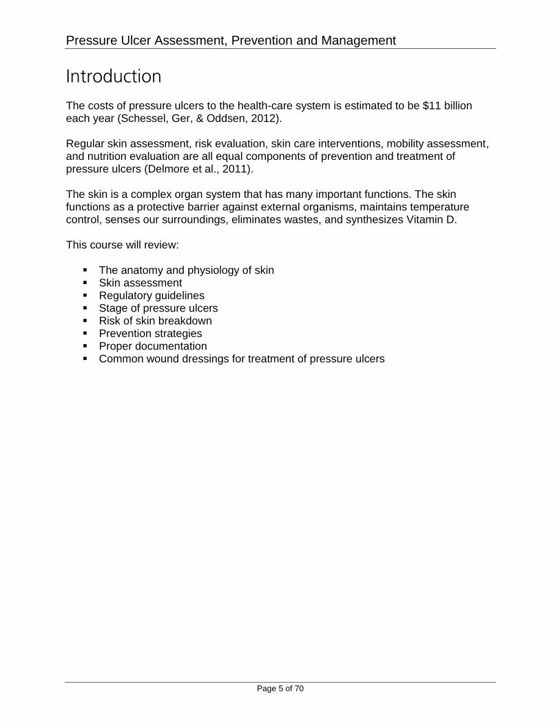
Introduction The costs of pressure ulcers to the health-care system is estimated to be $11 billion each year (Schessel, Ger, & Oddsen, 2012). Regular skin assessment, risk evaluation, skin care interventions, mobility assessment, and nutrition evaluation are all equal components of prevention and treatment of pressure ulcers (Delmore et al., 2011). The skin is a complex organ system that has many important functions. The skin functions as a protective barrier against external organisms, maintains temperature control, senses our surroundings, eliminates wastes, and synthesizes Vitamin D. This course will review:
The anatomy and physiology of skin Skin assessment Regulatory guidelines Stage of pressure ulcers Risk of skin breakdown Prevention strategies Proper documentation Common wound dressings for treatment of pressure ulcers

Pressure Ulcer Assessment, Prevention and Management
Page 6 of 70
Anatomy of Skin The skin forms an outer protective barrier, which contains many specialized cells and structures. The skin is also involved in maintaining optimal body temperature, gathers sensory information from the environment, and plays an active role in the body’s immunity. Three distinct layers of the skin are: Epidermis: Is the outer layer of skin, and consists of five layers, namely:
1. Stratum basale 2. Stratum spinosum 3. Stratum granulosum 4. Stratum licidum 5. Stratum corneum
Dermis: Is the innermost layer and is composed of two layers, namely the papillary and reticular layers. The upper, papillary layer contains a thin arrangement of collagen fibers, and the lower, reticular layer, is thicker and made of thick collagen fibers that are arranged parallel to the surface of the skin. This layer contains specialized cells, including hair follicles, sebaceous (oil) glands (apocrine glands), sweat glands (eccrine glands) and blood vessels and nerves. Subcutaneous Tissue: Is a layer of fat and connective tissue that houses larger blood vessels and nerves. This layer is important in the regulation of temperature of the skin itself and the body. The size of this layer varies throughout the body and from person to person. (Bryant & Nix, 2012)
Pressure Ulcer Assessment, Prevention and Management
Page 7 of 70
The Pressure Ulcer A pressure ulcer is localized injury to the skin and/or underlying tissue usually over a bony prominence, as a result of pressure, or pressure in combination with shear (National Pressure Ulcer Advisory Panel [NPUAP], 2014).

Pressure Ulcer Assessment, Prevention and Management
Page 8 of 70
Understanding Pressure Ulcers Why is it important to understand prevention, assessment, and documentation of pressure ulcers?
1. Reducing pressure ulcers is a national goal. 2. Pressure ulcers are both a high-cost and high-volume adverse event. 3. Due to the negative health and economic effects of pressure ulcers, prevention is
a priority.

Pressure Ulcer Assessment, Prevention and Management
Page 9 of 70
Healthy People 2020 Reducing the rate of pressure ulcer related hospitalizations among older adults is a national patient safety goal in the United States. It is a part of the Healthy People 2020 objectives for the Nation’s health. (U.S. Department of Health and Human Services, 2015).

Pressure Ulcer Assessment, Prevention and Management
Page 10 of 70
Regulatory Guidelines According to regulatory guidelines, pressure ulcers are now reportable to state and federal agencies. The information is placed in databases that can be accessed by the public (Centers for Medicare & Medicaid Services [CMS], 2015). The Center for Medicare and Medicaid (CMS) is working together with the National Quality Forum to improve hospital care and safety for the hospitalized patient (CSM, 2015).

Pressure Ulcer Assessment, Prevention and Management
Page 11 of 70
Never Events The term "Never Event" was first introduced in 2001 by Ken Kizer, MD, former CEO of the National Quality Forum (NQF), in reference to particularly shocking medical errors (such as wrong-site surgery) that should never occur. One of these events is hospital-acquired pressure ulcers (CMS, 2014 a&b). The NQF initially defined 27 such events in 2002. The list has been revised since then, most recently in 2011, and now consists of 29 events grouped into categories: surgical, product or device, patient protection, care management, environmental, radiologic, and criminal. (Agency for Healthcare Research and Quality [AHRQ], 2014).

Pressure Ulcer Assessment, Prevention and Management
Page 12 of 70
Never Events Targeted to Be Eliminated “Never Events” are targeted to be eliminated because they have been deemed high cost or high volume or both. These events result in the assignment of a case to an MS-DRG* that has a higher payment when present as a secondary diagnosis, and could reasonably have been prevented through the application of evidence-based guidelines. Hospital acquired conditions such as Stage III and Stage IV pressure ulcers are not reimbursable at the MS-DRG rate (CMS, 2014a&B). *The Medicare Severity - Diagnosis Related Groups (MS-DRGs) are payment groups designed for the Medicare population. Patients who have similar clinical characteristics and similar costs are assigned to a MS-DRG. The MS-DRG will be linked to a fixed payment amount based on the average cost of patients in the group.
Pressure Ulcer Assessment, Prevention and Management
Page 13 of 70
Reimbursement Implications US Centers for Medicare and Medicaid services challenge acute care hospitals to link quality and financial performance. This challenge has opened up opportunities for acute care hospitals to reduce or even eliminate hospital-acquired pressure ulcers. In FY 2016 CMS has committed to establishing, under the Affordable Care Act, a scoring methodology for hospitals related to a total hospital- acquired condition (HAC) score.
Those hospitals scoring in the top performing quartile will receive a one percent payment reduction.
Those hospitals scoring in the lowest performing quartile will receive a one percent payment penalty (CMS, 2014b)

Pressure Ulcer Assessment, Prevention and Management
Page 14 of 70
Present on Admission (POA) Indicator Requirement A pressure ulcer that is present at the time of admission is classified as POA (Present On Admission), and is defined as being present at the time the order for inpatient admission occurs. Conditions that develop during an outpatient encounter (including emergency department, observation, or outpatient surgery) are considered POA.

Pressure Ulcer Assessment, Prevention and Management
Page 15 of 70
Exempt from POA Indicator Requirement At this time, the following hospitals are EXEMPT from the POA indicator requirement:
• Critical Access Hospitals (CAHs) and Long-Term Care Hospitals (LTCHs). • Maryland Waiver Hospitals, Cancer Hospitals, and Children’s Inpatient
Facilities. • Rural Health Clinics and Federally Qualified Health Centers (FQHCs). • Religious Non-Medical Health Care Institutions. • Inpatient Psychiatric Hospitals, Inpatient Rehabilitation Facilities (IRFs),
and Veterans Administration/Department of Defense Hospitals. (CMS, 2014a)

Pressure Ulcer Assessment, Prevention and Management
Page 16 of 70
Skin Assessment The first step in a focused skin assessment is taking a thorough history. Generally, 80% of a patient’s assessment should focus on the medical history, focusing on what the patient and/or family member disclose about the patient’s skin and risk factors for skin breakdown.
Risk Factors for Skin Breakdown Incontinence Paralysis
Excessive perspiration or diaphoresis Poor nutrition
Wound drainage Confusion
Immobility Agitation
Inactivity Decreased level of consciousness
Paresthesia Pressure
(Bryant & Nix 2012)

Pressure Ulcer Assessment, Prevention and Management
Page 17 of 70
Skin Integrity Skin provides the body with a first line of defense against outside trauma or microbial invasion. Normal skin integrity can be compromised by many factors such as:
• Inflammation • Systemic disease-related factors • Burn-related factors • Allergies • Infections • Mechanical factors • Chemical factors • Vascular factors • Trauma-related insults (Bryant & Nix 2012)

Pressure Ulcer Assessment, Prevention and Management
Page 18 of 70
Focused History Questions Focused history questions that will guide an age-specific skin assessment are summarized in the following table.
Adults Geriatric Pediatric Past skin diseases Sun exposure Recent change in wart
or mole Sore that has not healed Signs of abuse
Dryness and itching Bruising tendency Longer healing time Nail texture changes Signs of abuse
Use/type of diaper cream or bathing products
Rashes or lesions Bruising Allergy Signs of abuse Injury history Sun exposure
(Bryant & Nix 2012)

Pressure Ulcer Assessment, Prevention and Management
Page 19 of 70
Physical Exam The skin is practically the only organ you can see. For that reason, you should closely examine the skin – ALL OF IT. A visual exam will enlighten you to many potential or evident skin problems that the patient may not be aware of. When you are visualizing the skin and see an actual or potential issue, compare this area to adjacent tissue. (Bryant & Nix 2012)
Look for localized areas
of heat or coolness. Heat may indicate underlying
inflammation, but coolness may indicate
underlying tissue damage. Any skin changes should be
documented and reported immediately.

Pressure Ulcer Assessment, Prevention and Management
Page 20 of 70
Indication of Pressure Ulcer Development The following signs may indicate impending pressure ulcer development:
• Persistent erythema • Non-blanching erythema • Blisters • Discoloration • Localized heat • Localized edema • Localized induration (Bryant & Nix 2012)

Pressure Ulcer Assessment, Prevention and Management
Page 21 of 70
Patients with Darkly Pigmented Skin When assessing the color of a patient’s skin, careful inspection is necessary. Note any bruising, cyanosis, pallor, or edema. You may note areas that are not uniform in color. When assessing for pressure ulcers in skin color for dark-skinned persons, consider the following guidelines:
Color remains unchanged when pressure is applied. Color changes occur at site of pressure which differs from the patient’s
usual skin color. Circumscribed area of intact skin may be warm to touch. As tissue
changes color, intact skin will feel cool to touch. Note gloves may diminish sensitivity to changes in skin temperature.
If patient previously had a pressure ulcer, that area of skin may be lighter
than original color. Localized area of skin may be purple/blue or violet (eggplant) instead of
red. Localized heat (inflammation) is detected by making comparisons to
surrounding skin. Localized area of warmth eventually will be replaced by area of coolness, which is a sign of tissue devitalization.
Edema (nonpitting swelling) may occur with induration and may appear
taut and shiny. Patient complains of discomfort at a site that is predisposed to pressure
ulcer development.
Pressure Ulcer Assessment, Prevention and Management
Page 22 of 70
Temperature, Moisture & Texture Skin temperature can range from cool to warm. Warm is always normal. Note if the overall skin’s temperature is cool or warm, or if it is localized. Normally, your patient’s skin should be dry with only a slight amount of moisture. Overly moist skin may be due to environmental conditions, anxiety, obesity, hyperthyroidism, fever, or diaphoresis. Dry skin affects approximately 59% to 85% of person’s older than 64 years of age. Many factors contribute to dry skin, including a low-humidity environment, the patient’s personal habits (smoking, alcohol intake, and poor nutrition), seasonal changes, chronic diseases, medications, and skin cleaners (Hess, 2010). Inspect the skin for a normally smooth, mobile texture. You can check skin turgor by grasping the skin on the top of the hand and gently pulling up. After letting go of the skin, the skin should “snap” back into place within three seconds. Skin that remains elevated or “tented” may be due to age related changes, dehydration, or a combination of both. (Bryant & Nix 2012)

Pressure Ulcer Assessment, Prevention and Management
Page 23 of 70
Edema When assessing edema it is useful to use an edema scale to guide your interpretation. This assessment is highly subjective and should be communicated at the patient’s bedside when possible so that each caregiver may interpret the degree of edema the same. Edema is often referred to as pitting or non-pitting edema. Although clinicians commonly grade pitting edema from 1+ to 4+ (mild to severe), there is no agreed upon definition of these grades. The four-point scale reflects the amount of time for the skin to return to normal after application or pressure (Koo, Reedy, & Smith, 2010). Again, it is a subjective measurement. In general, the scale shown may be useful in edema assessment. This type of grading tool will help individual clinicians record relative changes in edema in an individual patient (Bryant & Nix, 2012).
Grading Scale for Severity of Edema
1+ Slight pitting, no visible distortion; Disappears rapidly
2+ Somewhat deeper pit than in grade 1, but no readily detectable distortion; Disappears in 10–15 seconds
3+ Pit is noticeably deep and may last more than 1 minute; Dependent extremity looks fuller and swollen
4+ Pit is very deep and lasts as long as 2–5 minutes;
Dependent extremity is grossly distorted (Bryant & Nix, 2012)

Pressure Ulcer Assessment, Prevention and Management
Page 24 of 70
Wounds Assess any wounds or reddened areas of the skin. This assessment involves:
• Identification of the etiology of the wound • Location, size, and depth of the wound • Type of tissue present • Quality and quantity of exudates • Presence of infection • Condition of the wound margins
In addition, it is important to obtain a thorough evaluation of any past and current treatments that may impact the presentation. Determining the etiology of the patient’s wound is important so that systemic conditions can be enhanced to assist in healing. (Bryant & Nix 2012)

Pressure Ulcer Assessment, Prevention and Management
Page 25 of 70
Wound Etiology Acute wounds are usually obvious in the etiology (i.e., surgery, trauma) but the causative factors in chronic wounds can be less apparent. Often location is an indication of its cause. Chronic wounds are important to differentiate since their pathophysiology, and thus management pathways differ. Characteristic clinical location and appearance usually allows for clear distinction between ischemic, venous, and neuropathic ulcers (Armstrong & Meyr, 2013).

Pressure Ulcer Assessment, Prevention and Management
Page 26 of 70
Wound Etiology: Ulcer Types
Pressure Ulcers: Usually located over bony prominences. Areas of necrosis and
ulceration where soft tissue structures are compressed between osseous
prominences or hard external surfaces.
Malignant Ulcers: Tumors can present similar to chronic wounds.
Hypertension Ulcers: These are uncommon. Often associated with arterial
hypertension in patients with palpable pulses.
Ischemic Ulcers: Result of inadequate perfusion due to arterial obstruction.
Diabetic/Neuropathic Ulcers: Chronic ulceration in patients with diabetes is
multifactorial, due to a combination of diabetic neuropathy, autonomic
dysfunction, and vascular insufficiency. They happen at locations of the body
with repeated trauma such as plantar metatarsal heads. Areas of the foot are
often exposed to repetitive trauma (toes and sides of feet)
Venous Ulcer: Commonly located between the knee and ankle. Commonly from
deep vein thrombosis and venous valvular incompetence.
(Armstrong & Meyr, 2013). **This course will not focus on diabetic, arterial, or venous ulcers. It will also not focus on other skin issues such as: moles, lesions, or burns. It will focus on the pressure induced ulcer.

Pressure Ulcer Assessment, Prevention and Management
Page 27 of 70
Six Stages of Pressure Ulcers Recently in 2014, the National Pressure Ulcer Advisory Panel updated the definitions of the pressure ulcer staging. In order to prevent and treat pressure ulcers, it is important to understand the definitions of the following stages:
• Category/Stage 1 • Category/Stage 2 • Category/Stage 3 • Category/Stage 4 • Unstageable • Suspected deep tissue injury
Stage 1
• Non-blanchable erythema • Intact skin with non-blanchable redness of a localized
area usually over a bony prominence. • Darkly pigmented skin may not have visible blanching; it’s
color may differ from the surrounding area. • The area may be painful, firm, soft, warmer or cooler than
adjacent tissue. (NPUAP, 2014)
Stage 2
• Partial thickness skin loss • Loss of dermis presenting as a shallow open ulcer with a red pink wound bed,
without slough • Intact or open/ruptured serum-filled blister • Presents as a shiny or dry shallow
ulcer without slough or bruising. (Bruising indicates suspected deep tissue injury)
• This Category/Stage should not be used to describe skin tears, tape burns, perineal dermatitis, maceration or excoriation. (NPUAP, 2014)

Pressure Ulcer Assessment, Prevention and Management
Page 28 of 70
Stage 3
• Full thickness skin loss • Subcutaneous fat may be visible but bone, tendon or
muscle are not exposed • Slough may be present but does not obscure the depth
of the tissue loss • Undermining and tunneling may be present • Depth of Stage 3 varies by anatomical location • Shallow Stage 3 pressure ulcers can include
– Occiput, malleolus, bridge of nose, ears
The occiput, malleolus, bridge of nose, and ears do not have subcutaneous tissue and therefore Category/Stage III ulcers can be shallow. In contrast, areas of significant adiposity can develop extremely deep Category/Stage III pressure ulcers. Bone/tendon is not visible or directly palpable. (NPUAP, 2014)
Stage 4
• Full thickness tissue loss with exposed bone, tendon, or muscle.
• Slough or eschar may be present on some parts of the wound bed
• Tunneling and undermining are often present. • May be extending into muscle and/or supporting
structures making osteomyelitis possible (NPUAP, 2014)

Pressure Ulcer Assessment, Prevention and Management
Page 29 of 70
Unstageable Pressure Ulcer
• Depth unknown • Full thickness tissue loss in which the base of the
ulcer is covered by slough (yellow, tan, gray, green, or brown) and/or eschar (tan, brown, or black) in the wound bed
• Until enough slough and/or eschar is removed to expose the base of the wound, the true depth cannot be determined
• Stable (dry, adherent, intact without erythema or fluctuance) eschar serves as ‘the body’s natural (biological) cover’ and should not be removed.
(NPUAP, 2014)
Suspected Deep Tissue Injury Purple or maroon localized area of discolored intact skin or blood-filled blister due to damage of underlying soft tissue from pressure and/or shear The area may be preceded by tissue that is painful, firm, mushy, boggy, warmer, or cooler in comparison to adjacent tissue Evolution may be rapid exposing additional layers of tissue even with optimal treatment. (NPUAP, 2014)
Pressure Ulcer Assessment, Prevention and Management
Page 30 of 70
Ulcers Not Staged Staging should not be used to describe skin tears, tape burns, perineal dermatitis, maceration, or excoriation. Staging should not be used to describe surgical wounds, arterial ulcers, venous stasis ulcers, neuropathic wounds, or traumatic wounds.
(NPUAP, 2014)
Pressure ulcers are the only
wounds that are staged.

Pressure Ulcer Assessment, Prevention and Management
Page 31 of 70
Test Yourself 1 The following description is in a nurse's note: Patient has a Stage II skin tear on their right lateral ankle. There is partial thickness skin loss presenting as a shallow open area with a red pink wound bed, without slough. What is wrong with this nurse's note?
a. It is Stage I pressure ulcer. b. It is unstageable. c. You do not stage a skin tear.
You do not stage a skin tear; you only stage a pressure ulcer.

Pressure Ulcer Assessment, Prevention and Management
Page 32 of 70
Risk Assessment Why is it important to do a risk assessment for all patients?
• Used to identify patient’s potential for or actual risk for developing pressure ulcers.
• Assists with targeting appropriate interventions for prevention of pressure ulcers.
• Risk assessment tools should be conducted on admission and repeated regularly and as frequently as required by patient acuity.
• Reassessment must be completed if there is any change to the patient’s condition.
(NPUAP, 2014)

Pressure Ulcer Assessment, Prevention and Management
Page 33 of 70
Risk Assessment Tools If risk assessment tools are selected as a structured approach for skin/wound
assessment, additional factors (e.g., perfusion, skin status and other relevant risks)
should be considered as part of a comprehensive risk assessment. Regardless of how
the skin/wound assessment is structured, clinical judgment is essential.
Do not rely fully on the results of a risk assessment tool alone when assessing and individual’s pressure ulcer risk. When using a risk assessment tool, select a tool that is appropriate to the population, is valid and is reliable. Consider any individuals with an existing pressure ulcer (any category/stage) to be at risk of progression of the pressure ulcer and/or additional pressure ulcers. (NPUAP, 2014) A number of risk assessment tools have been developed, among them the most widely used in the adult population are:
Braden Scale
Norton Scale

Pressure Ulcer Assessment, Prevention and Management
Page 34 of 70
The Braden Scale
This tool can be used to identify patients at-risk for pressure ulcers. The Braden Scale was developed by Barbara Braden and Nancy Bergstrom in 1988 and has since been used widely in the general adult population. The scale consists of six subscales and the total scores range from 6-23. A lower Braden score indicates higher levels of risk for pressure ulcer development. Generally, a score of 18 or less indicates at-risk status. Instructions Complete the form by scoring each item from 1-4 (1 for low level of functioning and 4 for highest level of functioning) for the first five factors and 1-3 for the last risk factor. Use Use this tool in conjunction with clinical assessment to determine if a patient is at risk for developing pressure ulcers and plan the care accordingly. In addition to the overall score, abnormal score on any of the subscales should be addressed in the care plan.

Pressure Ulcer Assessment, Prevention and Management
Page 35 of 70
Braden Scale. http://www.bradenscale.com/images/bradenscale.pdf

Pressure Ulcer Assessment, Prevention and Management
Page 36 of 70
The Norton Scale
http://www.health.vic.gov.au/__data/assets/file/0010/233668/Norton-scale.pdf

Pressure Ulcer Assessment, Prevention and Management
Page 37 of 70
Ways to Prevent Pressure Ulcers The following sections will review in detail how to prevent pressure ulcers:
1. Skin assessment 2. Pressure reduction/repositioning 3. Support surfaces 4. Managing Incontinence 5. Nutrition support 6. Patient/caregiver education 7. Emerging Therapies for Prevention of Pressure Ulcers
Skin Assessment A head-to-toe skin inspection should occur:
on admission to an institution (within eight hours of admission or first visit in community setting)
as part of every risk assessment
ongoing based on the clinical setting and the individuals degree of risk
prior to discharge As a reminder, the skin assessment should include but not be limited to skin temperature, skin color, edema, skin texture/turgor, skin integrity, moisture status and change in tissue consistency in relation to surrounding tissues. Inspect Skin under and around medical devices at least twice daily for signs of pressure related injury on surrounding tissue. (NPUAP, 2014)
Pressure Redistribution/Repositioning Immobility is the most significant risk factor for pressure ulcer development. Patients with any degree of immobility should be closely monitored for pressure ulcer development. Repositioning involves moving a patient (i.e. changing their position from sitting or lying in bed), in order to redistribute pressure and make the patient more comfortable. This should be done at regular intervals.
• Failure to reposition will result in tissue ischemia and probable tissue damage.
• Frequency of repositioning will depend on the patient’s activity/mobility level, the patient’s tissue tolerance to pressure, and the patient’s overall skin and medical condition.
• Avoid positioning directly onto medical devices such as tubes or drains.
Pressure Ulcer Assessment, Prevention and Management
Page 38 of 70
• Rotate or reposition medical devices when possible and consider using a prophylactic dressing/padding under the device.
• Avoid positioning on bony prominences with existing pressure ulcers. • Repositioning should be at a 30-degree tilted side-lying position.
Avoidance of increased pressure positions such as 90-degree side-lying position or semi-recumbent position.
• Use transfer aides to reduce friction and shear.
― LIFT, don’t drag while repositioning. (NPUAP, 2014)
Support Services • A supportive surface is defined by National Pressure Ulcer Advisory Panel
(NPUAP) as “a specialized device for pressure redistribution designed for management of tissue loads, microclimate, and/or other therapeutic functions.”
• Examples of support surfaces include: mattress, integrated bed system, mattress replacement or overlay, or seat cushion or seat cushion overlay.
• Choose a support surface compatible with the care setting. • All individuals at risk for pressure ulcers should continue to be turned and
repositioned on a regular basis when a support surface is in place. • Limit the amount of linen between the individual and the support surface. • Ideally heels should be “floated” off the bed surface due to the small surface area
of the heel making it a challenge to try to redistribute the load from the heel through the use of pressure-redistribution device.
• Prolonged sitting results in a higher risk of pressure ulcer development.
― Using a pressure-redistribution seat cushion for individuals whose mobility is reduced and are at risk for pressure ulcer development.
― Limiting time an individual spends in a chair without pressure relief. (NPUAP, 2014)
Managing Incontinence Moisture from incontinence can contribute to pressure ulcer development by macerating the skin and increasing friction injuries. Pressure ulcers are four times more likely in incontinent patients than those who are continent.
• Patients who are incontinent should be cleaned as soon as possible after soiling.
Donut-type devices or rings have been shown to cause ischemia over
the pressure area causing more damage
than good.
Pressure Ulcer Assessment, Prevention and Management
Page 39 of 70
• Specialized incontinence cleansers or soaps that are neutral in PH and contain moisturizer are recommended.
• Do not leave patient on a bedpan longer than necessary. • Use of protective skin barriers for incontinent patients protects skin from
excessive moisture related to incontinence. • Pat skin when cleaning and drying; do not rub. • When documenting on skin breakdown with an incontinent individual it is
imperative to determine if skin breakdown is due to irritant dermatitis vs. pressure or possibly both.
(Wound Ostomy and Continence Nurses Society [WOCN], 2010)
Nutritional Support All individuals with a pressure ulcer or risk for pressure ulcer should be referred to a
dietitian for assessment and intervention of nutritional problems. Individuals should be assessed for their ability to eat independently. Enhanced foods and/or oral supplements should be offered between meals if
needed. Consider enteral or parenteral nutrition support when oral intake is inadequate if
appropriate. Maintain hydration (offer fluids on regular basis when not contraindicated). Provide adequate protein intake.
– 1.25-1.5grams protein/kg body weight daily for patient with an existing pressure ulcer.
(NPUAP, 2014)
Patient/Caregiver Education Pressure ulcer prevention is enhanced when both patients and their caregivers are included. Training in the correct methods of repositioning and use of equipment should be offered to all involved in the care of individuals at risk for pressure ulcers. Educate the patient and caregiver that Immobility is the most significant risk factor for pressure ulcer development. Teach the family/patient about the normal healing process and the signs and symptoms that should be brought to a professional’s attention (NPUAP, 2014).

Pressure Ulcer Assessment, Prevention and Management
Page 40 of 70
Emerging Therapies for Prevention of Pressure Ulcers New and emerging therapies for preventing pressure ulcers include: Microclimate Control
o local tissue and moisture control at the body/support surface interface. o management of the microclimate can provide an environment that is
conducive to prevention and tissue repair Fabrics and Textiles
o Use of silk-like fabrics rather than cotton or cotton blend fabrics to reduce shear and friction have decreased incidence of pressure ulcer development.
Polyurethane foam dressings to bony prominences (heels, sacrum) for prophylactic prevention of pressure ulcers.
o Polyurethane foam dressings have a greater ability to absorb moisture than film and hydrocolloid dressing and are often designed to have borders that lift easier.
Electrical Stimulation of muscles in individuals with spinal cord injury. o Believed to decrease tissue atrophy by increasing muscle mass, improving
blood flow and tissue oxygenation. o The periodic muscle contractions redistribute the loading and stiffness of the
deformed soft tissues.
(NPUAP, 2014)
Pressure Ulcer Assessment, Prevention and Management
Page 41 of 70
Test Yourself 2 If your patient has a pressure ulcer but no nutritional deficiency, it is still a good practice to give the patient extra vitamins, zinc, and iron.
a. True b. False
The correct answer is FALSE. Routine supplementation with vitamins A, E, or C, zinc, copper, or iron is not recommended (Little, 2012).

Pressure Ulcer Assessment, Prevention and Management
Page 42 of 70
How to Document for Pressure Ulcers The following slides will review how to document for pressure ulcers.
1. Pressure ulcer stage 2. Anatomical location 3. Wound measurements 4. Appearance of wound bed 5. Assessment of drainage 6. Condition of periwound skin 7. Wound care performed 8. Patients tolerance to wound care 9. Wound progress towards goal

Pressure Ulcer Assessment, Prevention and Management
Page 43 of 70
Pressure Ulcer Stage All assessments and skin inspection findings should be documented within 8 hours of admission or according to institution policy (NPUAP, 2014). Pressure ulcer assessment must include the stage of pressure ulcer: Stage 1, 2, 3, 4, unstageable, or suspected deep tissue injury. Pressure ulcers should never be reversed staged. Once layers of tissue and supporting structures are gone they are not replaced. Instead, the wound is filled with granulation tissue.
• Example: Once a pressure ulcer is a Stage 4 it will become a healing Stage 4 once it begins to granulate in.
(Bryant & Nix, 2012)

Pressure Ulcer Assessment, Prevention and Management
Page 44 of 70
Anatomical Location Correct terminology for location of pressure ulcers should be used at all times. This allows for an accurate description of the wound to colleagues. This also will assist in defining the etiology of the wound (Bryant & Nix 2012).

Pressure Ulcer Assessment, Prevention and Management
Page 45 of 70
Wound Measurements Always use a single-use, metric tape measure. Never measure using “coins” (dime-sized, quarter-sized, etc.).
Length of a wound is measured by placing a ruler at the point of greatest length (head-to-toe).
Width of a wound is measured by placing the ruler at the point of greatest width (side to side; right to left).
Depth is commonly obtained by placing a cotton-tipped applicator into the wound bed at the deepest point and placing a mark on the applicator at skin level (or simply using the examiners thumb and index finger) and using a ruler to determine the depth of the wound at the skin level mark.
(Bryant & Nix, 2012)
Measuring a cutaneous ulcer on the left forearm. Image provided courtesy of
Wikipedia and is in the public domain.
Pressure Ulcer Assessment, Prevention and Management
Page 46 of 70
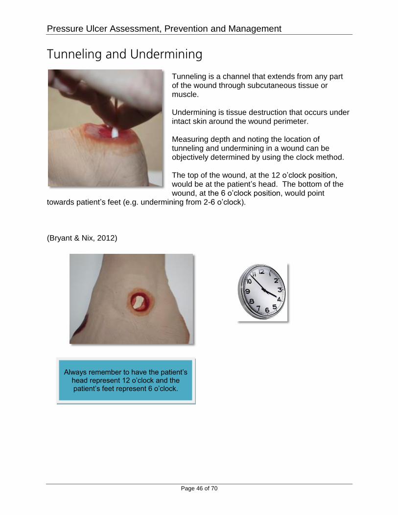
Tunneling and Undermining Tunneling is a channel that extends from any part of the wound through subcutaneous tissue or muscle. Undermining is tissue destruction that occurs under intact skin around the wound perimeter. Measuring depth and noting the location of tunneling and undermining in a wound can be objectively determined by using the clock method. The top of the wound, at the 12 o’clock position, would be at the patient’s head. The bottom of the wound, at the 6 o’clock position, would point
towards patient’s feet (e.g. undermining from 2-6 o’clock). (Bryant & Nix, 2012)
Always remember to have the patient’s head represent 12 o’clock and the patient’s feet represent 6 o’clock.
Pressure Ulcer Assessment, Prevention and Management
Page 47 of 70
Appearance of Wound Bed Types and amount of tissues in the wound bed should always be assessed and documented. Many pressure ulcers have a combination of different types of tissues in the wound bed. These combinations should be documented in percentages. Type and amount of granulation tissue in a wound bed will be indicative of where that pressure ulcer is in the healing phase. (Bryant & Nix, 2012)
Tissue Type & Thickness The wound bed tissue reveals the phase and progress of wound healing. There are tissue colors that can be seen in pressure ulcers such as pink, red, black, and yellow/beige:
• Epithelial tissue is "pearly pink" in color. • Granulation tissue is beefy red. • Necrotic tissue is usually black, brown, or tan and known as eschar. • Yellow necrotic tissue is known as slough (it can also be tan, gray, green,
or brown). (NPUAP, 2014)
Documentation Example:
80% of the wound bed contains yellow slough and 20% contains granulation
tissue.

Pressure Ulcer Assessment, Prevention and Management
Page 48 of 70
Assessment of Drainage The amount, type, and odor of wound drainage should always be assessed and documented. Amount: Assessed as none, light, moderate, or heavy. Type: Assessed as being clear, serous sanguineous, sanguineous, purulent, tan,
or bloody. Odor: Assessed as being absent, faint, moderate, or strong.
It is important to know that most wounds do have an odor and the type of dressing can affect the wound odor, as well as the presence of devitalized tissue.
(Bryant & Nix, 2012)

Pressure Ulcer Assessment, Prevention and Management
Page 49 of 70
Condition of Periwound Skin The integrity of periwound skin will ensure clues to the effectiveness of the treatment choice or dressing application. For example, maceration of the periwound skin could indicate poor application of dressing. If the dressing overlaps on the periwound skin or when exudate is allowed to pool on periwound skin, these indicate an inappropriate dressing application. Periwound skin should be assessed for:
• Color (erythema or white) • Temperature (cool or warm) • Texture (moist, indurated, boggy, or dry) • Integrity (candidiasis, epidermal stripping, pustules)
(Bryant & Nix, 2012)
Wound Margins
Pay close attention to wound margins, looking specifically for undermining or dead spaces.
Dead spaces are areas where the wound edges have come away from the wound base.
These areas may show signs of poor circulation such as grey or purple coloration.
Dead spaces should carefully be investigated to determine the extent of the undermining.

Pressure Ulcer Assessment, Prevention and Management
Page 50 of 70
Wound Care Performed The goals for wound healing are to:
Prevent infection Ensure proper cleansing Remove nonviable tissue Maintain proper moisture levels Eliminate dead space Odor control Minimize pain Protection of the wound and periwound skin
Documentation of the wound care should be clear and compatible with the wound care orders which are prescribed for the patient. The type of cleansing solution, type of dressing used, and the application of any secondary dressing should always be clearly documented each time the wound care is performed. (Bryant & Nix, 2012)

Pressure Ulcer Assessment, Prevention and Management
Page 51 of 70
Test Yourself 3 Which of the following is NOT a goal for wound healing?
a. Remove nonviable tissue b. Prevent infection c. Keep dead space d. Protection of the wound and periwound skin
The correct answer is that keeping dead space is NOT a goal for wound healing.

Pressure Ulcer Assessment, Prevention and Management
Page 52 of 70
Patients’ Tolerance to Wound Care Pain:
Assess for pressure-ulcer-related pain in adults using a validated scale.
Assessment of pain should include an assessment of body language and
nonverbal cues.
Wound pain can indicate deterioration, infection, or even inappropriate wound
treatments.
Optimize pressure ulcer care to ensure that it is coordinated with pain medication
administration.
Pain should be measured and rated prior to each dressing change and post
dressing change to determine if the appropriate interventions for pain
management were initiated during wound care.
(NPUAP, 2014)

Pressure Ulcer Assessment, Prevention and Management
Page 53 of 70
Wound Progress Towards Goal
• Pressure ulcers should be assessed at each dressing change for progress towards healing.
• Currently in clinical practice, monitoring for healing of pressure ulcers is based on the clinical judgement of the healthcare professional. There are pressure ulcer assessment tools to aid in progress of pressure ulcer healing.
– Pressure ulcer scale for healing (PUSH): 1. Validated tool developed by the NPUAP to monitor pressure ulcer
healing over time 2. Monitors 3 parameters considered most indicative of healing:
• Size (length & width), exudate amount, and tissue type • Record a sub score 0-5 (size) & 0-4 (exudate & tissue type);
total score calculated ranging from 0-17 (0 = healed) • Comparison of total scores over time provides an indication
of the improvement or deterioration in pressure ulcer healing • Photography has proven to be reliable and successful in capturing the pressure
ulcer condition over time. (Bryant & Nix, 2012 & NPUAP, 2014)

Pressure Ulcer Assessment, Prevention and Management
Page 54 of 70
Wound Dressing for Treatment of Pressure Ulcers Wound dressings are essential to management and treatment of pressure ulcers. Wound healing is optimized in a moist verses dry environment. Occlusive or semi-occlusive dressings maintain wound bed moisture to promote epithelialization and wound closure. Select a wound dressing based on the:
Ability to keep the wound bed moist
Need to address bacterial bioburden
Nature and volume of the wound exudate
Condition of the periulcer skin
Ulcer size, depth and location
Presence of tunneling and/or undermining (NPUAP, 2014)
As a pressure ulcer heals or deteriorates, the type of
wound dressing most appropriate to promote
healing will change.
Pressure Ulcer Assessment, Prevention and Management
Page 55 of 70
Hydrocolloid Dressings Wafer type dressing that contains gel-forming agents in an adhesive compound laminated onto a flexible, water resistant layer. Benefits:
Allow a moist healing environment Autolytic debridement Insulation Impermeable to bacteria and other contaminants Self-adherent & molds well to intact skin around the wound Can be worn for several days without needing to be changed
Indications:
Use for clean Category/ Stage 2 pressure ulcers in body areas where they will not roll or melt
Non-infected, shallow Category/Stage 3 Protection of intact skin or newly healed wound Not recommended for wounds with heavy exudate, sinus tracts or when infection
is present. (Morgan, 2013a & NPUAP, 2014)

Pressure Ulcer Assessment, Prevention and Management
Page 56 of 70
Transparent Film Dressings There is little to no research to support transparent dressing for treatment of pressure ulcers. Consider using a transparent film dressing:
For autolytic debridement when and individual is not immunocompromised
Secondary dressing for pressure ulcers treated with alginates or other wound filler that will likely remain in the ulcer bed for an extended period of time
Do not use as a interface layer over pressure ulcers with moderate to heavy exudate
Do not use as the over dressing for enzymatic debriding agent, gel or ointment
(NPUAP, 2014)

Pressure Ulcer Assessment, Prevention and Management
Page 57 of 70
Hydrogel Dressings Hydrogel dressings are a hydrated polymer (hydrogel) dressing that contain 90% water in a gel base. It helps regulate fluid exchange from the wound surface. Hydrogel dressings are available in three forms:
1. Amporphus hydrogel- free flowing gel, packaged in tubes, foil packets and spray bottles.
2. Impregnated hydrogel-amoprhous hydrogel saturated onto gauze pad, nonwoven sponge ropes and/or strips.
3. Sheet hydrogel- a gel supported by a thin fiber mesh. The dressing can overlap intact skin without causing trauma. It is available with/without adhesive borders and can be cut to fit wound size.
Benefits:
Soothing and reduce pain Rehydrate the wound bed Facilitate autolytic debridement Fill in dead space (amorphous and impregnated types) Can be used when infection is present.
Indications:
Shallow wounds that have minimal exudate Wounds with dry ulcer beds Painful pressure ulcers hydrogel sheets for wounds without depth and contours and/or body areas that
are at risk for dressing migration.
(Morgan 2013b & NPUAP, 2014)
Pressure Ulcer Assessment, Prevention and Management
Page 58 of 70
Alginate Dressings Alginates are a non-woven absorbent dressing derived from seaweed. They are placed into a wound in a dry form, and they absorb exudate to form a hydrophilic gel while still maintaining a moist wound environment. Alginates are manufactured in sheets or rope forms. Benefits:
Can be left on an ulcer for several days to decrease frequency of dressing changes
Indications:
Bleeding wounds (helps achieve hemostasis) Wounds with moderate to heavy exudate Full or partial thickness wounds with tunneling and or/undermining Clinically infected wounds when there is appropriate concurrent treatment of
infection.
Contradictions: Third-degree burns Minimal exudate or dry wounds
Tips: Gently remove alginate dressings from the wound by irrigating first If alginate dressing is still dry on dressing changes, consider lengthening time intervals for dressing changes or changing the type of dressing. (Morgan, 2012 & NPUAP, 2014)

Pressure Ulcer Assessment, Prevention and Management
Page 59 of 70
Foam Dressings Foam dressings absorb wound exudate from the wound bed.
Simple foam dressings- wick exudate away from the wound bed and translocate it to the surface of the wound dressing.
Complex foam dressings- absorb wound exudate by dispensing it throughout the wound dressing for retention away from the skin.
Gelling foam dressings manage excess wound exudate and protect surrounding skin from prolonged exposure to wound or body fluids.
Benefits:
Wounds with moderate to heavy exudate Decreases maceration of periwound tissue
Indications:
A Category/Stage 2 with exudate A shallow Category/Stage 3
(NPUAP, 2014)

Pressure Ulcer Assessment, Prevention and Management
Page 60 of 70
Vacuum Assisted Closure (VAC)
Also known as a Wound Vac, this system applies negative pressure to the ulcer using an open-cell polyurethane foam dressing. In general, negative pressure therapy or wound vacuum assisted closure (VAC) is indicated for Stage III and Stage IV pressure ulcers. Benefits:
Removes excess exudate and promotes a moist wound healing environment Promotes formation of granulation tissue Reduces bacterial count
Pressure Ulcer Assessment, Prevention and Management
Page 61 of 70
Silver-Impregnated Dressings The use of silver-impregnated dressings is intended to reduce bioburden. Bioburden is defined as the number of bacteria living on a surface. Silver-impregnated dressings should be used to reduce bioburden and discontinued once healing is noted. Silver impregnated dressings continue to be debated and currently there is little scientific literature to base recommendations on use of silver in wound care. Prophylactic use of silver dressings should be carefully considered. (NPUAP, 2014)

Pressure Ulcer Assessment, Prevention and Management
Page 62 of 70
Honey Impregnated Dressings Medicinal and healing properties of honey have been recognized for ages. Recently a resurgence of interest has occurred and grown in popularity for wound care products. Honey produces hydrogen peroxide, contains antioxidants and releases anti-inflammatory products. It helps in reduction of odor. Studies have shown an increased healing rate of those treated with honey for stage II and III pressure ulcers. (NPUAP, 2014)
CAUTION
Before applying honey dressing, ensure individual is not allergic to honey. Individuals who have bee or bee sting allergies are usually able to use properly irradiated honey products.

Pressure Ulcer Assessment, Prevention and Management
Page 63 of 70
Gauze Dressings Gauze dressings are made of cotton or synthetic fabric that is absorptive and permeable to water, water vapor and oxygen. Gauze dressings have been associated with:
Increased infection rate Retained dressing particles Pain (NPUAP, 2014)
CAUTION
Avoid use of wet-to-dry gauze dressings for open pressure ulcers.
Pressure Ulcer Assessment, Prevention and Management
Page 64 of 70
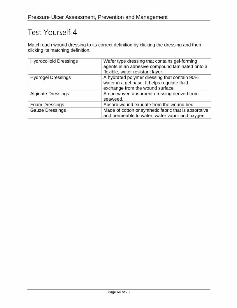
Test Yourself 4 Match each wound dressing to its correct definition by clicking the dressing and then clicking its matching definition.
Hydrocolloid Dressings Wafer type dressing that contains gel-forming agents in an adhesive compound laminated onto a flexible, water resistant layer.
Hydrogel Dressings A hydrated polymer dressing that contain 90% water in a gel base. It helps regulate fluid exchange from the wound surface.
Alginate Dressings A non-woven absorbent dressing derived from seaweed.
Foam Dressings Absorb wound exudate from the wound bed.
Gauze Dressings Made of cotton or synthetic fabric that is absorptive and permeable to water, water vapor and oxygen

Pressure Ulcer Assessment, Prevention and Management
Page 65 of 70
Test Yourself 5 Wound vacuum assisted closure (VAC) is used only on Stage IV and unstageable pressure ulcers.
a. TRUE b. FALSE
In general, negative pressure therapy or wound vacuum assisted closure (VAC) is indicated for Stage III and Stage IV pressure ulcers.

Pressure Ulcer Assessment, Prevention and Management
Page 66 of 70
Conclusion The costs of pressure ulcers to the health-care system is estimated to be $11 billion each year (Schessel, Ger, & Oddsen, 2012). In order to prevent a pressure ulcer and achieve positive outcomes, a multidisplinary team approach must be utilized. The physician, wound care specialist, nurse, physical therapist, and registered dietitian are all major components of this multidisciplinary team (Delmore, Lebovits, Baldock, Suggs & Ayello, 2011). Regular skin assessment, risk evaluation, skin care interventions, mobility assessment, and nutrition evaluation are all equal components of prevention and treatment of pressure ulcers (Delmore et al., 2011).

Pressure Ulcer Assessment, Prevention and Management
Page 67 of 70
References Agency for Healthcare Research and Quality [AHRQ] (2014). Never Events. Retrieved
from http://www.psnet.ahrq.gov/primer.aspx?primerID=3. Armstrong, D. & Meyr, A. (2013). Clinical Assessment of Wounds. Retrieved from
http://www.uptodate.com/contents/clinical-assessment-of-wounds Baxter, H. & Ballard K. (2001) Vacuum-assisted closure. Wound Care, (97)35. P. 51. Retrieved from http://www.nursingtimes.net/clinical-subjects/wound-care/vacuum-assisted-closure/200663.fullarticle . Bryant, R.A. & Nix, D.P. (2012). Acute & Chronic Wounds: Current Management
Concepts 4th ed. St. Louis, MO: Mosby. Centers for Medicare & Medicaid Services [CMS] (2014a). Present on Admission
Indicator Reporting by Acture Inpatient Prospective Payment System Hospitals. Retrieved from https://www.cms.gov/MLNProducts/downloads/wPOAFactSheet.pdf.
Centers for Medicare & Medicaid Services [CMS] (2014b). CMS to Improve Quality of
Care during Hospital Inpatient Stays. Retrieved from https://www.cms.gov/Newsroom/MediaReleaseDatabase/Fact-sheets/2014-Fact-sheets-items/2014-08-04-2.html
Delmore, B., Lebovits, S., Baldock, P., Suggs, B., & Ayello, E.A. (2011) Pressure ulcer
prevention program. Journal of Wound Ostomy & Continence Nursing , 38(5), 505-513.
Hess, C.T. (2010). Performing a skin assessment. Nursing 2010, 40(7), 66 Koo, L.W., Reedy, S., & Smith, J.K. (2010). Patient history key to diagnosing peripheral
edema. The Nurse Practitioner, 35(3), 44-52. Morgan, N. (2012). Calcium alginate. Wound Care Advisor 1(2), 26-27. Morgan, N. (2013a). What you need to know about hydrocolloid dressings. Wound Care
Advisor 2(3), 28-30. Morgan, N. (2013b). What you need to know about hydrogel dressings. Wound Care
Advisor 2(3), 21-23.

Pressure Ulcer Assessment, Prevention and Management
Page 68 of 70
National Pressure Ulcer Advisory Panel (2014). Prevention and Treatment of Pressure Ulcers: Clinical Practice Guideline. Osborne Park, Western Australia: Cambridge Media
Schessel, E.S., Ger, R., & Oddsen, R. (2012). The cost and outcomes of treating a
pressure ulcer in a patient with a deep pressure ulcer in a patient with quadriplegia. Ostomy Wound Management, 58, 2, 41-46.
U.S. Department of Health and Human Services (2015). 2020 Topics & Objectives:
Older Adults. Retrieved from http://www.healthypeople.gov/2020/topics-objectives/topic/older-adults/objectives
Wound Ostomy and Continence Nurses Society (2010). Guidelines for Prevention and
Management of Pressure Ulcers. Mount Laurel, NJ: WOCN.
Pressure Ulcer Assessment, Prevention and Management
Page 69 of 70
Disclaimer RN.com strives to keep its content fair and unbiased. The authors, planning committee, and reviewers have no conflicts of interest in relation to this course. Conflict of Interest is defined as circumstances a conflict of interest that an individual may have, which could possibly affect Education content about products or services of a commercial interest with which he/she has a financial relationship. There is no commercial support being used for this course. Participants are advised that the accredited status of RN.com does not imply endorsement by the provider or ANCC of any commercial products mentioned in this course. There is no "off label" usage of drugs or products discussed in this course. You may find that both generic and trade names are used in courses produced by RN.com. The use of trade names does not indicate any preference of one trade named agent or company over another. Trade names are provided to enhance recognition of agents described in the course. Note: All dosages given are for adults unless otherwise stated. The information on medications contained in this course is not meant to be prescriptive or all-encompassing. You are encouraged to consult with physicians and pharmacists about all medication issues for your patients.

Pressure Ulcer Assessment, Prevention and Management
Page 70 of 70
Ready to Take the Post-Test?
Close this window and click the TAKE THE TEST link to proceed to the test.
This publication is intended solely for the use of healthcare professionals taking this course, for credit, from RN.com. It is designed to assist healthcare professionals, including nurses, in addressing many issues associated with healthcare. The guidance provided in this publication is general in nature, and is not designed to address any specific situation. This publication in no way absolves facilities of their responsibility for the appropriate orientation of healthcare professionals. Hospitals or other organizations using this publication as a part of their own orientation processes should review the contents of this publication to ensure accuracy and compliance before using this publication. Hospitals and facilities that use this publication agree to defend and indemnify, and shall hold RN.com, including its parent(s), subsidiaries, affiliates, officers/directors, and employees from liability resulting from the use of this publication. The contents of this publication may not be reproduced without written permission from RN.com.