Prenatal Stress & Febrile Seizures Effects on the ...
173
i Prenatal Stress & Febrile Seizures Effects on the Epigenetic Mechanisms Involved in Cognitive Function Following Treatment with Searsia Chirindensis BY SADIYAH CASSIM 213541708 2016
Transcript of Prenatal Stress & Febrile Seizures Effects on the ...

i
Prenatal Stress & Febrile Seizures Effects on the Epigenetic Mechanisms
Involved in Cognitive Function Following Treatment with Searsia Chirindensis
BY
SADIYAH CASSIM
213541708
2016

ii
Prenatal Stress & Febrile Seizures Effects
on the Epigenetic Mechanisms Involved in Cognitive Function
Following Treatment with Searsia Chirindensis
SADIYAH CASSIM
213541708
Submitted in partial fulfilment for the degree
Masters in Medical Sciences
Department of Human Physiology
Faculty of Health Science
University of Kwa-Zulu Natal (Westville Campus)
DURBAN
2016

iii
Preface
The experimental studies carried out in this thesis were conducted in the Neuroscience Labs - and the
Biomedical Resource Centre of the School of Laboratory Medicine and Medical Sciences at the
University of Kwa-Zulu Natal (Westville Campus), under the supervision of Dr. M. V. Mabandla and
Ms L. Qulu.

iv
Declaration
I, Sadiyah Cassim (student number: 213541708), hereby declare that the dissertation entitled Prenatal
Stress & Febrile Seizures Effects on the Epigenetic Mechanisms Involved in Cognitive Function
Following Treatment with Searsia Chirindensis is the result of my own investigation and research
and that it has not been submitted in part or full for any other degree or to any other University or
Tertiary Institution. Where use was made of others work, it has been duly acknowledged. The
research done in this study was carried out under the supervision of Dr. M. V. Mabandla and Ms. L.
Qulu.
___________________________
Student: Sadiyah Cassim (213541708)
___________________________
Supervisor: Dr. M. V. Mabandla
DATE:_____________________
___________________________
Co- Supervisor: Ms. L. Qulu
DATE: _____________________
v
Acknowledgements
The author wishes to convey her sincere and genuine appreciation to every person who imparted their
support during the course of this study.
I would like to extend my full appreciation and sincere gratitude to Dr. M. V. Mabandla for his
valuable supervision, motivation, assistance, undivided attention, guidance and encouragement
during the course of the study. I further extend my appreciation and thanks for the mentoring,
patience and full support in general throughout my post-graduate career. Thank you for being my
father of research and academics.
I would also like to thank Ms. L. Qulu for her valuable input, patience and mentoring during these
tedious times of submission, as well as her motivation.
To my parents specifically, without your support I would have not made it this far. Thank you for
being patient and supportive of me during my long working hours and encouraging me during my
bad phases of health.
To Prof. W. M. U. Daniels, Ms. Z. M. Moosa, Mr M.W. Khan, Ms. K. McNeil, Ms. A.
Mohamed., Mr N. Soobryan, Ms. C. O. Subiah and Ms. K. Moodley for their time, assistance,
guidance, critique and advice for the duration of this study. Thank you for all the assistance and
support provided academically and emotionally.
I would like to thank Ms. Lavanya from Albert Luthuli Hospital for assisting me when the PCR
machine broke-down.
To the Neuroscience Team of UKZN, Department of Physiology, I thank you for your continuous
encouragement, assistance, support, advice and motivation.
University of KwaZulu-Natal, College of Health Sciences, thank you for the financial aid
provided during the course of this study.
To the Biomedical Resource Unit, Dr. S. Singh, Dr. L. Bester, Ms. R. Radebe and the support
staff for your assistance in animal handling, procedures and training where it was required.

vi
Abbreviations
α - alpha
β - beta
κ - kappa
γ - gamma
µ - micro
HSD-2 - hydroxysteriod-dehydrogenase 2
AChE - acetylcholine esterase
ACh - acetylcholine
ACTH - adrenocorticotropic hormone
AMPA - α-amino-3-hydroxy-5-methyl-4-isoxazolepropionic acid
BDNF - brain-derived neurotrophic factor
BGT-1 - betaine-GABA transporter 1
CA - cornus ammonis
CNS - central nervous system
CD – cluster of differentiation
COX-2 - cyclooxygenase-2
CPG - cytosine-phosphate-guanine
CRH - corticotrophin-releasing hormone
DNA - deoxynucleic acid
DNMTs - DNA methyltransferases
EAATs - excitatory amino acid transporters
EPSPs – excitatory post-synaptic potentials
FS - febrile seizure
GABA - γ-aminobutyric acid
GAD - glutamic acid decarboxylase
GC - glucocorticoids

vii
GND - gestational day
GR - glucocoticoid receptors
HATs - histone acetyltransferases
HDACs - histone deacetylases
HPA - hypothalamic-pituitary-adrenal
i.p. - intraperitoneally
IPSP - inhibitory post-synaptic potential
IL- interleukin
IL-r1a - interleukin-receptor 1 antagonist
KAR - kainate receptor
Kg - kilogram
LPS - lipopolysaccharides
LTD - long term depression
LTP - long term potentiation
mAChRs - muscarinic acetylcholine receptors
MAPK - mitogen-activated protein kinases
MeCP2 - methyl-CPG-binding protein 2
mg - milligram
MR - mineralocorticoid receptors
nAChRs - nicotinic acetylcholine receptors
NF - nuclear factor
NMDA - N-methyl-D-aspartate
NS - non-stressed
PBS - phosphate buffered saline
PFC - prefrontal cortex

viii
PGs - prostaglandins
PND - postnatal day
proBDNF - precursor protein brain-derived-neurotrophic factor
REST - repressor-element-1-silencing-transcriptional factor
RNA – ribonucleic acid
S - stress
SNARE - SNAP (soluble NSF attachment protein) Receptor
TGF - transforming growth factor
TLR - toll-like-receptor
TNF - tumour necrosis factor
Trk - tropomyosin-related kinase
UBE3 - ubiquitin-protein ligase 3
VGlut3 - vesicular glutamate transporter 3
ix
Table of Contents
Title pages and declarations………………...……………………………………….….….i-iv
Acknowledgements……….………………...……………………………………….……….v
Abbreviations…………………………………………………………………..….…….vi-viii
Table of content…………………………………………………………………………..ix-xi
List of tables……………………………………………………………………………..….xii
List of figures…………………………………………………………………………...xii-xiii
Study Outline...………………………………………………………………………….….xiv
Abstract……..…………………………………………………………………..…………..xv
Chapter One:
Literature Review
Background ......................................................................................................................... 1-2
1. Epidemiology ................................................................................................................... 3
2. Febrile Seizure ............................................................................................................ 4-7
2.1. Acetylcholine ............................................................................................................ 7
3. Effects of stress on the brain ............................................................................................ 8
3.1.Prenatal stress.......................................................................................................... 8-9
3.1.1. Effects of prenatal stress on the developing foetus ..................................... 9-10
3.2. Implications on Neurotransmitters within the Hippocampus ............................ 10-12
3.3.Role of BDNF ..................................................................................................... 12-13
4. Animal models .......................................................................................................... 13-14
5. Epigenetics ..................................................................................................................... 14
5.1.DNA Methylation .................................................................................................... 15
5.2.MeCP2 ................................................................................................................ 15-16

x
5.3.REST ................................................................................................................... 15-16
6. Treatments for Febrile seizures ...................................................................................... 16
5.1.The potentiating therapeutic effects of Searsia chirindensis.............................. 16-17
7. Aims ............................................................................................................................... 17
Chapter Two:
“Prenatal stress and early life febrile convulsions compromise hippocampal genes
MeCP2/REST function in mid-adolescent life of Sprague-Dawley rats”
Introduction ..................................................................................................................... 20-21
Material & Methods ........................................................................................................ 21-26
Results ............................................................................................................................. 26-32
Discussion ....................................................................................................................... 32-35
Conclusion ........................................................................................................................... 36
Acknowledgements .............................................................................................................. 36
References ....................................................................................................................... 37-43
Chapter Three:
“Febrile seizure induction impairs neuronal plasticity in prenatally stressed rat offspring”
Introduction ..................................................................................................................... 47-48
Material & Methods ........................................................................................................ 48-51
Results ............................................................................................................................. 52-55
Discussion ....................................................................................................................... 55-57
Conclusion ........................................................................................................................... 57
Acknowledgements .............................................................................................................. 59
References ...................................................................................................................... 59-62

xi
Chapter Four:
“The Therapeutic Effects of Searsia chirindensis, on Short Term Cognitive Impairment
Associated with Febrile Seizure Exposure in Prenatally Stressed Rats”
Introduction .................................................................................................................... 66-67
Material & Methods ........................................................................................................ 67-72
Results ............................................................................................................................. 73-77
Discussion ....................................................................................................................... 77-79
Conclusion ........................................................................................................................... 80
Acknowledgements .............................................................................................................. 80
References ....................................................................................................................... 81-83
Chapter Five:
Conclusion ........................................................................................................................... 84
Chapter Six:
References ...................................................................................................................... 85-97
List of Addendums ...................................................................................................... 98-107
Addendum A – “Prenatal stress and early life febrile convulsions compromise hippocampal
genes MeCP2/REST function in mid-adolescent life of Sprague-Dawley rats” .................. 98
Addendum B - “Febrile seizure induction impairs neuronal plasticity in prenatally stressed
rat offspring” ........................................................................................................................ 99
Addendum C - Protocol of qPCR ............................................................................... 100-104
Addendum D - Protocol of BDNF ELISA ......................................................................... 105
Addendum E - Protocol of AChE ELISA ......................................................................... 106

xii
Addendum F - Conference Outputs ................................................................................... 107
List of Tables
Chapter Two:
Table 1- Groups of pups ...................................................................................................... 24
Table 2- Seizure response score .......................................................................................... 25
Chapter Four:
Table 1- Seizure response score ........................................................................................... 69
List of Figures
Chapter One
Figure 1- Geographical distribution of developing countries ............................................. 3
Figure 2- Projected pathways of neuroinflammation ........................................................ 6
Figure 3- Mechanism & effect of prenatal stress ............................................................... 9
Chapter Two
Figure 1- Outline of prenatal protocol ................................................................................. 23
Figure 2- Outline of postnatal protocol ............................................................................... 24
Figure 3.a.-Graph displaying average time taken to locate the hidden platform in the
MWM .................................................................................................................................. 28
Figure 3.b.- Graph displaying average time spent in the quadrant of the hidden platform in
the MWM ............................................................................................................................ 29
Figure 4.a.- Graph displaying MeCP2 gene expression ...................................................... 30
Figure 4.b.- Graph displaying REST gene expression ........................................................ 31
Figure 5.a.- Graph displaying comparative analysis of overall gene expression in non-
stressed (NSS) and non-stressed febrile seizure (NSFS)...................................................... 31

xiii
Figure 5.b.- Graph displaying comparative analysis of overall gene expression in stressed
(SS) and stressed febrile seizure (SFS) ................................................................................ 32
Chapter Three
Figure 1- Graph displaying seizure onset and seizure duration ......................................... 52
Figure 2- Graph displaying number of entries into the closed arm of the EPM ................. 53
Figure 3- Graph displaying BDNF concentration ............................................................... 54
Figure 4- Graph displaying REST gene expression ............................................................ 55
Chapter Four
Figure 1- Linear diagram depicting the post-natal handling protocol ................................ 68
Figure 3.1.- Graph displaying average time taken to locate the hidden platform in the
MWM .................................................................................................................................. 73
Figure 3.2.- Graph displaying average time spent in the quadrant of the hidden platform in
the MWM ............................................................................................................................. 74
Figure 3.3- Graph displaying hippocampal AChE Enzyme concentration ......................... 75
Figure 3.4- Graph displaying the MeCP2 gene expression ................................................ 76
Figure 3.5- Graph displaying the REST gene expression ................................................... 77

xiv
Study Outline
This dissertation is presented in article format; with three studies and six chapters. A background has
been included in order to orientate the reader to the overall view of the problem statement as well as
our aims. Chapter one is a literature review which is a general discussion on key concepts investigated
in the study. Chapter two contains work published in “Neurobiology of Learning and Memory”.
Sadiyah Cassim, Lihle Qulu & Musa V. Mabandla. Neurobiology of Learning and Memory (2015)
125:195-201. DOI:10.1016/j.nlm.2015.09.002. The published version of the article has been attached
as addendum A. Chapter three entails study two of the project. This chapter is currently under review
in the journal “Annals of Neuroscience”. Sadiyah Cassim, Lihle Qulu & Musa V. Mabandla. The
submitted copy of the article has been attached as addendum B. Chapter four is the third study of the
project, this study is prepared in the article format required for the journal “Phytomedicine”. Chapter
five is a combined conclusion for all three studies conducted in this dissertation. Chapter six consists
of the reference list for all the work submitted.

xv
Abstract
Febrile seizures are of growing concern in the health fraternity within the African continent. These
seizures predominantly affect children between the ages of 3 months and 5 years of age. Stress during
pregnancy may influence the functioning of the hypothalamic-pituitary-adrenal (HPA) axis in the
developing foetus. This has a tendency to increase the brain’s vulnerability to a number of insults
including febrile seizures. Current treatment for febrile seizures often has undesirable side effects
hence the need to find more effective alternatives. Searsia chirindensis (Searsia) is commonly used to
treat conditions associated with the heart, rheumatism and brain abnormalities. The focus of this study
was to investigate whether exposure to prenatal stress and early life febrile seizures may affect the
expression of genes that play a role in neuronal plasticity. For this we looked at the expression of the
MeCP2 and REST genes in the hippocampus. Furthermore, we investigated whether neuronal
malformations and the resulting cognitive deficits associated with exposure to early life stress
influences the concentration of the neurotrophic factor, BDNF and whether further exposure to febrile
seizures exacerbates the stress effect. We also investigated whether treating febrile convulsions with
Searsia attenuates the negative effects caused by stress on febrile seizure development as well as any
cognitive effects that may be caused by the exposure to stress and febrile seizures. Fourteen day old
(PND 14) pups were divided into the following groups 1) Normally reared Sprague-Dawley offspring
injected with saline (NSS). (2) Prenatally stressed offspring injected with saline (SS). (3) Normally
reared offspring with febrile seizures (NSFS). (4) Prenatally stressed offspring with febrile seizures
(SFS). (5) Normally reared offspring treated with Searsia (NS-S). (6) Prenatally stressed offspring
treated with Searsia (S-S). (7) Normally reared offspring with febrile seizures and treated with
Searsia (NSFS-S). (8) Prenatally stressed offspring with febrile seizures and treated with Searsia
(SFS-S). Lipopolysaccharide and kainic acid were used to induce the febrile seizures. The Morris
water maze (MWM) was used to assess learning and memory function and the elevated plus maze
(EPM) was used to assess anxiety-like behaviour in these young rats. MeCP2/REST genes expression
and the concentration of BDNF as well as AChE were quantified using an immunoassay on
hippocampal tissue. Our results showed that exposure to prenatal stress (SS) and febrile seizures
(NSFS) may impair cognitive behavioural function in the short-term. However, in the NSFS animals,
there seems to be an attempt to counteract the effects of febrile seizures with time. Furthermore,
exposure to prenatal stress impeded the release of the neurotrophic factor, BDNF, while attenuating
REST gene expression in febrile seizure animals. These factors may contribute to the hindering of
neurogenic properties in the young, thus leading to neuronal plasticity deficits and possibly cognitive
malfunction in later life. Treatment with Searsia attenuated the transient cognitive impairments
present following a seizure by influencing the concentration of hippocampal acetylcholine and
activating MeCP2 gene expression. This suggests a role for Searsia in the management of febrile
seizures and in attenuating the development of chronic cognitive deficits.

1
Background
Febrile seizures arise from a systemic fever occurring in the body due to an infection which evokes the
excessive firing of neurons (Qulu, et al., 2012; Marchi, et al., 2013). Febrile seizure are triggered by
infections such as otitis media, otitis externa, sinusitis, pharyngitis and conjunctivitis (Alter, et al.,
2011). These infections trigger the activation of the immune system thus promoting the release of
various cytokines and leucocytes (Marchi, et al., 2013). This subsequently results in neurotransmitter
(glutamate and γ-aminobutyric acid (GABA) imbalance provoking a trigger of a seizure (Batten, 2013;
Lason, et al., 2013).
Furthermore, exposure to early life stress has been shown to result in the malformation of neuronal
development in young rats (McEwen, 2012). Prenatal stress is a critical component of early life stressors
which is a result of stress experienced by pregnant mothers in their last trimester of pregnancy
(Moisiadis & Matthews, 2014). This form of stress has been shown to exacerbate cytokine production
and seizure progression in rat offspring (Qulu, et al., 2012) resulting in hippocampal mass reduction
(Qulu, et al., 2015), increase in excitatory amino acids (glutamate and acetylcholine) that subsequently
affects neuronal function (McEwen, et al., 2015). The implications of neuronal loss particularly in the
hippocampus has been show to affect neuronal networks from histological to molecular aspects in brain
structure thereby affecting brain function (McEwen, 2012).
Furthermore, studies have shown that various genomic alterations may be a link to abnormal behaviour
and functioning in developing brains (Qureshi & Mehler, 2010; Lubin, 2012). Febrile seizures have
been shown to play an important role in influencing transcriptional factors and gene expressions viz.
GABAA receptor unit, REST, CREB, MeCP2 and BDNF, that contribute to changes in neuronal
networks particularly in the hippocampus (Qureshi & Mehler, 2010; Lubin, 2012; Roopra, et al., 2012).
Treatment regimens undertaken to treat various febrile seizures include sodium valproate,
carbamazepine and phenobarbitone (Lui, et al., 2015). However these treatments have adverse side
effects and financial implications, many have resorted to the use of traditional medicinal plants as a
source of alternative treatment (Lui, et al., 2015). Searsia chirindensis is amongst the few plants used
to treat various disorders inclusive of convulsions (Ojewole, 2008).
Therefore the focus of our study was to investigate:
1. The role of the MeCP2 and REST genes in the developing brain by investigating the effects
of early life exposure to neuronal insults (prenatal stress and febrile seizures) on learning
and memory as well as determining the role of MeCP2/REST genes activation on
hippocampal function.
2. Whether neuronal malformations and the resulting cognitive deficits associated with stress
are linked to changes in BDNF concentration and the REST gene factor expression. We

2
further investigated whether exposure to febrile seizures exacerbates the changes seen in
these factors, and
3. We investigated the effects of Searsia crude bark methanolic extract on prenatally stressed
and febrile seizure exposed rats that exhibit cognitive impairment and whether the crude
bark methanolic extract attenuates the changes observed in the hippocampus.

3
Chapter One
Literature Review
1. Epidemiology
Seizures are clinical symptoms that advocate a diagnosis for a condition that may be related to epilepsy
or clinical manifestation related to non-epileptic conditions such as febrile seizures (Marchi, et al.,
2014). Febrile seizures arise from a systemic fever occurring in the body due to an underlying infection
which evokes the excessive firing of impulses (Marchi, et al., 2014). Febrile seizures can be classified
into three categories viz. (1) simple febrile seizures, (2) complex febrile seizures and (3) status
epilepticus (Cappovilla, et al., 2009; Heida, et al., 2009). Simple febrile seizures occur predominantly
in children of 3 months to 5 years of age, with a seizure duration lasting between 1-2 minutes but not
more than 15 minutes and a clear focus of an infection is present (Cappovilla et al, 2009). Complex
febrile seizures are usually prolonged seizures that are focalised and last greater than 15 minutes with
chances of recurring within 24 hours (Cappovilla et al, 2009). Status epilepticus lasts greater than 30
minutes, with having two unprovoked seizures occurring 24 hours apart (Fisher, et al., 2014).
Infants and toddlers between the age of 3 months and 5 years old are more susceptible to a febrile
seizure (Cappovilla et al, 2009; Ackermann & Van Tooran, 2012). Factors such as poor sanitation and
inadequate medical facilities, as well as malnutrition are common issues affecting developing countries
(Idro, et al, 2008). Developing countries include predominantly parts of East Africa (Mozambique,
Ethiopia, Kenya and Sudan), followed by parts of West Africa (Nigeria, Democratic Republic of Congo
and Mali) and last, Southern Africa (Zimbabwe, Botswana and South Africa) (Figure 1). These
situations allow for easy contraction of infections, namely- gastroenteritis and respiratory tract
infections which subsequently activates the body’s immune responses resulting in systemic
inflammation (Heida, et al., 2009; Ackermann & Van Tooran, 2012).
South Africa has been shown to be affected by febrile seizures with Northern Province having a high
prevalence of approximately 6.7/1000 reported cases (Ackermann & Van Tooran, 2012). Thus it is
confirmed that febrile seizures are a growing concern in many facets of the health fraternity within
South Africa.

4
2. Febrile Seizures
In the 5th century BC, Aulus Cornelius Celsus, a Roman writer and physician, was the first person to
describe inflammation. He defined inflammation as a localized protective response caused by an injury
or destruction of tissue that serves to destroy or wall off both the infectious agent (bacteria, virus,
parasite or spores) and the tissue it affects (Scott, 2004; shah, 2012; Evans, el al., 2015). The
inflammatory response is evoked by physical, chemical or biological/infectious agents that result in
symptoms of heat (sometimes causing a fever), redness, swelling and pain (Scott, 2004; Stankov,
2012;Vezzani, et al., 2013; Evans, el al., 2015). The immune system is the bodies line of defence that
protects it from foreign invaders by triggering an inflammatory response that can be classified into
innate immunity (natural resistance/immunity the body has against pathogens) and adaptive immunity
(acquired immunity the body has to acquire to resist pathogens) (Hoeba, et al., 2004).
Innate immunity involves a non-specific reaction of the body to a particular pathogen (Hoeba, et al.,
2004). This natural immunity of the body is dependent on a group of proteins (cytokines) and phagocytic
cells (macrophages, neutrophils and dendritic cells) to recognize foreign invaders with the subsequent
activation to destroy them upon entry (Hoeba, et al., 2004). However, this first line of defence does not
produce memory cells to allow the body to become immune to a pathogen hence the interplay of the
adaptive immunity is required (Hoeba, et al., 2004). The adaptive immunity creates “immunological
memory” of a response to pathogen that enhances the body to defend itself in the long run (Hoeba, et
al., 2004).
Cytokines are signalling proteins that are grouped according to acute or chronic inflammation (Feghali
& Wright, 1997; Galic, et al. 2012). Acute inflammation is a hallmark of the immediate response of the
body to an infection by releasing cytokines; Interleukin (IL)-1β, Tumor Necrosis Factor-α (TNF-α), IL-
6, IL-8 and various other stimulating factors (Feghali & Wright, 1997). While chronic inflammation
include IL-3, IL-4, IL-5, IL-7, IL-9, IL-10, IL-13 and various other immune factors (Feghali & Wright,
1997). The cytokines are further divided into pro-inflammatory and anti-inflammatory factors (Feghali
Figure 1. Geographical illustration depicting the various developing countries (Adapted from Manarrazi Media Press, 2014).

5
& Wright, 1997; Galic, et al. 2012). Both IL-1β, IL-6 and TNF-α are cytokine factors produced at the
site of inflammation and their net effect is often taken into consideration by making a correlation
between the levels of expression and disease severity (Dinarello, 2000).
During a febrile seizure, an infection activates the immune system leading to an inflammatory response
with the subsequent release of immune cells such as macrophages and neutrophils (Dubé et al., 2009).
This subsequently results in the release of cytokines such as IL-6, TNF-α and predominantly, IL-1β
(figure 2) (Dubé et al., 2009). IL-1β is a pro-inflammatory cytokine that plays a pivotal role in the
progression of febrile seizures in response to the underlying infection (Heida et al., 2009). High levels
of circulating peripheral cytokines compromise the endothelial cells of the blood brain barrier resulting
in a leaky blood brain barrier (Heida et al., 2009). Levels of IL-1β are high and therefore its binding is
favoured and this subsequently sets off a sequelae of events that lead to the stimulation of the enzyme
cyclooxygenase 2 (COX-2) to catalyse the conversion of arachidonic acid into prostaglandin E2 (PGE2)
(figure 2) (Dubé et al., 2009; Heida et al., 2009). This results in the raise of the body’s thermostat “set
point” in the hypothalamus resulting in a rise in core body temperature (fever) (Heida et al., 2009). IL-
1β has a naturally occurring antagonist, the anti-inflammatory cytokine, IL-1 receptor antagonist (IL-
1Ra) which modulates the pro-inflammatory effects of IL-1β by binding to its receptors and inhibiting
the effects in normal function to bring about a homeostatic balance between pro- and anti-inflammatory
cytokines (Heida et al., 2009). However, during a febrile seizure both IL-1β and IL-R1 compete for the
same binding site favouring IL-1β resulting in a hyper secretion of IL-1β (Heida et al., 2009).
Furthermore, IL-1β phosphorylates glutamate receptors leading to an increase in cell permeability to
calcium ions and subsequently decreasing GABAA-mediated currents (Heida et al., 2009). This results
in excessive neuronal excitability which involves the imbalance of the neurotransmitters, glutamate
which is excitatory, and GABA which is inhibitory (figure 2) (Dubé et al., 2005, Heida et al., 2009). In
addition, excessive levels of IL-1β has also been shown to be neurotoxic particularly to the hippocampus
which subsequently affects cognitive function (Vezzani, et al., 2011; Marin & Kipnis, 2013).

6
Glutamate is an excitatory amino acid that plays a pivotal role in neuronal differentiation and migration
in developing circuitries of the brain (Danbolt, 2001, Ben-Ari & Holmes, 2006). It is particular
important for brain function that involves cognition, memory and learning (Danbolt, 2001). It is also
responsible for the direct signal transmission between neurons whereby it acts on ionotropic and
metabotropic glutamate receptors respectively (Danbolt, 2001, Ben-Ari & Holmes, 2006). Ionotropic
glutamate receptors are dependent on ionic gradients whereas metabotropic receptors facilitate their
action coupled to secondary G-protein messengers (Danbolt, 2001). The ionotropic receptors: α-amino-
3-hydroxy-5-methyl-4-isoxazolepropionic acid (AMPA), N-methyl-D-aspartate (NMDA) and kainate
(KARs) receptors (Popoli, et al., 2012). Glutamatergic transmission predominantly occurs within the
tripartite synapse of glutamate presenting cells (Popoli, et al., 2012). Glutamate is present in extremely
high levels to ensure that tight regulatory processes are in place to allow modulation of glutamate
receptors to optimally transmit impulses without causing excitotoxicity (Popoli, et al., 2012).
In relation to febrile seizures, the subsequent effect of exacerbated levels of IL-1β affects the NMDA
receptors resulting in the influx of calcium ions (Popoli, et al., 2012; Lason, et al., 2013). This effect
causes an increase in neuronal firing that triggers a seizure (Popoli, et al., 2012; Lason, et al., 2013).
Figure 2. Projected pathways of neuroinflammation. An infection causing a fever, triggers a systemic increase in
inflammatory markers thereby resulting in a leaky BBB. This results in part A (left), activating the COX-2 pathway
subsequently leading to a seizure and part B (right), directly affecting neuronal function, causing a seizure which
subsequently promotes the COX-2 pathway.
NEURON
ASTROCYTE
↓ GABA ↑ Glutamate
Seizure
↑ Ca2+ influx
Hyperexcitability
↓ Glutamate uptake
Infection ↑ Systemic cytokines (IL1β, IL1ra. TNF-α, IL-6)
Leaky Blood Brain Barrier
Cyclooxygenase-2 (COX-2)
Arachidonic acid
Prostaglandin E2
↑ Glutamate release. ↑ion channel permeability Neuronal hyperexciability
Blood Brain Barrier (Endothelial cells)
Fever

7
Subsequently resulting in excitotoxicity within neuronal cells thereby affecting neuronal network
functioning with detrimental effects on learning and memory in particular (Popoli, et al., 2012; Lason,
et al., 2013).
The hippocampus is involved in the learning and memory process with the cornus ammonis-1 region
and dentate gyrus expressing an abundant of AMPA and NMDA receptors, respectively (Riedel, et al.,
2003; Lason, et al., 2013). Long-term synaptic potentials are a possible mechanism involved for
learning and memory in which AMPA receptor-mediated depolarization “knocks off” magnesium ions
that block the NMDA receptors at resting membrane potential allowing for the influx of calcium ions
(Lason, et al., 2013). Depending on the calcium concentration intracellularly, this subsequently
activates protein kinases or phosphatases that lead to activation of various downstream signalling
pathways (Lason, et al., 2013). AMPA receptors generates fast excitatory post-synaptic potentials,
allowing for the permeability of sodium, potassium and limited calcium ions (Lason, et al., 2013).
NMDA receptors are responsible for the phosphorylation of AMPA receptors, which increases ion flow
thereby resulting in an increase in synaptic contacts via new dendritic out-growths (Riedel, et al., 2003;
Popoli, et al., 2012; Lason, et al., 2013). This subsequently contributes to learning and the consolidation
of short-term memory to long-term memory (Riedel, et al., 2003). In addition to excitatory amino acid,
glutamate, there is also an inhibitory amino acid that balances neuronal firing.
GABA is the main inhibitory amino acid of the central nervous system which acts predominantly on
chloride channels, GABAA ionotropic receptor, to initiate an inhibitory postsynaptic potential (IPSP)
(Lason, et al., 2013). This initiation acts as a negative feedback mechanism in prevention of excessive
excitatory discharges stimulated by glutamate in neurons particularly in events of a seizure (Lason, et
al., 2013). In contrast, impairment to the GABAergic system may compromise neural network function
thus contributing to seizure susceptibility (Lason, et al., 2013). Therefore, it is critical to maintain a
balance of GABA in the brain as Ben-Ari has postulated that increased glutamate and decreased GABA
in principal cells within the hippocampus are accompanied by persistent increase of intracellular levels
of chloride ions contributing to febrile seizure development. Thus it has been well established that
GABAA receptors are present intra- and extra-synaptically throughout the brain which are likely to
control seizure thresholds (Lason, et al., 2013). Furthermore, acetylcholine (ACh) has been shown to
influence the release GABA and glutamate respectively (Granger, et al., 2016).
2.1. The role of Acetylcholine (ACh) in neurotransmission
ACh is considered as an excitatory neurotransmitter that is responsible for the facilitation of learning
and memory formation and it increases alertness and attention (Granger, et al., 2016). The
hippocampus, an area of the limbic system within the brain, is highly innervated with extrinsic and
intrinsic inputs from cholinergic neurons (Granger, et al., 2016). This area is also rich with ionotropic
glutamate receptors and therefore it has been shown that ACh activates these receptors in addition to

8
nictonic acetylcholine receptors (nAChRs) and muscarinic acetylcholine receptors (mAChRs)
(Granger, et al., 2016). Activation nAChRs results in the increased release of glutamate and GABA
(Radcliffe, et al., 1999). This is due to nAChRs having a high permeability to calcium ions relative to
the NMDA receptors (Radcliffe, et al., 1999). ACh receptors regulate post-synaptic cellular excitability
and synaptic release of neurotransmitters at the presynaptic cell thereby directly altering synaptic
plasticity (Granger, et al., 2016). Glutamate receptor, VGlut3, is a co-transmitter with ACh particularly
in the striatum (Granger, et al., 2016). In the hippocampus, serotonergic neurons innervate the striatum
VGlut3 transporters thereby promoting the co-transmission of glutamate and ACh release making the
hippocampus the area of the brain susceptible to neuronal insults such as febrile seizures (Granger, et
al., 2016).
It has been further demonstrated that high levels of glucocorticoids influence neurotransmitter release
mainly in the prefrontal cortex (PFC) and the hippocampus (Popoli, et al., 2011). This suggests that
various forms of stress also influences neuronal function with subsequent malformation in plasticity
and functionality.
3. The effects of stress on brain function
Stress is a term often used to describe the ability of the human body and function to adapt in presence
of adverse life changing events viz. abuse, trauma and various underlying environmental factors
(McEwen, 2008). There are different types of stressors particularly in the evolving age of poverty,
malnutrition, famine and war, many children are subjected to loss of maternal care (maternal separation
stress), or the inability to cope with situations at a young age and sometimes having been exposed to
stress in utero (prenatal stress) (McEwen, 2008).
3.1. Prenatal Stress
Prenatal stress is a term used to describe the stress response experienced on the developing foetus by
an expectant mother (Lemaire, et al., 2000).
Stress is regulated via the hypothalamic-pituitary-adrenal (HPA) axis and is responsible for the
production and secretion of corticosteroids under basal and stressed conditions (Moisiadis & Matthews,
2014a). The paraventricular nucleus within the hypothalamus synthesizes corticotrophin-releasing
hormone (CRH) which stimulate adrenocorticotropin hormone (ACTH) from the anterior pituitary into
the blood circulation (Moisiadis & Matthews, 2014a). This results in the release of glucocorticoids
(cortisol in humans and corticosterone in rats) that bind specifically to glucocorticoid and
mineralocorticoid receptors in the hippocampus, the paraventricular nucleus and the anterior pituitary
(Moisiadis & Matthews, 2014a). Cortisol feedback is responsible for the feed-forward mechanism
which promotes the HPA-axis to activate the glucocorticoid and mineralocorticoid receptors within the
hippocampus, the paraventricular nucleus and the anterior pituitary when glucocorticoid levels drop

9
(Moisiadis & Matthews, 2014a). However, increase in glucocorticoid levels leads to a retardation in the
HPA activity (Moisiadis & Matthews, 2014a).
3.1.1. Effects of prenatal stress on the developing foetus
During pregnancy circulating maternal glucocorticoids cannot easily cross the placental membrane
(Barbazanges, et al., 1996; Seckl, 2004). Under normal physiological conditions, foetal glucocorticoid
concentrations are high during the last trimester of the gestation period, as it is required for the
development of organ systems in the foetus (Challis, et al., 2001). However, exposure to prenatal stress
exacerbates levels of foetal glucocorticoids by impairing the negative feedback mechanism involving
11β- hydroxysteriod-dehydrogenase type 2 (11β-HSD2) (Challis, et al., 2001). 11β-HSD2 is an enzyme
responsible for preventing the glucocorticoid effects on the developing HPA-axis of the foetus under
prolonged exposure to glucocorticoids (Seckl, 2004; Harris & Seckl, 2010; de Kloet, et al., 2014). 11β-
HSD2 catalyses the inactivation of glucocorticoids (cortisol and corticosterone) to inert 11-keto forms
(cortisone and 11-dehdrocorticosterone) (Moisiadis & Matthews, 2014a). Therefore the presence of this
enzyme acts as a shielding mechanism from higher than normal levels of exposure to glucocorticoids
present in the maternal circulation (Meaney, et al., 2007). Since this enzyme is least active during the
last trimester of pregnancy and cortisol in humans and corticosterone in rats, is required for lung
maturation and parturition, exposure to exacerbated levels of cortisol/corticosterone saturates the
binding to glucocorticoid receptors resulting in excess circulating glucocorticoids subsequently leading
to compromised neuronal development and function within the brain of the foetus (Welberg & Seckl,
2001; Harris & Seckl, 2010; de Kloet, et al., 2014).
Figure 4. Mechanism and effect of prenatal stress on developing circuitries in a new-born.
↑ CRH ↓11β- HSD2 expression & activity PLACENTA
PRENATAL
↑ Maternal CRH ↑ Cortisol
↑Foetal cortisol ↑CRH
Alteration in foetal hippocampus & HPA-axis
STRE
SSO
RS

10
Studies have shown that prolonged exposure to glucocorticoids has deleterious effects on various areas
of the brain especially in the developing foetus (McEwen & Sapolsky, 1995; Sapolsky, 2003;
Champagne, et al., 2009; Harris, et al., 2011). Glucocorticoids exerts its effects by binding to
glucocorticoid receptors (promotes negative feedback mechanism) and mineralocorticoid receptors
(promotes positive feedback mechanism) in the brain, particularly the hippocampus where these
receptors are in abundance (McEwen, 2008; Harris, et al., 2011). Binding influences many factors in
the developing brain specifically gene expression and transcription (Harris, et al., 2011). It is also
responsible for the promotion of correct brain development by initiating terminal maturation,
remodelling of axons and dendrites and overall neurogenesis (West & Greenberg, 2011). Mothers who
self-reported severe stress during pregnancy gave birth to offspring showing temperamental and
behavioural problems, as well as impaired cognition at the age of 7 years old (Gutteling, et al., 2005;
Gutteling, et al., 2006). Animal studies have also reported that prenatal stress (rodent restraint stress)
affected birth weight, cognitive function, anxiety-levels and altered functional brain development
(McEwen & Sapolsky, 1995; McEwen, et al., 2001; Qulu, et al., 2015a). There has also been reports of
abnormal physiologic function due to excessive exposure to glucocorticoids viz. immune incompetence,
inflammatory responses, bone and intermediary metabolic syndromes and cardiovascular malfunction
(Champagne, et al., 2009). Therefore, prenatal stress can be seen to be detrimental specifically to the
limbic circuitry of the brain.
Signals from the limbic circuitry reach afferent neurons in the hypothalamus, which promotes the
release of corticotrophin releasing hormone (CRH) and peptidergics namely vasopressin, prolactin and
growth hormone, which in return exerts its effects on behavioural, autonomous and neuroendocrine
responses to the stressor (Champagne, et al., 2009). Cortisol in humans and corticosterone in rodents,
is the end products of the HPA-axis and are responsible for promoting synaptic plasticity and adaptivity
to stressful events; allostasis (McEwen & Wingfield, 2003). Furthermore, glucocorticoid exposure have
also been shown to have an influence on neurotransmitter release in the prefrontol cortex (PFC) and the
hippocampus respectively (Popoli, et al., 2012).
3.2. Implications on Neurotransmitters within the Hippocampus
The hippocampus is a structure that lies deep in the limbic area of the brain which plays a pivotal role
in learning, spatial and navigational memory (McEwen, 2012). It comprises of the dentate gyrus, the
cornus ammonis (CA) 1, CA2 and CA3 layers, with each contributing to the facilitation of learning and
memory in the brain (McEwen, 2012). The hippocampus has been shown to be more susceptible to
neuronal insult in early life due to a high concentration of glucocorticoid receptors present in the CA1
and CA3 pyramidal neurons (Altman, et al., 1990; McEwen & Sapolsky, 1995; Sapolsky, 2003). These
neurons are particularly important to the hippocampus as they are responsible for plasticity that involve
long-term synaptic potentiation (LTP) and depression (LTD), dendritic remodelling and neurogenesis

11
(McEwen, 2001). LTP and LTD are mechanisms involved in synaptic plasticity that influences neuronal
changes whereby LTP promotes increased neuronal signalling resulting in enhanced synaptic strength
and LTD decreases neuronal signalling thereby suppressing synaptic strength (McEwen, 1999;
McEwen, 2001). This is particularly involved in consolidating short-term spatial memory to long-term
spatial memory in the hippocampus (Riedel, et al., 2003). The HPA-axis is closely related to the
functioning of the hippocampus in which adrenal steroids play a crucial role in hippocampal formation
(McEwen, 2001). Adrenal steroids influence LTP and LDP during neuronal response to stresses by
acting with excitatory amino acids in the regulation of neurogenesis entailing the remodelling of
dendrites in the CA3 region of the hippocampus (McEwen, 1999). Resulting in consequences often
contributing to cognitive impairments namely learning and memory impairment observed in prenatally
stressed rodents during behavioural tests (McEwen, 2001). Studies have shown that exposure to
prolonged stress periods stunts growth and brunching of dendrites resulting in a decrease in synaptic
plasticity (Hunter & McEwen, 2013). Subsequently leading to altered behaviour and cognitive
impairments (McEwen, 2000; Hunter & McEwen, 2013). The cognitive impairments may be assessed
in animal models using various behavioural testing methods viz. radial arm maze, novel object
recognition test and the Morris water maze, to name a few (Morris, 1984; D’Hooge & De Deyn, 2001).
While a measure of anxiety levels in a stress animal model can be assessed using the light/dark box,
open field test and elevated plus maze (Cannazziro, et al., 2006; Qulu, et al., 2012).
A study by Martin & Wellman (2011), has shown that the involvement of glutamate and various
glutamatergic receptors are responsible for the retardation of dendritic growth and branching. Excitatory
amino acids particularly are important factors of the CA3 region of the hippocampus (McEwen, 1999).
The excitatory amino acids are regulated by adrenal steroids activated by disturbance in the HPA-axis
(McEwen, 1999). Adrenal steroids possess both rapid and delayed effects which include non-genomic
mechanisms (facilitated by membrane receptors), indirect genomic mechanisms (facilitated by
membrane receptors and secondary messengers) and genomic mechanisms (facilitated by cytoplasmic
receptors that has a direct impact on the nucleus and act as transcriptional factors) (Popoli, et al., 2012).
High concentrations of glucocorticoid activation has been shown to result in increased intracellular
calcium ions, thereby exacerbating glutamate release within the hippocampus potentially causing
excitotoxicity (Popoli, et al., 2012). In addition, GABA and serotonin are other neurotransmitters also
involved in alterations in plasticity (McEwen, 2000). Various levels of adrenal steroids have varying
effects on the GABAa receptors in the CA3 region of the pyramidal cells (McEwen, 1999). Therefore
GABA has been thought to play a particularly important role in neuronal excitability (McEwen, 1999).
Studies have reported GABAergic transmission are calcium dependent and are also responsible for LTP
(Baroncelli, et al., 2011). However, emerging studies show the inter-relationship between glutamate
and GABA in maintaining balance in synaptic transmission thereby maintaining neuronal plasticity
(McEwen, 1999; 2001; 2008; Popoli. et al., 2012).

12
In the presence of adversities such as stress, seizures and brain trauma, a shift in the HPA-axis occurs
resulting in the imbalance between neurotransmitters glutamate/GABA and atrophy which are regarded
as common underlying factors that impair hippocampal plasticity (McEwen, 2000; Damaudery, et al.,
2007). However, a study by McEwen (2000) showed that repetitive exposure to stressors often does not
disrupt hippocampal function, instead promote neurogenesis or improved plasticity. Whereas exposure
to early life stress that does not result improved plasticity, has been linked to epigenetic and neurotropic
modifications that allow the hippocampus to become more vulnerable to neuronal insults (Damaudery,
et al., 2007).
3.3. Role of Brain-Derived Neurotrophic factor (BDNF)
BDNF, a neurotrophic factor, is a member of the neurotrophin family and is related to growth factors
(Murray & Holmes, 2011). It functions to support neuronal survival by promoting dendritic spine re-
organization, synaptic plasticity and neurogenesis particularly in the hippocampus (Murray & Holmes,
2011; Gray, et al., 2013). Studies have established that BDNF plays a crucial role in glutamatergic and
GABAergic synaptic maturation and LTP in neuronal activity (Gray, et al., 2013).
BDNF is first translated as a precursor protein (proBDNF) that is proteolytically cleaved to form mature
BDNF which functions upon binding to tropomyosin-related kinase B (TrkB) receptors thereby
activating various signalling pathways (Gray, et al., 2013). TrkB may also be activated by
glucocorticoids, thus in presence of stress, there are increased levels of glucocorticoids which surpass
the neuroprotective mechanism of BDNF subsequently leading to cell death in the hippocampus (Gray,
et al., 2013). The pathway in which glucocorticoids exerts its effect on BDNF production has not been
clearly established (Rothman & Mattson, 2014).
Furthermore, the role of BDNF involving the glutamatergic and GABAergic systems has been shown
to be directly involved in synaptic plasticity in which a positive feedback loop is present between the
pre- and post-synaptic termini that are responsible for the generation of LTP (Murry & Holmes, 2011).
Studies have shown that increased levels of intracellular calcium as a result of elevated NMDA receptor
activation, suggest that BDNF is a retrograde messenger that is responsible for producing regressive
action potentials (Murry & Holmes, 2011). These action potentials modulate short- and long-term
changes in synaptic activity and “knocks off” the magnesium ion from resting NMDA receptors
resulting in the dendritic release of BDNF thereby promoting a support mechanism for LTP and synaptic
plasticity (Murray & Holmes, 2011). Therefore, in general it can be postulated that increased BDNF
levels increases excitability by increasing synaptic plasticity, however, after a febrile seizure,
exacerbated levels of BDNF can lead to excitotoxicity resulting in cell death (Murray & Holmes, 2011).
Furthermore, BDNF regulates the signalling pathway (via the Trk pathway) between excitatory
pyramidal cells and inhibitory interneurons to maintain stability in neuronal circuitries particularly
under compromised conditions such as seizures (Cunha, et al., 2010). This form of autoregulatory

13
homeostasis plays a critical role in plasticity that contributes to cognitive function (Cunha, et al., 2010).
Although this link is poorly understood, emerging evidence suggest that cognitive malfunction may be
strongly related to alterations in the nuclear DNA-histone chromatin complex in genes that are
important in gene transcription in learning and memory (Cunha, et al., 2010).
4. Animal models of febrile seizures and prenatal stress
In order to mimic the human condition of febrile seizures in animal models, many resort to the use of
rodent models particularly to study the pathophysiology involved in febrile seizures (Heida & Pittman,
2005; Riazi, et al., 2010; Zhou, et al., 2014; Suchomelova, et al., 2015; Yagoubi, et al., 2015). The
common strain of rodent species used to mimic the febrile seizure model have been Sprague-Dawley or
Wistar rats (Heida & Pittman, 2005; Riazi, et al., 2010; Zhou, et al., 2014; Suchomelova, et al., 2015;
Yagoubi, et al., 2015).
There are various methodologies that have emerged to induce a febrile seizure; ranging from
administration of chemoconvulsant drugs, electrical stimulation, heated water bath or heated stream of
air (Heida & Pittman, 2005; Riazi, et al., 2010; Zhou, et al., 2014; Suchomelova, et al., 2015; Yagoubi,
et al., 2015). The study undertaken by Dubé and colleagues (2005) used a febrile seizure paradigm of
heated streamed air. Briefly, 14 and 15 day old rats were placed in glass containers and hyperthermia
was induced at 41˚C using regulated heated stream of air. After 2 minutes, this method would
subsequently trigger an onset of a febrile seizure (Dubé, et al., 2005). While Yagoubi and colleagues
(2015), placed 11 day old rats in glass bottles which were subsequently placed in 45-50˚C water bath
for 30 minutes until the rats core temperature exceeded 39.5˚C. Thereafter, upon observation of
myoclonic jerks, the rats were return to room temperature in which a febrile seizure would follow.
Furthermore, Dai, et al. (2014) established a complex febrile seizure model with the use of
pentylenetetrazol to induce a fever followed by maximal electroshock that evoked a seizure. Although
these models seem robust, each are assessing different pathophysiologies of the various types of febrile
seizures.
The model of complex febrile seizures as seen by Heida, et al. (2009) is a model of particular interest.
To induce febrile seizures in 14 day old rats, a combination of lipopolysaccharide and kainic acid was
used (Heida, et al., 2009). The reason for using this combination of drugs was that the observations
made from the use of pentylenetrazol, a GABAA antagonist and pilocarpine, a muscarinic cholinergic
agonist, induced prolonged phases of hypothermia in the absence of lipopolysaccharide. Therefore,
kainic acid, when administered alone was absent of hypothermia but produced an adequate
inflammatory response (Heida, et al., 2009). After considerable research on the dose dependent curve
that would provide febrile seizures in atleast 50% of animals without recurrent seizures, a final dose of
200µg/kg of lipopolysaccharide administered intraperitoneally followed by 1.75mg/kg of kainic acid
was adapted for this model (Heida, et al., 2009). This model provides clinical relevance regarding its

14
temperatures and seizure duration of at least 60 minutes and is therefore considered a favourable model
in many labs.
Furthermore, it is possible to mimic a prenatal stress animal model by exposing pregnant rats in their
last trimester of pregnancy (gestational day 14-21) to various forms of stressors (McEwen, 2001;
Moisiadis & Matthews, 2014a). These forms of stressors may vary from food deprivation, foot-shock,
rodent restrainers or administering increased doses of exogenous glucocorticoids directly into the
maternal circulation have proved to be an effective method to mimic a prenatally stressed rodent model
(McEwen, 2001; Moisiadis & Matthews, 2014a).
To assess learning and memory in rodent animal models, as previously mentioned, various behavioural
tasks may be performed viz. radial arm maze, novel object recognition test and the Morris water maze,
to name a few (Morris, 1984; D’Hooge & De Deyn, 2001). These tasks entail exposing an animal to a
training period in the behavioural apparatus to allow the animal to become familiar with the
environment (Morris, 1984; D’Hooge & De Deyn, 2001). Followed by a period with no training, a test
is followed to be able to assess if the animal was ble to recall the navigational and spatial memories
formed from exposure to the training period (Morris, 1984; D’Hooge & De Deyn, 2001).
While a measure of anxiety levels in a stress animal model can be assessed using the light/dark box,
open field test and elevated plus maze (Cannazziro, et al., 2006; Qulu, et al., 2012). These behavioural
apparatus does not include a training period but rather lengthy test period of approximately 5-10 minutes
(Cannazziro, et al., 2006; Qulu, et al., 2012). This time period allows the animal to overcome its novel
environment and the time spent on in corners of the open field test, the dark part of the light/dark box
or the closed arm of the elevated plus maze are indicators of an anxious animals (Cannazziro, et al.,
2006; Qulu, et al., 2012). Therefore these behavioural assessments are suited to assess the anxiety-like
behaviour of animals, particular those who are working with a stressed animal model (Cannazziro, et
al., 2006; Qulu, et al., 2012).
5. Epigenetics
Epigenetics is the modifications of the chromatin network that result from external factors that lead to
the alteration of gene expression without affecting the DNA sequence (Fagiolini, et al., 2009). These
modifications affect regulatory processes involved in CNS development, synaptic plasticity and
learning and memory (Kobow & Blümcke, 2011). Memory formation requires perpetual neuronal
synaptic connections (Heyward & Sweatt, 2015). These synaptic connections are often established
through synapse-to-nuclear signal transduction cascades that often result in stable changes in gene
expression (Adams & Sweatt, 2002). DNA-methylation is a typical underlying epigenetic mechanism
that is responsible for the formation of memories in mammals (Heyward & Sweatt, 2015).

15
5.1. DNA Methylation
DNA methylation is associated by the formation of a covalent bond between a methyl group to the
carbon situated on the cytosine pyrimidine ring (Bird, 2002). This subsequently forms a carbon-carbon
bond that allows the methyl-carbon group to be highly stable (Bird, 2002). In mammal species, DNA
methylation exhibits an exclusivity to binding to cytosine-phosphate-guanine (CpG) dinucleotides
(Heyward & Sweatt, 2015).
DNA methylation is a process that involves various associations with DNA methyltransferases
(DNMTs) post-transcriptionally to determine the stability status of gene expression (Kobow &
Blümcke, 2011; Foti & Roskams, 2011). DNMTs mediate the reaction involving the binding to the
methyl donor (Heyward & Sweatt, 2015). There are three conserved DNMTs in mammals; (1)
DNMT3A , DNMT3B (no CpG pairs a methylated) and DNMT1 (maintains symettrical methylation of
CpG pairs) (heyward & Sweatt, 2015). In addition, DNA methylation may also be associated with
transcriptional suppression (Heyward & Sweatt, 2015). Transcriptional suppression occurs via direct
interference with the transcription factor binding or via recruitment of transcriptional repression
complexes such as methyl-CpG-binding protein 2 (MeCP2) and histone deaceylases (HDACs) (Foti &
Roskams, 2011; Qureshi & Mehler, 2010; Martinowich, et al., 2003; Heyward & Sweatt, 2015).
5.2. Methyl-CpG Binding Protein 2 (MeCP2)
MeCP2 is abundant in the brain particularly during embryonic development (Qureshi & Mehler, 2010).
Starting as early as gestational day 14, MeCP2 is highly expressed in the brainstem (Mattarazzo, et al.,
2004). By gestational day 16, expression is predominant in the hippocampus and cortex suggesting its
role in neuronal plasticity (Mattarazzo, et al., 2004). MeCP2 is a repressor transcriptional factor that
determines neuronal cell fate by associating itself with corepressor transcriptional factors; Sin3a, NCoR
and c-Ski (Qureshi & Mehler, 2010). It has been suggested that MeCP2 plays a role in X-linked
disorders that often result in mental retardation, motor dysfunction, cognitive impairments and various
features linked to autism (Mattarazzo, et al., 2004). Furthermore, studies have shown that MeCP2 is
involved in neuronal maturation, cellular differentiation and synaptogenesis at critical time points in
neurodevelopment (Shahbazian, et al., 2002; Cohen, et al., 2003; Jung, et al., 2003).
The mechanism of action involving MeCP2 begins post-methylation of CpG-islands, as methylation
results in the active form of MeCP2, that once formed, recruits corepressors (Qureshi & Mehler, 2010).
Sin3a and HDAC 1 and 2, are some of the corepressors that once recruited by MeCP2, promotes histone
deacetylation and compacting of chromatin (Qureshi & Mehler, 2010). MeCP2 directly interacts with
c-terminals thereby modulating its gene expression (Mattarazzo, et al., 2004).

16
In addition, studies have linked MeCP2 to stress and stress-associated behaviours in which it has been
found that neuropeptide, corticotrophin-releasing hormone (CRH), plays a major in the HPA-axis,
increases during stressful periods facilitates the increase in MeCP2 expression (Guy, et al., 2011). This
suggests that MeCP2 does possess neurogenic properties to overcome compromising conditions (Guy,
et al., 2011). Furthermore, in recent studies MeCP2 has been implicated as a gene that regulates gene
transcription due to its role in contribution to seizures (Roopra, et al., 2012; Qureshi & Mehler, 2010).
Genomic region ubiquitin-protein ligase E (UBE3) and GABA receptor subunits are areas in which
MeCP2 can exert its effect by binding to methylated gene promoters, highlighting its role in seizures
due to its selective regulatory mechanisms (Qureshi & Mehler, 2010). Therefore, modifications to
MeCP2 has been shown to amplify excitatory outputs leading to cognitive dysfunction and memory
loss (Roopra, et al., 2012). MeCP2 is also an upstream regulator of BDNF, therefore when methylation
occurs at the CpG site, it suppresses all activity dependent on BDNF transcription which halts any
neurogenic activity (Martinowich, et al., 2003). This is seen as calcium channels are altered resulting
in depolarization of the presynaptic terminal which subsequently leads to the disassociation of BDNF
from MeCP2 (Martinowich, et al., 2003). This subsequently results in active chromatin remodelling
and transcription of altered gene expressions which is the ultimate consequence leading to neuronal
dysfunction (Martinowich, et al., 2003). Moreover a study conducted by Martinowich, et al. (2003)
confirmed that DNA methylation involving MeCP2, is a major contributor to chromatin remodelling
and long-term alterations in gene expressions in postmitotic neurons as it had been found that
dissassociation of MeCP2 and its corepressors from BDNF after depolarization allows for active
chromantin remodelling and gene activation by transcription coactivators. Another contributor
associated with chromatin remodelling is the corepressor: repressor element-1-silencing transcription
factor (REST) (Wu & Xie, 2006).
5.3. Repressor Element-1-Silencing Transcription (REST) factor
REST is a negative regulatory factor that is responsible for repressing gene transcription in various
neural cells (Roopra, et al., 2012; Wu & Xie, 2006; Griffith, et al., 2001). This transcriptional factor
comprises of two corepressors; CoREST and mSin3a attached to a ''zinc- finger'' DNA- binding domain
(Wu & Xie, 2006; Griffith, et al., 2001), Upon activation, the REST gene factor recruits MeCP2 and
histone deacetylases resulting in chromatin remodelling (Roopra, et al., 2012; Wu & Xie, 2006; Griffith,
et al., 2001). This is an important function of REST as it relates to the important role it plays at the level
of mRNA and protein (Roopra, et al., 2012). REST genes are actively transcribed in neural stem cells
and progenitor cells, therefore in developing neuronal networks, REST is increased particularly in the
hippocampus upon induction of a seizure (Roopra, et al., 2012). This occurs because cell death resulting
from seizures results in neural stem cells to “replace” the loss of cells that occur particularly in the
hippocampus (Roopra, et al., 2012). Thus subsequently leads to the disinhibition of the repressor gene
alterating gene expression (Roopra, et al., 2012; Wu & Xie, 2006). In studies conducted by Qureshi &

17
Mehler (2010), REST had shown to play a pivotal regulatory role in various implications in
epileptogenesis, namely in growth factors, gap junctions and neurosecretory vesicles. In addition, REST
is downregulated due to histone deacetylation post-exposure to kainic acid induced seizures (Qureshi
& Mehler, 2010).
The MeCP2 and REST genes are important regulators of transcription particularly in processes involved
in learning and memory (Martinowich, et al., 2003; Roopra, et al., 2012). In disease conditions such as
Rett Syndrome and epilepsy, these genes have been identified as key role players to epigenetic-related
cognitive dysfunction, however further research of these genes in febrile seizures are yet to be
established (Martinowich, et al., 2003; Roopra, et al., 2012).
6. Treatments for Febrile Seizures
Conventional drugs such as carbamazepine, sodium valporate acid and phenobarbitone are amongst the
common in treatment regimens in children presenting with febrile seizures (Liu. et al., 2015). However,
adverse effects related to the use of these drugs include deep brainstem retardation, lethargy, sleep apnea
and sometimes exacerbation of seizure occurrence leads to cognitive impairment (Liu. et al., 2015). In
addition to adverse effects, in the African continent, having febrile convulsions is often associated with
unnatural causes creating a stigma such that many avoid visiting local health facilities, whilst others
face financial adversities resulting in many resorting to the use an alternative affordable source of
treatment such as natural plant remedies (Ojewole, 2008; Glover, et al., 2010; Liu. et al., 2015).
6.1. The potentiating therapeutic effects of Searsia chirindensis
In South Africa specifically, many African communities use the bark of a "Red Current" tree as a
treatment and remedy to various conditions viz. hypertension, rheumatism and brain abnormalities
(Ojewole, 2008). Searsia chirindensis, belongs to the semi-deciduous to deciduous Anacardiaceae
family (Ojewole, 2008). Various species of this family occur throughout the globe, however S.
chirindensis is specific to Africa, particularly in Southern Africa within the province of KwaZulu-Natal
(Ojewole, 2008). This plant is a perennial species and prefers tropical to sub-tropical climates (Ojewole,
2008).
Searsia chirindensis (Searsia), previously called Rhus chirindensis, is a plant with many medicinal
properties that has been traditionally used by many people in Southern Africa (Ojewole, 2008). Searsia
also commonly known as “Red current”, belongs to the Anacardiaceae family that occurs naturally in
tropical and sub-tropical regions of Africa, thus making Searsia easily accessible to many (Ojewole,
2007; Ojewole, 2008). Various parts of the plant are used for different conditions, for instance, the sap
collected from the stem is used to treat cardiovascular complaints, whereas the bark is used to treat
rheumatism and mental disorders (Ojewole, 2007). Some of the active ingredients present in the crude
extract include flavonoids, which act as antioxidants; saponins that are regarded as anti-carcinogenic,

18
anti-oxidative and anti-diabetic; triterpenes which are regarded as precursors of steroids and possess
anti-inflammatory properties as well as, tannins, a polyphenol compound which contributes to
metabolism via its antioxidant properties (Ojewole, 2008). The anti-inflammatory effects of Searsia
shown by Qulu, et al. (2015), suggests that the anti-convulsive effects of Searsia may be due to its
ability to attenuate the increase in the concentration of pro-inflammatory cytokines such as IL-1β that
have been shown to play a role in febrile seizure occurrence.
7. Aims
It is possible to create an animal model of febrile seizures by injecting 14 day old rat pups with 200µg/kg
of the endotoxin that is part of the cell-wall in gram-negative bacteria, lipopolysacchride (Heida, et al.,
2009; Vasilche, 2015), thereby mimicking the inflammatory cascade as previously mentioned
increasing IL-1β levels. Subsequently, this is followed by a sub-lethal dose of kainic acid (1.75mg/kg),
a glutamate receptor agonist that binds at the kainate receptors, that are abundant in the hippocampus,
thereby triggering an excitatory postsynaptic potential that leads to a seizure (Heida, et al., 2009;
Vasilche, 2015).
Therefore, the aims of this study are to investigate; 1) The role of the MeCP2 and REST genes in the
developing brain by investigating the effects of early life exposure to neuronal insults (prenatal stress
and febrile seizures) on learning and memory as well as determining the role of MeCP2/REST genes
activation on hippocampal function. 2) Whether neuronal malformations and the resulting cognitive
deficits associated with stress are linked to changes in BDNF concentration and the REST gene factor
expression and whether exposure to febrile seizures exacerbates the changes seen in these factors, and
3) the effects of Searsia crude bark methanolic extract on prenatally stressed and febrile seizure
exposed rats that exhibit cognitive impairment and whether the crude bark methanolic extract attenuates
the changes observed in the hippocampus.

19
Chapter Two
Article 1
“Prenatal stress and early life febrile convulsions compromise hippocampal genes MeCP2/REST
function in mid-adolescent life of Sprague-Dawley rats”
The manuscript will begin with a title, authors and affiliations on a single page. As this study was in the
journal “Neurobiology of Learning and Memory” (Volume 125, 1 September 2015, pp 195-201) it is
written according to the authors guidelines for the journal. (Addendum A).

20
RESEARCH PAPER
Prenatal stress and early life febrile convulsions compromise
hippocampal genes MeCP2/REST function in mid-adolescent life
of Sprague-Dawley rats.
Sadiyah Cassim1, Lihle Qulu1 and Musa V. Mabandla1.
1Discipline of Human Physiology, School of Laboratory Medicine & Medical Sciences,
College of Health Sciences, University of KwaZulu-Natal.
Correspondence: Dr. Musa Mabandla
Tel: (+27) 31 260 8602
Email: [email protected]
21
Abstract
Early life neuronal insults exacerbate the development of febrile seizures and can result in
epigenetic changes in the hippocampus. The MeCP2 and REST genes play a pivotal role in
cognition as both contribute to neuronal function. In this study, cognitive function and
expression of the MeCP2 and REST genes in the hippocampus were investigated in four groups
viz. (1) Normally reared Sprague-Dawley offspring treated with saline (NSS). (2) Prenatally
stressed offspring treated with saline (SS). (3) Normally reared offspring with febrile seizures
(NSFS). (4) Prenatally stressed offspring with febrile seizures (SFS). A once-off exposure to
saline injections and febrile seizure induction were conducted on postnatal day (PND) 14.
Pregnant dams were subjected to 1 hour of restraint stress for 7 days. Behavioural tests were
conducted using the Morris-Water maze. There was a febrile seizure effect on learning and
memory in the non-stressed animals. However, febrile seizures did not exacerbate learning
deficits in the prenatally stressed animals. Gene analysis found a down-regulation in MeCP2
gene expression and an up-regulation of the REST gene in prenatally stressed animals.
Exposure to febrile seizure resulted in down-regulation of both MeCP2 and REST gene
expression in the non-stressed animals, but febrile seizures did not exacerbate the stress effect
on gene expression. This suggests that exposure to prenatal stress (SS) and febrile seizures
(NSFS) may impair cognitive behavioural function in the short-term. However, in the NSFS
animals, there seems to be an attempt to counteract the effects of febrile seizures with time.
Keywords: Prenatal stress; febrile seizures; epigenetics; MeCP2; REST; cognitive function

22
1.1. Background
Febrile convulsions are a growing concern in many facets of the health fraternity within South
Africa ([65];[1]). A febrile seizure may be categorised by acute episodes of synchronous firing
of neurons in the brain thereby altering neuronal functional balance on the central nervous
system (CNS) ([63];[51]). Febrile convulsions are shown to be exacerbated following exposure
to prenatal stress ([51]).
Prenatal stress is a term used to describe the stress response experienced by a pregnant women
due to various external factors (environmental factors, emotional and financial implications)
thereby affecting the developing foetus ([35]). The mechanism of action of stress involves the
release of glucocorticoids from the adrenal glands into the blood circulation of the expectant
mother ([61];[35]). Glucocorticoids can easily cross the placental membrane under normal
physiological conditions ([7];[58]). Foetal glucocorticoid concentrations are high during the
last trimester, as it is required for the development of organ systems in the developing foetus
([12]). However, exposure to prenatal stress further exacerbates glucocorticoid release thereby
impairing the negative feedback mechanism involving 11β- hydroxysteriod-dehydrogenase
type 2 (11β-HSD2) ([12]). 11β-HSD2 is an enzyme responsible for dampening glucocorticoid
effects on the developing HPA-axis in the foetus under prolonged exposure to glucocorticoids
([58];[28];[16]). Prolonged exposure to glucocorticoids has been shown to have deleterious
effects on various areas of the brain in the developing foetus ([12];[40];[11]). The hippocampus
is known to be more susceptible to neuronal insults in early life due to high expression of
glucocorticoid receptors ([39];[57];[12]).
Prenatal stress has also been linked to changes at cellular and molecular level involving DNA
methylation ([20]). DNA methylation has been shown to affect the hippocampus during
development by multiple mechanisms viz., inhibition of DNA methyltransferases (DNMTs)
resulting in reduced glutamate concentrations thereby affecting the transcriptional function of
gene Methyl-CpG-binding protein 2 (MeCP2) ([53];[26]). MeCP2 is a common gene found in
the brain that is susceptible to epigenetic related malfunctions particularly during the
gestational, neonatal and pubertal stages of mammalian development ([52];[26]). MeCP2
functions by binding to various co-repressors (Sin3a, NCOR,c-ski) which activate cAMP
response element-binding protein (CREB) simultaneously regulating Brain-Derived
Neurotrophic Factor (BDNF) transcription ([52]). MeCP2 is a multi-functional gene
(transcriptional regulation, DNA repair, chromosome segregation), which makes it susceptible

23
to mutation during neural development ([52];[26]). Symptoms related to MeCP2 mutations
include autistic features, seizures and mental retardation ([52];[26]). Furthermore, Repressor
Element-1 Silencing Transcription factor (REST) has been shown to be a pivotal player in
epigenetics ([52]). Initially it was thought that REST silences neuronal gene transcription in
supporting cells, however studies have emerged showing that REST is the master regulator of
neurogenesis along with gene silencing ([9]). REST mediates its action of chromatin and
histone modification through the recruitment of transcriptional repressors SIN3A/SIN3B and
methylated CpG binding proteins; MeCP2 ([37];[6]). Upon stimulation such as depolarization,
MeCP2 and other SIN3A leave the CpG site, allowing for higher level of gene expression ([6]).
Therefore in our study we aimed to better our understanding of the MeCP2 and REST genes in
the developing brain by investigating the effects of early life exposure to neuronal insults
(prenatal stress and febrile seizures) on learning and memory as well as determining the role
of MeCP2/REST genes activation on hippocampal function.
2.1. Materials & Methods
2.1.1. Animals
Sprague-Dawley rats were obtained from the Biomedical Resource Centre (BRC) of the
University of KwaZulu-Natal. The animals were kept under standard BRC conditions (20%
humidity at temperature of ±30˚C) and a 12hr light/dark cycle (lights on at 06h00) was
maintained. Food (Rodent Ripe Pellets, Meadow, South Africa) and water were available ad
libitium. All animals were housed in standard conventional polycarbonate 1291H techniplast
cages (425 x 266 x 185mm, floor space: 800m2). All experimental procedures were approved
by the Animals Ethics Research Committee of the University of KwaZulu-Natal
(073/14/animal) in accordance with guidelines of the National Institute of Health, USA.
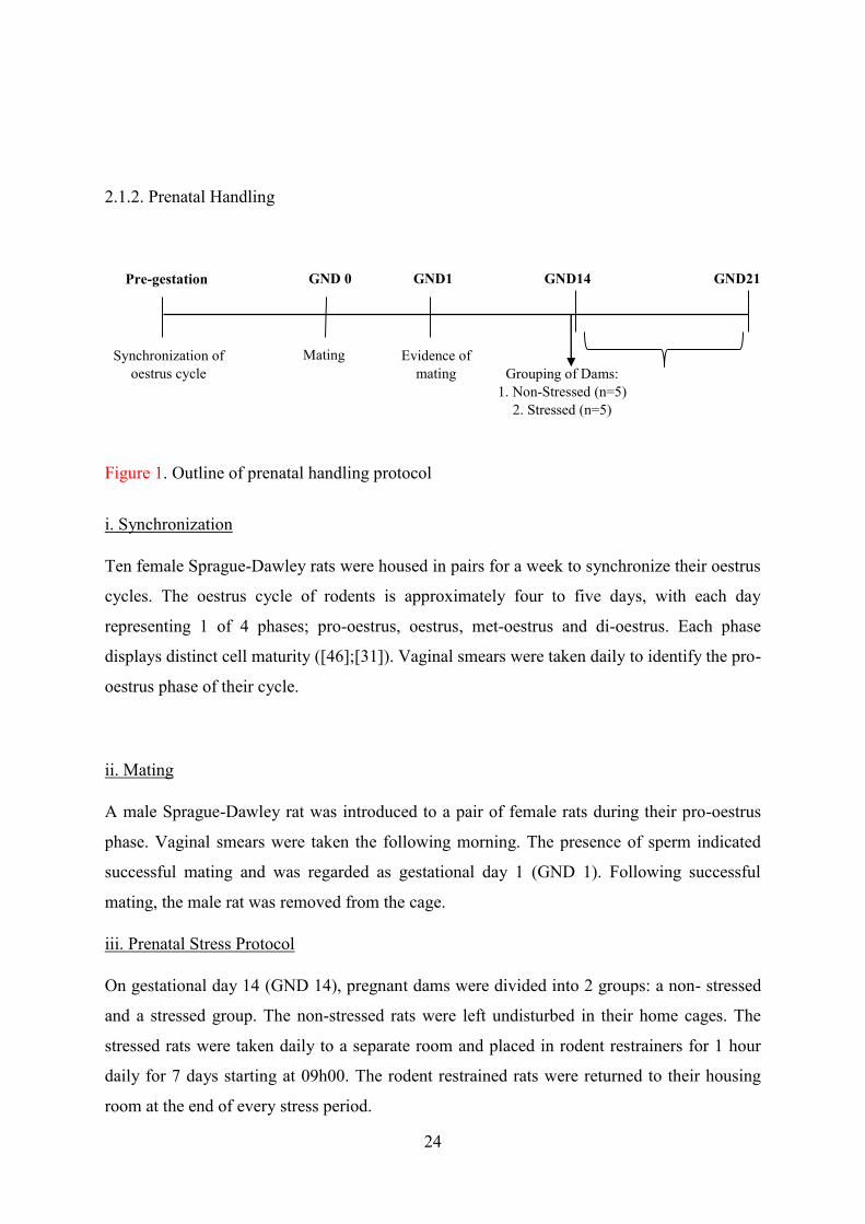
24
2.1.2. Prenatal Handling
i. Synchronization
Ten female Sprague-Dawley rats were housed in pairs for a week to synchronize their oestrus
cycles. The oestrus cycle of rodents is approximately four to five days, with each day
representing 1 of 4 phases; pro-oestrus, oestrus, met-oestrus and di-oestrus. Each phase
displays distinct cell maturity ([46];[31]). Vaginal smears were taken daily to identify the pro-
oestrus phase of their cycle.
ii. Mating
A male Sprague-Dawley rat was introduced to a pair of female rats during their pro-oestrus
phase. Vaginal smears were taken the following morning. The presence of sperm indicated
successful mating and was regarded as gestational day 1 (GND 1). Following successful
mating, the male rat was removed from the cage.
iii. Prenatal Stress Protocol
On gestational day 14 (GND 14), pregnant dams were divided into 2 groups: a non- stressed
and a stressed group. The non-stressed rats were left undisturbed in their home cages. The
stressed rats were taken daily to a separate room and placed in rodent restrainers for 1 hour
daily for 7 days starting at 09h00. The rodent restrained rats were returned to their housing
room at the end of every stress period.
Figure 1. Outline of prenatal handling protocol
Pre-gestation GND 0 GND1 GND14
Mating Synchronization of oestrus cycle
Evidence of mating
Grouping of Dams: 1. Non-Stressed (n=5)
2. Stressed (n=5)
GND21

25
2.1.3. Post-Natal Handling
i. Birth of pups
Following birth, the pups remained with their dams undisturbed until PND 14 after which they
were divided into the following groups (n=6 per group);
Table 1. Pups were grouped as follows:
Non- Stressed Animals (NS) Stressed Animals (S)
Saline (NSS) Saline (SS)
Febrile Seizure (LPS + KA) (NSFS) Febrile Seizure (LPS + KA) (SFS)
The pups were removed from their dams and placed in clean cages. Each cage, housing 6 pups
per experimental group was taken to the experimental room an hour prior to administration of
LPS and kainic acid so as to acclimatize to the new surroundings. The dams remained in the
home room, undisturbed during the duration of the experimental procedure.
ii. Induction of seizures
Control animals were injected with 2ml saline (0.9% NaCl, Adcock Ingram, South Africa) as
a vehicle. To induce a febrile seizure; 0.2ml of LPS (200 µg/kg, Sigma, USA) was injected
intra-peritoneally (i.p), after which pups were returned to their dams to allow for suckling and
grooming. 2.5 hours later, pups were once again removed from their dams and injected with
0.2ml kainic acid (1.75 mg/kg, i.p, Sigma, USA) ([29];[51]).
Figure 2. Outline of post-natal handling protocol
PND 1 PND 14 PND 17, 18 & 19 PND 21
Induction of seizures & Grouping: 1. Saline
2. LPS +KA
Birth of pups Behaviour test 2
Behavioural Training (Morris Water Maze)
(
PND 30
Behaviour test 1 & weaning

26
a) Assessment of Febrile Convulsions
The convulsive behaviour of each rat was observed for a period of 60 minutes and the degree
of convulsive behaviour was measured (Table 2) ([62];[29];[47]).
Table 2. Seizure response were scored as follows:
Stage Response
0 No response
1 Ear and facial twitching
2 Loss of postural control
3 Myoclonic jerks and rearing
4 Clonic convulsions with animal falling on its side
5 Repeated severe tonic- clonic convulsions or fatal convulsions
On PND 21, pups were weaned from their dams and the dams were returned to the animal
unit. On PND 21, learning ability was assessed on both male and female pups- whereas the
ability to remember (memory) was assessed on PND 30 male pups only. Only male pups were
used on PND 30 so as to negate the effects of hormonal changes associated with female rats
([48]).
2.2.1. Behavioural Tests
Morris Water Maze
Training in the Morris Water Maze (MWM) took place between PND 17-19. This was followed
by a test phase (PND 21 and PND 30) which assessed learning and memory deficits. This test
is one of many which determines the learning and recall ability of rats, and covers exploratory,
navigational, spatial and contextual memory ([45];[18];[23]). The MWM consists of a 1m
diameter pool, comprising of 4 quadrants. Each quadrant has a cue to assist the rat in finding
the hidden platform located in one of the quadrants of the pool. The method entails placing the
rat in a quadrant other than where the hidden platform is located, recording the time undertaken

27
for the rat to reach the hidden platform is considered as the animals ability to learn
([45];[18];[23]). The probe test is a post-test for learning which measures the ability of an
animal to remember the quadrant in which the hidden platform is located. The time spent in
the quadrant of the hidden platform is considered as the ability to remember (memory)
([45];[18];[23]).
Training procedure
Animals were exposed to three training sessions for a period of three days (PND 17-19).
Animals were taken to the behavioural room one hour prior to training to allow for
acclimatization to the new environment. For the training procedure, each rat was placed in a
quadrant, gently placed in the water, head facing the cue. The rat was given 120 sec to find the
hidden platform. If a rat failed to find the hidden platform in 120 sec, it was physically guided
by the experimenter towards the platform and was allowed 60 sec to explore the platform before
being returned to the home cage. Training resumed the following day for the next two days.
No training took place on PND 20.
Testing procedure
On PND 21, animals were removed from their rooms and taken to the behavioural room one
hour prior to the behavioural test to allow for animals to acclimatize. Thereafter, animals were
placed in a quadrant other than the hidden platform and the time to locate the hidden platform
was recorded. Animals were then returned to their cages before being returned to their room.
Animals were left undisturbed from PND 22-29. On PND 30, the probe test was conducted.
Prior to the test, the animals underwent the same acclimatization procedure previously
described. For the probe test, the platform was removed completely from the maze and the
animals were allowed 120s in the water. Time spent in the quadrant of the hidden platform was
timed. Following the probe test, the animals were returned to their home room. Animals were
sacrificed by decapitation 5 days later (PND 35), three weeks after seizure induction.
2.2.2. Sacrifice
On PND 35, the rats were taken to the autopsy room 1 hour before decapitation. The rats were
decapitated using a guillotine; trunk blood and hippocampal tissue were collected.
Hippocampal tissue was weighed prior to freezing in liquid nitrogen. All tissue material was
stored in a bio freezer at -80 ˚C.

28
2.2.3. Neurochemical Analysis
Real-Time PCR (qPCR)
Approximately 50mg of hippocampal tissue was weighed out (n=6 per group), homogenized
and suspended in 400µl of lysis buffer (Zymo Research, USA). Thereafter, samples were
homogenised using a sonicator and total RNA isolation was carried out as per manufacturers
guidelines (ZR RNA MiniPrepTM, USA). Purification of RNA isolates were conducted using a
NanoDrop. Purity of 1.5-2.01 was considered ideal for use in the construction of cDNA. cDNA
synthesis was done using the iScript™ cDNA Synthesis Kit (Biorad, South Africa), processing
guidelines were provided by the manufacturer. cDNA was run through the Thermocycler as
per conditions stipulated in the guidelines. The Fast start SYBR green kit (Roche Diagnostics,
USA) was used in accordance to the manufacturers protocol.
Primer sequences; MeCP2 gene- Forward primer: CGTCCCCTTGCCTGAAGGTTGGA,
Reverse primer: CTTTCCAGCAGAGCGACCAG; REST gene- Forward primer:
CAGTTGAACTGCCGTGGG, Reverse primer: CATCCGCTGTGACCGCTG; Beta actin
gene- Forward primer: GCTTCTTTGCAGCTCCTTCGT, Reverse primer:
CCAGCGCAGCGATATCG were reconstituted in RNA nuclease free water according to
manufacturers report (Inqaba) and was added to a master mix comprising of SYBR green dye,
nuclease free H2O and MgCl2. Thereafter cDNA was added into glass capillaries and run in the
Lightcycler 480 at optimised conditions.
2.2.4. Statistical Analysis
The data was analyzed using the software GraphPad Prism (version 5). Normality and Gaussian
distribution were determined using the Smirnov-Kolmogorov tests. A two-way factorial
analysis of variance was performed on behavioural and gene data with stress and febrile
seizures as between rat factors. Significant main effects were followed by Bonferonni post hoc
test. Differences were considered significant when p- value < 0.05.
3.1. Results
3.1.1. Morris Water Maze: Learning and Memory Assessment
The following groups were assessed for learning and memory deficits; non-stressed saline
(NSS), stressed saline (SS), non-stressed with febrile seizures (NSFS) and stressed with febrile

29
seizures (SFS). In the non-stressed (NS) groups we found a febrile seizure effect, as rats that
were exposed to febrile seizures took a longer time to locate the hidden platform *(NSS vs.
NSFS, F (1.0) = 5.15, p < 0.05).
A week later (PND 30) non-stressed with febrile seizure animals (NSS), stressed saline (SS),
non-stressed with febrile seizures (NSFS) and stressed with febrile seizures (SFS) were once
again assessed using the probe test. Our findings once again showed that in non- stressed (NS)
animals there was a febrile seizure effect as febrile seizure exposed rats spent less time in the
quadrant of the hidden platform *(NSS vs. NSFS, F (1.0) = 5.15, p < 0.05).
Learning Ability on PND 21
Saline
Febrile
Seizures
0
30
60
90
120Non-StressedStressed
*
Groups
Tim
e ta
ken
to lo
cate
hid
den
plat
form
(s)
Figure 3.1.a. Graph displaying average time taken to locate the hidden platform in the MWM in the following groups: non-stressed (NSS), stressed (SS), non-stressed febrile seizure (NSFS), stressed febrile seizures (SFS), n=6 per group.*(NSS vs. NSFS), p < 0.05.

30
3.1.2. RT-PCR: MeCP2 and REST genes
Real-time PCR was used to analyse the MeCP2 and REST genes. Groups analysed were non-
stressed with saline (NSS), stressed saline (SS), non-stressed with febrile seizures (NSFS) and
stressed with febrile seizures (SFS) animals. There was a stress effect as prenatally stressed
rats showed down-regulation of the MeCP2 gene *(NSS vs. SS, F (1.0) = 4.93, p < 0.05). There
was a febrile seizure effect in the non-stressed rats **(NSS vs. NSFS, F (1.0) = 4.93, p < 0.05).
Figure 3.1.b. Graph displaying average time spent in the quadrant of the hidden platform in the MWM in the following groups: non-stressed (NSS), stressed (SS), non-stressed febrile seizure (NSFS), stressed febrile seizures (SFS), n=6 per group. *(NSS vs. NSFS), p < 0.05.
Memory on PND 30
Saline
Febrile
Seizures
0
30
60
90
120Non-StressedStressed
*
Groups
Tim
e sp
ent i
n qu
adra
nt o
f hid
den
plat
form
(s)

31
In REST gene analysis, there was a febrile seizure effect in non-stressed rats as rats exposed
to febrile seizures showed down-regulation of the REST gene expression *(NSS vs. NSFS, F
(1.0) = 2.32, p < 0.05).
Figure 3.2.a. Graph displaying MeCP2 gene expression in the hippocampus in P30 rats in the following groups: non-stressed (NSS), stressed (SS), non-stressed febrile seizure (NSFS), stressed febrile seizures (SFS), n=6 per group. *(NSS vs. SS) and **(NSS vs. NSFS), p < 0.05.
Gene expression:MeCP2
Saline
Febrile
Seizures
0
1
2
3Non-StresssedStressed
* **
Groups
MeC
P2/B
-act
in

32
In non-stressed animals there was a significant interaction between the MeCP2 and REST
genes in febrile seizure exposed animals *( NSFS MeCP2 vs. NSFS REST, F (1.0) = 14.8, p <
0.05).
Figure 3.2.b. Graph displaying REST gene expression in the hippocampus in P30 rats in the following groups: non-stressed (NSS), stressed (SS), non-stressed febrile seizure (NSFS), stressed febrile seizures (SFS), n=6 per group. *(NSS vs. NSFS), p < 0.05.
Gene expression:REST
Saline
Febrile
Seizures
0
1
2
3Non-StressedStressed
*
Groups
RES
T/B
-act
in
Non- Stressed Animals(MeCP2 vs. REST)
NSSNSFS
0
1
2
3MeCP2REST
*
Groups
Targ
et G
ene/B
eta A
ctin
Figure 3.3.a. Graph displaying a comparative analysis of overall gene expression in non-stressed (NSS) and non-stressed febrile seizure (NSFS), n=6 per group. *(NSFS MeCP2 vs. NSFS REST), p < 0.05.

33
In stressed animals there was a significant interaction between the MeCP2 and REST genes in
saline and febrile seizure exposed animals *( SS MeCP2 vs. SS REST, F (1.0) = 3.23, p < 0.05)
and *(SFS MeCP2 vs. SFS REST, F (1.0) = 3.23, p < 0.05).
4.1. Discussion
In our study we investigated the effects of stress on learning and memory and whether any
effects present are influenced by the expression of the MeCP2 and REST genes. We also
investigated whether exposure to febrile seizures may influence learning and memory
behaviour as well as gene expression.
Exposure to early life stress (SS) did not seem to affect learning and while there was a tendency
for the prenatally stressed (SS) rats to struggle to find the quadrant of the hidden platform, the
stress effect was not significant, however a febrile seizure effect was present in the non-stressed
(NSS) animals. This effect was prolonged as rats tested 15 days post febrile seizure induction
also showed a presence of memory deficits. This suggests that there are learning and memory
deficits present in non-stressed animals exposed to febrile seizures (NSFS). These findings are
Stressed Animals(MeCP2 vs. REST)
SSSFS
0
1
2
3MeCP2REST
*
*
Targ
et G
ene/
Bet
a Ac
tin
Groups
Figure 3.3.b. Graph displaying a comparative analysis of overall gene expression in stressed (SS) and stressed febrile seizures (SFS), n=6 per group. *(SS MeCP2 vs. SS REST) and *(SFS MeCP2 vs. SFS REST), p < 0.05.

34
in agreement to literature that showed exposure to multiple forms of neuronal insults did not
affect cognitive function in the developing brain, however there was a degree of impairment
observed in adult rats that were exposed to early life multiple episodes of a febrile seizures
([14]). In our study we were able to observe cognitive impairment with a single episode of a
febrile seizure. These findings may be related to the neuroinflammation present in the brain
due to the presence of a fever which triggers a seizure. Studies have shown that high levels of
cytokine expression directly affects neuronal function viz. long-term potentiation, glutamate
release and activation of cell-signalling pathways ([43];[21]). These are related to synaptic
plasticity and are therefore important in cognition. Previous studies have shown that the
occurrence of a fever resulted in the release of various inflammatory markers ([53];[22]). A
common laboratory method to mimic a model of inflammation via fever is by injecting
lipopolysaccharide (LPS), a gram negative bacterial endotoxin that triggers the activation of
innate immunity via toll-like receptors (TLRs) promoting transcription of pro-inflammatory
(IL-1β, IL-6, TNFα) and anti-inflammatory (IL-1 receptor antagonist (ra), IL-10) cytokines
([34];[21]). A study showed that peripheral administration of LPS resulted in long-term
exposure to high levels of the pro-inflammatory cytokine IL-1β in circulation leading to
compromised neural function in the brain ([49]). Learning and memory impairments have been
reported in studies presenting acute exposure to various forms of peripheral inflammation
which resulted in prolonged brain excitability ([53];[64]). This subsequently led to excess
release of intracellular Ca2+ due to competitive binding between IL-1β and IL-1ra leading to
neuronal cell death and compromised cognitive function ([53];[48];[64]).
On PND 21, animals exposed to stress and febrile seizures (SFS) showed no significant
learning deficits from stressed animals without febrile seizures (SS), however both stressed
groups (SS and SFS) had a tendency to struggle to find the quadrant with the hidden platform
when compared to the non-stressed animals. The lack of difference in time taken to find the
quadrant with the hidden platform in stressed animals induced with febrile seizures (SFS)
suggests that the febrile seizure did not exacerbate learning impairments that may have been
present. This was also the case on PND 30 as we could not find differences in the recall ability
of the stressed animals (SS and SFS) when compared to the non-stressed animals without
exposure to febrile seizures (NSS).
Various studies have shown that early life neuronal insults can result in chronic physiologic
and neuronal alterations in later life ([60];[27];[21]). Findings from our behavioural results
were confirmed by gene analysis performed on hippocampal tissue on PND 35. We

35
investigated the roles of the MeCP2 and REST genes in learning and memory in a prenatally
stressed febrile seizure rat model.
We showed that the MeCP2 gene was significantly down-regulated in prenatally stressed
animals (SS) in comparison to non- stressed animals (NSS) thereby confirming the presence of
a stress effect. While we could not confirm definitive impairment in the behavioural tests,
possibly due to the number of animals tested, the gene expression results were more robust in
showing the presence of a stress effect. This suggests that prenatal stress had a direct effect on
the epigenetic modification in the developing brain. Studies have shown the deleterious effects
of various stressors in early life on brain development ([56];[39];[40];[41]). Early life stress
can cause selective methylation or demethylation on specific cytosine residues in the DNA
sequence which are shown to be influenced by 11β-HSD2 levels i.e. decrease in MeCP2 levels
results in the decrease of 11β-HSD2 enzyme, resulting in compromised brain function in the
foetus ([4]; [67]). Therefore our findings suggest that prenatal stress exposure alone has
deleterious effects on the developing brain which can result in long-term learning and memory
deficits.
Non-stressed animals exposed to febrile seizures (NSFS) showed a significant down-regulation
of the MeCP2 gene when compared to control animals (NSS) thereby confirming a febrile
seizure effect. These results may be related to spontaneous excitatory activity that occurred in
early life exposure to an acute seizure. A study showed that a decrease in MeCP2 gene is due
to a decrease in excitatory postsynaptic currents (EPSCs) ([15]). This was also presented in
another study that showed the MeCP2 gene regulates the amount of glutamate receptors in
hippocampal neurons ([13]). In our study, the acute bout of excitability, due to the injection of
the kainic acid, may have resulted in over-excitation of glutamate receptors. The injection of
this glutamate receptor agonist may have influenced the down-regulation of the MeCP2 gene.
Stressed animals exposed to febrile seizures (SFS) did not exhibit learning and memory deficits
when compared to control animals (SS) suggesting that exposure to febrile seizures did not
exacerbate the MeCP2 gene down-regulation. A study showed that stress increased arginine
vasopressin in the hypothalamus which significantly decreased MeCP2 gene expression ([45]).
We found no change in MeCP2 expression between stressed animals with and without febrile
seizure induction (SS and SFS). Therefore, exposure to a single early life stress may impair
cognition but further exposure to infections resulting in a febrile seizure does not exacerbate
these impairments.

36
The REST gene regulates its expression through recruitment of various co-repressors including
MeCP2 ([37];[6];[52]). Therefore if MeCP2 expression increases, REST will decrease. As
MeCP2 gene promotes neurogenesis and the REST gene acts as a repressor factor, an increase
in the MeCP2 gene expression acts as a protective feedback mechanism ([52]). Our findings
have shown that there was significant up-regulation in the REST gene expression in stressed
animals (SS), suggesting compromised neural plasticity. This concurs with our behavioural
result in which stressed animals showed tendencies of learning and memory impairments. A
study suggested that decreased expression of REST gene promotes cellular differentiation and
neurogenesis ([19]). While we did not measure neurogenesis in our study, we postulate that an
increase in REST gene expression affected plasticity thereby compromising hippocampal
function leading to learning and memory deficits.
A febrile seizure effect was present in non-stressed animals (NSFS) as there was a significant
down-regulation of the expression of the REST gene. As behavioural results showed impaired
cognitive function in these animals, down-regulation of REST gene suggests that the
hippocampal damage leading to cognitive impairment in these animals may be a transient
effect. Which we may confirm that febrile seizures are indeed a benign condition that does not
lead to permanent cognitive deficits. In order to create a febrile seizure animal model, animals
were exposed to lipopolysacchiride and kainic acid. Kainic acid, a glutamate agonist, promotes
increased intracellular Ca2+ concentrations, leading to excitotoxicity causing neuronal loss
([37]). Studies have previously shown that glutamate plays a role in regulation of REST gene
expression and that early life neural insults affect glutamate receptors by desensitizing the
receptors after prolonged exposure to glutamate thereby compromising hippocampal cell
survival ([54];[2]). These changes in expression of MeCP2 and REST genes also showed a
significant interaction suggesting that the relationship between both genes contributes to
changes in hippocampal functioning. A study showed the inter-dependent relationship of the
MeCP2 and REST genes suggesting that REST does silence MeCP2 under compromised
conditions ([37]).
Stressed animals that were exposed to febrile seizures (SFS) showed no significant changes in
the REST gene expression. This concurs with behavioural findings and MeCP2 gene
expression results. We suggest that the damage caused by exposure to prenatal stress was great
enough in hippocampal neurons, such that further exposure to a toxic insult did not further
exacerbate the damage.

37
5.1 Conclusion
Our findings suggest that exposure to febrile seizures early in life affects learning and memory
ability. However, it seems as if these cognitive deficits are transient as gene expression results
suggests that there may be an attempt to promote neural plasticity following the febrile seizure
episode. We also showed that exposure to stress early in life may affect the ability to promote
neural plasticity in the hippocampus. This effect is, however, not exacerbated by further
exposure to infections that lead to the development of febrile seizures.
Acknowledgements
We would like to thank the Biomedical Resource Centre for their assistance in this study. The
College of Health Sciences, University of KwaZulu-Natal and the National Research
Foundation (NRF) for their financial support. This paper forms part of a Masters degree for a
co-author (S. Cassim).
Disclosure
We hereby disclose that there are no known financial and personal conflict of interests in the
work of this project.

38
References
[1] Ackermann, S. & Van Toorn, R. Managing first-time seizures and epilepsy in
children. Continuing Medical Education. 2012; 30: 16-20.
[2] Abrajano, J. J., Qureshi, I. A., Gokahn, S., Zheng, D., Bergmann, A. &
Mehler, M. F. REST and CoREST modulate neuronal subtype
specification, maturation and maintenance. PLos One. 2009; 4: 1-12.
[3] Arnold, S.E. and Trojanowski, J.Q. Human fetal hippocampal development: I.
Cytoarchitecture, myeloarchitecture, and neuronal morphologic features. J.
Comp. Neurol. 1996; 367: 274–292
[4] Alikhani- Koopaei, R., Fouladkou, F., Frey, F. J. & Frey, B. M. Epigenetic
regulation of 11β-hydroxysteroid dehydrogenase type 2 expression. The
Journal of Clinical Investigation. 2004; 114: 1146-1157.
[5] Altman, J. and Bayer, S.A. Migration and distribution of two populations of
hippocampal granule cell precursors during the perinatal and postnatal periods.
J. Comp. Neurol. 1990; 301: 365–381.
[6] Ballas, N. & Mandel, G. The many faces of REST oversee epigenetic
programming of neuronal genes. Current Opinions in Neurobiology.
2005; 15: 500-506.
[7] Barbazanges, A., Piazza, P. V., Le Moal, M. & Maccari, S. Maternal
glucocorticoid secretion mediates long-term effects of prenatal stress. The
Journal of Neuroscience.1996; 16: 3943-3949.
[8] Benoit, J.D., Rakic, P. & Frick, K. M. 2015. Prenatal stress induces spatial
memory deficits and epigenetic changes in the hippocampus indicative of
heterochromatin formation and reduced gene expression. Behavioural Brain
Research. 2015; 281: 1-8.
[9] Bruce, W. A., Donaldson, I. J., Wood, I. C., Yerbury, S.A., Sadowski, M. I.,
Chapman, M., Gӧttgens, B. and Buckley, N. J. 2004. Genome- wide analysis
of repressor element 1 silencing transcription factor/ neuron- restrictive
silencing factor (REST/NRSF) target genes. PNAS. 2004; 101: 10458-10463.

39
[10] Cannizzaro, C., Plescia, F., Martire, M., Gagliano, M., Cannizzaro, G.,
Mantia,G. & Cannizzaro, E. Single intense prenatal stress decreases
emotionality and enhances learning performance in the adolescent rat
offspring: Interaction with a brief, daily maternal separation. Behavioural
Brain Research. 2006; 169: 128-136.
[11] Champagne, D. L., de Kloet, E. R. & Joëls, M. Fundamental aspects of the
impact of glucocorticoids on the (immature) brain. Seminars in Fetal &
Neonatal Medicine. 2009; 14: 136-142.
[12] Challis, J. R., Sloboda, D., Matthews, S. G., Holloway, A., Alfaidy, N., Patel,
F. A., Whittle, W., Fraser, M., Moss, T. J. M. & Newnham, J. The fetal
placental hypothalmic-pituitary-adrenal (HPA) axis, parturition and post
natal health. Molecular and Cellular Endocrinology. 2001; 185: 135-144.
[13] Chao, H., Zoghbi, H. Y. & Rosenmund, C. MeCP2 controls excitatory
synaptic strength by regulating glutamatergic synapse number. Neuron. 2007;
56: 58- 65.
[14] Chang, Y., Huang, A., Kuo, Y., Wang, S., Chang, Y. & Huang, C. Febrile
seizures impair memory and learning and cAMP response-element binding
protein activation. Annals of Neurology. 2003; 54: 706-718.
[15] Dani, V. S., Chang, Q., Maffei, A., Turrigiano, G. G., Jaenisch, R. & Nelson,
S. B. Reduced cortical activity due to a shift in the balance between
excitation and inhibition in a mouse model of Rett Syndrome. PNAS. 2005;
102: 12560-12565.
[16] De Kloet, E. R., Fitzsimons, C. P., Datson, N. A., Meijer, O. C. &
Vreugdenhil, E. Glucocorticoid signaling and stress-related limbic
susceptibility pathway about receptors, transcription machinery and
microRNA. Brain Research. 2009; 1293: 129-141.
[17] De Kloet, E. R., Claessens, S. E. F. & Kentrop, J. COntext modulates outcome
of perinatal glucocorticoid action in the brain. Frontiers in Endocrinology.
2014; 5: 1-13.
[18] D'Hooge, R. & De Deyn, P. P. Applications of Morris water maze in the study
of learning and memory. Brain Research. 2001; 36: 60-90.
[19] Ernsberger, U. Regulation of gene expression during early neuronal
differentiation: evidence for patterns conserved across neuron populations and
vertebrate classes. Cell Tissue Res. 2012; 348: 1-27.

40
[20] Fine, R., Zhang, M. D. & Stevens, H. E. Prenatal stress and inhibitory neuron
systems: implications for neuropyschiatric disorders. Molecular Psychiatry.
2014; 19: 641-651.
[21] Galic, M. A., Riazi, K., Heida, J. G., Mouihate, A., Fournier, N. M., Spencer,
S. J., Kalynchuk, L. E., Teskey, C. & Pittman, Q. J. Postnatal inflammation
increases seizure susceptibility in adult rats. The Journal of Neuroscience.
2008; 28: 6904-6913.
[22] Galic, M. A., Riazi, K. & Pittman, J. Q. Cytokines and brain excitability.
Frontiers in Neuroendocrinology. 2012; 33: 116- 125.
[23] Garthe, A. & Kempermann, G. An old test for new neurons: refining the
Morris water maze to study the functional revelance of adult hippocampal
neurogenesis. 2013; 7:63.
[24] Greer P. L. & Greenberg, M. E. From synapse to nucleus: calcium-dependent
gene transcription in the control of synapse development and function.
Neuron. 2008; 59: 546- 560.
[25] Griffith, E. C., Cowan, C. W. & Greenberg, M. E. REST acts through multiple
deacetylase complexes. Neuron. 2001; 31: 339- 340.
[26] Guy, J., Cheval, H., Selfridge, J. & Bird, A. The role of MeCP2 in the brain.
Annual Review of Cell and Developmental Biology. 2011; 27: 631-652.
[27] Harre, E. M., Galic, M. A., Mouihate, A., Noorbakhsh, F. & Pittman, Q. J.
Neonatal inflammation produces selective behavioural deficits and alters N-
methyl-D-aspartate receptor subunit mRNA in the adult rat brain. European
Journal of Neuroscience. 2008; 27: 644-653.
[28] Harris, A. & Seckl, J. Glucocorticoids, prenatal stress and the programming of
disease. Hormones and Behaviour. 2011; 59: 279- 289.
[29] Heida, J., Boissé, L. & Pittman, Q. J. Lipopolysaccharide-induced febrile
convulsions in the rat: short-term seuelae. Epilepsia. 2009; 45: 1317-1329.
[30] Huang, L. Early-life stress impacts the developing hippocampus and primes
seizure occurrence: cellular, molecular, and epigenetic mechanisms. Frontiers
in Molecular Neuroscience. 2014; 7: 1-15.
[31] Hubscher, C. H., Brooks, D. L. & Johnson, J. R. A quantitative method for
assessing stages of the rat estrous cycle. Biotechnic & Histochemistry. 2005;
80: 79-87.

41
[32] Jett, D. A. Chemical toxins that cause seizures. NeuroToxicology. 2012; 33:
1473- 1475.
[33] Kentner, A. C. & Pittman, Q. J. Minireview: early-life programming by
inflammation of the neuroendocrine system. Endocrinology. 2010; 151: 4602-
4606.
[34] Laflamme, N., Rivest, S. Toll-like receptor 4: the missing link of the cerebral
innate immunity response triggered by circulating gram-negative bacterial cell
wall components. FASEB J. 2001; 15: 155-163.
[35] Lemaire, V., Koehl, M., Le Moal, M & Abrous, D. N. Prenatal stress produces
learning deficits associated with an inhibition of neurogenesis in the
hippocampus. PNAS. 2000; 97: 11032- 11037.
[36] Lubin, F. D. Epileptogenesis: Can the science of epigenetics give us answers?
Epilepsy Currents. 2012; 12: 105-110.
[37] Lunyak, V.V., Burgess, R., Prefontaine, G. G., Nelson, C., Sze, S.,
Chenoweth, J., Schwarts, P., Pevzner, P.A., Glass, C., Mandel, G. &
Rosenfeld, M. G. Corepressor-dependent silencing of chromosomal regions
encoding neuronal genes. Science. 2002; 298: 1747- 1752.
[38] Martinowich, K., Hattori, D., Wu, H., Fouse, S., He, F., Hu, Y., Fan, G. &
Sun, Y.E. DNA methylation- related chromatin remodelling in activity-
dependent Bdnf gene regulation. Science. 2003; 302: 890-893.
[39] McEwen, B. S. & Sapolsky, R. M. Stress and Cognitive function. Cognitive
Neuroscience. 1995; 5: 205-216.
[40] McEwen, B. S. Plasticity of the hippocampus: adaptation tp chronic stress and
allostatic load. Annals of New York Academy of Sciences. 2001; 933: 265-277.
[41] McEwen, B. S. Central effects of stress hormones in health and disease:
Understanding the protective and damaging effects of stress and stress
mediators. European Journal of Pharmacology. 2008; 583: 174-185.
[42] Meyers, L. A., Ancel, F. D. & Lachmann, M. Evolution of genetic potential.
PLOS Computational Biology. 2005; 1:3.
[43] Moisiadis, V. G. & Matthews, S. G. Glucocorticoids and fetal programming
part 1: outcomes. Nature Reviews Endocrinology. 2014; 10: 391-402.
[44] Morris, R. 1984. Developments of a water-maze procedure for studying spatial
learning in the rat. Journal of Neurosciemce, 11, 47-60.

42
[45] Murgatoyd, C., Patchev, A. V., Wu, Y., Micale, V., Bockmuhl, Y., Dieter, F.,
Holsboer, F., Wotjak, C. T., Almeida, O. F. X. & Spengler, D. Dynamic DNA
methylation programs persistent adverse effects of early-life stress. Nature
Neuroscience. 1-10.
[46] Nelson, J. F., Felicio, L.S., Randall, P. K., Sims, C. & Finch, C. E. 1982. A
longitudinal study of estrous cyclicity in aging C57BL/6J mice: I; cycle
frequency, lenght and vaginal cytology. Biology of Reproduction. 1982; 27:
327-339.
[47] Ojewole, J. A. O. Anticonvulsant effect of Rhus chirindensis (Baker F.)
(Anacardiaceae) stem-bark aqueous extract in mice. Ethnopharmacology.
2008; 117: 130-135.
[48] Peper, J. S. & Dahl, R. E. The teenage brain: Surging hormones-Brain-
behavior interactions during puberty. Current Directions in Psychological
Science. 2013; 22: 134-139.
[49] Popoli, M., Yan, Z., McEwen, B. S. & Sanacora, G. The stressed synapse: The
impact of stress and glucocorticoids on glutamate transmission. Nature Review
Neuroscience. 2012; 13: 22-37.
[50] Qin, L., Wu, X., Block, M. L., Liu, Y., Breese, G. R., Hong, J.-S., Knapp, D.
J. and Crews, F. T. Systemic LPS causes chronic neuroinflammation and
progressive neurodegeneration. Glia. 2007; 55: 453–462.
[51] Qulu, L., Daniels, W.M.U., & Mabandla, M. Exposure to prenatal stress
enhances the development of seizures in young rats. Metabolic Brain
Disorders. 2012; 27: 399-404.
[52] Qureshi, I. A. & Mehler, M. F. Epigenetic mechanisms underlying human
epileptic disorders and the process of epileptogenesis. Neurobiology of
Disease. 2010; 39: 53-60.
[53] Riazi, K., Galic, M. A. & Pittman, Q. J. Contributions of peripheral
inflammation to seizure susceptibility: cytokines and brain excitiability.
Epilepsy Research. 2010; 89: 34-42.
[54] Rodenas- Ruano, A., Chávez, A., Cossio, M. J., Castillo, P. E. & Zukin, R. S.
REST-independent epigentic remodeling promotes the development switch in
synaptic NMDA receptors. Nature Neuroscience. 2012; 15: 1382- 1390.
[55] Roopra, A., Dingledine, R. & Hsieh, J. Epigenetics and epilepsy. Epilepsia.
2012; 53: 2-10.

43
[56] Sapolsky, R. M., Krey, L. C. & McEwen, B. S. Prolonged glucocorticoid
exposure reduces hippocampal neuron number: Implications for aging. The
Journal of Neuroscience. 1985; 5: 1222-1227.
[57] Sapolsky, R. M. Glucocorticoids, stress, and their adverse neurological effects:
relevance to aging. Exprimental Gerontology. 1999; 34: 721-732.
[58] Seckl, J. R. Prenatal glucocorticoids and long-term programming. European
Journal of Endocrinology. 2004; 151: U49- U62.
[59] Scantlebury, H. M. H. J. G. Febrile seizures and temporal lobe
epileptogenesis. Epilepsy Res. 2010; 89: 27-33.
[60] Spencer, S. J., Heida, J. G. & Pittman, Q. J. Early life immune challenge-
effects on behavioural indices of adult rat fear and anxiety. Behavioural Brain
Research. 2005; 164: 231-238.
[61] Vallѐe, M., Willy, M., Dellu, F., Le Moal, M., Simon, H. & Maccari, S.
Prenatal Stress Induces High Anxiety and Postnatal Handling Induces Low
Anxiety in Adult Offspring: Correlation with Stress-Induced
Corticosterone Secretion. The Journal of Neuroscience. 1997; 17: 2626-2636.
[62] Vezzani, A., Moneta, D., Richichi, C., Aliprandi, M., Burrows, S. J., Ravizza,
T., Perego, C. & De Simoni, M. G. Functional role of inflammatory cytokines
and antiinflammatory molecules in seizures and epileptogenesis. Epilepsia.
2002; 5: 30-35.
[63] Vezzani, A., Maroso, M., Balosso, S., Sanchez, M. & Tamas, B. IL-1
receptor/Toll-like receptor signaling in infection, inflammation, stress and
neurodegeneration couples hyperexcitability and seizures. Brain, Behaviour,
and Immunity. 2011; 25: 1281-1289.
[64] Vezzani, A. Epilepsy and inflammation in the brain: Overview and
pathophysiology. Epilepsy Currents. 2014; 14: 3-7.
[65] Wager, R. G. & Newton, C. R. Do helminths cause epilepsy? Parasite
Immunology. 2009; 31: 697-705.
[66] Waruiru, C., & Appleton, R. Febrile seizures: an update. Arch dis Child.2004;
89: 751-756.
[67] Weaver, I. C. G., D'Alessio, A. C., Brown, S. E., Hellstrom, I. C., Sergiy, D.,
Sharma, S., Szyf, M. & Meaney, M. J. The transcription factor nerve growth
factor-inducible protein A mediates epogenetic programming: altering

44
epigentic marks by immediate-ear;y genes. The Journal of Neuroscience.
2007; 27: 1756- 1768.

45
Chapter Three
Article 2
“Febrile seizure induction impairs neuronal plasticity in prenatally stressed rat offspring”
The manuscript will begin with a title, authors and affiliations on a single page. As this study will be in
the journal “Annals of Neuroscience” it is written according to the authors guidelines for the journal.
(Addendum B).

46
RESEARCH PAPER
Febrile seizure induction impairs neuronal plasticity in prenatally stressed
rat offspring.
Sadiyah Cassim1, Lihle Qulu1 and Musa V. Mabandla1
1Discipline of Human Physiology, School of Laboratory Medicine & Medical Sciences,
College of Health Sciences, University of KwaZulu-Natal.
_________________________________________________________________________
Correspondence: Dr. Musa Mabandla
Tel: (+27) 31 260 8602
Email: [email protected]

47
Abstract
Febrile seizures in young children are a growing concern in developing economies around the
world. Although this condition is regarded as benign, exposure to early life stress in these
children may affect neuronal functioning in due time. We investigated whether neuronal
malformation and the resulting cognitive deficits associated with stress are linked to changes
in BDNF and the REST gene factor concentration and expression respectively. Furthermore,
we investigated whether febrile seizure induction exacerbates the changes seen in these factors.
Febrile seizures were induced on postnatal day (PND) 14 in pups of dams that were stressed
during pregnancy. To assess the effects of prenatal stress on behaviour, behavioural
assessments were conducted on PND 21 followed by sacrifice by decapitation. Our results
show that there was a delay in the onset of seizures followed by a prolonged seizure duration
in animals exposed to prenatal stress. Furthermore, prenatal stress exposure resulted in anxiety-
like behaviour in young rats. We also found that prenatal stress resulted in increased
hippocampal BDNF concentration, whereas exposure to febrile seizures increased REST gene
expression in prenatally stressed animals. These findings suggest that exposure to prenatal
stress impedes neurotrophic factor release, while accentuating the REST gene expression in
febrile seizure animals. These factors may contribute to the hindering of neurogenic properties
in the young, thus leading to neuronal plasticity deficits and possibly cognitive malfunction in
later life.
Keywords: Anxiety-like behaviour, REST gene, BDNF, prenatal stress, febrile seizures,
neural plasticity

48
1.1 Background
Elevation in body temperature often follows the presence of an infection in the body (Heida, et
al., 2004; Wager, et al., 2009; Ackermann & Van Toorn, 2012). In children, infections are
common triggers of seizures (Heida, et al., 2004; Ackermann & Van Toorn, 2012; Wager, et
al., 2014). Seizures are categorised as epileptic (disorder of brain function that trigger episodic
and unpredictable seizure occurrence) or non-epileptic (once-off seizure that may be evoked
by electrical or chemical stimulations (McNamara, 1994). Febrile seizures are regarded as non-
epileptic seizures and may be classified as a simple, complex or generalized convulsion
(McNamara, 1994). Clinical findings suggest that non-epileptic convulsions lead to motor
disorientation and transient cognitive deficits in patients (Hoefnagels, et al., 1991). Stress has
been shown to aggravate seizure progression in a kainic acid induced seizure rodent model
(Qulu, et al., 2012). More than 10% of children who had previously been exposed to various
forms of early life stressors had exacerbated seizure occurrences (Wood, et al., 2004). Clinical
findings have further suggested that early exposure to stress affects anxiety levels thereby
decreasing immune function resulting in children being more susceptible to febrile convulsions
(Koh, et al., 1998; Ackermann & Van Toorn, 2012).
Furthermore, it has been shown that exposure to various forms of stressors prenatally, affects
the hypothalamic-pituitary-adrenal (HPA) axis particularly of the developing foetus (Qulu, et
al., 2012; Moisiadis, et al., 2014). As a consequence, the developing neuronal circuitries in the
foetus are affected due to exposure to high concentrations of circulating maternal
glucocorticoids (Wilson, et al., 2013; Moisiadis, et al., 2014). This subsequently affects the
limbic area of the brain, specifically the hippocampus, as there is a high prevalence of cortisol
receptors present (McEwen, 2001; Wilson, et al., 2013). Therefore, disruption in hippocampal
function contributes to various anxiety disorders and cognitive deficits (McEwen, 2001;
Wilson, et al., 2013; Moisiadis, et al., 2014).
Qulu, et al. (2015), has shown that prenatal stress reduces hippocampal mass in young rats.
This may be a result of neuronal malformation involving various factors that involve disruption
in DNA methylation affecting neurotrophic factor concentrations and multiple down- and up-
stream signalling pathways altering neuronal function and structure (Qureshi & Mehler, 2010;
Guy, et al., 2011). Brain-derived neurotrophic factor (BDNF), a crucial player involved in
neuronal plasticity, may result in the manifestation of various neuronal abnormalities and
disorders when compromised (Karpova, 2014). BDNF has been closely associated with

49
epigenetic factors involved in DNA methylation (Karpova, 2014). Although methyl-CpG-
binding proteins actively manipulate the function of BDNF, RE1-silencing transcriptional
(REST) factor is essentially required to regulate transcription suggesting its involvement in
neurogenesis (Pruunsild, et al., 2011). It has been shown that febrile seizures have a transient
effect on cognitive function by altering the balance between methyl-CpG-binding proteins and
the REST gene factor in the long term (Cassim, et al, 2015).
Therefore the aim of this study was to investigate whether neuronal malformations and the
resulting cognitive deficits associated with stress are linked to changes in BDNF concentration
and the REST gene factor expression. We further investigated whether exposure to febrile
seizures exacerbates the changes seen in these factors.
2.1. Material and Methods
2.1.1. Animals
Male and female Sprague-Dawley rats were obtained from the Biomedical Resource Centre
(BRC) of the University of KwaZulu-Natal. The animals were housed under standard BRC
conditions (20% humidity at temperature of ±23˚C) and a 12 hour light/dark cycle (lights on at
06h00) was maintained. Food and water were available ad libitium. All experimental
procedures were approved by the Animal Ethics Research Committee of the University of
KwaZulu- Natal (073/14/animal) in accordance with guidelines of the National Institute of
Health, USA.
2.1.2. Prenatal Handling
a. Mating
Ten female Sprague-Dawley rats were housed in pairs for a week to synchronize their oestrus
cycles (Nelson et al., 1982; Hubscher et al., 2005). Vaginal smears were taken daily to identify
when the rats were in pro-oestrus (Shorr, 1941; Moosa, et al., 2014). When females reached
pro-oestrus, a male rat was introduced to a pair of females, followed by the taking of vaginal
smears the next morning. The presence of sperm indicated a successful mating and was
regarded as gestational day (GND) 1. Following successful mating, the male rat was removed
from the cage.

50
b. Prenatal Stress Protocol
On GND 14, pregnant dams were divided into 2 groups: a non-stressed and a stressed group.
The non-stressed rats were left undisturbed in their home cages. The stressed rats were taken
daily to a separate room and placed in rodent retainers for 1 hour, for 7 days starting at 09h00.
The rodent restrained rats were returned to their home room at the end of every stress period
(Qulu, et al., 2012; Cassim, et al., 2015).
2.1.3. Post-Natal Handling
Following birth, the pups remained with their dams undisturbed until post-natal day (PND) 14,
after which they were divided into the following groups (n=6 per group per cage);
a. Non-stressed saline (NSS)
b. Prenatally stressed saline (SS)
c. Non-stressed febrile seizure (NSFS)
d. Prenatally stressed febrile seizure (SFS)
The experimental protocol was performed in the same way as mentioned by Cassim, et al.
(2015). Briefly, control animals were injected with 0.2 ml/g saline (0.9% NaCl, Adcock
Ingram, South Africa) as a vehicle. To induce a febrile seizure (groups c and d); 0.2 ml/kg of
LPS (200 µg/kg, Sigma, USA) was injected intra-peritoneally (i.p) and pups were returned to
their dams to allow for suckling and grooming to avoid dehydration and hypothermia. Two and
a half hours later, pups were once again removed from their dams and injected with 0.2 ml/g
kainic acid (1.75 mg/kg, i.p, Sigma, USA) to induce a febrile convulsion (Heida, et al., 2004;
Qulu, et al., 2012; Cassim, et al., 2015). The convulsive behaviour of each rat was observed
and recorded for a period of 60 minutes. The various stages of convulsive behaviour were
assessed following the guidelines provided in Heida, et al. (2004) and Ojewole (2008).
Thereafter, pups were returned to their dams.
2.2.1. Behavioural Test
Elevated Plus Maze (EPM)
The elevated plus-maze (EPM) is a test used to assess the anxiety-like behaviour of an animal
exposed to various stressors (Cannazziro, et al., 2006). On PND 21, the rats were assessed for

51
anxiety-like behaviour using the elevated plus maze (Cannazziro, et al., 2006). The test entails
placing the rat in the central area of the maze facing the open arm and the degree of anxiety-
like behaviour is measured by the number of entries into the preferred arm (Cannazziro, et al.,
2006). Animals were allowed 5 minutes in the apparatus and preference for the closed arm was
regarded as a measure of anxiety-like behaviour. An entry was scored when all four paws
entered into each arm.
2.2.1. Sacrifice
Rats were sacrificed by decapitation. Hippocampal tissue was collected and weighed prior to
freezing in liquid nitrogen. All tissue material was stored in a bio-freezer at -80 ˚C.
2.2.3. Neurochemical Analysis
Real-Time PCR (qPCR)
The qPCR analysis was done as previously described (Cassim, et al., 2015). Briefly,
hippocampal tissue (50mg) was homogenized and suspended in 400µl of lysis buffer (Zymo
Research, USA). This was followed by RNA extraction using the guidelines stipulated by the
manufacturer (ZR RNA MiniPrepTM, USA). Purification of RNA isolates was conducted using
a NanoDrop. Purified RNA isolates were used in conjunction with the iScript™ cDNA
Synthesis Kit (Biorad, South Africa), to construct cDNA using a Thermocycler as per
conditions stipulated by the manufacturer. The Fast start SYBR green kit (Roche Diagnostics,
USA), cDNA and primers were used to amplify our gene of interest at optimised conditions in
the Lightcycler 480 (Roche Diagnostics, USA).
The primer sequences used:
Primer sequences;
a. REST gene-
Forward primer: CAGTTGAACTGCCGTGGG,
Reverse primer: CATCCGCTGTGACCGCTG;
b. Beta actin gene-
Forward primer: GCTTCTTTGCAGCTCCTTCGT,
Reverse primer: CCAGCGCAGCGATATCG
(Inqaba, South Africa).

52
2.2.4. Immunoassay (ELISA)
BDNF protein was measured with a conventional enzyme-linked immunosorbent assay
(ELISA) system. The BDNF immunoassay (Cusabio Biotech Co., Ltd., China) was performed
according to the manufacturer’s protocol. The harvested hippocampal tissue was rinsed and
stored in 1 ml of PBS overnight at -20°C. After two freeze thaw cycles, homogenates were
centrifuged at 5000xg at 7°C. The supernatant was used to determine the BDNF concentration.
The sensitivity of the assay was 0.312 ng/ml- 20 ng/ml, and the cross-reactivity with other
related neurotrophic factors was minimum. The intra- and inter-assay precisions were <10%,
respectively.
2.2.5. Statistical Analysis
The data was analyzed using the software GraphPad Prism (version 5). Normality and Gaussian
distribution were determined using the Kolmogorov-Smirnov test. A two-way factorial
analysis of variance was performed with stress and febrile seizures as between rat factors.
Significant main effects were followed by the Bonferonni post hoc test. Differences were
considered significant when p- value < 0.05.

53
3.1. Results
3.1.1. Onset of seizure and seizure duration
Seizures were induced on PND 14 in non-stressed (NSFS) and stressed (SFS) animals. There
was a stress effect as animals exposed to prenatal stress exhibited with a delay in seizure onset
but had seizures of prolonged duration *(NSFS onset of seizure vs. SFS onset of seizure, F(1.0)
= 132.2, p < 0.05) and **(NSFS seizure duration vs. SFS seizure duration, F(1.0) = 32.6, p <
0.05).
Onset of Seizure and Duration of Seizure
0 20 40 60 80 100
Non-Stressed
Stressed
Onset of SeizureDuration of seizure
* **
Time (min)
Gro
ups
3.1.2. Elevated Plus Maze: Anxiety-like behaviour
Anxiety-like behaviour was assessed on the following groups of animals on PND 21: non-
stressed saline (NSS), stressed saline (SS), non-stressed with febrile seizures (NSFS) and
stressed with febrile seizures (SFS). There was a stress effect in both the saline and the febrile
seizure groups as these animals spent more time in the closed arms *(NSS vs. SS, F (3.0) = 97.13,
p < 0.05) and *(NSFS vs. SFS, F(3.0) = 97.13, p < 0.05).
Figure 1. Graph displaying seizure onset and seizure duration in the following groups: non-stressed (NSFS) and
stressed (SFS), n=6 per group.*(NSFS onset of seizure vs. SFS onset of seizure) and ** (NSFS duration of seizure vs.
SFS duration of seizure), p < 0.05.

54
3.1.3. Brain-Derived-Neurotrophic Factor (BDNF)
To assess a marker of neural plasticity within the hippocampus, BDNF protein concentration
was measured in the following groups: non-stressed saline (NSS), stressed saline (SS), non-
stressed with febrile seizures (NSFS) and stressed with febrile seizures (SFS). Animals exposed
to prenatal stress showed a stress effect with high concentration of BDNF levels when
compared to non-stressed animals *(NSS vs. SS, F(1.0) = 4.903, p < 0.05)
Elevated Plus Maze: Closed Arm
Saline
Febrile
Seizures
0
2
4
6
8
10Non-StressedPrenatally stressed
Groups
# en
trie
s in
to a
rm
*
**
Figure 2. Graph displaying number of entries into the closed arm of the EPM in the following groups: non- stressed
(NSS), stressed (SS), non-stressed febrile seizure (NSFS), stressed febrile seizures (SFS), n=6 per group.*(NSS vs SS)
and **(NSFS vs. SFS), p < 0.05.

55
3.1.4. qPCR: REI-1-Silencing Transcription (REST)
The REST factor expression was analysed in the following groups: non-stressed saline (NSS),
stressed saline (SS), non-stressed with febrile seizures (NSFS) and stressed with febrile
seizures (SFS) animals.
Brain-Derived-Neurotrophic Factor (BDNF)
Saline
Febrile
Seizures
0.0
0.5
1.0
1.5
2.0Non-StressedStressed
*
Groups
Con
cent
ratio
n (n
g/m
l)
Figure 3. Graph displaying BDNF concentration in the following groups: non- stressed (NSS), non-stressed febrile
seizure (NSFS), n=5 per group in duplicate. *(NSS vs SS), p < 0.05.

56
There was a stress effect on the REST gene expression in the febrile seizure groups *(NSFS
vs. SFS, F(1.0) = 5.57, p < 0.05).
4.1. Discussion
In our study we investigated the effects of stress and febrile seizure induction on young rats
and whether these effects influence the concentration of BDNF and REST gene factor
expression in the hippocampus.
Our findings show that animals exposed to prenatal stress had a delay in seizure onset that was
followed by a prolonged seizure duration. Stress exerts its effects via various mechanisms
(Weinstock, et al., 2008). Exposure to pro-inflammatory cytokine release (IL-1β, TNF-α, IL-
6) is a typical response to stress exposure, which increases pro-inflammatory response in the
brain resulting in neuronal malfunction and subsequently altered behaviour in rodents (Qulu,
et al., 2012; Salim, et al., 2012; Diz-Chaves, et al., 2013). Studies have shown that interleukin-
1β results in competitive binding to interleukin-1 receptor type 1, thereby resulting in an
REST gene expression
Saline
Febrile Seizu
res0
1
2Non- StressedStressed
*
Groups
REST
/B-a
ctin
Figure 4. Graph displaying REST gene expression in the following groups: non- stressed (NSS), stressed (SS), non-
stressed febrile seizure (NSFS), stressed febrile seizures (SFS), n=6 per group.*(NSFS vs. SFS), p < 0.05.

57
increase in concentration of the neurotransmitter glutamate and an inhibition of the GABAergic
system (Viviani, et al., 2003). The subsequent imbalance between the excitatory
neurotransmitter glutamate and the inhibitory neurotransmitter, γ-aminobutyric acid (GABA),
leads to excitotoxicity due to inhibition of Ca2+ release causing hyperexcitability thereby
exaggerating seizure duration and intensity (Viviani, et al., 2003).
Furthermore, our findings showed that exposure to prenatal stress resulted in anxiety-like
behaviour in the elevated plus maze (EPM). This confirms a stress effect as stressed animals
showed a preference for the closed arms of the EPM. These findings are related to previous
studies showing that exposure to prenatal stress results in anxiety-like behaviour (McEwen,
2007; Benoit, et al., 2015). Rodents are nocturnal animals and therefore feel most comfortable
in places. It has been shown that in the absence of prenatal stress, rodents are highly explorative
in novel environments (Balcombe, 2006). Findings by Weinstock (2008), showed that
offspring that were exposed to stress during gestational days 17-21, spent less time in the open
arms of the EPM when compared to control animals. In our study, febrile seizure induction did
not cause or enhance anxiety-like behaviour in the non-stressed and stressed animals. his
suggests that febrile seizures may not have a prolonged effect on HPA axis activity.
Exposure to prenatal stress resulted in an increase in the concentration of BDNF in stressed
animals (SS). BDNF is a crucial neurotrophic peptide that has been shown to be involved in
many regulatory pathways that contribute to neuronal functioning (Calabrese, et al., 2009;
Yoshii & Constantine-Paton, 2009). Exposure to high levels of glucocorticoids during
gestation is known to affect the feed-forward mechanism of the HPA-axis which de-sensitizes
glucocorticoid receptors in the hippocampus, particularly the dentate gyrus, in brains of rodents
(Boyle, 2006; McEwen, 2007; Benoit, et al., 2015). This subsequently promotes remodelling
of the hippocampus by increasing the levels of extracellular glutamate, resulting in excitotoxic
damage and possible cell death via the apoptotic pathway (McEwen, et al., 2012). It is therefore
possible that an increase in BDNF concentration at PND21 is an attempt to promote cell
survival and regeneration in the presence of the prenatal stress effect. It has been suggested that
the presence of high levels of BDNF is an attempt to promote neurogenesis which subsequently
dampens the stress effect (McEwen, et al, 2015). This has been related to epigenetic and post-
translation modifications, as acetylation and methylation have led to changes in BDNF levels
resulting in anxiety-like behaviour (McEwen, et al., 2015).

58
In non-stressed animals, febrile seizures did not affect changes in BDNF concentrations in
comparison to the control (NSS) group. These findings suggest that the febrile seizure effect
did not result in damage to neurons in these rats. This may be partly due to the fact that febrile
seizures are a form of an acute stressor on neuronal networks (Calabrese, et al., 2009) and
therefore no permanent changes should be expected as neurotrophic markers aid in the
“defensive pathways” to promote neurogenesis (Calabrese, et al., 2009). Thus, in the stressed
febrile seizure group of animals, we did not see a rise in the levels of BDNF, as these animals
were primarily prenatally stressed. This suggests that prenatal stress is a form of a chronic
stress and no neurogenic properties may overcome its detrimental effects.
The REST gene factor expression showed no significant change between non-stressed animals
(NSS) and stressed animals (SS). However tendencies of down-regulation in the REST gene
factor were seen in the stress group. While this was not a true effect (possibly due to sample
size), the REST gene factor down-regulation is usually a sign of neurogenesis (Palm, et al.,
1998; Pruunslid, et al., 2011; Cassim, et al., 2015) which supports our BDNF findings.
In the febrile seizure groups a stress effect was present as there was a significant up-regulation
in the REST gene factor expression in the stressed animals when compared to non-stressed
animals. This suggests that stress in these animals impedes neurogenesis and neuronal
plasticity, which is supported by our findings on BDNF concentration results. Palm, et al.
(1998), showed that the REST gene factor is typically regulated during a seizure as a
neuroprotective mechanism, however overexpression of this gene suggests that these animals
are susceptible to cognitive impairment in later life. Furthermore, there was no change in the
non-stressed febrile seizure exposed animals when compared to the control group, suggesting
that our once-off febrile seizure exposure is an acute stressor with only transient effects on
neuronal function. These findings reiterate that the stress effect may enhance the febrile seizure
effect resulting in a delay in neurogenesis and thus may compromise cognitive function at a
later stage.
5.1. Conclusion
Our findings suggest that the prenatal stress effects delay in the onset of seizures and prolongs
their duration. Exposure to stress also led to an increase in BDNF concentration (non-seizure
group) and an increase in the REST gene expression in rats exposed to febrile seizures. In the
face of the non-elevated BDNF concentration, a known neuroprotective and neurogenic

59
peptide, the increase in the expression of the REST gene may suggest a repression on neural
plasticity which may delay cognitive function in the affected animals.
Acknowledgements
We would like to thank the Biomedical Resource Centre for their assistance in this study. The
College of Health Sciences, University of KwaZulu-Natal for their financial support. This
paper forms part of a Masters degree for a co-author; Ms Sadiyah Cassim.
Disclosure
We hereby disclose that there are no known financial and personal conflict of interests in the
work of this project.

60
References
1. Ackermann, S. & Van Toorn, R. 2012. Managing first-time seizures and epilepsy in
children. Continuing Medical Education, 30, 16-20.
2. Balcombe, J. P. 2006. Laboratory environments and rodents’ behavioural needs: a
review. Laboratory Animals, 40, 217-235.
3. Barkley, R. A., Fletcher, K., Fischer, M. & Smallish, L. 2002. The persistence of
attention-deficit/hyperactivity disorder into young adulthood as a function of
reporting source and definition of disorder. Journal of Abnormal Psychology,
111:2, 279-289.
4. Benoit, J. D., Rakic, P. & Frick, K. M. 2015. Prenatal stress induces spatial memory
deficits and epigenetic changes in the hippocampus indicative of
heterochromatin formation and reduced gene expression. Behavioural Brain
Research, 281, 1-8.
5. Botterill, J. J., Brymer, K. J., Caruncho, H. J. & Kalynchuk, L. E. 2015. Aberrant
hippocampal neurogenesis after limbic kindling: Relationship to BDNF and
hippocampal-dependent memory. Epilepsy & Behavior, 47, 83-92.
6. Boyle, M. P., Kolber, B. J., Vogt, S. K., Wozniak, D. F. & Muglia, L. J. 2006.
Forebrain glucocorticoid receptors modulate anxiety-associated locomotor
activation and adrenal responsiveness. Journal of Neurosciecnce, 26, 1971-
1978.
7. Calabrese, F., Molteni, R., Racagni, G. & Riva, M. A. 2009. Neural plasticity: A link
between stress and mood disorders. Psychoneuroendocrinology, 345, S208-
S216.
8. Cannazziro, C., Plescia, F., Martire, M., Gagliano, M., Cannizzaro, G., Mantia, G. &
Cannizzaro, E. 2006. Single, intense prenatal stress decreases emotionality and
enhances learning performance in adolescent rat offspring: interaction with a
brief, daily maternal separation. Behavioural Brain Research, 169, 128-136.
9. Cantwell, D. P. & Baker, L. 1991. Association between attention deficit-hyperactivity
disorder and learning disorders. Journal of Learning Disabilities, 24, 88-95.
10. Cassim, S., Qulu, L. & Mabandla, M. V. 2015. Prenatal stress and early life febrile
convulsions compromise hippocampal genes MeCP2/REST function in mid-
adolescent life of Sprague-Dawley rats. Neurobiology of Learning and Memory,
6313, 1-7.

61
11. Diz-Chaves, Y., Astiz, M., Bellini, M. J. & Garcia-Segura, L. M. 2013. Prenatal stress
increases the expression of proinflammatory cytokines and exacerbates the
inflammatory response to LPS in the hippocampal formation of adult male
mice. Brain, Behaviour, and Immunity, 28, 196-206.
12. Guy, J., Cheval, H., Selfridge, J. & Bird, A. 2011. The role of MeCP2 in the brain.
Annual Review of Cell and Developmental Biology, 27, 631-652.
13. Heida, J. G., Boisse & Pittman, Q. J. 2004. Lipopolysaccharide-induced febrile
convulsions in the rat: short-term sequelae. Epilepsia, 45:11, 1317-1329.
14. Hubscher, C. H., Brooks, D. L. & Johnson, J. R. 2005. A quantitative method for
assessing stages of the rat estrous cycle. Biotechnic & Histochemistry, 80:2,
79-87.
15. Hoefnagels, W. A., J., Padberg, G. W., Overweg, J., van der Velde, E. A. & Roos, R.
A. C. 1991. Transient loss of consciousness: the value of the history for
distinguishing seizure from syncope. Journal of Neurology, 238:1, 39-43.
16. Karpova, N. N. 2014. Role of BDNF epigenetics in activity-dependent neuronal
plasticity. Neuropharmacology, 76: 709-718.
17. Koh, K.P. & Lee, B. K. 1998. Reduced Lymphocyte Proliferation and lnterleukin-2
Production in Anxiety Disorders. Psychosomatic Medicine, 60, 479-483.
18. Palm, K., Belluardo, N., Metsis & Timmusk, T. 1998. Neuronal expression of zinc
finger transcription factor REST/NRSF/XBR gene. The Journal of
Neuroscience, 18:4, 1280-1296.
19. McEwen, B. S. 2001. Plasticity of the hippocampus: adaptation tp chronic stress and
allostatic load. Annals of New York Academy of Sciences, 933, 265-277.
20. McEwen, B. 2007. Physiology and neurobiology of stress and adaptation: central role
of the brain. Physiology Reviews, 87, 873-904.
21. McEwen, B. S., Eiland, L., Hunter, R. G. & Miller, M. M. 2012. Stress and anxiety:
Structural plasticity and epigenetic regulation as a consequence of stress.
Neuropharmacology, 62:1, 3-12.
22. McEwen, B. S., Bowles, N. P., Gray, J. D., Hill, M. N., Hunter, R. G., Karatsoreos, I.
N. & Nasca, C. 2015. Mechanisms of stress in the brain. Nature Neurocience,
18:10, 1353-1363.
23. McNamara, J. O. 1994. Temporal lobe epilepst and BDNF receptor, TrkB. Jasper’s
Basic Mechanisms of the Epilepsies, 4, 1-22.

62
24. Moisiadis, V. G. & Matthews, S. G. 2014. Glucocorticoids and fetal programming
part 2: mechanisms. Nature Reviews Endocrinology, 10, 403-411.
25. Moosa, Z. M., Daniels, W. M. U. & Mabandla, M. V. 2014. The effects of prenatal
methylmercury exposure on trace element and antioxidant levels in rats
following 6-hydroxydopamine-induced neuronal insult. Metabolic Brain
Diseases, 1-11.
26. Nelson, J. F., Felicio, L.S., Randall, P. K., Sims, C. & Finch, C. E. 1982. A
longitudinal study of estrous cyclicity in aging C57BL/6J mice: I; cycle
frequency, lenght and vaginal cytology. Biology of Reproduction, 27:2, 327-
339.
27. Ojewole, J. A. O. 2008. Anticonvulsant effect of Rhus chirindensis (Baker F.)
Anacardiaceae) stem-bark aqueous extract in mice. Journal of Ethno-
Pharmacology, 117, 130-135.
28. Pruunsild, P., Sepp, M., Orav, E., Koppel, I. & Timmusk, T. 2011. Identification of cis-
elements and transcription factors regulating neuronal activity-dependent
transcription of human BDNF gene. The Journal of Neuroscience, 31:9, 3295-
3308.
29. Qulu, L., Daniels, W.M.U., & Mabandla, M. 2012. Exposure to prenatal stress
enhances the development of seizures in young rats. Metabolic Brain
Disorders, 27, 399-404.
30. Qulu, L., Daniels, W. M. U. & Mabandla, M. V. 2015. Exposure to prenatal stress has
deleterious effects on hippocampal function in a febrile seizure rat model. Brain
Research, 1-8.
31. Qureshi, I. A. & Mehler, M. F. 2010. Epigenetic mechanisms underlying human
epileptic disorders and the process of epileptogenesis. Neurobiology of
Disease, 39, 53-60
32. Salim, S., Chugh, G. & Asghar, M. 2012. Inflammation in anxiety. Advances in
Protein Chemistry and Structural Biology, 88, 1-17.
33. Shorr, E. 1941. A new technic for staining vaginal smears: III, a single differential stain.
apparatus and laboratory methods, 94:2449, 545-546.
34. Viviani, B., Bartesaghi, F. & Gardoni, F. 2003. Interleukin-1β enhances NMDA
receptor-mediated intracellular calcium increase through activation of Src
family of kinases. Journal of Neuroscience. 23:25, 8692-8700

63
35. Wager, R. G. & Newton, C. R. Do helminths cause epilepsy? 2014. Parasite
Immunology, 31: 697-705
36. Weinstock, M. 2008. The long-term behavioural consequences of prenatal stress.
Neuroscience and Biobehavioural Reviews, 32, 1073-1086.
37. Wilson, C. A., Vazdarjanova, A. & Terry Jr, A. V. 2013. Exposure to variable stress
in rats: effects on anxiety-related behaviours, innate and contextual fear, and
fear extinction. Behavioural Brain Research, 238, 279-288
38. Wood, B. L., Haque, S., Weinstock, A. & Miller, B. 2004. Pediatric stress-related
seizures: conceptualization, evaluation, and treatment of nonepileptic seizures
in children and adolescents. Current Opinions in Pediatrics, 16:5, 523-531.
39. Yoshii, A. & Constantine-Paton, M. 2010. Postsynaptic BDNF-TrkB signalling in
synapse maturation, plasticity, and disease. Developmental Neurobiology, 304-
322.

64
Chapter Four
Article 3
“Methanolic crude extract, Searsia chirindensis, attenuates the febrile seizure effect on cognitive
function in prenatally stressed adolescent Sprague-Dawley rats”
The manuscript will begin with a title, authors and affiliations on a single page. As this study has been
prepared for the journal “Phytomedicine” it is written according to the authors guidelines for the
journal.

65
RESEARCH PAPER
The Therapeutic Effects of Searsia chirindensis, on Short Term Cognitive
Impairment Associated with Febrile Seizure Exposure in Prenatally
Stressed Rats.
Sadiyah Cassim1, Lihle Qulu1 and Musa V. Mabandla1
1Discipline of Human Physiology, School of Laboratory Medicine & Medical Sciences,
College of Health Sciences, University of KwaZulu-Natal.
Correspondence: Dr. Musa Mabandla
Tel: (+27) 31 260 8602
Email: [email protected]

66
Abstract
Drugs used in for the treatment of febrile seizures (FS) may have adverse side-effects on
cognitive function in young children. In developing countries traditional beliefs and a lack of
financial resources results in many resorting to the use of medicinal plants as an alternative.
Searsia chirindensis (Searsia) is a medicinal plant used in many communities and has been
shown to attenuate FS in rats. We investigated the effects of Searsia injection on prenatally
stressed rats exposed to FS and whether this extract has an effect on cognitive function.
Following FS induction and treatment with Searsia, the rats were assessed for learning and
memory impairment in the Morris-Water maze. Injection of Searsia in animals in absence of a
seizure impaired learning ability but not in animals exposed to stress and/or FS. No changes
were present in memory recall ability in animals. Acetylcholinesterase (AChE) concentrations
was increased in prenatally stressed rats but decreased in stressed FS animals. This decrease
was attenuated by Searsia. The MeCP2 gene expression was up-regulated in non-stressed and
Searsia treated animals while REST gene expression was down-regulated in non-stressed FS
animals with/or without Searsia treatment. This suggests that Searsia may attenuate transient
cognitive impairment following a seizure by activating the cholinergic pathway in the
hippocampus and the MeCP2/REST gene expression. Therefore Searsia may play a role in the
management and attenuation of the short-term cognitive impairment associated with a febrile
seizure.
Keywords: Febrile seizures, REST gene, acetylcholinesterase, MeCP2 gene, neuronal
plasticity, Searsia chirindensis

67
1.1. Background
A common consequence of compromised neuronal function within the hippocampus is
impaired learning and memory (McEwen, 2001; Cassim, et al., 2015). The hippocampus is an
area of the brain within the limbic system that is susceptible to insult due to the high expression
of mineralocorticoid and glucocorticoid receptors (McEwen, 2001). Neuronal insults resulting
from acute stress (once off exposure to a seizure insult) or chronic stress (continuous restraint
stress) affect neuronal network functioning (McEwen, 2001). Acetylcholine (ACh) has been
shown to influence synaptic neuronal plasticity thereby altering the concentration of
neurotransmitters such as glutamate and γ-amino butyric acid (GABA) that determine the fate
of cell survival or death in neuronal networks (Picciotto, et al., 2012). Exposure to prenatal
stress and febrile seizures has been shown to transiently impair cognitive function in adolescent
rats by altering the balance between transcriptional factors methyl-CpG-binding protein 2
(MeCP2) and RE1-silencing-transcriptional (REST) factor (Cassim, et al., 2015).
In humans, febrile seizures are typically remedied with synthetic therapeutic interventions such
as carbamazepine, phenobarbital and sodium valporate (Chang, 2014; Lui, et al., 2015).
However, after considerable research, studies have shown that the use of these drugs result in
adverse side-effects that may lead to mental retardation and the partial loss of cognitive
function (Chang, 2014; Lui, et al., 2015). Furthermore, poor economic and health care
conditions in developing countries results in many being unable to afford the treatment
regimens for convulsion related conditions such as febrile seizures, thus many people resort to
the use of traditional remedies which include various plant and herbal extracts.
Searsia chirindensis (Searsia), previously called Rhus chirindensis, is a plant with many
medicinal properties that has been traditionally used by many people in Southern Africa
(Ojewole, 2008). Searsia also commonly known as Red current, belongs to the Anacardiaceae
family that occurs naturally in tropical and sub-tropical regions of Africa, thus making Searsia
easily accessible to many (Ojewole, 2007; Ojewole, 2008). Various parts of the plant are used
for different conditions, for instance, the sap collected from the stem is used to treat
cardiovascular complaints, whereas the bark is used to treat rheumatism and mental disorders
(Ojewole, 2007). Some of the active ingredients present in the crude extract include flavonoids,
which act as antioxidants; saponins that are regarded as anti-carcinogenic, anti-oxidative and
anti-diabetic; triterpenes which are regarded as precursors of steroids and possess anti-
inflammatory properties as well as, tannins, a polyphenol compound which contributes to

68
metabolism via its antioxidant properties (Ojewole, 2008). The anti-inflammatory effects of
Searsia shown by Qulu, et al. (2015), suggests that the anti-convulsive effects of Searsia may
be due to its ability to attenuate the increase in the concentration of pro-inflammatory cytokines
such as Interluekin-1β (IL-1β) that have been shown to play a role in febrile seizure occurrence.
In this study we investigated whether Searsia crude bark methanolic extract injection attenuates
the transient cognitive impairment that follows febrile seizure induction and whether
MeCP2/REST gene expression, as well as acetylcholine concentration play a role in this effect.
2.1. Materials & Methods
2.1.1. Animals
Male and female Sprague-Dawley rats were obtained from the Biomedical Resource Centre
(BRC) of the University of KwaZulu-Natal. The animals were housed under standard BRC
conditions (temperature of ±23˚C and 20% humidity) and a 12 hour light/dark cycle (lights on
at 06h00) was maintained. Food and water were available ad libitium. All experimental
procedures were approved by the Animal Ethics Research Committee of the University of
KwaZulu-Natal (Ethical Number: 73/14/animal) and were in accordance with guidelines of the
National Institute of Health, USA.
2.1.2. Prenatal Handling
Mating
Ten female Sprague-Dawley rats were housed in pairs for a week so as to synchronise their
oestrus cycles. Vaginal smears were taken daily so as to identify when the rat’s cycle was in
pro-oestrus At pro-oestrus, a male rat was placed in the cage and the following morning, the
presence of sperm positive cells in the vaginal smear was regarded as an indicator of successful
mating (Shorr, 1941; Nelson et al., 1982; Hubscher et al., 2005; Moosa, et al., 2013). This was
regarded as gestational day (GND) 1. Following a successful mating, the male rat was removed
from the cage.
Prenatal Stress Protocol
The prenatal stress protocol was followed as per Cassim, et al. (2015). Briefly, on GND 14,
pregnant dams were divided into 2 groups: a non-stressed and a stressed group. The stressed
rats were taken daily to a separate room and placed in rodent restrainers for 1 hour, for 7 days

69
starting at 09h00. The restrained rats were returned to their home room at the end of every
stress period protocol.
2.1.3. Post-Natal Handling
Following birth, the pups remained with their dams undisturbed until PND 14 after which they
were divided into the following groups (n=6 per group);
Non-stressed animals
Non-stressed animals injected with saline (NSS)
Non-stressed with injected with lipopolysaccharide (LPS) and kainic acid to induce a
febrile seizure (NSFS)
Non-stressed animals injected with Searsia (NS-S)
Non-stressed animals injected with lipopolysaccharide and kainic acid to a febrile
seizure and then treated with Searsia (NSFS-S)
Prenatally stressed animals
Stressed animals injected with saline (SS)
Prenatally stressed animals injected with lipopolysaccharide and kainic acid to induce
a febrile seizure (SFS)
Stressed animals injected with Searsia (S-S)
Stressed animals injected with lipopolysaccharide and kainic acid to induce a febrile
seizure and then treated with Searsia (SFS-S)
PND 1 PND 14 PND 17, 18 & 19 PND 21
Induction of seizures & Grouping: 1. Saline
2. LPS +KA 3. Searsia chirindensis
Birth of pups Behaviour test 2
(Probe test)
Behavioural Training (Morris Water Maze)
(
PND 30
Behaviour test 1 & weaning
Figure 1. Linear flow diagram depicting the post-natal handling protocol
PND 35
Sacrifice
(Decapitation)

70
The experimental protocol was performed as described by Cassim, et al. (2015). Briefly,
control animals (NSS and SS) were injected with 0.2 ml/g saline (0.9% NaCl, Adcock Ingram,
South Africa). To induce a febrile seizure on postnatal day (PND) 14, NSFS, SFS, NSFS-S and
SFS-S rats were injected with 0.2 ml/kg of LPS (200 µg/kg, Sigma, USA). Following injection,
the pups were returned to their dams to allow for suckling and grooming as so to avoid
dehydration and hypothermia. Two and a half hours later, the pups were once again removed
from their dams and injected with 0.2 ml/g kainic acid (1.75 mg/kg, i.p, Sigma, USA) to induce
a febrile convulsion (Heida, et al., 2004; Qulu, et al., 2012). Once the pups had reached stage
3 of seizures i.e. myoclonic jerks throughout the body of the rat (Table 1) (Heida, et al., 2004;
Ojewole, 2008), 0.2 ml/g of the Searsia chirindensis crude bark extract working solution (1000
mg/kg, i.p.) was administered. Following Searsia injection, convulsive behaviour of each rat
was monitored and recorded for a period of 60 minutes. The various stages of convulsive
behaviour were assessed following the guidelines provided in Heida, et al. (2004) and Ojewole
(2008) (Table 1). Thereafter, pups were returned to their dams.
Table 2. Stages involved in the progression of a febrile seizure (Heida, et al., 2004; Ojewole, 2008)
Stage Response
0 No response
1 Ear and facial twitching
2 Loss of postural control
3 Myoclonic jerks and rearing
4 Clonic convulsions with animal falling on its side
5 Repeated severe tonic- clonic convulsions or fatal convulsions
2.2.1. Searsia chirindensis
a. Plant material
Fresh stem-bark pieces of Searsia chirindensis were collected from University of KwaZulu-
Natal’s Howard College Campus between May and August. The stem-bark was identified by
a botanist from the University’s Botany Department as that of Searsia chirindensis (family:

71
Anacardiaceous). A voucher specimen of the plant (voucher number: 4594000, assertion no.
1228/ward herbarium) was deposited in the University’s Botany Departmental Herbarium.
b. Preparation of Searsia chirindensis stem-bark methanol extract
Fresh pieces of the stem-bark were air-dried at room temperature. One kilogram (1 kg) of the
air-dried Searsia chirindensis stem-bark pieces were pounded and milled into fine powder
using a commercial grinder. The stem-bark powder was extracted once, with 0.5 litres of
methanol (Sigma, USA) to 250g of Searsia c. powder using filter paper over a period of 48
hours (with occasional shaking and tampering) at room temperature (23±1 ◦C). This resulted
in a yield of 250g of thick viscous extract which was freeze-dried under reduced pressure in a
rotary evaporator at 60±1 ◦C (Cole-Parmer, South Africa). Following freeze-drying we ended
up with a 30g yield of a red brown-copper, powdery, crude Searsia chirindensis stem-bark
extract. Without any further purification, the crude extract was used in this study.
On the day the rats were injected, aliquoted portions per animal (1000mg/kg) were dissolved
in 15ml of saline working solution (0.9% NaCl, Adcock Ingram, South Africa) and injected
into our rats.
2.2.2. Behavioural Tests
Morris Water Maze
This test assesses the learning and recall ability of rats which covers exploratory, navigational,
spatial and contextual memory (Morris, 1984; D'Hooge & De Deyn, 2001; Garthe &
Kempermann, 2013).
Training in the Morris Water Maze (MWM) took place between PND 17-19. This was followed
by two test phases on PND 21 and PND 30 which assessed learning and memory deficits
respectively.

72
Training and testing procedures
Animals were subjected to three training sessions over a period of three days (PND 17-19).
Animals were taken to the behavioural room one hour prior to training to allow for
acclimatisation to the new environment. For the training procedure and tests, the protocol
followed was described in Cassim, et al., 2015. Briefly, 21 day old rats were placed in a
quadrant other than the hidden platform and the time to locate the hidden platform was
recorded. Animals were then returned to their cages before being returned to their room.
Animals were left undisturbed from PND 22-29. The probe test was conducted on PND 30.
The animals underwent the same acclimatisation procedure previously described prior to the
probe test. For the probe test, the platform was removed completely from the maze and the
animals were allowed 120 seconds in the water. Time spent in the quadrant of the hidden
platform was timed. Following the probe test, the animals were returned to their home room.
Animals were sacrificed by decapitation 5 days later (PND 35), three weeks after seizure
induction.
2.2.3. Sacrifice
Rats were sacrificed by decapitation on PND 35. Hippocampal tissue was collected and
weighed prior to freezing in liquid nitrogen. All tissue material was stored in a bio-freezer at
-80 ˚C.
2.2.4. Neurochemical Analysis
a. Real-Time PCR (qPCR)
The qPCR analysis was done as previously described (Cassim, et al., 2015). Briefly,
hippocampal tissue (50mg) was homogenized and suspended in 400µl of lysis buffer (Zymo
Research, USA). This was followed by RNA extraction using the guidelines stipulated by the
manufacturer (ZR RNA MiniPrepTM, USA). Purification of RNA isolates was conducted using
a NanoDrop. Purified RNA isolates were used in conjunction with the iScript™ cDNA
Synthesis Kit (Biorad, South Africa), to construct cDNA using a Thermocycler as per
conditions stipulated by the manufacturer. The Fast start SYBR green kit (Roche Diagnostics,

73
USA), cDNA and primers were used to amplify our gene of interest at optimised conditions in
the Lightcycler 480 (Roche Diagnostics, USA).
The following primer sequences were used:
Primer sequences:
MeCP2 gene- Forward primer: CGTCCCCTTGCCTGAAGGTTGGA,
Reverse primer: CTTTCCAGCAGAGCGACCAG;
REST gene- Forward primer: CAGTTGAACTGCCGTGGG,
Reverse primer: CATCCGCTGTGACCGCTG;
Beta actin gene- Forward primer: GCTTCTTTGCAGCTCCTTCGT,
Reverse primer: CCAGCGCAGCGATATCG
(Inqaba, South Africa).
b. Immunoassay (ELISA)
Acetylcholineesterase (AChE) was measured with a conventional enzyme-linked
immunosorbent assay (ELISA) system. The AChE immunoassay (ELabSciences, WuHan,
China) was performed according to the manufacturer’s protocol. The harvested hippocampal
tissue was rinsed and incubated in 1 ml of PBS overnight at -20°C. After two freeze thaw
cycles, homogenates were centrifuged at 5000xg at 7°C using a Heraeus Labofuge 200 (USA).
The supernatant was used to determine the AChE concentration according to the protocol
provided by the manufacturer (ELab. The sensitivity of the assay was 0.78 ng/ml-50 ng/ml,
and the cross-reactivity was minimum. The intra- and inter-assay precisions were <10%,
respectively.
2.2.5. Statistical Analysis
The data was analyzed using the software GraphPad Prism (version 5). Normality and Gaussian
distribution were determined using the Kolmogorov-Smirnov test. A two-way factorial
analysis of variance was performed with stress and febrile seizures, stress and Searsia and
febrile seizures and Searsia as between rat factors. Significant main effects were followed by
the Bonferonni post hoc test. p < 0.05 was considered significant.

74
3.1 Results
3.1.1. Morris Water Maze
The following 8 groups were assessed for learning deficits on PND 21; non-stressed saline
(NSS), stressed saline (SS), non-stressed with exposure to febrile seizures (NSFS), stressed
with exposure to febrile seizures (SFS), non-stressed with Searsia only (NSS-S), stressed with
Searsia only (SS-S), non-stressed with exposure to febrile seizures (NSFS-S) and Searsia and
stressed with exposure to febrile seizures and Searsia (SFS-S). A Searsia effect was present
in the non-stressed (NS) groups as these rats took a longer time to locate the hidden platform
*(NSS vs. NS-S, F (1.0) = 5.33, p < 0.05).
Learning Ability on PND 21
Saline FS
Searsi
a
FS-Searsi
a0
40
80
120 Non-StressedStressed
*
Groups
Tim
e ta
ken
to lo
cate
hid
den
plat
form
(s)
Just over a week later (PND 30), the groups were assessed for memory deficits using the probe
tests. There were no significant changes observed in all groups.
Figure 3.1. Graph displaying average time taken to locate the hidden platform in the MWM in the following groups:
non-stressed saline (NSS), stressed saline (SS), non-stressed with exposure to febrile seizures (NSFS), stressed with
exposure to febrile seizures (SFS), non-stressed with Searsia only (NS-S), stressed with Searsia only (S-S), non-
stressed with exposure to febrile seizures (NSFS-S) and Searsia and stressed with exposure to febrile seizures and
Searsia (SFS-S), n=6 per group.*(NSS vs. NS-S), p < 0.05.
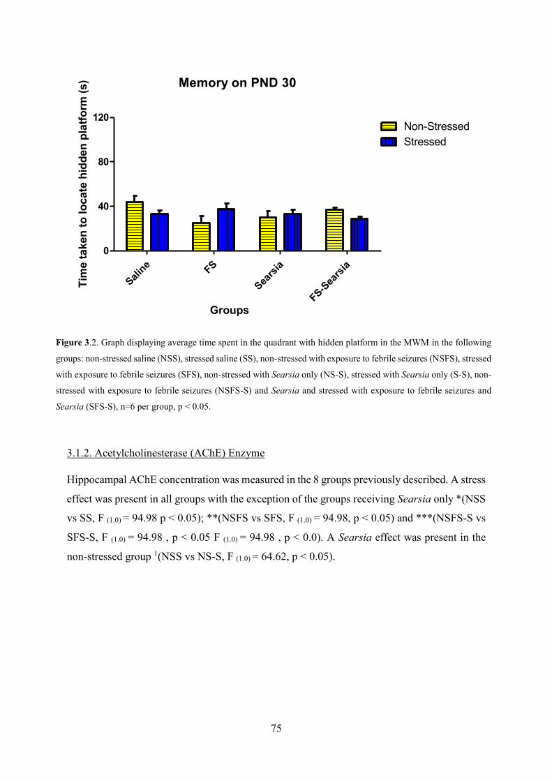
75
Memory on PND 30
Saline FS
Searsi
a
FS-Searsi
a0
40
80
120Non-StressedStressed
Groups
Tim
e ta
ken
to lo
cate
hid
den
plat
form
(s)
3.1.2. Acetylcholinesterase (AChE) Enzyme
Hippocampal AChE concentration was measured in the 8 groups previously described. A stress
effect was present in all groups with the exception of the groups receiving Searsia only *(NSS
vs SS, F (1.0) = 94.98 p < 0.05); **(NSFS vs SFS, F (1.0) = 94.98, p < 0.05) and ***(NSFS-S vs
SFS-S, F (1.0) = 94.98 , p < 0.05 F (1.0) = 94.98 , p < 0.0). A Searsia effect was present in the
non-stressed group 1(NSS vs NS-S, F (1.0) = 64.62, p < 0.05).
Figure 3.2. Graph displaying average time spent in the quadrant with hidden platform in the MWM in the following
groups: non-stressed saline (NSS), stressed saline (SS), non-stressed with exposure to febrile seizures (NSFS), stressed
with exposure to febrile seizures (SFS), non-stressed with Searsia only (NS-S), stressed with Searsia only (S-S), non-
stressed with exposure to febrile seizures (NSFS-S) and Searsia and stressed with exposure to febrile seizures and
Searsia (SFS-S), n=6 per group, p < 0.05.

76
Acetylcholinesterase Enzyme
Saline FS
Searsi
a
FS-Searsi
a0.0
0.5
1.0
1.5
2.0Non-StressedStressed
*
**
***
1
1
Groups
AChE
(ng/
ml)
3.1.3. RT-PCR: MeCP2 and REST genes
MeCP2 gene expression was analysed in the 8 groups described above. A stress effect was
present in all groups except the group of animals exposed to febrile seizures *(NSS vs. SS, F
(1.0) = 6.80, p < 0.05); **(NS-S vs. S-S, F (1.0) = 6.80, p < 0.05) and ***(NSFS-S vs. SFS-S, F
(1.0) = 13.39, p < 0.05). A Searsia effect was present in the non-stressed febrile seizure rats and
the non-stressed febrile seizure with Searsia exposure rats 1(NSFS vs. NSFS-S, F (1.0) = 18.32,
p < 0.05).
Figure 3.3. Graph displaying hippocampal AChE Enzyme concentration on PND 35 the following groups: non-
stressed saline (NSS), stressed saline (SS), non-stressed with exposure to febrile seizures (NSFS), stressed with
exposure to febrile seizures (SFS), non-stressed with Searsia only (NS-S), stressed with Searsia only (S-S), non-
stressed with exposure to febrile seizures (NSFS-S) and Searsia and stressed with exposure to febrile seizures and
Searsia (SFS-S), n=6 per group.*(NSS vs. SS), **(NSFS vs. SFS), ***(NSFS-S vs. SFS-S), 1(NSS vs NS-S), p <
0.05).

77
MeCP2 gene expression
Saline FS
Searsi
a
FS-Searsi
a0
2
4
6Non-StressedStressed
*
*****
1
Groups
MeC
P2/B
-act
in
The REST gene expression was analysed in the same groups as the MeCP2 gene expression.
A febrile seizure effect was present in the non-stressed groups *(NSS vs. NSFS, F (1.0) = 5.39,
p < 0.05) and **(NS-S vs. NSFS-S, F (1.0) = 5.39, p < 0.05). A Searsia effect was present in the
non-stressed and prenatally stressed animals groups exposed to febrile seizures ***(NSFS vs.
NSFS-S, F (1.0) = 5.39, p < 0.05) and 1(SFS vs. SFS-S, F (1.0) = 5.39, p < 0.05).
Figure 3.4. Graph displaying the MeCP2 gene expression on PND 35 the following groups: non-stressed saline (NSS),
stressed saline (SS), non-stressed with exposure to febrile seizures (NSFS), stressed with exposure to febrile seizures
(SFS), non-stressed with Searsia only (NS-S), stressed with Searsia only (S-S), non-stressed with exposure to febrile
seizures (NSFS-S) and Searsia and stressed with exposure to febrile seizures and Searsia (SFS-S), n=6 per
group..*(NSS vs. SS), ** (NS-S vs. S-S), *** (NSFS-S vs SFS-S) and 1(NSFS vs NSFS-S), p < 0.05).

78
REST
Saline FS
Searsi
a
FS-Searsi
a0
2
4
6Non-StressedStressed
*
******
**
11
Groups
RES
T/B
-act
in
4.1. Discussion
In our study we investigated the effects of the Searsia crude bark extract on learning and
memory function in prenatally stressed febrile seizure exposed rats. We further investigated
whether Searsia treatment affected the concentration of the acetylcholinesterase enzyme and
the expression of MeCP2 and REST genes in the hippocampus.
Assessment in the Morris water maze (MWM) showed that injecting Searsia in the absence of
a febrile seizure in non-stressed animals resulted in poor learning ability. This suggests that
injecting Searsia in the absence of convulsions may impair the neuronal plasticity needed for
learning new tasks. While there was a tendency for prenatally stressed animals and febrile
seizure exposed rats to take longer to locate the hidden platform, this effect was not severe as
in the non-stressed rats treated with Searsia. Treatment with Searsia in rats exposed to febrile
seizures did not affect the ability to learn. Previously, we had shown that Searsia treatment
during a seizure dampens seizure severity and attenuates Interleukin-1β (IL-1β) concentration
Figure 3.5. Graph displaying the REST gene expression on PND 35 the following groups: non-stressed saline (NSS),
stressed saline (SS), non-stressed with exposure to febrile seizures (NSFS), stressed with exposure to febrile seizures
(SFS), non-stressed with Searsia only (NS-S), stressed with Searsia only (S-S), non-stressed with exposure to febrile
seizures (NSFS-S) and Searsia and stressed with exposure to febrile seizures and Searsia (SFS-S), n=6 per
group..*(NSS vs. NSFS), **(NS-S vs. NSFS-S), ***(NSFS vs NSFS-S) and 1(SFS vs SFS-S), p < 0.05).

79
(Qulu, et al., 2015). Therefore the improved learning ability in the febrile seizure exposed rats
treated with Searsia may suggest a protective effect against neuroinflammation and potential
apoptotic cell death.
Just over a week after the learning assessments were performed, these animals were subjected
to the probe test to assess memory recall ability. As suggested by the time period the animals
spent in the quadrant with the hidden platform, no lingering effects of exposure to febrile
seizures and Searsia were observed. This suggests that the changes and tendencies seen at PND
21 were transient and that treatment with Searsia does not have chronic effects on hippocampal
neuronal plasticity. We suggest that the effects on learning seen in the non-stressed rats injected
with Searsia only (NS-S) may be due to the crude extract’s anti-inflammatory properties which
may cause inhibition in the release of pro-inflammatory cytokines involved with memory
formation. Tumor necrosis factor-α (TNF-α) and IL-1β are cytokines that are produced in the
brain under normal conditions and they are regarded pivotal for normal synaptic functioning
(Marin & Kipnis, 2013; Vezzani, et al., 2016). TNF-α acts on the TNFR2 receptor to promote
neurogenesis (Iosif, et al., 2006) while the IL-1 receptor contributes to learning tasks in the
Morris water maze (Goshen, et al., 2007). These studies concluded that impairment or
reduction in these signalling pathways resulted in cognitive deficits (Iosif, et al., 2006; Goshen,
et al., 2007). As previously discussed, Searsia has anti-inflammatory properties and has been
shown to directly influence the concentration of IL-1β (Qulu, et al., 2015).
The acetylcholinesterase (AChE) enzyme is responsible for the degradation of the
neurotransmitter, Acetylcholine (ACh) that influences synaptic transmission and induces
synaptic plasticity in neurons (Picciotto, et al., 2012). We showed that exposure to prenatal
stress increased hippocampal AChE concentration. This suggests an increase in ACh
degradation in the synaptic cleft. It has been shown that an increase in ACh degradation results
in decreased plasticity following exposure to prenatal stress (Picciotto, et al., 2012).
Furthermore, we showed that a febrile seizure effect (decrease in AChE concentration) in non-
stressed animals treated with Searsia was present, however treatment in stressed animals
resulted in an increase in the AChE concentration. This suggests that ACh degradation was
reduced in the non-stressed animals while it was increased in stressed animals. We therefore
infer that Searsia treatment in non-stressed animals promoted neuronal plasticity while in the
stressed animals it did not attenuate the stress effect on cognitive function (less neuronal
plasticity). ACh signalling is regulated by chloride transporters (Picciotto, et al., 2012). These

80
chloride transporters are important as their effects of GABA bidding to a cell results in neuronal
hyperpolarisation decreasing calcium ion entry into the cell (Picciotto, et al., 2012; Granger, et
al., 2015). Hyperpolarisation of GABA subsequently inhibits neurons by reducing electrical
activity (Picciotto, et al., 2012; Granger, et al., 2015), therefore treatment with Searsia resulted
in an increase in ACh concentration (decrease AChE concentration) in the pre-synaptic cleft
thus attenuating febrile seizure convulsions as seen in Qulu, et al. (2015). This suggests that
ACh has a dual role in seizure pathology i.e. by increasing ACh concentration (decreasing
AChE concentration) it plays an important function in seizure attenuation which may explain
the shorter seizure duration in non-stressed animals as described in Qulu, et al. (2015).
Hippocampal MeCP2 gene expression showed down-regulation in the expression of MeCP2 in
the stressed rats except in the untreated febrile seizure groups. As MeCP2 functions to promote
neurogenesis and neuronal plasticity, this suggests that these functions are slowed by exposure
to stress. This supports studies that have shown that exposure to early life stress such as prenatal
stress may result in the development of cognitive deficits (McEwen, 2000; McEwen, et al.,
2012; Qulu, et al., 2012; Cassim, et al., 2015).In the absence of treatment, both non-stressed
and stressed febrile seizure groups exhibited down-regulation in the expression of the MeCP2
gene. This may suggest that while the febrile seizure effect is transient, it however does slow
down MeCP2 related functions on neuronal plasticity. Treatment with Searsia in non-stressed
febrile seizure exposed rats resulted in the up-regulation of MeCP2 gene expression. Previously
we showed that exposure to febrile seizures down-regulated MeCP2 gene expression in the
hippocampus (Cassim, et al. 2015), hence an up-regulation of MeCP2 in these animals
following injection with Searsia suggests that in addition to attenuating seizure development,
Searsia also promotes neuronal plasticity in vulnerable neurons. In our study, Searsia was not
able to reverse the deleterious effects of stress on the hippocampal neuronal circuitry nor
plasticity, as these effects occurred in utero, weeks before the injection with Searsia.
In the non-stressed rats exposed to febrile seizures (untreated and treated) the REST gene
expression was down-regulated, suggesting that in these animals neuronal plasticity is being
favoured. These findings reiterate that prenatal stress has detrimental effects on neuronal
plasticity whereas febrile seizures transiently affects neuronal function however, exposure to
Searsia crude bark extract attenuates these effects by promoting potentiating therapeutic effects
on neuronal plasticity.

81
5.1 Conclusion
The outcomes of our findings suggest that Searsia is able to attenuate febrile seizures by
influencing the release of AChE. Exposure to prenatal stress compromises neuronal function
and plasticity, subsequently affecting the animals’ ability to learn. This effect is also present in
following exposure to febrile seizures but is transient in these rats and may be influenced by
the concentration of cytokines. In the presence of febrile seizures, Searsia injection seems to
counteract the impaired ability to learn. This effect together with its ability to attenuate seizure
progression suggests that Searsia may have potential for use as an adjunct treatment for febrile
seizures.
Acknowledgements
We would like to thank the Biomedical Resource Centre for their assistance in this study. The
College of Health Sciences, University of KwaZulu-Natal and the National Research
Foundation (NRF) for their financial support.
Disclosure
We hereby disclose that there are no known financial and personal conflict of interests in the
work of this project.

82
References
1. Cassim, S., Qulu, L. & Mabandla, M. V. 2015. Prenatal stress and early life febrile
convulsions compromise hippocampal genes MeCP2/REST function in mid-
adolescent life of Sprague-Dawley rats. Neurobiology of Learning and Memory,
6313, 1-7.
2. Chang, P. Walker, M. C. & Williams, R. S. B. 2014. Seizure-induced reduction in PIP3
levels contributes to seizure-activity and is reduced by valproic acid.
Neurobiology of disease, 62, 296-306.
3. D'Hooge, R. & De Deyn, P. P. Applications of Morris water maze in the study of
learning and memory. Brain Research. 2001; 36: 60-90.
4. Garthe, A. & Kempermann, G. An old test for new neurons: refining the Morris
water maze to study the functional revelance of adult hippocampal
neurogenesis. 2013; 7:63.
5. Goshen, I., Kreisel, T., Ounallah-Saad, H., Renbaum, P., Zalzstein, Y., Ben-Hur, T.,
Levy-Lahad, E. & Yirmiya, R. 2007. A dual role for interleukin-1 in
hippocampal-dependent memory processes. Psychoneuroendocrinology, 32,
1106–1115.
6. Granger, A. J., Mulder, N., Saunders, A. & Sabatini, B. L. Cotransmission od
acetylcholine and GABA. Neuropharmacology, 100, 40-46.
7. Heida, J. G., Boisse & Pittman, Q. J. 2004. Lipopolysaccharide-induced febrile
convulsions in the rat: short-term sequelae. Epilepsia, 45:11, 1317-1329.
8. Hubscher, C. H., Brooks, D. L. & Johnson, J. R. 2005. A quantitative method for
assessing stages of the rat estrous cycle. Biotechnic & Histochemistry, 80:2,
79-87.
9. Iosif, R. E., Ekdahl, C. T., Ahlenius, H., Pronk, C.J., Bonde, S., Kokaia, Z., Jacobsen,
S. E. & Lindvall, O. 2006. Tumor necrosis factor receptor 1 is a negative
regulator of progenitor proliferation in adult hippocampal neurogenesis.
Journal of Neuroscience, 26, 9703–9712.
10. Lui, Y. W., Li, D.,Yang. L. & Huang, S. 2015. Short-term use of antiepileptic drugs is
neurotoxic to the immature brain. Neural Regen Res. 10:4, 599–604.
11. Marin, I. & Kipnis, J. 2013. Learning and memory…and the immune system. Learning
& Memory, 20: 601-606.
12. McEwen, B. S. 2000. The neurobiology of stress: from serendipity to clinical relevance.

83
Brain Research Interactive, 886: 172-189.
13. McEwen, B. S. 2001. Plasticity of the hippocampus: adaptation tp chronic stress and
allostatic load. Annals of New York Academy of Sciences, 933, 265-277.
14. McEwen, B. 2007. Physiology and neurobiology of stress and adaptation: central role
of the brain. Physiology Reviews, 87, 873-904.
15. Moosa, Z. M., Daniels, W. M. U. & Mabandla, M. V. 2014. The effects of prenatal
methylmercury exposure on trace element and antioxidant levels in rats
following 6-hydroxydopamine-induced neuronal insult. Metabolic Brain
Diseases, 1-11.
16. Morris, R. 1984. Developments of a water-maze procedure for studying spatial
learning in the rat. Journal of Neurosciemce, 11, 47-60
17. Nelson, J. F., Felicio, L.S., Randall, P. K., Sims, C. & Finch, C. E. 1982. A
longitudinal study of estrous cyclicity in aging C57BL/6J mice: I; cycle
frequency, lenght and vaginal cytology. Biology of Reproduction, 27:2, 327-
339.
18. Ojewole, J. A. O. 2007. Analgesic, anti-inflammatory and hypoglcaemic effects of Rhus
chirindensis (Baker F.) [Anacrdiaceae] stem-bark aqueous extract in mice and
rats. Journal of Ethnopharmacology, 113, 338-345.
19. Ojewole, J. A. O. 2008. Anticonvulsant effect of Rhus chirindensis (Baker F.)
Anacardiaceae) stem-bark aqueous extract in mice. Journal of Ethno-
Pharmacology, 117, 130-135.
20. Picciotto, M. R., Higley, M. J. & Mineur, Y. S. 2012. Acetylcholine as a
neuromodulator: cholinergic signaling shapes nervous system function and
behaviour. Neuron Reviews, 76, 116-129.
21. Qulu, L., Daniels, W.M.U., & Mabandla, M. 2012. Exposure to prenatal stress
enhances the development of seizures in young rats. Metabolic Brain
Disorders, 27, 399-404.
22. Qulu, L., Daniels, W. M. U., Russell, V. & Mabandla, M. V. 2015. Searsia chirindensis
reverses the potentiating effect of prenatal stress on the development of febrile
seizures and decreased plasma interleukin-1β levels. Neuroscience Research,
article in press.
23. Shorr, E. 1941. A new technic for staining vaginal smears: III, a single differential stain.
apparatus and laboratory methods, 94:2449, 545-546.
24. Vezzani, A., Fujinami, R. S., White, H. S., Preux, P. M., Blümcke, I., Sander, J. W, &

84
Löscher, W. 2016. Infections, inflammation and epilepsy. Acta
Neuropathology, 131, 211-234.

85
Chapter Five
Conclusion
Febrile seizures results from a common infection that evokes a fever that subsequently triggers the firing
of neurons resulting in a seizure. This condition is exacerbated by exposure to stress during pregnancy.
In these prenatally stressed rats, seizures were more vigorous and took longer to abate than in non-
stressed rats. Exposure to stress and febrile seizures also affected the ability to learn and remember in
the Morris water maze. However, these cognitive deficits were transient as the gene expression results
suggested that there may be an attempt to promote neural plasticity following the febrile seizure episode.
We also showed that exposure to stress early in life may affect the ability to promote neural plasticity
in the hippocampus as evidenced by the increase in the MeCP2 gene expression and corresponding
decrease in the expression of the REST gene. These genes work together in promoting neuronal
plasticity (and hence learning and memory) with an increase in the MeCP2 gene expression promoting
plasticity.. Exposure to stress also led to an increase in BDNF concentration (non-seizure group) and
an increase in the REST gene expression in rats exposed to febrile seizures. In the face of the non-
elevated BDNF concentration, a known neuroprotective and neurogenic peptide, the increase in the
expression of the REST gene may suggest a repression on neural plasticity which may delay cognitive
function in the affected animals. Treatment with Searsia chirindensis attenuated febrile seizures by
influencing the release of AChE and increasing the expression of the MeCP2 gene. Exposure to prenatal
stress compromises neuronal function and plasticity, subsequently affecting the animals’ ability to learn.
In the presence of febrile seizures, Searsia injection seems to counteract the impaired ability to learn.
This effect together with its ability to attenuate seizure progression suggests that Searsia may have
potential as an adjunct treatment for febrile seizures.
In conclusion our study confirms that febrile seizures remain a benign condition but immediate
following of a seizure episode, there seems to be a transient impairment in cognitive function related to
learning and memory function. While treatment with Searsia is able to attenuate seizure development
in all animals, it however does not seem to reverse neuronal malformation present in prenatally stressed
animals. We therefore conclude that the crude bark extract of Searsia chirindensis contains bioactive
ingredients that may be used as an adjunct treatment for the management of febrile seizures.

86
Chapter Six
References
1. Ackermann, S. & Van Toorn, R. 2012. Managing first-time seizures and
epilepsy in children. Continuing Medical Education, 30, 16-20.
2. Abrajano, J. J., Qureshi, I. A., Gokahn, S., Zheng, D., Bergmann, A. & Mehler, M.
F. REST and CoREST modulate neuronal subtype specification, maturation and
maintenance. PLos One. 2009; 4: 1-12.
3. Aggarwal, B. B., Samanta, A. & Feldmann, M. 2000. TNF alpha and the TNF
receptor superfamily: structure- function relationship. Microscience Research
Technology. 50, 184-195.
4. Akira, S. 2006. TLR Signalling. Current Top Microbioogyl Immunology, 311,
1-16.
5. Alikhani- Koopaei, R., Fouladkou, F., Frey, F. J. & Frey, B. M. Epigenetic
regulation of 11β-hydroxysteroid dehydrogenase type 2 expression. The Journal of
Clinical Investigation. 2004; 114: 1146-1157.
6. Altman, J. and Bayer, S.A. 1990. Migration and distribution of two populations
of hippocampal granule cell precursors during the perinatal and postnatal periods. J.
Comp. Neurol. 301, 365–381.
7. Arnold, S.E. and Trojanowski, J.Q. 1996. Human fetal hippocampal
development: I. Cytoarchitecture, myeloarchitecture, and neuronal morphologic
features. J. Comp. Neurol. 367, 274–292.
8. Balcombe, J. P. 2006. Laboratory environments and rodents’ behavioural needs:
a review. Laboratory Animals, 40, 217-235
9. Ballas, N. & Mandel, G. The many faces of REST oversee epigenetic
programming of neuronal genes. Current Opinions in Neurobiology. 2005; 15:
500-506.
10. Barbazanges, A., Piazza, P. V., Le Moal, M. & Maccari, S. 1996. Maternal
glucocorticoid secretion mediates long-term effects of prenatal stress. The Journal of
Neuroscience, 16:12, 3943-3949.
11. Barkley, R. A., Fletcher, K., Fischer, M. & Smallish, L. 2002. The persistence
of attention-deficit/hyperactivity disorder into young adulthood as a function of
reporting source and definition of disorder. Journal of Abnormal Psychology,
111:2, 279-289.
12. Baroncelli, L., Chiara, B., Spolidoro, M., Begenisic, T., Maffei, L & Sale, A.
2011. Brain Plasticity and disease: A Matter of Inhibition. Neural Plasticity, 1-11.
13. Batten, S. R. 2013. Glutamate dysregulation and hippocampal dysfunction in

87
epileptogenesis. Theses and Dissertations--Medical Sciences, 1, 1-88.
14. Ben-Ari, Y. 2006. Seizures beget seizures: the quest for GABA as a key player.
Critical Review Neurobilogy. 18, 135-144.
15. Benoit, J.D., Rakic, P. & Frick, K. M. 2015. Prenatal stress induces spatial
memory deficits and epigenetic changes in the hippocampus indicative of
heterochromatin formation and reduced gene expression. Behavioural Brain Research,
281, 1-8.
16. Bezzi, P., Domercq, M. & Brrambilla, L. 2001. CXCR4- activated astrocyte
glutamate release via TNFa: amplification by microglia triggers neurotoxicity. Nature
Neuroscience. 4:7, 702-710.
17. Bird, A. DNA methylation patterns and epigenetic memory. 2002. Genes &
Development, 16; 6-21.
18. Botterill, J. J., Brymer, K. J., Caruncho, H. J. & Kalynchuk, L. E. 2015. Aberrant
hippocampal neurogenesis after limbic kindling: Relationship to BDNF and
hippocampal-dependent memory. Epilepsy & Behavior, 47, 83-92.
19. Boyle, M. P., Kolber, B. J., Vogt, S. K., Wozniak, D. F. & Muglia, L. J. 2006.
Forebrain glucocorticoid receptors modulate anxiety-associated locomotor
activation and adrenal responsiveness. Journal of Neurosciecnce, 26, 1971-
1978.
20. Bruce, W. A., Donaldson, I. J., Wood, I. C., Yerbury, S.A., Sadowski, M. I.,
Chapman, M., Gӧttgens, B. and Buckley, N. J. 2004. Genome- wide analysis of
repressor element 1 silencing transcription factor/ neuron- restrictive silencing factor
(REST/NRSF) target genes. PNAS, 101, 10458-10463.
21. Calabrese, F., Molteni, R., Racagni, G. & Riva, M. A. 2009. Neural plasticity:
A link between stress and mood disorders. Psychoneuroendocrinology, 345, S208-
S216.
22. Cannizzaro, C., Plescia, F., Martire, M., Gagliano, M., Cannizzaro, G., Mantia,
G., & Cannizzaro, E. 2006. Single, intense prenatal stress decreases emotionality and
enhances learning performance in the adolescent rat offspring: Interaction with a brief,
daily maternal separation. Behavioural Brain Research. 169, 128-136.
23. Cantwell, D. P. & Baker, L. 1991. Association between attention deficit-
hyperactivity disorder and learning disorders. Journal of Learning Disabilities, 24, 88-
95.
24. Cappovilla, G. Mastrangelo, M., Romeo, A. & Vigevano, F. 2009.
Recommendation for the management of “febrile seizures’: Ad Hoc task of LICE
guidelines commission. Epilepsia, 1, 2-6.
25. Carlwzon Jr., W. A., Duman, R. S. & Nestler, E. J. 2005. The many faces of

88
CREB. TRENDS in Neuroscience, 28, 436-445.
26. Cassim, S., Qulu, L. & Mabandla, M. V. 2015. Prenatal stress and early life \
febrile convulsions compromise hippocampal genes MeCP2/REST function in
mid-adolescent life of Sprague-Dawley rats. Neurobiology of Learning and Memory,
6313, 1-7.
27. Champagne, D. L., de Kloet, E. R. & Joëls, M. Fundamental aspects of the
impact of glucocorticoids on the (immature) brain. Seminars in Fetal & Neonatal
Medicine. 2009; 14: 136-142.
28. Challis, J. R., Sloboda, D., Matthews, S. G., Holloway, A., Alfaidy, N., Patel, F.
A., Whittle, W., Fraser, M., Moss, T. J. M. & Newnham, J. The fetal
placental hypothalmic-pituitary-adrenal (HPA) axis, parturition and post natal health.
Molecular and Cellular Endocrinology. 2001; 185: 135-144.
29. Chao, H., Zoghbi, H. Y. & Rosenmund, C. MeCP2 controls excitatory synaptic
strength by regulating glutamatergic synapse number. Neuron. 2007; 56: 58- 65.
30. Chang, Y., Huang, A., Kuo, Y., Wang, S., Chang, Y. & Huang, C. Febrile
seizures impair memory and learning and cAMP response-element binding
protein activation. Annals of Neurology. 2003; 54: 706-718.
31. Cohen, D. R. S., Matarazzo, V., Palmer, A. M., Yajun, T., Ok-Hee, J., Pevsner,
J. & Ronett, G. V. 2003. Expression of MeCP2 in olfactory receptor neurons in
developmentally regulated and occurs before synaptogenesis. Molecular and Celular
Neuroscience, 22, 417-429.
32. Crino, P. B. 2007. Gene expression, genetics, and genomics in epilepsy: Some
answers, more questions. Epilepsia, 48, 42-50.
33. Cunha, C., Bramilla, R. & Thomas, K. L. 2010. A simple role for BDNF in
learning and memory? Frontiers in Molecular Neuroscience, 3:1, 1-14.
34. Danbolt, N. C. 2001. Glutamate uptake. Progress in Neurobiology, 65:1, 1-105.
35. Dani, V. S., Chang, Q., Maffei, A., Turrigiano, G. G., Jaenisch, R. & Nelson, S.
B. Reduced cortical activity due to a shift in the balance between excitation and
inhibition in a mouse model of Rett Syndrome. PNAS. 2005; 102: 12560-12565.
36. David, J., Cacheaux, L. P., Ivens, S., Lapilover, E., Heinimann, U., Kaufer, D.,
& Friedman, A. 2009. Astrocytic dysfunction in epileptogenesis: Consequences of
altered potassium and glutamate homeostasis? The Journal of Neuroscience, 29,
10588-10599.
37. D'Hooge, R. & De Deyn, P. P. Applications of Morris water maze in the study of
learning and memory. Brain Research. 2001; 36: 60-90.

89
38. De Kloet, E. R., Fitzsimons, C. P., Datson, N. A., Meijer, O. C. &
Vreugdenhil, E. Glucocorticoid signaling and stress-related limbic
susceptibility pathway about receptors, transcription machinery and microRNA.
Brain Research. 2009; 1293: 129-141.
39. De Kloet, E. R., Claessens, S. E. F. & Kentrop, J. 2014. Context modulates
outcome of perinatal glucocorticoid action in the brain. Frontiers in Endocrinology, 5:
100, 1-13.
40. Dinarello, C. A. 2000. Proinflammatory cytokines. Chest Journal, 118:2, 503-
508.
41. Dingledine, R. 2011. Glutamatergic mechanisms related to epilepsy: Ionotropic
receptors. Jasper's Basic Mechanisms of the Epilepsies, 4, 1-11.
42. Diz-Chaves, Y., Astiz, M., Bellini, M. J. & Garcia-Segura, L. M. 2013. Prenatal
stress increases the expression of proinflammatory cytokines and exacerbates the
inflammatory response to LPS in the hippocampal formation of adult male mice. Brain,
Behaviour, and Immunity, 28, 196-206.
43. Dubé, C., Vezzani, A., Behrens, M., Bartfai, T. & Baram, T. Z. 2005.
Interleukin-1β contributes to the generation of experimental febrile seizures. Annals
Neurology, 57:1, 152-155.
44. Ernsberger, U. Regulation of gene expression during early neuronal
differentiation: evidence for patterns conserved across neuron populations and
vertebrate classes. Cell Tissue Res. 2012; 348: 1-27.
45. Evans, S. S., Repasky, E. A. & Fisher, D. T. 2015. Fever and the thermal
regulation of immunity: the immune system feels the heat. Nature Reviews
Immunology, 15, 335-349.
46. Fagiolini, M., Jensen, C. L. & Champagne, F. A. 2009. Epigenetic influences
the brain development and plasticity. Current Opinion in Neurobiology, 19, 207-212.
47. Feghali, C. A. & Wright, T. M. 1997. Cytokines in acute and chronic
inflammation. Frontiers in Bioscience, 2: d12-d26.
48. Fine, R., Zhang, M. D. & Stevens, H. E. Prenatal stress and inhibitory neuron
systems: implications for neuropyschiatric disorders. Molecular Psychiatry. 2014; 19:
641-651.
49. Fisher, R. S., Acevedo, C., Arzimanoglou, A., Bogacz, A., Cross, H., Elger, C.
E., Engel Jr, J., Glynn, M., Hesdorffer, D. C., Lee, B. I., Mathern, G. W., Moshé, S. L.,
Perucca, E., Scheffer, I. E., Tomson, T., Watanabe, M. & Wiebe, S. 2014. ILAE official
report: a practical clinical definition of epilepsy. Epilepsia, 55:4, 475-482.
50. Foster, J., Mauger, A. R., Chrisma, C. R., Thomasson, K. & Taylor, L. 2015. Is

90
prostoglandin E2 involved in the thermogenic response to environmental cooling in
healthy humans? Medical Hypothesis, article in press.
51. Foti, S. B & Roskams, A.J. 2011. A tale of two epiphenomena: The complex
interplay of epigenetics and epilepsy. Underlying mechanisms of Epilepsy, 12, 220-
240.
52. Galic, M. A., Riazi, K., Heida, J. G., Mouihate, A., Fournier, N. M., Spencer, S.
J., Kalynchuk, L. E., Teskey, C. & Pittman, Q. J. 2008. Postnatal
inflammation increases seizure susceptibility in adult rats. The Journal of
Neuroscience. 28: 6904-6913.
53. Galic, M. A., Riazi, K. & Pittman, J. Q. 2012. Cytokines and brain excitability.
Frontiers in Neuroendocrinology, 33, 116- 125.
54. Garthe, A. & Kempermann, G. An old test for new neurons: refining the Morris
water maze to study the functional revelance of adult hippocampal
neurogenesis. 2013; 7:63.
55. Glover, V., O;Connor, T. G. & O’Donnell, K. 2010. Prenatal stress and the
programming of the HPA axis. Neuroscience and Bopbehavioural reviews. 35, 17-22.
56. Granger, A. J., Mulder, N., Saunders, A. & Sabatini, B. L. Cotransmission od
acetylcholine and GABA. Neuropharmacology, 100, 40-46.
57. Gray, J.D., Milner, T. A. & McEwen, B. S. 2013. Dynamic plasticity: hThe role
of glucocorticoids, brain-derived neurotrophic factor and other trophic factors.
Neuroscience, 239, 214-227.
58. Greer P. L. & Greenberg, M. E. 2008. From synapse to nucleus: calcium-
dependent gene transcription in the control of synapse development and function.
Neuron, 59, 546- 560.
59. Griffith, E. C., Cowan, C. W. & Greenberg, M. E. 2001. REST acts through
multiple deacetylase complexes. Neuron, 31, 339- 340.
60. Gutteling, B. M., de Weerth, C., Willemsen-Swinkels, S. H. N., Huizink, A.
C., Mulder, E. J. H., Visser, G. H. A. & Buitelaar, J. K. 2005. The effects of prenatal
stress on temperament and problem behaviour of 27-month-old toddlers. European
childe & adolescent psychiatry, 14:1, 41-51.
61. Gutteling, B. M., de Weerth, C., Zandbelt, N., Mulder, E. J. H., Visser, G. H.
A. & Buitelaar, J. K. 2006. Does maternal prenatal stress adversely affect the child’s
learning and memory at age six? Journal of abnormal child psychology, 34:6, 787-
796.
62. Guy, J., Cheval, H., Selfridge, J. & Bird, A. The role of MeCP2 in the brain.
Annual Review of Cell and Developmental Biology. 2011; 27: 631-652.
63. Harre, E. M., Galic, M. A., Mouihate, A., Noorbakhsh, F. & Pittman, Q. J.

91
Neonatal inflammation produces selective behavioural deficits and alters N-methyl-
D-aspartate receptor subunit mRNA in the adult rat brain. European Journal of
Neuroscience. 2008; 27: 644-653.
64. Harris, A. & Seckl, J. 2010. Glucocorticoids, prenatal stress and the
programming of disease. Hormones and Behaviour. 59, 279-289.
65. Heida, J. G., Boisse & Pittman, Q. J. 2004. Lipopolysaccharide-induced febrile
convulsions in the rat: short-term sequelae. Epilepsia, 45:11, 1317-1329.
66. Heida, J. G. & Pittman, Q. J. 2005. Causal links between brain cytokines and
experimental febrile convulsions in the rat. Epilepsia, 46:12, 1906-1913.
67. Heida, J., Boissé, L. & Pittman, Q. J. Lipopolysaccharide-induced febrile
convulsions in the rat: short-term seuelae. Epilepsia. 2009; 45: 1317-1329.
68. Heyward, F. D. & Sweatt, J. D. DNA methylation in memory formation: Emerging
Insights. 2015, 1-15.
69. Huang, L. 2014. Early-life stress impacts the developing hippocampus and
primes seizure occurrence: cellular, molecular, and epigenetic mechanisms. Frontiers
in Molecular Neuroscience. 7: 8, 1-15.
70. Hoeba, K., Janssen, E. & Beutler, B. 2004. Ther interface between innate and
adaptive immunity. Nature immunology, 5:10, 971-975.
71. Hoefnagels, W. A., J., Padberg, G. W., Overweg, J., van der Velde, E. A. &
Roos, R. A. C. 1991. Transient loss of consciousness: the value of the history for
distinguishing seizure from syncope. Journal of Neurology, 238:1, 39-43
72. Hubscher, C. H., Brooks, D. L. & Johnson, J. R. A quantitative method for
assessing stages of the rat estrous cycle. Biotechnic & Histochemistry. 2005; 80: 79-
87.
73. Hunter, R. G. & McEwen, B. S. 2013. Stress and anxiety across the lifespan:
structural plasticity and epigenetic regulation. Epigenomics, 5:2, 1-18.
74. Idro, R., Gwer, S., Kahindi, M., Gatakaa, H., Kazungu, T., Ndiritu, M.,
Maitland, K., Neville, B. R., Kager, P. A. & Newton, C. R. J. C. 2008. The incidence,
aetiology and outcomes of acute seizures in children admitted to a rural Kenyan
district hospital. Biomed Central Pediatrics, 8:5, 1-11.
75. Jessberger, S., Nakashima, K., Clemenson Jr., G. D., Mejia, E., Mathews, E.,
Ure, K., Ogawa, S., Sinton, C. M., Gage, F. H. & Hsieh, J. 2007. Epigenetic
modulation of seizure- induced neurogenesis and cognitive decline. The Journal of
Neuroscience, 27, 5967- 5975.
76. Jett, D. A. Chemical toxins that cause seizures. NeuroToxicology. 2012; 33:
1473-1475.
77. Jung, B. P., Jugloff, D. G. M., Zhang, G., Logan, R., Brown, S. & Eubanks, J.

92
H. 2003. The expression of methyl CpG factor MeCP2 correlates with cellular
differentiation in the developing rat brain and in cultured cells. Developmental
Neurobiology, 55:1, 86-96.
78. Kabow, K. & Blümcke, I. 2011. The methylation hypothesis: Do epigenetic
chromatin modifications play a role in epileptogenesis? Epilepsia, 52, 15-19.
79. Karpova, N. N. 2014. Role of BDNF epigenetics in activity-dependent neuronal
plasticity. Neuropharmacology, 76: 709-718.
80. Kentner, A. C. & Pittman, Q. J. Minireview: early-life programming by
inflammation of the neuroendocrine system. Endocrinology. 2010; 151: 4602-4606.
81. Koh, K.P. & Lee, B. K. 1998. Reduced Lymphocyte Proliferation and
lnterleukin-2 production in Anxiety Disorders. Psychosomatic Medicine, 60, 479-483.
82. Laflamme, N., Rivest, S. Toll-like receptor 4: the missing link of the cerebral
innate immunity response triggered by circulating gram-negative bacterial cell wall
components. FASEB J. 2001; 15: 155-163.
83. Lasón, W., Chlebicka, M. & Rejdek, K. 2013. Research advances in basic
mechanisms of seizures and antiepileptic drug action. Pharmacological Reports. 65,
787-801.
84. Larsson, A. K., Idborg, H., Hebriksson, M. A., Martinsson, T., Johnsen, J. L.,
Korotkova, M., Kogner, P. & Jakobsson, P. 2015. COX/mPGES-1/PGE2 pathway
depicts an inflammatory dependent high-risk neuroblastoma subset. PNAS, 112:26,
8070-8075.
85. Lemaire, V., Koehl, M., Le Moal, M & Abrous, D. N. 2000. Prenatal stress
produces learning deficits associated with an inhibition of neurogenesis in the
hippocampus. PNAS, 97: 20, 11032- 11037.
86. Lubin, F. D. 2012. Epileptogenesis: Can the science of epigenetics give us
answers? Epilepsy Currents, 12, 105-110.
87. Lunyak, V.V., Burgess, R., Prefontaine, G. G., Nelson, C., Sze, S., Chenoweth,
J., Schwarts, P., Pevzner, P.A., Glass, C., Mandel, G. & Rosenfeld, M. G. Corepressor-
dependent silencing of chromosomal regions encoding neuronal genes. Science. 2002;
298: 1747- 1752.
88. Lui, Y. W., Li, D.,Yang. L. & Huang, S. 2015. Short-term use of antiepileptic
drugs is neurotoxic to the immature brain. Neural Regen Res. 10:4, 599–604.
89. Madsen, K. K., White, H. S. & Schousboe, A. 2010. Neuronal and non-neuronal
GABA transporters as targets of antiepiletic drugs. Pharmacology Therapy. 125, 394-
401.
90. Marchi, N., Angelov, L., Masaryk, T., Fazio, V., Granata, T., Hernandez, N.,

93
Hallene, K., Diglow, T., Franic, L., Najm, I, and Janigro, D. 2007. Seizure- promoting
effect of bloob-brain barrier disruption. Epilepsia, 48, 732-742
91. Marchi, N., Granata, T., & Janigro, D. 2013. Inflammatory pathways of seizure
disorders. Trends in Neuroscience, 1-11.
92. Marchi, N., Granata, T. & Janigro, D. 2014. Inflammatory pathways of seizure
disorders. Cell Press, 37:2, 55-65.
93. Martin, K. P. & Wellman, C. L. 2011. NMDA receptor blockade alters stress-
induced dendritic remodeling in medial prefrontal cortex. Cerebral cortex, 21:10,
2366-2373.
94. Martinowich, K., Hattori, D., Wu, H., Fouse, S., He, F., Hu, Y., Fan, G. & Sun,
Y.E. 2003. DNA methylation- related chromatin remodelling in activity- dependent
Bdnf gene regulation. Science, 302, 890-893.
95. Mattarazzo, V., Cohen, D., Palmer, A. M., Simppson, P. J., Khokhar, B., Pan,
S. & Ronnett, G. V. 2004. The transcriptional repressor MeCP2 regulates terminal
neuronal differentiation. Molecular and Cellular Neuroscience, 27, 44-58.
96. McEwen, B. S. & Sapolsky, R. M. Stress and Cognitive function. Cognitive
Neuroscience. 1995; 5: 205-216.
97. McEwen, B. S. 1999. Protective and damagingg effects of stress mediators.
Seminars in Medicine of the Beth Israel Deaconess Medical Center, 338:3, 171-179.
98. McEwen, B. S. 2000. The neurobiology of stress: from srendipity to clinical
relevance. Brain Research Interactive, 172--189.
99. McEwen, B. S. Plasticity of the hippocampus: adaptation tp chronic stress and
allostatic load. Annals of New York Academy of Sciences. 2001; 933: 265-277.
100. McEwen. B. S. 2003. Mood disorders and allostatic load. Society of Biological
Psychiatry, 54, 200-207.
101. McEwen, B. S. & Wingfield, J. C. 2003. The concept of allostasis in biology
and biomedicine. Horm Behav. 43:1, 2-15.
102. McEwen, B. 2007. Physiology and neurobiology of stress and adaptation:
central role of the brain. Physiology Reviews, 87, 873-904
103. McEwen, B. S. 2008. Central effects of stress hormones in health and disease:
Understanding the protective and damaging effects of stress and stress
mediators. European Journal of Pharmacology. 583, 174-185.
104. McEwen, B. S., Eiland, L., Hunter, R. G. & Miller, M. M. 2012. Stress and
anxiety: Structural plasticity and epigenetic regulation as a consequence of
stress. Neuropharmacology, 62:1, 3-12.
105. McEwen, B. S., Bowles, N. P., Gray, J. D., Hill, M. N., Hunter, R. G.,
Karatsoreos, I. N. & Nasca, C. 2015. Mechanisms of stress in the brain. Nature

94
Neuroscience, 18:10, 1353-1363.
106. McNamara, J. O. 1994. Temporal lobe epilepst and BDNF receptor, TrkB.
Jasper’s Basic Mechanisms of the Epilepsies, 4, 1-22.
107. Meyers, L. A., Ancel, F. D. & Lachmann, M. Evolution of genetic potential.
PLOS Computational Biology. 2005; 1:3.
108. Moisiadis, V. G. & Matthews, S. G. Glucocorticoids and fetal programming
part 1: outcomes. Nature Reviews Endocrinology. 2014; 10: 391-402.
109. Moosa, Z. M., Daniels, W. M. U. & Mabandla, M. V. 2014. The effects of
prenatal methylmercury exposure on trace element and antioxidant levels in rats
following 6-hydroxydopamine-induced neuronal insult. Metabolic Brain
Diseases, 1-11.
110. Morris, R. 1984. Developments of a water-maze procedure for studying spatial
learning in the rat. Journal of Neurosciemce, 11, 47-60.
111. Murgatoyd, C., Patchev, A. V., Wu, Y., Micale, V., Bockmuhl, Y., Dieter, F.,
Holsboer, F., Wotjak, C. T., Almeida, O. F. X. & Spengler, D. Dynamic DNA
methylation programs persistent adverse effects of early-life stress. Nature
Neuroscience. 1-10.
112. Murray, P. S. & Holmes, P. V. 2011. An overview of Brain-derived
neurotrophic factor and implications for excitotoxic vulnerability in the hippocampus.
International journal of peptides, 1-12.
113. Nelson, J. F., Felicio, L.S., Randall, P. K., Sims, C. & Finch, C. E. 1982. A
longitudinal study of estrous cyclicity in aging C57BL/6J mice: I; cycle
frequency, lenght and vaginal cytology. Biology of Reproduction. 1982; 27:
327-339.
114. Ojewole, J. A. O. 2007. Analgesic, anti-inflammatory and hypoglcaemic effects
of Rhus chirindensis (Baker F.) [Anacrdiaceae] stem-bark aqueous extract in mice and
rats. Journal of Ethnopharmacology, 113, 338-345.
115. Ojewole, J. A. O. Anticonvulsant effect of Rhus chirindensis (Baker F.)
(Anacardiaceae) stem-bark aqueous extract in mice. Ethnopharmacology.
2008; 117: 130-135.
116. Palm, K., Belluardo, N., Metsis & Timmusk, T. 1998. Neuronal expression of
zinc finger transcription factor REST/NRSF/XBR gene. The Journal of Neuroscience,
18:4, 1280-1296.
117. Peper, J. S. & Dahl, R. E. The teenage brain: Surging hormones-Brain- behavior
interactions during puberty. Current Directions in Psychological Science. 2013; 22:
134-139.

95
118. Popoli, M., Yan, Z., McEwen, B. S. & Sanacora, G. The stressed synapse: The
impact of stress and glucocorticoids on glutamate transmission. Nature Review
Neuroscience. 2012; 13: 22-37.
119. Pruunsild, P., Sepp, M., Orav, E., Koppel, I. & Timmusk, T. 2011. Identification
of cis-elements and transcription factors regulating neuronal activity-dependent
transcription of human BDNF gene. The Journal of Neuroscience, 31:9, 3295-
3308.
120. Qin, L., Wu, X., Block, M. L., Liu, Y., Breese, G. R., Hong, J.-S., Knapp, D. J.
and Crews, F. T. 2007. Systemic LPS causes chronic neuroinflammation and
progressive neurodegeneration. Glia, 55, 453–462.
121. Qulu, L., Daniels, W.M.U., & Mabandla, M. 2012. Exposure to prenatal stress
enhances the development of seizures in young rats. Metabolic Brain Disorders,
27, 399-404.
122. Qulu, L., Daniels, W.M.U., & Mabandla, M. 2015. Exposure to prenatal stress
has deleterious effects on hippocampal function in a febrile seizure rat model.
Brain Research, 1-8.
123. Qulu, L., Daniels, W. M. U., Russell, V. & Mabandla, M. V. 2015. Searsia
chirindensis reverses the potentiating effect of prenatal stress on the development of
febrile seizures and decreased plasma interleukin-1β levels. Neuroscience Research,
article in press.
124. Qureshi, I. A. & Mehler, M. F. 2010. Epigenetic mechanisms underlying human
epileptic disorders and the process of epileptogenesis. Neurobiology of
Disease, 39, 53-60.
125. Radcliffe, K. A., Fisher, J. L., Gray, R. & Dani, J. A. 1999. Nicotinic
modulation of glutamate and GABA synaptic transmission in hippocampal neurons.
Annals New York Academy of Science, 591-610.
126. Riazi, K., Galic, M. A. & Pittman, Q. J. Contributions of peripheral
inflammation to seizure susceptibility: cytokines and brain excitiability. Epilepsy
Research. 2010; 89: 34-42.
127. Riedel, G., Platt, B & Micheau, J. 2003. Glutamate receptor function in learning and
memory. Behavioural Brain Research, 140:1, 1-47.
128. Rodenas- Ruano, A., Chávez, A., Cossio, M. J., Castillo, P. E. & Zukin, R. S.
REST-independent epigentic remodeling promotes the development switch in
synaptic NMDA receptors. Nature Neuroscience. 2012; 15: 1382- 1390.
129. Rojas, A., Jiang, J., Ganesh, T., Yang, M., Leluthi, N., Gueorguieva, P. &
Dingledine, R. 2014. Cyclooxygenase-2 in epilepsy. Epilepsia, 55:1, 17-25.
130. Roopra, A., Dingledine, R. & Hsieh, J. 2012. Epigenetics and epilepsy.

96
Epilepsia, 53, 2-10.
131. Rothman, S. M. & Mattson, M. P. 2014. Activity-dependent, stress-responsive
BDNF signaling and the quest for optimal brain health and resilience throughout the
lifespan. Neuroscience, 239, 228-240.
132. Salim, S., Chugh, G. & Asghar, M. 2012. Inflammation in anxiety. Advances in
Protein Chemistry and Structural Biology, 88, 1-17.
133. Sapolsky, R. M., Krey, L. C. & McEwen, B. S. Prolonged glucocorticoid
exposure reduces hippocampal neuron number: Implications for aging. The
Journal of Neuroscience. 1985; 5: 1222-1227.
134. Sapolsky, R. M. Glucocorticoids, stress, and their adverse neurological effects:
relevance to aging. Exprimental Gerontology. 1999; 34: 721-732.
135. Sapolsky, R. M. 2003. Stress and plasticity in the limbic system. Neurochem
Res, 28:11, 1735-1742.
136. Seckl, J. R. Prenatal glucocorticoids and long-term programming. European
Journal of Endocrinology. 2004; 151: U49- U62.
137. Scott, A., Khan, K. M. Cook, J. L. & Duronio, V. 2005. What is
“inflammation”? Are we ready to move beyond Celsus? Inflammation, 248- 249.
138. Scantlebury, H. M. H. J. G. 2010. Febrile seizures and temporal lobe
epileptogenesis. Epilepsy Res, 89, 27-33.
139. Shahbazian, M. D. & Zoghbi, H. Y. 2002. Rett syndrome and MeCP: linking
epigenetics and neuronal function. American Journal of Human Genetics, 71:6, 1259-
1272.
140. Shorr, E. 1941. A new technic for staining vaginal smears: III, a single
differential stain. apparatus and laboratory methods, 94:2449, 545-546.
141. Spencer, S. J., Heida, J. G. & Pittman, Q. J. Early life immune challenge-
effects on behavioural indices of adult rat fear and anxiety. Behavioural Brain
Research. 2005; 164: 231-238.
142. Stankov, S. V. 2012. Definition of Inflammation, causes of inflammation and
possible anti-inflammatory strategies. The open Inflammation Journal, 5, 1-9.
143. Suchomelova, L., Lopez-Meraz, M. L., Niquet, J., Kubova, H. & Wasterlain,
C. G. 2015. Hyperthermia aggravates status epilepticus-induced epileptogenesis and
neuronal loss in immature rats. Neuroscience, 305, 209-224.
144. Takemiya, T., Matsumura, K. & Yamagata, K. 2007. Roles of prostaglandin
synthesis in excitotoxic brain diseases. Neurochemistry International. 51:2-4,
112-120.
145. Vallѐe, M., Willy, M., Dellu, F., Le Moal, M., Simon, H. & Maccari, S. 1997.
Prenatal Stress Induces High Anxiety and Postnatal Handling Induces Low

97
Anxiety in Adult Offspring: Correlation with Stress-Induced Corticosterone
Secretion. The Journal of Neuroscience, 17:7, 2626-2636.
146. Vasilche, A. M. 2015. Talking to the brain at the blood-brain barrier through
inflammation-induced prostaglandins E2. Linkoping University, 1435, 1-101.
147. Vezzani, A., Moneta, D., Richichi, C., Aliprandi, M., Burrows, S. J., Ravizza, T.,
Perego, C. & De Simoni, M. G. 2002. Functional role of inflammatory
cytokines and antiinflammatory molecules in seizures and epileptogenesis. Epilepsia.
5: 30-35.
148. Vezzani, A., Maroso, M., Balosso, S., Sanchez, M. & Tamas, B. IL-1
receptor/Toll-like receptor signaling in infection, inflammation, stress and
neurodegeneration couples hyperexcitability and seizures. Brain, Behaviour, and
Immunity. 2011; 25: 1281-1289.
149. Vezzani, A., Aronica, E., Mazarati, A. & Pittman, Q. J. 2013. Epilepsy and
brain inflammation. Experimental Neurology, 244, 11-21.
150. Vezzani, A. Epilepsy and inflammation in the brain: Overview and
pathophysiology. Epilepsy Currents. 2014; 14: 3-7.
151. Vezzani, A., Fujinami, R. S., White, H. S., Preux, P. M., Blümcke, I., Sander,
J. W, & Löscher, W. 2016. Infections, inflammation and epilepsy. Acta
Neuropathology, 131, 211-234.
152. Viviani, B., Bartesaghi, F. & Gardoni, F. 2003. Interleukin-1b enhances NMDA
receptor-mediated intracellular calcium increase through activation of Src
family of kinases. Journal of Neuroscience. 23:25, 8692-8700.
153. Wager, R. G. & Newton, C. R. Do helminths cause epilepsy? Parasite
Immunology. 2009; 31: 697-705.
154. Wang, L. Y., Yang, L. D. & Huang, S. 2015. Short-term use of antiepileptic
drugs is neurotoxic to the immature brain. Neural Regen Res. 10:4, 599–604.
155. Ware, C. F. 2005. Network communication: lymphotoxins, LIGHT, and TNF.
Annu Rev Immunology. 23, 787- 819.
156. Waruiru, C., & Appleton, R. 2004. Febrile seizures: an update. Arch dis Child,
89, 751-756.
157. Weaver, I. C. G., D'Alessio, A. C., Brown, S. E., Hellstrom, I. C., Sergiy, D.,
Sharma, S., Szyf, M. & Meaney, M. J. The transcription factor nerve growth
factor-inducible protein A mediates epogenetic programming: altering epigentic
marks by immediate-ear;y genes. The Journal of Neuroscience. 2007; 27: 1756- 1768
158. Weinstock, M. 2008. The long-term behavioural consequences of prenatal
stress. Neuroscience and Biobehavioural Reviews, 32, 1073-1086.
159. Welberg, L. A. M. & Seckl, J. R. 2001. Prenatal stress, glucocorticoids and the

98
programming of the brain. Journal of Neuroendocrinology, 13, 113-128.
160. West, A. E. & Greenberg, M. E. 2011. Neuronal activity- regulated gene
transcriptio in synapse development and cognitive function. Cold Spring Harbor
Perspectives in Biology, 1-22.
161. Wilson, C. A., Vazdarjanova, A. & Terry Jr, A. V. 2013. Exposure to variable
stress in rats: effects on anxiety-related behaviours, innate and contextual fear,
and fear extinction. Behavioural Brain Research, 238, 279-288
162. Wood, B. L., Haque, S., Weinstock, A. & Miller, B. 2004. Pediatric stress-
related seizures: conceptualization, evaluation, and treatment of nonepileptic
seizures in children and adolescents. Current Opinions in Pediatrics, 16:5, 523-
531.
163. Wu, J. & Xie, X. 2006. Comparitive sequence analysis reveals an intricate
network among REST, CREB and miRNA in mediating neuronal gene
expression. Genome Biology, 7, R85.1- R85.14.
164. Xu, M., Sulkowski, Z. L., Parekh, P., Khan, A., Chen, T., Midha, S., Iwasaki,
T., Shimokawa, N., Koibuchi, N., Zavacki, A. M. and Sulkowska- Sajdel, E. M. 2013.
Effects of perinatal lipopolysacchiride (LPS) exposure on the developing rat brain;
modelling the effect of maternal infection on the developing human CNS. Cerebellum,
12, 572-286.
165. Yagoubi, N., Yosra, J & Sakly, M. 2015. Hyperthermia-induced febrile seizures
have moderate and transient effects on spatial learning in immature rats. Behavioural
Neurology, 1-6.
166. Yoshii, A. & Constantine-Paton, M. 2010. Postsynaptic BDNF-TrkB signalling
in synapse maturation, plasticity, and disease. Developmental Neurobiology, 304-322.
167. Zheng, X., Zhang, H., Luo, Q. & Zhu, J. Kainic acid- induced
neurodegenerative model: Potentials and limitarions. 2010. Journal of Biomedicine and
Biotechnology, 2011, 1-10.
168. Zhou, J., Wang, F., Zhang, J., Yang, Y. & Fu, R. 2014. Repeated febrile
convulsions impair hippocampal neurons and cause synaptic damage in immature rats:
neuroprotective effect of fructose-1,6-diphosphate. Neural Regeneration Research,
9:9, 937-942.

99
Addendum A
Prenatal stress and early life febrile convulsions compromise
hippocampal genes MeCP2/REST function in mid-adolescent life
of Sprague-Dawley rats.

100
Addendum B
Febrile seizure induction impairs neuronal plasticity in prenatally stressed rat
offspring.

101
Addendum C
Protocol of qPCR
1. Gene Expression studies
There are three important steps in this part of the study:
1. RNA isolation
2. cDNA Synthesis
3. Real-time RT PCR
1.1 RNA Isolation
RNA was isolated, from the tissues using the RNeasy Mini Kit. The kit contains the
following reagents and apparatus:
Lysis buffer
RNeasy Spin Columns
Buffer (Prep)
Buffer (Wash)
RNase Free Water
In addition to these materials supplied in the kit the following is also needed:
96 – 100% Ethanol
70% Ethanol
Collection Tubes (1.5 and 2.0 ml)

102
The procedure followed is explained below:
1. The tissues that were stored in the Bio-freezer at -75 ͦC were removed and
approximately 40 to 50 mg of tissue was weighed out.
2. The tissue was placed in a 2.0ml Eppendorf tube with 700 µl of lysis reagent and
then disrupted using a Sonicator (important to note that this was done on ice to
maintain the integrity of the RNA).
3. The tubes of tissue were centrifuged at 12 000 rpm for 15 min at 4 ͦ C, after which the
centrifuged was heated to room temperature (15 – 25 ͦ C).
4. 600 µl of the upper aqueous phase was transferred to a new micro centrifuge tube.
5. An equal volume (600 µl) of ethanol was added to the tube, no centrifugation or
mixing was done at this step.
6. 700 µl of the sample was then added to an Rneasy Mini spin column that is placed in
a 2 ml collection tube (was supplied in the kit) and was then centrifuged for 15s at 10
000 rpm at room temperature. The flow-through was discarded.
7. 700 µl of Buffer (Wash) was added to the spin column and spun at 10 00 rpm for 15s,
the flow-through was discarded this step was done to wash the membrane.
8. 500 µl of Buffer (Prep) was added to the spin column and spun at 10 000 rpm for 15s,
the flow-through was discarded.
9. Step 12 was repeated only increasing the centrifugation time to 2 min.
10. 30 µ of RNase-Free water was added to elute the RNA and was spun at 10 000 rpm
for 1, after centrifuging a second volume (30 µl ) of RNase-Free water was added and
spun down again the RNA was then collected from the collection tube and stored in
the Bio-Freezer until it was time for cDNA synthesis.

103
1.2 Assay to determine the Purity and Concentration of RNA
RNA concentration and purity was determined using the NanoDrop ND- 1000
Spectrophotometer. The spectrophotometer was blanked using 2 µl of distilled water, 2 µl of
the RNA sample was then placed on the pedestal absorbance was read at 230 nm, 260 nm and
280 nm. The ratio of the absorbance at 260 nm and 280 nm (𝐴 260 𝑛𝑚
𝐴 280 𝑛𝑚) as this is an indication
of the purity of RNA, samples with values between 1.7 – 2.1 are considered to contain RNA
of good purity. The software that accompanies the NanoDrop is able to calculate the
concentration of RNA in ng/µl.
1.3 cDNA Synthesis
Reverse transcriptase PCR was used to synthesize cDNA from the RNA isolated in the
previous step. This was done using the BIORAD iScript cDNA Synthesis Kit. The kit
contains all the reagents needed for the reaction process and is shown in the table below:
Table 1.3.1: showing contents of cDNA synthesis kit and volumes required for Reaction mixture
Reagent
Volume (µl)
Nuclease Free Water
8
5x iScript Reaction mix
4
iScript Reverse Transcriptase
1
RNA Template (isolated in 3.4.2)
7
Total Volume
20

104
The reagents mentioned above were added to a thin-walled PCR tube and incubated in the
Applied Biosystems GeneAmp 9700 thermocycler under the following conditions:
5 minutes at 25 ͦ C
30 minutes at 42 ͦ C
5 minutes 85 ͦ C
A final Hold at 4 ͦ C
The tubes were then stored in Bio-freezer at – 75 ͦC to be used in the Real-time PCR
detection of our desired genes.
1.4 Real -Time PCR Detection
The real time detection was done using the Roche Diagnostics Lightcycler. The Fast Start
SYBR green kit was used, the kit also from Roche Diagnostics and contains the reagents for
the PCR reaction including nuclease free water, 𝑀𝑔𝐶𝑙2 and SYBR green (the fluorescent
dye) we had to order our own primers and use our own cDNA that was synthesized in 3.4.3.
In order to conduct the reaction a number of reagents need to be added to the reaction; this
mixture of reagents is called the master mix and is prepared according to the table below:
Table 1.4.2: showing reagents and volumes used in the preparation of the Master Mix
Reagent
Volume (µl)
Nuclease Free Water
5.5
𝑀𝑔𝐶𝑙2 2.0

105
Forward Primer
0.25
Reverse Primer
0.25
Fast Start SYBR Green I
1.0
cDNA (made in 3.4.3 ) 1.0
The cDNA for each of the control group were pooled together and run in the Lightcylcer. A
standard curve was constructed using the cDNA from the control groups and dilutions were
done according the table shown below:
Table 1.4.3: showing Serial Dilutions for Standard Curve
Dilution Series Volume of cDNA
(µl)
Volume of Nuclease
Free Water (µl)
Concentration (U)
𝑆1
2 µl from each
control
0 1000
𝑆2
4 µl of 𝑆1 16 200
𝑆3
2 µl of 𝑆1 18 100
𝑆4
4 µl of 𝑆3 16 20
𝑆5
2 µl of 𝑆3 18 10
𝑆6
4 µl of 𝑆5 16 2
𝑆7
2 µl of 𝑆5 18 1

106
Addendum D
Protocol of BDNF ELISA
\

107
Addendum E
Protocol of AChE ELISA

108
Addendum F
Conference Output
1. International Society of Neuroscientists in Africa (SONA) (year 2015)
Oral presentation- Prenatal stress and early life febrile convulsions compromise
hippocampal genes MeCP2/REST function in mid-adolescent life of Sprague-
Dawley rats.
2. University of Pretoria Neuroscience Day (year 2015)
Oral presentation- Prenatal stress and early life febrile convulsions
compromise hippocampal genes MeCP2/REST function in mid-adolescent life
of Sprague-Dawley rats.
3. College of Health Sciences, University of Kwa-Zulu Natal, Research Symposium
(year 2015)
Oral presentation- Prenatal stress and early life febrile convulsions
compromise hippocampal genes MeCP2/REST function in mid-adolescent life
of Sprague-Dawley rats.
4. Biological Psychiatry and Neuroscience Conference (year 2015)
(Abstract accepted for Oral presentation)
Prenatal stress and early life febrile convulsions compromise hippocampal
genes MeCP2/REST function in mid-adolescent life of Sprague-Dawley rats.

Neurobiology of Learning and Memory 125 (2015) 195–201
Contents lists available at ScienceDirect
Neurobiology of Learning and Memory
journal homepage: www.elsevier .com/ locate/ynlme
Prenatal stress and early life febrile convulsions compromisehippocampal genes MeCP2/REST function in mid-adolescent lifeof Sprague-Dawley rats
http://dx.doi.org/10.1016/j.nlm.2015.09.0021074-7427/� 2015 Elsevier Inc. All rights reserved.
⇑ Corresponding author.E-mail address: [email protected] (M.V. Mabandla).
Sadiyah Cassim, Lihle Qulu, Musa V. Mabandla ⇑Discipline of Human Physiology, School of Laboratory Medicine & Medical Sciences, College of Health Sciences, University of KwaZulu-Natal, South Africa
a r t i c l e i n f o
Article history:Received 10 June 2015Revised 6 August 2015Accepted 1 September 2015Available online 7 September 2015
Keywords:Prenatal stressFebrile seizuresEpigeneticsMeCP2RESTCognitive function
a b s t r a c t
Early life neuronal insults exacerbate the development of febrile seizures and can result in epigeneticchanges in the hippocampus. The MeCP2 and REST genes play a pivotal role in cognition as bothcontribute to neuronal function. In this study, cognitive function and expression of the MeCP2 andREST genes in the hippocampus were investigated in four groups of Sprague Dawley rats offspring viz.(1) Normally reared treated with saline (NSS). (2) Prenatally stressed treated with saline (SS). (3)Normally reared with febrile seizures (NSFS). (4) Prenatally stressed with febrile seizures (SFS).Pregnant dams were subjected to 1 h of restraint stress for 7 days starting on gestational day 14.Following birth, a once-off exposure to saline injections or febrile seizure induction was conducted onpostnatal day (PND) 14. Behavioural tests were conducted using the Morris-Water maze on PND 21and 30. Our results showed a febrile seizure effect on learning and memory in the non-stressed animals.However, febrile seizures did not exacerbate learning deficits in the prenatally stressed animals. Geneanalysis found a down-regulation in MeCP2 gene expression and an up-regulation of the REST gene inprenatally stressed animals. Exposure to febrile seizure resulted in down-regulation of both MeCP2and REST gene expression in the non-stressed animals, but febrile seizures did not exacerbate the stresseffect on gene expression. This suggests that exposure to prenatal stress (SS) and febrile seizures (NSFS)may impair cognitive behavioural function. However, in the NSFS animals, there seems to be an attemptto counteract the effects of febrile seizures with time.
� 2015 Elsevier Inc. All rights reserved.
1. Background
Febrile convulsions are a growing concern in many facets of thehealth fraternity within South Africa (Ackermann & Van Toorn,2012; Wager & Newton, 2009). A febrile seizure may be cate-gorised by acute episodes of synchronous firing of neurons in thebrain thereby altering neuronal functional balance on the centralnervous system (CNS) (Qulu, Daniels, & Mabandla, 2012; Vezzani,Maroso, Balosso, Sanchez, & Tamas, 2011). Febrile convulsionsare shown to be exacerbated following exposure to prenatal stress(Qulu et al., 2012) (see Figs. 1–5b).
Prenatal stress is a term used to describe the stress responseexperienced by pregnant women due to various external factors(environmental factors, emotional and financial implications)thereby affecting the developing foetus (Lemaire, Koehl, Le Moal,& Abrous, 2000). The mechanism of action of stress involves the
release of glucocorticoids from the adrenal glands into the bloodcirculation of the expectant mother (Lemaire et al., 2000; Vallèeet al., 1997). Glucocorticoids can easily cross the placentalmembrane under normal physiological conditions (Barbazanges,Piazza, Le Moal, & Maccari, 1996; Seckl, 2004). Foetal glucocorti-coid concentrations are high during the last trimester, as it isrequired for the development of organ systems in the developingfoetus (Challis et al., 2001). However, exposure to prenatal stressfurther exacerbates glucocorticoid release thereby impairing thenegative feedback mechanism involving 11b-hydroxysteriod-dehydrogenase type 2 (11b-HSD2) (Challis et al., 2001). 11b-HSD2 is anenzyme responsible for dampening glucocorticoid effects on thedeveloping HPA-axis in the foetus under prolonged exposure toglucocorticoids (De Kloet, Fitzsimons, Datson, Meijer, &Vreugdenhil, 2009; Harris & Seckl, 2011; Seckl, 2004). Prolongedexposure to glucocorticoids has been shown to have deleteriouseffects on various areas of the brain in the developing foetus(Challis et al., 2001; Champagne, de Kloet, & Joëls, 2009;McEwen, 2001). The hippocampus is known to be more susceptible

Pre-gestation GND 0 GND1 GND14
Mating Synchronization of oestrus cycle
Evidence of mating Grouping of Dams:
1. Non-Stressed (n=5) 2. Stressed (n=5)
GND21
Fig. 1. Outline of prenatal handling protocol.
PND 1 PND 14 PND 17, 18 & 19 PND 21
Induction of seizures & Grouping:1. Saline
2. LPS +KA
Birth of pups Behaviour test 2
Behavioural Training (Morris Water Maze)
PND 30
Behaviour test 1 & weaning
Fig. 2. Outline of post-natal handling protocol.
Learning Ability on PND 21
Saline
Febrile
Seizures
0
30
60
90
120Non-StressedStressed
*
Groups
Tim
e ta
ken
to lo
cate
hid
den
plat
form
(s)
Fig. 3a. Graph displaying average time taken to locate the hidden platform in theMWM in the following groups: non-stressed (NSS), stressed (SS), non-stressedfebrile seizure (NSFS), stressed febrile seizures (SFS), n = 6 per group. *(NSS vs.NSFS), p < 0.05.
Memory on PND 30
Saline
Febrile
Seizures
0
30
60
90
120Non-StressedStressed
*
Groups
Tim
e sp
ent i
n qu
adra
nt o
f hid
den
plat
form
(s)
Fig. 3b. Graph displaying average time spent in the quadrant of the hiddenplatform in the MWM in the following groups: non-stressed (NSS), stressed (SS),non-stressed febrile seizure (NSFS), stressed febrile seizures (SFS), n = 6 per group.*(NSS vs. NSFS), p < 0.05.
196 S. Cassim et al. / Neurobiology of Learning and Memory 125 (2015) 195–201
to neuronal insults in early life due to high expression of glucocor-ticoid receptors (Challis et al., 2001; McEwen & Sapolsky, 1995;Sapolsky, 1999).
Prenatal stress has also been linked to changes at cellular andmolecular level involving DNA methylation (Fine, Zhang, &Stevens, 2014). DNA methylation has been shown to affect thehippocampus during development by multiple mechanisms viz.,inhibition of DNAmethyltransferases (DNMTs) resulting in reducedglutamate concentrations thereby affecting the transcriptionalfunction of gene Methyl-CpG-binding protein 2 (MeCP2) (Guy,Cheval, Selfridge, & Bird, 2011; Riazi, Galic, & Pittman, 2010).MeCP2 is a common gene found in the brain that is susceptible toepigenetic related malfunctions particularly during the gestational,neonatal and pubertal stages of mammalian development (Guyet al., 2011; Qureshi & Mehler, 2010). MeCP2 functions by bindingto various co-repressors (Sin3a, NCOR, c-ski) which activate cAMPresponse element-binding protein (CREB) simultaneouslyregulating Brain-Derived Neurotrophic Factor (BDNF) transcription(Qureshi & Mehler, 2010). MeCP2 is a multi-functional gene(transcriptional regulation, DNA repair, chromosome segregation),whichmakes it susceptible to mutation during neural development
(Guy et al., 2011; Qureshi & Mehler, 2010). Symptoms related toMeCP2 mutations include autistic features, seizures and mentalretardation (Guy et al., 2011; Qureshi & Mehler, 2010). Further-more, Repressor Element-1 Silencing Transcription factor (REST)has been shown to be a pivotal player in epigenetics (Qureshi &Mehler, 2010). Initially it was thought that REST silences neuronalgene transcription in supporting cells, however studies haveemerged showing that REST is the master regulator of neurogenesisalong with gene silencing (Bruce et al., 2004). REST mediates itsaction of chromatin and histone modification through the recruit-ment of transcriptional repressors SIN3A/SIN3B and methylatedCpG binding proteins; MeCP2 (Ballas & Mandel, 2005; Lunyaket al., 2002). Upon stimulation such as depolarisation, MeCP2 andother SIN3A leave the CpG site, allowing for higher level of geneexpression (Ballas & Mandel, 2005).
Therefore in our study we aimed to better our understanding ofthe MeCP2 and REST genes in the developing brain by investigatingthe effects of early life exposure to neuronal insults (prenatal stressand febrile seizures) on learning and memory as well as determin-ing the role of MeCP2/REST genes activation on hippocampalfunction.

Gene expression:MeCP2
Saline
Febrile
Seizures
0
1
2
3Non-StresssedStressed
* **
Groups
MeC
P2/B
-act
in
Fig. 4a. Graph displaying MeCP2 gene expression in the hippocampus of P30 rats inthe following groups: non-stressed (NSS), stressed (SS), non-stressed febrile seizure(NSFS), stressed febrile seizures (SFS), n = 6 per group. *(NSS vs. SS) and **(NSS vs.NSFS), p < 0.05.
Gene expression:REST
Saline
Febrile
Seizures
0
1
2
3Non-StressedStressed
*
Groups
RES
T/B
-act
in
Fig. 4b. Graph displaying REST gene expression in the hippocampus of P30 rats inthe following groups: non-stressed (NSS), stressed (SS), non-stressed febrile seizure(NSFS), stressed febrile seizures (SFS), n = 6 per group. *(NSS vs. NSFS), p < 0.05.
Stressed Animals(MeCP2 vs. REST)
SSSFS
0
1
2
3MeCP2
REST
*
*
Targ
et G
ene/
Bet
a A
ctin
Groups
Fig. 5b. Graph displaying a comparative analysis of overall gene expression instressed (SS) and stressed febrile seizures (SFS), n = 6 per group. *(SS MeCP2 vs. SSREST) and *(SFS MeCP2 vs. SFS REST), p < 0.05.
Non- Stressed Animals(MeCP2 vs. REST)
NSSNSFS
0
1
2
3MeCP2REST
*
Groups
Targ
et G
ene/
Bet
a A
ctin
Fig. 5a. Graph displaying a comparative analysis of overall gene expression innon-stressed (NSS) and non-stressed febrile seizure (NSFS), n = 6 per group. *(NSFSMeCP2 vs. NSFS REST), p < 0.05.
Table 1Pups were grouped as follows.
Non-stressed animals (NS) Stressed animals (S)Saline (NSS) Saline (SS)Febrile seizure (LPS + KA) (NSFS) Febrile seizure (LPS + KA) (SFS)
S. Cassim et al. / Neurobiology of Learning and Memory 125 (2015) 195–201 197
2. Materials & methods
2.1. Animals
Sprague-Dawley rats were obtained from the BiomedicalResource Centre (BRC) of the University of KwaZulu-Natal. The ani-mals were kept under standard BRC conditions (20% humidity attemperature of ±30 �C) and a 12 h light/dark cycle (lights on at06h00) was maintained. Food (Rodent Ripe Pellets, Meadow, SouthAfrica) and water were available ad libitium. All animals werehoused in standard conventional polycarbonate 1291H techniplastcages (425 � 266 � 185 mm, floor space: 800 m2). All experimen-tal procedures were approved by the Animals Ethics ResearchCommittee of the University of KwaZulu-Natal (073/14/animal)in accordance with guidelines of the National Institute of Health,USA.
2.2. Prenatal handling
2.2.1. SynchronisationTen female Sprague-Dawley rats were housed in pairs for a
week to synchronise their oestrus cycles. The oestrus cycle of
rodents is approximately four to five days, with each dayrepresenting 1 of 4 phases; pro-oestrus, oestrus, met-oestrus anddi-oestrus. Each phase displays distinct cell maturity (Hubscher,Brooks, & Johnson, 2005; Nelson, Felicio, Randall, Sims, & Finch,1982). Vaginal smears were taken daily to identify the pro-oestrus phase of their cycle.
2.2.2. MatingA male Sprague-Dawley rat was introduced to a pair of female
rats during their pro-oestrus phase. Vaginal smears were takenthe following morning. The presence of sperm indicated successfulmating and was regarded as gestational day 1 (GND 1). Followingsuccessful mating, the male rat was removed from the cage.
2.2.3. Prenatal stress protocolOn gestational day 14 (GND 14), pregnant dams were divided
into 2 groups: a non-stressed and a stressed group. The non-stressed rats were left undisturbed in their home cages. Thestressed rats were taken daily to a separate room and placed in

Table 2Seizure response were scored as follows.
Stage Response
0 No response1 Ear and facial twitching2 Loss of postural control3 Myoclonic jerks and rearing4 Clonic convulsions with animal falling on its side5 Repeated severe tonic-clonic convulsions or fatal convulsions
198 S. Cassim et al. / Neurobiology of Learning and Memory 125 (2015) 195–201
rodent restrainers for 1 h daily for 7 days starting at 09h00. Therodent restrained rats were returned to their housing room atthe end of every stress period.
2.3. Post-natal handling
2.3.1. Birth of pupsFollowing birth, the pups remained with their dams undis-
turbed until PND 14 after which they were divided into the follow-ing groups (n = 6 per group);
The pups were removed from their dams and placed in cleancages. Each cage, housing 6 pups per experimental group wastaken to the experimental room an hour prior to administrationof LPS and kainic acid so as to acclimatise to the new surroundings.The dams remained in the home room, undisturbed during theduration of the experimental procedure.
2.3.2. Induction of seizuresControl animals were injected with 2 ml saline (0.9% NaCl,
Adcock Ingram, South Africa) as a vehicle. To induce a febrileseizure; 0.2 ml of LPS (200 lg/kg, Sigma, USA) was injected intra-peritoneally (i.p), after which pups were returned to their damsto allow for suckling and grooming. 2.5 h later, pups were onceagain removed from their dams and injected with 0.2 ml kainicacid (1.75 mg/kg, i.p, Sigma, USA) (Heida, Boissé, & Pittman,2009; Qulu et al., 2012).
2.3.2.1. Assessment of febrile convulsions. The convulsive behaviourof each rat was observed for a period of 60 min and the degree ofconvulsive behaviour was measured (Table 1) (Heida et al., 2009;Ojewole, 2008; Vezzani et al., 2002).
Learning ability was assessed on both male and female pups onpostnatal day 21 as studies have shown that there are no sex dif-ferences at this age (Peper & Dahl, 2013). However, for the memoryassessment on PND 30 only male pups were used (see Table 2).
2.4. Behavioural tests
2.4.1. Morris water mazeTraining in the Morris Water Maze (MWM) took place between
PND 17-19. This was followed by a test phase (PND 21 and PND 30)which assessed learning and memory deficits. This test is one ofmany which determines the learning and recall ability of rats,and covers exploratory, navigational, spatial and contextualmemory (D’Hooge & De Deyn, 2001; Garthe & Kempermann,2013; Morris, 1984). The MWM consists of a 1 m diameter pool,comprising of 4 quadrants. Each quadrant has a cue to assist therat in finding the hidden platform located in one of the quadrantsof the pool. The method entails placing the rat in a quadrant otherthan where the hidden platform is located, recording the timeundertaken for the rat to reach the hidden platform is consideredas the animals ability to learn (D’Hooge & De Deyn, 2001;Garthe & Kempermann, 2013; Morris, 1984). The probe test is apost-test for learning which measures the ability of an animal toremember the quadrant in which the hidden platform is located.The time spent in the quadrant of the hidden platform is
considered as the ability to remember (memory) (D’Hooge & DeDeyn, 2001; Garthe & Kempermann, 2013; Morris, 1984).
2.4.2. Training procedureAnimals were exposed to three training sessions for a period of
three days (PND 17-19). Animals were taken to the behaviouralroom one hour prior to training to allow for acclimatisation tothe new environment. For the training procedure, each rat wasplaced in a quadrant, gently placed in the water, head facing thecue. The rat was given 120 s to find the hidden platform. If a ratfailed to find the hidden platform in 120 s, it was physically guidedby the experimenter towards the platform and was allowed 60 s toexplore the platform before being returned to the home cage.Training resumed the following day for the next two days. Notraining took place on PND 20.
2.4.3. Testing procedureOn PND 21, animals were removed from their rooms and taken
to the behavioural room one hour prior to the behavioural test toallow for animals to acclimatise. Thereafter, animals were placedin a quadrant other than the hidden platform and the time to locatethe hidden platform was recorded. Animals were then returned totheir cages before being returned to their room. Animals were leftundisturbed from PND 22-29. On PND 30, the probe test was con-ducted. Prior to the test, the animals underwent the same acclima-tisation procedure previously described. For the probe test, theplatform was removed completely from the maze and the animalswere allowed 120 s in the water. Time spent in the quadrant of thehidden platform was timed. Following the probe test, the animalswere returned to their home room. Animals were sacrificed bydecapitation 5 days later (PND 35), three weeks after seizureinduction.
2.5. Sacrifice
On PND 35, the rats were taken to the autopsy room 1 h beforedecapitation. The rats were decapitated using a guillotine; trunkblood and hippocampal tissue were collected. Hippocampal tissuewas weighed prior to freezing in liquid nitrogen. All tissue materialwas stored in a bio freezer at �80 �C.
2.6. Neurochemical analysis
2.6.1. Real-time PCR (qPCR)Approximately 50 mg of hippocampal tissue was weighed out
(n = 6 per group), homogenised and suspended in 400 ll of lysisbuffer (Zymo Research, USA). Thereafter, samples were homoge-nised using a sonicator and total RNA isolation was carried out asper manufacturers guidelines (ZR RNA MiniPrepTM, USA). Purifica-tion of RNA isolates were conducted using a NanoDrop. Purity of1.5–2.01 was considered ideal for use in the construction of cDNA.cDNA synthesis was done using the iScriptTM cDNA Synthesis Kit(Biorad, South Africa), processing guidelines were provided bythe manufacturer. cDNA was run through the Thermocycler asper conditions stipulated in the guidelines. The Fast start SYBRgreen kit (Roche Diagnostics, USA) was used in accordance to themanufacturers protocol.
Primer sequences; MeCP2 gene-Forward primer: CGTCCCCTTGCCTGAAGGTTGGA, Reverse primer: CTTTCCAGCAGAGCGACCAG;REST gene-Forward primer: CAGTTGAACTGCCGTGGG, Reverseprimer: CATCCGCTGTGACCGCTG; Beta actin gene-Forward primer:GCTTCTTTGCAGCTCCTTCGT, Reverse primer: CCAGCGCAGCGA-TATCG were reconstituted in RNA nuclease free water accordingto manufacturers report (Inqaba) and was added to a master mixcomprising of SYBR green dye, nuclease free H2O and MgCl2.

S. Cassim et al. / Neurobiology of Learning and Memory 125 (2015) 195–201 199
Thereafter cDNA was added into glass capillaries and run in theLightcycler 480 at optimised conditions.
2.7. Statistical analysis
The data was analysed using the software GraphPad Prism(version 5). Normality and Gaussian distribution were determinedusing the Smirnov–Kolmogorov tests. A two-way factorial analysisof variance was performed on behavioural and gene data withstress and febrile seizures as between rat factors. Significant maineffects were followed by Bonferonni post hoc test. Differences wereconsidered significant when p-value <0.05.
3. Results
3.1. Morris water maze: learning and memory assessment
The following groups were assessed for learning and memorydeficits; non-stressed saline (NSS), stressed saline (SS), non-stressed with febrile seizures (NSFS) and stressed with febrileseizures (SFS). In the non-stressed (NS) groups we found a febrileseizure effect, as rats that were exposed to febrile seizures took alonger time to locate the hidden platform *(NSS vs. NSFS,F(1.0) = 5.15, p < 0.05).
A week later (PND 30) non-stressed with febrile seizure animals(NSS), stressed saline (SS), non-stressed with febrile seizures(NSFS) and stressed with febrile seizures (SFS) were once againassessed using the probe test. Our findings once again showed thatin non-stressed (NS) animals there was a febrile seizure effect asfebrile seizure exposed rats spent less time in the quadrant ofthe hidden platform *(NSS vs. NSFS, F(1.0) = 5.15, p < 0.05).
3.2. RT-PCR: MeCP2 and REST genes
Real-time PCR was used to analyse the MeCP2 and REST genes.Groups analysed were non-stressed with saline (NSS), stressedsaline (SS), non-stressed with febrile seizures (NSFS) and stressedwith febrile seizures (SFS) animals. There was a stress effect asprenatally stressed rats showed down-regulation of the MeCP2gene *(NSS vs. SS, F(1.0) = 4.93, p < 0.05). There was a febrile seizureeffect in the non-stressed rats (NSS vs. NSFS, F(1.0) = 4.93, p < 0.05).
In REST gene analysis, there was a febrile seizure effect innon-stressed rats as rats exposed to febrile seizures showeddown-regulation of the REST gene expression *(NSS vs. NSFS,F(1.0) = 2.32, p < 0.05).
In non-stressed animals there was a significant interactionbetween the MeCP2 and REST genes in febrile seizure exposedanimals *(NSFS MeCP2 vs. NSFS REST, F(1.0) = 14.8, p < 0.05).
In stressed animals, there was a significant interaction betweenthe MeCP2 and REST genes in saline and febrile seizure exposedanimals *(SS MeCP2 vs. SS REST, F(1.0) = 3.23, p < 0.05) and *(SFSMeCP2 vs. SFS REST, F(1.0) = 3.23, p < 0.05).
4. Discussion
In our study we investigated the effects of stress on learningand memory and whether any effects present are influenced bythe expression of the MeCP2 and REST genes. We also investigatedwhether exposure to febrile seizures may influence learning andmemory behaviour as well as gene expression.
A febrile seizure effect was present in the non-stressed (NSFS)animals on PND 21 as these rats struggled to find the hidden plat-form. This effect was prolonged as rats tested 15 days post febrileseizure induction also showed a presence of memory deficits.These findings are in agreement with studies that showed that
exposure to neuronal insults early in life result in a degree of cog-nitive impairment later in life (Chang et al., 2003). In our study wewere able to observe cognitive impairment with a single episode offebrile seizure induction. These findings may be related to the neu-roinflammation present in the brain due to the presence of a feverthat triggers a seizure. Studies have shown that high levels of cyto-kine expression directly affect neuronal function viz. long-termpotentiation, glutamate release and activation of cell-signallingpathways (Galic et al., 2008; Moisiadis & Matthews, 2014). Theseare related to synaptic plasticity and are therefore important incognition. Previous studies have shown that the occurrence of afever resulted in the release of various inflammatory markers(Galic, Riazi, & Pittman, 2012; Riazi et al., 2010). A common labo-ratory method to mimic a model of inflammation via fever is byinjecting lipopolysaccharide (LPS), a gram negative bacterial endo-toxin that triggers the activation of innate immunity via toll-likereceptors (TLRs) promoting transcription of pro-inflammatory(IL-1b, IL-6, TNFa) and anti-inflammatory (IL-1 receptor antago-nist, IL-10) cytokines (Galic et al., 2008; Laflamme & Rivest,2001). It has been shown that peripheral administration of LPSresults in long-term exposure to high levels of the pro-inflammatory cytokine IL-1b in circulation compromising neuralfunction in the brain (Popoli, Yan, McEwen, & Sanacora, 2012).Acute exposure to various forms of peripheral inflammation havebeen shown to result in prolonged brain excitability which leadsto learning and memory impairments (Riazi et al., 2010; Vezzani,2014). This subsequently results in excess release of intracellularCa2+ due to competitive binding between IL-1b and IL-1ra leadingto neuronal cell death and compromised cognitive function(Peper & Dahl, 2013; Riazi et al., 2010; Vezzani, 2014).
While we did not find differences in learning deficits in thestress and febrile seizures (SFS) animals and the stressed animalswithout febrile seizures (SS), both groups had a tendency to strug-gle to find the quadrant with the hidden platform when comparedto the non-stressed (NSS) animals. The absence of a difference inthe time taken to find the quadrant with the hidden platform bythe stressed animals induced with febrile seizures (SFS) suggeststhat the seizures did not exacerbate learning impairments thatmay have been present. Similarly on PND 30, the recall ability ofthe stressed groups (SS and SFS) was also impaired as these groupsstruggled to find the quadrant with the hidden platform whencompared to the non-stressed animals without exposure to febrileseizures (NSS).
Various studies have shown that early life neuronal insults canresult in chronic physiologic and neuronal alterations in later life(Galic et al., 2008; Harre, Galic, Mouihate, Noorbakhsh, &Pittman, 2008; Spencer, Heida, & Pittman, 2005). Findings fromour behavioural results were confirmed by gene analysis per-formed on hippocampal tissue on PND 35. We investigated theroles of the MeCP2 and REST genes in learning and memory in aprenatally stressed febrile seizure rat model.
We showed that the MeCP2 gene was significantly down-regulated in prenatally stressed animals (SS) in comparison tonon-stressed animals (NSS) thereby confirming the presence of astress effect. While we could not confirm definitive impairmentin the behavioural tests, possibly due to the number of animalstested, the gene expression results were more robust in showingthe presence of a stress effect. This suggests that prenatal stresshad a direct effect on the epigenetic modification in the developingbrain. Studies have shown the deleterious effects of various stres-sors in early life on brain development (McEwen, 2001, 2008;McEwen & Sapolsky, 1995; Sapolsky, Krey, & McEwen, 1985). Earlylife stress can cause selective methylation or demethylation onspecific cytosine residues in the DNA sequence which are shownto be influenced by 11b-HSD2 levels i.e. decrease in MeCP2 levelsresults in the decrease of the 11b-HSD2 enzyme, resulting in

200 S. Cassim et al. / Neurobiology of Learning and Memory 125 (2015) 195–201
compromised brain function in the foetus (Alikhani-Koopaei,Fouladkou, Frey, & Frey, 2004; Weaver et al., 2007). Thereforeour findings suggest that prenatal stress exposure alone hasdeleterious effects on the developing brain which can result inlong-term learning and memory deficits.
Non-stressed animals exposed to febrile seizures (NSFS)showed a significant down-regulation of the MeCP2 gene whencompared to control animals (NSS) thereby confirming a febrileseizure effect. These results may be related to spontaneousexcitatory activity that occurred in early life exposure to an acuteseizure. It has been shown that a decrease in MeCP2 geneexpression is due to a decrease in excitatory postsynaptic currents(EPSCs) (Dani et al., 2005). It has also been shown that the MeCP2gene regulates the amount of glutamate receptors in hippocampalneurons (Chao, Zoghbi, & Rosenmund, 2007). In our study the acutebout of excitability due to the injection of kainic acid may haveresulted in over-excitation of glutamate receptors. The injectionof this glutamate receptor agonist may have influenced thedown-regulation of the MeCP2 gene.
Stressed animals exposed to febrile seizures (SFS) did notexhibit learning and memory deficits when compared to controlstressed animals (SS) suggesting that exposure to febrile seizuresdid not exacerbate the MeCP2 gene down-regulation. Stressincreases arginine vasopressin in the hypothalamus which resultsin a decrease in MeCP2 gene expression (Murgatroyd et al.,2009). We found no change in MeCP2 expression between stressedanimals with and without febrile seizure induction (SS and SFS).Therefore, exposure to a single early life stress may impair cogni-tion but further exposure to infections resulting in febrile seizuresdo not exacerbate these impairments.
The REST gene regulates its expression through recruitment ofvarious co-repressors including MeCP2 (Ballas & Mandel, 2005;Lunyak et al., 2002; Qureshi & Mehler, 2010). Therefore if MeCP2expression increases, REST will decrease. As MeCP2 gene promotesneurogenesis and the REST gene acts as a repressor factor, anincrease in the MeCP2 gene expression acts as a protective feed-back mechanism (Qureshi & Mehler, 2010). Our findings haveshown that there was significant up-regulation in the REST geneexpression in stressed animals (SS), suggesting compromised neu-ral plasticity. This concurs with our behavioural result in whichstressed animals showed tendencies of learning and memoryimpairments. It has also been suggested that decreased expressionof REST gene promotes cellular differentiation and neurogenesis(Ernsberger, 2012). While we did not measure neurogenesis inour study, we postulate that an increase in REST gene expressionaffected plasticity thereby compromising hippocampal functionleading to learning and memory deficits.
A febrile seizure effect was present in non-stressed animals(NSFS) as there was a significant down-regulation of the expressionof the REST gene. As behavioural results showed impairedcognitive function in these animals, down-regulation of the RESTgene suggests that the hippocampal damage leading to cognitiveimpairment in these animals may be a transient effect. This mayconfirm that febrile seizures are indeed a benign condition thatdoes not lead to permanent cognitive deficits. In order to create afebrile seizure animal model, animals were exposed to lipopolysac-charide and kainic acid. Kainic acid is a glutamate agonist thatpromotes increased intracellular Ca2+ concentrations, leading toexcitotoxicity and eventually neuronal loss (Lunyak et al., 2002).Studies have previously shown that glutamate plays a role in theregulation of REST gene expression and that early life neural insultsaffect glutamate receptors by desensitizing the receptors after pro-longed exposure to glutamate thereby compromising hippocampalcell survival (Abrajano et al., 2009; Rodenas-Ruano, Chávez, Cossio,Castillo, & Zukin, 2012). These changes in the expression of theMeCP2 and REST genes also showed a significant interaction
suggesting that the relationship between both genes contributesto changes in hippocampal functioning. The inter-dependencybetween the two genes results in the REST gene silencing MECP2gene expression in stressful conditions (Lunyak et al., 2002).
Stressed animals that were exposed to febrile seizures (SFS)showed no significant changes in the REST gene expression. Thisconcurs with behavioural findings and MeCP2 gene expressionresults. We suggest that the damage caused by exposure toprenatal stress was great enough in hippocampal neurons, suchthat further exposure to a toxic insult did not further exacerbatethe damage.
5. Conclusion
Our findings suggest that exposure to febrile seizures early inlife affects learning and memory ability. However, it seems as ifthese cognitive deficits are transient as gene expression resultssuggests that there may be an attempt to promote neural plasticityfollowing the febrile seizure episode. We also showed thatexposure to stress early in life may affect the ability to promoteneural plasticity in the hippocampus. This effect is, however, notexacerbated by further exposure to infections that lead to thedevelopment of febrile seizures.
Disclosure
We hereby disclose that there are no financial and personalconflict of interests.
Acknowledgments
We would like to thank the Biomedical Resource Centre fortheir assistance in this study as well as the College of HealthSciences of the University of KwaZulu-Natal and the NationalResearch Foundation (NRF) of South Africa for financial support.
References
Abrajano, J. J., Qureshi, I. A., Gokahn, S., Zheng, D., Bergmann, A., & Mehler, M. F.(2009). REST and CoREST modulate neuronal subtype specification, maturationand maintenance. PLoS ONE, 4, 1–12.
Ackermann, S., & Van Toorn, R. (2012). Managing first-time seizures and epilepsy inchildren. Continuing Medical Education, 30, 16–20.
Alikhani-Koopaei, R., Fouladkou, F., Frey, F. J., & Frey, B. M. (2004). Epigeneticregulation of 11b-hydroxysteroid dehydrogenase type 2 expression. The Journalof Clinical Investigation, 114, 1146–1157.
Ballas, N., & Mandel, G. (2005). The many faces of REST oversee epigeneticprogramming of neuronal genes. Current Opinions in Neurobiology, 15, 500–506.
Barbazanges, A., Piazza, P. V., Le Moal, M., & Maccari, S. (1996). Maternalglucocorticoid secretion mediates long-term effects of prenatal stress. TheJournal of Neuroscience, 16, 3943–3949.
Bruce, W. A., Donaldson, I. J., Wood, I. C., Yerbury, S. A., Sadowski, M. I., Chapman, M.,... Buckley, N. J. (2004). Genome-wide analysis of repressor element 1 silencingtranscription factor/neuron-restrictive silencing factor (REST/NRSF) targetgenes. PNAS, 2004(101), 10458–10463.
Challis, J. R., Sloboda, D., Matthews, S. G., Holloway, A., Alfaidy, N., Patel, F. A., ...Newnham, J. (2001). The fetal placental hypothalamic-pituitary-adrenal (HPA)axis, parturition and post natal health.Molecular and Cellular Endocrinology, 185,135–144.
Champagne, D. L., de Kloet, E. R., & Joëls, M. (2009). Fundamental aspects of theimpact of glucocorticoids on the (immature) brain. Seminars in Fetal & NeonatalMedicine, 14, 136–142.
Chang, Y., Huang, A., Kuo, Y., Wang, S., Chang, Y., & Huang, C. (2003). Febrile seizuresimpair memory and learning and cAMP response-element binding proteinactivation. Annals of Neurology, 54, 706–718.
Chao, H., Zoghbi, H. Y., & Rosenmund, C. (2007). MeCP2 controls excitatory synapticstrength by regulating glutamatergic synapse number. Neuron, 56, 58–65.
Dani, V. S., Chang, Q., Maffei, A., Turrigiano, G. G., Jaenisch, R., & Nelson, S. B. (2005).Reduced cortical activity due to a shift in the balance between excitation andinhibition in a mouse model of Rett Syndrome. PNAS, 102, 12560–12565.
De Kloet, E. R., Fitzsimons, C. P., Datson, N. A., Meijer, O. C., & Vreugdenhil, E. (2009).Glucocorticoid signaling and stress-related limbic susceptibility pathway aboutreceptors, transcription machinery and microRNA. Brain Research, 1293,129–141.

S. Cassim et al. / Neurobiology of Learning and Memory 125 (2015) 195–201 201
D’Hooge, R., & De Deyn, P. P. (2001). Applications of Morris water maze in the studyof learning and memory. Brain Research, 36, 60–90.
Ernsberger, U. (2012). Regulation of gene expression during early neuronaldifferentiation: Evidence for patterns conserved across neuron populationsand vertebrate classes. Cell and Tissue Research, 348, 1–27.
Fine, R., Zhang, M. D., & Stevens, H. E. (2014). Prenatal stress and inhibitory neuronsystems: Implications for neuropsychiatric disorders. Molecular Psychiatry, 19,641–651.
Galic, M. A., Riazi, K., Heida, J. G., Mouihate, A., Fournier, N. M., Spencer, S. J., ...Pittman, Q. J. (2008). Postnatal inflammation increases seizure susceptibility inadult rats. The Journal of Neuroscience, 28, 6904–6913.
Galic, M. A., Riazi, K., & Pittman, J. Q. (2012). Cytokines and brain excitability.Frontiers in Neuroendocrinology, 33, 116–125.
Garthe, A., & Kempermann, G. (2013). An old test for new neurons: Refining theMorris water maze to study the functional relevance of adult hippocampalneurogenesis. Frontier Neuroscience, 7(63), 1–11.
Guy, J., Cheval, H., Selfridge, J., & Bird, A. (2011). The role of MeCP2 in the brain.Annual Review of Cell and Developmental Biology, 27, 631–652.
Harre, E. M., Galic, M. A., Mouihate, A., Noorbakhsh, F., & Pittman, Q. J. (2008).Neonatal inflammation produces selective behavioural deficits and alters N-methyl-D-aspartate receptor subunit mRNA in the adult rat brain. EuropeanJournal of Neuroscience, 27, 644–653.
Harris, A., & Seckl, J. (2011). Glucocorticoids, prenatal stress and the programmingof disease. Hormones and Behaviour, 59, 279–289.
Heida, J., Boissé, L., & Pittman, Q. J. (2009). Lipopolysaccharide-induced febrileconvulsions in the rat: Short-term sequelae. Epilepsia, 45, 1317–1329.
Hubscher, C. H., Brooks, D. L., & Johnson, J. R. (2005). A quantitative methodfor assessing stages of the rat estrous cycle. Biotechnic & Histochemistry, 80,79–87.
Laflamme, N., & Rivest, S. (2001). Toll-like receptor 4: The missing link of thecerebral innate immunity response triggered by circulating gram-negativebacterial cell wall components. The FASEB Journal, 15, 155–163.
Lemaire, V., Koehl, M., Le Moal, M., & Abrous, D. N. (2000). Prenatal stress produceslearning deficits associated with an inhibition of neurogenesis in thehippocampus. PNAS, 97, 11032–11037.
Lunyak, V. V., Burgess, R., Prefontaine, G. G., Nelson, C., Sze, S., Chenoweth, J., ...Rosenfeld, M. G. (2002). Corepressor-dependent silencing of chromosomalregions encoding neuronal genes. Science, 298, 1747–1752.
McEwen, B. S. (2001). Plasticity of the hippocampus: Adaptation tp chronicstress and allostatic load. Annals of New York Academy of Sciences, 933,265–277.
McEwen, B. S. (2008). Central effects of stress hormones in health and disease:Understanding the protective and damaging effects of stress and stressmediators. European Journal of Pharmacology, 583, 174–185.
McEwen, B. S., & Sapolsky, R. M. (1995). Stress and cognitive function. CognitiveNeuroscience, 5, 205–216.
Moisiadis, V. G., & Matthews, S. G. (2014). Glucocorticoids and fetal programmingpart 1: Outcomes. Nature Reviews Endocrinology, 10, 391–402.
Morris, R. (1984). Developments of a water-maze procedure for studying spatiallearning in the rat. Journal of Neuroscience, 11, 47–60.
Murgatroyd, C., Patchev, A. V., Wu, Y., Micale, V., Bockmuhl, Y., Fischer, D., ...Spengler, D. (2009). Dynamic DNA methylation programs persistent adverseeffects of early-life stress. Nature Neuroscience, 12, 1559–1566.
Nelson, J. F., Felicio, L. S., Randall, P. K., Sims, C., & Finch, C. E. (1982). A longitudinalstudy of estrous cyclicity in aging C57BL/6J mice: I; cycle frequency, length andvaginal cytology. Biology of Reproduction, 1982(27), 327–339.
Ojewole, J. A. O. (2008). Anticonvulsant effect of Rhus chirindensis (Baker F.)(Anacardiaceae) stem-bark aqueous extract in mice. Ethnopharmacology, 117,130–135.
Peper, J. S., & Dahl, R. E. (2013). The teenage brain: Surging hormones-Brain-behavior interactions during puberty. Current Directions in Psychological Science,22, 134–139.
Popoli, M., Yan, Z., McEwen, B. S., & Sanacora, G. (2012). The stressed synapse: Theimpact of stress and glucocorticoids on glutamate transmission. Nature ReviewNeuroscience, 13, 22–37.
Qulu, L., Daniels, W. M. U., & Mabandla, M. (2012). Exposure to prenatal stressenhances the development of seizures in young rats. Metabolic Brain Disorders,27, 399–404.
Qureshi, I. A., & Mehler, M. F. (2010). Epigenetic mechanisms underlying humanepileptic disorders and the process of epileptogenesis. Neurobiology of Disease,39, 53–60.
Riazi, K., Galic, M. A., & Pittman, Q. J. (2010). Contributions of peripheralinflammation to seizure susceptibility: Cytokines and brain excitability.Epilepsy Research, 89, 34–42.
Rodenas-Ruano, A., Chávez, A., Cossio, M. J., Castillo, P. E., & Zukin, R. S. (2012). REST-independent epigenetic remodeling promotes the development switch insynaptic NMDA receptors. Nature Neuroscience, 15, 1382–1390.
Sapolsky, R. M. (1999). Glucocorticoids, stress, and their adverse neurologicaleffects: Relevance to aging. Experimental Gerontology, 34, 721–732.
Sapolsky, R. M., Krey, L. C., & McEwen, B. S. (1985). Prolonged glucocorticoidexposure reduces hippocampal neuron number: Implications for aging. TheJournal of Neuroscience, 5, 1222–1227.
Seckl, J. R. (2004). Prenatal glucocorticoids and long-term programming. EuropeanJournal of Endocrinology, 151, U49–U62.
Spencer, S. J., Heida, J. G., & Pittman, Q. J. (2005). Early life immune challenge-effectson behavioural indices of adult rat fear and anxiety. Behavioural Brain Research,164, 231–238.
Vallèe, M., Willy, M., Dellu, F., Le Moal, M., Simon, H., & Maccari, S. (1997). Prenatalstress induces high anxiety and postnatal handling induces low anxiety in adultoffspring: Correlation with stress-induced corticosterone secretion. The Journalof Neuroscience, 17, 2626–2636.
Vezzani, A. (2014). Epilepsy and inflammation in the brain: Overview andpathophysiology. Epilepsy Currents, 14, 3–7.
Vezzani, A., Maroso, M., Balosso, S., Sanchez, M., & Tamas, B. (2011). IL-1 receptor/Toll-like receptor signaling in infection, inflammation, stress andneurodegeneration couples hyperexcitability and seizures. Brain, Behaviour,and Immunity, 25, 1281–1289.
Vezzani, A., Moneta, D., Richichi, C., Aliprandi, M., Burrows, S. J., Ravizza, T., ... DeSimoni, M. G. (2002). Functional role of inflammatory cytokines andantiinflammatory molecules in seizures and epileptogenesis. Epilepsia, 5, 30–35.
Wager, R. G., & Newton, C. R. (2009). Do helminths cause epilepsy? ParasiteImmunology, 31, 697–705.
Weaver, I. C. G., D’Alessio, A. C., Brown, S. E., Hellstrom, I. C., Sergiy, D., Sharma, S., ...Meaney, M. J. (2007). The transcription factor nerve growth factor-inducibleprotein A mediates epigenetic programming: Altering epigentic marks byimmediate-early genes. The Journal of Neuroscience, 27, 1756–1768.

RESEARCH PAPER
Febrile seizure induction impairs neuronal plasticity in prenatally stressed
rat offspring.
Sadiyah Cassim1, Lihle Qulu1 and Musa V. Mabandla1
1Discipline of Human Physiology, School of Laboratory Medicine & Medical Sciences,
College of Health Sciences, University of KwaZulu-Natal.
_________________________________________________________________________
Correspondence: Dr. Musa Mabandla
Tel: (+27) 31 260 8602
Email: [email protected]

2
Abstract
Febrile seizures in young children are a growing concern in developing economies around the
world. Although this condition is regarded as benign, exposure to early life stress in these
children may affect neuronal functioning in due time. We investigated whether neuronal
malformation and the resulting cognitive deficits associated with stress are linked to changes
in BDNF and the REST gene factor concentration and expression respectively. Furthermore,
we investigated whether febrile seizure induction exacerbates the changes seen in these factors.
Febrile seizures were induced on postnatal day (PND) 14 in pups of dams that were stressed
during pregnancy. To assess the effects of prenatal stress on behaviour, behavioural
assessments were conducted on PND 21 followed by sacrifice by decapitation. Our results
show that there was a delay in the onset of seizures followed by a prolonged seizure duration
in animals exposed to prenatal stress. Furthermore, prenatal stress exposure resulted in anxiety-
like behaviour in young rats. We also found that prenatal stress resulted in increased
hippocampal BDNF concentration, whereas exposure to febrile seizures increased REST gene
expression in prenatally stressed animals. These findings suggest that exposure to prenatal
stress impedes neurotrophic factor release, while accentuating the REST gene expression in
febrile seizure animals. These factors may contribute to the hindering of neurogenic properties
in the young, thus leading to neuronal plasticity deficits and possibly cognitive malfunction in
later life.
Keywords: Anxiety-like behaviour, REST gene, BDNF, prenatal stress, febrile seizures,
neural plasticity

3
1.1 Background
Elevation in body temperature often follows the presence of an infection in the body (Heida, et
al., 2004; Wager, et al., 2009; Ackermann & Van Toorn, 2012). In children, infections are
common triggers of seizures (Heida, et al., 2004; Ackermann & Van Toorn, 2012; Wager, et
al., 2014). Seizures are categorised as epileptic (disorder of brain function that trigger episodic
and unpredictable seizure occurrence) or non-epileptic (once-off seizure that may be evoked
by electrical or chemical stimulations (McNamara, 1994). Febrile seizures are regarded as non-
epileptic seizures and may be classified as a simple, complex or generalized convulsion
(McNamara, 1994). Clinical findings suggest that non-epileptic convulsions lead to motor
disorientation and transient cognitive deficits in patients (Hoefnagels, et al., 1991). Stress has
been shown to aggravate seizure progression in a kainic acid induced seizure rodent model
(Qulu, et al., 2012). More than 10% of children who had previously been exposed to various
forms of early life stressors had exacerbated seizure occurrences (Wood, et al., 2004). Clinical
findings have further suggested that early exposure to stress affects anxiety levels thereby
decreasing immune function resulting in children being more susceptible to febrile convulsions
(Koh, et al., 1998; Ackermann & Van Toorn, 2012).
Furthermore, it has been shown that exposure to various forms of stressors prenatally, affects
the hypothalamic-pituitary-adrenal (HPA) axis particularly of the developing foetus (Qulu, et
al., 2012; Moisiadis, et al., 2014). As a consequence, the developing neuronal circuitries in the
foetus are affected due to exposure to high concentrations of circulating maternal
glucocorticoids (Wilson, et al., 2013; Moisiadis, et al., 2014). This subsequently affects the
limbic area of the brain, specifically the hippocampus, as there is a high prevalence of cortisol
receptors present (McEwen, 2001; Wilson, et al., 2013). Therefore, disruption in hippocampal
function contributes to various anxiety disorders and cognitive deficits (McEwen, 2001;
Wilson, et al., 2013; Moisiadis, et al., 2014).
Qulu, et al. (2015), has shown that prenatal stress reduces hippocampal mass in young rats.
This may be a result of neuronal malformation involving various factors that involve disruption
in DNA methylation affecting neurotrophic factor concentrations and multiple down- and up-
stream signalling pathways altering neuronal function and structure (Qureshi & Mehler, 2010;
Guy, et al., 2011). Brain-derived neurotrophic factor (BDNF), a crucial player involved in
neuronal plasticity, may result in the manifestation of various neuronal abnormalities and
disorders when compromised (Karpova, 2014). BDNF has been closely associated with

4
epigenetic factors involved in DNA methylation (Karpova, 2014). Although methyl-CpG-
binding proteins actively manipulate the function of BDNF, RE1-silencing transcriptional
(REST) factor is essentially required to regulate transcription suggesting its involvement in
neurogenesis (Pruunsild, et al., 2011). It has been shown that febrile seizures have a transient
effect on cognitive function by altering the balance between methyl-CpG-binding proteins and
the REST gene factor in the long term (Cassim, et al, 2015).
Therefore the aim of this study was to investigate whether neuronal malformations and the
resulting cognitive deficits associated with stress are linked to changes in BDNF concentration
and the REST gene factor expression. We further investigated whether exposure to febrile
seizures exacerbates the changes seen in these factors.
2.1. Material and Methods
2.1.1. Animals
Male and female Sprague-Dawley rats were obtained from the Biomedical Resource Centre
(BRC) of the University of KwaZulu-Natal. The animals were housed under standard BRC
conditions (20% humidity at temperature of ±23˚C) and a 12 hour light/dark cycle (lights on at
06h00) was maintained. Food and water were available ad libitium. All experimental
procedures were approved by the Animal Ethics Research Committee of the University of
KwaZulu- Natal (073/14/animal) in accordance with guidelines of the National Institute of
Health, USA.
2.1.2. Prenatal Handling
a. Mating
Ten female Sprague-Dawley rats were housed in pairs for a week to synchronize their oestrus
cycles (Nelson et al., 1982; Hubscher et al., 2005). Vaginal smears were taken daily to identify
when the rats were in pro-oestrus (Shorr, 1941; Moosa, et al., 2014). When females reached
pro-oestrus, a male rat was introduced to a pair of females, followed by the taking of vaginal
smears the next morning. The presence of sperm indicated a successful mating and was
regarded as gestational day (GND) 1. Following successful mating, the male rat was removed
from the cage.

5
b. Prenatal Stress Protocol
On GND 14, pregnant dams were divided into 2 groups: a non-stressed and a stressed group.
The non-stressed rats were left undisturbed in their home cages. The stressed rats were taken
daily to a separate room and placed in rodent retainers for 1 hour, for 7 days starting at 09h00.
The rodent restrained rats were returned to their home room at the end of every stress period
(Qulu, et al., 2012; Cassim, et al., 2015).
2.1.3. Post-Natal Handling
Following birth, the pups remained with their dams undisturbed until post-natal day (PND) 14,
after which they were divided into the following groups (n=6 per group per cage);
a. Non-stressed saline (NSS)
b. Prenatally stressed saline (SS)
c. Non-stressed febrile seizure (NSFS)
d. Prenatally stressed febrile seizure (SFS)
The experimental protocol was performed in the same way as mentioned by Cassim, et al.
(2015). Briefly, control animals were injected with 0.2 ml/g saline (0.9% NaCl, Adcock
Ingram, South Africa) as a vehicle. To induce a febrile seizure (groups c and d); 0.2 ml/kg of
LPS (200 µg/kg, Sigma, USA) was injected intra-peritoneally (i.p) and pups were returned to
their dams to allow for suckling and grooming to avoid dehydration and hypothermia. Two and
a half hours later, pups were once again removed from their dams and injected with 0.2 ml/g
kainic acid (1.75 mg/kg, i.p, Sigma, USA) to induce a febrile convulsion (Heida, et al., 2004;
Qulu, et al., 2012; Cassim, et al., 2015). The convulsive behaviour of each rat was observed
and recorded for a period of 60 minutes. The various stages of convulsive behaviour were
assessed following the guidelines provided in Heida, et al. (2004) and Ojewole (2008).
Thereafter, pups were returned to their dams.
2.2.1. Behavioural Test
Elevated Plus Maze (EPM)
The elevated plus-maze (EPM) is a test used to assess the anxiety-like behaviour of an animal
exposed to various stressors (Cannazziro, et al., 2006). On PND 21, the rats were assessed for
6
anxiety-like behaviour using the elevated plus maze (Cannazziro, et al., 2006). The test entails
placing the rat in the central area of the maze facing the open arm and the degree of anxiety-
like behaviour is measured by the number of entries into the preferred arm (Cannazziro, et al.,
2006). Animals were allowed 5 minutes in the apparatus and preference for the closed arm was
regarded as a measure of anxiety-like behaviour. An entry was scored when all four paws
entered into each arm.
2.2.1. Sacrifice
Rats were sacrificed by decapitation. Hippocampal tissue was collected and weighed prior to
freezing in liquid nitrogen. All tissue material was stored in a bio-freezer at -80 ˚C.
2.2.3. Neurochemical Analysis
Real-Time PCR (qPCR)
The qPCR analysis was done as previously described (Cassim, et al., 2015). Briefly,
hippocampal tissue (50mg) was homogenized and suspended in 400µl of lysis buffer (Zymo
Research, USA). This was followed by RNA extraction using the guidelines stipulated by the
manufacturer (ZR RNA MiniPrepTM, USA). Purification of RNA isolates was conducted using
a NanoDrop. Purified RNA isolates were used in conjunction with the iScript™ cDNA
Synthesis Kit (Biorad, South Africa), to construct cDNA using a Thermocycler as per
conditions stipulated by the manufacturer. The Fast start SYBR green kit (Roche Diagnostics,
USA), cDNA and primers were used to amplify our gene of interest at optimised conditions in
the Lightcycler 480 (Roche Diagnostics, USA).
The primer sequences used:
Primer sequences;
a. REST gene-
Forward primer: CAGTTGAACTGCCGTGGG,
Reverse primer: CATCCGCTGTGACCGCTG;
b. Beta actin gene-
Forward primer: GCTTCTTTGCAGCTCCTTCGT,
Reverse primer: CCAGCGCAGCGATATCG
(Inqaba, South Africa).

7
2.2.4. Immunoassay (ELISA)
BDNF protein was measured with a conventional enzyme-linked immunosorbent assay
(ELISA) system. The BDNF immunoassay (Cusabio Biotech Co., Ltd., China) was performed
according to the manufacturer’s protocol. The harvested hippocampal tissue was rinsed and
stored in 1 ml of PBS overnight at -20°C. After two freeze thaw cycles, homogenates were
centrifuged at 5000xg at 7°C. The supernatant was used to determine the BDNF concentration.
The sensitivity of the assay was 0.312 ng/ml- 20 ng/ml, and the cross-reactivity with other
related neurotrophic factors was minimum. The intra- and inter-assay precisions were <10%,
respectively.
2.2.5. Statistical Analysis
The data was analyzed using the software GraphPad Prism (version 5). Normality and Gaussian
distribution were determined using the Kolmogorov-Smirnov test. A two-way factorial
analysis of variance was performed with stress and febrile seizures as between rat factors.
Significant main effects were followed by the Bonferonni post hoc test. Differences were
considered significant when p- value < 0.05.

8
3.1. Results
3.1.1. Onset of seizure and seizure duration
Seizures were induced on PND 14 in non-stressed (NSFS) and stressed (SFS) animals. There
was a stress effect as animals exposed to prenatal stress exhibited with a delay in seizure onset
but had seizures of prolonged duration *(NSFS onset of seizure vs. SFS onset of seizure, F(1.0)
= 132.2, p < 0.05) and **(NSFS seizure duration vs. SFS seizure duration, F(1.0) = 32.6, p <
0.05).
Onset of Seizure and Duration of Seizure
0 20 40 60 80 100
Non-Stressed
Stressed
Onset of SeizureDuration of seizure
* **
Time (min)
Gro
ups
3.1.2. Elevated Plus Maze: Anxiety-like behaviour
Anxiety-like behaviour was assessed on the following groups of animals on PND 21: non-
stressed saline (NSS), stressed saline (SS), non-stressed with febrile seizures (NSFS) and
stressed with febrile seizures (SFS). There was a stress effect in both the saline and the febrile
seizure groups as these animals spent more time in the closed arms *(NSS vs. SS, F (3.0) = 97.13,
p < 0.05) and *(NSFS vs. SFS, F(3.0) = 97.13, p < 0.05).
Figure 1. Graph displaying seizure onset and seizure duration in the following groups: non-stressed (NSFS) and
stressed (SFS), n=6 per group.*(NSFS onset of seizure vs. SFS onset of seizure) and ** (NSFS duration of seizure vs.
SFS duration of seizure), p < 0.05.

9
3.1.3. Brain-Derived-Neurotrophic Factor (BDNF)
To assess a marker of neural plasticity within the hippocampus, BDNF protein concentration
was measured in the following groups: non-stressed saline (NSS), stressed saline (SS), non-
stressed with febrile seizures (NSFS) and stressed with febrile seizures (SFS). Animals exposed
to prenatal stress showed a stress effect with high concentration of BDNF levels when
compared to non-stressed animals *(NSS vs. SS, F(1.0) = 4.903, p < 0.05)
Elevated Plus Maze: Closed Arm
Saline
Febrile
Seizures
0
2
4
6
8
10Non-StressedPrenatally stressed
Groups
# en
trie
s in
to a
rm
*
**
Figure 2. Graph displaying number of entries into the closed arm of the EPM in the following groups: non- stressed
(NSS), stressed (SS), non-stressed febrile seizure (NSFS), stressed febrile seizures (SFS), n=6 per group.*(NSS vs SS)
and **(NSFS vs. SFS), p < 0.05.

10
3.1.4. qPCR: REI-1-Silencing Transcription (REST)
The REST factor expression was analysed in the following groups: non-stressed saline (NSS),
stressed saline (SS), non-stressed with febrile seizures (NSFS) and stressed with febrile
seizures (SFS) animals.
Brain-Derived-Neurotrophic Factor (BDNF)
Saline
Febrile
Seizures
0.0
0.5
1.0
1.5
2.0Non-StressedStressed
*
Groups
Con
cent
ratio
n (n
g/m
l)
Figure 3. Graph displaying BDNF concentration in the following groups: non- stressed (NSS), non-stressed febrile
seizure (NSFS), n=5 per group in duplicate. *(NSS vs SS), p < 0.05.

11
There was a stress effect on the REST gene expression in the febrile seizure groups *(NSFS
vs. SFS, F(1.0) = 5.57, p < 0.05).
4.1. Discussion
In our study we investigated the effects of stress and febrile seizure induction on young rats
and whether these effects influence the concentration of BDNF and REST gene factor
expression in the hippocampus.
Our findings show that animals exposed to prenatal stress had a delay in seizure onset that was
followed by a prolonged seizure duration. Stress exerts its effects via various mechanisms
(Weinstock, et al., 2008). Exposure to pro-inflammatory cytokine release (IL-1β, TNF-α, IL-
6) is a typical response to stress exposure, which increases pro-inflammatory response in the
brain resulting in neuronal malfunction and subsequently altered behaviour in rodents (Qulu,
et al., 2012; Salim, et al., 2012; Diz-Chaves, et al., 2013). Studies have shown that interleukin-
1β results in competitive binding to interleukin-1 receptor type 1, thereby resulting in an
REST gene expression
Saline
Febrile Seizu
res0
1
2Non- StressedStressed
*
Groups
REST
/B-a
ctin
Figure 4. Graph displaying REST gene expression in the following groups: non- stressed (NSS), stressed (SS), non-
stressed febrile seizure (NSFS), stressed febrile seizures (SFS), n=6 per group.*(NSFS vs. SFS), p < 0.05.

12
increase in concentration of the neurotransmitter glutamate and an inhibition of the GABAergic
system (Viviani, et al., 2003). The subsequent imbalance between the excitatory
neurotransmitter glutamate and the inhibitory neurotransmitter, γ-aminobutyric acid (GABA),
leads to excitotoxicity due to inhibition of Ca2+ release causing hyperexcitability thereby
exaggerating seizure duration and intensity (Viviani, et al., 2003).
Furthermore, our findings showed that exposure to prenatal stress resulted in anxiety-like
behaviour in the elevated plus maze (EPM). This confirms a stress effect as stressed animals
showed a preference for the closed arms of the EPM. These findings are related to previous
studies showing that exposure to prenatal stress results in anxiety-like behaviour (McEwen,
2007; Benoit, et al., 2015). Rodents are nocturnal animals and therefore feel most comfortable
in places. It has been shown that in the absence of prenatal stress, rodents are highly explorative
in novel environments (Balcombe, 2006). Findings by Weinstock (2008), showed that
offspring that were exposed to stress during gestational days 17-21, spent less time in the open
arms of the EPM when compared to control animals. In our study, febrile seizure induction did
not cause or enhance anxiety-like behaviour in the non-stressed and stressed animals. his
suggests that febrile seizures may not have a prolonged effect on HPA axis activity.
Exposure to prenatal stress resulted in an increase in the concentration of BDNF in stressed
animals (SS). BDNF is a crucial neurotrophic peptide that has been shown to be involved in
many regulatory pathways that contribute to neuronal functioning (Calabrese, et al., 2009;
Yoshii & Constantine-Paton, 2009). Exposure to high levels of glucocorticoids during
gestation is known to affect the feed-forward mechanism of the HPA-axis which de-sensitizes
glucocorticoid receptors in the hippocampus, particularly the dentate gyrus, in brains of rodents
(Boyle, 2006; McEwen, 2007; Benoit, et al., 2015). This subsequently promotes remodelling
of the hippocampus by increasing the levels of extracellular glutamate, resulting in excitotoxic
damage and possible cell death via the apoptotic pathway (McEwen, et al., 2012). It is therefore
possible that an increase in BDNF concentration at PND21 is an attempt to promote cell
survival and regeneration in the presence of the prenatal stress effect. It has been suggested that
the presence of high levels of BDNF is an attempt to promote neurogenesis which subsequently
dampens the stress effect (McEwen, et al, 2015). This has been related to epigenetic and post-
translation modifications, as acetylation and methylation have led to changes in BDNF levels
resulting in anxiety-like behaviour (McEwen, et al., 2015).

13
In non-stressed animals, febrile seizures did not affect changes in BDNF concentrations in
comparison to the control (NSS) group. These findings suggest that the febrile seizure effect
did not result in damage to neurons in these rats. This may be partly due to the fact that febrile
seizures are a form of an acute stressor on neuronal networks (Calabrese, et al., 2009) and
therefore no permanent changes should be expected as neurotrophic markers aid in the
“defensive pathways” to promote neurogenesis (Calabrese, et al., 2009). Thus, in the stressed
febrile seizure group of animals, we did not see a rise in the levels of BDNF, as these animals
were primarily prenatally stressed. This suggests that prenatal stress is a form of a chronic
stress and no neurogenic properties may overcome its detrimental effects.
The REST gene factor expression showed no significant change between non-stressed animals
(NSS) and stressed animals (SS). However tendencies of down-regulation in the REST gene
factor were seen in the stress group. While this was not a true effect (possibly due to sample
size), the REST gene factor down-regulation is usually a sign of neurogenesis (Palm, et al.,
1998; Pruunslid, et al., 2011; Cassim, et al., 2015) which supports our BDNF findings.
In the febrile seizure groups a stress effect was present as there was a significant up-regulation
in the REST gene factor expression in the stressed animals when compared to non-stressed
animals. This suggests that stress in these animals impedes neurogenesis and neuronal
plasticity, which is supported by our findings on BDNF concentration results. Palm, et al.
(1998), showed that the REST gene factor is typically regulated during a seizure as a
neuroprotective mechanism, however overexpression of this gene suggests that these animals
are susceptible to cognitive impairment in later life. Furthermore, there was no change in the
non-stressed febrile seizure exposed animals when compared to the control group, suggesting
that our once-off febrile seizure exposure is an acute stressor with only transient effects on
neuronal function. These findings reiterate that the stress effect may enhance the febrile seizure
effect resulting in a delay in neurogenesis and thus may compromise cognitive function at a
later stage.
5.1. Conclusion
Our findings suggest that the prenatal stress effects delay in the onset of seizures and prolongs
their duration. Exposure to stress also led to an increase in BDNF concentration (non-seizure
group) and an increase in the REST gene expression in rats exposed to febrile seizures. In the
face of the non-elevated BDNF concentration, a known neuroprotective and neurogenic

14
peptide, the increase in the expression of the REST gene may suggest a repression on neural
plasticity which may delay cognitive function in the affected animals.
Acknowledgements
We would like to thank the Biomedical Resource Centre for their assistance in this study. The
College of Health Sciences, University of KwaZulu-Natal for their financial support. This
paper forms part of a Masters degree for a co-author; Ms Sadiyah Cassim.
Disclosure
We hereby disclose that there are no known financial and personal conflict of interests in the
work of this project.

15
References
1. Ackermann, S. & Van Toorn, R. 2012. Managing first-time seizures and epilepsy in
children. Continuing Medical Education, 30, 16-20.
2. Balcombe, J. P. 2006. Laboratory environments and rodents’ behavioural needs: a
review. Laboratory Animals, 40, 217-235.
3. Barkley, R. A., Fletcher, K., Fischer, M. & Smallish, L. 2002. The persistence of
attention-deficit/hyperactivity disorder into young adulthood as a function of
reporting source and definition of disorder. Journal of Abnormal Psychology,
111:2, 279-289.
4. Benoit, J. D., Rakic, P. & Frick, K. M. 2015. Prenatal stress induces spatial memory
deficits and epigenetic changes in the hippocampus indicative of
heterochromatin formation and reduced gene expression. Behavioural Brain
Research, 281, 1-8.
5. Botterill, J. J., Brymer, K. J., Caruncho, H. J. & Kalynchuk, L. E. 2015. Aberrant
hippocampal neurogenesis after limbic kindling: Relationship to BDNF and
hippocampal-dependent memory. Epilepsy & Behavior, 47, 83-92.
6. Boyle, M. P., Kolber, B. J., Vogt, S. K., Wozniak, D. F. & Muglia, L. J. 2006.
Forebrain glucocorticoid receptors modulate anxiety-associated locomotor
activation and adrenal responsiveness. Journal of Neurosciecnce, 26, 1971-
1978.
7. Calabrese, F., Molteni, R., Racagni, G. & Riva, M. A. 2009. Neural plasticity: A link
between stress and mood disorders. Psychoneuroendocrinology, 345, S208-
S216.
8. Cannazziro, C., Plescia, F., Martire, M., Gagliano, M., Cannizzaro, G., Mantia, G. &
Cannizzaro, E. 2006. Single, intense prenatal stress decreases emotionality and
enhances learning performance in adolescent rat offspring: interaction with a
brief, daily maternal separation. Behavioural Brain Research, 169, 128-136.
9. Cantwell, D. P. & Baker, L. 1991. Association between attention deficit-hyperactivity
disorder and learning disorders. Journal of Learning Disabilities, 24, 88-95.
10. Cassim, S., Qulu, L. & Mabandla, M. V. 2015. Prenatal stress and early life febrile
convulsions compromise hippocampal genes MeCP2/REST function in mid-
adolescent life of Sprague-Dawley rats. Neurobiology of Learning and Memory,
6313, 1-7.

16
11. Diz-Chaves, Y., Astiz, M., Bellini, M. J. & Garcia-Segura, L. M. 2013. Prenatal stress
increases the expression of proinflammatory cytokines and exacerbates the
inflammatory response to LPS in the hippocampal formation of adult male
mice. Brain, Behaviour, and Immunity, 28, 196-206.
12. Guy, J., Cheval, H., Selfridge, J. & Bird, A. 2011. The role of MeCP2 in the brain.
Annual Review of Cell and Developmental Biology, 27, 631-652.
13. Heida, J. G., Boisse & Pittman, Q. J. 2004. Lipopolysaccharide-induced febrile
convulsions in the rat: short-term sequelae. Epilepsia, 45:11, 1317-1329.
14. Hubscher, C. H., Brooks, D. L. & Johnson, J. R. 2005. A quantitative method for
assessing stages of the rat estrous cycle. Biotechnic & Histochemistry, 80:2,
79-87.
15. Hoefnagels, W. A., J., Padberg, G. W., Overweg, J., van der Velde, E. A. & Roos, R.
A. C. 1991. Transient loss of consciousness: the value of the history for
distinguishing seizure from syncope. Journal of Neurology, 238:1, 39-43.
16. Karpova, N. N. 2014. Role of BDNF epigenetics in activity-dependent neuronal
plasticity. Neuropharmacology, 76: 709-718.
17. Koh, K.P. & Lee, B. K. 1998. Reduced Lymphocyte Proliferation and lnterleukin-2
Production in Anxiety Disorders. Psychosomatic Medicine, 60, 479-483.
18. Palm, K., Belluardo, N., Metsis & Timmusk, T. 1998. Neuronal expression of zinc
finger transcription factor REST/NRSF/XBR gene. The Journal of
Neuroscience, 18:4, 1280-1296.
19. McEwen, B. S. 2001. Plasticity of the hippocampus: adaptation tp chronic stress and
allostatic load. Annals of New York Academy of Sciences, 933, 265-277.
20. McEwen, B. 2007. Physiology and neurobiology of stress and adaptation: central role
of the brain. Physiology Reviews, 87, 873-904.
21. McEwen, B. S., Eiland, L., Hunter, R. G. & Miller, M. M. 2012. Stress and anxiety:
Structural plasticity and epigenetic regulation as a consequence of stress.
Neuropharmacology, 62:1, 3-12.
22. McEwen, B. S., Bowles, N. P., Gray, J. D., Hill, M. N., Hunter, R. G., Karatsoreos, I.
N. & Nasca, C. 2015. Mechanisms of stress in the brain. Nature Neurocience,
18:10, 1353-1363.
23. McNamara, J. O. 1994. Temporal lobe epilepst and BDNF receptor, TrkB. Jasper’s
Basic Mechanisms of the Epilepsies, 4, 1-22.
17
24. Moisiadis, V. G. & Matthews, S. G. 2014. Glucocorticoids and fetal programming
part 2: mechanisms. Nature Reviews Endocrinology, 10, 403-411.
25. Moosa, Z. M., Daniels, W. M. U. & Mabandla, M. V. 2014. The effects of prenatal
methylmercury exposure on trace element and antioxidant levels in rats
following 6-hydroxydopamine-induced neuronal insult. Metabolic Brain
Diseases, 1-11.
26. Nelson, J. F., Felicio, L.S., Randall, P. K., Sims, C. & Finch, C. E. 1982. A
longitudinal study of estrous cyclicity in aging C57BL/6J mice: I; cycle
frequency, lenght and vaginal cytology. Biology of Reproduction, 27:2, 327-
339.
27. Ojewole, J. A. O. 2008. Anticonvulsant effect of Rhus chirindensis (Baker F.)
Anacardiaceae) stem-bark aqueous extract in mice. Journal of Ethno-
Pharmacology, 117, 130-135.
28. Pruunsild, P., Sepp, M., Orav, E., Koppel, I. & Timmusk, T. 2011. Identification of cis-
elements and transcription factors regulating neuronal activity-dependent
transcription of human BDNF gene. The Journal of Neuroscience, 31:9, 3295-
3308.
29. Qulu, L., Daniels, W.M.U., & Mabandla, M. 2012. Exposure to prenatal stress
enhances the development of seizures in young rats. Metabolic Brain
Disorders, 27, 399-404.
30. Qulu, L., Daniels, W. M. U. & Mabandla, M. V. 2015. Exposure to prenatal stress has
deleterious effects on hippocampal function in a febrile seizure rat model. Brain
Research, 1-8.
31. Qureshi, I. A. & Mehler, M. F. 2010. Epigenetic mechanisms underlying human
epileptic disorders and the process of epileptogenesis. Neurobiology of
Disease, 39, 53-60
32. Salim, S., Chugh, G. & Asghar, M. 2012. Inflammation in anxiety. Advances in
Protein Chemistry and Structural Biology, 88, 1-17.
33. Shorr, E. 1941. A new technic for staining vaginal smears: III, a single differential stain.
apparatus and laboratory methods, 94:2449, 545-546.
34. Viviani, B., Bartesaghi, F. & Gardoni, F. 2003. Interleukin-1β enhances NMDA
receptor-mediated intracellular calcium increase through activation of Src
family of kinases. Journal of Neuroscience. 23:25, 8692-8700

18
35. Wager, R. G. & Newton, C. R. Do helminths cause epilepsy? 2014. Parasite
Immunology, 31: 697-705
36. Weinstock, M. 2008. The long-term behavioural consequences of prenatal stress.
Neuroscience and Biobehavioural Reviews, 32, 1073-1086.
37. Wilson, C. A., Vazdarjanova, A. & Terry Jr, A. V. 2013. Exposure to variable stress
in rats: effects on anxiety-related behaviours, innate and contextual fear, and
fear extinction. Behavioural Brain Research, 238, 279-288
38. Wood, B. L., Haque, S., Weinstock, A. & Miller, B. 2004. Pediatric stress-related
seizures: conceptualization, evaluation, and treatment of nonepileptic seizures
in children and adolescents. Current Opinions in Pediatrics, 16:5, 523-531.
39. Yoshii, A. & Constantine-Paton, M. 2010. Postsynaptic BDNF-TrkB signalling in
synapse maturation, plasticity, and disease. Developmental Neurobiology, 304-
322.

1
Rat brain derived neurotrophic facor (BDNF) ELISA Kit
Catalog Number. CSB-E04504r
For the quantitative determination of rat brain derived neurotrophic facor (BDNF) concentrations in serum, plasma, tissue homogenates.
This package insert must be read in its entirety before using this product.
If You Have Problems
Technical Service Contact information
Phone: 86-27-87582341 Fax: 86-27-87196150 Email: [email protected] Web: www.cusabio.com
In order to obtain higher efficiency service, please ready to supply the lot number of the kit to us (found on the outside of the box).

2
PRINCIPLE OF THE ASSAY
This assay employs the quantitative sandwich enzyme immunoassay technique. Antibody specific for BDNF has been pre-coated onto a microplate. Standards and samples are pipetted into the wells and any BDNF present is bound by the immobilized antibody. After removing any unbound substances, a biotin-conjugated antibody specific for BDNF is added to the wells. After washing, avidin conjugated Horseradish Peroxidase (HRP) is added to the wells. Following a wash to remove any unbound avidin-enzyme reagent, a substrate solution is added to the wells and color develops in proportion to the amount of BDNF bound in the initial step. The color development is stopped and the intensity of the color is measured.
DETECTION RANGE
0.312 ng/ml-20 ng/ml.
SENSITIVITY
The minimum detectable dose of rat BDNF is typically less than 0.078 ng/ml. The sensitivity of this assay, or Lower Limit of Detection (LLD) was defined as the lowest protein concentration that could be differentiated from zero. It was determined the mean O.D value of 20 replicates of the zero standard added by their three standard deviations.
SPECIFICITY
This assay has high sensitivity and excellent specificity for detection of rat BDNF. No significant cross-reactivity or interference between rat BDNF and analogues was observed.
Note: Limited by current skills and knowledge, it is impossible for us to complete the cross-reactivity detection between rat BDNF and all the analogues, therefore, cross reaction may still exist.

3
PRECISION
Intra-assay Precision (Precision within an assay): CV%<8% Three samples of known concentration were tested twenty times on one plate to assess.
Inter-assay Precision (Precision between assays): CV%<10% Three samples of known concentration were tested in twenty assays to assess.
LIMITATIONS OF THE PROCEDURE
FOR RESEARCH USE ONLY. NOT FOR USE IN DIAGNOSTIC PROCEDURES.
The kit should not be used beyond the expiration date on the kit label. Do not mix or substitute reagents with those from other lots or sources. If samples generate values higher than the highest standard, dilute the
samples with Sample Diluent and repeat the assay. Any variation in Sample Diluent, operator, pipetting technique, washing
technique, incubation time or temperature, and kit age can cause variation in binding.
This assay is designed to eliminate interference by soluble receptors, binding proteins, and other factors present in biological samples. Until all factors have been tested in the Immunoassay, the possibility of interference cannot be excluded.

4
MATERIALS PROVIDED
Reagents Quantity Assay plate (12 x 8 coated Microwells) 1(96 wells) Standard (Freeze dried) 2 Biotin-antibody (100 x concentrate) 1 x 120 μl HRP-avidin (100 x concentrate) 1 x 120 μl Biotin-antibody Diluent 1 x 15 ml HRP-avidin Diluent 1 x 15 ml Sample Diluent 1 x 50 ml Wash Buffer (25 x concentrate) 1 x 20 ml TMB Substrate 1 x 10 ml Stop Solution 1 x 10 ml Adhesive Strip (For 96 wells) 4 Instruction manual 1
STORAGE
Unopened kit Store at 2 - 8°C. Do not use the kit beyond the expiration date
Opened kit
Coated assay plate
May be stored for up to 1 month at 2 - 8°C. Try to keep it in a sealed aluminum foil bag,
and avoid the damp. Standard May be stored for up to 1 month at 2 - 8° C. If
don’t make recent use, better keep it store at -20°C .
Biotin-antibody HRP-avidin
Biotin-antibody Diluent
May be stored for up to 1 month at 2 - 8°C.
HRP-avidin Diluent Sample Diluent
Wash Buffer TMB
Substrate Stop Solution
*Provided this is within the expiration date of the kit.

5
OTHER SUPPLIES REQUIRED
Microplate reader capable of measuring absorbance at 450 nm, with the correction wavelength set at 540 nm or 570 nm.
An incubator which can provide stable incubation conditions up to 37°C±0.5°C.
Squirt bottle, manifold dispenser, or automated microplate washer. Absorbent paper for blotting the microtiter plate. 100 ml and 500 ml graduated cylinders. Deionized or distilled water. Pipettes and pipette tips. Test tubes for dilution.
PRECAUTIONS
The Stop Solution provided with this kit is an acid solution. Wear eye, hand, face, and clothing protection when using this material.

6
SAMPLE COLLECTION AND STORAGE
Serum Use a serum separator tube (SST) and allow samples to clot for two hours at room temperature or overnight at 4°C before centrifugation for 15 minutes at 1000 ×g. Remove serum and assay immediately or aliquot and store samples at -20°C or -80°C. Avoid repeated freeze-thaw cycles.
Plasma Collect plasma using EDTA, or heparin as an anticoagulant. Centrifuge for 15 minutes at 1000 x g, 2 - 8°C within 30 minutes of collection. Assay immediately or aliquot and store samples at -20°C or -80°C. Avoid repeated freeze-thaw cycles. Centrifuge the sample again after thawing before the assay.
Tissue Homogenates 100mg tissue was rinsed with 1X PBS, homogenized in 1 ml of 1X PBS and stored overnight at -20°C. After two freeze-thaw cycles were performed to break the cell membranes, the homogenates were centrifuged for 5 minutes at 5000 x g, 2 - 8°C. The supernate was removed and assayed immediately. Alternatively, aliquot and store samples at -20°C or -80°C. Centrifuge the sample again after thawing before the assay. Avoid repeated freeze-thaw cycles.

7
Note: 1. CUSABIO is only responsible for the kit itself, but not for the samples
consumed during the assay. The user should calculate the possible amount of the samples used in the whole test. Please reserve sufficient samples in advance.
2. Samples to be used within 5 days may be stored at 2-8°C , otherwise samples must be stored at -20°C (≤1month) or -80°C (≤2month) to avoid loss of bioactivity and contamination.
3. Grossly hemolyzed samples are not suitable for use in this assay. 4. If the samples are not indicated in the manual, a preliminary experiment to
determine the validity of the kit is necessary. 5. Please predict the concentration before assaying. If values for these are
not within the range of the standard curve, users must determine the optimal sample dilutions for their particular experiments.
6. Tissue or cell extraction samples prepared by chemical lysis buffer may cause unexpected ELISA results due to the impacts of certain chemicals.
7. Owing to the possibility of mismatching between antigen from other resource and antibody used in our kits (e.g., antibody targets conformational epitope rather than linear epitope), some native or recombinant proteins from other manufacturers may not be recognized by our products.
8. Influenced by the factors including cell viability, cell number and also sampling time, samples from cell culture supernatant may not be detected by the kit.
9. Fresh samples without long time storage are recommended for the test. Otherwise, protein degradation and denaturalization may occur in those samples and finally lead to wrong results.

8
REAGENT PREPARATION
Note: Kindly use graduated containers to prepare the reagent. Please don't
prepare the reagent directly in the Diluent vials provided in the kit. Bring all reagents to room temperature (18-25°C) before use for 30min. Prepare fresh standard for each assay. Use within 4 hours and discard
after use. Making serial dilution in the wells directly is not permitted. Please carefully reconstitute Standards according to the instruction, and
avoid foaming and mix gently until the crystals have completely dissolved. To minimize imprecision caused by pipetting, use small volumes and ensure that pipettors are calibrated. It is recommended to suck more than 10μl for once pipetting.
Distilled water is recommended to be used to make the preparation for reagents or samples. Contaminated water or container for reagent preparation will influence the detection result.
1. Biotin-antibody (1x) - Centrifuge the vial before opening. Biotin-antibody requires a 100-fold dilution. A suggested 100-fold dilution is 10 μl of Biotin-antibody + 990 μl of Biotin-antibody Diluent.
2. HRP-avidin (1x) - Centrifuge the vial before opening. HRP-avidin requires a 100-fold dilution. A suggested 100-fold dilution is 10 μl of HRP-avidin + 990 μl of HRP-avidin Diluent.
3. Wash Buffer(1x)- If crystals have formed in the concentrate, warm up to room temperature and mix gently until the crystals have completely dissolved. Dilute 20 ml of Wash Buffer Concentrate (25 x) into deionized or distilled water to prepare 500 ml of Wash Buffer (1 x).

9
4. Standard Centrifuge the standard vial at 6000-10000rpm for 30s. Reconstitute the Standard with 1.0 ml of Sample Diluent. Do not substitute other diluents. This reconstitution produces a stock solution of 20 ng/ml. Mix the standard to ensure complete reconstitution and allow the standard to sit for a minimum of 15 minutes with gentle agitation prior to making dilutions. Pipette 250 μl of Sample Diluent into each tube (S0-S6). Use the stock solution to produce a 2-fold dilution series (below). Mix each tube thoroughly before the next transfer. The undiluted Standard serves as the high standard (20 ng/ml). Sample Diluent serves as the zero standard (0 ng/ml).
Tube S7 S6 S5 S4 S3 S2 S1 S0
ng/ml 20 10 5 2.5 1.25 0.625 0.312 0

10
ASSAY PROCEDURE
Bring all reagents and samples to room temperature before use. Centrifuge the sample again after thawing before the assay. It is recommended that all samples and standards be assayed in duplicate.
1. Prepare all reagents, working standards, and samples as directed in the previous sections.
2. Refer to the Assay Layout Sheet to determine the number of wells to be used and put any remaining wells and the desiccant back into the pouch and seal the ziploc, store unused wells at 4°C.
3. Add 100μl of standard and sample per well. Cover with the adhesive strip provided. Incubate for 2 hours at 37°C. A plate layout is provided to record standards and samples assayed.
4. Remove the liquid of each well, don’t wash. 5. Add 100μl of Biotin-antibody (1x) to each well. Cover with a new
adhesive strip. Incubate for 1 hour at 37°C. (Biotin-antibody (1x) may appear cloudy. Warm up to room temperature and mix gently until solution appears uniform.)
6. Aspirate each well and wash, repeating the process two times for a total of three washes. Wash by filling each well with Wash Buffer (200μl) using a squirt bottle, multi-channel pipette, manifold dispenser, or autowasher, and let it stand for 2 minutes, complete removal of liquid at each step is essential to good performance. After the last wash, remove any remaining wash Buffer by aspirating ordecanting. Invert the plate and blot it against clean paper towels.
7. Add 100μl of HRP-avidin (1x) to each well. Cover the microtiter plate with a new adhesive strip. Incubate for 1 hour at 37°C.
8. Repeat the aspiration/wash process for five times as in step 6. 9. Add 90μl of TMB Substrate to each well. Incubate for 15-30 minutes at
37°C. Protect from light. 10. Add 50μl of Stop Solution to each well, gently tap the plate to ensure
thorough mixing.
11
11. Determine the optical density of each well within 5 minutes, using a microplate reader set to 450 nm. If wavelength correction is available, set to 540 nm or 570 nm. Subtract readings at 540 nm or 570 nm from the readings at 450 nm. This subtraction will correct for optical imperfections in the plate. Readings made directly at 450 nm without correction may be higher and less accurate.
*Samples may require dilution. Please refer to Sample Preparation section.
Note: 1. The final experimental results will be closely related to validity of the
products, operation skills of the end users and the experimental environments.
2. Samples or reagents addition: Please use the freshly prepared Standard. Please carefully add samples to wells and mix gently to avoid foaming. Do not touch the well wall as possible. For each step in the procedure, total dispensing time for addition of reagents or samples to the assay plate should not exceed 10 minutes. This will ensure equal elapsed time for each pipetting step, without interruption. Duplication of all standards and specimens, although not required, is recommended. To avoid cross-contamination, change pipette tips between additions of each standard level, between sample additions, and between reagent additions. Also, use separate reservoirs for each reagent.
3. Incubation: To ensure accurate results, proper adhesion of plate sealers during incubation steps is necessary. Do not allow wells to sit uncovered for extended periods between incubation steps. Once reagents have been added to the well strips, DO NOT let the strips DRY at any time during the assay. Incubation time and temperature must be observed.
4. Washing: The wash procedure is critical. Complete removal of liquid at each step is essential to good performance. After the last wash, remove any remaining Wash Solution by aspirating or decanting and remove any drop of water and fingerprint on the bottom of the plate. Insufficient washing will result in poor precision and falsely elevated absorbance reading. When using an automated plate washer, adding a 30 second soak period following the addition of wash buffer, and/or rotating the plate 180 degrees between wash steps may improve assay precision.

12
5. Controlling of reaction time: Observe the change of color after adding TMB Substrate (e.g. observation once every 10 minutes), TMB Substrate should change from colorless or light blue to gradations of blue. If the color is too deep, add Stop Solution in advance to avoid excessively strong reaction which will result in inaccurate absorbance reading.
6. TMB Substrate is easily contaminated. TMB Substrate should remain colorless or light blue until added to the plate. Please protect it from light.
7. Stop Solution should be added to the plate in the same order as the TMB Substrate. The color developed in the wells will turn from blue to yellow upon addition of the Stop Solution. Wells that are green in color indicate that the Stop Solution has not mixed thoroughly with the TMB Substrate.

13
ASSAY PROCEDURE SUMMARY
*Samples may require dilution. Please refer to Sample Preparation section.

14
CALCULATION OF RESULTS
Using the professional soft "Curve Expert 1.3" to make a standard curve is recommended, which can be downloaded from our web.
Average the duplicate readings for each standard and sample and subtract the average zero standard optical density. Create a standard curve by reducing the data using computer software capable of generating a four parameter logistic (4-PL) curve-fit. As an alternative, construct a standard curve by plotting the mean absorbance for each standard on the x-axis against the concentration on the y-axis and draw a best fit curve through the points on the graph. The data may be linearized by plotting the log of the BDNF concentrations versus the log of the O.D. and the best fit line can be determined by regression analysis. This procedure will produce an adequate but less precise fit of the data. If samples have been diluted, the concentration read from the standard curve must be multiplied by the dilution factor.

6th Edition, revised in June, 2015
(FOR RESEARCH USE ONLY. DO NOT USE IT IN CLINICAL DIAGNOSIS !) Rat AChE (Acetylcholinesterase) ELISA Kit Synonyms:ACEE; ARACHE; N-ACHE; YT; acetylhydrolase Catalog No: E-EL-R0355 96T This manual must be read attentively and completely before using this product. May you have any problems, please contact our Technical Service Center for help. Phone: 86-27-87805095 Email: [email protected]
[email protected] Website: www.elabscience.com Please kindly provide us the lot number (on the outside of the box) of the kit for more efficient service.
Copyright ©2012 Elabscience Biotechnology Co.,Ltd All Rights Reserved
Elabscience www.elabscience.com

6th Edition, revised in June, 2015
www.elabscience.com 2
Intended use This ELISA kit applies to the in vitro quantitative determination of Rat AChE concentrations in serum, plasma and other biological fluids. Sensitivity The minimum detectable dose of Rat AChE is 0.469ng/mL (The sensitivity of this assay, or lowest detectable limit (LDL) was defined as the lowest protein concentration that could be differentiated from zero). Detection Range 0.781-50ng/mL Specificity This kit recognizes natural and recombinant Rat AChE. No significant cross-reactivity or interference between Rat AChE and analogues was observed. Note: Limited by existing techniques, cross reaction may still exist, as it is impossible for us to complete the cross-reactivity detection between Rat AChE and all the analogues. Repeatability Coefficient of variation were<10%. Statement: Thank you for choosing our products. This product is produced by using raw material from world-renowned manufacturer and professional manufacturing technology of ELISA kits. Please read the instructions carefully before use and check all the reagent compositions! If in doubt, please contact Elabscience Biotechnology Co., Ltd. Storage: All the reagents in the kit should be stored according to the labels on vials. Unused wells should be returned to the foil pouch with the desiccant pack and resealed along entire edge of zip-seal. Substrate Reagent shouldn't be kept at -20℃ (Check!). Exposure of reagents to strong light should be avoided in the process of incubation and storage. All the taps of reagents should be tightened to prevent evaporation and microbial contamination. If not to store reagents according to above suggestions, erroneous results may occur.

6th Edition, revised in June, 2015
www.elabscience.com 3
Kit Components: Item Specifications Storage
Micro ELISA Plate 8 wells ×12 strips 4℃/-20℃#
Reference Standard 2 vials 4℃/-20℃#
Reference Standard & Sample Diluent 1vial 20mL 4℃
Concentrated Biotinylated Detection Ab 1vial 120μL 4℃/-20℃#
Biotinylated Detection Ab Diluent 1vial 10mL 4℃
Concentrated HRP Conjugate 1vial 120μL 4℃(shading light)
HRP Conjugate Diluent 1vial 10mL 4℃
Concentrated Wash Buffer (25×) 1vial 30mL 4℃
Substrate Reagent 1vial 10mL 4℃(shading light)
Stop Solution 1vial 10mL 4℃
Plate Sealer 5pieces
Manual 1 copy
Certificate of Analysis 1 copy
#: keep the kit at 4℃ if it’s used within 30 days, keep at -20℃ for longer storage.
Test principle
This ELISA kit uses Sandwich-ELISA as the method. The micro ELISA plate provided in this kit has been pre-coated with an antibody specific to AChE. Standards or samples are added to the appropriate micro ELISA plate wells and combined with the specific antibody. Then a biotinylated detection antibody specific for AChE and Avidin-Horseradish Peroxidase (HRP) conjugate is added to each micro plate well successively and incubated. Free components are washed away. The substrate solution is added to each well. Only those wells that contain AChE, biotinylated detection antibody and Avidin-HRP conjugate will appear blue in color. The enzyme-substrate reaction is terminated by the addition of a sulphuric acid solution and the color turns yellow. The optical density (OD) is measured spectrophotometrically at a wavelength of 450 nm ± 2 nm. The OD value is proportional to the concentration of AChE. You can calculate the concentration of AChE in the samples by comparing the OD of the samples to the standard curve.

6th Edition, revised in June, 2015
www.elabscience.com 4
Sample collection and storage Samples should be clear and transparent and be centrifuged to remove suspended solids. Serum: Allow samples to clot for 2 hours at room temperature or overnight at 4℃ before centrifugation for 15 minutes at 1000×g. Collect the supernatant and carry out the assay immediately. Blood collection tubes should be disposable, non-pyrogenic, and non-endotoxin. Plasma: Collect plasma using EDTA or heparin as an anticoagulant. Centrifuge samples for 15 minutes at 1000×g at 2 - 8℃ within 30 minutes of collection. Collect the supernatant and carry out the assay immediately. Hemolysis samples are not suitable for ELISA assay! Cell culture supernate: Centrifuge supernate for 20 minutes to remove insoluble impurity and cell debris at 1000×g at 2 - 8°C. Collect the clear supernate and carry out the assay immediately. Tissue homogenates: You’d better get detailed references from other literatures before assay aiming at different tissue types. For general information, hemolysis blood may affect the result, so you should mince the tissues to small pieces and rinse them in ice-cold PBS (0.01M, pH=7.4) to remove excess blood thoroughly. Tissue pieces should be weighed and then homogenized in PBS (the volume depends on the weight of the tissue) with a glass homogenizer on ice. To further break the cells, you can sonicate the suspension with an ultrasonic cell disrupter or subject it to freeze-thaw cycles. The homogenates are then centrifugated for 5minutes at 5000×g to get the supernate. Other biological fluids: Centrifuge samples for 20 minutes at 1000×g at 2 - 8℃. Collect the supernatant and carry out the assay immediately.
Note: 1. Samples should be used within 7 days when stored at 2-8℃, otherwise samples must be divided
and stored at -20℃ (≤1month) or -80℃ (≤6months) to avoid the loss of bioactivity and contamination. Avoid repeated freeze-thaw cycles.
2. Please take the samples to room temperature (18-25℃) without extra heating before performing the assay.
3. Please predict the concentration before assaying. If the sample concentration is not within the range of the standard curve, users must determine the optimal sample dilutions for their particular experiments.
Sample preparation 1. Elabscience is only responsible for the kit itself, but not for the samples consumed during the
experiment. The user should calculate the possible amount of the samples needed in the whole test. Reserving sufficient samples in advance is recommended.
2. If the samples are not mentioned in this manual, a pre-experiment to determine the validity of the kit is necessary.
3. Tissue or cell extraction samples prepared by chemical lysis buffer may cause unexpected Elisa results due to the impacts of certain chemicals.
4. Due to the possibility of mismatching between antigen from other origins and antibodies used in

6th Edition, revised in June, 2015
www.elabscience.com 5
our kits, some native or recombinant proteins from other manufacturers may not be detected by our kits.
5. Influenced by factors including cell viability, cell number or sampling time, molecular from cells culture supernatant may not be detected by the kit.
6. Grossly hemolyzed samples are not suitable for use in the assay. 7. Fresh samples without long time storage are recommended for the test. Otherwise, protein
degradation and denaturalization may occur in those samples and finally lead to wrong results.
Other supplies required Microplate reader with 450nm wavelength filter High-precision transferpettor, EP tubes and disposable pipette tips 37℃ Incubator Deionized or distilled water Absorbent paper Loading slot for Wash Buffer
Reagent preparation Bring all reagents to room temperature (18-25℃) before use.
Wash Buffer - Dilute 30 mL of Concentrated Wash Buffer into 750 mL of Wash Buffer with deionized
or distilled water. Put unused solution back at 4℃. If crystals have formed in the concentrate, you can
warm it with 40℃ water bath (Heating temperature should not exceed 50℃) and mix it gently until the
crystals have completely dissolved. The solution should be cooled to room temperature before use.
Standard – Prepare standard within 15 minutes before use. Centrifuge at 10,000×g for 1 minute, and
reconstitute the Standard with 1.0mL of Reference Standard &Sample Diluent. Tighten the lid, let it
stand for 10 minutes and turn it upside down for several times. After it dissolves fully, mix it
thoroughly with a pipette. This reconstitution produces a stock solution of 50ng/mL. Then make serial
dilutions as needed (making serial dilution in the wells directly is not permitted). The recommended
concentrations are as follows:50、25、12.5、6.25、3.125、1.563、0.781、0 ng/mL. If you want to make
standard solution at the concentration of 25ng/mL, you should take0.5mL standard at 50ng/mL, add it
to an EP tube with 0.5mL Reference Standard &Sample Diluent, and mix it. Procedures to prepare the
remained concentrations are all the same. The undiluted standard serves as the highest standard
(50ng/mL). The Reference Standard &Sample Diluent serves as the zero (0 ng/mL).
(Standards can also be diluted according to the actual amount,such as 200μL/tube)

6th Edition, revised in June, 2015
www.elabscience.com 6
1 2 3 4 5 6 7 8 tube 50 25 12.5 6.25 3.125 1.563 0.781 0 ng/mL
Biotinylated Detection Ab – Calculate the required amount before experiment (100μL/well). In actual
preparation, you should prepare 100~200μL more. Centrifuge the stock tube before use, dilute the
concentrated Biotinylated Detection Ab to the working concentration using Biotinylated Detection Ab
Diluent (1:100).
Concentrated HRP Conjugate – Calculate the required amount before experiment (100μL/well). In
actual preparation, you should prepare 100~200μL more. Dilute the Concentrated HRP Conjugate to
the working concentration using Concentrated HRP Conjugate Diluent (1:100).
Substrate Reagent: As it is sensitive to light and contaminants, so you shouldn't open the vial until you
need it! The needed dosage of the reagent can be aspirated with sterilized tips and the unused residual
reagent shouldn't be dumped back into the vial again.
Note: Please don't prepare the reagent directly in the Diluent vials provided in the kit. Contaminated
water or container for reagent preparation will influence the result. Washing Procedure: 1. Automated Washer: Add 350μL wash buffer into each well, the interval between injection and
suction should be set about 60s. 2. Manual wash: Add 350μLWash Buffer into each well, soak it for 1~2minutes. After the last wash,
decant any remaining Wash Buffer by inverting the plate and blotting it dry by rapping it firmly against clean and toweling absorbent paper on a hard surface.

6th Edition, revised in June, 2015
www.elabscience.com 7
Assay procedure Bring all reagents and samples to room temperature before use. Centrifuge the sample again after thawing before the assay. All the reagents should be mixed thoroughly by gently swirling before pipetting. Avoid foaming. It’s recommended that all samples and standards be assayed in duplicate. 1. Add Sample: Add 100μL of Standard, Blank, or Sample per well. The blank well is added with
Reference Standard & Sample diluent. Solutions are added to the bottom of micro ELISA plate well, avoid inside wall touching and foaming as possible. Mix it gently. Cover the plate with sealer we provided. Incubate for 90 minutes at 37℃.
2. Biotinylated Detection Ab: Remove the liquid of each well, don't wash. Immediately add 100μL of Biotinylated Detection Ab working solution to each well. Cover with the Plate sealer. Gently tap the plate to ensure thorough mixing. Incubate for 1 hour at 37°C.
3. Wash: Aspirate each well and wash, repeating the process three times. Wash by filling each well with Wash Buffer (approximately 350μL) (a squirt bottle, multi-channel pipette, manifold dispenser or automated washer are needed). Complete removal of liquid at each step is essential. After the last wash, remove remained Wash Buffer by aspirating or decanting. Invert the plate and pat it against thick clean absorbent paper.
4. HRP Conjugate: Add 100μL of HRP Conjugate working solution to each well. Cover with the Plate sealer. Incubate for 30 minutes at 37°C.
5. Wash: Repeat the wash process for five times as conducted in step 3. 6. Substrate: Add 90μL of Substrate Solution to each well. Cover with a new Plate sealer. Incubate
for about 15 minutes at 37°C. Protect the plate from light. The reaction time can be shortened or extended according to the actual color change, but not more than 30minutes. When apparent gradient appeared in standard wells, user should terminate the reaction.
7. Stop: Add 50μLof Stop Solution to each well. Then, the color turns to yellow immediately. The order to add stop solution should be the same as the substrate solution.
8. OD Measurement: Determine the optical density (OD value) of each well at once, using a micro-plate reader set to 450 nm. User should open the micro-plate reader in advance, preheat the instrument, and set the testing parameters.
9. After experiment, put all the unused reagents back into the refrigerator according to the specified storage temperature respectively until their expiry.
Important Note:
1. ELISA Plate: The just opened ELISA Plate may appear water-like substance, which is normal and will not have any impact on the experimental results.
2. Add Sample: The interval of sample adding between the first well and the last well should not be too long, otherwise will cause different pre-incubation time, which will significantly affect the experiment’s accuracy and repeatability. For each step in the procedure, total dispensing time for

6th Edition, revised in June, 2015
www.elabscience.com 8
addition of reagents or samples to the assay plate should not exceed 10 minutes. Parallel measurement is recommended.
3. Incubation: To prevent evaporation and ensure accurate results, proper adhesion of plate sealers during incubation steps is necessary. Do not allow wells to sit uncovered for extended periods between incubation steps. Do not let the strips dry at any time during the assay. Strict compliance with the given incubation time and temperature.
4. Washing: The wash procedure is critical. Insufficient washing will result in poor precision and falsely elevated absorbance readings. Residual liquid in the reaction wells should be patted dry against absorbent paper in the washing process. But don't put absorbent paper into reaction wells directly. Note that clear the residual liquid and fingerprint in the bottom before measurement, so as not to affect the micro-titer plate reader.
5. Reagent Preparation: As the volume of Concentrated Biotinylated Detection Ab and Concentrated HRP Conjugate is very small, liquid may adhere to the tube wall or tube cap when being transported. You better hand-throw it or centrifugal it for 1 minute at 1000rpm. Please pipette the solution for 4-5 times before pippeting. Please carefully reconstitute Standards, working solutions of Detection Ab and HRP Conjugate according to the instructions. To minimize imprecision caused by pipetting, ensure that pipettors are calibrated. It is recommended to suck more than 10μL for once pipetting. Do not reuse standard solution, working solution of Detection Ab and HRP Conjugate, which have been diluted. If you need to use standard repeatedly, you can divide the standard into a small pack according to the amount of each assay, keep them at -20~-80℃ and avoid repeated freezing and thawing.
6. Reaction Time Control: Please control reaction time strictly following this product description! 7. Substrate: Substrate Solution is easily contaminated. Please protect it from light. 8. Stop Solution: As it is an acid solution, please pay attention to the protection of your eyes, hands,
face and clothes when using this solution. 9. Mixing: You’d better use micro-oscillator at the lowest frequency, as sufficient and gentle mixing
is particularly important to reaction result. If there is no micro-oscillator available, you can knock the ELISA plate frame gently with your finger before reaction.
10. Security: Please wear lab coats and latex gloves for protection. Especially detecting samples of blood or other body fluid, please perform following the national security columns of biological laboratories.
11. Do not use components from different batches of kit (washing buffer and stop solution can be an exception).
12. To avoid cross-contamination, change pipette tips between adding of each standard level, between sample adding, and between reagent adding. Also, use separate reservoirs for each reagent. Otherwise, the results will be inaccurate!

6th Edition, revised in June, 2015
www.elabscience.com 9
Calculation of results Average the duplicate readings for each standard and samples, then subtract the average zero standard optical density. Create a standard curve by plotting the mean OD value for each standard on the y-axis against the concentration on the x-axis and draw a best fit curve through the points on the graph. It is recommended to use some professional software to do this calculation, such as curve expert 1.3 or 1.4. In the software interface, a best fitting equation of standard curve will be calculated using OD values and concentrations of standard sample. The software will calculate the concentration of samples after entering the OD value of samples. If samples have been diluted, the concentration calculated from the standard curve must be multiplied by the dilution factor. If the OD of the sample surpasses the upper limit of the standard curve, you should re-test it after appropriate dilution. The actual concentration is the calculated concentration multiplied dilution factor. SUMMARY
1. Add 100μL standard or sample to each well. Incubate 90mintues at 37℃
2. Remove the liquid. Add 100μL Biotinylated Detection Ab. Incubate 1 hour at 37℃
3. Aspirate and wash 3 times
4. Add 100μL HRP Conjugate. Incubate 30 minutes at 37℃
5. Aspirate and wash 5 times
6. Add 90μL Substrate Reagent. Incubate 15 minutes at 37℃
7. Add 50μL Stop Solution. Read at 450nm immediately
8. Calculation of results

6th Edition, revised in June, 2015
www.elabscience.com 10
Troubleshooting
Problem Causes Solutions
Poor standard curve
Inaccurate pipetting Check pipettes
Improper standard dilution Ensure briefly spin the vial of standard and dissolve the powder thoroughly by a gentle mix.
Wells not completely aspirated Completely aspirate wells between steps.
Low signal
Too brief incubation times Ensure sufficient incubation time;
Incorrect assay temperature Use recommended incubation temperature. Bring substrate to room temperature before use.
Inadequate reagent volumes Check pipettes and ensure correct preparation
Improper dilution
Deep color but low value
Plate reader settings not optimal Verify the wavelength and filter setting in the plate reader.
Open the Plate Reader ahead to pre-heat
Large CV Inaccurate pipetting Check pipettes
High background
Concentration of detector too high Use recommended dilution factor.
Plate is insufficiently washed Review the manual for proper wash. If using a plate washer, check that all ports are unobstructed.
Contaminated wash buffer Make fresh wash buffer
Low sensitivity
Improper storage of the ELISA kit
All the reagents should be stored according to the instructions
Stop solution not added Stop solution should be added to each well before measurement

6th Edition, revised in June, 2015
www.elabscience.com 11
Declaration: 1. Limited by current conditions and scientific technology, we can't completely conduct the
comprehensive identification and analysis on all the raw material provided. So there might be some qualitative and technical risks for users using the kit.
2. The final experimental results will be closely related to the validity of products, operation skills of the operators and the experimental environments. Please make sure that sufficient samples are available.
3. To get the best results, please only use the reagents supplied by the manufacturer and strictly comply with the instructions in the description!
4. Incorrect results may led by wrong operations during the reagents preparation and loading, as well as incorrect parameter setting for Micro-plate reader. Please read the instruction carefully and adjust the instrument prior to the experiment.
5. Even the same operator might get different results in two separate experiments. In order to get better reproducible results, the operation of every step in the assay should be controlled.
6. Every kit has strictly passed QC test. However, results from end users might be inconsistent with our data due to some unexpected reasons such as transportation conditions, different lab equipments, and so on. Intra-assay variance among kits from different batches might arise from above reasons, too.
7. Valid period: 6 months.
Copyright ©2015-2016 Elabscience Biotechnology Co.,Ltd All Rights Reserved