Epilepsy, hippocampal sclerosis and febrile seizures ...
13
Thomas Jefferson University Thomas Jefferson University Jefferson Digital Commons Jefferson Digital Commons Department of Neurology Faculty Papers Department of Neurology 10-1-2013 Epilepsy, hippocampal sclerosis and febrile seizures linked by Epilepsy, hippocampal sclerosis and febrile seizures linked by common genetic variation around SCN1A. common genetic variation around SCN1A. Dalia Kasperaviciute NIHR University College London Hospitals Claudia B Catarino NIHR University College London Hospitals Mar Matarin NIHR University College London Hospitals Costin Leu NIHR University College London Hospitals Jan Novy NIHR University College London Hospitals See next page for additional authors Follow this and additional works at: https://jdc.jefferson.edu/neurologyfp Part of the Neurology Commons Let us know how access to this document benefits you Recommended Citation Recommended Citation Kasperaviciute, Dalia; Catarino, Claudia B; Matarin, Mar; Leu, Costin; Novy, Jan; Tostevin, Anna; Leal, Bárbara; Hessel, Ellen V S; Hallmann, Kerstin; Hildebrand, Michael S; Dahl, Hans-Henrik M; Ryten, Mina; Trabzuni, Daniah; Ramasamy, Adaikalavan; Alhusaini, Saud; Doherty, Colin P; Dorn, Thomas; Hansen, Jörg; Krämer, Günter; Steinhoff, Bernhard J; Zumsteg, Dominik; Duncan, Susan; Kälviäinen, Reetta K; Eriksson, Kai J; Kantanen, Anne-Mari; Pandolfo, Massimo; Gruber- Sedlmayr, Ursula; Schlachter, Kurt; Reinthaler, Eva M; Stogmann, Elisabeth; Zimprich, Fritz; Théâtre, Emilie; Smith, Colin; O'Brien, Terence J; Meng Tan, K; Petrovski, Slave; Robbiano, Angela; Paravidino, Roberta; Zara, Federico; Striano, Pasquale; Sperling, Michael R; Buono, Russell J; Hakonarson, Hakon; Chaves, João; Costa, Paulo P; Silva, Berta M; da Silva, António M; de Graan, Pierre N E; Koeleman, Bobby P C; Becker, Albert; Schoch, Susanne; von Lehe, Marec; Reif, Philipp S; Rosenow, Felix; Becker, Felicitas; Weber, Yvonne; Lerche, Holger; Rössler, Karl; Buchfelder, Michael; Hamer, Hajo M; Kobow, Katja; Coras, Roland; Blumcke, Ingmar; Scheffer, Ingrid E; Berkovic, Samuel F; Weale, Michael E.; UK Brain Expression Consortium; Delanty, Norman; Depondt, Chantal; Cavalleri, Gianpiero L; Kunz, Wolfram S; and Sisodiya, Sanjay M, "Epilepsy, hippocampal sclerosis and febrile seizures linked by common genetic variation around SCN1A." (2013). Department of Neurology Faculty Papers. Paper 62. https://jdc.jefferson.edu/neurologyfp/62 This Article is brought to you for free and open access by the Jefferson Digital Commons. The Jefferson Digital Commons is a service of Thomas Jefferson University's Center for Teaching and Learning (CTL). The Commons is a showcase for Jefferson books and journals, peer-reviewed scholarly publications, unique historical collections from the University archives, and teaching tools. The Jefferson Digital Commons allows researchers and interested readers anywhere in the world to learn about and keep up to date with Jefferson scholarship. This article has been accepted for inclusion in Department of Neurology Faculty Papers by an authorized administrator of the Jefferson Digital Commons. For more information, please contact: [email protected].
Transcript of Epilepsy, hippocampal sclerosis and febrile seizures ...
Thomas Jefferson University Thomas Jefferson University
Jefferson Digital Commons Jefferson Digital Commons
Department of Neurology Faculty Papers Department of Neurology
10-1-2013
Epilepsy, hippocampal sclerosis and febrile seizures linked by Epilepsy, hippocampal sclerosis and febrile seizures linked by
common genetic variation around SCN1A. common genetic variation around SCN1A.
Dalia Kasperaviciute NIHR University College London Hospitals
Claudia B Catarino NIHR University College London Hospitals
Mar Matarin NIHR University College London Hospitals
Costin Leu NIHR University College London Hospitals
Jan Novy NIHR University College London Hospitals
See next page for additional authors
Follow this and additional works at: https://jdc.jefferson.edu/neurologyfp
Part of the Neurology Commons
Let us know how access to this document benefits you
Recommended Citation Recommended Citation
Kasperaviciute, Dalia; Catarino, Claudia B; Matarin, Mar; Leu, Costin; Novy, Jan; Tostevin, Anna;
Leal, Bárbara; Hessel, Ellen V S; Hallmann, Kerstin; Hildebrand, Michael S; Dahl, Hans-Henrik M;
Ryten, Mina; Trabzuni, Daniah; Ramasamy, Adaikalavan; Alhusaini, Saud; Doherty, Colin P; Dorn,
Thomas; Hansen, Jörg; Krämer, Günter; Steinhoff, Bernhard J; Zumsteg, Dominik; Duncan,
Susan; Kälviäinen, Reetta K; Eriksson, Kai J; Kantanen, Anne-Mari; Pandolfo, Massimo; Gruber-
Sedlmayr, Ursula; Schlachter, Kurt; Reinthaler, Eva M; Stogmann, Elisabeth; Zimprich, Fritz;
Théâtre, Emilie; Smith, Colin; O'Brien, Terence J; Meng Tan, K; Petrovski, Slave; Robbiano, Angela;
Paravidino, Roberta; Zara, Federico; Striano, Pasquale; Sperling, Michael R; Buono, Russell J;
Hakonarson, Hakon; Chaves, João; Costa, Paulo P; Silva, Berta M; da Silva, António M; de Graan,
Pierre N E; Koeleman, Bobby P C; Becker, Albert; Schoch, Susanne; von Lehe, Marec; Reif, Philipp
S; Rosenow, Felix; Becker, Felicitas; Weber, Yvonne; Lerche, Holger; Rössler, Karl; Buchfelder,
Michael; Hamer, Hajo M; Kobow, Katja; Coras, Roland; Blumcke, Ingmar; Scheffer, Ingrid E;
Berkovic, Samuel F; Weale, Michael E.; UK Brain Expression Consortium; Delanty, Norman;
Depondt, Chantal; Cavalleri, Gianpiero L; Kunz, Wolfram S; and Sisodiya, Sanjay M, "Epilepsy,
hippocampal sclerosis and febrile seizures linked by common genetic variation around SCN1A."
(2013). Department of Neurology Faculty Papers. Paper 62.
https://jdc.jefferson.edu/neurologyfp/62
This Article is brought to you for free and open access by the Jefferson Digital Commons. The Jefferson Digital Commons is a service of Thomas Jefferson University's Center for Teaching and Learning (CTL). The Commons is a showcase for Jefferson books and journals, peer-reviewed scholarly publications, unique historical collections from the University archives, and teaching tools. The Jefferson Digital Commons allows researchers and interested readers anywhere in the world to learn about and keep up to date with Jefferson scholarship. This article has been accepted for inclusion in Department of Neurology Faculty Papers by an authorized administrator of the Jefferson Digital Commons. For more information, please contact: [email protected].
Authors Authors Dalia Kasperaviciute, Claudia B Catarino, Mar Matarin, Costin Leu, Jan Novy, Anna Tostevin, Bárbara Leal, Ellen V S Hessel, Kerstin Hallmann, Michael S Hildebrand, Hans-Henrik M Dahl, Mina Ryten, Daniah Trabzuni, Adaikalavan Ramasamy, Saud Alhusaini, Colin P Doherty, Thomas Dorn, Jörg Hansen, Günter Krämer, Bernhard J Steinhoff, Dominik Zumsteg, Susan Duncan, Reetta K Kälviäinen, Kai J Eriksson, Anne-Mari Kantanen, Massimo Pandolfo, Ursula Gruber-Sedlmayr, Kurt Schlachter, Eva M Reinthaler, Elisabeth Stogmann, Fritz Zimprich, Emilie Théâtre, Colin Smith, Terence J O'Brien, K Meng Tan, Slave Petrovski, Angela Robbiano, Roberta Paravidino, Federico Zara, Pasquale Striano, Michael R Sperling, Russell J Buono, Hakon Hakonarson, João Chaves, Paulo P Costa, Berta M Silva, António M da Silva, Pierre N E de Graan, Bobby P C Koeleman, Albert Becker, Susanne Schoch, Marec von Lehe, Philipp S Reif, Felix Rosenow, Felicitas Becker, Yvonne Weber, Holger Lerche, Karl Rössler, Michael Buchfelder, Hajo M Hamer, Katja Kobow, Roland Coras, Ingmar Blumcke, Ingrid E Scheffer, Samuel F Berkovic, Michael E. Weale, UK Brain Expression Consortium, Norman Delanty, Chantal Depondt, Gianpiero L Cavalleri, Wolfram S Kunz, and Sanjay M Sisodiya
This article is available at Jefferson Digital Commons: https://jdc.jefferson.edu/neurologyfp/62
BRAINA JOURNAL OF NEUROLOGY
Epilepsy, hippocampal sclerosis and febrileseizures linked by common genetic variationaround SCN1ADalia Kasperaviciute_ ,1,* Claudia B. Catarino,1,2,* Mar Matarin,1,* Costin Leu,1,* Jan Novy,1,2
Anna Tostevin,1,2 Barbara Leal,3,4 Ellen V. S. Hessel,5 Kerstin Hallmann,6,7 Michael S. Hildebrand,8
Hans-Henrik M. Dahl,8 Mina Ryten,9,10 Daniah Trabzuni,9,10,11 Adaikalavan Ramasamy,9,10,12
Saud Alhusaini,13,14 Colin P. Doherty,15 Thomas Dorn,16 Jorg Hansen,16 Gunter Kramer,16
Bernhard J. Steinhoff,17 Dominik Zumsteg,18 Susan Duncan,19 Reetta K. Kalviainen,20,21
Kai J. Eriksson,22 Anne-Mari Kantanen,20 Massimo Pandolfo,23 Ursula Gruber-Sedlmayr,24
Kurt Schlachter,25 Eva M. Reinthaler,26 Elisabeth Stogmann,26 Fritz Zimprich,26 Emilie Theatre,27,28
Colin Smith,29 Terence J. O’Brien,30,31 K. Meng Tan,30,31 Slave Petrovski,30,31,32
Angela Robbiano,33 Roberta Paravidino,33 Federico Zara,33 Pasquale Striano,34
Michael R. Sperling,35 Russell J. Buono,36 Hakon Hakonarson,37 Joao Chaves,38
Paulo P. Costa,3,4,39 Berta M. Silva,3,4 Antonio M. da Silva,4,38 Pierre N. E. de Graan,5
Bobby P. C. Koeleman,40 Albert Becker,41 Susanne Schoch,41 Marec von Lehe,42 Philipp S. Reif,43
Felix Rosenow,43 Felicitas Becker,44 Yvonne Weber,44 Holger Lerche,44 Karl Rossler,45
Michael Buchfelder,45 Hajo M. Hamer,46 Katja Kobow,47 Roland Coras,47 Ingmar Blumcke,47
Ingrid E. Scheffer,8,48,49 Samuel F. Berkovic,8 Michael E. Weale,12 UK Brain ExpressionConsortium9,10,† Norman Delanty,13,50 Chantal Depondt,23 Gianpiero L. Cavalleri,13
Wolfram S. Kunz6,7 and Sanjay M. Sisodiya1,2
1 NIHR University College London Hospitals Biomedical Research Centre, Department of Clinical and Experimental Epilepsy, UCL Institute of
Neurology, Queen Square, London, WC1N 3BG, UK
2 Epilepsy Society, Chalfont-St-Peter, SL9 0RJ, UK
3 Immunogenetics Laboratory, University of Porto, 4050-313 Porto, Portugal
4 UMIB - Instituto Ciencias Biomedicas Abel Salazar, University of Porto, 4099-003 Porto, Portugal
5 Rudolf Magnus Institute of Neuroscience, Department of Neuroscience and Pharmacology, University Medical Centre Utrecht, 3584 CG Utrecht,
The Netherlands
6 Department of Epileptology, University of Bonn, 53105 Bonn, Germany
7 Life & Brain Centre, University of Bonn, 53105 Bonn, Germany
8 Epilepsy Research Centre, Austin Health, University of Melbourne, Melbourne VIC 3084, Australia
9 Department of Molecular Neuroscience, UCL Institute of Neurology, London, WC1N 3BG, UK
10 Reta Lila Weston Institute, UCL Institute of Neurology, London, WC1N 3BG, UK
11 Department of Genetics, King Faisal Specialist Hospital and Research Centre, Riyadh, 11211, Saudi Arabia
12 Department of Medical and Molecular Genetics, King’s College London, Guy’s Hospital, London, SE1 9RT, UK
13 Molecular and Cellular Therapeutics Department, Royal College of Surgeons in Ireland, Dublin 2, Ireland
14 Brain Morphometry Laboratory, Neurophysics Department, Beaumont Hospital, Dublin 9, Ireland
15 Department of Neurology, St James’ Hospital, Dublin 8, Ireland
16 Swiss Epilepsy Centre, 8008 Zurich, Switzerland
17 Kork Epilepsy Centre, 77694 Kehl-Kork, Germany
18 Department of Neurology, University Hospital Zurich, 8091 Zurich, Switzerland
19 Edinburgh and South East Scotland Epilepsy Service, Western General Hospital Edinburgh, EH4 2XU, Scotland, UK
doi:10.1093/brain/awt233 Brain 2013: 136; 3140–3150 | 3140
Received February 14, 2013. Revised June 28, 2013. Accepted July 2, 2013. Advance Access publication September 6, 2013� The Author (2013). Published by Oxford University Press on behalf of the Guarantors of Brain.
This is an Open Access article distributed under the terms of the Creative Commons Attribution License (http://creativecommons.org/licenses/by/3.0/), which permits unrestricted reuse,
distribution, and reproduction in any medium, provided the original work is properly cited.
20 Kuopio Epilepsy Centre, Kuopio University Hospital, 70211 Kuopio, Finland
21 Department of Neurology, Institute of Clinical Medicine, University of Eastern Finland, 70211 Kuopio, Finland
22 Paediatric Neurology Unit, Tampere University Hospital and Paediatric Research Centre, University of Tampere, 33521 Tampere, Finland
23 Department of Neurology, Hopital Erasme, Universite Libre de Bruxelles, 1070 Brussels, Belgium
24 Department of Paediatrics, Medical University of Graz, 8036 Graz, Austria
25 Department of Paediatrics, LKH Bregenz, 6900 Bregenz, Austria
26 Department of Clinical Neurology, Medical University of Vienna, 1090 Vienna, Austria
27 Groupe Interdisciplinaire de Genoproteomique Appliquee (GIGA-R) and Faculty of Veterinary Medicine, University of Liege, 4000 Liege, Belgium
28 Unit of Gastroenterology, Centre Hospitalier Universitaire, University of Liege, 4000 Liege, Belgium
29 Department of Neuropathology, MRC Sudden Death Brain Bank Project, University of Edinburgh, Wilkie Building, Edinburgh, EH8 9AG, UK
30 Departments of Medicine and Neurology, Royal Melbourne Hospital, University of Melbourne, Melbourne VIC 3050, Australia
31 Melbourne Brain Centre, University of Melbourne, Melbourne VIC 3084, Australia
32 Department of Medicine, Austin Health, University of Melbourne, Melbourne VIC 3084, Australia
33 Department of Neurosciences, Laboratory of Neurogenetics, University of Genoa, ‘G. Gaslini’ Institute, 16147 Genova, Italy
34 Paediatric Neurology and Muscular Diseases Unit, Department of Neurosciences, Rehabilitation, Ophthalmology, Genetics, Maternal and Child
Health, University of Genoa, ‘G. Gaslini’ Institute, 16147 Genova, Italy
35 Department of Neurology, Thomas Jefferson University, Philadelphia, PA 19107, USA
36 Department of Biomedical Science, Cooper Medical School of Rowan University, Camden, NJ 08103, USA
37 Centre for Applied Genomics, The Children’s Hospital of Philadelphia, Perelman School of Medicine, University of Pennsylvania, Philadelphia, PA
19104-4318, USA
38 Department of Neurological Disorders and Senses, Hospital Santo Antonio / Centro Hospitalar do Porto, 4099-001 Porto, Portugal
39 Instituto Nacional de Saude Dr. Ricardo Jorge (INSA), 4049-019 Porto, Portugal
40 Department of Medical Genetics, University Medical Centre Utrecht, 3584 CG Utrecht, The Netherlands
41 Department of Neuropathology, University of Bonn, 53105 Bonn, Germany
42 Department of Neurosurgery, University of Bochum, 44892 Bochum, Germany
43 Epilepsy-Centre Hessen, Department of Neurology, University Hospitals and Philipps-University Marburg, 35043 Marburg, Germany
44 Department of Neurology and Epileptology, Hertie Institute for Clinical Brain Research, University of Tubingen, 72076 Tubingen, Germany
45 Department of Neurosurgery, University Hospital Erlangen, 91054 Erlangen, Germany
46 Department of Neurology, Epilepsy Centre, University Hospital Erlangen, 91054 Erlangen, Germany
47 Department of Neuropathology, University Hospital Erlangen, 91054 Erlangen, Germany
48 Florey Institute of Neuroscience and Mental Health, Melbourne VIC 3010, Australia
49 Department of Paediatrics, University of Melbourne, Royal Children’s Hospital, Melbourne VIC 3052, Australia
50 Department of Neurology, Beaumont Hospital, Dublin 9, Ireland
*These authors contributed equally to this work.†The list of members of the UK Brain Expression Consortium is available at http://ukbec.wordpress.com.
Correspondence to: Sanjay M. Sisodiya, PhD, FRCP
Department of Clinical and Experimental Epilepsy, UCL Institute of Neurology, 33 Queen Square London,
WC1N 3BG,
UK
E-mail: [email protected]
Epilepsy comprises several syndromes, amongst the most common being mesial temporal lobe epilepsy with hippocampal
sclerosis. Seizures in mesial temporal lobe epilepsy with hippocampal sclerosis are typically drug-resistant, and mesial temporal
lobe epilepsy with hippocampal sclerosis is frequently associated with important co-morbidities, mandating the search for better
understanding and treatment. The cause of mesial temporal lobe epilepsy with hippocampal sclerosis is unknown, but there is
an association with childhood febrile seizures. Several rarer epilepsies featuring febrile seizures are caused by mutations in
SCN1A, which encodes a brain-expressed sodium channel subunit targeted by many anti-epileptic drugs. We undertook a
genome-wide association study in 1018 people with mesial temporal lobe epilepsy with hippocampal sclerosis and 7552 control
subjects, with validation in an independent sample set comprising 959 people with mesial temporal lobe epilepsy with hippo-
campal sclerosis and 3591 control subjects. To dissect out variants related to a history of febrile seizures, we tested cases with
mesial temporal lobe epilepsy with hippocampal sclerosis with (overall n = 757) and without (overall n = 803) a history of febrile
seizures. Meta-analysis revealed a genome-wide significant association for mesial temporal lobe epilepsy with hippocampal
sclerosis with febrile seizures at the sodium channel gene cluster on chromosome 2q24.3 [rs7587026, within an intron of the
SCN1A gene, P = 3.36 � 10�9, odds ratio (A) = 1.42, 95% confidence interval: 1.26–1.59]. In a cohort of 172 individuals with
febrile seizures, who did not develop epilepsy during prospective follow-up to age 13 years, and 6456 controls, no association
was found for rs7587026 and febrile seizures. These findings suggest SCN1A involvement in a common epilepsy syndrome, give
new direction to biological understanding of mesial temporal lobe epilepsy with hippocampal sclerosis with febrile seizures, and
open avenues for investigation of prognostic factors and possible prevention of epilepsy in some children with febrile seizures.
SCN1A and MTLEHS with FS Brain 2013: 136; 3140–3150 | 3141
Keywords: mesial temporal lobe epilepsy; mesial temporal sclerosis; SCN1A; association; complex genetics
Abbreviations: MTLEHS = mesial temporal lobe epilepsy with hippocampal sclerosis; MTLEHS + FS = MTLEHS with febrile seizures;MTLEHS–FS = MTLEHS without febrile seizures
IntroductionMesial temporal lobe epilepsy with hippocampal sclerosis
(MTLEHS) is typically a serious epilepsy syndrome and the most
common drug-resistant epilepsy (Berg et al., 2010). It is associated
with burdensome co-morbidities, such as memory and psychiatric
disorders. MTLEHS is the epilepsy most considered for therapeutic
neurosurgery. Although surgery is a proven therapy, only �50%
of patients have sustained postoperative seizure freedom (de Tisi
et al., 2011), and surgery can have important adverse conse-
quences. Better treatment options, or even prevention, of
MTLEHS are therefore needed, but rational therapy for MTLEHS
remains elusive because its causes are obscure (O’Dell et al.,
2012).
MTLEHS is associated with a history of febrile seizures in child-
hood (Pittau et al., 2009; O’Dell et al., 2012). About 3% of chil-
dren have febrile seizures; why only some go on to develop
epilepsy, including MTLEHS, is unknown. There are a number of
rare, genetically-determined, epilepsy syndromes in which febrile
seizures are a prominent feature, such as Dravet syndrome and
‘genetic epilepsy with febrile seizures plus’ (GEFS + ) (Oliva et al.,
2012). MTLEHS has rarely been described in families with GEFS +
(Abou-Khalil et al., 2001) or familial febrile seizures (Mantegazza
et al., 2005) associated with SCN1A mutations. In familial mesial
temporal lobe epilepsy, some family members may have hippo-
campal sclerosis (Labate et al., 2011). A cluster of families with
mesial temporal lobe epilepsy with hippocampal changes has been
described in Brazil (Andrade-Valenca et al., 2008). Together, this
evidence implies genetic susceptibility to MTLEHS, although its
heritability is unknown.
We hypothesized that MTLEHS, or MTLEHS with febrile seiz-
ures, as common epilepsy syndromes, might be associated with
common genetic variation, and tested this ‘common disease-
common variant’ hypothesis in a genetic association study.
Materials and methodsAll aspects of the study were approved by the relevant institutional
review board. All participants gave written informed consent.
SubjectsPatients were recruited during clinical appointments. MTLEHS was
defined as in Wieser (2004). The diagnosis was made and/or reviewed
by a consultant epileptologist who was part of this study, with access
to history and investigation results. Patients with bilateral hippocampal
sclerosis or dual pathology were excluded. One thousand and eighteen
patients were included in the discovery stage and 959 patients in the
replication. The number of patients by country is shown in Table 1,
with further details in Supplementary Table 1. A history of presence or
absence of febrile seizures was accepted only if contemporary medical
records or a parental account was available; otherwise it was
considered unknown, and not eligible for analysis. Population-based
controls (n = 7552) were included in the discovery stage, and 3591 in
the replication (Table 1 and Supplementary Table 1).
We also studied 542 individuals who had had febrile seizures but by
the last follow-up had not had unprovoked seizures. These came from
three groups: a German group; an Austrian group and the ALSPAC
(Avon Longitudinal Study of Parents and Children) cohort, the latter
followed to age 13 years (Supplementary material); MTLEHS after
febrile seizures almost always develops by the age of 15 (Neligan
et al., 2012). These cases were compared with 7387 control subjects
from three relevant populations (Table 1). For the German and
Austrian samples, the same controls as in the MTLEHS study were
used.
To minimize population stratification, only individuals of white
European ancestry were included. In the discovery stage, a combin-
ation of self-identified ancestry and EIGENSTRAT principal component
methods was used to determine European ancestry. In the replication
and febrile seizures analyses, only self-reported white individuals of
European ancestry were included. More detailed ancestry data were
available from all sources except Austria, allowing exclusion of individ-
uals self-reported as coming from countries other than those where
they were recruited.
Genotyping and quality controlIn the discovery stage, all but the Austrian samples and Belgian con-
trols comprised a subset of a previously described data set
(Kasperaviciute et al., 2010), genotyped on Illumina genome-wide
genotyping chips, mostly on Illumina Human610-Quadv1/Human1-
2M-DuoCustom. One hundred and fifty-seven Austrian patients and
332 controls were genotyped on Illumina HumanCNV370duo, and
285 Belgian controls were genotyped on Illumina HumanHap300 gen-
otyping chips. Gender and relatedness checks were performed on all
samples. The cluster plots of the top-associated single nucleotide poly-
morphisms were inspected manually. Details are given in
Kasperaviciute et al. (2010) and in the online Supplementary material.
For replication analysis, several methods were used for genotyping.
Statistical analysisIn the discovery stage, genome-wide association analysis was per-
formed using PLINK. Only single nucleotide polymorphisms present
on both Illumina Human610-Quadv1 and Human1-2M-DuoCustom
were analysed. In the discovery stage, we performed logistic regression
using an additive model, including all significant EIGENSTRAT axes
(assessed using the Tracy-Widom statistic with P5 0.05) as covariates.
Only single nucleotide polymorphisms with minor allele frequency
of5 1% were analysed. Since the replication samples did not have
genome-wide data available to calculate EIGENSTRAT axes, we per-
formed stratified analysis using the Cochran-Mantel-Haenszel test for
2 � 2 � 8 stratified case-control subsamples deriving from eight differ-
ent recruitment countries and self-identified ancestry, using R. The
Woolf test was used to assess effect heterogeneity. Meta-analysis of
discovery and replication studies was performed using the inverse vari-
ance-weighted fixed-effects model as implemented in the GWAMA
3142 | Brain 2013: 136; 3140–3150 D. Kasperaviciut_e et al.
software (Magi and Morris, 2010). We considered an association to be
genome-wide significant at P5 5 � 10�8.
To fine map the association signal in the discovery stage, we
imputed single nucleotide polymorphisms in the 10 Mb region
surrounding rs7587026. Imputation was performed using MINIMAC
(Howie et al., 2012), and 1000 Genomes Project data (1000 Genomes
Project Consortium et al., 2010) as the reference data set. Subsequent
association analysis was performed using MACH2DAT (Li et al., 2010)
using significant EIGENSTRAT axes as covariates.
Power calculations were performed using Genetic Power Calculator
(Purcell et al., 2003).
Expression analysisWe tested association between genotypes of the two top single nu-
cleotide polymorphisms rs7587026 and rs11692675 and SCN1A exons
and gene expression in the middle temporal cortex (Brodmann areas
20 and 21) from 78 patients with MTLEHS who had undergone sur-
gical resection, compared with 78 neurologically normal individuals
from the MRC Sudden Death Brain and Tissue Bank. We specifically
chose not to study the hippocampus to avoid confounding due to
tissue changes such as cell loss and gliosis. All samples were randomly
hybridized to Affymetrix Human Exon 1.0 ST arrays. Differential
expression of SCN1A transcripts incorporating the ‘neonatal’ or
‘adult’ exon 5 form (5N or 5A exon, respectively), and expression of
non-coding exons 1a and 1b (GenBank accession numbers DQ993522
and DQ993523, respectively) (Martin et al., 2007) in the 5’ region of
SCN1A, were tested by quantitative RT-PCR as they are not covered
by the array. Details are provided in the Supplementary material.
Further, we tested whether the associated single nucleotide poly-
morphisms have an effect on expression or splicing of any genes in the
genome in post-mortem tissue of nine brain regions from 134 control
individuals (Supplementary material).
Results
Genome-wide association analysesWe performed a two-stage study. For discovery, we first investi-
gated genome-wide association between all MTLEHS and 531 164
single nucleotide polymorphisms in 1018 MTLEHS cases and 7552
controls from seven populations of European descent (Table 1 and
Supplementary Table 1). Using logistic regression analysis and cor-
recting for population stratification, suggestive association
emerged for three single nucleotide polymorphisms in a region
of strong linkage disequilibrium on chromosome 2q24.3 encom-
passing SCN1A and other sodium channel genes (Supplementary
Fig. 1). The most strongly associated single nucleotide polymorph-
ism, rs11692675, is within intron 3 of the SCN1A full-length
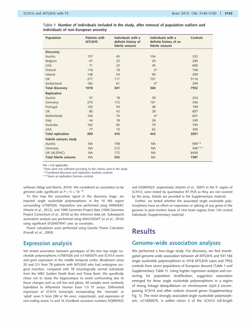
Table 1 Number of individuals included in the study, after removal of population outliers andindividuals of non-European ancestry
Population Patients withMTLEHS
Individuals with adefinite history offebrile seizures
Individuals with adefinite history of nofebrile seizures
Controls
Discovery
Austria 157 45 104 332
Belgium 67 23 20 285
USA 71 23 45 605
Finland 116 18 0* 746
Ireland 148 54 90 209
UK 277 117 101 5116
Switzerland 182 61 0* 259
Total discovery 1018 341 360 7552
Replication
Austria 57 18 39 254
Germany 273 112 161 346
Portugal 102 54 48 190
UK 80 42 28 857
Netherlands 164 74 0* 601
Italy 44 18 26 249
Australia 162 83 79 794
USA 77 15 62 300
Total replication 959 416 443 3591
Febrile seizures study
Austria NA 158 NA 585**
Germany NA 212 NA 346***
UK (ALSPAC) NA 172 NA 6456
Total febrile seizures NA 542 NA 7387
NA = not applicable.*Data were not collected according to the criteria used in the study.**Combined discovery and replication Austrian controls.***Same as replication German controls.
SCN1A and MTLEHS with FS Brain 2013: 136; 3140–3150 | 3143
transcript variant (NM_001202435.1) {P = 5.26 � 10�8, odds ratio
for G allele [OR(G)] 1.31, 95% confidence interval (CI) 1.19–1.44;
Table 2}. Two other single nucleotide polymorphisms within
SCN1A intron 1, had similarly low P-values: rs7587026
(r2 = 0.806 with rs11692675 in CEU population based on 1000
Genomes data set), P = 1.19 � 10�7 [OR(A) = 1.31, 95% CI:
1.19–1.45]; and rs580041 (r2 = 0.806 with rs11692675),
P = 5.74 � 10�7 [OR(A) = 1.29, 95% CI: 1.17–1.43].
SCN1A encodes brain-expressed voltage-gated sodium channel
type I, alpha subunit. It bears the largest number of known epi-
lepsy-related mutations, some associated with febrile seizures
(Oliva et al., 2012). The common SCN1A single nucleotide poly-
morphism rs3812718, affecting splicing (Heinzen et al., 2007), has
also been associated with febrile seizures (Schlachter et al., 2009),
though replication has failed (Petrovski et al., 2009). Retrospective
studies show association between MTLE and febrile seizures
(Pittau et al., 2009; O’Dell et al., 2012). Whether febrile seizures
cause MTLEHS (Koyama et al., 2012) or whether pre-existing
hippocampal abnormalities predispose to febrile seizures (Cendes,
2004), which may then also be injurious, is unknown. Clinical
differences between patients with and without a history of febrile
seizures suggest MTLEHS is heterogeneous (Thom et al., 2010).
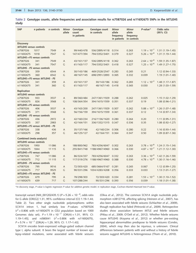
Table 2 Genotype counts, allele frequencies and association results for rs7587026 and rs11692675 SNPs in the MTLEHSstudy
SNP n patients n controls Minorallele
Genotypecountin patients
Genotype countin controls
Minorallelefrequencyin patients
Minorallelefrequencyin controls
P-value* Odds ratio(95% CI)
Discovery
MTLEHS versus controls
rs7587026 1017 7549 A 99/440/478 536/2895/4118 0.314 0.263 1.19 � 10–7 1.31 (1.19–1.45)
rs11692675 1018 7547 G 147/477/394 794/3352/3401 0.379 0.327 5.26 � 10–8 1.31 (1.19–1.44)
MTLEHS + FS versus controls
rs7587026 341 7549 A 43/161/137 536/2895/4118 0.362 0.263 2.64 � 10–8 1.59 (1.35–1.87)
rs11692675 341 7547 G 61/163/117 794/3352/3401 0.418 0.327 1.25 � 10–6 1.49 (1.27–1.75)
MTLEHS�FS versus controls
rs7587026 359 6544 A 30/143/186 469/2528/2547 0.283 0.265 0.21 1.12 (0.94–1.33)
rs11692675 360 6542 G 48/167/145 698/2951/2893 0.365 0.332 0.039 1.19 (1.01–1.40)
MTLEHS + FS versus MTLEHS�FS
rs7587026 341 359 A 43/161/137 30/143/186 0.362 0.283 1.12 � 10–3 1.48 (1.17–1.87)
rs11692675 341 360 G 61/163/117 48/167/145 0.418 0.365 0.030 1.28 (1.03–1.59)
Replication
MTLEHS versus controls
rs7587026 933 3537 A 89/360/484 247/1361/1929 0.288 0.262 0.025 1.15 (1.02–1.29)
rs11692675 826 3568 G 108/364/354 394/1615/1559 0.351 0.337 0.19 1.08 (0.96–1.21)
MTLEHS + FS versus controls
rs7587026 406 3537 A 43/163/200 247/1361/1929 0.307 0.262 5.88 � 10–3 1.26 (1.07–1.48)
rs11692675 371 3568 G 56/156/159 394/1615/1559 0.361 0.337 0.12 1.14 (0.97–1.34)
MTLEHS�FS versus controls
rs7587026 436 2972 A 42/160/234 216/1136/1620 0.280 0.264 0.20 1.11 (0.95–1.31)
rs11692675 357 2983 G 42/164/151 336/1332/1315 0.347 0.336 0.35 1.08 (0.92–1.27)
MTLEHS + FS versus MTLEHS�FS
rs7587026 338 436 A 35/137/166 42/160/234 0.306 0.280 0.22 1.16 (0.93–1.44)
rs11692675 298 357 G 46/125/127 42/164/151 0.364 0.347 0.50 1.09 (0.87–1.36)
Combined (meta-analysis)
MTLEHS versus controls
rs7587026 1950 11 086 A 188/800/962 783/4256/6047 0.302 0.263 3.78 � 10–8 1.24 (1.15–1.34)
rs11692675 1844 11 115 G 255/841/748 1188/4967/4960 0.366 0.330 4.87 � 10–7 1.21 (1.12–1.30)
MTLEHS + FS versus controls
rs7587026 747 11 086 A 86/324/337 783/4256/6047 0.332 0.263 3.36 � 10–9 1.42 (1.26–1.59)
rs11692675 712 11 115 G 117/319/276 1188/4967/4960 0.388 0.330 4.78 � 10–6 1.30 (1.16–1.46)
MTLEHS�FS versus controls
rs7587026 795 9516 A 72/303/420 685/3664/5167 0.281 0.265 0.067 1.12 (0.99–1.25)
rs11692675 717 9525 G 90/331/296 1034/4283/4208 0.356 0.333 0.033 1.13 (1.01–1.27)
MTLEHS + FS versus MTLEHS�FS
rs7587026 679 795 A 78/298/303 72/303/420 0.334 0.281 1.53 � 10–3 1.30 (1.10–1.52)
rs11692675 639 717 G 107/288/244 90/331/296 0.393 0.356 0.039 1.18 (1.01–1.38)
*In discovery stage, P-value is logistic regression P-value for additive genetic model; in replication stage, Cochran-Mantel-Haenszel test P-value.
3144 | Brain 2013: 136; 3140–3150 D. Kasperaviciut_e et al.
This evidence motivated our pre-analysis collection of febrile
seizure data, and our previous study of febrile seizures (Petrovski
et al., 2009). We performed analysis of patients in the discovery
cohort with a known history of presence of childhood febrile seiz-
ures (MTLEHS + FS, n = 341) (Table 2 and Fig. 1). The strongest
association was for rs7587026, P = 2.64 � 10�8 [OR(A) = 1.59,
95% CI: 1.35–1.87] and rs580041, P = 8.91 � 10�7 [OR(A)
= 1.56, 95% CI: 1.33–1.84], whereas the signal for rs11692675
was slightly weaker, P = 1.25 � 10�6 [OR(G) = 1.49, 95% CI:
1.27–1.75]. No association was seen in patients with MTLEHS
without febrile seizures (MTLEHS�FS), despite similar sample size.
To refine the association signal, we performed regional imput-
ation in the discovery data set of a 10 Mb region surrounding
rs7587026 using the 1000 Genomes reference panel. Two single
nucleotide polymorphisms had slightly lower P-values in the
MTLEHS + FS analysis than the original single nucleotide poly-
morphisms [rs16851603 (P = 2.23 � 10�8) and rs3919196
(P = 2.26 � 10�8)], but neither were significantly stronger than
the original associations, and these signals reflected regional link-
age disequilibrium structure (Supplementary Fig. 2). No known
functional variants in SCN1A, nor in other genes in the region,
were in high linkage disequilibrium with rs7587026. The associ-
ation signal is localized within one linkage disequilibrium block that
also spans the promoter and 5’ UTR region of SCN1A
(Supplementary Fig. 2).
Replication and combined analysesWe selected the two top single nucleotide polymorphisms,
rs7587026 and rs11692675, for replication in an independent
sample of 959 patients with MTLEHS, of whom 416 had
MTLEHS + FS, and 3591 population-matched controls of
European descent from eight populations (Table 1 and
Supplementary Table 1). We did not study rs580041 because of
its perfect linkage disequilibrium with rs7587026 in white
Europeans (r2 = 1). We detected an association between
rs7587026 and MTLEHS + FS, P = 5.88 � 10�3 [OR(A) = 1.26,
95% CI: 1.07–1.48; Table 2]; this value remains significant at a
revised alpha threshold of 6.3 � 10�3 after Bonferroni correction
for multiple comparisons in the replication cohort. No association
was detected for MTLEHS�FS.
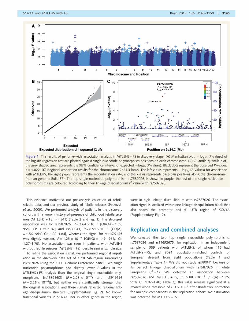
Figure 1 The results of genome-wide association analysis in MTLEHS + FS in discovery stage. (A) Manhattan plot, � log10 (P-values) of
the logistic regression test are plotted against single nucleotide polymorphism positions on each chromosome. (B) Quantile-quantile plot,
the grey shaded area represents the 95% confidence interval of expected � log10 (P-values). Black dots represent the observed P-values;
� = 1.022. (C) Regional association results for the chromosome 2q24.3 locus. The left y-axis represents � log10 (P-values) for association
with MTLEHS, the right y-axis represents the recombination rate, and the x-axis represents base-pair positions along the chromosome
(human genome Build 37). The top single nucleotide polymorphism, rs7587026, is shown in purple, the rest of the single nucleotide
polymorphisms are coloured according to their linkage disequilibrium r2 value with rs7587026.
SCN1A and MTLEHS with FS Brain 2013: 136; 3140–3150 | 3145
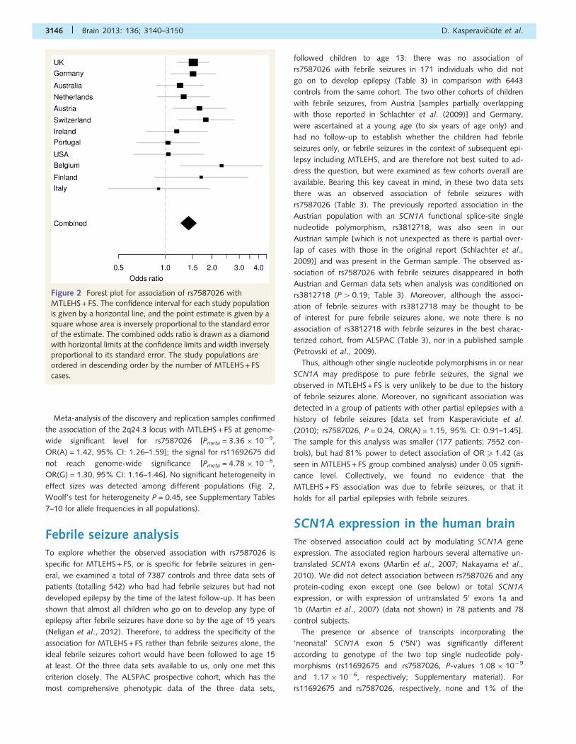
Meta-analysis of the discovery and replication samples confirmed
the association of the 2q24.3 locus with MTLEHS + FS at genome-
wide significant level for rs7587026 [Pmeta = 3.36 � 10�9,
OR(A) = 1.42, 95% CI: 1.26–1.59]; the signal for rs11692675 did
not reach genome-wide significance [Pmeta = 4.78 � 10�6,
OR(G) = 1.30, 95% CI: 1.16–1.46]. No significant heterogeneity in
effect sizes was detected among different populations (Fig. 2,
Woolf’s test for heterogeneity P = 0.45, see Supplementary Tables
7–10 for allele frequencies in all populations).
Febrile seizure analysisTo explore whether the observed association with rs7587026 is
specific for MTLEHS + FS, or is specific for febrile seizures in gen-
eral, we examined a total of 7387 controls and three data sets of
patients (totalling 542) who had had febrile seizures but had not
developed epilepsy by the time of the latest follow-up. It has been
shown that almost all children who go on to develop any type of
epilepsy after febrile seizures have done so by the age of 15 years
(Neligan et al., 2012). Therefore, to address the specificity of the
association for MTLEHS + FS rather than febrile seizures alone, the
ideal febrile seizures cohort would have been followed to age 15
at least. Of the three data sets available to us, only one met this
criterion closely. The ALSPAC prospective cohort, which has the
most comprehensive phenotypic data of the three data sets,
followed children to age 13: there was no association of
rs7587026 with febrile seizures in 171 individuals who did not
go on to develop epilepsy (Table 3) in comparison with 6443
controls from the same cohort. The two other cohorts of children
with febrile seizures, from Austria [samples partially overlapping
with those reported in Schlachter et al. (2009)] and Germany,
were ascertained at a young age (to six years of age only) and
had no follow-up to establish whether the children had febrile
seizures only, or febrile seizures in the context of subsequent epi-
lepsy including MTLEHS, and are therefore not best suited to ad-
dress the question, but were examined as few cohorts overall are
available. Bearing this key caveat in mind, in these two data sets
there was an observed association of febrile seizures with
rs7587026 (Table 3). The previously reported association in the
Austrian population with an SCN1A functional splice-site single
nucleotide polymorphism, rs3812718, was also seen in our
Austrian sample [which is not unexpected as there is partial over-
lap of cases with those in the original report (Schlachter et al.,
2009)] and was present in the German sample. The observed as-
sociation of rs7587026 with febrile seizures disappeared in both
Austrian and German data sets when analysis was conditioned on
rs3812718 (P40.19; Table 3). Moreover, although the associ-
ation of febrile seizures with rs3812718 may be thought to be
of interest for pure febrile seizures alone, we note there is no
association of rs3812718 with febrile seizures in the best charac-
terized cohort, from ALSPAC (Table 3), nor in a published sample
(Petrovski et al., 2009).
Thus, although other single nucleotide polymorphisms in or near
SCN1A may predispose to pure febrile seizures, the signal we
observed in MTLEHS + FS is very unlikely to be due to the history
of febrile seizures alone. Moreover, no significant association was
detected in a group of patients with other partial epilepsies with a
history of febrile seizures [data set from Kasperaviciute et al.
(2010); rs7587026, P = 0.24, OR(A) = 1.15, 95% CI: 0.91–1.45].
The sample for this analysis was smaller (177 patients; 7552 con-
trols), but had 81% power to detect association of OR5 1.42 (as
seen in MTLEHS + FS group combined analysis) under 0.05 signifi-
cance level. Collectively, we found no evidence that the
MTLEHS + FS association was due to febrile seizures, or that it
holds for all partial epilepsies with febrile seizures.
SCN1A expression in the human brainThe observed association could act by modulating SCN1A gene
expression. The associated region harbours several alternative un-
translated SCN1A exons (Martin et al., 2007; Nakayama et al.,
2010). We did not detect association between rs7587026 and any
protein-coding exon except one (see below) or total SCN1A
expression, or with expression of untranslated 5’ exons 1a and
1b (Martin et al., 2007) (data not shown) in 78 patients and 78
control subjects.
The presence or absence of transcripts incorporating the
‘neonatal’ SCN1A exon 5 (‘5N’) was significantly different
according to genotype of the two top single nucleotide poly-
morphisms (rs11692675 and rs7587026, P-values 1.08 � 10�9
and 1.17 � 10�6, respectively; Supplementary material). For
rs11692675 and rs7587026, respectively, none and 1% of the
Figure 2 Forest plot for association of rs7587026 with
MTLEHS + FS. The confidence interval for each study population
is given by a horizontal line, and the point estimate is given by a
square whose area is inversely proportional to the standard error
of the estimate. The combined odds ratio is drawn as a diamond
with horizontal limits at the confidence limits and width inversely
proportional to its standard error. The study populations are
ordered in descending order by the number of MTLEHS + FS
cases.
3146 | Brain 2013: 136; 3140–3150 D. Kasperaviciut_e et al.
individuals with the GG and AA genotype showed SCN1A
transcripts in the neonatal form, compared with 83% and 81%
with the genotype AA or CC. This alternative splicing event is
influenced by rs3812718 (Heinzen et al., 2007). The association
of alternative splicing with rs922224 (r2 = 1 with rs3812718) was
stronger, P = 2.33 � 10�31. The level of expression of SCN1A
exon 5N was also significantly different according to genotype
(P = 1.62 � 10�11 for rs11692675, 2.70 � 10�6 for rs7587026,
7.40 � 10�34 for rs922224). In conditional analyses including all
three single nucleotide polymorphisms, only rs922224 remained
significant (P = 1.08 � 10�25). Finally, expression quantitative
trait loci analyses for subsets of patients according to a known
history of presence (n = 46) or absence (n = 27) of febrile seizures
in childhood for rs11692675 or rs7587026 showed significant dif-
ferences in the level of expression of 5N exon according to geno-
type in both MTLEHS + FS and MTLEHS�FS. Including both
rs11692675 or rs11692675 and rs922224 in the regression
models, only rs922224 remained significant in both MTLEHS + FS
and MTLEHS�FS groups (Supplementary material).
We cannot exclude the possibility that rs7587026 (or another
single nucleotide polymorphism in the high linkage disequilibrium
region) may act as an additional splicing controller to rs3812718,
but our data are consistent with rs7587026 having no solo effect
on 5N splicing. We also did not detect any correlation using a
significance level of P55 � 10�5 between rs7587026 and
expression/splicing of any other genes across the genome
(Supplementary material).
DiscussionWe show that common variation in and near SCN1A may increase
susceptibility to MTLEHS + FS. Our previously published larger
genome-wide association study for a broader range of focal
epilepsies did not identify any single-single nucleotide polymorph-
ism association (Kasperaviciute et al., 2010), but the findings
here demonstrate that associated variants may exist for more
narrowly-defined syndromes. Because the biology of most of the
epilepsies is poorly understood, there are few a priori data upon
which to base selection of the range of phenotypes to include in
studies of possible genetic causation. Our findings suggest that
focussing on clinically recognized syndromes or constellations
(Berg et al., 2010) may prove fruitful by reducing heterogeneity
before genomic analyses.
Our association seems to be specific for MTLEHS + FS, with no
association for MTLEHS�FS, febrile seizures alone or non-
MTLEHS partial epilepsies with febrile seizures. Our findings sug-
gest that there is genetic susceptibility to MTLEHS, and that it, or
hippocampal sclerosis, may not necessarily be only acquired. The
results support the concept of heterogeneity in MTLEHS, beyond
that already documented clinico-pathologically (Tassi et al., 2009;
Thom et al., 2010; Blumcke et al., 2012). However, further work
will be needed to confirm the specificity of our findings, as we did
not formally establish a significant difference in odds ratios be-
tween MTLEHS + FS and MTLEHS�FS. It would also be interesting
to explore, in a suitably-powered study, whether there is any
association with MTLE without hippocampal sclerosis.
The notably weaker association in the replication stage could be
due to several factors, the most important of which is the ‘win-
ner’s curse’ (Ioannidis et al., 2009); there may be a large number
of weak but real associations in the data, some of which achieve
genome-wide significance in a particular study through random
stochastic chance, but will not do so in another study. The asso-
ciation in our discovery cohort was replicated in the second inde-
pendent sample, but it is nevertheless important that other studies
are undertaken to further replicate our findings. Other limitations
of our study are the lack of genome-wide data in the replication
sample, preventing direct population stratification assessment,
though self-identification closely corresponds to genetically-deter-
mined ancestry (Lao et al., 2008; Wang et al., 2010), a phenom-
enon we confirmed in the discovery stage, and the small size of
some of the replication groups, reducing replication power, and
magnifying effects of undetected population admixture.
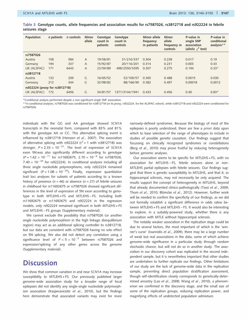
Table 3 Genotype counts, allele frequencies and association results for rs7587026, rs3812718 and rs922224 in febrileseizures stage
Population n patients n controls Minorallele
Genotypecount inpatients
Genotypecount incontrols
Minor allelefrequencyin patients
Minorallelefrequency incontrols
P-value insingle SNPassociation(allelic �2 test)
P-value inconditionalanalysis**
rs7587026
Austria 158 584 A 19/58/81 31/216/337 0.304 0.238 0.017 0.19
Germany 194 337 A 15/92/87 20/116/201 0.314 0.231 0.003 0.43
UK (ALSPAC) 171 6443 A 23/59/89 498/2550/3395 0.307 0.275 0.194 0.33*
rs3812718
Austria 133 209 G 16/65/52 52/100/57 0.365 0.488 0.0015 0.030
Germany 212 344 G 32/98/82 88/166/90 0.382 0.497 0.00018 0.0012
rs922224 (proxy for rs3812718)
UK (ALSPAC) 172 6456 G 34/81/57 1371/3144/1941 0.433 0.456 0.40 0.83*
*Conditional analysis performed despite a non-significant single SNP association.
**In conditional analyses, rs7587026 was conditioned for rs3812718 (or its proxy, rs922224, for the ALSPAC cohort), while rs3812718 and rs922224 were conditioned forrs7587026.
SCN1A and MTLEHS with FS Brain 2013: 136; 3140–3150 | 3147
As for many genome-wide association studies, we could not
narrow the association to a single gene or functional variant.
There are other genes designated ‘SCNxA’ in the vicinity:
SCN3A, SCN2A, SCN9A and SCN7A (this last does not show
any sodium channel activity in exogenous expression systems)
(Meisler et al., 2010). Among these genes, SCN2A has the most
published evidence to support its role in the epilepsies. We cannot
exclude the possibility that the association is driven by deleterious
variants in these or other nearby genes. SCN1A, however,
emerges as the most plausible candidate, due both to its proximity
to the associated region and its role in other epilepsies with febrile
seizures. Notably, our association is with a syndrome involving
hippocampal damage, whereas typically no hippocampal damage
is observed in patients with Dravet syndrome caused by deleteri-
ous changes affecting SCN1A (Catarino et al., 2011), suggesting
that SCN1A might influence epileptogenesis through various
mechanisms.
The location of the associated variants within SCN1A and over-
lapping its promoter regions (Long et al., 2008), was suggestive of
possible roles in SCN1A expression modulation. In fact, we did not
detect a definitive effect on expression of SCN1A or its exons in
temporal neocortex. However, this analysis may have been con-
founded by many factors: effects may be brain-region or cell-
population specific, as in SCN1A-related Dravet syndrome,
where consequences are only found in interneurons (Ogiwara
et al., 2007); our whole-tissue expression analysis would not
detect such subtle signals. Moreover, noting the febrile seizures
association, the effects may be temporally or spatially restricted,
acting only in childhood or/and in the stress of febrile seizures
(Koyama et al., 2012). Further studies will be needed to explore
possible functional effects.
The detected association could act in different ways, predispos-
ing to MTLEHS + FS as a distinct syndrome, or to the specific de-
velopment of MTLEHS in the context of remote febrile seizures. If
the association does indeed relate to SCN1A and function of the
encoded protein, new lines of investigation may prove possible in
the context of the existing deep knowledge of SCN1A, experimen-
tal models of MTLE and in vitro study of mechanisms of hippo-
campal dysfunction in epilepsy, as well as intriguing reports of the
role of SCN1A in many epilepsies, such as the suggestion that
mutations in SCN1A in Dravet Syndrome may protect against
hippocampal sclerosis (Auvin et al., 2008; Catarino et al., 2011).
Stratifying by febrile seizures type could also prove illuminating, as
prolonged, lateralized or repeated febrile seizures within a short
interval may have different effects to ‘uncomplicated’ febrile seiz-
ures. Our retrospective febrile seizures data were insufficiently
resolved to permit such analysis. This is an important avenue for
further investigation, because no predictors exist for the develop-
ment of epilepsy in the 3% of all the children who have febrile
seizures, and because established MTLEHS can have devastating
consequences. Eventual reliable prediction of significant risk of
MTLEHS after febrile seizures could lead to novel preventative
measures in at-risk individuals: here, we note that SCN1A encodes
an important anti-epileptic drug target and that it is possible to
pharmacologically prevent the development of epilepsy after
febrile seizures in an animal model (Koyama et al., 2012). Our
findings suggest that further work on SCN1A variation may
contribute to understanding the risk of developing MTLEHS after
febrile seizures.
AcknowledgementsWe thank all patients, their families and physicians for participat-
ing in this study. We thank Drs. Goldstein, Heinzen and Radtke for
use of data from patients at Duke University School of Medicine,
and for their comments. We thank Drs. D. Lowenstein and A-E.
Lehesjoki, and the ILAE Consortium on Complex Epilepsies
(including Carolien de Kovel, Thomas Sander, Dennis Dlugos,
Warren Lo, Tom Ferraro, Mike Johnson, Tony Marson, Douglas
Speed, Patrick Kwan, Larry Baum, Stacey Cherny, Zhi Wei, Larry
Brody, Pak Sham, David Balding and Aarno Palotie) for their sup-
port of the work, and for their comments. We thank Prof J. Hardy
for his support. We thank the members of the Dutch Collaborative
Epilepsy Surgery Program for their cooperation. We acknowledge
the Italian League against Epilepsy (LICE) collaborative group for
its support. We gratefully acknowledge Professor John Hopper
(School of Population Health, The University of Melbourne) for
providing the control samples for the Melbourne cohort and Dr.
Marian Todaro for processing the DNA samples for the Melbourne
cohort; Dr. Liisa Myllykangas (Folkhalsan Institute of Genetics and
Department of Pathology, University of Helsinki) and Dr. Pentti
Tienari (Molecular Neurology Programme, Biomedicum, University
of Helsinki and Department of Neurology, Helsinki University
Central Hospital) for providing us the Vantaa85 + Study GWAS
genotypes; Professor Edouard Louis (MD, PhD), Head of the
Gastroenterology Unit, University Hospital Centre (CHU) of
Liege and Professor Michel Georges (DVM, PhD), Head of the
Animal Genomics Unit, Faculty of Veterinary Medicine
and Groupe Interdisciplinaire de Genoproteomique Appliquee
(GIGA-R) for providing us the Belgian control cohort; Drs. P. C.
van Rijen and P. H. Gosselaar, Department of Neurosurgery,
Rudolf Magnus Institute of Neuroscience, University Medical
Centre Utrecht, The Netherlands; Christian Hengsbach for expert
assistance with recruitment for the Tubingen MTLE cohort;
Susanne Beyer and Ulrike Strube for the technical assistance in
SNP genotyping for the Bonn MTLE cohort; and the whole
ALSPAC team, which includes interviewers, computer and labora-
tory technicians, clerical workers, research scientists, volunteers,
managers, receptionists and nurses. We would like to thank
AROS Applied Biotechnology AS company laboratories and
Affymetrix for their valuable input. We are grateful to the
Banner Sun Health Research Institute Brain and Body Donation
Program of Sun City, Arizona for the provision of human biospeci-
mens contributing to gene expression analysis of nine brain re-
gions from 134 control individuals. The Brain and Body
Donation Program is supported by the National Institute of
Neurological Disorders and Stroke (U24 NS072026 National
Brain and Tissue Resource for Parkinson’s Disease and Related
Disorders), the National Institute on Aging (P30 AG19610
Arizona Alzheimer’s Disease Core Centre), the Arizona
Department of Health Services (contract 211002, Arizona
Alzheimer’s Research Centre), the Arizona Biomedical Research
Commission (contracts 4001, 0011, 05-901 and 1001 to the
3148 | Brain 2013: 136; 3140–3150 D. Kasperaviciut_e et al.
Arizona Parkinson’s Disease Consortium) and the Michael J. Fox
Foundation for Parkinson’s Research.
FundingSupported by the Wellcome Trust (grant 084730 to S.M.S., N.D,
C.D.); the UK Medical Research Council (MRC grants G0400126
to S.M.S., G1100616 to C.S., G0901254 to J.H. and M.E.W., and
Training Fellowship G0802462 to M.R.); the King Faisal Specialist
Hospital and Research Centre (KFSH & RC grant to D.T.); the
University College London Hospitals Charities and the Clinical
Research and Development Committee (UCLH/CRDC grant F136
to S.M.S.); the National Institute for Health Research (NIHR grant
08-08-SCC to S.M.S.); the National Society for Epilepsy; the
National Institute for Health Research University College London
Hospitals Biomedical Research Centre; Marie Curie International
Re-integration Grant (FP7-PEOPLE-2009-RG grant No 256545 to
M.M. and S.M.S.); the European Commission (FP7 project EpiPGX,
grant 279062 to H.L., S.M.S and W.S.K.; FP6 project Epicure,
grant LSHM-CT-2006-037315 to H.L., P.S.R. and F.R.); the
Robert Bosch Foundation, Stuttgart, and the University of
Tubingen (IZEPHA project 18-0-0, grant to H.L.); the German
Federal Ministry of Education and Research, National Genome
Research Network (NGFNplus: EMINet grant 01GS08123 to
H.L., S.S., A.J.B.); BONFOR (S.S., A.J.B.); the German Federal
Ministry of Education and Research (independent research
groups in neuroscience); the German Research Foundation
(EUROCORES program, EuroEPINOMICS-RES grant RO3396/2-1
to P.S.R. and F.R. and EuroEPINOMICS-RES grant DFG Bl421/3-1
to I.B. and K.K.); the Austrian Science Fund (project FWF I643,
grant to F. Zimprich); the National Health and Medical Research
Council (NHMRC grant 628952 to S.F.B.); the Royal Melbourne
Hospital Neuroscience Foundation (grant to T.J.O’B.); the
Foundation of Science and Technology, Lisbon (FCT grant PIC/
IC/83297/2007 to B.M.S.); the National Institutes of Health,
USA (NIH grants R01-NS-49306-01 to R.J.B. and R01-NS-
064154-01 to R.J.B. and H.H.); the collection of the Irish patient
cohort was supported by the Irish Higher Education Authority
Programme for Research in Third Level Institutions (PRTLI3)
through a Science Foundation Ireland Research Frontiers
Programme award (08/RFP/GEN1538) and a Medical Research
Charities Group of Ireland/Health Research Board award (2009/
001) from Brainwave–the Irish Epilepsy Association.
GlaxoSmithKline funded the recruitment and phenotypic data col-
lection of the GenEpA Consortium samples used in this study and
contributed to the genotyping costs associated with their study.
The collection of the Belgian patients was supported by the Fonds
National de la Recherche Scientifique, grant n. FC 63574/
3.4.620.06 F, and the Fonds Erasme, Universite Libre de
Bruxelles. The collection of the Belgian control cohort was sup-
ported by the Walloon Region and the French-Speaking
Community of Belgium, the Belgian Science Policy and the
University of Liege. J.N. is supported by the Swiss National
Science Foundation-Fellowships for prospective researchers and
the SICPA Foundation, Prilly, Switzerland. Computing facilities
used at King’s College London were supported by the National
Institute for Health Research (NIHR) Biomedical Research Centre
based at Guy’s and St Thomas’ NHS Foundation Trust and King’s
College London. Funding support for the ALSPAC was provided by
The UK Medical Research Council (grant refs: 74882) the
Wellcome Trust (grant refs: 076467) and the University of Bristol.
This study makes use of data generated by the Wellcome Trust
Case-Control Consortium (a full list of the investigators who con-
tributed to the generation of the data is available from www.
wtccc.org.uk; funding for the project was provided by the
Wellcome Trust under award 076113 and 085475) and the Irish
Amyotrophic Lateral Sclerosis Study (funding support was pro-
vided by Muscular Dystrophy Association, USA, Irish Institute of
Clinical Neurosciences Travel Award, and National Institutes of
Health, USA; additional genotyping was provided by the
National Institute of Neurological Disorders and Stroke (NINDS);
the dataset used for the analyses described in this manuscript were
obtained from the NINDS Database found at http://www.ncbi.
nlm.nih.gov/gap through dbGaP accession number
phs000127.v1.p1).
Conflict of interestY.W. has served on scientific advisory boards for UCB Pharma and
has received funding for travel and speaker honoraria from UCB,
Desitin Pharmaceuticals, GmbH, and Eisai Inc.; H.L. has served on
scientific advisory boards for Eisai Inc., GlaxoSmithKline, Pfizer Inc,
UCB, and Valeant Pharmaceuticals International, has received
funding for travel from GlaxoSmithKline, Medtronic, Pfizer and
UCB and speaker honoraria from Desitin Pharmaceuticals,
GmbH, Eisai Inc., GlaxoSmithKline, Pfizer, and UCB, and has
received research support from Sanofi-Aventis, UCB, DFG,
BMBF, and the EU; J.C. has received funding from the Tecnifar
group (BICE Tecnifar Grant 2009); F.R. has received within the last
two years honoraria as scientific advisor from GSK, EISAI, UCB and
Pfizer, and has received speaker honoraria from UCB, GSK, Eisai,
Desitin and Medtronic and educational grants from Nihon-
Kohden, UCB, Medtronics, Cyberonics and Cerbomed (F.R. has,
however, no conflicts of interest regarding this study); P.S.R. has
received travel grants from UCB; G.L.C has received research
funding from UCB and speaker honoraria from Eisai; S.M.S has
received research funding or personal/institutional honoraria from
UCB Pharma, GlaxoSmithKline and Eisai Inc; H.M.H. has served on
the scientific advisory board of Eisai, Pfizer, GlaxoSmithKline and
UCB Pharma, has served on the speakers’ bureau of Desitin, Eisai,
GlaxoSmithKline and UCB Pharma and received research funding
from Desitin, Janssen-Cilag, GlaxoSmithKline and UCB Pharma;
I.B. received speaker honoraria from Desitin Pharmaceuticals,
GmbH, Eisai Inc., and UCB, and has received research support
from Boehringer-Ingelheim; R.K.K. has served on scientific advis-
ory boards for UCB Pharma, Eisai, Lundbeck, GlaxoSmithKline,
and Fennomedical and has received funding for travel and speaker
honoraria from UCB Pharma, Eisai, GlaxoSmithKline, Medtronic,
Pfizer, Orion, Fennomedical and institutional research funding
from UCB Pharma and GlaxoSmithKline; A-M.K has received
funding for travel from UCB Pharma, Eisai, Abbott and Biogen;
S.F.B. is an inventor on a patent for SCN1A testing held by
SCN1A and MTLEHS with FS Brain 2013: 136; 3140–3150 | 3149
Bionomics Inc and licensed to various diagnostic companies. All
other authors declare that they have no conflicts of interest.
Supplementary materialSupplementary material is available at Brain online.
References1000 Genomes Project Consortium, Abecasis GR, Altshuler D, Auton A,
Brooks L, Durbin R, et al. A map of human genome variation from
population-scale sequencing. Nature 2010; 467: 1061–73.Abou-Khalil B, Ge Q, Desai R, Ryther R, Bazyk A, Bailey R, et al. Partial
and generalized epilepsy with febrile seizures plus and a novel SCN1A
mutation. Neurology 2001; 57: 2265–72.
Andrade-Valenca LP, Valenca MM, Velasco TR, Carlotti CG Jr,
Assirati JA, Galvis-Alonso OY, et al. Mesial temporal lobe epilepsy:
clinical and neuropathologic findings of familial and sporadic forms.
Epilepsia 2008; 49: 1046–54.
Auvin S, Dulac O, Vallee L. Do SCN1A mutations protect from hippo-
campal sclerosis? Epilepsia 2008; 49: 1107–8.
Berg AT, Berkovic SF, Brodie MJ, Buchhalter J, Cross JH, van Emde
Boas W, et al. Revised terminology and concepts for organization of
seizures and epilepsies: report of the ILAE Commission on
Classification and Terminology, 2005-2009. Epilepsia 2010; 51:
676–85.
Blumcke I, Coras R, Miyata H, Ozkara C. Defining clinico-neuropatho-
logical subtypes of mesial temporal lobe epilepsy with hippocampal
sclerosis. Brain Pathol 2012; 22: 402–11.
Catarino CB, Liu JY, Liagkouras I, Gibbons VS, Labrum RW, Ellis R, et al.
Dravet syndrome as epileptic encephalopathy: evidence from long-
term course and neuropathology. Brain 2011; 134: 2982–3010.
Cendes F. Febrile seizures and mesial temporal sclerosis. Curr Opin
Neurol 2004; 17: 161–4.De Tisi J, Bell GS, Peacock JL, McEvoy AW, Harkness WF, Sander JW,
et al. The long-term outcome of adult epilepsy surgery, patterns of
seizure remission, and relapse: a cohort study. Lancet 2011; 378:
1388–95.
Heinzen EL, Yoon W, Tate SK, Sen A, Wood NW, Sisodiya SM, et al.
Nova2 interacts with a cis-acting polymorphism to influence the pro-
portions of drug-responsive splice variants of SCN1A. Am J Hum
Genet 2007; 80: 876–83.
Howie B, Fuchsberger C, Stephens M, Marchini J, Abecasis GR. Fast and
accurate genotype imputation in genome-wide association studies
through pre-phasing. Nat Genet 2012; 44: 955–9.
Ioannidis JP, Thomas G, Daly MJ. Validating, augmenting and refining
genome-wide association signals. Nat Rev Genet 2009; 10: 318–29.
Kasperaviciute D, Catarino CB, Heinzen EL, Depondt C, Cavalleri GL,
Caboclo LO, et al. Common genetic variation and susceptibility to
partial epilepsies: a genome-wide association study. Brain 2010; 133:
2136–47.
Koyama R, Tao K, Sasaki T, Ichikawa J, Miyamoto D, Muramatsu R,
et al. GABAergic excitation after febrile seizures induces ectopic
granule cells and adult epilepsy [Internet]. Nat Med 2012; 18:
1271–8. Available from: http://www.ncbi.nlm.nih.gov/pubmed/
22797810 (13 September 2012, date last accessed).
Labate A, Gambardella A, Andermann E, Aguglia U, Cendes F,
Berkovic SF, et al. Benign mesial temporal lobe epilepsy. Nat Rev
Neurol 2011; 7: 237–40.
Lao O, Lu TT, Nothnagel M, Junge O, Freitag-Wolf S, Caliebe A, et al.
Correlation between genetic and geographic structure in Europe. Curr
Biol 2008; 18: 1241–8.
Li Y, Willer CJ, Ding J, Scheet P, Abecasis GR. MaCH: using sequence
and genotype data to estimate haplotypes and unobserved genotypes.
Genet Epidemiol 2010; 34: 816–34.Long YS, Zhao QH, Su T, Cai YL, Zeng Y, Shi YW, et al. Identification of
the promoter region and the 5’-untranslated exons of the human volt-
age-gated sodium channel Nav1.1 gene (SCN1A) and enhancement of
gene expression by the 5’-untranslated exons. J. Neurosci Res 2008;
86: 3375–81.
Magi R, Morris AP. GWAMA: software for genome-wide association
meta-analysis. BMC Bioinformatics 2010; 11: 288.Mantegazza M, Gambardella A, Rusconi R, Schiavon E, Annesi F,
Cassulini RR, et al. Identification of an Nav1.1 sodium channel
(SCN1A) loss-of-function mutation associated with familial simple fe-
brile seizures. Proc Natl Acad.Sci USA 2005; 102: 18177–82.
Martin MS, Tang B, Ta N, Escayg A. Characterization of 5’ untranslated
regions of the voltage-gated sodium channels SCN1A, SCN2A, and
SCN3A and identification of cis-conserved noncoding sequences.
Genomics 2007; 90: 225–35.
Meisler MH, O’Brien JE, Sharkey LM. Sodium channel gene family: epi-
lepsy mutations, gene interactions and modifier effects. J Physiol
(Lond.) 2010; 588: 1841–8.
Nakayama T, Ogiwara I, Ito K, Kaneda M, Mazaki E, Osaka H, et al.
Deletions of SCN1A 5’ genomic region with promoter activity in
Dravet syndrome. Hum Mutat 2010; 31: 820–9.
Neligan A, Bell GS, Giavasi C, Johnson AL, Goodridge DM, Shorvon SD,
et al. Long-term risk of developing epilepsy after febrile seizures: a
prospective cohort study. Neurology 2012; 78: 1166–70.O’Dell CM, Das A, Wallace G 4th, Ray SK, Banik NL. Understanding the
basic mechanisms underlying seizures in mesial temporal lobe epilepsy
and possible therapeutic targets: a review. J Neurosci Res 2012; 90:
913–24.
Ogiwara I, Miyamoto H, Morita N, Atapour N, Mazaki E, Inoue I, et al.
Nav1.1 localizes to axons of parvalbumin-positive inhibitory inter-
neurons: a circuit basis for epileptic seizures in mice carrying an
Scn1a gene mutation. J Neurosci 2007; 27: 5903–14.
Oliva M, Berkovic SF, Petrou S. Sodium channels and the neurobiology
of epilepsy. Epilepsia 2012; 53: 1849–59.
Petrovski S, Scheffer IE, Sisodiya SM, O’Brien TJ, Berkovic SF. Lack of
replication of association between scn1a SNP and febrile seizures.
Neurology 2009; 73: 1928–30.
Pittau F, Bisulli F, Mai R, Fares JE, Vignatelli L, Labate A, et al. Prognostic
factors in patients with mesial temporal lobe epilepsy. Epilepsia 2009;
50 (Suppl 1): 41–4.
Purcell S, Cherny SS, Sham PC. Genetic power calculator: design of link-
age and association genetic mapping studies of complex traits.
Bioinformatics 2003; 19: 149–50.
Schlachter K, Gruber-Sedlmayr U, Stogmann E, Lausecker M, Hotzy C,
Balzar J, et al. A splice site variant in the sodium channel gene SCN1A
confers risk of febrile seizures. Neurology 2009; 72: 974–8.Tassi L, Meroni A, Deleo F, Villani F, Mai R, Russo GL, et al. Temporal
lobe epilepsy: neuropathological and clinical correlations in 243 surgi-
cally treated patients. Epileptic Disord 2009; 11: 281–92.
Thom M, Mathern GW, Cross JH, Bertram EH. Mesial temporal lobe
epilepsy: how do we improve surgical outcome? Ann Neurol 2010;
68: 424–34.
Wang H, Haiman CA, Kolonel LN, Henderson BE, Wilkens LR, Le
Marchand L, et al. Self-reported ethnicity, genetic structure and the
impact of population stratification in a multiethnic study. Hum Genet
2010; 128: 165–77.
Wieser HG. ILAE Commission Report. Mesial temporal lobe epilepsy with
hippocampal sclerosis. Epilepsia 2004; 45: 695–714.
3150 | Brain 2013: 136; 3140–3150 D. Kasperaviciut_e et al.