Postcholecystectomy Syndrome: From Pathophysiology to … · cholecystectomy pain ranged from 14%...
7
Review Article Open Access Madacsy et al., Pancreat Disord Ther 2015, 5:3 DOI: 10.4172/2165-7092.1000162 Volume 5 • Issue 3 • 1000162 Pancreat Disord Ther ISSN: 2165-7092 PDT, an open access journal Postcholecystectomy Syndrome: From Pathophysiology to Differential Diagnosis - A Critical Review Madacsy L*, Dubravcsik Z and Szepes A Department of Gastroenterology, Bács-Kiskun Teaching Hospital of the University of Szeged, Kecskemét, Hungary Keywords: Postcholecystectomy syndrome; Sphincter of oddi dysfunction; Functional abdominal pain Introduction Cholecystectomy has an excellent therapeutic outcome. Up to 15-20 % of cholecystectomized patients, however, continue to have a variety of gastrointestinal symptoms such as fatty food intolerance, nausea and vomiting, heartburn, flatulence, indigestion, diarrhea, mild occasional abdominal pain attacks and severe RUQ pain with extreme post-cholecystectomy distress. e term used to describe this condition is post-cholecystectomy syndrome (PCS), which was originally suggested by Pribram [1]. Post-cholecystectomy syndrome (PCS) defined as symptoms of biliary colic or persistent right upper quadrant (RUQ) abdominal pain with or without dyspepsia, which are similar to that experienced by the patient before cholecystectomy. Although a large volume of medical information accumulated since the first description, patients with PCS, continue to be present as a diagnostic and therapeutic challenge. Our current knowledge about the pathophysiology of PCS has significantly improved since the introduction of the endoscopic retrograde cholangiopancreatography (ERCP) and the endoscopic sphincter of Oddi manometry (ESOM) in the diagnostic opportunities. e aim of this paper was to review the literature critically on the magnitude of the problem and the possible pathophysiological explanations of PCS. Magnitude of the Problem e reported frequency of PCS varies widely in the literature. Several previous investigations have evaluated the effects of cholecystectomy on the symptoms, but their results were contradictory, which caused by the variations of the study design. It is also difficult to precisely analyze the current literature in regards to the frequency of post-cholecystectomy symptoms because the lack of clear distinction between biliary pain and dyspepsia. e reported frequency of post- cholecystectomy pain ranged from 14% to 34% in the reviewed literature, but postoperative dyspepsia was a more frequent symptom that occurred in up to 54% of cholecystectomized patients. In an old study published by Burnett. 126 patients retrospectively interviewed one year aſter cholecystectomy, and demonstrated that 75% of patients were relieved of all symptoms [2]. In an excellent landmark study by Bodwall, two thousand patients were followed up symptomatically for 2-5 years aſter cholecystectomy, and the results were evaluated by computer-assisted analysis of multiple variables. eir results suggested that female sex, lower age, longer preoperative symptomatic period, the absence of gallbladder inflammation, and a functioning acalculous gallbladder were associated with higher frequency of PCS. e accurate interpretation of 351 intravenous cholangiography by the same working group has provided up to date evidence that patients with more severe symptoms of PCS associated with a larger common bile duct diameter [3]. Gunn emphasized that although dyspeptic symptoms were present in 88% of all patients preoperatively, as much as 69% of patients were symptomatically improved by cholecystectomy [4]. Bates published a prospective controlled study in 295 patients referred to cholecystectomy. ey claimed that before the operation symptoms of dyspepsia were far more frequent in the gallstone group, and cholecystectomy reduced the dyspeptic symptoms to an incidence that was almost equal to the level of the age-matched control group [5]. Our working group demonstrated a positive correlation between post-cholecystectomy pain and the presence of dyspeptic symptoms aſter the operation [6]. Ros pointed out that the overall results of cholecystectomy mainly depended on the fact what was the patient’s expectation of the procedure [7]. In contrast, the type of the surgical access has not influenced the symptomatic outcome (neither open nor laparoscopic approach) [8]. e majority of PCS patients only suffered from dyspepsia with mild and occasional pain attacks that caused by functional motility disturbances of the upper gut and the sphincter of Oddi (SO). Only 2-5% of the PCS patients with continuous severe distress, intense right upper quadrant pain and recurrent cholangitis accounted from all cholecystectomized adults. In this group, the diagnostic strategy should be focused on the exploration of any organic causes [9]. Possible Pathophysiological Explanations PCS may be caused by organic or functional diseases of the gastrointestinal tract. It can be further classified into two groups for systematic evaluation. Patients with symptoms of non-biliary tract origin and those with symptoms of biliary tract origin. If organic Abstract Cholecystectomy has an excellent therapeutic outcome. Up to 15-20 % of cholecystectomized patients however, continue to have a variety of gastrointestinal symptoms. Post-cholecystectomy syndrome (PCS) can be defined as symptoms of biliary colic or persistent right upper quadrant (RUQ) abdominal pain with or without dyspepsia, which are similar to that experienced by the patient before cholecystectomy. PCS continue to be present as a diagnostic and therapeutic challenge. The aim of this paper was to critically review the literature about the magnitude of the problem and the possible pathophysiological explanations of PCS. *Corresponding author: Madacsy L, Department of Gastroenterology, Bács- Kiskun Teaching Hospital of the University of Szeged, Kecskemét, Hungary, Tel: +3662544000; E-mail: [email protected] Received September 02, 2015; Accepted November 16, 2015; Published November 19, 2015 Citation: Madacsy L, Dubravcsik Z, Szepes A (2015) Postcholecystectomy Syndrome: From Pathophysiology to Differential Diagnosis - A Critical Review. Pancreat Disord Ther 5: 162. doi:10.4172/2165-7092.1000162 Copyright: © 2015 Madacsy L, et al. This is an open-access article distributed under the terms of the Creative Commons Attribution License, which permits unrestricted use, distribution, and reproduction in any medium, provided the original author and source are credited. Pancreatic Disorders & Therapy P a n c r e a t i c D i s o r d e r s & T h e r a p y ISSN: 2165-7092
Transcript of Postcholecystectomy Syndrome: From Pathophysiology to … · cholecystectomy pain ranged from 14%...

Review Article Open Access
Madacsy et al., Pancreat Disord Ther 2015, 5:3 DOI: 10.4172/2165-7092.1000162
Volume 5 • Issue 3 • 1000162Pancreat Disord TherISSN: 2165-7092 PDT, an open access journal
Postcholecystectomy Syndrome: From Pathophysiology to Differential Diagnosis - A Critical ReviewMadacsy L*, Dubravcsik Z and Szepes A
Department of Gastroenterology, Bács-Kiskun Teaching Hospital of the University of Szeged, Kecskemét, Hungary
Keywords: Postcholecystectomy syndrome; Sphincter of oddidysfunction; Functional abdominal pain
IntroductionCholecystectomy has an excellent therapeutic outcome. Up to
15-20 % of cholecystectomized patients, however, continue to havea variety of gastrointestinal symptoms such as fatty food intolerance,nausea and vomiting, heartburn, flatulence, indigestion, diarrhea,mild occasional abdominal pain attacks and severe RUQ pain withextreme post-cholecystectomy distress. The term used to describethis condition is post-cholecystectomy syndrome (PCS), which wasoriginally suggested by Pribram [1]. Post-cholecystectomy syndrome(PCS) defined as symptoms of biliary colic or persistent right upperquadrant (RUQ) abdominal pain with or without dyspepsia, whichare similar to that experienced by the patient before cholecystectomy.Although a large volume of medical information accumulated sincethe first description, patients with PCS, continue to be present as adiagnostic and therapeutic challenge. Our current knowledge aboutthe pathophysiology of PCS has significantly improved since theintroduction of the endoscopic retrograde cholangiopancreatography(ERCP) and the endoscopic sphincter of Oddi manometry (ESOM) inthe diagnostic opportunities. The aim of this paper was to review theliterature critically on the magnitude of the problem and the possiblepathophysiological explanations of PCS.
Magnitude of the ProblemThe reported frequency of PCS varies widely in the literature.
Several previous investigations have evaluated the effects of cholecystectomy on the symptoms, but their results were contradictory, which caused by the variations of the study design. It is also difficult to precisely analyze the current literature in regards to the frequency of post-cholecystectomy symptoms because the lack of clear distinction between biliary pain and dyspepsia. The reported frequency of post-cholecystectomy pain ranged from 14% to 34% in the reviewed literature, but postoperative dyspepsia was a more frequent symptom that occurred in up to 54% of cholecystectomized patients. In an old study published by Burnett. 126 patients retrospectively interviewed one year after cholecystectomy, and demonstrated that 75% of patients were relieved of all symptoms [2]. In an excellent landmark study by Bodwall, two thousand patients were followed up symptomatically for 2-5 years after cholecystectomy, and the results were evaluated by computer-assisted analysis of multiple variables. Their results suggested that female sex, lower age, longer preoperative symptomatic period, the absence of gallbladder inflammation, and a functioning acalculous
gallbladder were associated with higher frequency of PCS. The accurate interpretation of 351 intravenous cholangiography by the same working group has provided up to date evidence that patients with more severe symptoms of PCS associated with a larger common bile duct diameter [3]. Gunn emphasized that although dyspeptic symptoms were present in 88% of all patients preoperatively, as much as 69% of patients were symptomatically improved by cholecystectomy [4]. Bates published a prospective controlled study in 295 patients referred to cholecystectomy. They claimed that before the operation symptoms of dyspepsia were far more frequent in the gallstone group, and cholecystectomy reduced the dyspeptic symptoms to an incidence that was almost equal to the level of the age-matched control group [5]. Our working group demonstrated a positive correlation between post-cholecystectomy pain and the presence of dyspeptic symptoms after the operation [6]. Ros pointed out that the overall results of cholecystectomy mainly depended on the fact what was the patient’s expectation of the procedure [7]. In contrast, the type of the surgical access has not influenced the symptomatic outcome (neither open nor laparoscopic approach) [8]. The majority of PCS patients only suffered from dyspepsia with mild and occasional pain attacks that caused by functional motility disturbances of the upper gut and the sphincter of Oddi (SO). Only 2-5% of the PCS patients with continuous severe distress, intense right upper quadrant pain and recurrent cholangitis accounted from all cholecystectomized adults. In this group, the diagnostic strategy should be focused on the exploration of any organic causes [9].
Possible Pathophysiological ExplanationsPCS may be caused by organic or functional diseases of the
gastrointestinal tract. It can be further classified into two groups for systematic evaluation. Patients with symptoms of non-biliary tract origin and those with symptoms of biliary tract origin. If organic
AbstractCholecystectomy has an excellent therapeutic outcome. Up to 15-20 % of cholecystectomized patients however,
continue to have a variety of gastrointestinal symptoms. Post-cholecystectomy syndrome (PCS) can be defined as symptoms of biliary colic or persistent right upper quadrant (RUQ) abdominal pain with or without dyspepsia, which are similar to that experienced by the patient before cholecystectomy. PCS continue to be present as a diagnostic and therapeutic challenge. The aim of this paper was to critically review the literature about the magnitude of the problem and the possible pathophysiological explanations of PCS.
*Corresponding author: Madacsy L, Department of Gastroenterology, Bács-Kiskun Teaching Hospital of the University of Szeged, Kecskemét, Hungary, Tel:+3662544000; E-mail: [email protected]
Received September 02, 2015; Accepted November 16, 2015; Published November 19, 2015
Citation: Madacsy L, Dubravcsik Z, Szepes A (2015) Postcholecystectomy Syndrome: From Pathophysiology to Differential Diagnosis - A Critical Review. Pancreat Disord Ther 5: 162. doi:10.4172/2165-7092.1000162
Copyright: © 2015 Madacsy L, et al. This is an open-access article distributed under the terms of the Creative Commons Attribution License, which permits unrestricted use, distribution, and reproduction in any medium, provided the original author and source are credited.
Pancreatic Disorders & Therapy Panc
reati
c Disorders & Therapy
ISSN: 2165-7092

Citation: Madacsy L, Dubravcsik Z, Szepes A (2015) Postcholecystectomy Syndrome: From Pathophysiology to Differential Diagnosis - A Critical Review. Pancreat Disord Ther 5: 162. doi:10.4172/2165-7092.1000162
Page 2 of 7
Volume 5 • Issue 3 • 1000162Pancreat Disord TherISSN: 2165-7092 PDT, an open access journal
diseases are suspected, such as peptic ulcer, common bile duct stone, a benign or malignant tumor of the biliary tract or the head of the pancreas than the management is self-evident. However, functional causes of the PCS are more frequent in clinical practice and because of the lack of objective diagnostic criteria; it’s more difficult to diagnose and to treat properly.
Organic Extrabiliary DiseasesDifferential diagnosis can be difficult since symptoms similar
to PCS may originate from other organic diseases of the esophagus, stomach, small and large bowel or the pancreas. Careful case history, physical examination, laboratory studies, abdominal ultrasound, abdominal computer tomography and gastrointestinal endoscopy may be useful to identify an underlying organic disorder. Disease of the dorsal spine may manifest in a disabling, chronic abdominal pain and represent an important and treatable cause of PCS, which is unfortunately infrequently diagnosed [10]. The pain originated from the vertebral column can be discogenic and zygapophysial- joint (facet) mediated or it can be a result of spinal root compression [11]. The early phase of herpes zoster may also induce neuropathy and radiating burning pain a few days before the typical skin rash appears. Upper abdominal pain, which can be reproduced by pressure on the lower costal margin, may be caused by costochondritis or lesions of the intercostal nerves [12]. Regardless of the exact pathomechanism, the diagnosis requires clinical suspicion. Complete neurological and rheumatological examination should be always performed in these patients.
Organic Biliary DiseasesResidual or de novo formed common bile duct stones are the most
common organic biliary causes of PCS. After cholecystectomy bile duct stones can occur in 5-15% of the patients depending on the population studied, whether operative or preoperative cholangiography was applied and the period of the follow-up [13]. Although retained bile duct stones are far more frequent, some stones are clearly formed (de novo) in the common bile duct [14]. Pathological elevation of liver enzymes (bilirubin and alkaline phosphatase) and a dilated common bile duct (CBD) can predict the presence of stones in the majority of cholecystectomized patients. In contrast, the absence of these clinical manifestations has a 95% negative predictive value for choledocholithiasis [15]. Magnetic resonance cholangiopancreatography (MRCP) is the first choice of diagnostic method to demonstrate CBD stones in these patients. In contrast, ERCP enables therapeutic intervention, such as endoscopic sphincterotomy and immediate stone extraction at the time of diagnosis [15]. Benign postoperative bile duct strictures are infrequent causes of PCS, and the majority of such strictures are initiated by accidental surgical trauma during cholecystectomy. While the incidence of duct injuries during open cholecystectomy was around 0.1%, recent reports suggested that the laparoscopic approach increases the frequency by ten times [16]. Commonly observed symptoms caused by bile duct strictures are fever, chills, and jaundice that are the characteristic signs of recurrent cholangitis [24]. ERCP and endoscopic biliary multiple plastic stenting after balloon dilatation can provide good and long-term biliary drainage in the majority of patients [17]. Benign or malignant tumors of the Vater papilla and the periampullary area can mimic symptoms related to a PCS, such as biliary colic and jaundice. The villous adenoma and the adenocarcinoma of the Vater papilla are represented in 5% and 0.2% of all gastrointestinal tumors [18]. ERCP is the most accurate diagnostic test since it provides direct visualization of the papilla of Vater with an access for biopsy and endoscopic papillectomy [19]. Choledochal cyst is a cystic dilatation of
the common bile duct and is a rather rare condition. It can be classified into three types: (I) the segmental cystic dilatation of the common bile duct, (II) the solitary common bile duct diverticulum arising laterally, (III) the dilatation of the intra-duodenal portion of the common bile duct (choledochocele). It can be associated with the anomalous connection of the pancreatic and biliary duct (ACPBD), which may be demonstrated by ERCP, MRCP or by endoscopic ultrasonography. ACPBD can be visualized as a long common channel and a high connection between the pancreas and bile duct outside of the sphincter zone. The majority of patients with choledochal cysts are asymptomatic, but they may be presented with abdominal pain, nausea, vomiting, biliary colic, and jaundice. Gallstones and pancreatitis have been associated with this entity in 25 and 30% of the cases. There is an increased risk of development of cholangiocarcinoma within the cyst and also for gallbladder carcinoma due to pancreaticobiliary reflux of the pancreatic juice that must be taken into consideration during the follow-up. ERCP and endoscopic manometry seem to be useful in clarifying the pathophysiology of symptomatic choledochocele and ACPBD [20-23]. Several reports suggested that juxtapapillary diverticulum (JPD) might cause symptoms or diseases of the biliary and pancreatic duct. It has been proved, that the incidence of cholangitis and common bile duct stones were increased in patients with JPD, but there was no similar relationship between the gallbladder stones and JPD [24]. Common bile duct stones associated with JPD are usually pigment stones, which are thought to be associated with infections and stasis. In patients with JPD endoscopic manometry usually demonstrated decreased SO baseline pressure, which may be responsible for the increased risk of ascending cholangitis, but the compression of the distal bile duct by the distended diverticulum may cause obstructive symptoms as well [25]. Several authors have suggested that the cystic duct stump or gallbladder remnant left behind after cholecystectomy; so-called cystic duct stump may be responsible for PCS. Cystic duct syndrome (CDS) described in 1950 by Garlock in cholecystectomized patients with recurrent cholangitis [26]. Since the majority of their patients had common bile duct stones, they were convinced that the cystic duct remnant can form calculi, which are responsible for the symptoms. Ten years later, Glenn F. suggested, that’s a long cystic duct remnant can be a sole cause of postcholecystectomy pain, and indicated a satisfactory outcome after the operative removal of the cystic duct stump [27]. Unfortunately, in controlled studies no significant difference was found between symptomatic and asymptomatic cholecystectomized subjects in the frequency of cystic duct remnant [28]. Therefore, CDS should not be considered as a reliable cause of PCS.
Functional Extrabiliary Causes It is obvious that irritable bowel syndrome (IBS) may occur after
cholecystectomy, and, therefore, dyspeptic symptoms in patients with PCS may be caused by altered gut motility. Additionally, one paper has been shown a strong correlation between the presence of IBS and SO motility disorders [29]. RUQ pain can be also evoked by balloon distension in the upper gut as well as in the right-sided colon [30]. Altered antroduodenal motility, antroduodenal dyscoordination, and even duodenal wall spasm have been documented after cholecystectomy in patients with PCS [31]. Since there is a well-known synchronization between the SO motility and the fasting phases of the duodenal motor activity), therefore, malfunction of the duodenal pace- maker may be one possible pathophysiological factor in the development of PCS. As a coincidence or as a consequence pathologically increased amount of duodenogastric bile reflux has been demonstrated after cholecystectomy, and also in association with PCS [32]. Although the prevalence of antral gastritis after cholecystectomy

Citation: Madacsy L, Dubravcsik Z, Szepes A (2015) Postcholecystectomy Syndrome: From Pathophysiology to Differential Diagnosis - A Critical Review. Pancreat Disord Ther 5: 162. doi:10.4172/2165-7092.1000162
Page 3 of 7
Volume 5 • Issue 3 • 1000162Pancreat Disord TherISSN: 2165-7092 PDT, an open access journal
is significantly increased, probably caused by bile acids, but the role of the coexistent Helicobacter pylori infection is uncertain since the results are controversial [33]. Gastroesophageal reflux disease may also be exacerbated in patients with PCS and associated with a significant fall in the lower oesophageal sphincter pressure and a higher rate of bile reflux into the esophagus. Enhanced colon motility has been evoked after administration of exogenous CCK in patients with IBS, which might be responsible for some abdominal complaints after feeding. Finally, after cholecystectomy the pattern of the biliary excretion is also changed, and bile acids may induce an exaggerated motility and chronic minimal inflammatory response of the colon [34]. Psychosocial status may be an important factor too, for they can influence the threshold of the patient for seeking medical advice. Although there is a clear association between the Prevalence of functional gastrointestinal diseases and some psychiatric disorders such as psychosocial problems, anxiety syndrome, panic disorder and sexual victimization, but no specific psychiatric profile characterizes patients with PCS [35]. Altered pain perception and increased pain sensitivity might also have an influence on the development of functional abdominal pain and PCS [36].
Functional Biliary CausesSphincter of Oddi dysfunction (SOD), although extensively
studied, is an uncommon clinical entity, accounts for only 1.5 - 3% of PCS patients [37]. Episodes of moderate to severe steady pain located in the epigastrium or the RUQ recurring more than three months are the most common presenting symptoms in patients with SOD. The RUQ pain may radiate to the epigastrium or the back, starts 15-30 min after a meal, lasts more than 60 minutes, and evoked by fatty food. The clinical suspicion is enhanced by the transient elevations of liver enzymes (AST/ALT/AP), and a dilated common bile or pancreatic duct in an absence of an obvious structural cause [38]. On the basis of the pathogenic mechanism SO dysfunction’s (SODs) can be further classified into two groups as follows. SO stenosis is a structural narrowing of the SO, which may be caused by inflammation, fibrosis or smooth muscle hypertrophy; and SO dyskinesia, which is a functional motility disorder of the SO, and thought to be caused by neuromuscular incoordination [39]. Traditionally, endoscopic retrograde cholangiopancreatography (ERCP) played a major role in the diagnostic evaluation of patients with SOD. The diagnostic cholangiographic criteria for SOD are dilated common bile duct (> 12 mm) and delayed contrast drainage time (> 45 min) without stone or obvious organic cause (Figure 1). Unfortunately, these ERCP findings prove to be more prevalent in patients with SO stenosis than in SO dyskinesia [40]. A useful way of classifying of these patients is to divide them into the following three groups by clinical presentation, laboratory results, and ERCP findings. SOD group I patients present with abdominal pain, abnormally elevated liver or pancreatic enzymes, dilated common bile or main pancreatic duct and delayed contrast drainage time. SOD group II patients have abdominal pain and at least one more above mentioned criteria. SOD group III patients have only abdominal pain and none of the other criteria. Since the risk of post-ERCP pancreatitis is relatively high in patients having SOD, recently we suggested that non-invasive classification of SOD. Abdominal ultrasound, endoscopic ultrasonography or MRCP (common bile duct anatomy and diameter) and hepatobiliary scintigraphy (delayed isotope contrast drainage time) may be useful before the clinical decision of further invasive diagnostic investigations [41] (Figures 2 and 3). The introduction of the endoscopic SO manometry into the clinical practice regarded as a major step both in the exploration of the physiological motor function of the human SO and also in the diagnosis
of SOD [42,43]. Several motility abnormalities have been described, such as increased baseline pressure, increased the amplitude of phasic contractions, increased number of retrograde contractions, increased frequency of phasic contractions, and increased the pressure of the common bile duct and paradoxical response to cholecystokinin administration [44]. The manometrical evidence of an elevated SO
Figure 1: Typical cholangiographic presentation of SOD biliary type I patient during ERCP.
Figure 2: EUS findings in a patient with cholangitis due to SO stenosis, but without CBD stones.
Figure 3: Slow transpapillary bile transit demonstrated by hepatobiliary scintigraphy in a cholecystectomized patient having SO stenosis.

Citation: Madacsy L, Dubravcsik Z, Szepes A (2015) Postcholecystectomy Syndrome: From Pathophysiology to Differential Diagnosis - A Critical Review. Pancreat Disord Ther 5: 162. doi:10.4172/2165-7092.1000162
Page 4 of 7
Volume 5 • Issue 3 • 1000162Pancreat Disord TherISSN: 2165-7092 PDT, an open access journal
basal pressure proved to be the most important diagnostic finding. Elevated basal pressure (manometric features of SO stenosis) had an excellent reproducibility [45,46] and regarded as a reliable predictor of the satisfactory therapeutic outcome in those patients, who were scheduled for endoscopic sphincterotomy [47]. In patients with SOD biliary type I or SO stenosis documented by ESOM, an endoscopic sphincterotomy are the therapy of choice since significant long-term symptomatic relief could be achieved [41]. In contrast, in a recent randomized trial, no significant differences in the symptomatic outcome could be demonstrated in between abnormal and normal SO manometry groups in those patients with SO dyskinesia (SOD biliary type III) [48]. Reduction of the elevated basal pressure and an acceleration of the trans papillary bile flow following administration of amyl nitrite demonstrated with endoscopic manometry and quantitative hepatobiliary scintigraphy, respectively, suggest functional dyskinesia rather than organic stenosis of the SO [49,50] (Figure 4). The reported frequencies of abnormal SO manometry in patients of SOD biliary group I, II and III were 85.7%, 55.1% and 28.1% respectively [51]. Manometric abnormalities of the SO were not only demonstrated in PCS but also in patients with intact gallbladder and acalculous biliary pain, which strongly support the evidence that SO dyskinesia had to exist before cholecystectomy [52]. Conclusively removal of the gallbladder without endoscopic sphincterotomy produces a high probability of the development of PCS in these patients by losing the reservoir and pressure equalizer function of the GB [53]. Cholecystokinin (CCK) is a potent inhibitor of SO basal and phasic activity. The release of CCK after food intake is the most important factor in the inhibition of the SO during the postprandial period, allowing the movement of an increased bile flow across the SO with decreased resistance. After intravenous administration of CCK, the inhibitory effect lasts 2-6 minutes, and afterward the SO activity returns to normal [54]. The mechanism of this dominant inhibitory action of CCK on the human SO appears to be via stimulation of nitric oxide and vasoactive intestinal polypeptide mediated non-adrenergic-non-cholinergic (NANC) inhibitory neurons, overriding the direct smooth muscle stimulatory effect of CCK. The imbalance between the dominant inhibitory effect and the direct smooth muscle excitatory action of CCK on the SO (denervation pattern) in patients with SOD may lead to inappropriate spasm i.e. the paradoxical response of the SO after CCK administration [55]. It has also been suggested that the lack of inhibitory
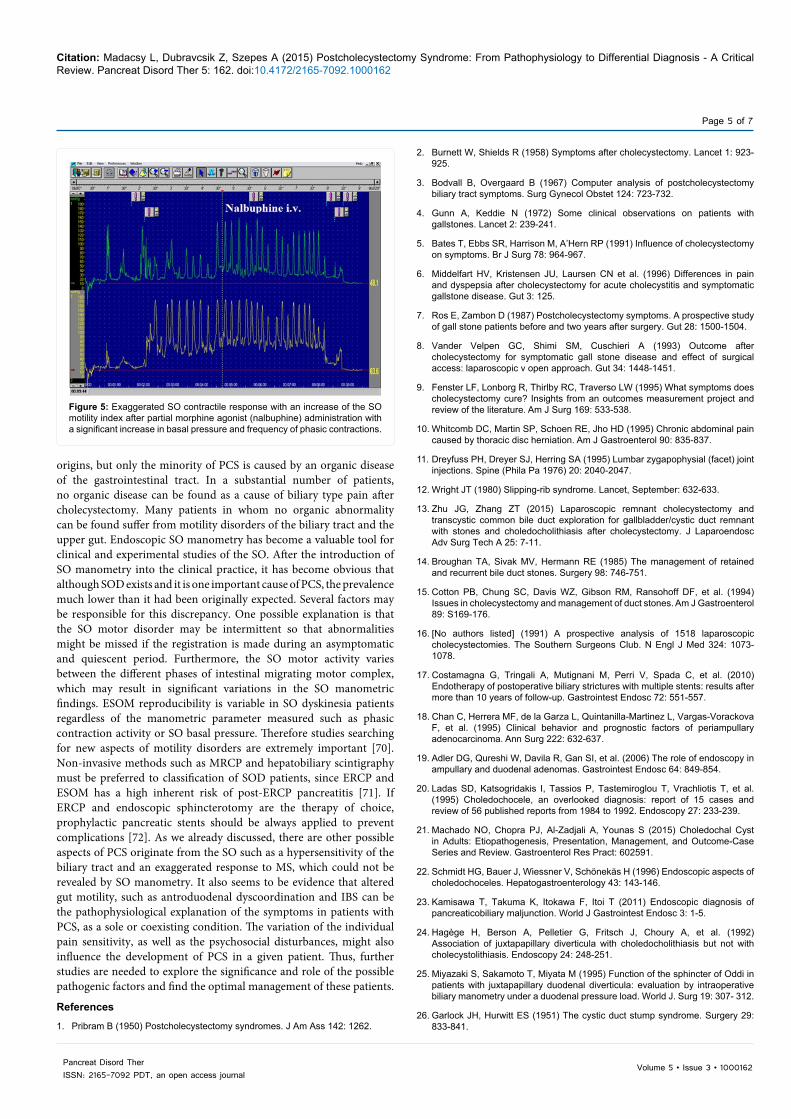
action of the CCK on the SO can be the consequence of denervation of the SO and cholecystectomy might be responsible for this, by directly dissecting the cholecysto-sphincteric nerve bundles. Although in a few patients with PCS, the reproduction of biliary pain was also observed at the time of paradoxical SO spasm during CCK administration, the frequency of this phenomenon is rather low for being an exclusive pathomechanism or a reliable provocation test [56]. There are early observations, which demonstrated that experimental distension of the common bile duct induces localized RUQ or epigastrial pain accompanied by nausea and vomiting [57]. Many patients with PCS have experienced the reproduction of biliary-type pain upon the injection of a small amount of contrast material into the common bile duct during diagnostic ERCP, thought to be caused by hypersensitivity of the biliary tract. Sometimes the cannulation of the papilla of Vater can be painful. ERCP filling pain proved to be highly reproducible regardless whether contrast material or physiologic saline has been injected. It was also claimed, that the pain had vanished after the aspiration of the contrast material. Simultaneous manometric measurements in the common bile duct revealed a marked pressure rise at the time of the evoked pain [58]. Unfortunately, controlled studies with endoscopic sphincter of Oddi manometry failed to show any correlation between positive ERCP filling pain test and manometric diagnosis of SOD [59]. Although pain during ERCP proved to have no diagnostic value for SOD, it can be regarded as a good indicator of an oversensitive biliary tract, which has a low compliance to volume changes and might act as a pain trigger zone. Although the presence of opioid receptors in the SO area is confirmed by immuno- histochemical studies, but their exact physiological role has not been established yet [60]. It is a well-known fact that morphine sulfate (MS) has a stimulatory effect on the human SO. Very small doses of MS (2.5 ug/kg) increases the frequency and the amplitude of phasic contractions, whereas larger doses can cause SO spasm with a substantial elevation of the SO basal pressure. The effect of MS can be antagonized by naloxone, but not by atropine [61]. This phenomenon suggests a direct smooth muscle stimulatory effect of MS by facilitating the Ca++ influx through the voltage dependent or receptor operated calcium channels. In patients with PCS, a marked rise in CBD pressure was demonstrated using endoscopic manometry during MS administration, which was associated with biliary pain [62]. Provocation tests based on MS administration, such as the Debray and the Nardi test have been used for several years in the evaluation of patients with SO dyskinesia [63,64], but the diagnostic usefulness of these tests has been criticized because of their low specificity and sensitivity [65]. In those patients with biliary pain and prolonged SO spasm due to the MS administration, a significant elevation of the CBD pressure and the stoppage of the bile flow over the SO can be demonstrated using intraoperative and endoscopic SO manometry and quantitative hepatobiliary scintigraphy. Therefore, MS augmentation of these diagnostic tests may improve the sensitivity and specificity of SO provocation tests [66,67] (Figure 5). It has been also proved, that the frequencies of abnormal SO manometric results were significantly higher in those patients with positive Nardi provocation test [68]. Finally, it can be hypothesized, that there is a subgroup of patients with PCS, who have exaggerated SO contractile response after MS administration, which is not always accompanied by basal motility disturbances on the SO manometry. Therefore, it seems likely, that factors influencing motor responses to morphine provocation tests are not related either to those which determine the basal SO motility or the responses to intravenous CCK [69].
ConclusionsObviously, PCS symptoms could have several pathophysiological
Figure 4: Reduction of elevated SO basal pressure after amyl nitrite administration on ESOM in a patient with SOD biliary type II.
Citation: Madacsy L, Dubravcsik Z, Szepes A (2015) Postcholecystectomy Syndrome: From Pathophysiology to Differential Diagnosis - A Critical Review. Pancreat Disord Ther 5: 162. doi:10.4172/2165-7092.1000162
Page 5 of 7
Volume 5 • Issue 3 • 1000162Pancreat Disord TherISSN: 2165-7092 PDT, an open access journal
origins, but only the minority of PCS is caused by an organic disease of the gastrointestinal tract. In a substantial number of patients, no organic disease can be found as a cause of biliary type pain after cholecystectomy. Many patients in whom no organic abnormality can be found suffer from motility disorders of the biliary tract and the upper gut. Endoscopic SO manometry has become a valuable tool for clinical and experimental studies of the SO. After the introduction of SO manometry into the clinical practice, it has become obvious that although SOD exists and it is one important cause of PCS, the prevalence much lower than it had been originally expected. Several factors may be responsible for this discrepancy. One possible explanation is that the SO motor disorder may be intermittent so that abnormalities might be missed if the registration is made during an asymptomatic and quiescent period. Furthermore, the SO motor activity varies between the different phases of intestinal migrating motor complex, which may result in significant variations in the SO manometric findings. ESOM reproducibility is variable in SO dyskinesia patients regardless of the manometric parameter measured such as phasic contraction activity or SO basal pressure. Therefore studies searching for new aspects of motility disorders are extremely important [70]. Non-invasive methods such as MRCP and hepatobiliary scintigraphy must be preferred to classification of SOD patients, since ERCP and ESOM has a high inherent risk of post-ERCP pancreatitis [71]. If ERCP and endoscopic sphincterotomy are the therapy of choice, prophylactic pancreatic stents should be always applied to prevent complications [72]. As we already discussed, there are other possible aspects of PCS originate from the SO such as a hypersensitivity of the biliary tract and an exaggerated response to MS, which could not be revealed by SO manometry. It also seems to be evidence that altered gut motility, such as antroduodenal dyscoordination and IBS can be the pathophysiological explanation of the symptoms in patients with PCS, as a sole or coexisting condition. The variation of the individual pain sensitivity, as well as the psychosocial disturbances, might also influence the development of PCS in a given patient. Thus, further studies are needed to explore the significance and role of the possible pathogenic factors and find the optimal management of these patients.
References
1. Pribram B (1950) Postcholecystectomy syndromes. J Am Ass 142: 1262.
2. Burnett W, Shields R (1958) Symptoms after cholecystectomy. Lancet 1: 923-925.
3. Bodvall B, Overgaard B (1967) Computer analysis of postcholecystectomy biliary tract symptoms. Surg Gynecol Obstet 124: 723-732.
4. Gunn A, Keddie N (1972) Some clinical observations on patients with gallstones. Lancet 2: 239-241.
5. Bates T, Ebbs SR, Harrison M, A’Hern RP (1991) Influence of cholecystectomy on symptoms. Br J Surg 78: 964-967.
6. Middelfart HV, Kristensen JU, Laursen CN et al. (1996) Differences in pain and dyspepsia after cholecystectomy for acute cholecystitis and symptomatic gallstone disease. Gut 3: 125.
7. Ros E, Zambon D (1987) Postcholecystectomy symptoms. A prospective study of gall stone patients before and two years after surgery. Gut 28: 1500-1504.
8. Vander Velpen GC, Shimi SM, Cuschieri A (1993) Outcome after cholecystectomy for symptomatic gall stone disease and effect of surgical access: laparoscopic v open approach. Gut 34: 1448-1451.
9. Fenster LF, Lonborg R, Thirlby RC, Traverso LW (1995) What symptoms does cholecystectomy cure? Insights from an outcomes measurement project and review of the literature. Am J Surg 169: 533-538.
10. Whitcomb DC, Martin SP, Schoen RE, Jho HD (1995) Chronic abdominal pain caused by thoracic disc herniation. Am J Gastroenterol 90: 835-837.
11. Dreyfuss PH, Dreyer SJ, Herring SA (1995) Lumbar zygapophysial (facet) joint injections. Spine (Phila Pa 1976) 20: 2040-2047.
12. Wright JT (1980) Slipping-rib syndrome. Lancet, September: 632-633.
13. Zhu JG, Zhang ZT (2015) Laparoscopic remnant cholecystectomy and transcystic common bile duct exploration for gallbladder/cystic duct remnant with stones and choledocholithiasis after cholecystectomy. J Laparoendosc Adv Surg Tech A 25: 7-11.
14. Broughan TA, Sivak MV, Hermann RE (1985) The management of retained and recurrent bile duct stones. Surgery 98: 746-751.
15. Cotton PB, Chung SC, Davis WZ, Gibson RM, Ransohoff DF, et al. (1994) Issues in cholecystectomy and management of duct stones. Am J Gastroenterol 89: S169-176.
16. [No authors listed] (1991) A prospective analysis of 1518 laparoscopic cholecystectomies. The Southern Surgeons Club. N Engl J Med 324: 1073-1078.
17. Costamagna G, Tringali A, Mutignani M, Perri V, Spada C, et al. (2010) Endotherapy of postoperative biliary strictures with multiple stents: results after more than 10 years of follow-up. Gastrointest Endosc 72: 551-557.
18. Chan C, Herrera MF, de la Garza L, Quintanilla-Martinez L, Vargas-Vorackova F, et al. (1995) Clinical behavior and prognostic factors of periampullary adenocarcinoma. Ann Surg 222: 632-637.
19. Adler DG, Qureshi W, Davila R, Gan SI, et al. (2006) The role of endoscopy in ampullary and duodenal adenomas. Gastrointest Endosc 64: 849-854.
20. Ladas SD, Katsogridakis I, Tassios P, Tastemiroglou T, Vrachliotis T, et al. (1995) Choledochocele, an overlooked diagnosis: report of 15 cases and review of 56 published reports from 1984 to 1992. Endoscopy 27: 233-239.
21. Machado NO, Chopra PJ, Al-Zadjali A, Younas S (2015) Choledochal Cyst in Adults: Etiopathogenesis, Presentation, Management, and Outcome-Case Series and Review. Gastroenterol Res Pract: 602591.
22. Schmidt HG, Bauer J, Wiessner V, Schönekäs H (1996) Endoscopic aspects of choledochoceles. Hepatogastroenterology 43: 143-146.
23. Kamisawa T, Takuma K, Itokawa F, Itoi T (2011) Endoscopic diagnosis of pancreaticobiliary maljunction. World J Gastrointest Endosc 3: 1-5.
24. Hagège H, Berson A, Pelletier G, Fritsch J, Choury A, et al. (1992) Association of juxtapapillary diverticula with choledocholithiasis but not with cholecystolithiasis. Endoscopy 24: 248-251.
25. Miyazaki S, Sakamoto T, Miyata M (1995) Function of the sphincter of Oddi in patients with juxtapapillary duodenal diverticula: evaluation by intraoperative biliary manometry under a duodenal pressure load. World J. Surg 19: 307- 312.
26. Garlock JH, Hurwitt ES (1951) The cystic duct stump syndrome. Surgery 29: 833-841.
Figure 5: Exaggerated SO contractile response with an increase of the SO motility index after partial morphine agonist (nalbuphine) administration with a significant increase in basal pressure and frequency of phasic contractions.

Citation: Madacsy L, Dubravcsik Z, Szepes A (2015) Postcholecystectomy Syndrome: From Pathophysiology to Differential Diagnosis - A Critical Review. Pancreat Disord Ther 5: 162. doi:10.4172/2165-7092.1000162
Page 6 of 7
Volume 5 • Issue 3 • 1000162Pancreat Disord TherISSN: 2165-7092 PDT, an open access journal
27. Glenn F, Whitsell Jc (1961) The surgical treatment of cystic duct remnants. Surg Gynecol Obstet 113: 711-719.
28. Bodvall B, Overgaard B (1966) Cystic duct remnant after cholecystectomy: incidence studied by cholegraphy in 500 cases, and significance in 103 reoperations. Ann Surg 163: 382-390.
29. Evans PR, Dowsett JF, Bak YT, Chan YK, Kellow JE (1995) Abnormal sphincter of Oddi response to cholecystokinin in postcholecystectomy syndrome patients with irritable bowel syndrome. The irritable sphincter. Dig Dis Sci 40: 1149-1156.
30. Perdikis G, Wilson P, Hinder R, Redmond E, Wetscher G, et al. (1994) Altered antroduodenal motility after cholecystectomy. Am J Surg 168: 609-614.
31. Koussayer T, Ducker TE, Clench MH, Mathias JR (1995) Ampulla of Vater/duodenal wall spasm diagnosed by antroduodenal manometry. Dig Dis Sci 40: 1710-1719.
32. Wilson P, Jamieson JR, Hinder RA, Anselmino M, Perdikis G, et al. (1995) Pathologic duodenogastric reflux associated with persistence of symptoms after cholecystectomy. Surgery 117: 421-428.
33. Farsakh NA, Roweily E, Steitieh M, Butchoun R, Khalil B (1995) Prevalence of Helicobacter pylori in patients with gall stones before and after cholecystectomy: a longitudinal study. Gut 36: 675-678.
34. Harvey RF, Read AE (1973) Effect of cholecystokinin on colonic motility and symptoms in patients with the irritable-bowel syndrome. Lancet 1: 1-3.
35. Drossman DA, Funch-Jensen P (1992) Functional dyspepsia. Psychosocial factors in functional dyspepsia. Eur J Gastroenetrol Hepatol 4: 602-607.
36. Kurucsai G, Joó I, Fejes R, Székely A, Székely I, et al. (2008) Somatosensory hypersensitivity in the referred pain area in patients with chronic biliary pain and a sphincter of Oddi dysfunction: new aspects of an almost forgotten pathogenetic mechanism. Am J Gastroenterol 103: 2717-2725.
37. Bar-Meir S, Halpern Z, Bardan E, Gilat T (1984) Frequency of papillary dysfunction among cholecystectomized patients. Hepatology 4: 328-330.
38. Hogan WJ, Geenen JE, Dodds WJ (1987) Dysmotility disturbances of the biliary tract: classification, diagnosis, and treatment. Semin Liver Dis 7: 302-310.
39. Varró V, Lonovics J (1989) Sphincter of Oddi dyskinesia: pathology and clinical aspects. In Bianchi Porro G., ed.: Topics in digestive disease. New York: Raven Press 357-383.
40. Sherman S, Troiano FP, Hawes RH, O’Connor KW, Lehman GA (1991) Frequency of abnormal sphincter of Oddi manometry compared with the clinical suspicion of sphincter of Oddi dysfunction. Am J Gastroenterol 86: 586-590.
41. Madácsy L, Fejes R, Kurucsai G, Joó I, Székely A, et al. (2006) Characterization of functional biliary pain and dyspeptic symptoms in patients with sphincter of Oddi dysfunction: effect of papillotomy. World J Gastroenterol 12: 6850-6856.
42. Csendes A, Kruse A, Funch-Jensen P, Oster MJ, Ornsholt J, et al. (1979) Pressure measurements in the biliary and pancreatic duct systems in controls and in patients with gallstones, previous cholecystectomy, or common bile duct stones. Gastroenterology 77: 1203-1210.
43. Geenen JE, Hogan WJ, Dodds WJ, Stewart ET, Arndorfer RC (1980) Intraluminal pressure recording from the human sphincter of Oddi. Gastroenterology 78: 317-324.
44. Toouli J, Roberts-Thomson IC, Dent J, Lee J (1985) Manometric disorders in patients with suspected sphincter of Oddi dysfunction. Gastroenterology 88: 1243-1250.
45. Madácsy L, Middelfart HV, Matzen P, Hojgaard L, Funch-Jensen P (2000) Quantitative hepatobiliary scintigraphy and endoscopic sphincter of Oddi manometry in patients with suspected sphincter of Oddi dysfunction: assessment of flow-pressure relationship in the biliary tract. Eur J Gastroenterol Hepatol 12: 777-86.
46. Thune A, Scicchitano J, Roberts-Thomson I, Toouli J (1991) Reproducibility of endoscopic sphincter of Oddi manometry. Dig Dis Sci 36: 1401-1405.
47. Geenen JE, Hogan WJ, Dodds WJ, Toouli J, Venu RP (1989) The efficacy of endoscopic sphincterotomy after cholecystectomy in patients with sphincter-of-Oddi dysfunction. N Engl J Med 320: 82-87.
48. Cotton PB, Durkalski V, Romagnuolo J, Pauls Q, Fogel E, et al. (2014) Effect of endoscopic sphincterotomy for suspected sphincter of Oddi dysfunction on pain-related disability following cholecystectomy: the EPISOD randomized clinical trial. JAMA 311: 2101-2109.
49. Bertalan V, Madácsy L, Pávics L, Lonovics J (2004) Scintigraphic sign of functional biliary obstruction is pathognomic for sphincter of Oddi dysfunction. Hepatogastroenterology 51: 76-81.
50. Szepes A, Dubravcsik Z, Madácsy L (2013) [The effect of endoscopic sphincterotomy on the motility of the gallbladder and of the sphincter of Oddi in patients with acalculous biliary pain syndrome]. Orv Hetil 154: 306-313.
51. Bartha I, Carlson JM, Brumme CJ, McLaren PJ, Brumme ZL, et al. (2013) A genome-to-genome analysis of associations between human genetic variation, HIV-1 sequence diversity, and viral control. Elife 2: e01123.
52. Tanaka M, Ikeda S, Nakayama F (1984) Change in bile duct pressure responses after cholecystectomy: a loss of gallbladder as a pressure reservoir. Gastroenterology 87: 1154-1159.
53. Toouli J, Hogan WJ, Geenen JE, Dodds WJ, Arndorfer RC (1982) Action of cholecystokinin-octapeptide on sphincter of Oddi basal pressure and phasic wave activity in humans. Surgery 92: 497-503.
54. Hogan WJ, Geenen J, Dodds W (1982) Paradoxical motor response to cholecystokinin (CCK -OP) in patients with suspected sphincter of Oddi dysfunction. Gastroenterology 82: 1085.
55. Rolny P, Arlebäck A, Funch-Jensen P, Kruse A, Ravnsbaeck J, et al. (1986) Paradoxical response of sphincter of Oddi to intravenous injection of cholecystokinin or ceruletide. Manometric findings and results of treatment in biliary dyskinesia. Gut 27: 1507-1511.
56. Zollinger R (1933) Observations following distension of the gallbladder and common bile duct in man. Proc. Soc. Exper. Biol. & Med 30: 1260-1261.
57. Lasson A, Fork FT, Trägårdh B, Zederfeldt B (1988) The postcholecystectomy syndrome: bile ducts as pain trigger zone. Scand J Gastroenterol 23: 265-271.
58. Schmalz MJ, Geenen JE, Hogan WJ, Dodds WJ, Venu RP, et al. (1990) Pain on common bile duct injection during ERCP: does it indicate sphincter of Oddi dysfunction? Gastrointest Endosc 36: 458-461.
59. Venu R, Toouli J, Geenen JE (1983) Effect of morphine on motor activity of the human spjhincter of Oddi. Gastroenterology 84: 1342.
60. Toouli J, Collinson T, Bushell M (1983) Effect of morphine on sphincter of Oddi motility. Gastroenterology 86: 5-12.
61. Tanaka M, Ikeda S, Nakayama F (1983) Continous measurement of coomon bile duct pressure with an indwelling microtransducer catheter introduced by duodenoscopy: new diagnostic aid for postcholecystectomy diskinesia - a preliminary report. Gastrointest Endosc 29: 83-88.
62. Debray PC, Hardouin JP, Fablet J (1962) Le test choleretique-morphine. Son enteret dans les affections des voies biliaries et dans les migraines. Gastroenterologia 97: 137-148.
63. Nardi GL, Acosta JM (1966) Papillitis as a cause of pancreatitis and abdominal pain: role of evocative test, operative pancreatography and histologic evaluation. Ann Surg 164: 611-621.
64. Steinberg WM, Salvato RF, Toskes PP (1980) The morphine-prostigmine provocative test - is it useful for making clinical decisions? Gastroenterol 78: 728-731.
65. Madura JA, McCammon RL, Paris JM, Jesseph JE (1981) The Nardi test and biliary manometry in the diagnosis of pancreaticobiliary sphincter dysfunction. Surgery 90: 588-595.
66. Madácsy L, Velösy B, Lonovics J, Csernay L (1995) Evaluation of results of the prostigmine-morphine test with quantitative hepatobiliary scintigraphy: a new method for the diagnosis of sphincter of Oddi dyskinesia. Eur J Nucl Med 22: 227-232.
67. Roberts-Thomson IC, Pannall PR, Toouli J (1989) Relationship between morphine responses and sphincter of Oddi motility in undefined biliary pain after cholecystectomy. J Gastroenterol Hepatol 4: 317-324.
68. Madácsy L, Bertalan V, Szepes A, Lonovics J (2003) Effect of nalbuphine on the motility of the sphincter of Oddi in patients with suspected sphincter of Oddi dysfunction. Gastrointest Endosc 57: 319-323.
69. Kunwald P, Drewes AM, Kjaer D, Gravesen FH, McMahon BP, et al. (2010) A new distensibility technique to measure sphincter of Oddi function. Neurogastroenterol Motil 22: 978-983.
70. Yaghoobi M, Pauls Q, Durkalski V, Romagnuolo J, Fogel EL, et al. (2015) Incidence and predictors of post-ERCP pancreatitis in patients with suspected sphincter of Oddi dysfunction undergoing biliary or dual sphincterotomy: results

Citation: Madacsy L, Dubravcsik Z, Szepes A (2015) Postcholecystectomy Syndrome: From Pathophysiology to Differential Diagnosis - A Critical Review. Pancreat Disord Ther 5: 162. doi:10.4172/2165-7092.1000162
Page 7 of 7
Volume 5 • Issue 3 • 1000162Pancreat Disord TherISSN: 2165-7092 PDT, an open access journal
from the EPISOD prospective multicenter randomized sham-controlled study. Endoscopy 47: 884-890.
71. Madácsy L, Kurucsai G, Fejes R, Székely A, Székely I (2009) Prophylactic
pancreas stenting followed by needle-knife fistulotomy in patients with sphincter of Oddi dysfunction and difficult cannulation: new method to prevent post - ERCP pancreatitis. Dig Endosc: 8-13.