PLEASE SCROLL DOWN FOR ARTICLE - UPMC · a Laboratoire de Psychologie et Neurosciences Cognitives...
18
PLEASE SCROLL DOWN FOR ARTICLE This article was downloaded by: [Plaza, Monique] On: 16 March 2009 Access details: Access Details: [subscription number 909314491] Publisher Psychology Press Informa Ltd Registered in England and Wales Registered Number: 1072954 Registered office: Mortimer House, 37-41 Mortimer Street, London W1T 3JH, UK Neurocase Publication details, including instructions for authors and subscription information: http://www.informaworld.com/smpp/title~content=t713658146 Speaking without Broca's area after tumor resection Monique Plaza a ; Peggy Gatignol a ; Marianne Leroy a ; Hugues Duffau a a Laboratoire de Psychologie et Neurosciences Cognitives (UMR CNRS 8189), Université Paris Descartes, Service de Neurochirurgie, CHU Gui de Chauliac, Montpellier, France First Published on: 09 March 2009 To cite this Article Plaza, Monique, Gatignol, Peggy, Leroy, Marianne and Duffau, Hugues(2009)'Speaking without Broca's area after tumor resection',Neurocase, To link to this Article: DOI: 10.1080/13554790902729473 URL: http://dx.doi.org/10.1080/13554790902729473 Full terms and conditions of use: http://www.informaworld.com/terms-and-conditions-of-access.pdf This article may be used for research, teaching and private study purposes. Any substantial or systematic reproduction, re-distribution, re-selling, loan or sub-licensing, systematic supply or distribution in any form to anyone is expressly forbidden. The publisher does not give any warranty express or implied or make any representation that the contents will be complete or accurate or up to date. The accuracy of any instructions, formulae and drug doses should be independently verified with primary sources. The publisher shall not be liable for any loss, actions, claims, proceedings, demand or costs or damages whatsoever or howsoever caused arising directly or indirectly in connection with or arising out of the use of this material.
Transcript of PLEASE SCROLL DOWN FOR ARTICLE - UPMC · a Laboratoire de Psychologie et Neurosciences Cognitives...

PLEASE SCROLL DOWN FOR ARTICLE
This article was downloaded by: [Plaza, Monique]On: 16 March 2009Access details: Access Details: [subscription number 909314491]Publisher Psychology PressInforma Ltd Registered in England and Wales Registered Number: 1072954 Registered office: Mortimer House,37-41 Mortimer Street, London W1T 3JH, UK
NeurocasePublication details, including instructions for authors and subscription information:http://www.informaworld.com/smpp/title~content=t713658146
Speaking without Broca's area after tumor resectionMonique Plaza a; Peggy Gatignol a; Marianne Leroy a; Hugues Duffau a
a Laboratoire de Psychologie et Neurosciences Cognitives (UMR CNRS 8189), Université Paris Descartes,Service de Neurochirurgie, CHU Gui de Chauliac, Montpellier, France
First Published on: 09 March 2009
To cite this Article Plaza, Monique, Gatignol, Peggy, Leroy, Marianne and Duffau, Hugues(2009)'Speaking without Broca's area aftertumor resection',Neurocase,
To link to this Article: DOI: 10.1080/13554790902729473
URL: http://dx.doi.org/10.1080/13554790902729473
Full terms and conditions of use: http://www.informaworld.com/terms-and-conditions-of-access.pdf
This article may be used for research, teaching and private study purposes. Any substantial orsystematic reproduction, re-distribution, re-selling, loan or sub-licensing, systematic supply ordistribution in any form to anyone is expressly forbidden.
The publisher does not give any warranty express or implied or make any representation that the contentswill be complete or accurate or up to date. The accuracy of any instructions, formulae and drug dosesshould be independently verified with primary sources. The publisher shall not be liable for any loss,actions, claims, proceedings, demand or costs or damages whatsoever or howsoever caused arising directlyor indirectly in connection with or arising out of the use of this material.

NEUROCASE2009, iFirst, 1–17
© 2009 Psychology Press, an imprint of the Taylor & Francis Group, an Informa business
http://www.psypress.com/neurocase DOI: 10.1080/13554790902729473
NNCS Speaking without Broca’s area after tumor resection
Broca’s Area Resection Monique Plaza, Peggy Gatignol, Marianne Leroy, and Hugues Duffau
Laboratoire de Psychologie et Neurosciences Cognitives (UMR CNRS 8189), Université Paris Descartes, Service de Neurochirurgie, CHU Gui de Chauliac, Montpellier, France
We present the case of a right-handed patient who received surgical treatment for a left frontal WHO grade IIglioma invading the left inferior and middle frontal gyri, the head of the caudate nucleus, the anterior limb of theinternal capsule and the anterior insula, in direct contact also with the anterior-superior part of the lentiformnucleus. The tumor resection was guided by direct electrical stimulation on brain areas, while the patient wasawake. Adding a narrative production task to the neuropsychological assessment, we compared pre-, peri- andpost-surgical language skills in order to analyze the effects of the tumor infiltration and the consequences of theleft IFG resection, an area known to be involved in various language and cognitive processes. We showed that thetumor infiltration and its resection did not lead to the severe impairments predicted by the localization modelsassigning a significant role in language processing to the left frontal lobe, notably Broca’s area. We showed thatslow tumor evolution – the patient had been symptom-free for a long time – enabled compensatory mechanismsto process most language functions endangered by the tumor infiltration. However, a subtle fragility was observedin two language devices, i.e., reported speech and relative clauses, related to minor working memory deficits. Thiscase study of a patient speaking without Broca’s area illustrates the efficiency of brain plasticity, and shows thenecessity to broaden pre-, peri-, post-surgery language and cognitive assessments.
Keywords: Language; Inferior frontal gyrus; Working memory; Plasticity; Tumor; Neurosurgery.
INTRODUCTION
Historically, language has been considered aslocalized in two major brain areas, Broca’santerior frontal area – for production – andWernicke’s posterior temporal area – for comprehen-sion. The Lichtheim–Geshwind model (Geshwind,1967; Lichtheim, 1885) was based on clinicalinformation from aphasia consecutive to suddentrauma or vascular stroke, and anatomicalinformation from post-mortem observations. Assevere language impairments were observed incases of temporal and left frontal lesions, espe-cially when Broca’s and Wernicke’s areas wereinvolved, the first functional model of languageadvocated a temporal frontal network. Today,
the complex networks underlying language can beexplored through neuroimaging techniques basedon fMRI and diffusion tensor tractography,bringing support to less static and more connec-tionist approaches. Thus, areas originally thoughtto be specifically specialized for language havebeen shown to be involved in different cognitiveand perceptual processing not necessarily relatedto language, and speech does not exclusivelyemanate from the Wernicke-Broca’s languagenetwork (Démonet, Thierry, & Cardebat, 2005;Etard et al., 2000; Ffytche & Catani, 2005;Vigneau et al., 2006).
Neuropsychological studies on the conse-quences of acquired brain lesions in children andadults have suggested that the classical aphasia
Address correspondence to Monique Plaza, PhD, Laboratoire de Psychologie et Neurosciences Cognitives (UMR CNRS 8189),Université Paris Descartes, 71 Avenue Edouard Vaillant, 92773 Boulogne-Billancourt cedex, France. (E-mail: [email protected]).
Downloaded By: [Plaza, Monique] At: 16:15 16 March 2009

2 PLAZA ET AL.
model shows great variability in outcomes,according to age, etiology, lesion site, lesion sizeand initial impairment severity (e.g., Anderson,Morse, Catroppa, Haritou, & Rosenfeld, 2004;Anderson, Catroppa, Morse, Haritou, & Rosen-feld, 2005; Aram, 1999; Bates et al., 2001; Clark,Manes, Anotun, Sahabian, & Robbins, 2003;Coelho, Max, & Tranel, 2005; Damasio &Damasio, 1989). While sudden lesions affectingspecialized areas often result in severe aphasia,the clinical pattern is different in the case ofslow-growing lesions such as Low GradeGliomas (LGG). Here slow tumor evolutionallows for compensatory mechanisms todevelop, with the brain recruiting other areas forthe processing of the endangered functions, i.e.,perilesional and/or controlateral homologousregions (Belin et al., 1996; Blasi et al., 2002;Crosson et al., 2005; Desmurget, Bonnetblanc, &Duffau, 2006; Léger et al., 2002; Musso et al.,1999). Neurosurgical resection under electricalstimulation of WHO grade II Glioma highlightsuch processes which can be directly observedwithin cortical and sub-cortical areas (Bonnetblanc,Desmurget, & Duffau, 2006; Duffau, 2005, 2006).This surgery technique adds useful data to thoseobtained through functional imaging techniques,enriching neuropsychological case and groupstudies (e.g., Plaza, Gatignol, Cohen, Berger, &Duffau, 2007).
In the case presented here, FV, a right-handedadult aged 27, received surgical treatment for aWHO grade II Glioma (LGG). The tumorinvaded the left inferior and middle frontal gyri,the head of the caudate nucleus, the anterior limbof the internal capsule and the anterior insula. Thetumor was also in direct contact with the anterior-superior part of the lentiform nucleus. The surgi-cal resection concerned the pars triangularis, thepars orbitalis and the anterior part of the parsopercularis, i.e., Broca’s area (see below for clini-cal details). The clinical and theoretical objectiveof this case study was to analyze the effects of thetumor infiltration and the consequences of the leftinferior frontal gyrus resection since this area isknown to be activated by various linguistic, cogni-tive and sensory-motor tasks (Bonnetblanc et al.,2006; Bookheimer, 2002; Devlin, Matthews, &Rushworth, 2003; Greewe et al., 2006; Grodzinsky,2006; Heim et al., 2005; Koechlin & Jubault, 2006;Musso et al., 1999; Smith & Jonides, 1999;Tettamanti et al., 2002; Tettamanti & Wenninger,2006; Voets et al., 2006). Research on brain-damaged
adults and children has shown that narrative compe-tence is particularly sensitive to disruptions infrontal attention processes, causing deficits incomplex language assembly procedures and inexecutive function (e.g., Alexander, 2006; Bateset al., 2001; Chapman et al., 1992; Liles, 1993;Plaza, 1998; Plaza, Guitton, & Le Normand, 1998;Reilly, Bates, & Marchman, 1998; Reilly, Losh,Bellugi, & Wulfeck, 2004). A narrative productiontask was thus added to the classical neuropsycho-logical assessment to better evaluate thepatient’s linguistic profile. Narratives provide aquasi-naturalistic measure of spontaneous lan-guage as well as a relevant context for compar-ing several aspects of discourse in typical andatypical populations. On the basis of longitudi-nal analysis of the patient’s performances, wehypothesized that (a) the tumor infiltration mayhave been compensated for by plasticity mecha-nisms and (b) the resection of the left IFG resec-tion would lead to subtle impairments, innarrative production more specifically. Finally,this case study raises the interesting question ofhow Broca’s area can be ‘replaced’ by otherstructures for its cognitive and language process-ing skills.
METHODS
The patient
FV was a 27-year-old right-handed man workingas a computer engineer. Right-handedness wasdocumented using the Edinburgh inventory ques-tionnaire (Oldfield, 1971). He had had wordretrieval difficulties for 4–5 years, in markedincrease for the past 2 years when he first consultedfor generalized seizures. The neurological examina-tion was normal, without either somosatosensoryor motor deficit, but the MRI revealed a tumorinvading the left frontal lobe. Though put on anti-epileptic medication, his partial seizures and lan-guage difficulties intensified. The patient thenaccepted to undergo surgical resection with electri-cal stimulation technique while awake. Elevenmonths elapsed between the first seizure and thesurgical tumor removal. After surgery, FV had 70speech therapy sessions for 6 months, trainingworking and episodic memory, verbal and nonverbal reasoning, flexibility and inhibition mecha-nisms. Speech therapy began 1 month aftersurgery.
Downloaded By: [Plaza, Monique] At: 16:15 16 March 2009

BROCA’S AREA RESECTION 3
Procedure
Neuroimaging
Pre- and postoperative anatomical MRI wereperformed to establish the exact lesion locationand the extent of the surgical cavity.
Surgical mapping
Intra-operative mapping was done under localanesthesia with direct electrical stimulation tech-nique already described by the authors (Vigneauet al., 2006). Sensory-motor and language func-tions were assessed. First the patient was asked tocount repetitively from 1 to 10 in order to identifythe areas essential to speech production, namelythose indicating complete anarthria when stimu-lated. Second, the DO 80 picture naming test(Metz-Lutz et al., 1991) was used to detect anomiaand naming impairment. Each site was tested threetimes, three trials being enough to establishwhether a cortical site is essential for a particularcognitive function (Ojemann, Ojemann, Lettich, &Berger, 1989). To avoid seizures, the same corticalsite was never stimulated twice in a row. To ensuresuccessful tumor removal while sparing functionalareas, the limits of the resection were progressivelyset so as to preserve functional pathways in theimmediate vicinity of the surgical cavity. Such aprocedure minimizes residual morbidity whileenhancing the quality of the resection — thusimproving patient survival by minimizing the ana-plastic transformation of WHO grade II glioma(Duffau, 2005).
Pre-operative neuropsychological assessment procedure
Ten days before surgery, FV was administeredan extensive neuropsychological assessmentincluding the following tests: WAIS-R 7-SF(Ward, 1990; Wechsler, 1981); Gröber and Buschke(Gröber, Buschke, Bang, & Dresner, 1988); Rey–Osterreith complex figure (Rey, 1959, 1970); BostonNaming Test (Kaplan, Goodglass, & Weintraub,1983); Verbal fluency (Cardebat, Doyon, Puel,Goulet, & Joanette, 1990); Chapman–Cook speed ofreading test; Weintraub and Mesulam CancellationTest (Weintraub & Mesulam, 1985); Geometric fig-ures and cube of BEC 96 (Signoret et al., 1988); TheHooper Visual Orientation Test (Hooper, 1958);Stroop Test [Modified version: Chatelois, Pineau,Belleville, & Peretz, 1993; Stroop, 1935); Wisconsin
Card Sorting Test (Berg, 1948; Heaton, Chelure,Talley, Kay, & Curtiss, 2002); Ruff Figural FluencyTest (Ruff, Light, & Evans, 1987); Corsi blocks(Milner, 1971); Ruff 2 and 7 (Ruff, 1992). Theneuropsychological assessment systematicallyincluded three language tests (Boston Naming Test,Chapman–Cook Naming Test and Verbal fluencyTest) allowing the neuropsychologist to correlateverbal and non verbal skills, i.e., ‘left’ and ‘right’hemispheric processing.
Longitudinal language assessment procedure
Language processes were assessed on the daybefore and after surgery with the following tasks:
1. the standardized Boston Diagnosis AphasiaExamination, BDAE (Goodglass & Caplan,1983), adapted in French by Mazaux andOrgogozo (1982), administered on the daybefore, as well as 5 days after surgery and3 months later;
2. the written narrative task of the BDAE, ‘Cook-ies’, which in one minute requires the subject towrite a short story from a picture representinga scene;
3. the Picture naming test DO 80 (Metz-Lutzet al., 1991) consisting of 80 black and whitedrawings of objects controlled for frequency,familiarity, age of acquisition and level of edu-cation, administered on the day before surgery,during surgery, as well as 5 days after surgeryand 3 months later;
4. the experimental narrative ask elicited by thewordless picture book Frog, where are you?(Mayer, 1969) used in previous studies by theauthors (Plaza, 1998; Plaza et al., 1998), andadministered on the day before surgery, 5 and12 days after surgery as well as 3 months later.This storybook represents a boy and his doglooking for their frog which escaped during thenight. While looking for the frog in the forest,the boy and his dog meet several animals thatinterfere with their search. In the end, the boyand his dog find the frog which is with a friendand several baby frogs. The story ends with theboy and his dog heading home with a babyfrog as their new companion. As he lookedthrough the pictures, the patient was asked totell the story in his own way so that anybodylistening to the registered story could under-stand it. The experimenter kept silent during
Downloaded By: [Plaza, Monique] At: 16:15 16 March 2009

4 PLAZA ET AL.
the session. All recordings were timed, regis-tered and transcribed orthographically usingthe convention of the CHAT system to facili-tate lexical analysis (different words used andtype/token ratio) by the CLAN computer pro-gram (MacWhinney & Snow, 1991). For eachtranscript, as we have done in previous data,overall story length, lexical, syntactic, cohe-sion, descriptive coherence and evaluativedevices were scored.
Overall story length was scored from the totalnumber of propositions used by the narrator.A proposition corresponding to a single event isdefined as composed of a verb and itsarguments.
Lexical analysis represented the number oftokens, different words, and the ratio of nouns andverbs compared to the total number of words.
Syntactic analysis scored three types ofclauses, i.e., sequential (e.g., ‘the boy sleeps inthe room’), subordinate and coordinate (e.g., ‘theboy and the dog saw that the frog escaped’, ‘thedog was afraid because he saw the bees flyingtowards him’, ‘the boy is looking into the holewhile the dog is smelling the flowers’, and rela-tive clauses (e.g., ‘the boy’s dog, who is lookingat the tree, suddenly falls’).
Cohesion referred to the 12 situations composingthe story, i.e., (1) it is night time; (2) the boy andthe dog are looking at the frog; (3) they are sleep-ing; (4) the frog is escaping; (5) they wake up anddiscover that the frog has escaped; (6) the dog isfalling through the window; (7) they are lookingfor the frog in the forest; (8) they meet bees; (9) anowl; (10) a reindeer; (11) they discover the frog’sfamily; and (12) they are heading home with ababy frog.
Descriptive coherence took into account the 65events suggested by the pictures.
Evaluative devices were composed of (a) reportedspeech (indirect, e.g., ‘he asks the dog to be quiet’and direct, e.g., ‘he asks the dog “be quiet”,please’), (b) negative qualifiers (e.g., ‘he did not seethe reindeer’), (c) causal connectors (since,because, as) and (d) frames of mind (desire, inten-tion, perception, emotion and knowledge). We cal-culated the ratio of each device compared to thetotal number of propositions.
Besides, a qualitative analysis was conducted onerrors or speech difficulties (e.g., paraphasia, inco-herence or planning disorder) in the patient’s andcontrol group’s productions.
Control group and statistical data
Since the narrative task is experimental and notstandardized yet, the patient’s narrative produc-tion was compared with that of 10 control subjectsmatched in age (26–29 years, mean age: 27; 3) andsocio-professional status (post-graduate studies).The means and standard deviations were com-puted for each measure in the control group, serv-ing as a basis to establish the patient’s z scores.
RESULTS
Intraoperative functional data
After the incision of the dura matter and the identi-fication of the tumor boundaries through ultra-sonography, electrical functional mapping wasperformed on the awake patient. Several significantcortical sites were detected with stimulation (Figure 1)i.e., in 5, primary somatosensory finger area, induc-ing dysesthesia (retrocentral gyrus); in 12, primarymotor face area, inducing facial movements (pre-central gyrus); in 10 and 11, premotor ventral area,eliciting complete speech arrest (rolandic opercu-lum); in 13, dorsal premotor cortex, elicitingsemantic paraphasia. The inferior frontal gyrus wasremoved up to the anterior and middle part of theinsula, which was also partly removed withoutimpairing language processing (Figure 2). After theresection of the pars orbitalis (BA 47) and the parstriangularis (BA 45), the anterior half of the parsopercularis (BA 44) was also removed without elic-iting any language disorders. The cortical limit forthe resection was established according to the elo-quent sites previously identified by the stimulation.Sub-cortically, after opening the frontal horn of theventricle, a deep functional boundary was set by thehead of the caudate nucleus, in 41, where the stimu-lation induced persevering responses. Next to thislandmark, the pathway coming from the premotorventral cortex, in 42 was detected with anarthriainduced by the stimulation, and the resection wentfurther, in the fibers composing the posterior wall ofthe surgical cavity. As to the deep postero-superiorboundary of the resection, the limit was set at theinferior occipito-frontal fasciculus, connected to thedorso-lateral prefrontal cortex, in 40, where thestimulation elicited semantic paraphasia. The ante-rior frontopolar cortex was completely removed.Thus, the resection stopped within functionalboundaries, both at cortical and sub-cortical levels.
Downloaded By: [Plaza, Monique] At: 16:15 16 March 2009

BROCA’S AREA RESECTION 5
Figure 1. (Preoperative MRI) reveals a left precentral glioma, invading the left inferior and middle frontal gyri, the head of the cau-date nucleus, the anterior limb of the internal capsule, the anterior insula and regions in contact with the anterior-superior part of thelentiform nucleus.
Figure 2. (At the beginning of the brain mapping) shows the sites directly involved in speech production as detected by electricalstimulation.
Downloaded By: [Plaza, Monique] At: 16:15 16 March 2009
6 PLAZA ET AL.
Pre- and post-neuropsychological data
Neuropsychological assessment before surgery (Table 1)
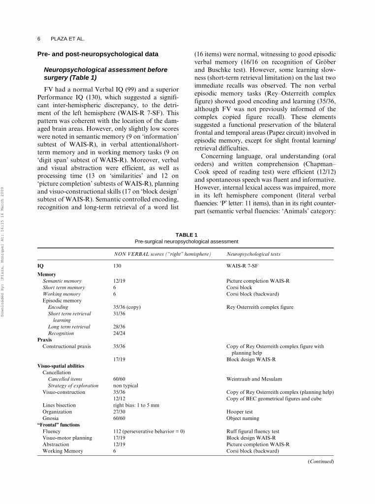
FV had a normal Verbal IQ (99) and a superiorPerformance IQ (130), which suggested a signifi-cant inter-hemispheric discrepancy, to the detri-ment of the left hemisphere (WAIS-R 7-SF). Thispattern was coherent with the location of the dam-aged brain areas. However, only slightly low scoreswere noted in semantic memory (9 on ‘information’subtest of WAIS-R), in verbal attentional/short-term memory and in working memory tasks (9 on‘digit span’ subtest of WAIS-R). Moreover, verbaland visual abstraction were efficient, as well asprocessing time (13 on ‘similarities’ and 12 on‘picture completion’ subtests of WAIS-R), planningand visuo-constructional skills (17 on ‘block design’subtest of WAIS-R). Semantic controlled encoding,recognition and long-term retrieval of a word list
(16 items) were normal, witnessing to good episodicverbal memory (16/16 on recognition of Gröberand Buschke test). However, some learning slow-ness (short-term retrieval limitation) on the last twoimmediate recalls was observed. The non verbalepisodic memory tasks (Rey–Osterreith complexfigure) showed good encoding and learning (35/36,although FV was not previously informed of thecomplex copied figure recall). These elementssuggested a functional preservation of the bilateralfrontal and temporal areas (Papez circuit) involved inepisodic memory, except for slight frontal learning/retrieval difficulties.
Concerning language, oral understanding (oralorders) and written comprehension (Chapman–Cook speed of reading test) were efficient (12/12)and spontaneous speech was fluent and informative.However, internal lexical access was impaired, morein its left hemisphere component (literal verbalfluencies: ‘P’ letter: 11 items), than in its right counter-part (semantic verbal fluencies: ‘Animals’ category:
TABLE 1 Pre-surgical neuropsychological assessment
NON VERBAL scores (“right” hemisphere) Neuropsychological tests
IQ 130 WAIS-R 7-SF
MemorySemantic memory 12/19 Picture completion WAIS-RShort term memory 6 Corsi blockWorking memory 6 Corsi block (backward)Episodic memory
Encoding 35/36 (copy) Rey Osterreith complex figureShort term retrieval
learning31/36
Long term retrieval 28/36Recognition 24/24
PraxisConstructional praxis 35/36 Copy of Rey Osterreith complex figure with
planning help17/19 Block design WAIS-R
Visuo-spatial abilitiesCancellation
Cancelled items 60/60 Weintraub and MesulamStrategy of exploration non typical
Visuo-construction 35/36 Copy of Rey Osterreith complex (planning help)12/12 Copy of BEC geometrical figures and cube
Lines bisection right bias: 1 to 5 mmOrganization 27/30 Hooper testGnosia 60/60 Object naming
“Frontal” functionsFluency 112 (perseverative behavior = 0) Ruff figural fluency testVisuo-motor planning 17/19 Block design WAIS-RAbstraction 12/19 Picture completion WAIS-RWorking Memory 6 Corsi block (backward)
(Continued)
Downloaded By: [Plaza, Monique] At: 16:15 16 March 2009

BROCA’S AREA RESECTION 7
21 items). Lexical access assessment showed ano-mias, semantic paraphasias, circumlocutions anddelayed responses (42/60 on the Boston NamingTest). Besides, FV reported lexical access troublesfor 4 or 5 years previous to surgery, aggravated thepast 2 years. A ‘surface’ dysgraphia during irregu-lar and low frequency word dictation was alsoobserved (14/21), whereas non-word reading andrepetition were preserved. These results suggested afrontal inferior and temporal superior perturba-tion in the left hemisphere.
Calculation abilities were efficient but slow (9 on‘arithmetic’ subtest of WAIS-R), due to verbalworking memory limitation (backward digit span).FV operated on a visual mode (he used a visualdiagram for solving mental arithmetic problems).Neither limb (significant, non significant, reflexive,non reflexive gestures), nor visuo-constructional(Rey–Osterreith complex figure copy withplanning help), melokinetic (keyboarding) praxistroubles, pathological psychomotor slowness(‘digit symbol’ subtest of WAIS-R), were observed
TABLE 1 (Continued)
VERBAL scores (“left” hemisphere) Neuropsychological tests
IQ 99 WAIS-R 7-SFMemory
Semantic memory 9/19 Information WAIS-RShort term memory 9/19 Digit Span WAIS-R
5 Digit SpanWorking memory 9/19 Digit Span WAIS-R
5 Digit Span (backward)Episodic memory
Encoding 16/16 Grober and BuschkeShort term retrieval
learning15 (11 + 4); 16 (12 + 4);
16 (13 + 3)/16Long term retrieval 16 (15 + 1)/16Recognition 16/16
LanguageLexical access
Spontaneous speech okNaming 42/60 Boston Naming Test
(4 semantic paraphasias; 7 anomias; 4 circumlocutions; 3 delayed responses)
Semantic fluency 21 Animal categoryLiteral fluency 11 (+2 perseverations) “P” letter
ComprehensionOral understanding ok OrdersWritten comprehension 12 Chapman-speed of reading test
TranscodageWord Reading 16/16 Self made listsNon-word reading 6/6Word dictation score 14/21Word dictation
phonological score21/21
Non-word dictation 6/6Non-word repetition 6/6
Calculation 9/19 Arithmetic WAIS-R2/2 Writing calculation on failed
“Frontal” functionsInhibition 89″; no error Stroop testFlexibility 93″; no error Stroop testFluency 21 Animals category
11 (+2 perseverations) “P” letterAbstraction 13/19 Similarities WAIS-RWorking Memory 5 Digit Span (backward)
Downloaded By: [Plaza, Monique] At: 16:15 16 March 2009
8 PLAZA ET AL.
suggesting parietal, premotor and motor func-tional preservation. Despite an atypical visualexploration strategy (Weintraub and MesulamCancellation Test), and a discrete bias to theright (1–5 ml, i.e., non pathological) during linebisection, no neglect, agnosia, visual organiza-tion (Hooper Test), visuo-constructional orplanning deficits (Rey complex figure) wereobserved. The neuro-visual assessment suggestedfunctional right hemisphere (post-rolandic)preservation.
In summary, the impairment was not of a heavyquantitative nature but involved light deficits inverbal fluency (‘P’ letter and ‘Animals’ category),auditory attentional and verbal working memory(‘digit span’ WAIS-R subtest), especially during themental arithmetic task (increased resolution timesin ‘arithmetic’ WAIS-R subtest). All the other‘executive’ skills were efficient (Stroop test;Wisconsin Card Sorting test; Ruff Figural FluencyTest; ‘Block design’, ‘Similarities’, ‘Pictures com-pletion’ subtests of WAIS-R; Backward Corsiblocks; Ruff 2 and 7).
Neuropsychological assessment after surgery
Generally, apart from difficult clinical situa-tions, the neuropsychological assessment onlyintervenes eight months to 1 year after surgery. FVhad no complaint after surgery and was not pre-occupied by his cognitive health.
BDAE, DO 80 on Day −1, Day +5, Day +3 months
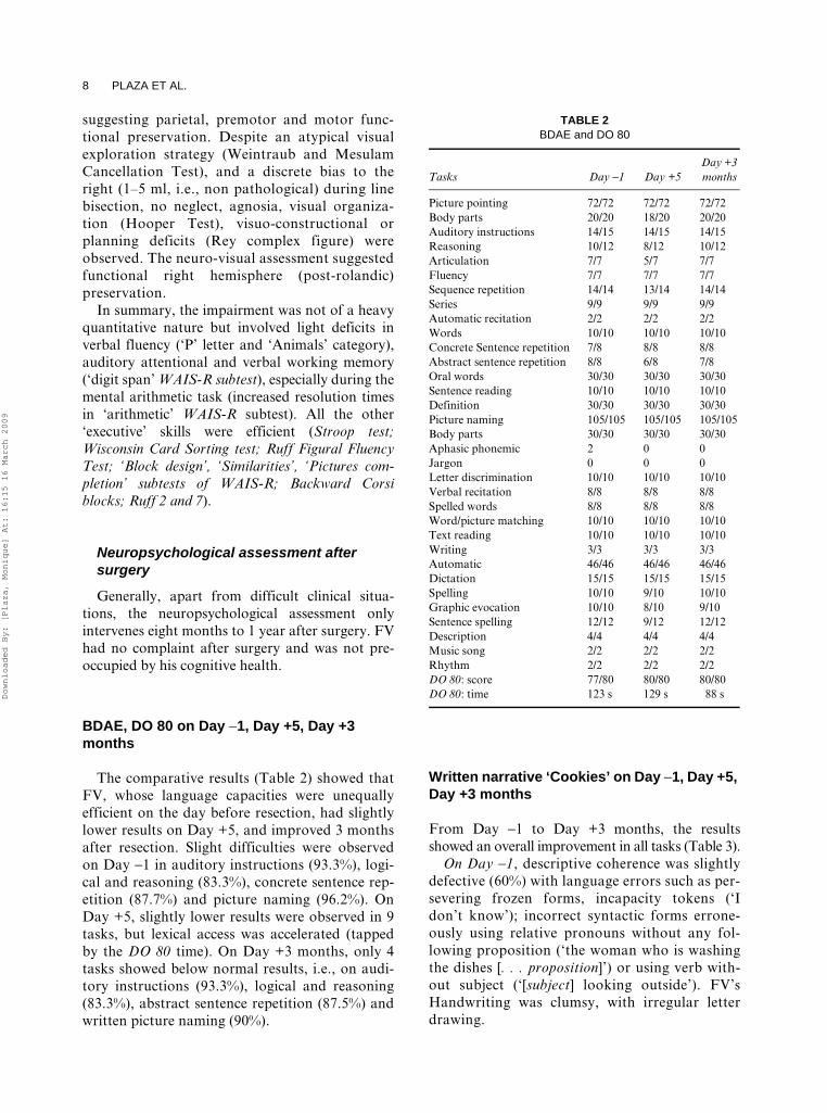
The comparative results (Table 2) showed thatFV, whose language capacities were unequallyefficient on the day before resection, had slightlylower results on Day +5, and improved 3 monthsafter resection. Slight difficulties were observedon Day −1 in auditory instructions (93.3%), logi-cal and reasoning (83.3%), concrete sentence rep-etition (87.7%) and picture naming (96.2%). OnDay +5, slightly lower results were observed in 9tasks, but lexical access was accelerated (tappedby the DO 80 time). On Day +3 months, only 4tasks showed below normal results, i.e., on audi-tory instructions (93.3%), logical and reasoning(83.3%), abstract sentence repetition (87.5%) andwritten picture naming (90%).
Written narrative ‘Cookies’ on Day −1, Day +5, Day +3 months
From Day −1 to Day +3 months, the resultsshowed an overall improvement in all tasks (Table 3).
On Day -1, descriptive coherence was slightlydefective (60%) with language errors such as per-severing frozen forms, incapacity tokens (‘Idon’t know’); incorrect syntactic forms errone-ously using relative pronouns without any fol-lowing proposition (‘the woman who is washingthe dishes [. . . proposition]’) or using verb with-out subject (‘[subject] looking outside’). FV’sHandwriting was clumsy, with irregular letterdrawing.
TABLE 2 BDAE and DO 80
Tasks Day -1 Day +5Day +3 months
Picture pointing 72/72 72/72 72/72Body parts 20/20 18/20 20/20Auditory instructions 14/15 14/15 14/15Reasoning 10/12 8/12 10/12Articulation 7/7 5/7 7/7Fluency 7/7 7/7 7/7Sequence repetition 14/14 13/14 14/14Series 9/9 9/9 9/9Automatic recitation 2/2 2/2 2/2Words 10/10 10/10 10/10Concrete Sentence repetition 7/8 8/8 8/8Abstract sentence repetition 8/8 6/8 7/8Oral words 30/30 30/30 30/30Sentence reading 10/10 10/10 10/10Definition 30/30 30/30 30/30Picture naming 105/105 105/105 105/105Body parts 30/30 30/30 30/30Aphasic phonemic 2 0 0Jargon 0 0 0Letter discrimination 10/10 10/10 10/10Verbal recitation 8/8 8/8 8/8Spelled words 8/8 8/8 8/8Word/picture matching 10/10 10/10 10/10Text reading 10/10 10/10 10/10Writing 3/3 3/3 3/3Automatic 46/46 46/46 46/46Dictation 15/15 15/15 15/15Spelling 10/10 9/10 10/10Graphic evocation 10/10 8/10 9/10Sentence spelling 12/12 9/12 12/12Description 4/4 4/4 4/4Music song 2/2 2/2 2/2Rhythm 2/2 2/2 2/2DO 80: score 77/80 80/80 80/80DO 80: time 123 s 129 s 88 sD
ownloaded By: [Plaza, Monique] At: 16:15 16 March 2009

BROCA’S AREA RESECTION 9
On Day +5, narrative coherence improved (80%)but the patient made one semantic error (mention-ing two boys while the characters were a boy and agirl), he incorrectly used relative pronouns (e.g., ‘Inthe meantime the mother who is washing the dishes[lacking proposition]’) and he omitted one comple-ment pronoun (‘he is giving [it] to a little girl’).
On Day +3 months, narrative coherence was per-fect (100%) but the patient omitted one verb (‘whilethe mother was washing the dishes without [paying]attention’), he also madeone semantic error (‘chair’instead of ‘stool’). His handwriting was harmonious.
ORAL NARRATIVE PRODUCTION
Global and lexical patterns of narrative production
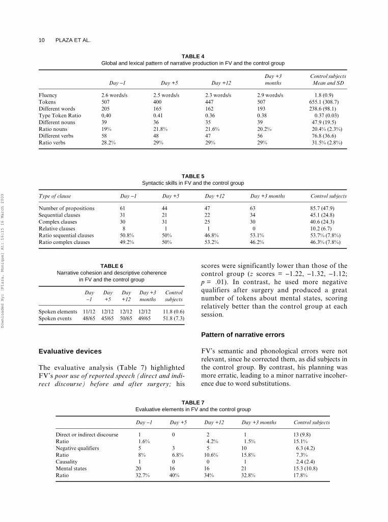
The comparative intra-subject results (Table 4) showed(a) a stability of verbal fluency (words/sec), adecrease in discourse (number of tokens) and lexi-con diversity (number of different words used bythe patient) immediately after surgery and (b) ageneral improvement 3 months later. The nounand verb ratios remained stable. The inter-subjectcomparison showed that FV’s results were withinnormal limits in all measures at each testing session.
Syntactic skills
The comparative intra-subject results (Table 5)showed (a) a stability of complex clause use, adecrease in discourse content (number of proposi-tions), and number of sequential clauses and aboveall a significant decrease in the use of relativeclauses, immediately after surgery and (b) a generalimprovement except for relatives 3 months later.The inter-subject comparison showed that FV’sresults were within normal limits in all measures ateach session, except for relative clause use, where hescored significantly lower than the control group(z score = −1.37; p = .01).
Narrative cohesion and descriptive coherence
All situations were reliably accounted for by FV atany time, and by the control group as well (Table 6).Similarly, concerning descriptive coherence, FVrecounted most events suggested by the pictures.Like most subjects in the control group, he showeddescriptive coherence but for five details systemi-cally omitted at each session. But since those fivedetails were also omitted by most subjects in thecontrol group, FV did not differ on this point.
TABLE 3 Narrative written BDAE task elicited by the picture ‘Cookies’ in 1 min
Day -1 Day +5 Day +3 months
Je ne sais 1pas ce que fait la femme (I don’t know 1what the woman is doing).
Ce sont deux petits garçons 1 (They are two little boys1).
Au moment où la mère faisait la vaisselle sans attention 1 (While the mother washed the dishes without attention1)
Le petit garçon attend je sais pas 1quoi (The little boy waits I don’t know 1what).
Un prend un gâteau (One takes a cake). En regardant par la fenêtre (Looking through the window).
Le tabouret est en train de 2tomber (The stool is falling).
Il est en train de 2 donner à une petite fille (he is giving 2 a little girl).
L’eau de l’évier se mit à déborder (The water in the sink started to overflow).
Et la femme qui3 est en train de 2faire la vaisselle (And the woman who is washing the dishes).
Pendant ce temps, la mère qui 3fait la vaisselle (In the meantime, the mother who3 washes the dishes).
Pendant ce temps, son fils et sa fille essayaient de voler des gâteaux dans le placard du haut (In the meantime, her son and daughter tried to steal cakes in the top cupboard ).
En regardant dehors (Looking outside). Est en train de regarder dehors (Is look-ing outside).
Son fils avait dû monter sur une chaise 2 (her son had to stand on a chair2)
Et que3 son évier est en train de 2déborder (And that her sink is overflowing).
Pendant que son évier déborde (While her sink overflows).
Qui 3 était en train de basculer (that 3 was falling over).
Narrative coherence: 6/10 Narrative coherence: 8/10 Narrative coherence: 10/101 and 2 : perseverative forms. 1 :
Incapacity tokens (« I don’t know »).3 syntactic erroneous forms with « qui »
(who) and « que » (that)
1 semantic error : the characters are a boy and a girl.
2 pronoun omission (« le », it)3 relative clause correctly written
1 verb omission : « without paying attention »2 semantic paraphasia : « chair » instead of
« stool »3 relative clause correctly written
Irregular handwriting Regular handwriting Regular handwriting
Downloaded By: [Plaza, Monique] At: 16:15 16 March 2009
10 PLAZA ET AL.
Evaluative devices
The evaluative analysis (Table 7) highlightedFV’s poor use of reported speech (direct and indi-rect discourse) before and after surgery; his
scores were significantly lower than those of thecontrol group (z scores = −1.22, −1.32, −1.12;p = .01). In contrast, he used more negativequalifiers after surgery and produced a greatnumber of tokens about mental states, scoringrelatively better than the control group at eachsession.
Pattern of narrative errors
FV’s semantic and phonological errors were notrelevant, since he corrected them, as did subjects inthe control group. By contrast, his planning wasmore erratic, leading to a minor narrative incoher-ence due to word substitutions.
TABLE 4 Global and lexical pattern of narrative production in FV and the control group
Day -1 Day +5 Day +12Day +3 months
Control subjects Mean and SD
Fluency 2.6 words/s 2.5 words/s 2.3 words/s 2.9 words/s 1.8 (0.9)Tokens 507 400 447 507 655.1 (308.7)Different words 205 165 162 193 238.6 (98.1)Type Token Ratio 0,40 0.41 0.36 0.38 0.37 (0.03)Different nouns 39 36 35 39 47.9 (19.5)Ratio nouns 19% 21.8% 21.6% 20.2% 20.4% (2.3%)Different verbs 58 48 47 56 76.8 (36.6)Ratio verbs 28.2% 29% 29% 29% 31.5% (2.8%)
TABLE 5 Syntactic skills in FV and the control group
Type of clause Day -1 Day +5 Day +12 Day +3 months Control subjects
Number of propositions 61 44 47 63 85.7 (47.9)Sequential clauses 31 21 22 34 45.1 (24.8)Complex clauses 30 31 25 30 40.6 (24.3)Relative clauses 8 1 1 0 10.2 (6.7)Ratio sequential clauses 50.8% 50% 46.8% 53.1% 53.7% (7.8%)Ratio complex clauses 49.2% 50% 53.2% 46.2% 46.3% (7.8%)
TABLE 6 Narrative cohesion and descriptive coherence
in FV and the control group
Day -1
Day +5
Day +12
Day +3 months
Control subjects
Spoken elements 11/12 12/12 12/12 12/12 11.8 (0.6)Spoken events 48/65 45/65 50/65 49/65 51.8 (7.3)
TABLE 7 Evaluative elements in FV and the control group
Day -1 Day +5 Day +12 Day +3 months Control subjects
Direct or indirect discourse 1 0 2 1 13 (9.8)Ratio 1.6% 4.2% 1.5% 15.1%Negative qualifiers 5 3 5 10 6.3 (4.2)Ratio 8% 6.8% 10.6% 15.8% 7.3%Causality 1 0 0 1 2.4 (2.4)Mental states 20 16 16 21 15.3 (10.8)Ratio 32.7% 40% 34% 32.8% 17.8%
Downloaded By: [Plaza, Monique] At: 16:15 16 March 2009

BROCA’S AREA RESECTION 11
DISCUSSION
In the present paper, a detailed evaluation of thepatient’s pre- and post-surgical profile wasenriched by narrative production data not regu-larly provided by classical neuropsychologicalassessment. On the basis of a longitudinal analysisof the patient’s performances, we originallyhypothesized that (a) the tumor infiltration mayhave led to impairments partially or completelycompensated for by plasticity mechanisms and (b)the left IFG resection would lead to subtle impair-ments in narrative production, more specifically.
Before resection: tumor consequences
Before surgery, FV displayed minor neuropsycho-logical deficits, as shown by PIQ/VIQ discrepancywith low scores in verbal fluency, learning/retrieval difficulties, verbal working memory limi-tation (digit span) and lexical access trouble(Boston Naming Test: anomias, semantic parapha-sias, circumlocutions, delayed responses). Justbelow average results in DO 80 (96.2%) wereobserved as well as in three BDAE tasks requiringauditory working memory, i.e., concrete sentencerepetition (87.5%), auditory instructions (93.3%),logical and reasoning (83.3%). In narrative produc-tion, five lexical disturbances were observed, i.e.,one semantic paraphasia, three preposition substi-tutions and one phonological error. The observedlexical disturbances could have been expected,since the left inferior frontal gyrus is specificallyinvolved in individual word processing (Fiebach &Friederici, 2004). However, the patient displayedefficient verbal speed in spite of slightly weakerlexical access. He also correctly processed allaspects of narrative, notably recruiting languageassembly procedures such as syntactic strategiesand lexical repertoire, respecting script cohesion(the 12 situations composing the story) anddescriptive coherence (picture/speech matching),using evaluative devices – notably negative qualifi-ers (which introduce the virtual axis) and mentalstates, as described by Theory of Mind. In con-trast, the BDAE written narrative task (elicited bythe single picture ‘Cookies’ under time constraint)highlighted difficulties in descriptive coherence,syntactic planning and handwriting. Thus, thecomplex coordination of visual picture analysis,lexical/syntactic retrieval and handwriting wasimpaired.
As shown in Figures 3 and 4, the surgical resec-tion was very large, close to removing the left infe-rior frontal gyrus, which was completely ‘silent’ fornaming during stimulation. The sites directlyinvolved in speech naming production were thepremotor ventral area (complete speech arrest),the dorsal premotor cortex (semantic paraphasia),the head of the caudate nucleus (perseveringresponses), the premotor ventral cortex (anar-thria), the inferior occipito-frontal fasciculus, con-nected to the dorso-lateral prefrontal cortex(semantic paraphasia). Thus, before resection,plasticity mechanisms recruiting adjacent and insome cases controlateral regions (Knecht et al.,2000) allowed the brain to compensate for the leftinferior frontal gyrus incapacitation due to tumourgrowth.
Compensatory mechanisms: their efficiency and limits
After the tumor resection which preserved regionsdirectly involved in speech naming production, thepatient quickly retrieved good language abilities.On Day +5, some BDAE scores were slightly belownormal limits (due to surgical after-effects, asobserved in all patients) but most scores were aver-age 3 months later. The naming speed increaseobserved on Day +3 months in the DO 80 picturenaming test was parallel to the speech accelerationobserved in the narrative task from Day −1 to Day+3 months. Speech acceleration could be partiallyattributed to retest effect, though its amplitudesuggests a motor and phonological planningimprovement following tumor removal, speech-therapy and brain decompression, linked to thearrest of epilepsy episodes. The patient experiencedgeneralized seizures 11 months before surgery anddespite anti-epileptic drugs, namely Valproat andGabapentin, partial seizures intensified, accompa-nied with language difficulties. After surgical resec-tion, epilepsy stopped, which led to a reduceprescription of anti-epileptic drugs.
The narrative task longitudinal analysis high-lighted the patient’s stable syntactic competenceillustrated by the number of correct complex utter-ances, the preservation of verbs as action markersbetween pre- and post-assessments. Lexical abilitieswere shown to improve after a period of impover-ishment (−20% at Day +5 and Day + 12), and over-all story length increased after a fall at Day +5 andDay + 12. The patient also preserved script cohesion
Downloaded By: [Plaza, Monique] At: 16:15 16 March 2009

12 PLAZA ET AL.
and descriptive coherence, efficiently using negativequalifiers and evoking mental states. Moreover, thenarrative evaluative assessment showed that FV wasable to express his own point of view, introduceemotional factors, specify virtual and potential fea-tures and then give a personal meaning to the story(Bamberg & Damrad-Frye, 1991). In the writtennarrative task, descriptive coherence and syntacticstrategy, which were defective before resection, dra-matically improved afterwards.
However, the patient’s narrative productionremained impaired in subtle ways. At the syntacticlevel, FV partially ‘lost’ the use of the relative pro-nominal strategy which allows speakers to producecomplex sentences including more than two subjectsites (e.g., ‘The boy’s dog, which has put its headinto the box, goes to the window and falls out-side’). This relative clause deficit suggested a for-mal linguistic vulnerability which could beexplained by the resource limitation hypothesis.
At Day +5 and Day +3 months, FV had justbelow average results in BDAE tasks requiringworking memory involvement, i.e., auditoryinstructions (93.3% at both evaluations), abstractsentence repetition (respectively, 75 and 87.5%),logics and reasoning (respectively, 66.6 and 83.3%).In parallel, during narrative production, he tendedto avoid sentences including various subject sites.The left IFG, specifically the upper part of BA 44,is known to be critically involved in verbal-auditoryworking memory (Paulesu et al., 1993; Wallentin,Ropstorff, Glover, & Burgess, 2006). Length andcomplexity of relative clauses increase demands onsyntactic production. The speaker’s performance isdetermined by the fact that language processing is asequential process in time. It depends on the dis-tance between an element original position in thesyntactic hierarchy and its actual position in thesentence. Once a word has been produced as amoved element, the speaker has to keep this
Figure 3. Post resection figure shows that the resection concerned the left frontal lobe, notably the pars triangularis, the pars orbitalisand the anterior part of the pars opercularis.
Downloaded By: [Plaza, Monique] At: 16:15 16 March 2009

BROCA’S AREA RESECTION 13
element active in the working memory specializedfor syntactic features. Processing demands increaseas a function of the distance between non-localdependencies. The core region of BA 44/45 appar-ently supporting these processes in syntactic pars-ing (Friederici, 2006), seems to be involved in thesame way in syntactic production too. In the writ-ten narrative task, difficulties with relative andembedded clauses were observed on Day −1, anddisappeared on Day +5 and Day +3 months. Thetask undoubtedly increased the difficulty involvedin the visual processing of one single picture, thehandwriting and the overall sentence planningunder time constraint.
Along with impaired lexical access (Faidiga &Craighero, 2006), Broca’s aphasics show disturbedsyntactic production, with grammatically simplifiedspeech, missing function words and morphemes(Caplan, 2006). Globally this was not FV’s case.
However, in the written narrative task, he omittedsome words and in the oral task, he did not use manycausal conjunctions, although he produced manycomplex clauses. He only used one causal conjunc-tion before surgery, and another 3 months later.Conjunctions are grammatical function words thatunderlie inter-sentential connections. They arecomplex tools, for they do not have a referent in anextra-linguistic context and are used for bothsemantic and pragmatic functions (Bloom, Lahey,Hood., Lifter, & Fiess, 1980). Developmental stud-ies showed that young children initially join sen-tences by juxtaposing them without conjunction(Miller, 1981) since for children, conjunctionsfunction as index items without autonomousmeaning (Orsolini, 1993). Clinical studies alsoshowed that dyslexic children tend to use a similar‘economic’ pattern without conjunctions (Plaza,1998), just as aphasic adults, and this can be
Figure 4. Post resection MRI shows that the resection concerned the left frontal lobe, notably the pars triangularis, the pars orbitalisand the anterior part of the pars opercularis.
Downloaded By: [Plaza, Monique] At: 16:15 16 March 2009

14 PLAZA ET AL.
related to working memory limitations. FV tried tocompensate for his vulnerability about causal con-nectors by using sequential clauses introduced by‘then’, implicitly requiring the auditor to infer cau-sality. But, since the control group did not usemore causal connectors, the lack of causal con-junctions from Day −1 to Day +3 months couldnot be considered as atypical in our patient.
At the evaluative level, FV did not use reportedspeech (direct and indirect). The rarity or absenceof reported speech suggested that FV did not rep-resent speech within speech. The animated subjectsof the story (the boy, the dog, the frog, the stag, theowl, the bees . . .) did not have any inner speechand they did not talk to each other, although theywere endowed with a large range of perceptions,desires and emotions. We hypothesized that thepoorness of reported speech could correspond tothe ‘social-emotional change’ observed in thepatient’s behavior post surgery. He presented aslight anosognosia and did not acknowledge hisdifficulties at first and he also appeared more indif-ferent than preoperatively.
The discrepant pattern of evaluative devices inFV’s narrative shows that Broca’s area is involved inauditory verbal working memory, and could berequired in the subtle process of representing speechwithin speech. As suggested by recent data, the supe-rior part of the IFG (BA 44) is involved in the acti-vation of internal speech representations, notablymobilized for correct identification of speech stimuli(Zekveld, Heslenfeld, Festen, & Schoonhoven,2006). Though Broca’s area is not ‘necessarily’involved either in the process of translating socialintent into speech (Gentilucci, Bernardis, Crisi, &Volta, 2006) or in the linguistic representation ofactions (Hamzei et al., 2003; Rizzolatti, Focassi, &Gallese, 2001), its resection could be presumed tolead to transitory disturbances of ‘mirroring’speech devices within narrative discourse.
CONCLUSION
Despite a massive resection of the inferior frontalgyrus, the patient did not exhibit the severe lan-guage impairments predicted by the localizationtheory of the brain. The present case confirms therelevance of connectionist approaches based onstudies of slow-growth tumors, which demonstratethat compensatory mechanisms start beforesurgery, in reaction to tumor infiltration, andconsolidate during and after surgical procedures
(Bonnetblanc et al., 2006). The patient recoveredhis functional preoperative status within 3 monthsfollowing surgery, with no neurological deficit, andcould resume a normal socio-professional life. Thepatient’s positive outcome can be attributed to theslow evolution of his tumor and the ensuing com-pensatory processes the results of which wereclearly revealed by cortical and sub-cortical electri-cal stimulation. Whereas the compensating role ofthe controlateral right hemisphere has alreadybeen observed especially in slow-growing lesionsuch a LGG (Desmurget et al., 2007), the presentidentification of several structures essential to lan-guage within the remaining left hemisphere supportthe crucial role of perilesional areas in functionalcompensation. It is worth noting that cortical sitesalone (i.e., the postero-inferior part of the parsopercularis, the ventral and dorsal premotor cor-tex, the dorso-lateral prefrontal cortex, the poste-rior insula) cannot account for the patient’slanguage recovery following surgical resection.Indeed, sub-cortical mapping also showed that thepreservation of both white matter pathways anddeep grey nuclei prevented the occurrence of per-manent postoperative aphasia. Such cerebral plas-ticity brings strong support to connectionist brainprocessing models, claiming to the existence of par-allelly distributed cortical/sub-cortical networks(Devlin et al., 2003).
But the case also demonstrates the need toundertake fine-grained language and neuropsycho-logical analyses before, during and after surgery, inorder to specify the remaining disturbances anddefine new rehabilitation techniques. Thus, futurestudies should be undertaken to develop the assess-ment of syntactic skills, working memory and emo-tional processing during peroperative session, inthe hope of preventing the subtle deficits notedhere. In pre- and postoperative evaluations, theaddition of a narrative production task to the neu-ropsychological assessment is of particular interest,usefully complementing the standardized assess-ments, by focusing on both narrative microstruc-ture (syntax, lexicon, story length), macrostructure(initiation, maintenance and resolution of storycomponents, i.e., executive processing) and psy-chosocial/emotional features. Narrative produc-tion appears as a ‘marker’ of subtle impairmentsthat could otherwise go unnoticed.
Original manuscript received 20 August 2008Revised manuscript accepted 19 December 2008
First published online
Downloaded By: [Plaza, Monique] At: 16:15 16 March 2009
BROCA’S AREA RESECTION 15
REFERENCES
Alexander, M. (2006). Impairments of procedures forimplementing complex language are due to disrup-tion of frontal attention processes. Journal of Interna-tional Neuropsychological Society, 12, 236–247.
Anderson, V. A., Morse, S. A., Catroppa, C., Haritou, F.,& Rosenfeld, J. V. (2004). Thirty month outcomefrom early childhood injury: A prospective analysisof neurobehavioral recovery. Brain, 124, 2608–2620.
Anderson, V., Catroppa, C., Morse, S., Haritou, F., &Rosenfeld, J. (2006). Understanding predictors of func-tional recovery and outcome 30 months following earlychildhood head injury. Neuropsychology, 20(1), 42–57.
Aram, D. (1999). Neuroplasticity: evidence from unilat-eral brain lesions in children. In S. Broman & J. M.Fletcher (Eds), The changing nervous system: Neuro-behavioral consequences of early brain disorders (pp.254–273). New York: Oxford University Press.
Bamberg, M., & Damrad-Frye, R. (1991). On the abilityto provide evaluative comments: further explorationsof children’s narrative competencies. Journal of ChildLanguage, 18(3), 689–710.
Bates, E., Reilly, J., Wulfeck, B., Dronckers, N., Opie,M., Fenson, J., Kriz, S., Jeffries, R., Miller, L., &Herbst, K. (2001). Differential effects of unilaterallesions on language production in children andadults. Brain and Language, 79, 223–265.
Belin, P., Van Eckhout, P., Zilbovicius, M., Remy, P.,François, P., Guillaume, S., Chain, F., Rancurel, G., &Samson. Y. (1996). Recovery from nonfluent aphasiaafter melodic intonation therapy: a PET study. Neu-rology, 47(6), 1504–1511.
Blasi, V., Young, A. C., Tansy, A. P., Petersen, S. E.,Snyder, A. Z., & Corbetta, M. (2002).Word retrievallearning modulates right frontal cortex in patientswith left frontal damage. Neuron, 36(1), 159–170.
Bloom, L., Et Lahey, M., Hood, L., Lifter, K., & Fiess, K.(1980). Complex sentences: Acquisition of syntacticconnectives and the semantic relation they encode.Journal of Child Language, 7, 235–261.
Bonnetblanc, F., Desmurget, M., & Duffau, H. (2006).Gliomes de bas grade et plasticité cérébrale. Implica-tions fondamentales et cliniques. Médecine/Sciences,22, 389–339.
Bookheimer, S. (2002). Functional MRI of language:New approaches to understanding the cortical organ-ization of semantic processing. Annual Review ofNeuroscience, 25, 151–188.
Caplan, D. (2006). Why is Broca’s area involved in syn-tax ? Cortex, 42, 469–471.
Cardebat, D., Doyon, B., Puel, M., Goulet, P., &Joanette, Y. (1990). Evocation lexicale formelleet sémantique chez les sujets normaux: performanceset dynamique de production en fonction du sexe, del’âge et du niveau d’étude. Acta Neurologica Belgica,90, 207–217.
Chapman, S., Culhane, K., Levin, H., Harward, H.,Mendelsohn, D., Ewing-Cobbs, L., Fletcher, J. M.,Bruce, D. (1992). Narrative discourse after closedhead injury in children and adolescents. Brain andLanguage, 43(1), 42–65.
Chatelois, J., Pineau, H., Belleville, S., & Peretz, I.(1993). A computerized memory test battery basedon the cognitive approach. Canadian Psychology,34, 45–63.
Clark, L., Manes, F., Anotun, N., Sahabian B. J., &Robbins T. W. (2003). The contribution of lesionlaterality and lesion volume to decision-makingimpairment following left-frontal lobe damage.Neuropsychologia, 41, 1474–1483.
Coelho, S., Max, J., & Tranel, D. (2005). A matchedlesion analysis of childhood versus adult-onset braininjury due to unilateral stroke. Cognitive BehavioralNeurology, 18(1), 5–17.
Cooper, D. (2006). Broca’s arrow: Evolution, predictionand language in the brain. The Anatomical Record,289B, 9–24.
Crosson, B., Moore, A. B., Gopinath, K., White, K. D.,Wierrenga, C. E., Gaiefsky, M. E., Fabrizio, K. S.,Peck, K. K., Soltysik, D., Milsted, C., Briggs, R. W.,Conway, T. W., & Gonzalez Rothi, L. J.. (2005).Role of the right and left hemispheres in recovery offunction during treatment of intention in aphasia.Journal of Cognitive Neuroscience, 17(3), 392–406.
Damasio, H., & Damasio, A. (1989). Lesion Analysis inNeuropsychology. Oxford: Oxford University Press.
Démonet, J. F., Thierry, G., & Cardebat, D. (2005).Renewal of the neurophysiology of language: Func-tional neuroimaging. Physiological Review, 85, 49–95.
Desmurget, M., Bonnetblanc, F., & Duffau, H. (2007).Contrasting acute and slow-growing lesions: A newdoor to brain plasticity. Brain, 130, 898–914.
Devlin, J., Matthews, P., & Rushworth, M. (2003).Semantic processing in the left inferior frontal cortex:A combined functional magnetic resonance imagingand transcranial magnetic stimulation study. Journalof Cognitive Neuroscience, 15, 71–84.
Duffau, H.. (2005). Lessons from brain mapping in low-grade glioma surgery: Insights into relationshipsbetween tumor and brain plasticity. Lancet Neuro-logy, 4, 476–4864.
Duffau, H. (2006). Brain plasticity: From pathophysio-logical mechanisms to therapeutic applications. Jour-nal of Clinical Neurosciences, 13(9), 885–897.
Etard, O., Mellet, E., Papathanassiou, D., Benali, K.,Houdé, O., Mazoyer, B., & Tzourio-Mazoyer, N.(2000). Picture naming without Broca’s andWernicke’s area. NeuroReport, 22(3), 617–622.
Faidiga, L., & Craighero, L. (2006). Hand actions andspeech representation in Broca’s area. Cortex, 42,486–490.
Fiebach, C., & Friederici, A. (2004). Processing concretewords: fMRI evidence against a specific right-hemis-phere involvement. Neuropsychologia, 42, 62–70.
Ffytche, D. H., & Catani, M.. (2005). Beyond localiza-tion: from hodology to function. Philosophical Trans-actions of the Royal Society, 360, 767–779.
Friederici, A. (2006). Broca’s area and the ventral pre-motor cortex in language: functional differentiationand specificity. Cortex, 42, 472–475.
Gentilucci, M., Bernardis, P., Crisi, G., & Volta, R.(2006). Repetitive transcranial stimulation of Broca’sarea ffects verbal responses to gesture observation.Journal of Cognitive Neuroscience, 18(7), 1059–1074.
Downloaded By: [Plaza, Monique] At: 16:15 16 March 2009

16 PLAZA ET AL.
Geshwind, N. (1967). The variety of naming errors. Cor-tex, 3, 97–112.
Goodglass, H., & Kaplan, E. (1983) Boston DiagnosticAphasia Examination. San Antonio TX: The Psycho-logical Corporation.
Greewe, T., Bornkessel, I., Zysset, S., Wiese, R., VonCramon, D., & Schlesewsky, M. (2006). Linguisticprominence and Broca’s area: the influence of ani-macy as a linearization principle. NeuroImage, 32(3),1395–402.
Gröber, E., Buschke, H., Bang, S., & Dresner, R. (1988).Screening for dementia by memory testing. Neurol-ogy, 38, 900–903.
Grodzinsky, Y. (2006). The language faculty, Broca’sregion and the mirror system. Cortex, 42, 464–468.
Hamzei, F., Rinjntjes, M., Dettmers, C., Glauche, V.,Weiller, C., & Buchel, C. (2003). The human actionrecognition system and its relationship to Broca’sarea: an fMRI study. Neuroimage, 19, 637–644.
Heaton, R. K., Chelure, G. J., Talley, J. L., Kay, G. G., &Curtiss, G. (2003). Test de classement des cartes duWisconsin. Paris: Editions du Centre de PsychologieAppliquée.
Heim, S., Alter, K., Ischebeck, A., Amunts, K., Eickhoff,S., Mohlberg, H., Zilles, K., Von Cramon, D., &Friederici, A. (2005). The role of the left Brodmann’sareas 44 and 5 in reading words and pseudowords.Cognitive Brain Research, 25, 982–993.
Hooper, H. E. (1958). The Hooper visual orientation test.Manual. Los Angeles: Western Psychological Services.
Kaplan, E., Goodglass, H., & Weintraub, S. (1983).Boston Naming test. Philadelphia, PA: Lea & Fabiger.
Koechlin, E., & Jubault, T. (2006). Broca’s area and thehierarchical organization of human behavior. Neu-ron, 50(6), 963–974.
Léger, A., Démonet, J. F., Ruff, S., Aithamon, B., Touy-eras, B., Puel, M., Boulanouar, K., & Cardebat, D.(2002). Neural substrates of spoken language rehabil-itation in an aphasic patient: a fMRI study. Neuroim-age, 17(1), 174–183.
Lichtheim, L. (1885). Uber Aphasie. Deutsches ArchivesKlinical Medicine, 36, 204–268.
Liles, B. (1993). Narrative discourse in children with lan-guage disorders and children with normal language:A critical review of the literature. Journal of Speechand Hearing Research, 36, 868–882.
MacWhinney, L., & Snow, K. (1991). The CHILDESProject: Tools for analyzing talk. Hillsdale, NJ: Law-rence Erlbaum.
Mayer, M. (1969). Frog where are you? New York: Pen-guin Book.
Mazaux, J. M., & Orgogozo, J. M. (1982). Echelle d’éval-uation de l’aphasie adaptée du Boston DiagnosticAphasia Examination. Paris: E.A.P. Editions Psycho-techniques.
Metz-Lutz, M. N., Kremin, H., Deloche, G., Hanne-quin, D., Ferrand, I., Perrier, D., Quint, S., Dordain,M., Brunel, G., Cardebat, C., Larroque, C., Lota, A.M., Pichard, B., & Blavier, A. (1991). Standardisa-tion d’un test de dénomination orale: contrôle deseffets de l’âge, du sexe et du niveau de scolarité chezles sujets adultes normaux. Revue de neuropsycholo-gie, 1(1), 73–95.
Miller, J. F. (1981). Assessing language production in chil-dren. London: Edward Arnold.
Milner, B. (1971). Interhemispheric differences in thelocalization of psychological processes in man.British Medical Bulletin, 27, 272–276.
Musso, M., Weiller, C., Kiebel, S., Muller, S. P., Bullau, P.,& Rijntjes, M. (1999). Training-induced plasticity inaphasia. Brain, 122(9), 1781–1790.
Ojemann, G., Ojemann, J., Lettich, E., & Berger, M.(1989). Cortical language localisation in left, domi-nant hemisphere. An electrical stimulation mappinginvestigation in 117 patients. Journal of Neurosur-gery, 71(3), 316–326.
Oldfield, R. C. (1971). The assessment and analysis ofhandedness: the Edinburgh inventory. Neuropsycho-logia, 9, 97–113.
Orsolini, M. (1993). Because in children’s discourse.Applied Psycholinguistics, 14, 89–120.
Paulesu, E., Frith, U., Snowling, M., Gallagher, A.,Morton, J. Frackowiak, R. S., & Frith, C. D. (1993).The neural correlates of the verbal component ofworking memory. Nature, 362, 342–345.
Plaza, M. (1998). Reference and evaluation in the narra-tive speech of a group of French-speaking dyslexicchildren. Proceedings of the International Congress ofthe International Society of Applied Psycholinguistics(Porto), pp. 665–668.
Plaza, M., Guitton, C., & Le Normand, M.-T. (1998).Vulnerability of conjunction and verb tense use in thenarrative speech of dyslexic children. In W. Ziegler &K. Deger (Eds), Clinical Phonetics and Linguistics(pp. 41–46). London: Whurr Publishers Ltd.
Plaza, M., Gatignol, P., Cohen, H., Berger, B., &Duffau, H. (2007). A discrete area with in the LeftDorsolateral prefrontal cortex involved in incongru-ence visual/verbal judgment. Cerebral Cortex, 18(6),1253–1259.
Reilly, J., Bates, E., & Marchman, V. (1998). Narrativediscourse in children with early focal brain injury.Brain and Language, 61, 335–375.
Reilly, J., Losh, M., Bellugi, U., & Wulfeck, B. (2004).‘Frog, where are you?’ Narratives in children with spe-cific language impairment, early focal brain injury, andWilliams syndrome. Brain and Language, 88, 229–247.
Rey, A. (1959–1970). Test de copie d’une figure complexe.Manuel. Paris, Centre de Psychologie Appliquée.
Rizzolatti, G., Focassi, L., & Gallese, V.. (2001). Neuro-physiological mechanisms underlying the under-standing and imitation of action. Nature ReviewsNeuroscience, 2, 661–670.
Ruff, R. M., Light, R. H., & Evans, R. W. (1987). TheRuff Figural Fluency Test: A normative study withadults. Developmental Neuropsychology, 3, 37–51.
Ruff, R. M., Niemann, H., Allen C. C., Farrow, C. E., &Wylie, T. (1992). The Ruff 2 and 7 Selective Atten-tion Test: A neuropsychological application. Percep-tual and Motor Skills, 75, 1311–1319.
Signoret, J. L., Bonvarlet, M., Benoît, N., Bolgert, F.,Eustache, F., Leger, J. M. (1988). Batterie d’estima-tion des états démentiels; description et validation. InD. Leys & H. Petit. La maladie d’Alzheimer et seslimites. Congrès de Psychiatrie et de Neurologie deLangue Française (pp. 265–270), Masson.
Downloaded By: [Plaza, Monique] At: 16:15 16 March 2009

BROCA’S AREA RESECTION 17
Smith, E., & Jonides, J. (1999). Storage and executiveprocesses in the frontal lobes. Science, 283, 1657–1661.
Stroop, J. R. (1935). Studies of interference in serial ver-bal reactions. Journal of Experimental Psychology,18, 643–662.
Tettamanti, M., & Weniger, D. (2006). Broca’s area: asupramodal hierarchical processor? Cortex, 42,491–494.
Tettamanti, M., Alkhadi, M., Moro, A., Perani, D.,Kollias, S., & Weniger, D. (2002). Neural correlatesfor the acquisition of natural language syntax. NeuroImage, 17, 700–709.
Vigneau, M., Beaucousin, V., Herve, P. Y., Duffau, H.,Crivello, F., Houde, O., Mazoyer, B., & Tzourio-Mazoyer, N. (2006). Meta-analyzing left hemispherelanguage areas: phonology, semantics, and sentenceprocessing. Neuroimage, 30(4), 1414–1432.
Voets, N., Adcock, J., Flitney, D., Behrens, T., Hart, Y.,Stacey, R., Carpenter, K., & Mathhews, P. (2006).Distinct right frontal lobe activation in language
processing following left hemisphere injury. Brain,129, 754–766.
Wallentin, M., Roepstorff, A., Glover, R., & Burgess, N.(2006). Parallel memory systems for talking aboutlocation and age in precuneus, caudate and Broca’sregion. NeuroImage, 32(4), 1850–1864.
Ward, L. C. (1990). Prediction of verbal, performance,and full scale IQs from seven subtests of the WAIS-R.Journal of Clinical Psychology, 46(4), 436–440.
Wechsler, D. (1981). Wechsler adult intelligence scale-revised. New York: Psychological Corporation.
Weintraub, S., & Mesulam, M. M. (1985). Mental stateassessment of young and elderly adults in behavioralneurology. In M.-M. Mesulam (Ed.), Principles ofbehavioral neurology (pp. 71–123). Philadelphia, PA:F.A. Davis.
Zekveld, A., Heslenfeld, D., Festen, J. M., &Schoonhoven, R. (2006). Top-down and bottom-upprocesses in speech comprehension. Neuroimage,32(4), 1826–1836.
Downloaded By: [Plaza, Monique] At: 16:15 16 March 2009