
lated' heterozygotes.
7
HETERODUPLEX HETEROZYGOTES IN BACTERIOPHAGE T4 INVOLVING MUTATIONS OF VARIOUS DIMENSIONS* BY JOHN W. DRAKE DEPARTMENT OF MICROBIOLOGY, UNIVERSITY OF ILLINOIS, URBANA Communicated by S. Spiegelman, January 6, 1966 Single-factor genetic crosses in bacteriophage T4 produce two distinct types of heterozygous progeny.' One kind probably corresponds to a terminal redundancy of the chromosome. The other kind, variously termed a heteroduplex heterozygote, an internal heterozygote, or a recombinational heterozygote, arises as an intermediate in recombination (Fig. 1). Internal heterozygotes are preferentially accumu- lated"' 2, 10 12 in the presence of FUDR, which inhibits DNA replication and there- fore inhibits the conversion of internal heterozygotes into homozygous progeny. Furthermore, only certain types of mutational lesions are efficiently recovered from internal heterozygotes." 2 If a cross is performed between two different mutants, one of which (point mutation) contains a base pair substitution and the other an extended deletion, the heterozygous particles which emerge usually contain the point mutant. This result indicates that internal heterozygotes cannot easily ac- commodate mutations of extended dimensions, presumably because such hetero- zygotes would contain single-stranded loops. At present it is not clear whether deletion mutations fail to form internal heterozygotes initially, or whether the heterozygotes are formed but are later repaired by a mechanism leading to homo- zygosity.3 The sign mutants4 of bacteriophage T4 consist5 of additions and/or deletions of small numbers of base pairs; they are also called "reading-frame mutants" and "acridine-type mutants." An extensive fine-scale map6 of the rIl mutants in the B cistron strongly suggests that sign mutants possess extended dimensions (Fig. 2). An analysis of the ability of internal heterozygotes to encompass sign mutations should therefore yield information about the physical dimensions of the mutations, and also about putative single-stranded regions within the DNA molecule. Materials and Methods.-Strains of bacteriophage T4B and its rIl mutants,7 and of Escherichia coli, were obtained from the collection of Dr. Sydney Brenner and Mrs. Leslie Barnett in Cam- bridge. All incubations were at 37°. E. coli strain Bw plates r phages as large plaques, and r+ phages as small plaques. Strain BB does not distinguish r and r+ phages, and is used to grow stocks and for crosses. Strain OP33 is a K-12(X) derivative which transmits ril mutants at a very low frequency. Strain QA1 is a K-12(X) derivative which permits the growth only of certain rII mutants of the amber class, such as rX417. The various rII mutants employed have been ex- tensively mapped,6 and a preliminary and condensed map appears in Figure 2. All of the mutants except r187 and r196b are capable of reversion. FUDR crosses were performed according to Shalitin and Stahl.2 BB cells in M9- casamino acids medium were infected with 5 of each parental phage. At 2 min, FUDR was added to 4 X 10⁻⁵ M, and uracil to 2 X 10⁻⁴ M. The phage-cell complexes were aerated continuously thereafter. At 9 min, chloramphenicol was added to 125 jug/ml. At 90 min the complexes were chilled and cen- trifuged at low speed. They were resuspended in the same medium without chloramphenicol (but with FUDR and uracil) at about 107/ml and incubated without aeration for an additional 60 min. Lysis was completed with chloroform. Burst sizes ranged from 1 to 10, but were usually about 5. Enrichment plating for heterozygotes was also performed according to Shalitin and Stahl.2 OP33 cells were suspended in buffer containing 4 X 10⁻² M KCN. The lysate from an FUDR cross was adsorbed to the cells at a total multiplicity of 0.01 or less. After 4 min of adsorption, during which 506 Downloaded by guest on December 3, 2021
Transcript of lated' heterozygotes.
HETERODUPLEX HETEROZYGOTES IN BACTERIOPHAGE T4INVOLVING MUTATIONS OF VARIOUS DIMENSIONS*
BY JOHN W. DRAKE
DEPARTMENT OF MICROBIOLOGY, UNIVERSITY OF ILLINOIS, URBANA
Communicated by S. Spiegelman, January 6, 1966
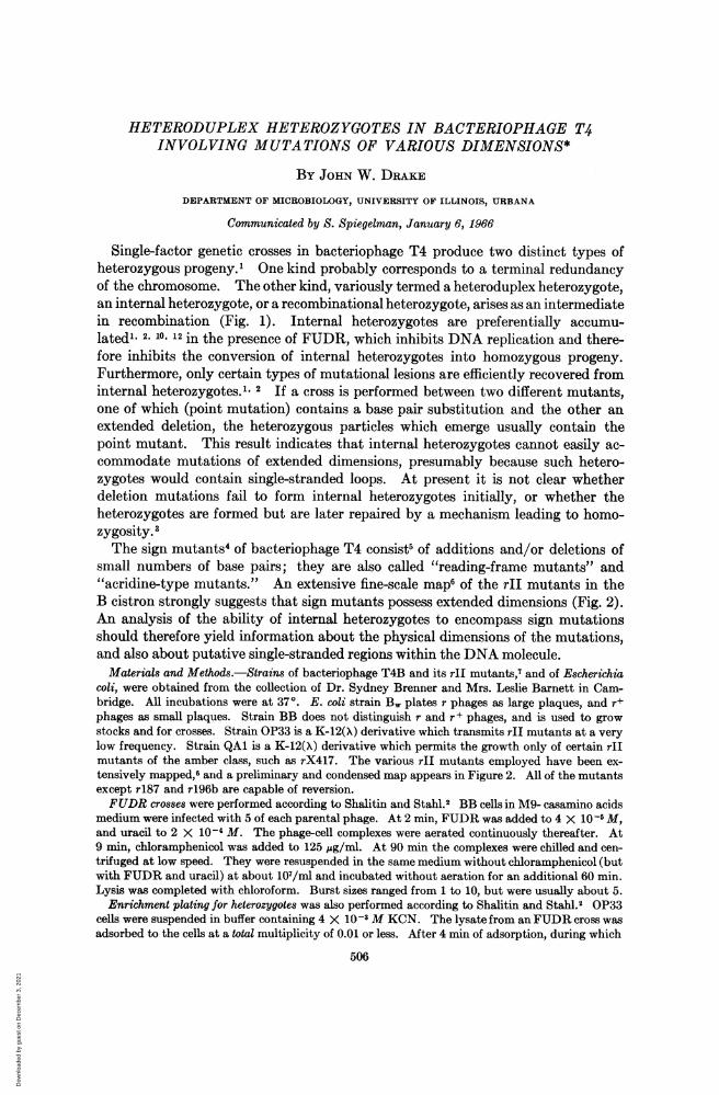
Single-factor genetic crosses in bacteriophage T4 produce two distinct types ofheterozygous progeny.' One kind probably corresponds to a terminal redundancyof the chromosome. The other kind, variously termed a heteroduplex heterozygote,an internal heterozygote, or a recombinational heterozygote, arises as an intermediatein recombination (Fig. 1). Internal heterozygotes are preferentially accumu-lated"' 2, 10 12 in the presence of FUDR, which inhibits DNA replication and there-fore inhibits the conversion of internal heterozygotes into homozygous progeny.Furthermore, only certain types of mutational lesions are efficiently recovered frominternal heterozygotes." 2 If a cross is performed between two different mutants,one of which (point mutation) contains a base pair substitution and the other anextended deletion, the heterozygous particles which emerge usually contain thepoint mutant. This result indicates that internal heterozygotes cannot easily ac-commodate mutations of extended dimensions, presumably because such hetero-zygotes would contain single-stranded loops. At present it is not clear whetherdeletion mutations fail to form internal heterozygotes initially, or whether theheterozygotes are formed but are later repaired by a mechanism leading to homo-zygosity.3The sign mutants4 of bacteriophage T4 consist5 of additions and/or deletions of
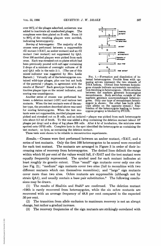
small numbers of base pairs; they are also called "reading-frame mutants" and"acridine-type mutants." An extensive fine-scale map6 of the rIl mutants in theB cistron strongly suggests that sign mutants possess extended dimensions (Fig. 2).An analysis of the ability of internal heterozygotes to encompass sign mutationsshould therefore yield information about the physical dimensions of the mutations,and also about putative single-stranded regions within the DNA molecule.
Materials and Methods.-Strains of bacteriophage T4B and its rIl mutants,7 and of Escherichiacoli, were obtained from the collection of Dr. Sydney Brenner and Mrs. Leslie Barnett in Cam-bridge. All incubations were at 37°. E. coli strain Bw plates r phages as large plaques, and r+phages as small plaques. Strain BB does not distinguish r and r+ phages, and is used to growstocks and for crosses. Strain OP33 is a K-12(X) derivative which transmits ril mutants at a verylow frequency. Strain QA1 is a K-12(X) derivative which permits the growth only of certain rIImutants of the amber class, such as rX417. The various rII mutants employed have been ex-tensively mapped,6 and a preliminary and condensed map appears in Figure 2. All of the mutantsexcept r187 and r196b are capable of reversion.FUDR crosses were performed according to Shalitin and Stahl.2 BB cells in M9- casamino acids
medium were infected with 5 of each parental phage. At 2 min, FUDR was added to 4 X 10⁻⁵ M,and uracil to 2 X 10⁻⁴ M. The phage-cell complexes were aerated continuously thereafter. At9 min, chloramphenicol was added to 125 jug/ml. At 90 min the complexes were chilled and cen-trifuged at low speed. They were resuspended in the same medium without chloramphenicol (butwith FUDR and uracil) at about 107/ml and incubated without aeration for an additional 60 min.Lysis was completed with chloroform. Burst sizes ranged from 1 to 10, but were usually about 5.
Enrichment plating for heterozygotes was also performed according to Shalitin and Stahl.2 OP33cells were suspended in buffer containing 4 X 10⁻² M KCN. The lysate from anFUDR cross wasadsorbed to the cells at a total multiplicity of 0.01 or less. After 4 min of adsorption, during which
506
Dow
nloa
ded
by g
uest
on
Dec
embe
r 3,
202
1
VOL. 55, 1966 GENETICS: J. W. DRAKE 507
over 95% of the phages adsorbed, antiserum was laadded to inactivate all unadsorbed phages. The PaRNTWALcomplexes were then plated on B cells. From 10to 80% of the resulting plaques were mottled, rbindicating heterozygosity. I}(ated s
Scoring of heterozygotes: The majority of the MUTrbcrosses were performed between a suppressible ra +rI mutant (rX417, an amber mutant) and an rIl + INrNALmutant (test mutant) not suppressed by QAL. *r, ETERZYGmorEOver 100 mottled plaques were picked from each Re.piotion ofcross. Each was streaked out on plates which had hterozygot.been previously poured with soft agar containing 'WILDOF2 drops of a mixture of overnight cultures of B + rband QAL cells in the ratio 2:1. (The use of this ,__'_rArmixed indicator was suggested by Mrs. Leslie rbBarnett.) Virtually all of the heterozygotes con- FIG. 1.-Formation and dissolution of in-tamned wild-type phages plus one but not both ternal heterozygotes. Double lines with op-wildtype, plus onebutnot h posing arrows represent the two strands ofof the parental r phages, in agreement with the DNA helices. Dotted lines between homolo-results of Hertel.8 Each genotype formed a dis- gous strands indicate asymmetric recombina-tinctive plaque type on the mixed indicator, and tion resulting in heterozygosis. (Both switchesscoring was unambiguous. between the markers generate homozygousrecombinants. Both switches outside of theAnother series of crosses was performed be- markers generate nonrecombinant heterozy-tween a deletion mutant (r187) and various test gotes. Only one type of recombinant hetero-mutants. When the test mutants were of the am- zygote is shown; the other has both wild-ber type, the procedure described above was used type alleles on the opposite strand.) Rep-lication of the heterozygote leading to homo-for scoring heterozygotes. When the test mu- zygous progeny is blocked byFUDR.tants were not suppressible, mottled plaques werepicked and streaked out on B cells, and an isolated r plaque was picked from each heterozygoteinto about 0.2 ml of broth. To this was added a drop containing the deletion mutant (about 107phages per drop) and a drop of log phase BB cells. After 4 hr of incubation, the mixtures werespotted onto OP33 cells. Complete lysis in the spot identified the heterozygote as containing thetest mutant; no lysis, as containing the deletion mutant.These tests were shown to be reliable in reconstruction experiments.
Results.-Crosses were first performed between an amber mutant, rX417, and aseries of test mutants. Only the first 100 heterozygotes to be scored were recordedfor each test mutant. The mutants are arranged in Figure 3 in order of their in-creasing rates of recovery from heterozygotes. The dotted lines delimit the rangewithin which 95 per cent of the values would fall, if rX417 and the test mutant wereequally frequently represented. The symbol used for each mutant indicates atleast roughly its genetic extent. Thus "small" sign mutants cover only one site(see Fig. 2); "medium" sign mutants cover two sites (fail to recombine with twodifferent mutants which can themselves recombine); and "large" sign mutantscover more than two sites. Ochre mutants are suppressible (although not bystrain QA1), and usually contain a base pair substitution.9 The following conclu-sions may be drawn.
(1) The results of Shalitin and Stahl2 are confirmed. The deletion mutantr196b is rarely recovered from heterozygotes, while the six ochre mutants arerecovered with an average frequency of 48.8 per cent compared to the expected50 per cent.
(2) The transition from allele exclusion to maximum recovery is not an abruptchange, but rather a gradual increase.
(3) The recovery frequencies of the sign mutants are strikingly correlated with
Dow
nloa
ded
by g
uest
on
Dec
embe
r 3,
202
1
508 GENETICS: J. W. DRAKE PROC. N. A. S.
FC82 ]FC6 FC9 FC148
EEE 1 Q
I~ ~FC1IFI C
FC7
F___ __FC18fC23|Cl
ll l | ~~~FC35 }
N87017 FC42
FC4O
f9C96 1 _ -
FC36
196bi
FC231 J FC217 FC222 a
FC34
I FC215 |
187
FIG. 2.-Left end of the B cistron. This is a preliminary and condensed version of a muchmore extensive map prepared by Mrs. Leslie Barnett and Dr. Sydney Brenner. Nonrecombiningsites are drawn as overlapping. The named sites are those employed in the present study. Manyof the sites arose repeatedly, and thus represent hot spots. Sites falling within the line are sign(0) mutants (ochres and ambers). Sites above the line have sign (+), and below the line sign (-).
their genetic extent. Most of the "large" sign mutants are infrequently recoveredfrom heterozygotes. The "small" mutants are a more heterogeneous set, but areconcentrated at the higher recovery frequencies.
(4) Several "small" sign mutants are nevertheless infrequently recovered fromheterozygotes, including some (r1074, rFC223) lying in densely marked regions ofthe map.Any monotonically arranged set of data will tend to increase gradually, even if
actually representing an abruptly changing function, because of accumulated varia-tions in the measurements. It is therefore important to verify the shape of thegradually rising portion of the curve in Figure 3. This was accomplished by per-forming crosses between a deletion mutant, r187, and a series of test mutants se-
Dow
nloa
ded
by g
uest
on
Dec
embe
r 3,
202
1

VOL. -55, 1966 GENETICS: J. W. DRAKE 509
ON
50-~~~~~~~~~~~~~002L
40.Z 0~~~00
>. 30
> 0
W0 00
Irg
Q~~~~~~~~~~~~~~~0=mediumI0
0M
CD
NN CJ C
NN I(N CD NW N
TEST MUTANT
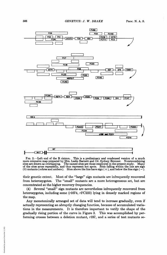
FIG. 3.-Marker recovery frequencies from heterozygotes. Heterozygotes were obtainedfrom FUDR crosses between the test mutants and the amber mutant rX417, and scored for thepresence of the test mutant. The expected recovery frequency for a true point mutant is 50% ±+10% (two standard deviations) for sample sizes of 100. Open squares, "large" sign mutantscovering more than two sites; open circles, "medium" sign mutants covering just two sites;solid circles, "small" sign mutants occupying a single site; solid squares, ochre mutants.
lected to span the critical region. In this case, the test mutants tend to pre-dominate over the deletion in heterozygotes, the more so the smaller the mutantlesion. The results are shown in Figure 4. The rise in heterozygote participationvalues in the r187 crosses orders the mutants in very nearly the same sequence asthat shown in Figure 3, except that rFC9 was recovered slightly less frequentlythan expected. Four additional amber mutants were also recovered at frequenciesclose to 100 per cent.Two "large" sign mutants (rJ158 and rFC211) from the first set of crosses were
later discovered to map as identical short deletions. They also exhibited the samefrequencies of recovery from heterozygotes (20 and 21%, respectively).The cross between r187 and r196b revealed that the former was significantly less
frequently represented than the latter among the resulting heterozygotes. The'frequency of heterozygotes on the selection plates was also relatively much smallerthan what was customarily observed in crosses between base pair substitutionmutants.
Discussiont.-The method: Of the two types of T4 heterozygotes, internal hetero-zygotes are preferentially accumulated in FUDR crosses.'I. Deletion mutationsare rarely recovered from internal heterozygotes. The use of FUDR thereforegreatly magnifies the differences between point mutants and mutants of largerextent. However, heterozygotes containing deletion mutations are selected to alimited extent from FUDR crosses by the methods employed here.' Several percent of heterozygotes also appeared on the selection plates from the cross betweenr187 and r196b. It is not yet clear whether these heterozygotes represent relativelyrare internal heterozygotes, in addition to terminal redundancy heterozygotes.The heterozygote enrichment plating method should result in the loss of half of
Dow
nloa
ded
by g
uest
on
Dec
embe
r 3,
202
1
510 GENETICS: J. W. DRAKE PROC. N. A. S.
FIG. 4.-Marker recovery fre-.50 quencies from heterozygotes.
> 90 o Heterozygotes were obtainedZ o 40 from FUDR crosses between the
0 ° test mutants and the deletionS08-0 mutant r487, and scored for the
presence of the test mutant.20,The solid line and the right-
hand scale reproduce the data of70-, Fig. 3. Symbols as in Fig. 3,.10 except that solid squares rep-
CIO__._._,_._.._,resent amber rather than ochre
mutants.
TEST MUTANT
the internal heterozygotes, since the only phages which will multiply in OP33 cellsare those which possess both r+ alleles on the strand of DNA which specifies messen-ger RNA.10 However, there is presently no reason to suspect that any allele wouldbe recovered at reduced frequency from either strand of DNA.In each of the crosses recorded here, the standard mutant and the test mutant
were compared within a common lysate, so that physiological differences betweencrosses may be excluded as sources of the observed mutant-specific heterozygote re-covery frequencies. The reproducibility of the results was further confirmed bothby comparing two "large" sign mutants which later turned out to map as identicalshort deletions, and also by comparing crosses against a standard deletion mutantwith crosses against a standard ochre mutant.
The extent of mutational lesions: The original investigation of sign mutants5characterized them by means of an algebra which assigned to each a sign, usually(+) or (-), but sometimes (0). Double mutants composed of one (+) and one
(-) mutation constitute a mutually suppressing pair. The most simple molecularconfiguration for sign mutants would be additions and deletions of single basepairs. It is clear from the present study, however, that many sign mutants behaveas if their mutational lesions were much more extensive. The data suggest a moreor less continuous distribution of lesion sizes, from true point mutations to con-ventionally described deletions. However, nearly all of the sign mutants testedhere were of spontaneous origin. Mutants induced by proflavin" or by ultravioletirradiation4 might exhibit different size distributions.
Heterozygote' frequencies have been reported'2 to decline as markers progressivelyfarther from the rII A/B cistron division were tested, although this conclusion wasbased on small-sized samples. However, this apparent decline might have arisenfrom the use of fortuitously arranged rIl sign mutants with differently sized lesions.When recovery frequencies from the present study were plotted against map order,the result was a wild scatter without any downward trend away from the cistrondivision.
It should be possible to distinguish between addition and deletion mutants by thetests employed here. Small deletions would fail to recombine with two or moredifferent sites, and would be infrequently recovered from heterozygotes; manymutants of this type were encountered. Small additions would map as points,since they would possess the DNA base sequences required for recombination withany other portion of the cistron, but would nevertheless be infrequently recovered
Dow
nloa
ded
by g
uest
on
Dec
embe
r 3,
202
1
VOL. 55, 1966 GENETICS: J. W. DRAKE 511
from heterozygotes. The more densely mapped the region around such a mutant,the more reliable would be its classification. Mutants r1074 and rFC223 are puta-tive examples of addition-type sign mutants. Streisinger" has analyzed the aminoacid sequence of a mutant T4 protein resulting from a pair of mutually suppressingsign mutants. The total number of amino acids in the affected region remainedconstant. One of the sign mutants must therefore have been an addition and theother a deletion of the same number of base pairs.A third class of sign mutants might also be revealed by these tests: additions and
deletions of sign (0). All of the sign (0) mutants in this portion of the B cistroncontain chain-terminating9 amber and ochre codons. These might arise either bybase pair substitutions, or by additions or deletions of multiples of three base pairs.Mutants of the latter type might be infrequently recovered from heterozygotes, andmight also cover two or more sites on the map. Both the ochre mutation rX511and the amber mutation r2074 appear to cover two sites on the preliminary mapshown in Figure 2. However, none of the six ochre or four amber mutants be-haved like extended sites in heterozygote participation tests.The methods developed here should be useful for quantitatively estimating the
extent of sign mutant lesions, especially where additional markers are not availablefor fine-scale mapping or for measurements of outside marker contraction or ex-pansion. The method may also allow sizing of larger deletions: whatever thenature of the heterozygotes containing r187 and r196b, the relative sizes of the twoprobably affect their relative recovery frequencies from heterozygotes.
The DNA of internal heterozygotes encompassing sign mutations presumably con-tains short, looped-out, single-stranded regions. Taking the base pair content'4of T4 as about 2 X 10⁵, the total map length" as close to 2400 units, the length of theB cistron"6 as 6.5 units, the region of the B cistron shown in Figure 2 as about 40per cent of the whole cistron, estimated from the total number of sites,7 and thelonger deletion-type sign mutants of Figure 2 as up to 10 per cent of this region, thenthe looped-out region would contain up to 22 bases. Single-stranded regionsof this magnitude are probably too small to detect unambiguously at present.Summary.-The DNA of an internal heterozygote contains different genetic in-
formation on opposite strands in a limited region of the molecule. True pointmutations resulting from a base pair substitution will readily form internal hetero-zygotes during genetic recombination, but extended deletions will not, presumablybecause of distorted pairing between complementary strands of the DNA. TherII sign mutations of bacteriophage T4 arise from additions or deletions of smallnumbers of base pairs, and sometimes map as very small deletions. Sign mutantsparticipate in internal heterozygotes at various characteristic frequencies which aremeasures of the extent of the mutational lesions. Criteria were developed for de-ciding whether mutants of any algebraic sign contain additions or deletions of basepairs. The DNA of internal heterozygotes encompassing sign mutations must con-tain very short single-stranded loop-outs.The author is very grateful to Drs. Sydney Brenner and Francis Crick for providing him with
a constantly stimulating environment during his visit to their laboratory in 1964-65. The presentwork evolved from discussions in Cambridge with Dr. Frank Stahl. The author is also very grate-ful to Mrs. Leslie Barnett for supplying him with most of the phage and cell stocks.
* This investigation was initiated at the Medical Research Council Laboratory of Molecular
Dow
nloa
ded
by g
uest
on
Dec
embe
r 3,
202
1
512 GENETICS: KUBITSCHEK AND HENDERSON PROC. N. A. S.
Biology, Hills Road, Cambridge, while the author was a Guggenheim fellow. It was supportedin part by U.S. Public Health Service research grant AI-04886 from the National Institute of Al-lergy and Infectious Diseases.
1 S6chaud, J., G. Streisinger, J. Emrich, J. Newton, H. Lanford, H. Reinhold, and M. M. Stahl,these PROCEEDINGS, 54, 1333 (1965).
2 Shalitin, C., and F. W. Stahl, these PROCEEDINGS, 54, 1340 (1965).3 A repair mechanism which removed single-stranded loops would convert deletion mutation
heterozygotes into homozygous mutants, and addition mutation heterozygotes into homozygouswild-type phages. The result would be a curious type of weak, allele-specific selection duringmixed growth of sign mutants and wild-type phages.
4Drake, J. W., J. Mol. Biol., 6, 268 (1963).6 Crick, F. H. C., L. Barnett, S. Brenner, and R. J. Watts-Tobin, Nature, 192, 1227 (1961).6 The author is indebted to Mrs. Leslie Barnett and to Dr. Sydney Brenner for making avail-
able the results of their ultrafine-scale mapping experiments, and for permission to present theabbreviated map of Fig. 2.
7Benzer, S., these PROCEEDINGS, 47, 403 (1961).8 Hertel, R., Z. Vererbungslehre, 94, 436 (1963).9 Brenner, S., A. 0. W. Stretton, and S. Kaplan, Nature, 206, 994 (1965).
10 Hertel, R., Z. Vererbungslehre, 96, 105 (1965).11 Brenner, S., S. Benzer, and L. Barnett, Nature, 182, 983 (1958).12 Berger, H., Genetics, 52, 729 (1965).13 Terzaghi, E., Y. Okada, G. Streisinger, J. Emrich, M. Inouye, and A. Tsugita, personal
communication.14 Hershey, A. D., J. Dixon, and M. Chase, J. Gen. Physiol., 36, 777 (1953).15 Stahl, F. W., R. S. Edgar, and J. Steinberg, Genetics, 50, 539 (1964).16 Edgar, R. S., R. P. Feynman, S. Klein, I. Lielausis, and C. M. Steinberg, Genetics, 47, 179
(1962).
DNA REPLICATION
BY H. E. KUBITSCHEK* AND T. R. HENDERSONt
DIVISION OF BIOLOGICAL AND MEDICAL RESEARCH, ARGONNE NATIONAL LABORATORY,ARGONNE, ILLINOIS, AND BIOCHEMISTRY DEPARTMENT,
UNIVERSITY OF ARKANSAS SCHOOL OF MEDICINE, LITTLE ROCK
Communicated by George W. Beadle, January 17, 1966
As reported earlier, segregational division could not be detected in continuous,glucose-limited cultures of strains derived from Escherichia coli B when mutation toresistance to bacteriophage T5 was induced with acridine orange and visible light,methylene blue and light, or with 2-aminopurine.1 2 Since chromosome replicationappears to be initiated more or less randomly from different starting points in the Band other F- strains, the absence of segregation cannot be explained as mutation ofthe T5 locus with or just prior to replication and cell division. In addition, mutantfrequencies were so low in these experiments that it is extremely unlikely that bothstrands were mutated during exposure to the mutagen. Furthermore, it is im-probable that the absence of segregation could be attributed to a later alteration ofthe second strand, leading to mutation of both strands before cell division. Such analteration might be expected to occur in the presence of DNA "repair" processesthat permit the mutant code to be copied from the altered strand to its complement.
Dow
nloa
ded
by g
uest
on
Dec
embe
r 3,
202
1


![Rolf Brück, Emitec GmbH · 2015-03-11 · SV.SCR = 58.000 1/hr S-tube ... 94,6 % 91,1 % 0 200 400 600 800 1000 1200 1400 0 30 60 90 0 3 time [s] lated] lated] Total NOx Reduction](https://static.fdocuments.net/doc/165x107/5e999df0ef561b52da11bad2/rolf-brck-emitec-gmbh-2015-03-11-svscr-58000-1hr-s-tube-946-911.jpg)


