Pediatric Sensorineural Hearing Loss, Part 1: REVIEW … · 2011-05-12 · Pediatric Sensorineural...
7
REVIEW ARTICLE Pediatric Sensorineural Hearing Loss, Part 1: Practical Aspects for Neuroradiologists B.Y. Huang C. Zdanski M. Castillo SUMMARY: SNHL is a major cause of childhood disability worldwide, affecting 6 in 1000 children. For children with prelingual hearing loss, early diagnosis and treatment is critical to optimizing speech and language development, academic achievement, and social and emotional development. Cross-sec- tional imaging has come to play an important role in the evaluation of children with SNHL because otolaryngologists routinely order either CT or MR imaging to assess the anatomy of the inner ears, to identify causes of hearing loss, and to provide prognostic information related to potential treatments. In this article, which is the first in a 2-part series, we describe the basic clinical approach to imaging of children with SNHL, including the utility of CT and MR imaging of the temporal bones; we review the most recent proposed classification of inner ear malformations; and we discuss nonsyndromic con- genital causes of childhood SNHL. ABBREVIATIONS: ABR auditory brain stem response; BCNC bony cochlear nerve canal; CISS constructive interference in steady state; CMV cytomegalovirus; CN cochlear nerve; CND cochlear nerve deficiency; FN facial nerve; FSE fast spin-echo; IAC internal auditory canal; IP-I incomplete partition type I; IP-II incomplete partition type II; SCC semicircular canal; SNHL sensorineural hearing loss; VA vertebral artery S NHL is a major cause of childhood disability worldwide, with an estimated prevalence of 1 in 2000 neonates and 6 in 1000 children by 18 years of age. 1 Early diagnosis and treat- ment of SNHL in children is critical because it is well recog- nized that a delay in identification of hearing impairment can adversely affect speech and language development, academic achievement, and social and emotional development. 2 The in- stitution of universal neonate hearing screening in the United States has altered the management of childhood SNHL by sub- stantially lowering the average age at diagnosis from 24 –30 months before the introduction of screening programs to 2–3 months. 3 Cross-sectional imaging has become an integral tool in the clinical evaluation of SNHL. Otolaryngologists now routinely order CT or MR imaging examinations of the inner ear in infants and young children to identify a potential etiology for hearing loss, to define the anatomy of the temporal bone and the central auditory pathway, to identify abnormalities that may predict hearing loss progression or prognosis, and to identify additional intracranial abnormalities that may require further work-up and/or intervention. 4 The aim of this 2-part series is to review the clinical and imaging evaluation of children presenting with SNHL, with an emphasis on aspects that neuroradiologists should know. Here we describe our typical clinical approach to imaging of children with SNHL, discuss the primary imaging techniques used for evaluation of the inner ear, review the classification of congenital inner ear malformations, and discuss common nonsyndromic causes of congenital SNHL. In the second part of the series, we will discuss syndromic congenital and ac- quired causes of SNHL that typically demonstrate inner ear abnormalities on imaging. Work-Up of Childhood Hearing Loss and Choice of Imaging Technique In most of the United States, neonate hearing screening is mandated by law. After a neonate fails hearing screening how- ever, the evaluation process can be extremely variable because there are presently no definitive universal recommendations from federal agencies or medical professional societies for the comprehensive work-up of a neonate who fails hearing screening. At our institution, a complete medical evaluation and a natural sleep diagnostic ABR test are performed. If a natural sleep study is not possible, a sedated or operative ABR is per- formed. If hearing loss is confirmed, the work-up centers around establishing a definitive cause, identifying potentially life-threatening conditions, and identifying other sensory def- icits. This includes a complete ophthalmologic examination to rule out visual loss, retinitis pigmentosa (seen in Usher syn- drome), or other eye problems; a 12-lead electrocardiogram to rule out prolonged QT or Jervell and Lange-Nielsen syn- drome; genetic testing for connexin-related hearing loss; CMV testing of Guthrie card samples; genetics consultation; and cross-sectional imaging of the temporal bones and the central nervous system. 4 Controversy exists as to the ideal algorithm for imaging children with newly diagnosed SNHL. Historically, CT has been the study of choice, but MR imaging has become in- creasingly popular as concerns mount over the risks asso- ciated with ionizing radiation exposure, particularly in children. 5 Trimble et al 6 have suggested that dual-tech- nique imaging with high-resolution temporal bone CT and MR imaging identifies a substantially larger number of ab- normalities in children being evaluated for cochlear im- plantation than either technique alone; however, whether the additional diagnostic yield of this strategy justifies the From the Departments of Radiology (B.Y.H., M.C.) and Otolaryngology/Head and Neck Surgery (C.Z.), University of North Carolina School of Medicine, Chapel Hill, North Carolina. Please address correspondence to Benjamin Y. Huang, MD, MPH, 101 Manning Dr, CB#7510, Chapel Hill, NC 27599-7510; e-mail: [email protected] Indicates open access to non-subscribers at www.ajnr.org DOI 10.3174/ajnr.A2498 REVIEW ARTICLE AJNR Am J Neuroradiol ●:● ● 2012 www.ajnr.org 1 Published May 12, 2011 as 10.3174/ajnr.A2498 Copyright 2011 by American Society of Neuroradiology.
Transcript of Pediatric Sensorineural Hearing Loss, Part 1: REVIEW … · 2011-05-12 · Pediatric Sensorineural...
REVIEW ARTICLE
Pediatric Sensorineural Hearing Loss, Part 1:Practical Aspects for Neuroradiologists
B.Y. HuangC. ZdanskiM. Castillo
SUMMARY: SNHL is a major cause of childhood disability worldwide, affecting 6 in 1000 children. Forchildren with prelingual hearing loss, early diagnosis and treatment is critical to optimizing speech andlanguage development, academic achievement, and social and emotional development. Cross-sec-tional imaging has come to play an important role in the evaluation of children with SNHL becauseotolaryngologists routinely order either CT or MR imaging to assess the anatomy of the inner ears, toidentify causes of hearing loss, and to provide prognostic information related to potential treatments.In this article, which is the first in a 2-part series, we describe the basic clinical approach to imaging ofchildren with SNHL, including the utility of CT and MR imaging of the temporal bones; we review themost recent proposed classification of inner ear malformations; and we discuss nonsyndromic con-genital causes of childhood SNHL.
ABBREVIATIONS: ABR � auditory brain stem response; BCNC � bony cochlear nerve canal; CISS �constructive interference in steady state; CMV � cytomegalovirus; CN � cochlear nerve; CND �cochlear nerve deficiency; FN � facial nerve; FSE � fast spin-echo; IAC � internal auditory canal;IP-I � incomplete partition type I; IP-II � incomplete partition type II; SCC � semicircular canal;SNHL � sensorineural hearing loss; VA � vertebral artery
SNHL is a major cause of childhood disability worldwide,with an estimated prevalence of 1 in 2000 neonates and 6
in 1000 children by 18 years of age.1 Early diagnosis and treat-ment of SNHL in children is critical because it is well recog-nized that a delay in identification of hearing impairment canadversely affect speech and language development, academicachievement, and social and emotional development.2 The in-stitution of universal neonate hearing screening in the UnitedStates has altered the management of childhood SNHL by sub-stantially lowering the average age at diagnosis from 24 –30months before the introduction of screening programs to 2–3months.3
Cross-sectional imaging has become an integral tool in theclinical evaluation of SNHL. Otolaryngologists now routinelyorder CT or MR imaging examinations of the inner ear ininfants and young children to identify a potential etiology forhearing loss, to define the anatomy of the temporal bone andthe central auditory pathway, to identify abnormalities thatmay predict hearing loss progression or prognosis, and toidentify additional intracranial abnormalities that may requirefurther work-up and/or intervention.4
The aim of this 2-part series is to review the clinical andimaging evaluation of children presenting with SNHL, with anemphasis on aspects that neuroradiologists should know.Here we describe our typical clinical approach to imaging ofchildren with SNHL, discuss the primary imaging techniquesused for evaluation of the inner ear, review the classification ofcongenital inner ear malformations, and discuss commonnonsyndromic causes of congenital SNHL. In the second partof the series, we will discuss syndromic congenital and ac-
quired causes of SNHL that typically demonstrate inner earabnormalities on imaging.
Work-Up of Childhood Hearing Loss and Choice ofImaging TechniqueIn most of the United States, neonate hearing screening ismandated by law. After a neonate fails hearing screening how-ever, the evaluation process can be extremely variable becausethere are presently no definitive universal recommendationsfrom federal agencies or medical professional societies for thecomprehensive work-up of a neonate who fails hearingscreening.
At our institution, a complete medical evaluation and anatural sleep diagnostic ABR test are performed. If a naturalsleep study is not possible, a sedated or operative ABR is per-formed. If hearing loss is confirmed, the work-up centersaround establishing a definitive cause, identifying potentiallylife-threatening conditions, and identifying other sensory def-icits. This includes a complete ophthalmologic examination torule out visual loss, retinitis pigmentosa (seen in Usher syn-drome), or other eye problems; a 12-lead electrocardiogram torule out prolonged QT or Jervell and Lange-Nielsen syn-drome; genetic testing for connexin-related hearing loss; CMVtesting of Guthrie card samples; genetics consultation; andcross-sectional imaging of the temporal bones and the centralnervous system.4
Controversy exists as to the ideal algorithm for imagingchildren with newly diagnosed SNHL. Historically, CT hasbeen the study of choice, but MR imaging has become in-creasingly popular as concerns mount over the risks asso-ciated with ionizing radiation exposure, particularly inchildren.5 Trimble et al6 have suggested that dual-tech-nique imaging with high-resolution temporal bone CT andMR imaging identifies a substantially larger number of ab-normalities in children being evaluated for cochlear im-plantation than either technique alone; however, whetherthe additional diagnostic yield of this strategy justifies the
From the Departments of Radiology (B.Y.H., M.C.) and Otolaryngology/Head and NeckSurgery (C.Z.), University of North Carolina School of Medicine, Chapel Hill, North Carolina.
Please address correspondence to Benjamin Y. Huang, MD, MPH, 101 Manning Dr,CB#7510, Chapel Hill, NC 27599-7510; e-mail: [email protected]
Indicates open access to non-subscribers at www.ajnr.org
DOI 10.3174/ajnr.A2498
REVIEWA
RTICLE
AJNR Am J Neuroradiol ●:● � ● 2012 � www.ajnr.org 1
Published May 12, 2011 as 10.3174/ajnr.A2498
Copyright 2011 by American Society of Neuroradiology.
added cost of routinely performing both studies or evenalters outcomes remains an area of debate.
Both modalities have their relative strengths and weak-nesses. MR imaging avoids ionizing radiation and providessuperior soft-tissue contrast. Advances in MR imaging tech-nology, including high-field-strength units, improved coiltechnology, and parallel imaging, also allow increasingly high-resolution images of the inner ear and brain.7-10 High-resolu-tion temporal bone CT is better for assessing bone detail andcan be performed at lower cost and in less time, resulting inless frequent need for sedation or anesthesia compared withMR imaging.6,10
At our institution, MR imaging is the preferred initial im-aging test performed in children with newly diagnosed SNHL.Our pediatric temporal bone MR imaging protocol, which isperformed in approximately 20 minutes, includes either high-resolution FSE T2-weighted images (TR/TE/NEX, 1000/136ms/1; echo-train length, 21; flip angle, 180°; FOV, 140 mm;matrix size, 192) or CISS images (TR/TE/NEX, 5.42–12.25/2.42–5.9 ms/1–2; flip angle, 50°– 80°; FOV, 120 –180 mm; ma-trix size, 256) through the temporal bones, as well as standardspin-echo T1-weighted images, FSE T2-weighted images, flu-id-attenuated inversion recovery, and diffusion-weighted im-ages through the entire brain. The CISS sequence generatesnear-isotropic voxels measuring 0.5– 0.7 mm in length, which
is adequate to visualize the anatomy of the fluid-filled innerear and the nerves within the IACs. Intravenous contrast is notroutinely administered in children unless there is a clinicalsuspicion of neoplasm or an infectious or inflammatory causeof hearing loss.
CT is reserved for the following cases: 1) The SCC defects orcochleovestibular anomalies are identified, so that the courseof the facial nerve and cochlear anatomy can be delineatedbefore cochlear implantation; 2) inner ear obstruction is evi-dent, to determine whether the process is fibrous or osseous;3) the IAC is narrow, precluding adequate visualization of thecochlear nerve, and it is necessary to determine patency of thebony cochlear nerve canal; or 4) if there is a conductive com-ponent to the hearing loss, to determine the integrity of theossicular chain.4,11 Our routine pediatric temporal bone CTprotocol includes both direct axial and coronal contiguoussequential acquisitions by using a collimation of 0.6 – 0.75mm, 120 kV(peak), and variable mAs with current modula-tion (CARE Dose; Siemens Medical Systems, Erlangen, Ger-many) at a reference milliampere-second of 200. Scans extendfrom the top of the petrous apex to the mastoid tip in the axialdirection and from the anterior tip of the petrous apex to theposterior margin of the mastoid in the coronal direction. Inpatients unable to tolerate direct coronal scanning, an axial
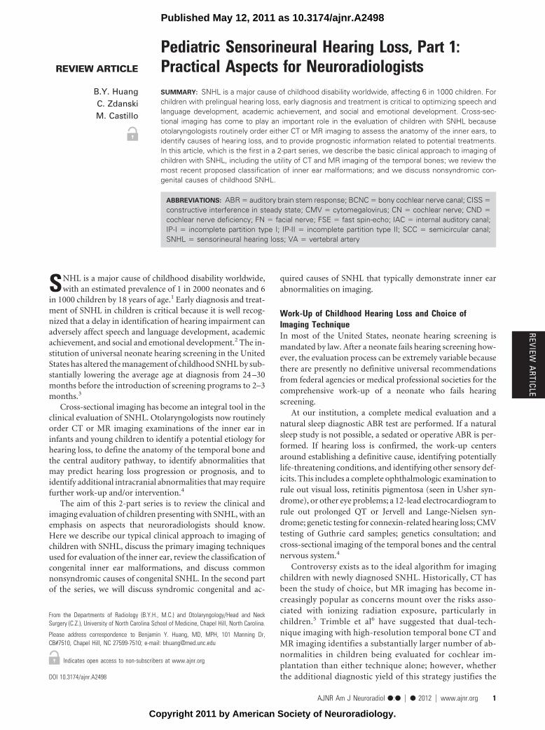
Fig 1. Labyrinthine aplasia. Axial (A) and coronal (B) right temporal bone CT images demonstrate the complete absence of normal inner ear structures. Notice the diminished size of theinner ear edifice and absence of a well-formed internal auditory canal, which help to distinguish this from labyrinthitis ossificans.
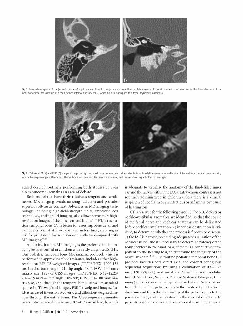
Fig 2. IP-II. Axial CT (A) and CISS (B) images through the right temporal bone demonstrate cochlear dysplasia with a deficient modiolus and fusion of the middle and apical turns, resultingin a bulbous-appearing cochlear apex. The vestibule and semicircular canals are normal, and the vestibular aqueduct is not enlarged.
2 Huang � AJNR ● � ● 2012 � www.ajnr.org
spiral acquisition is performed, with axial and coronal imagesets reconstructed at a section thickness of 0.6 – 0.75 mm.
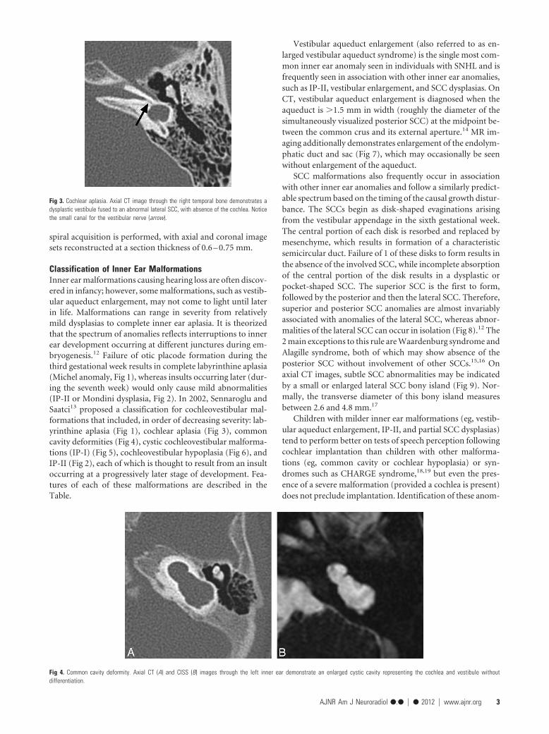
Classification of Inner Ear MalformationsInner ear malformations causing hearing loss are often discov-ered in infancy; however, some malformations, such as vestib-ular aqueduct enlargement, may not come to light until laterin life. Malformations can range in severity from relativelymild dysplasias to complete inner ear aplasia. It is theorizedthat the spectrum of anomalies reflects interruptions to innerear development occurring at different junctures during em-bryogenesis.12 Failure of otic placode formation during thethird gestational week results in complete labyrinthine aplasia(Michel anomaly, Fig 1), whereas insults occurring later (dur-ing the seventh week) would only cause mild abnormalities(IP-II or Mondini dysplasia, Fig 2). In 2002, Sennaroglu andSaatci13 proposed a classification for cochleovestibular mal-formations that included, in order of decreasing severity: lab-yrinthine aplasia (Fig 1), cochlear aplasia (Fig 3), commoncavity deformities (Fig 4), cystic cochleovestibular malforma-tions (IP-I) (Fig 5), cochleovestibular hypoplasia (Fig 6), andIP-II (Fig 2), each of which is thought to result from an insultoccurring at a progressively later stage of development. Fea-tures of each of these malformations are described in theTable.
Vestibular aqueduct enlargement (also referred to as en-larged vestibular aqueduct syndrome) is the single most com-mon inner ear anomaly seen in individuals with SNHL and isfrequently seen in association with other inner ear anomalies,such as IP-II, vestibular enlargement, and SCC dysplasias. OnCT, vestibular aqueduct enlargement is diagnosed when theaqueduct is �1.5 mm in width (roughly the diameter of thesimultaneously visualized posterior SCC) at the midpoint be-tween the common crus and its external aperture.14 MR im-aging additionally demonstrates enlargement of the endolym-phatic duct and sac (Fig 7), which may occasionally be seenwithout enlargement of the aqueduct.
SCC malformations also frequently occur in associationwith other inner ear anomalies and follow a similarly predict-able spectrum based on the timing of the causal growth distur-bance. The SCCs begin as disk-shaped evaginations arisingfrom the vestibular appendage in the sixth gestational week.The central portion of each disk is resorbed and replaced bymesenchyme, which results in formation of a characteristicsemicircular duct. Failure of 1 of these disks to form results inthe absence of the involved SCC, while incomplete absorptionof the central portion of the disk results in a dysplastic orpocket-shaped SCC. The superior SCC is the first to form,followed by the posterior and then the lateral SCC. Therefore,superior and posterior SCC anomalies are almost invariablyassociated with anomalies of the lateral SCC, whereas abnor-malities of the lateral SCC can occur in isolation (Fig 8).12 The2 main exceptions to this rule are Waardenburg syndrome andAlagille syndrome, both of which may show absence of theposterior SCC without involvement of other SCCs.15,16 Onaxial CT images, subtle SCC abnormalities may be indicatedby a small or enlarged lateral SCC bony island (Fig 9). Nor-mally, the transverse diameter of this bony island measuresbetween 2.6 and 4.8 mm.17
Children with milder inner ear malformations (eg, vestib-ular aqueduct enlargement, IP-II, and partial SCC dysplasias)tend to perform better on tests of speech perception followingcochlear implantation than children with other malforma-tions (eg, common cavity or cochlear hypoplasia) or syn-dromes such as CHARGE syndrome,18,19 but even the pres-ence of a severe malformation (provided a cochlea is present)does not preclude implantation. Identification of these anom-
Fig 4. Common cavity deformity. Axial CT (A) and CISS (B) images through the left inner ear demonstrate an enlarged cystic cavity representing the cochlea and vestibule withoutdifferentiation.
Fig 3. Cochlear aplasia. Axial CT image through the right temporal bone demonstrates adysplastic vestibule fused to an abnormal lateral SCC, with absence of the cochlea. Noticethe small canal for the vestibular nerve (arrow).
AJNR Am J Neuroradiol ●:● � ● 2012 � www.ajnr.org 3
alies is important in surgical planning, however, because thepresence of cochlear malformations may make implant place-ment more challenging and also increases the risk for peri-lymph/CSF leak, postimplantation meningitis, and potentialelectrode misplacement, including placement into the IAC. Inaddition, the facial nerve course is aberrant in approximately15%–32% of patients with cochleovestibular anomalies, afinding that influences surgical planning and occasionally pre-cludes placement of the cochleostomy in the optimal positionin relation to the round window.18,19
CNDCND refers to the absence or reduction in caliber of the cochlearnerve and is observed in 12%–18% of ears affected with
SNHL.10,20 In pediatric patients, CND is usually congenital, but itcan occasionally develop subsequent to birth due to atrophy ofthe nerve in patients who previously demonstrated normal hear-ing in the affected ear.21 Because cochlear implants are generallycontraindicated in CND,22,23 it is important to identify this con-dition in children being considered for implantation.
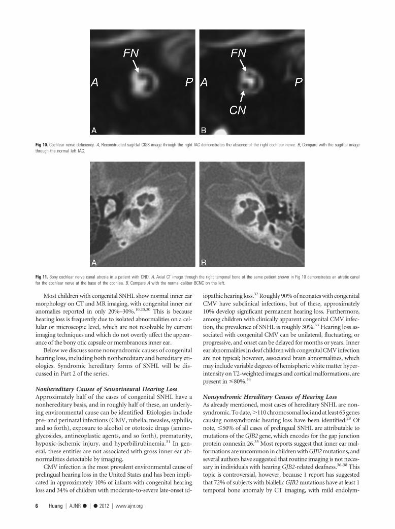
MR imaging is the most sensitive technique for diagnosingCND. On CISS images, the normal cochlear nerve is easily iden-tifiable within the IAC. Oblique sagittal reconstructions orientedperpendicular to the long axis of the IAC are useful for visualizingthe cochlear nerve, which is situated in the anterior inferior quad-rant of the canal, as well as the intracanalicular segment of thefacial nerve and the superior and inferior divisions of the vestib-ular nerve (Fig 10). CND can be seen in isolation (with the ves-
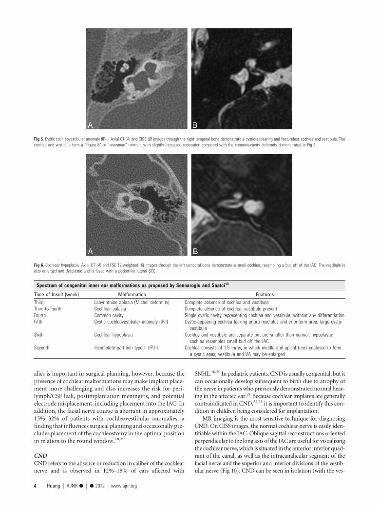
Fig 5. Cystic cochleovestibular anomaly (IP-I). Axial CT (A) and CISS (B) images through the right temporal bone demonstrate a cystic-appearing and featureless cochlea and vestibule. Thecochlea and vestibule form a “figure 8” or “snowman” contour, with slightly increased separation compared with the common cavity deformity demonstrated in Fig 4.
Fig 6. Cochlear hypoplasia. Axial CT (A) and FSE T2-weighted (B) images through the left temporal bone demonstrate a small cochlea, resembling a bud off of the IAC. The vestibule isalso enlarged and dysplastic and is fused with a pocketlike lateral SCC.
Spectrum of congenital inner ear malformations as proposed by Sennaroglu and Saatci13
Time of Insult (week) Malformation FeaturesThird Labyrinthine aplasia (Michel deformity) Complete absence of cochlea and vestibuleThird-to-fourth Cochlear aplasia Complete absence of cochlea; vestibule presentFourth Common cavity Single cystic cavity representing cochlea and vestibule, without any differentiationFifth Cystic cochleovestibular anomaly (IP-I) Cystic-appearing cochlea lacking entire modiolus and cribriform area; large cystic
vestibuleSixth Cochlear hypoplasia Cochlea and vestibule are separate but are smaller than normal; hypoplastic
cochlea resembles small bud off the IACSeventh Incomplete partition type II (IP-II) Cochlea consists of 1.5 turns, in which middle and apical turns coalesce to form
a cystic apex; vestibule and VA may be enlarged
4 Huang � AJNR ● � ● 2012 � www.ajnr.org

tibular nerve divisions present) or in conjunction with aplasia ofthe vestibular nerve (complete absence of the eighth cranialnerve). Commonly, there will be associated narrowing of the IAC(defined as an IAC diameter of �4 mm), and it has been theo-rized that this occurs because the IAC depends on the presence ofvestibulocochlear nerve cells to form normally.21,24
CT is less sensitive than MR imaging for the detection of CNDbut may demonstrate secondary signs, such as an absent or ste-notic BCNC (normal between 1.4 and 3.0 mm)25 or a stenoticIAC (Fig 11). Children with narrow IACs on CT perform worsefollowing implantation than those with normal caliber IACs, pre-sumably because the cochlear nerve is likely to be absent or smallwhen the IAC is narrow.19 Cochlear nerve�deficient ears maydemonstrate normal caliber BCNCs in �23% of cases9 and nor-mal sized IACs in �73% of cases.26
Congenital Hearing LossCongenital hearing loss is defined as hearing loss present atbirth and is generally divided into genetic and nongeneticforms. It is estimated that in �50% of patients, SNHL can belinked to a genetic cause, of which approximately 75%– 80%demonstrate autosomal recessive inheritance; 15%–20%, au-tosomal dominant inheritance; and 1%–2%, X-linked inheri-tance. Approximately 30% of inherited forms of hearing lossare syndromic, and the remaining 70% are considered non-syndromic.27 To date, �300 syndromic forms of hearing losshave been described.28 Of the nongenetic forms of SNHL, anenvironmental cause can be identified in half and the remain-der are considered idiopathic.29
Fig 9. SCC dysplasia with a small lateral SCC bony island. The transverse diameter of thelateral SCC (double arrow) only measures 1.9 mm (normal, 2.6 – 4.8 mm). The vestibule isalso enlarged and dysplastic, and the cochlea is hypoplastic.
Fig 7. Enlarged vestibular aqueduct. A, Axial CT image through the right temporal bone demonstrates enlargement of the vestibular aqueduct, which has a fanlike configuration (arrow).Notice how the width of the aqueduct at its midpoint is larger than that of the adjacent SCCs. The cochlea is also mildly dysplastic (IP-II). B, Axial CISS image through the left temporalbone in a different patient demonstrates marked enlargement of the endolymphatic duct and sac (asterisk).
Fig 8. Lateral SCC dysplasia. Axial (A) and coronal (B) right temporal bone images demonstrate a dysplastic pocketlike lateral SCC, which is fused to the vestibule and missing its centralbony island. On the coronal image, the lateral SCC is truncated. Both the superior and posterior SCCs are present.
AJNR Am J Neuroradiol ●:● � ● 2012 � www.ajnr.org 5
Most children with congenital SNHL show normal inner earmorphology on CT and MR imaging, with congenital inner earanomalies reported in only 20%–30%.10,20,30 This is becausehearing loss is frequently due to isolated abnormalities on a cel-lular or microscopic level, which are not resolvable by currentimaging techniques and which do not overtly affect the appear-ance of the bony otic capsule or membranous inner ear.
Below we discuss some nonsyndromic causes of congenitalhearing loss, including both nonhereditary and hereditary eti-ologies. Syndromic hereditary forms of SNHL will be dis-cussed in Part 2 of the series.
Nonhereditary Causes of Sensorineural Hearing LossApproximately half of the cases of congenital SNHL have anonhereditary basis, and in roughly half of these, an underly-ing environmental cause can be identified. Etiologies includepre- and perinatal infections (CMV, rubella, measles, syphilis,and so forth), exposure to alcohol or ototoxic drugs (amino-glycosides, antineoplastic agents, and so forth), prematurity,hypoxic-ischemic injury, and hyperbilirubinemia.31 In gen-eral, these entities are not associated with gross inner ear ab-normalities detectable by imaging.
CMV infection is the most prevalent environmental cause ofprelingual hearing loss in the United States and has been impli-cated in approximately 10% of infants with congenital hearingloss and 34% of children with moderate-to-severe late-onset id-
iopathic hearing loss.32 Roughly 90% of neonates with congenitalCMV have subclinical infections, but of these, approximately10% develop significant permanent hearing loss. Furthermore,among children with clinically apparent congenital CMV infec-tion, the prevalence of SNHL is roughly 30%.33 Hearing loss as-sociated with congenital CMV can be unilateral, fluctuating, orprogressive, and onset can be delayed for months or years. Innerear abnormalities in deaf children with congenital CMV infectionare not typical; however, associated brain abnormalities, whichmay include variable degrees of hemispheric white matter hyper-intensity on T2-weighted images and cortical malformations, arepresent in �80%.34
Nonsyndromic Hereditary Causes of Hearing LossAs already mentioned, most cases of hereditary SNHL are non-syndromic. To date,�110 chromosomal loci and at least 65 genescausing nonsyndromic hearing loss have been identified.28 Ofnote, �50% of all cases of prelingual SNHL are attributable tomutations of the GJB2 gene, which encodes for the gap junctionprotein connexin 26.35 Most reports suggest that inner ear mal-formations are uncommon in children with GJB2 mutations, andseveral authors have suggested that routine imaging is not neces-sary in individuals with hearing GJB2-related deafness.36-38 Thistopic is controversial, however, because 1 report has suggestedthat 72% of subjects with biallelic GJB2 mutations have at least 1temporal bone anomaly by CT imaging, with mild endolym-
Fig 10. Cochlear nerve deficiency. A, Reconstructed sagittal CISS image through the right IAC demonstrates the absence of the right cochlear nerve. B, Compare with the sagittal imagethrough the normal left IAC.
Fig 11. Bony cochlear nerve canal atresia in a patient with CND. A, Axial CT image through the right temporal bone of the same patient shown in Fig 10 demonstrates an atretic canalfor the cochlear nerve at the base of the cochlea. B, Compare A with the normal-caliber BCNC on the left.
6 Huang � AJNR ● � ● 2012 � www.ajnr.org
phatic fossa enlargement and modiolar hypoplasia being themost common findings.39
By far, the most common inner ear malformation seen in chil-dren with nonsyndromic hearing loss is vestibular aqueduct en-largement. The prevalence of enlarged vestibular aqueducts inchildren with SNHL is estimated to be 10%–15%.40 The malfor-mation is bilateral in approximately 90% of cases, and patientsmay present with profound congenital SNHL, progressive SNHL,or fluctuating SNHL. Initially, hearing loss may be of the high-frequency variety, and some patients may experience acute hear-ing decline related to episodes of minor head injury, overexertion,or barometric pressure changes (eg, related to air travel, deep seadiving, or Valsalva maneuver).14,41 In approximately 63% ofnonsyndromic individuals with vestibular aqueduct enlarge-ment, a mutation at the PDS locus on chromosome 7q31—whichis the same gene responsible for Pendred syndrome—can beidentified, suggesting a genetic basis for most cases of themalformation.41
ConclusionsCauses of hearing loss in the pediatric population are numer-ous and varied, often making evaluation and management ofthe condition quite challenging for otolaryngologists. Imagingplays a central role in the work-up of childhood SNHL becauseit depicts anomalies in the structures of the inner ear, fre-quently provides clues as to the etiology of the hearing loss,and may detect associated comorbid conditions. Although CTand MR imaging are both viable imaging choices, we preferMR imaging due to its ability to directly assess not only theinner ear but also the cranial nerves and brain. In cases ofcongenital hearing impairment, inner ear malformations andCND are fairly common, and identification of these anomaliesis critical when surgical intervention is being considered be-cause they may complicate or even preclude cochlear implan-tation in a child with SNHL.
References1. Billings KR, Kenna MA. Causes of pediatric sensorineural hearing loss: yester-
day and today. Arch Otolaryngol Head Neck Surg 1999;125:517–212. American Academy of Pediatrics, Joint Committee on Infant Hearing. Year
2007 position statement: principles and guidelines for early hearing detectionand intervention programs. Pediatrics 2007;120:898 –921
3. Harrison M, Roush J, Wallace J. Trends in age of identification and interven-tion in infants with hearing loss. Ear Hear 2003;24:89 –95
4. Buchman CA, Adunka OF, Zdanski CJ, et al. Hearing loss in children: theotologist’s perspective. In: Seewald RC, Bamford JM, eds. A Sound FoundationThrough Early Amplification: Proceedings of the Fourth International Conference.Basel, Switzerland: Phonak; 2008:63–77
5. Brenner DJ, Hall EJ. Computed tomography: an increasing source of radiationexposure. N Engl J Med 2007;357:2277– 84
6. Trimble K, Blaser S, James AL, et al. Computed tomography and/or magneticresonance imaging before pediatric cochlear implantation? Developing an in-vestigative strategy. Otol Neurotol 2007;28:317–24
7. Buchman CA, Roush PA, Teagle HF, et al. Auditory neuropathy characteristicsin children with cochlear nerve deficiency. Ear Hear 2006;27:399 – 408
8. Adunka OF, Roush PA, Teagle HF, et al. Internal auditory canal morphology inchildren with cochlear nerve deficiency. Otol Neurotol 2006;27:793– 801
9. Adunka OF, Jewells V, Buchman CA. Value of computed tomography in theevaluation of children with cochlear nerve deficiency. Otol Neurotol2007;28:597– 604
10. Parry DA, Booth T, Roland PS. Advantages of magnetic resonance imaging
over computed tomography in preoperative evaluation of pediatric cochlearimplant candidates. Otol Neurotol 2005;26:976 – 82
11. Davidson HC. Imaging evaluation of sensorineural hearing loss. Semin Ultra-sound CT MR 2001;22:229 – 49
12. Jackler RK, Luxford WM, House WF. Congenital malformations of the innerear: a classification based on embryogenesis. Laryngoscope 1987;97(3 pt 2 suppl40):2–14
13. Sennaroglu L, Saatci I. A new classification for cochleovestibular malforma-tions. Laryngoscope 2002;112:2230 – 41
14. Swartz JD. An overview of congenital/developmental sensorineural hearingloss with emphasis on the vestibular aqueduct syndrome. Semin UltrasoundCT MR 2004;25:353– 68
15. Higashi K, Matsuki C, Sarashina N. Aplasia of posterior semicircular canal inWaardenburg syndrome type II. J Otolaryngol 1992;21:262– 64
16. Koch B, Goold A, Egelhoff J, et al. Partial absence of the posterior semicircularcanal in Alagille syndrome: CT findings. Pediatr Radiol 2006;36:977–79
17. Purcell DD, Fischbein NJ, Patel A, et al. Two temporal bone computed tomog-raphy measurements increase recognition of malformations and predict sen-sorineural hearing loss. Laryngoscope 2006;116:1439 – 46
18. Buchman CA, Copeland BJ, Yu KK, et al. Cochlear implantation in childrenwith congenital inner ear malformations. Laryngoscope 2004;114:309 –16
19. Papsin BC. Cochlear implantation in children with anomalous cochleovestib-ular anatomy. Laryngoscope 2005;115(1 pt 2 suppl 106):1–26
20. McClay JE, Booth TN, Parry DA, et al. Evaluation of pediatric sensorineuralhearing loss with magnetic resonance imaging. Arch Otolaryngol Head NeckSurg 2008;134:945–52
21. Glastonbury CM, Davidson HC, Harnsberger HR, et al. Imaging findings ofcochlear nerve deficiency. AJNR Am J Neuroradiol 2002;23:635– 43
22. Gray RF, Ray J, Baguley DM, et al. Cochlear implant failure due to unexpectedabsence of the eighth nerve: a cautionary tale. J Laryngol Otol 1998;112:646 – 49
23. Maxwell AP, Mason SM, O’Donoghue GM. Cochlear nerve aplasia: its impor-tance in cochlear implantation. Am J Otol 1999;20:335–37
24. Walton J, Gibson WP, Sanli H, et al. Predicting cochlear implant outcomes inchildren with auditory neuropathy. Otol Neurotol 2008;29:302– 09
25. Stjernholm C, Muren C. Dimensions of the cochlear nerve canal: a radioana-tomic investigation. Acta Otolaryngol 2002;122:43– 48
26. Huang BY, Roche JP, Buchman CA, et al. Brain stem and inner ear abnormal-ities in children with auditory neuropathy spectrum disorder and cochlearnerve deficiency. AJNR Am J Neuroradiol 2010;31:1972–79. Epub 2010 Jul 1
27. Lalwani AK, Castelein CM. Cracking the auditory genetic code: nonsyndromichereditary hearing impairment. Am J Otol 1999;20:115–32
28. Morton CC, Nance WE. Newborn hearing screening: a silent revolution.N Engl J Med 2006;354:2151– 64
29. Hone SW, Smith RJ. Medical evaluation of pediatric hearing loss: laboratory,radiographic, and genetic testing. Otolaryngol Clin North Am 2002;35:751– 64
30. Coticchia JM, Gokhale A, Waltonen J, et al. Characteristics of sensorineural hear-ing loss in children with inner ear anomalies. Am J Otolaryngol 2006;27:33–38
31. Morzaria S, Westerberg BD, Kozak FK. Systematic review of the etiology ofbilateral sensorineural hearing loss in children. Int J Pediatr Otorhinolaryngol2004;68:1193–98
32. Barbi M, Binda S, Caroppo S, et al. A wider role for congenital cytomegalovirusinfection in sensorineural hearing loss. Pediatr Infect Dis J 2003;22:39 – 42
33. Peckham CS, Stark O, Dudgeon JA, et al. Congenital cytomegalovirus infection: acause of sensorineural hearing loss. Arch Dis Child 1987;62:1233–37
34. Kimani JW, Buchman CA, Booker JK, et al. Sensorineural hearing loss in a pediat-ric population: association of congenital cytomegalovirus infection with intra-cranial abnormalities. Arch Otolaryngol Head Neck Surg 2010;136:999–1004
35. Denoyelle F, Marlin S, Weil D, et al. Clinical features of the prevalent form ofchildhood deafness, DFNB1, due to a connexin-26 gene defect: implicationsfor genetic counseling. Lancet 1999;353:1298 –303
36. Greinwald JH Jr, Hartnick CJ. The evaluation of children with sensorineuralhearing loss. Arch Otolaryngol Head Neck Surg 2002;128:84 – 87
37. Preciado DA, Lim LH, Cohen AP, et al. A diagnostic paradigm for childhood idio-pathic sensorineural hearing loss. Otolaryngol Head Neck Surg 2004;131:804–09
38. Azaiez H, Smith RJ. In reference to temporal bone imaging in GJB2 deafness.Laryngoscope 2007;117:1127, author reply 1127–29
39. Propst EJ, Blaser S, Stockley TL, et al. Temporal bone imaging in GJB2 deafness.Laryngoscope 2006;116:2178 – 86
40. Arcand P, Desrosiers M, Dube J, et al. The large vestibular aqueduct syndromeand sensorineural hearing loss in the pediatric population. J Otolaryngol1991;20:247–50
41. Reardon W, OMahoney CF, Trembath R, et al. Enlarged vestibular aqueduct:a radiological marker of Pendred syndrome, and mutation of the PDS gene.QJM 2000;93:99 –104
AJNR Am J Neuroradiol ●:● � ● 2012 � www.ajnr.org 7


![Tuli Sensorineural Pada Pasien Geriatrik (Presbikusis) [Autosaved]](https://static.fdocuments.net/doc/165x107/577c77e31a28abe0548de074/tuli-sensorineural-pada-pasien-geriatrik-presbikusis-autosaved.jpg)