3.1 The Cellular Level of Organization 2 4 3.1 The Cellular Level of Organization • The word...
23
8/30/2013 1 1 Chapter 03 Lecture and Animation Outline Copyright © The McGraw-Hill Companies, Inc. Permission required for reproduction or display. See separate PowerPoint slides for all figures and tables pre-inserted into PowerPoint without notes and animations. To run the animations you must be in Slideshow View. Use the buttons on the animation to play, pause, and turn audio/text on or off. Please Note: Once you have used any of the animation functions (such as Play or Pause), you must first click on the slide’s background before you can advance to the next slide. 2 3.1 The Cellular Level of Organization • The cell marks the boundary between the nonliving and the living. • It is the structural and functional unit of an organism. • It is the smallest structure capable of performing all the functions necessary for life. 3 3.1 The Cellular Level of Organization • Three fundamentally different cell types exist. – Prokaryotic cells lack membrane-enclosed structures. – Eukaryotic cells possess membrane-enclosed structures. – Archaean cells posses qualities of both prokaryotes and eukaryotes.
Transcript of 3.1 The Cellular Level of Organization 2 4 3.1 The Cellular Level of Organization • The word...

8/30/2013
1
1
Chapter 03
Lecture and
Animation Outline
Copyright © The McGraw-Hill Companies, Inc. Permission required for reproduction or display.
See separate PowerPoint slides for all figures and tables pre-inserted into PowerPoint without notes and
animations.
To run the animations you must be in Slideshow View. Use the buttons on the animation to play, pause, and turn
audio/text on or off.
Please Note: Once you have used any of the animation functions (such as Play or Pause), you must first click on the slide’s background before you can advance to the next slide.
2
3.1 The Cellular Level of
Organization
• The cell marks the boundary between the
nonliving and the living.
• It is the structural and functional unit of an
organism.
• It is the smallest structure capable of
performing all the functions necessary for life.
3
3.1 The Cellular Level of
Organization
• Three fundamentally different cell types exist.
– Prokaryotic cells lack membrane-enclosed
structures.
– Eukaryotic cells possess membrane-enclosed structures.
– Archaean cells posses qualities of both prokaryotes and eukaryotes.

8/30/2013
2
4
3.1 The Cellular Level of
Organization• The word “cell” entered biology in the 17th century.
• Anton van Leeuwenhoek is recognized for inventing one of the earliest microscopes and
observing a first cell.
• Robert Hooke confirmed earlier findings and coined the term “cell.”
• Matthias Schleiden stated that plants are composed of cells.
• Theodore Schwann stated that animals are
composed of cells.
5
The Cell Theory
• All organisms are composed of one or
more cells.
• Cells are the basic living unit of structure
and function in organisms.
• All cells come only from other cells.
6
Cell Size
• Cell size varies, but are quite small.
• A frog’s egg is about 1 millimeter (1mm) in
diameter.
– Large enough to be seen by naked eye
• Most cells are smaller than 1mm.
• Some cells are as small as 1 micrometer (1µm).

8/30/2013
3
7
Sizes of Living Things
10 m1 m0.1 m1 cm1 mm100 µm10 µm100 nm10 nm1 nm0.1 nm
mousefrog egg
human eggmost bacteria
virus
protein
atomant
electron microscope
light microscope
human eye
human
blue whale
chloroplastrose
1 km100 m1 µm
aminoacids
plant andanimalcells
ostrichegg
Copyright © The McGraw-Hill Companies, Inc. Permission required for reproduction or display.
Figure 3.1
8
Cell Size
• Cells being small is an advantage for multicellular organisms:
– Nutrients such as glucose can enter the cell.
– Wastes such as CO2 can exit cell.
– Therefore surface area affects the ability to get materials in and out of a cell.
– As cells increase in volume, the proportionate amount of surface area decreases.
9
Cell Size
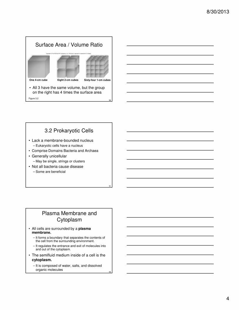
• For a cube-shaped cell:
– The volume increases by the cube of the sides (height X width X depth).
– The surface area increases the square of the sides and the number of sides (height X width X 6).
– If a cell doubles in size, its surface area increases
fourfold, while the volume increases eightfold.
8/30/2013
4
10
Surface Area / Volume Ratio
• All 3 have the same volume, but the group
on the right has 4 times the surface area
One 4-cm cube Eight 2-cm cubes Sixty-four 1-cm cubes
Copyright © The McGraw-Hill Companies, Inc. Permission required for reproduction or display.
Figure 3.2
11
3.2 Prokaryotic Cells
• Lack a membrane-bounded nucleus
– Eukaryotic cells have a nucleus
• Comprise Domains Bacteria and Archaea
• Generally unicellular
– May be single, strings or clusters
• Not all bacteria cause disease
– Some are beneficial
12
Plasma Membrane and
Cytoplasm
• All cells are surrounded by a plasma membrane.
– It forms a boundary that separates the contents of the cell from the surrounding environment.
– It regulates the entrance and exit of molecules into and out of the cytoplasm.
• The semifluid medium inside of a cell is the
cytoplasm.
– It is composed of water, salts, and dissolved organic molecules

8/30/2013
5
13
Copyright © The McGraw-Hill Companies, Inc. Permission required for reproduction or display.
proteinmolecules
phospholipidbilayer
14
Bacterial Anatomy
• Cell Wall
– Located outside of plasma membrane consisting of Peptidoglycan
• Capsule
– A gelatinous sheath that surrounds the cell wall of some bacteria
• Flagellum
– A long thin appendage for movement in some bacteria
• Fimbriae
– Short appendages in some bacteria that help attach to appropriate surface
15
Bacterial Anatomy
• Nucleoid
– A region of the cytoplasm where a single bacterial chromosome is located
– Region not surrounded by a membrane
• Ribosomes
– Used for protein synthesis
• Thylakoids
– Membranes of flattened disks that contain light-sensitive pigments in cyanobacteria
8/30/2013
6
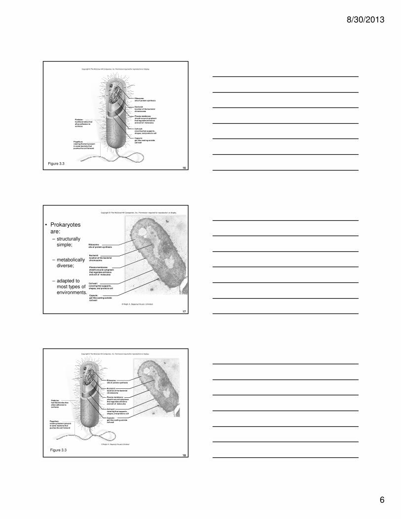
16
Fimbriae:
hairlike bristles that
allow adhesion to
surfaces
Flagellum:
rotating filament present
in some bacteria that
pushes the cell forward
Ribosome:
site of protein synthesis
Nucleoid:
location of the bacterial
chromosome
Plasma membrane:
sheath around cytoplasm
that regulates entrance
and exit of molecules
Cell wall:
covering that supports ,
shapes, and protects cell
Capsule:
gel-like coating outside
cell wall
Copyright © The McGraw-Hill Companies, Inc. Permission required for reproduction or display.
Figure 3.3
17
• Prokaryotes are:
– structurally simple;
– metabolically diverse;
– adapted to most types of environments.
Ribosome:site of protein synthesis
Nucleoid:location of the bacterialchromosome
Plasma membrane:sheath around cytoplasmthat regulates entranceand exit of molecules
Cell wall:covering that supports ,shapes, and protects cell
Capsule:gel-like coating outsidecell wall
Copyright © The McGraw-Hill Companies, Inc. Permission required for reproduction or display.
© Ralph A. Slepecky/Visuals Unlimited
18
Fimbriae:
hairlike bristles that
allow adhesion to
surfaces
Flagellum:
rotating filament present
in some bacteria that
pushes the cell forward
Ribosome:
site of protein synthesis
Nucleoid:
location of the bacterial
chromosome
Plasma membrane:
sheath around cytoplasm
that regulates entrance
and exit of molecules
Cell wall:
covering that supports ,
shapes, and protects cell
Capsule:
gel-like coating outside
cell wall
Copyright © The McGraw-Hill Companies, Inc. Permission required for reproduction or display.
© Ralph A. Slepecky/Visuals Unlimited
Figure 3.3

8/30/2013
7
19
20
3.3 Eukaryotic Cells
• Eukaryotic cells:
– are structurally complex;
– have a nucleus;
– possess membrane-bound organelles;
– make up animals, plants, fungi and protists.
21
Cell Walls
• Some eukaryotic cells have cell walls.
• Plant cells may have a primary and secondary
cell wall.
– Cellulose and chitin make up fungi cell walls.
• Also found in algae (protist) cell walls
– Lignin is found in secondary cell walls.
– Fungi cell walls
• Some cellulose
• Some chitin (also found in insect exoskeletons)

8/30/2013
8
22
23
24

8/30/2013
9
25
Organelles of Eukaryotic Cells
• The term organelle originally referred to
only membranous structures.
• It now refers to any well-defined cell structure
that performs a particular function(s).
• The cell is analogous to a factory where raw
materials enter, whereupon different departments turn them into various products.
– Must also get rid of wastes
26
Cytoskeleton:
Actin filaments
Nucleus:
Rough ER
Ribosomes
Golgi apparatus
Centrioles*
Cytoplasm Peroxisome
*not in plant cells
Intermediate filamentsSmooth ER
Endoplasmic Reticulum:
Microtubules
Centrosome
Mitochondrion
Polyribosome
Nucleolus
Chromatin
Nuclear envelope
Lysosome*
Vesicle
Copyright © The McGraw-Hill Companies, Inc. Permission required for reproduction or display.
Figure 3.4
27
Copyright © The McGraw-Hill Companies, Inc. Permission required for reproduction or display.
Central vacuole*
Smooth ER
Cytoplasm
*not in animal cells
Cell wall*
Cell wall of adjacent cell
Chloroplast*
Mitochondrion
Microtubules
Plasma membrane
Actin filaments
Granum*
Ribosomes
Rough ER
Endoplasmic
Reticulum:
Centrosome
Nucleus:
Nuclear envelope
Chromatin
Nuclear pore
Golgi apparatus
Peroxisome
Nucleolus
Figure 3.5

8/30/2013
10
28
The Nucleus
• The nucleus is a prominent structure with a
diameter of ~5µm.
– Stores genetic material, DNA• Every cell in an individual contains the same DNA.
• DNA governs the characteristics and metabolism of a cell.
– Contains chromatin• Consists of DNA and associated proteins
• Undergoes coiling and condenses into chromosomes
29
The Nucleus
• Nucleoplasm, a semifluid medium in the nucleus
• Nucleolus
– Where ribosomal RNA (rRNA) is made
• Nuclear Envelope—a double membrane that separates the nucleus from the cytoplasm
• Nuclear pores—openings that permit transport
of protein and ribosomal subunits
30
nuclear pore
Nuclear envelope:
inner membrane
outer membrane chromatin
nucleoplasm
nucleolus
phospholipid
nuclearpore
nuclearenvelope
Copyright © The McGraw-Hill Companies, Inc. Permission required for reproduction or display.
Figure 3.6

8/30/2013
11
31
Ribosomes
• Site of protein synthesis– Use messenger RNA (mRNA) as template
• Composed of two subunits (large and small)– Subunits consist of rRNA and protein molecules
• Where found– In groups of polyribosomes, several
ribosomes associated with a single mRNA
– attached to endoplasmic reticulum
– free in cytoplasm
32
Endomembrane System
• Consists of the nuclear envelope, the endoplasmic reticulum, the Golgi apparatus,
and several vesicles (tiny membranous sacs)
• Acts as the transportation and product-processing section of the cell
• Compartmentalizes cell so that enzymatic
reaction restricted to specific cell sections
33
Endoplasmic Reticulum
nuclear enveloperibosomes
0.08 µm
rough
endoplasmic
reticulum
smooth
endoplasmic
reticulum
Copyright © The McGraw-Hill Companies, Inc. Permission required for reproduction or display.
© R. Bolender & D. Fawcett/Visuals Unlimited
Figure 3.7

8/30/2013
12
34
Endoplasmic Reticulum
• Rough ER
– Studded with ribosomes
– Processing, folding and modification of proteins
• Smooth ER
– Has no attached ribosomes
– Synthesizes phospholipids and steroids
– Stores calcium ions
– Various other functions, depending on cell type
35
Golgi Apparatus
• Consists of a stack of three to twenty slightly curved sacs.
• In animal cells, one side is directed toward the ER, and other side is directed toward the
plasma membrane.
• Often referred to as the shipping center of the
cell.
• Apparatus collects, sorts, packages, and
distributes materials such as proteins and lipids.
36
Golgi Apparatus
• Apparatus receives proteins and also lipid-filled vesicles that bud from the ER.
• Proteins made in rough ER have tags that serve as “zip codes” to direct Golgi apparatus
where to send them.
• Lipids and proteins are modified in transit
through the Golgi before being repackaged into secretory vesicles
• Contents are discharged out of the cell by secretion.

8/30/2013
13
37
Copyright © The McGraw-Hill Companies, Inc. Permission required for reproduction or display.
plasmamembrane
secretion
enzyme
lysosomecontains digestive enzymesthat break down worn-outcell parts or substancesentering the cell at theplasma membrane
secretory vesiclefuses with the plasmamembrane as secretionoccurs
Golgi apparatusmodifies lipids and proteinsfrom the ER; sorts themand packages them invesicles
transport vesicleshuttles lipids to variouslocations such as theGolgi apparatus
lipid
transport vesicleshuttles proteins tovarious locations such asthe Golgi apparatus
protein
ribosome
rough endoplasmic reticulumfolds and processes proteinsand packages them in vesicles;vesicles commonly go tothe Golgi apparatus
Nucleus
smooth endoplasmicreticulumsynthesizes lipids andalso per forms variousother functions
incoming vesiclebrings substances intothe cell that are digestedwhen the vesicle fuseswith a lysosome
Figure 3.8
38
Lysosomes
• Membrane-enclosed vesicles formed by
Golgi
– Contain hydrolytic digestive enzymes
– Act as garbage disposals of the cell
– Break down unwanted, foreign substances or worn-out parts of cells
– Bring macromolecules into the cell
39
Please note that due to differing operating systems, some animations will not appear until the presentation is viewed in presentation mode (Slide Show view). You may see blank slides in the “Normal” or “Slide Sorter” views.
All animations will appear after viewing in Slide Show mode and playing each animation. Most animations will require the latest version of the Flash Player, which is available at http://get.adobe.com/flashplayer.
8/30/2013
14
40
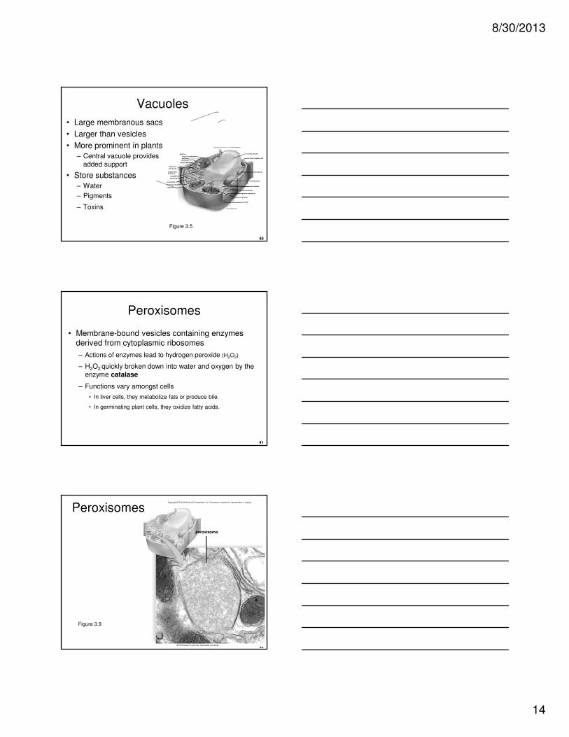
Vacuoles
• Large membranous sacs
• Larger than vesicles
• More prominent in plants
– Central vacuole provides added support
• Store substances
– Water
– Pigments
– Toxins
Figure 3.5
central vacuole*
smooth ER
cytoplasm
*not in animal cells
cell wall*
cell wall of adjacent cell
chloroplast*
mitochondrion
microtubules
plasma membrane
actin filaments
granum*
ribosomes
rough ER
Endoplasmic
Reticulum:
centrosome
Nucleus:
nuclear envelope
chromatin
nuclear pore
Golgi apparatus
peroxisome
nucleolus
41
Peroxisomes
• Membrane-bound vesicles containing enzymes derived from cytoplasmic ribosomes
– Actions of enzymes lead to hydrogen peroxide (H2O2)
– H2O2 quickly broken down into water and oxygen by the enzyme catalase
– Functions vary amongst cells
• In liver cells, they metabolize fats or produce bile.
• In germinating plant cells, they oxidize fatty acids.
42
Copyright © The McGraw-Hill Companies, Inc. Permission required for reproduction or display.
peroxisome
© EM Research Services, Newcastle University
Peroxisomes
Figure 3.9
8/30/2013
15
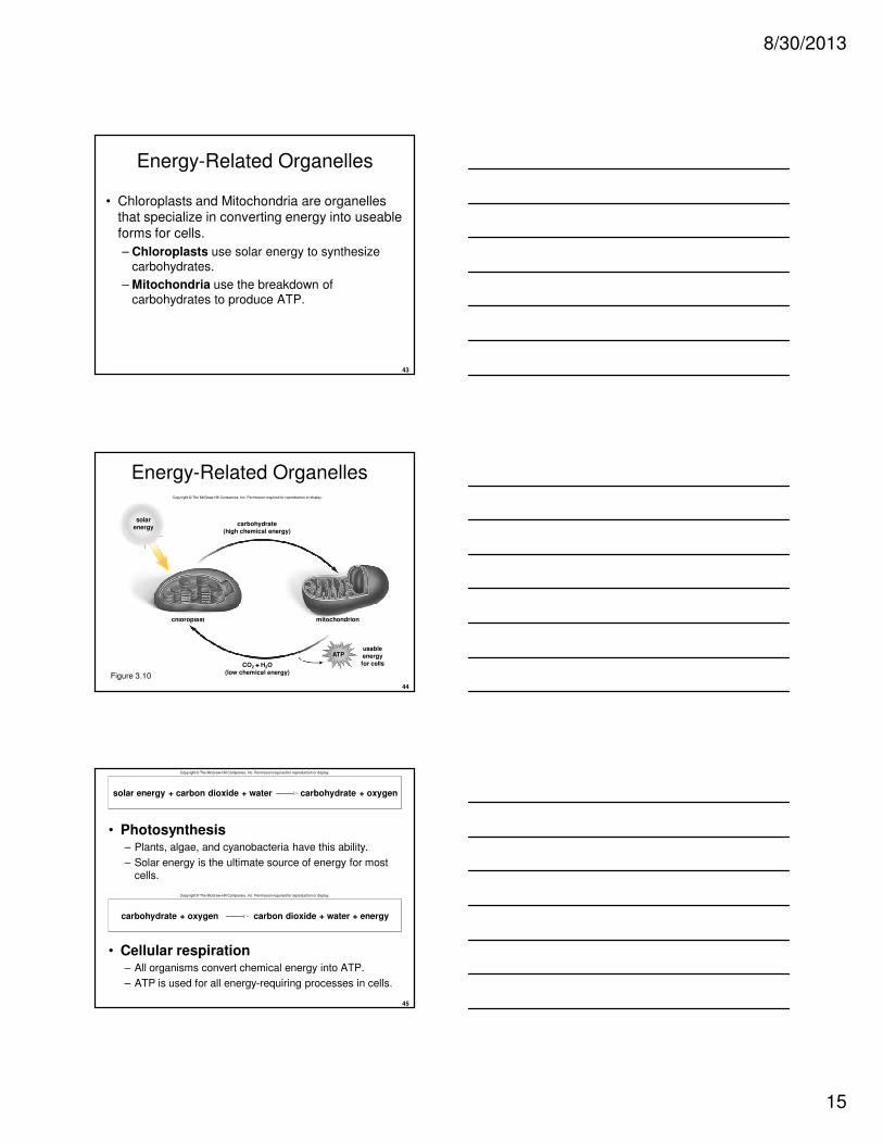
43
• Chloroplasts and Mitochondria are organelles that specialize in converting energy into useable
forms for cells.
– Chloroplasts use solar energy to synthesize carbohydrates.
– Mitochondria use the breakdown of carbohydrates to produce ATP.
Energy-Related Organelles
44
Energy-Related OrganellesCopyright © The McGraw-Hill Companies, Inc. Permission required for reproduction or display.
carbohydrate(high chemical energy)
chloroplast mitochondrion
usableenergyfor cellsCO2 ++++ H2O
(low chemical energy)
solarenergy
ATP
Figure 3.10
45
• Photosynthesis– Plants, algae, and cyanobacteria have this ability.
– Solar energy is the ultimate source of energy for most cells.
• Cellular respiration– All organisms convert chemical energy into ATP.
– ATP is used for all energy-requiring processes in cells.
Copyright © The McGraw-Hill Companies, Inc. Permission required for reproduction or display.
solar energy + carbon dioxide + water carbohydrate + oxygen
carbohydrate + oxygen carbon dioxide + water + energy
Copyright © The McGraw-Hill Companies, Inc. Permission required for reproduction or display.
8/30/2013
16
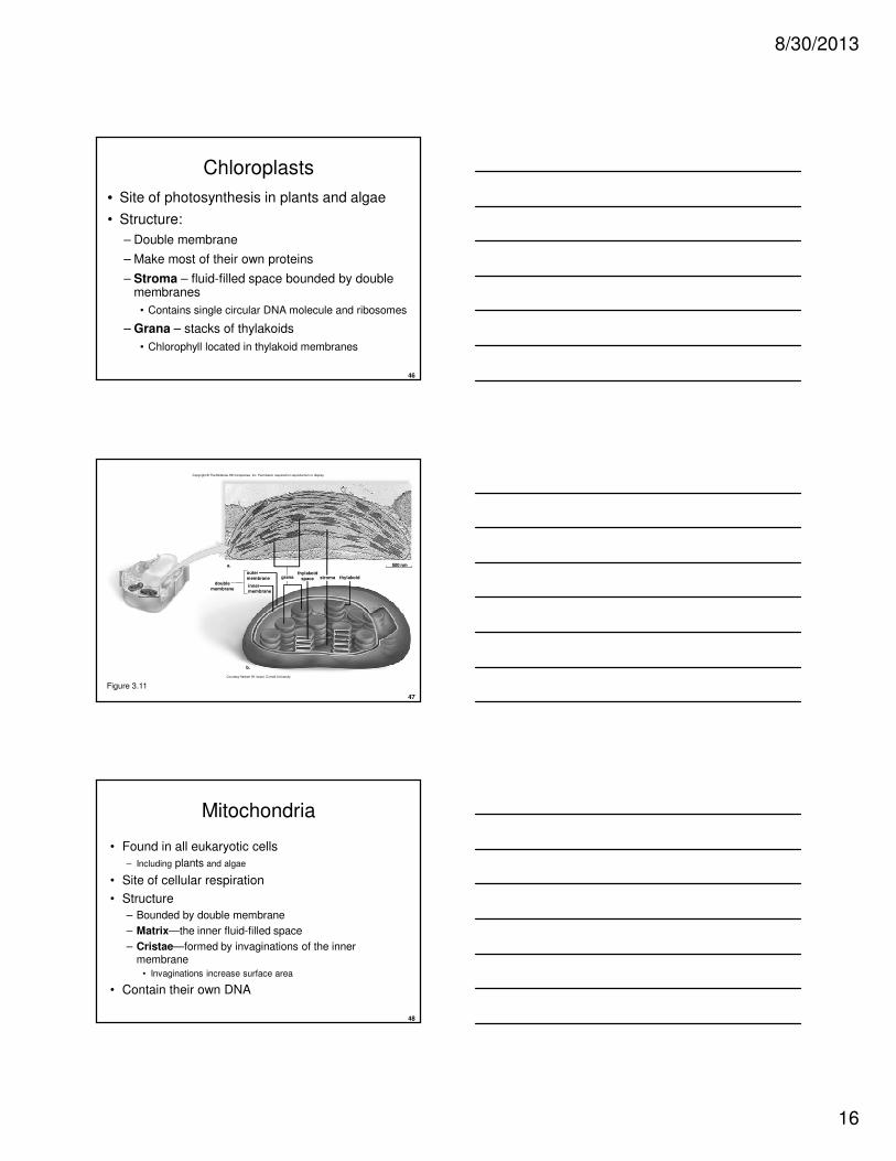
46
Chloroplasts
• Site of photosynthesis in plants and algae
• Structure:
– Double membrane
– Make most of their own proteins
– Stroma – fluid-filled space bounded by double membranes
• Contains single circular DNA molecule and ribosomes
– Grana – stacks of thylakoids
• Chlorophyll located in thylakoid membranes
47
Copyright © The McGraw-Hill Companies, Inc. Permission required for reproduction or display.
grana thylakoidstroma
a.
b.
500 nm
doublemembrane
outermembrane
innermembrane
thylakoidspace
Courtesy Herbert W. Israel, Cornell University
Figure 3.11
48
Mitochondria
• Found in all eukaryotic cells
– Including plants and algae
• Site of cellular respiration
• Structure
– Bounded by double membrane
– Matrix—the inner fluid-filled space
– Cristae—formed by invaginations of the inner membrane
• Invaginations increase surface area
• Contain their own DNA

8/30/2013
17
49
Copyright © The McGraw-Hill Companies, Inc. Permission required for reproduction or display.
cristae matrix
a.
b.
200 nm
doublemembrane
outermembrane
innermembrane
Courtesy Dr. Keith PorterFigure 3.12
50
3.4 The Cytoskeleton
• Consists of three interconnecting proteins
– Actin filaments
– Intermediate filaments
– Microtubules
• Maintains cell shape
• Assists in movement of cell and organelles
• Dynamic—assembled and disassembled as
needed
51
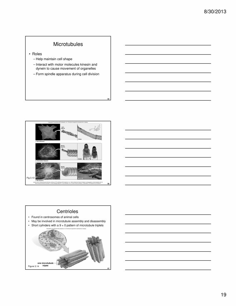
Actin Filaments
• Two long, thin, flexible actin chains twisted in helix
• Roles
– Provide structure as dense web under plasma membrane
– Form projections in intestinal cells as microvilli
– Allow for formation of pseudopods in amoeboid movement
Copyright © The McGraw-Hill Companies, Inc. Permission required for reproduction or display.
a. Actin filaments
actin
subunit
Chara
a(actin): © M. Schliwa/Visuals Unlimited; a(Chara): © The McGraw-Hill Companies, Inc. /Dennis Strete and Darrell Vodopich, photographers
Figure 3.13

8/30/2013
18
52
Actin Filaments
• Actin interacts with motor molecules for movement
– Example: muscle cells
• In the presence of ATP, myosin pulls actin along
Copyright © The McGraw-Hill Companies, Inc. Permission required for reproduction or display.
tail head
PADP +
actin filament
membrane
myosin
moleculesATP
53
Intermediate Filaments• Intermediate in size between actin filaments and
microtubules
• Functions:
– Support nuclear envelope
– Help form cell-to-cell junctions, such as those holding
skin cells tightly together
– Strengthen human hair
human
fibrous
subunits
b. Intermediate filaments
Copyright © The McGraw-Hill Companies, Inc. Permission required for reproduction or display.
b(intermediate): © K.G. Murti/Visuals Unlimited; b(humans): © Amos Morgan/Getty RF
Figure 3.13
54
Microtubules
• Hollow cylinders made of globular tubulin (α and β)
• Assembly– Controlled by Microtubule Organizing Center (MTOC)
– Most important MTOC is centrosome
c. Microtubules
tubulin
subunit
chameleon
Copyright © The McGraw-Hill Companies, Inc. Permission required for reproduction or display.
c(microtubules): © K.G. Murti/Visuals Unlimited; c(chameleon): © Photodisc/Vol. 6/Getty RF
Figure 3.13
8/30/2013
19
55
Microtubules
• Roles
– Help maintain cell shape
– Interact with motor molecules kinesin and dynein to cause movement of organelles
– Form spindle apparatus during cell division
56
a. Actin filaments
human
actin
subunit
Chara
Copyright © The McGraw-Hill Companies, Inc. Permission required for reproduction or display.
fibrous
subunits
b. Intermediate filaments
c. Microtubules
tubulin
subunit
chameleon
a(actin): © M. Schliwa/Visuals Unlimited; a(Chara): © The McGraw-Hill Companies, Inc. /Dennis Strete and Darrell Vodopich, photographers; b(intermediate): © K.G. Murti/Visuals Unlimited; b(humans): © Amos Morgan/Getty RF; c(microtubules): © K.G. Murti/Visuals Unlimited; c(chameleon): © Photodisc/Vol. 6/Getty RF
Fig 3.13
57
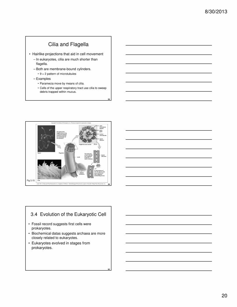
Centrioles• Found in centrosomes of animal cells
• May be involved in microtubule assembly and disassembly
• Short cylinders with a 9 + 0 pattern of microtubule triplets
one microtubule
triplet
Copyright © The McGraw-Hill Companies, Inc. Permission required for reproduction or display.
Figure 3.14
8/30/2013
20
58
Cilia and Flagella
• Hairlike projections that aid in cell movement
– In eukaryotes, cilia are much shorter than
flagella.
– Both are membrane-bound cylinders.
• 9 + 2 pattern of microtubules
– Examples
• Paramecia move by means of cilia.
• Cells of the upper respiratory tract use cilia to sweep
debris trapped within mucus.
59
Copyright © The McGraw-Hill Companies, Inc. Permission required for reproduction or display.
Flagellum
shaft
Sperm
Cilia
flagellum
Flagellum cross section 25 nm
The shaft of the
flagellum has a ring
of nine microtubule
doublets anchoredto a central pair of
microtubules.
In the presence of
ATP, the dynein side
arms reach out to
their neighbors,and bending occurs.
ATP
dynein
side arms
The side arms
of each doublet
are composed
of dynein, amotor molecule.
dynein
side arm
central
microtubules
radial
spoke
outer
microtubule
doublet
plasma
membrane
(cilia): © Dr. G. Moscoso/Photo Researchers, Inc.; (flagellum): © William L. Dentler/Biological Photo Service; (sperm): © David M. Phillips/Photo Researchers, Inc.
Fig 3.15
60
3.4 Evolution of the Eukaryotic Cell
• Fossil record suggests first cells were prokaryotes.
• Biochemical datas suggests archaea are more
closely related to eukaryotes.
• Eukaryotes evolved in stages from
prokaryotes.

8/30/2013
21
61
3.4 Evolution of the Eukaryotic Cell
• Endosymbiotic theory-Mitochondria and
chloroplasts derived from prokaryotes that were taken up by a larger cell
– Mitochondria were originally heterotrophic bacteria
– Chloroplasts were originally cyanobacteria
– After entering the host cell, the bacteria begun living together cooperatively
62
Originalprokaryotic cell
DNA
1. Cell gains a nucleus by theplasma membrane invaginatingand surrounding the DNAwith a double membrane.
2. Cell gains an endomembranesystem by proliferationof membrane.
Copyright © The McGraw-Hill Companies, Inc. Permission required for reproduction or display.
63
photosyntheticbacterium
Animal cellhas mitochondria,but not chloroplasts.
mitochondrion
aerobicbacterium
3. Cell gains mitochondria.
4. Cell gains chloroplasts.
Copyright © The McGraw-Hill Companies, Inc. Permission required for reproduction or display.

8/30/2013
22
64
1. Cell gains a nucleus by theplasma membrane invaginatingand surrounding the DNAwith a double membrane.
2. Cell gains an endomembranesystem by proliferationof membrane.
3. Cell gains mitochondria.
4. Cell gains chloroplasts.
chloroplast
Plant cellhas both mitochondriaand chloroplasts.
photosyntheticbacteriumAnimal cell
has mitochondria,but not chloroplasts.
mitochondrion
aerobicbacterium
Originalprokaryotic cell
DNA
Copyright © The McGraw-Hill Companies, Inc. Permission required for reproduction or display.
65
Supporting Evidence for
Hypothesis
1. Both organelles are similar to bacteria in size and structure
2. Both organelles are bounded by a double membrane
– The outer membrane may be derived from the engulfing vesicle
– The inner one may be derived from the plasma membrane of the original prokaryote
66
Supporting Evidence for
Hypothesis3. Both organelles contain a limited amount of
genetic material and divide by splitting.
– Their DNA is a circular loop like that of prokaryotes.
4. Although most of the proteins within these organelles are produced by the eukaryotic host, they have their own ribosomes and produce some
proteins .
– Their ribosomes resemble those of prokaryotes

8/30/2013
23
67
Supporting Evidence for
Hypotheis
5. The RNA (ribonucleic acid) base sequence
of the ribosomes in chloroplasts and mitochondria also suggests a prokaryotic
origin of these organelles.