3 Cellular Level of Organization
148
© 2011 Pearson Education, Inc. PowerPoint ® Lecture Presentations prepared by Alexander G. Cheroske Mesa Community College at Red Mountain 3 Cellular Level of Organizatio n
-
Upload
garrison-meadows -
Category
Documents
-
view
142 -
download
6
description
3 Cellular Level of Organization. Section 1: An Introduction to Cells. Learning Outcomes 3.1 Describe the cell and its organelles, including the composition and function of each. 3.2 Describe the chief structural features of the plasma membrane. - PowerPoint PPT Presentation
Transcript of 3 Cellular Level of Organization

© 2011 Pearson Education, Inc.
PowerPoint® Lecture Presentations prepared byAlexander G. CheroskeMesa Community College at Red Mountain
3Cellular Level of Organization
© 2011 Pearson Education, Inc.
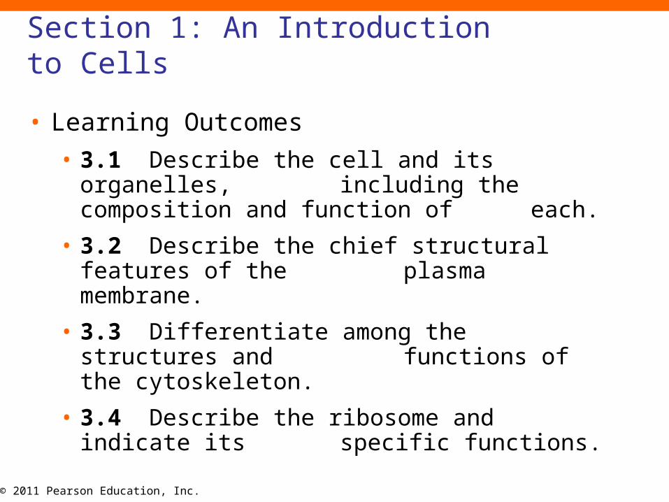
Section 1: An Introduction to Cells
• Learning Outcomes
• 3.1 Describe the cell and its organelles, including the composition and function
of each.
• 3.2 Describe the chief structural features of the plasma membrane.
• 3.3 Differentiate among the structures and functions of the cytoskeleton.
• 3.4 Describe the ribosome and indicate its specific functions.
© 2011 Pearson Education, Inc.
Section 1: An Introduction to Cells
• Learning Outcomes
• 3.5 Describe the Golgi apparatus and indicate its specific functions.
• 3.6 Describe mitochondria, indicate their functions, and explain their
significance to cellular function.
© 2011 Pearson Education, Inc.
Section 1: An Introduction to Cells
• Typical cell
• Smallest living unit in the body
• ~0.1 mm in diameter
• Could not be examined until invention of microscope in 17th century
Animation: Your Cells

© 2011 Pearson Education, Inc.
Section 1: An Introduction to Cells
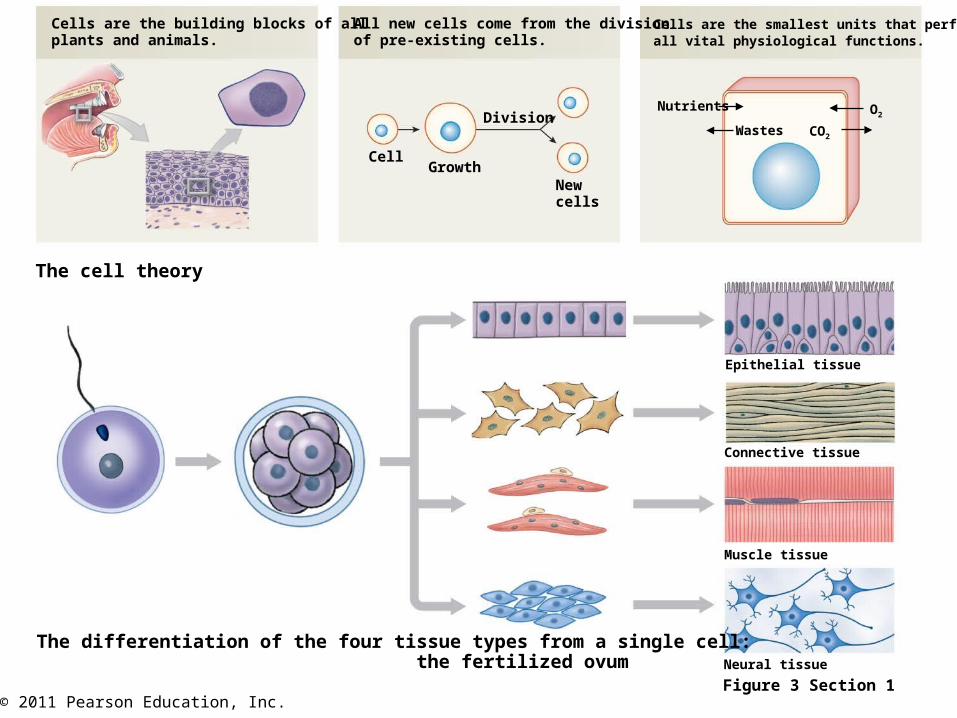
• Cell theory
1. Cells are building blocks of all plants and animals
2. All new cells come from division of preexisting cells
3. Cells are smallest unit that perform all vital physiological functions
© 2011 Pearson Education, Inc.Figure 3 Section 1
Cells are the building blocks of allplants and animals.
All new cells come from the divisionof pre-existing cells.
Cells are the smallest units that performall vital physiological functions.
Cell
Division
GrowthNewcells
Nutrients
Wastes
O2
CO2
The cell theory
The differentiation of the four tissue types from a single cell: the fertilized ovum
Epithelial tissue
Connective tissue
Muscle tissue
Neural tissue

© 2011 Pearson Education, Inc.
Section 1: An Introduction to Cells
• Each cell maintains homeostasis
• Coordinated activities of cells allow homeostasis at higher organizational levels
© 2011 Pearson Education, Inc.
Section 1: An Introduction to Cells
• Cells vary in structure and function but all descend from a single fertilized ovum
• Fertilized ovum contains genetic potential to become any cell
• Cell divisions occur creating smaller, different parcels of cytoplasm
• Cytoplasmic differences turn off/on specific genes in DNA and daughter cells become specialized
• = Differentiation
• Differentiated cells are responsible for all body functions
© 2011 Pearson Education, Inc.
Section 1: An Introduction to Cells
• Extracellular fluid
• Watery medium surrounding cells
• Called interstitial fluid (interstitium, something standing between) in most tissues
© 2011 Pearson Education, Inc.
Module 3.1: Smallest living units of life
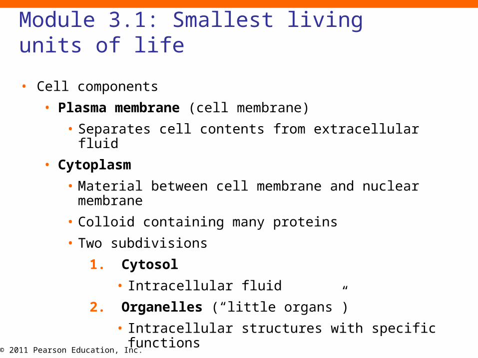
• Cell components
• Plasma membrane (cell membrane)
• Separates cell contents from extracellular fluid
• Cytoplasm
• Material between cell membrane and nuclear membrane
• Colloid containing many proteins
• Two subdivisions
1. Cytosol
• Intracellular fluid
2. Organelles (“little organs”)
• Intracellular structures with specific functions

© 2011 Pearson Education, Inc.
Module 3.1: Smallest living units of life
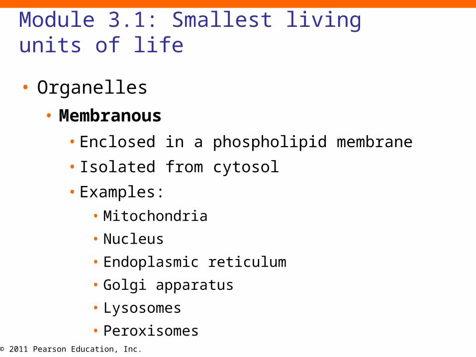
• Organelles
• Nonmembranous
• Not completely enclosed by membranes
• In direct contact with cytosol
• Examples:
• Cytoskeleton
• Microvilli
• Centrioles
• Cilia
• Ribosomes
© 2011 Pearson Education, Inc.
Module 3.1: Smallest living units of life
• Organelles
• Membranous
• Enclosed in a phospholipid membrane
• Isolated from cytosol
• Examples:
• Mitochondria
• Nucleus
• Endoplasmic reticulum
• Golgi apparatus
• Lysosomes
• Peroxisomes
© 2011 Pearson Education, Inc.
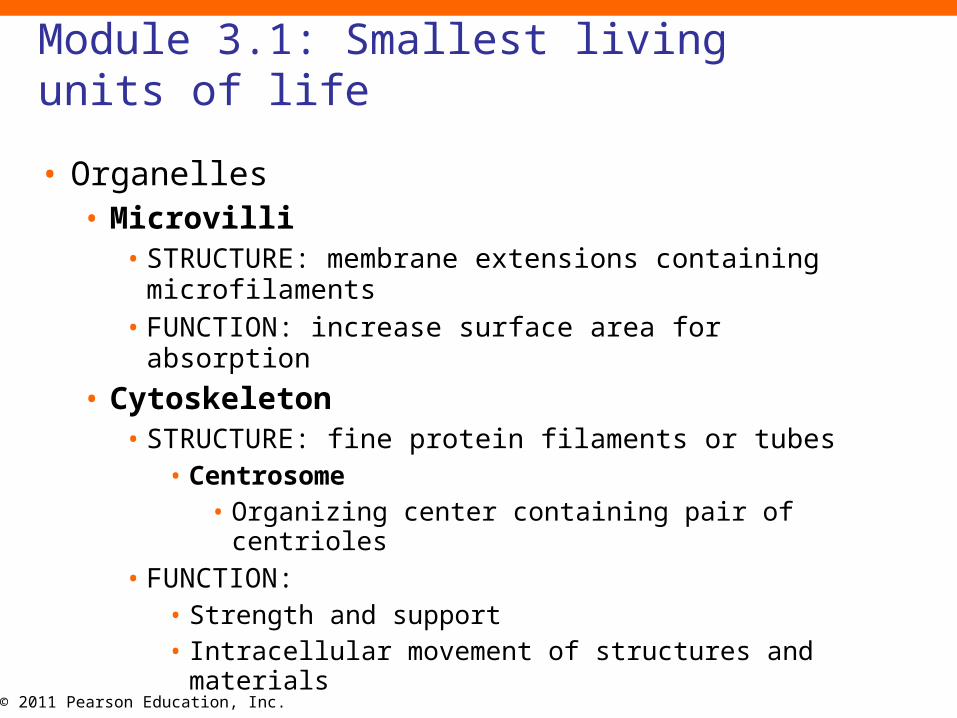
Module 3.1: Smallest living units of life
• Organelles• Microvilli
• STRUCTURE: membrane extensions containing microfilaments
• FUNCTION: increase surface area for absorption
• Cytoskeleton• STRUCTURE: fine protein filaments or tubes
• Centrosome • Organizing center containing pair of centrioles
• FUNCTION: • Strength and support• Intracellular movement of structures and materials
© 2011 Pearson Education, Inc.
Module 3.1: Smallest living units of life
• Organelles
• Ribosomes
• STRUCTURE: RNA and proteins
• Fixed: attached to endoplasmic reticulum
• Free: scattered in cytoplasm
© 2011 Pearson Education, Inc.
Module 3.1: Smallest living units of life
• Organelles
• Peroxisome
• STRUCTURE: vesicles containing degradative enzymes
• FUNCTION:
• Catabolism of fats/other organic compounds
• Neutralization of toxic compounds
• Lysosome
• STRUCTURE: vesicles containing digestive enzymes
• FUNCTION:
• Removal of damaged organelles or pathogens

© 2011 Pearson Education, Inc.
Module 3.1: Smallest living units of life
• Organelles
• Golgi apparatus
• STRUCTURE: stacks of flattened membranes (cisternae) containing chambers
• FUNCTION: storage, alteration, and packaging of synthesized products
• Mitochondria
• STRUCTURE:
• Double membrane
• Inner membrane contains metabolic enzymes
• FUNCTION: production of 95% of cellular ATP
© 2011 Pearson Education, Inc.
Module 3.1: Smallest living units of life
• Organelles
• Nucleus
• STRUCTURE:• Fluid nucleoplasm containing enzymes, proteins,
DNA, and nucleotides
• Surrounded by double membrane
• FUNCTION:• Control of metabolism
• Storage/processing of genetic information
• Control of protein synthesis
Animation: Nucleus
© 2011 Pearson Education, Inc.
Module 3.1: Smallest living units of life
• Organelles
• Endoplasmic reticulum (ER)
• STRUCTURE: membranous sheets and channels
• FUNCTION: synthesis of secretory products, storage, and transport
• Smooth ER
• No attached ribosomes
• Synthesizes lipids and carbohydrates
• Rough ER
• Attached ribosomes
• Modifies/packages newly synthesized proteins
© 2011 Pearson Education, Inc.
Module 3.1 Review
a. Distinguish between the cytoplasm and cytosol.
b. Describe the functions of the cytoskeleton.
c. Identify the membranous organelles and describe their functions.
© 2011 Pearson Education, Inc.
Module 3.2: Plasma membrane
• Plasma membrane
• Selectively permeable membrane that controls:
• Entry of ions and nutrients
• Elimination of wastes
• Release of secretions

© 2011 Pearson Education, Inc.
Module 3.2: Plasma membrane
• Plasma membrane components
• Glycocalyx
• Superficial membrane carbohydrates
• Components of complex molecules
• Proteoglycans (carbohydrates with protein attached)
• Glycoproteins (protein with carbohydrates attached)
• Glycolipids (lipids with carbohydrates attached)
• Functions
• Cell recognition
• Binding to extracellular structures
• Lubrication of cell surface
© 2011 Pearson Education, Inc.
Module 3.2: Plasma membrane
• Plasma membrane components (continued)• Integral proteins
• Part of cell membrane
• Cannot be removed without damaging cell
• Often span entire cell membrane• = Transmembrane proteins
• Can transport water or solutes
• Peripheral proteins• Attached to cell membrane surface
• Removable
• Fewer than integral proteins

© 2011 Pearson Education, Inc.Figure 3.2 1
Structure of the plasma membrane
EXTRACELLULAR FLUID
Glycolipid
Glycocalyx(extracellular
carbohydrates)Integral protein
with channel
CYTOPLASM
Integralglycoproteins
Peripheral proteins Cytoskeleton(microfilaments)
= 2 nm
Integral (transmembrane) proteins

© 2011 Pearson Education, Inc.
Module 3.2: Plasma membrane
• Plasma membrane structure
• Thin (6–10 nm) and delicate
• Phospholipid bilayer
• Mostly comprised of phospholipid molecules in two layers
• Hydrophilic heads at membrane surface
• Hydrophobic tails on the inside
• Isolates cytoplasm from extracellular fluid
Animation: Cell Membrane Barrier

© 2011 Pearson Education, Inc.Figure 3.2 2
Cholesterol
Hydrophilicheads
Hydrophobictails
The phospholipid bilayer that forms theplasma membrane

© 2011 Pearson Education, Inc.
Module 3.2: Plasma membrane
• Plasma membrane functions
• Physical isolation
• Regulation of exchange with external environment
• Sensitivity to environment
• Structural support
• Lipid bilayer provides isolation
• Proteins perform most other functions
© 2011 Pearson Education, Inc.Figure 3.2 3
© 2011 Pearson Education, Inc.Figure 3.2 3

© 2011 Pearson Education, Inc.
Module 3.2 Review
a. List the general functions of the plasma membrane.
b. Which structural component of the plasma membrane is mostly responsible for its ability to isolate a cell from its external environment?
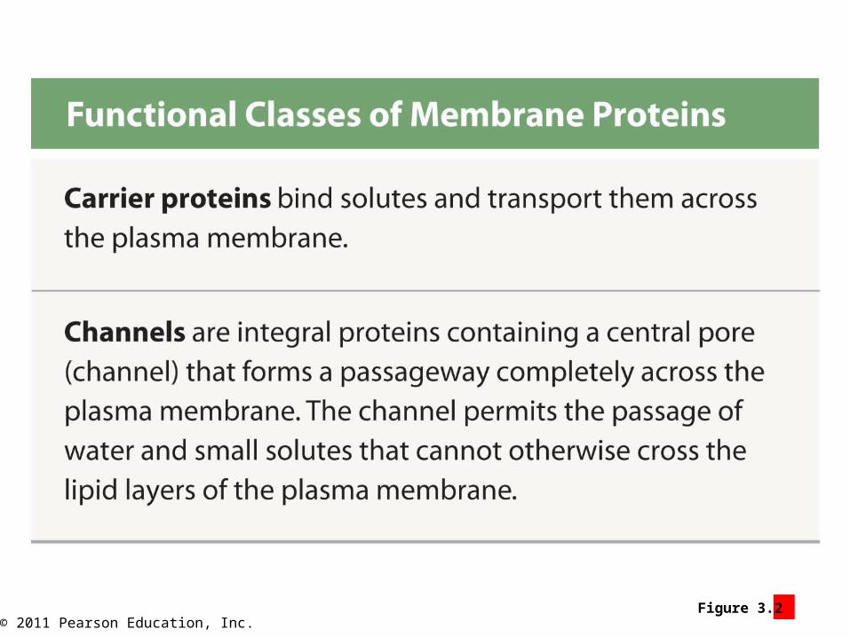
c. Which type of integral protein allows water and small ions to pass through the plasma membrane?
© 2011 Pearson Education, Inc.
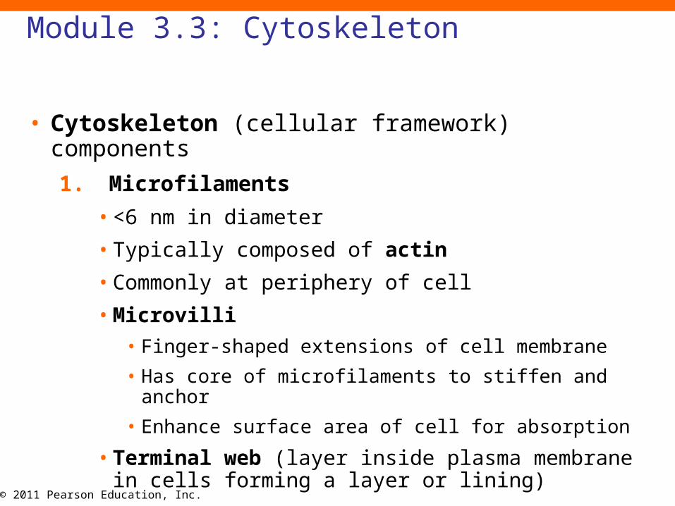
Module 3.3: Cytoskeleton
• Cytoskeleton (cellular framework) components
1. Microfilaments
• <6 nm in diameter
• Typically composed of actin
• Commonly at periphery of cell
• Microvilli
• Finger-shaped extensions of cell membrane
• Has core of microfilaments to stiffen and anchor
• Enhance surface area of cell for absorption
• Terminal web (layer inside plasma membrane in cells forming a layer or lining)

© 2011 Pearson Education, Inc.
Module 3.3: Cytoskeleton
• Cytoskeleton (cellular framework) components (continued)
2. Intermediate filaments
• 7–11 nm in diameter
• Strongest and most durable cytoskeletal elements
3. Microtubules
• ~25 nm in diameter
• Largest components of cytoskeleton
• Extend outward from centrosome (near nucleus)
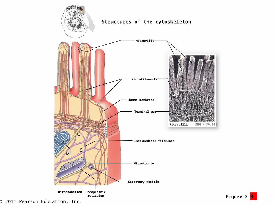
© 2011 Pearson Education, Inc.Figure 3.3 1
Structures of the cytoskeleton
Microvilli
Microfilaments
Plasma membrane
Terminal web
Intermediate filaments
Microtubule
Secretory vesicle
Endoplasmicreticulum
Mitochondrion
Microvilli SEM X 30,000

© 2011 Pearson Education, Inc.
Module 3.3: Cytoskeleton
• Centrioles
• Cylindrical structures
• Composed of microtubules (9 groups of triplets)
• Two in each centrosome
• Control movement of DNA strands during cell division
• Cells without centrioles cannot divide
• Red blood cells
• Skeletal muscle cells
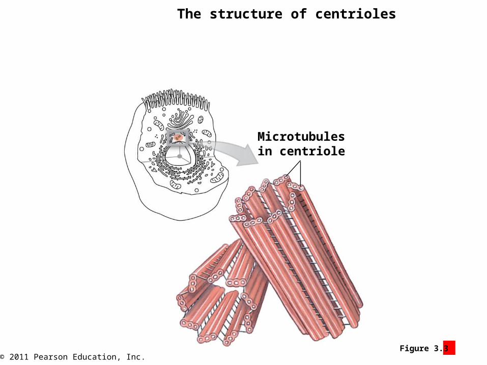
© 2011 Pearson Education, Inc.Figure 3.3 3
The structure of centrioles
Microtubulesin centriole
© 2011 Pearson Education, Inc.
Module 3.3: Cytoskeleton
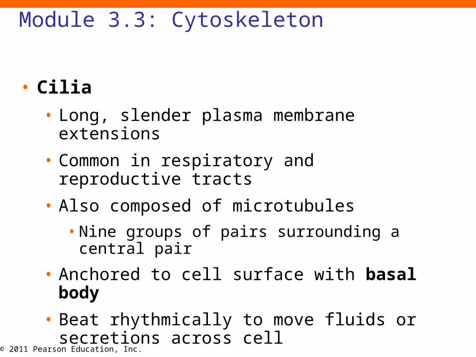
• Cilia
• Long, slender plasma membrane extensions
• Common in respiratory and reproductive tracts
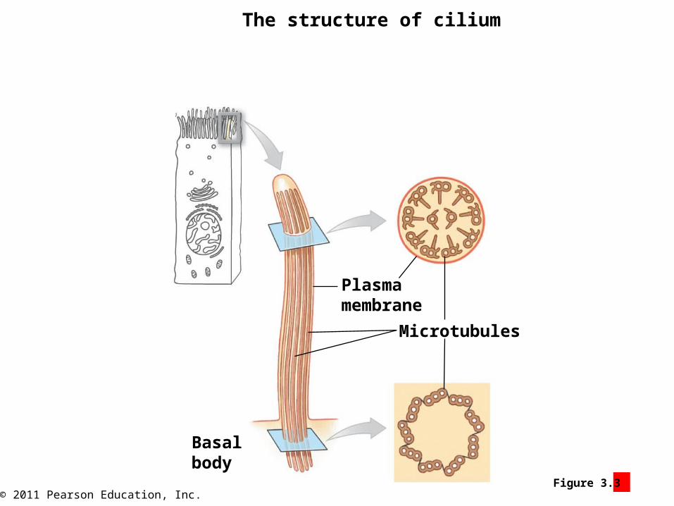
• Also composed of microtubules
• Nine groups of pairs surrounding a central pair
• Anchored to cell surface with basal body
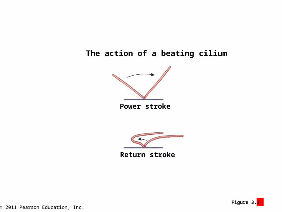
• Beat rhythmically to move fluids or secretions across cell
© 2011 Pearson Education, Inc.Figure 3.3 4
The structure of cilium
Basalbody
Plasmamembrane
Microtubules
© 2011 Pearson Education, Inc.Figure 3.3 5
The action of a beating cilium
Power stroke
Return stroke

© 2011 Pearson Education, Inc.
Module 3.3 Review
a. List the three basic components of the cytoskeleton.
b. Which cytoskeletal component is common to both centrioles and cilia?
c. What is the function of cilia?
© 2011 Pearson Education, Inc.
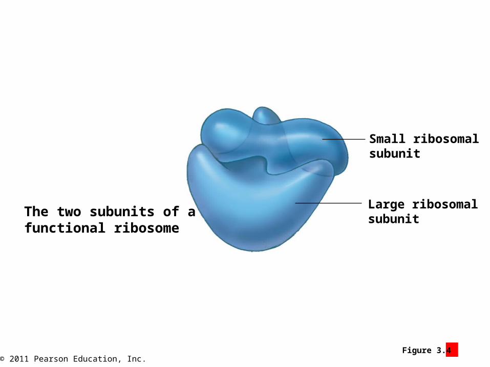
Module 3.4: Ribosomes
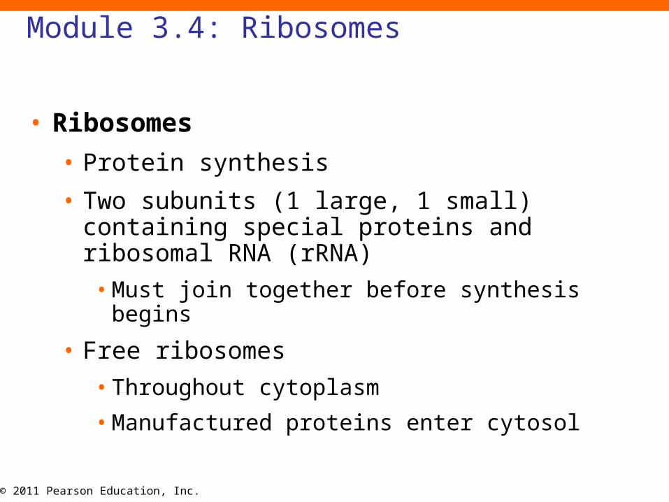
• Ribosomes
• Protein synthesis
• Two subunits (1 large, 1 small) containing special proteins and ribosomal RNA (rRNA)
• Must join together before synthesis begins
• Free ribosomes
• Throughout cytoplasm
• Manufactured proteins enter cytosol
© 2011 Pearson Education, Inc.Figure 3.4 1
The two subunits of a functional ribosome
Small ribosomalsubunit
Large ribosomalsubunit
© 2011 Pearson Education, Inc.
Module 3.4: Ribosomes
• Endoplasmic reticulum (ER)
• Network of intracellular membranes attached to nucleus
• Forms hollow tubes, sheets, and chambers (cisternae, singular, cisterna, reservoir for water)
© 2011 Pearson Education, Inc.
Module 3.4: Ribosomes
• Endoplasmic reticulum (ER)
• Two types
1. Smooth (SER)
• Lacks ribosomes
• Tubular cisternae
2. Rough (RER)
• Has attached (fixed) ribosomes
• Modification of newly synthesized proteins
• Export to Golgi apparatus
• Proportion of SER to RER depends on the cell and its functions

© 2011 Pearson Education, Inc.Figure 3.4 2 – 3
The structure of the endoplasmic reticulum (ER)
Nuclearenvelope
Cisternae
Tubularcisternae
Smoothendoplasticreticulum (SER)

© 2011 Pearson Education, Inc.Figure 3.4 3

© 2011 Pearson Education, Inc.
Module 3.4: Ribosomes
• Functions of SER
• Synthesis of phospholipids and cholesterol
• Synthesis of steroid hormones
• Synthesis and storage of glycerides in liver and fat cells
• Synthesis and storage of glycogen in skeletal and liver cells
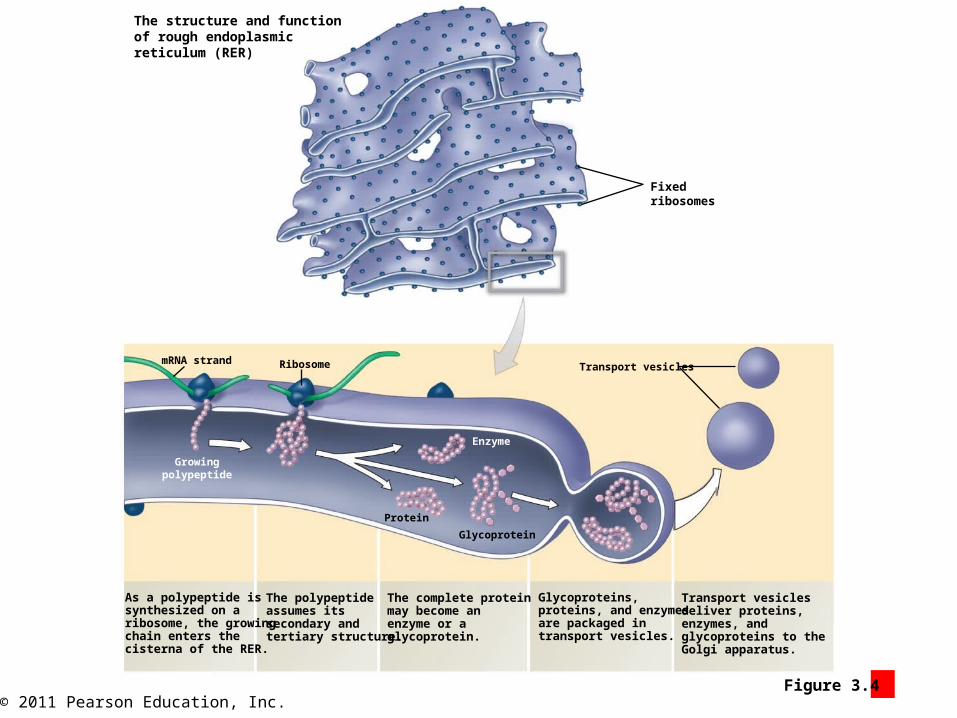
© 2011 Pearson Education, Inc.Figure 3.4 4
mRNA strand Ribosome Transport vesicles
Fixedribosomes
Growingpolypeptide
Enzyme
Protein
Glycoprotein
As a polypeptide issynthesized on aribosome, the growingchain enters thecisterna of the RER.
The polypeptideassumes itssecondary andtertiary structure.
The complete proteinmay become anenzyme or aglycoprotein.
Transport vesiclesdeliver proteins,enzymes, andglycoproteins to the Golgi apparatus.
The structure and functionof rough endoplasmicreticulum (RER)
Glycoproteins,proteins, and enzymesare packaged intransport vesicles.
© 2011 Pearson Education, Inc.
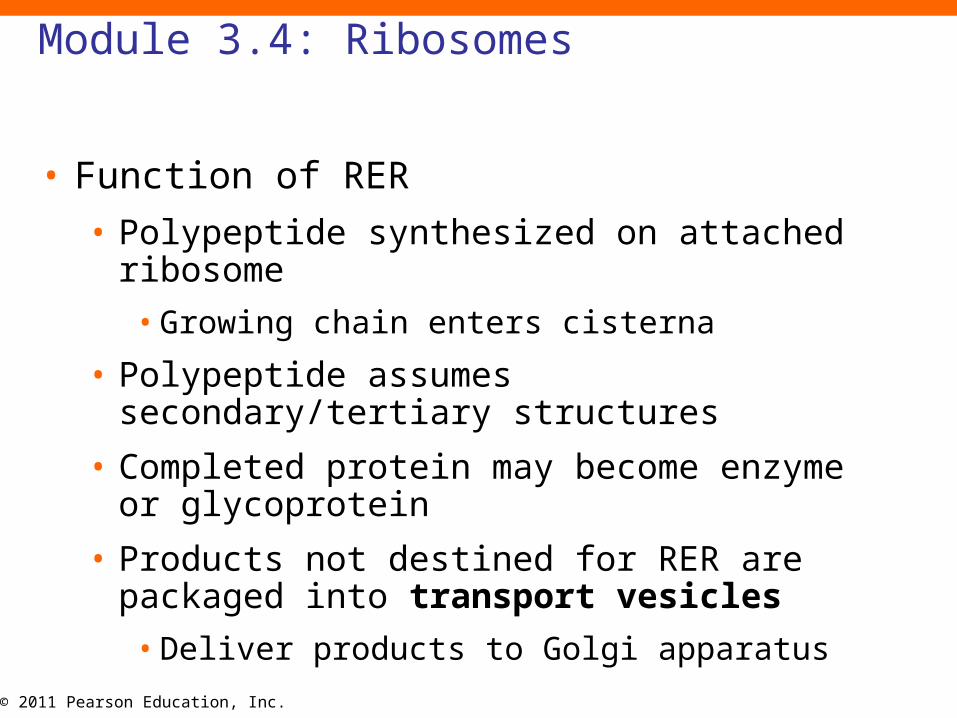
Module 3.4: Ribosomes
• Function of RER
• Polypeptide synthesized on attached ribosome
• Growing chain enters cisterna
• Polypeptide assumes secondary/tertiary structures
• Completed protein may become enzyme or glycoprotein
• Products not destined for RER are packaged into transport vesicles
• Deliver products to Golgi apparatus
© 2011 Pearson Education, Inc.
Module 3.4 Review
a. Describe the immediate cellular destinations of newly synthesized proteins from free ribosomes and fixed ribosomes.
b. Describe the structure of smooth endoplasmic reticulum.
c. Why do certain cells in the ovaries and testes contain large amounts of smooth endoplasmic reticulum (SER)?
© 2011 Pearson Education, Inc.
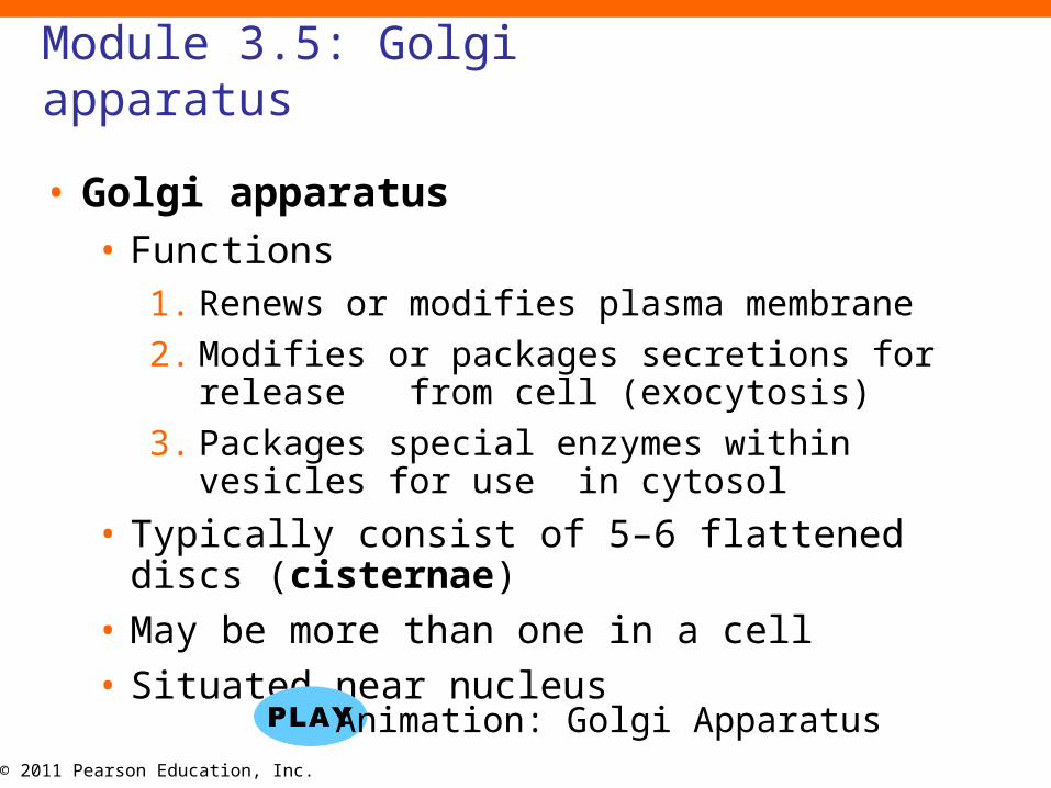
Module 3.5: Golgi apparatus
• Golgi apparatus• Functions
1. Renews or modifies plasma membrane
2. Modifies or packages secretions for release from cell (exocytosis)
3. Packages special enzymes within vesicles for use in cytosol
• Typically consist of 5–6 flattened discs (cisternae)
• May be more than one in a cell
• Situated near nucleus
Animation: Golgi Apparatus
© 2011 Pearson Education, Inc.
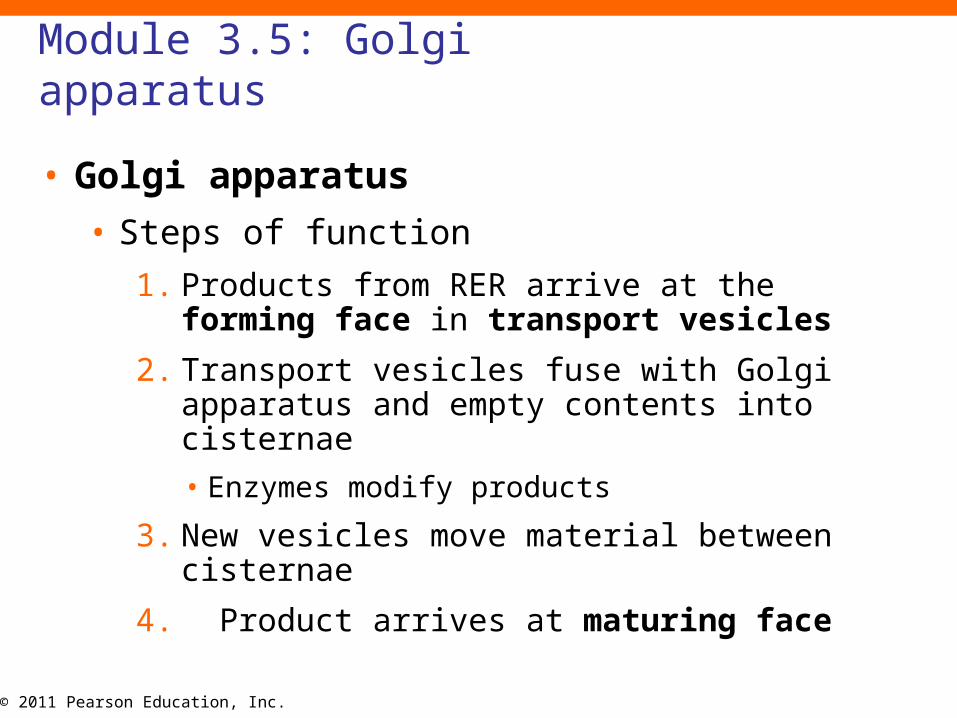
Module 3.5: Golgi apparatus
• Golgi apparatus
• Steps of function
1. Products from RER arrive at the forming face in transport vesicles
2. Transport vesicles fuse with Golgi apparatus and empty contents into cisternae
• Enzymes modify products
3. New vesicles move material between cisternae
4. Product arrives at maturing face
© 2011 Pearson Education, Inc.
Module 3.5: Golgi apparatus
• Golgi apparatus• Products
• Membrane renewal vesicles• Add to plasma membrane
• Secretory vesicles• Contain products to be discharged from the cell
• Fuse with plasma membrane and release contents into extracellular environment
• Enzymes for cytosol• Contained within lysosomes (lyso-, a loosening
+ soma, body)
• Isolate damaging chemical reactions
© 2011 Pearson Education, Inc.
Module 3.5: Golgi apparatus
• Lysosomes
• Isolated intracellular location for toxic chemicals involved in breakdown and recycling of large organic molecules
• Three basic functions
1. May fuse with another organelle to activate digestive enzymes
2. May fuse with another vesicle containing fluid or solid extracellular materials
3. May break down with cell injury or death causing autolysis (enzymes destroy cytoplasm)
• “Suicide packets”
© 2011 Pearson Education, Inc.Figure 3.5 2
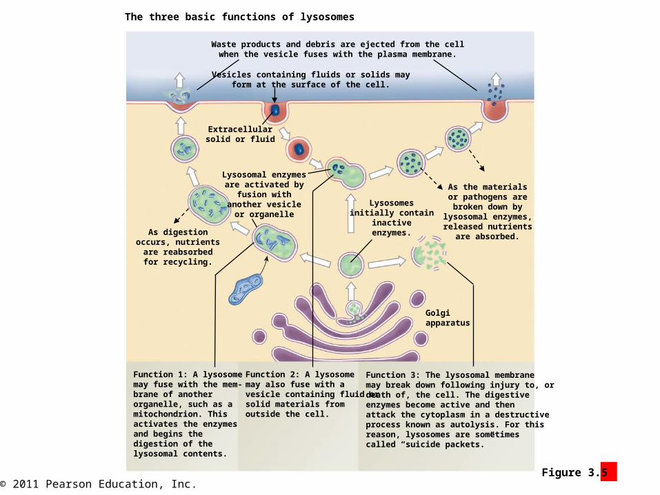
The three basic functions of lysosomes
Waste products and debris are ejected from the cellwhen the vesicle fuses with the plasma membrane.
Vesicles containing fluids or solids mayform at the surface of the cell.
Extracellularsolid or fluid
Lysosomal enzymesare activated by
fusion withanother vesicle
or organelle
As digestionoccurs, nutrientsare reabsorbedfor recycling.
Lysosomesinitially contain
inactiveenzymes.
As the materialsor pathogens arebroken down by
lysosomal enzymes,released nutrients
are absorbed.
Golgiapparatus
Function 1: A lysosomemay fuse with the mem-brane of anotherorganelle, such as amitochondrion. Thisactivates the enzymesand begins thedigestion of thelysosomal contents.
Function 2: A lysosomemay also fuse with avesicle containing fluid orsolid materials fromoutside the cell.
Function 3: The lysosomal membranemay break down following injury to, ordeath of, the cell. The digestiveenzymes become active and thenattack the cytoplasm in a destructiveprocess known as autolysis. For thisreason, lysosomes are sometimescalled “suicide packets.”
© 2011 Pearson Education, Inc.
Module 3.5: Golgi apparatus
• Membrane flow
• Continuous movement and exchange of materials between organelles using vesicles
• Can replace parts of cell membrane to allow cell to grow, mature, or respond to changing environment
© 2011 Pearson Education, Inc.
Module 3.5 Review
a. List the three major functions of the Golgi apparatus.
b. The Golgi apparatus produces lysosomes. What do these lysosomes contain?
c. Describe three functions of lysosomes.
© 2011 Pearson Education, Inc.
Module 3.6: Mitochondria
• Mitochondria (mitos, thread + chondrion, granule)
• Produce energy (ATP) for cells through the breakdown of carbohydrates (glucose)
• Vary widely in shape and number
• Red blood cells have none
• Cardiac muscle cells are 30% mitochondria by volume
Animation: Mitochondria
© 2011 Pearson Education, Inc.Figure 3.6 2
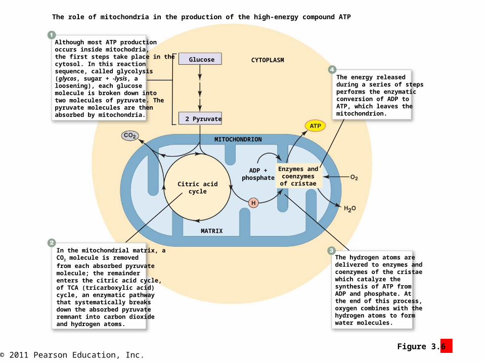
The role of mitochondria in the production of the high-energy compound ATP
Although most ATP productionoccurs inside mitochodria,the first steps take place in thecytosol. In this reactionsequence, called glycolysis(glycos, sugar + -lysis, aloosening), each glucosemolecule is broken down intotwo molecules of pyruvate. Thepyruvate molecules are thenabsorbed by mitochondria.
In the mitochondrial matrix, aCO2 molecule is removedfrom each absorbed pyruvatemolecule; the remainderenters the citric acid cycle,of TCA (tricarboxylic acid)cycle, an enzymatic pathwaythat systematically breaksdown the absorbed pyruvateremnant into carbon dioxideand hydrogen atoms.
The hydrogen atoms aredelivered to enzymes andcoenzymes of the cristaewhich catalyze thesynthesis of ATP fromADP and phosphate. Atthe end of this process,oxygen combines with thehydrogen atoms to formwater molecules.
The energy releasedduring a series of stepsperforms the enzymaticconversion of ADP toATP, which leaves themitochondrion.
CYTOPLASMGlucose
2 Pyruvate
Citric acidcycle
Enzymes andcoenzymesof cristae
MITOCHONDRION
MATRIX
ADP +phosphate
© 2011 Pearson Education, Inc.
Module 3.6: Mitochondria
• Mitochondria
• Double membrane
• Outer (surrounds organelle)
• Inner (contains numerous folds called cristae)
• Encloses liquid (matrix)
• Cristae increase surface area for energetic reactions
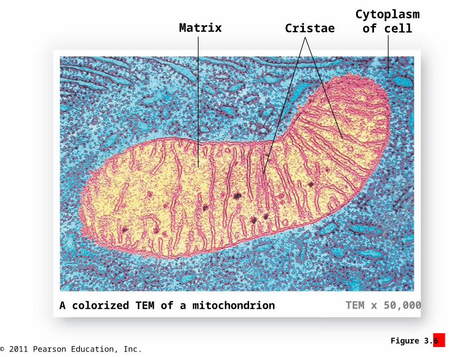
© 2011 Pearson Education, Inc.Figure 3.6 1
Matrix CristaeCytoplasm
of cell
A colorized TEM of a mitochondrion TEM x 50,000

© 2011 Pearson Education, Inc.
Module 3.6: Mitochondria
• Steps of ATP production
1. Glycolysis (glycos, sugar + -lysis, a loosening)
• Occurs in cytosol
• 1 glucose 2 pyruvate
• Pyruvate absorbed into mitochondria
2. In mitochondrial matrix
• CO2 removed from pyruvate
• Enters citric acid (or TCA, tricarboxylic acid) cycle
1.Systematically removes CO2 and hydrogen atoms
© 2011 Pearson Education, Inc.
Module 3.6: Mitochondria
• Steps of ATP production (continued)
3. Enzymes and coenzymes use hydrogen atoms to catalyze ATP from ADP
• Also forms H2O
4. ATP leaves mitochondrion
© 2011 Pearson Education, Inc.
Module 3.6 Review
a. Describe the structure of a mitochondrion.
b. Most of a cell’s ATP is produced within its mitochondria. What gas do mitochondria require to produce ATP?
c. What does the presence of many mitochondria imply about a cell’s energy requirements?

© 2011 Pearson Education, Inc.
Section 2: Nucleus
• Learning Outcomes
• 3.7 Explain the functions of the cell nucleus, and discuss the nature and
importance of the genetic code.
• 3.8 Summarize the process of protein synthesis.
• 3.9 Summarize the process of transcription.
• 3.10 Summarize the process of translation.
© 2011 Pearson Education, Inc.
Section 2: Nucleus
• Nucleus
• Usually largest cellular structure
• Control center for cellular operations
• Can direct synthesis of >100,000 different proteins
• Coded in sequence of nucleotides
• Determines cell structure/function
• Usually only one per cell
• Exceptions:
• Multiple: skeletal muscle cell
• None: mature red blood cells
© 2011 Pearson Education, Inc.
Section 2: Nucleus
• The nucleus directs cellular responses to environmental (ECF) changes
• Short-term adjustments
• Enzyme activity changes
• Long-term adjustments
• Changes in enzymes produced
• Changes in cell structure from changes in structural proteins
• Often occur as part of growth, development, and aging

© 2011 Pearson Education, Inc.Figure 3 Section 2
The role of the nucleus in preservinghomeostasis at the cellular level
Changesin the
compositionof theECF
Binding tomembranereceptors
Diffusionthrough
membranechannels
EXTRACELLULAR FLUID (ECF)
Plasma membrane
SHORT-TERMADJUSTMENTS
LONG-TERMADJUSTMENTS
CYTOPLASM
Binding to nuclearreceptors that alter
genetic activity
DNA innucleus
Enzymeactivation orinactivation
Changes in the bio-chemical processesunder way in the cellresulting from thesynthesis of additionalenzymes, fewerenzymes, or differentenzymes
Changes in thephysical structure ofthe cell due to altera-tions in the rates ortypes of structuralproteins synthesized
© 2011 Pearson Education, Inc.
Module 3.7: Nuclear structure
• Nuclear structures and functions
• Nuclear envelope
• Separates nucleus from cytoplasm
• Double membrane
• Perinuclear space (peri-, around)
• Space between layers
• Nuclear pores
• Allow passage of small molecules and ions
© 2011 Pearson Education, Inc.
Module 3.7: Nuclear structure
• Nuclear structures and functions (continued)
• Nucleoplasm
• Fluid contents of nucleus
• Fine filaments
• Ions
• Enzymes
• RNA and DNA nucleotides
• Small amounts of RNA
• DNA

© 2011 Pearson Education, Inc.
Module 3.7: Nuclear structure
• Nuclear structures and functions (continued)
• Nucleoli (singular, nucleolus)
• Transient, clear nuclear organelles
• Composed of:
• RNA
• Enzymes
• Proteins (histones)
• Form around DNA instructions for forming proteins/RNA
• Assemble RNA subunits
• Many found in large, protein-producing cells
• Liver
• Nerve
• Muscle

© 2011 Pearson Education, Inc.Figure 3.7 1
The structure of the nucleus
Nuclear envelope
Perinuclearspace
Nuclear pores
Nucleoplasm
Nucleolus

© 2011 Pearson Education, Inc.
Module 3.7: Nuclear structure
• DNA
• Instructions for protein synthesis
• Strands coiled
• Wrap around histone molecules forming nucleosomes
• Loosely coiled (chromatin) in nondividing cells
• Tightly coiled (chromosomes) in dividing cells
• To begin, two copies of each chromosome held together at centromere
• 23 paired chromosomes in somatic (general body) cells
• One each from mother/father
• Carry instructions for proteins and RNA
• Also some regulatory and unknown functions

© 2011 Pearson Education, Inc.Figure 3.7 2
The coiled structure of DNA in the nucleus of a nondividing cell
Nucleus of nondividing cell
Chromatin
Nucelosome
Histones DNA doublehelix
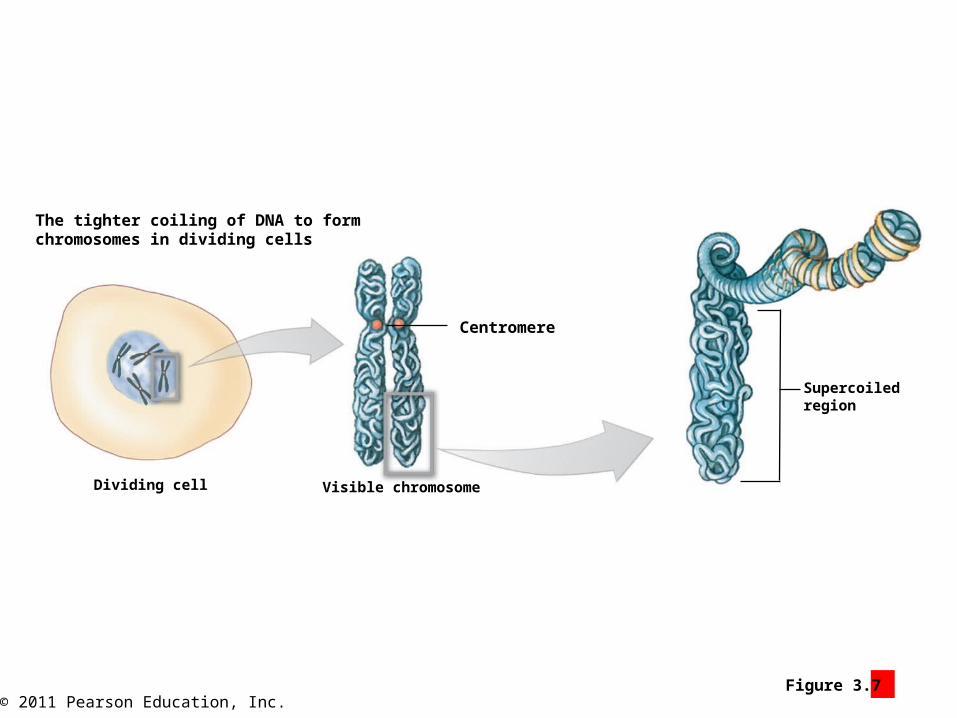
© 2011 Pearson Education, Inc.Figure 3.7 3
The tighter coiling of DNA to formchromosomes in dividing cells
Dividing cell
Centromere
Visible chromosome
Supercoiledregion
© 2011 Pearson Education, Inc.
Module 3.7 Review
a. What molecule in the nucleus contains instructions for making proteins?
b. Describe the contents and the structure of the nucleus.
c. How many chromosomes are contained within a typical somatic cell?

© 2011 Pearson Education, Inc.
Module 3.8: Protein synthesis
• DNA
• Long parallel chains of nucleotides
• Chains held by hydrogen bonds
• Four nitrogenous bases
1. Adenine (A)
2. Thymine (T)
3. Cytosine (C)
4. Guanine (G)
• Genetic code (sequence of nucleotides)
• Triplet code (three nucleotides specify single amino acid)
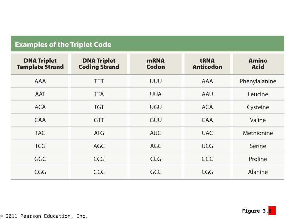
© 2011 Pearson Education, Inc.Figure 3.8 2
© 2011 Pearson Education, Inc.
Module 3.8: Protein synthesis
• DNA (continued)
• Gene
• Functional unit of heredity
• All the DNA nucleotides needed to produce a specific protein
• Size varies (~3003000 nucleotides)

© 2011 Pearson Education, Inc.
Module 3.8: Protein synthesis
• Gene activation
• Removal of histones and DNA uncoiling
• Messenger RNA (mRNA)
• Assembled by enzymes
• Connecting complementary RNA nucleotides
• (A, G, C, U)
• Contains information in triplets (codons)
• Leaves nucleus through pores
• Transfer RNA (tRNA)
• Contains triplets (anticodons) that bind to mRNA codons
• Each type carries a specific amino acid linked to form a polypeptide

© 2011 Pearson Education, Inc.
Module 3.8: Protein synthesis
Animation: Protein Synthesis: RNA Polymerase
Animation: Protein Synthesis: Transcription and Translation
© 2011 Pearson Education, Inc.Figure 3.8 3 – 6
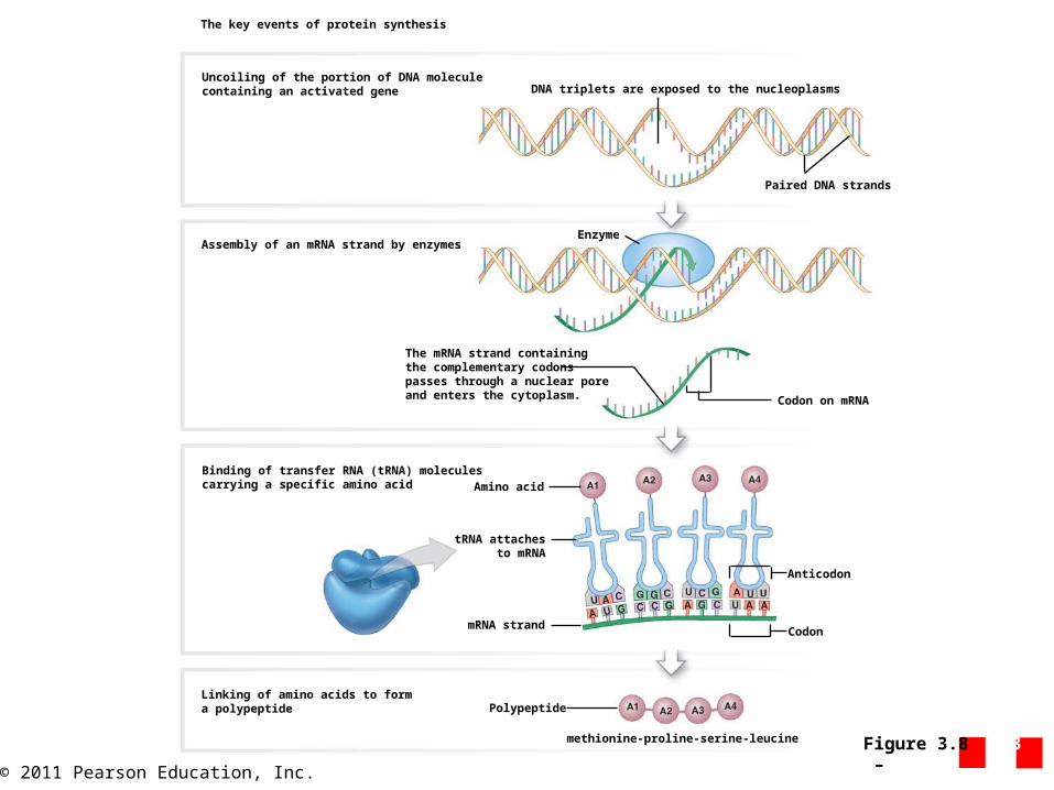
The key events of protein synthesis
Uncoiling of the portion of DNA moleculecontaining an activated gene
Assembly of an mRNA strand by enzymes
Binding of transfer RNA (tRNA) moleculescarrying a specific amino acid
Linking of amino acids to forma polypeptide Polypeptide
methionine-proline-serine-leucine
mRNA strand
tRNA attachesto mRNA
Amino acid
Anticodon
Codon
Codon on mRNA
The mRNA strand containingthe complementary codonspasses through a nuclear poreand enters the cytoplasm.
Enzyme
Paired DNA strands
DNA triplets are exposed to the nucleoplasms

© 2011 Pearson Education, Inc.Figure 3.8 6
A summary of how DNA codes for a protein
The DNAtriplets
determine thesequence of
mRNA codons.
The mRNAcodons
determinethe sequence
of tRNAs.
The sequence of tRNAsdetermines the
sequence of aminoacids in the polypeptide
or protein.

© 2011 Pearson Education, Inc.
Module 3.8 Review
a. List the three types of RNA involved in protein synthesis.
b. What is a gene?
c. Why is the genetic code described as a triplet code?
© 2011 Pearson Education, Inc.
Module 3.9: Transcription
• Transcription (“to copy” or “rewrite”)
• Production of RNA from DNA template
• All three types of RNA are formed
• Example:
• mRNA (information for synthesizing proteins)
© 2011 Pearson Education, Inc.
Module 3.9: Transcription
• Steps of transcription
1. Gene activation
• Occurs at control segment (1st segment of gene)
• Template strand (One DNA strand used to synthesize RNA)
2. RNA polymerase (enzyme)
• Binds to promoter
• Assembles mRNA strand
• Complementary to DNA
• Example: (DNA triplet TAC = mRNA AUG)
• Hydrogen bonds between nucleotides
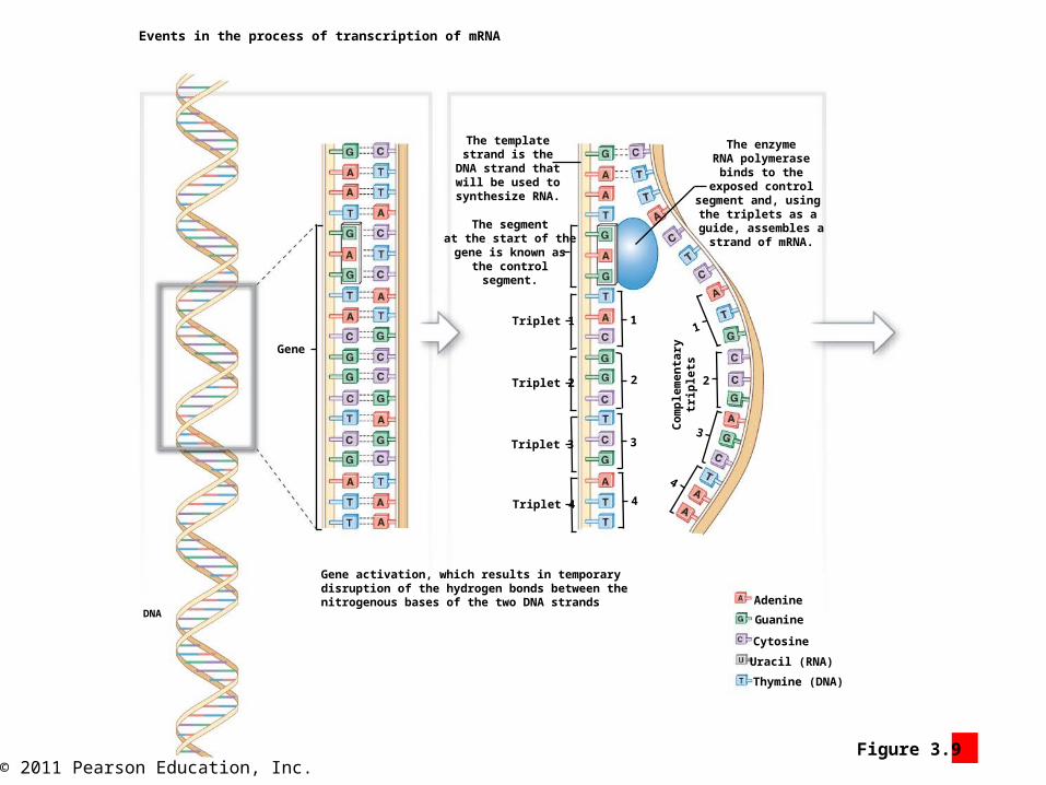
© 2011 Pearson Education, Inc.Figure 3.9 1
Events in the process of transcription of mRNA
Gene
DNA
Gene activation, which results in temporarydisruption of the hydrogen bonds between thenitrogenous bases of the two DNA strands
The templatestrand is the
DNA strand thatwill be used to
synthesize RNA.
The segmentat the start of thegene is known as
the controlsegment.
Triplet 1
Triplet 2
Triplet 3
Triplet 4
Co
mp
lem
enta
rytr
iple
ts
1
2
3
4
1
2
3
4
Adenine
Guanine
Cytosine
Uracil (RNA)
Thymine (DNA)
The enzymeRNA polymerase
binds to theexposed control
segment and, using the triplets as a
guide, assembles astrand of mRNA.

© 2011 Pearson Education, Inc.
Module 3.9: Transcription
• Steps of transcription (continued)
3. Transcription ends
• Stop codon reached
• mRNA detaches
• Complementary DNA strands reassociate (hydrogen bonding between complementary base pairs)
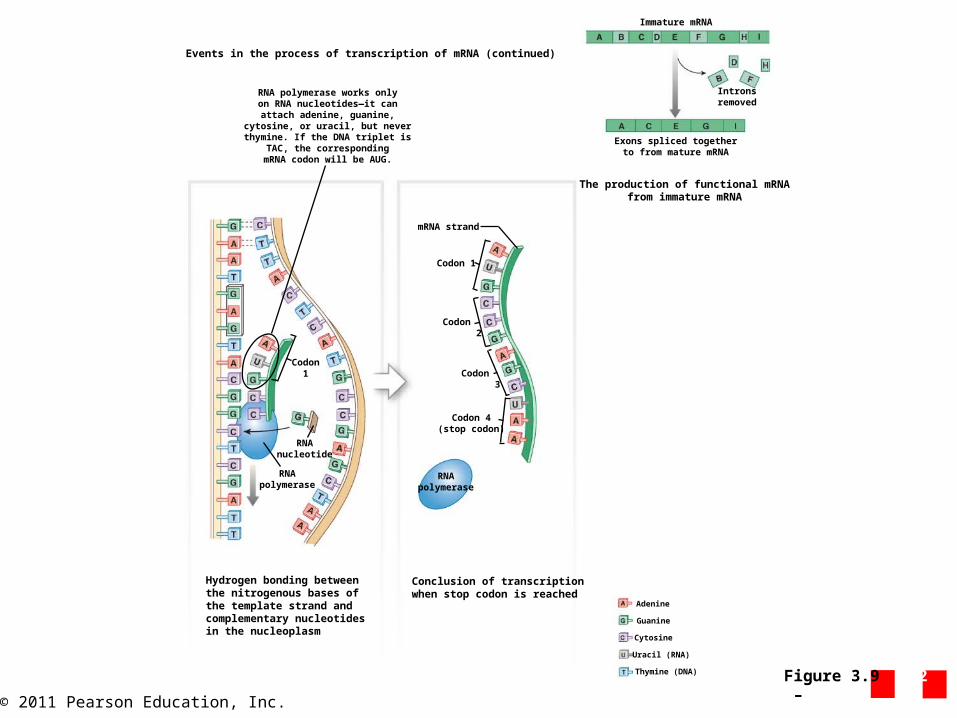
© 2011 Pearson Education, Inc.Figure 3.9 2 – 3
Hydrogen bonding betweenthe nitrogenous bases ofthe template strand andcomplementary nucleotidesin the nucleoplasm
Conclusion of transcriptionwhen stop codon is reached
RNApolymerase
RNAnucleotide
Codon 1
RNA polymerase works onlyon RNA nucleotides—it can
attach adenine, guanine,cytosine, or uracil, but neverthymine. If the DNA triplet is
TAC, the correspondingmRNA codon will be AUG.
mRNA strand
Codon 1
Codon 2
Codon 3
Codon 4(stop codon)
RNApolymerase
The production of functional mRNAfrom immature mRNA
Immature mRNA
Intronsremoved
Exons spliced togetherto from mature mRNA
Adenine
Guanine
Cytosine
Uracil (RNA)
Thymine (DNA)
Events in the process of transcription of mRNA (continued)
© 2011 Pearson Education, Inc.
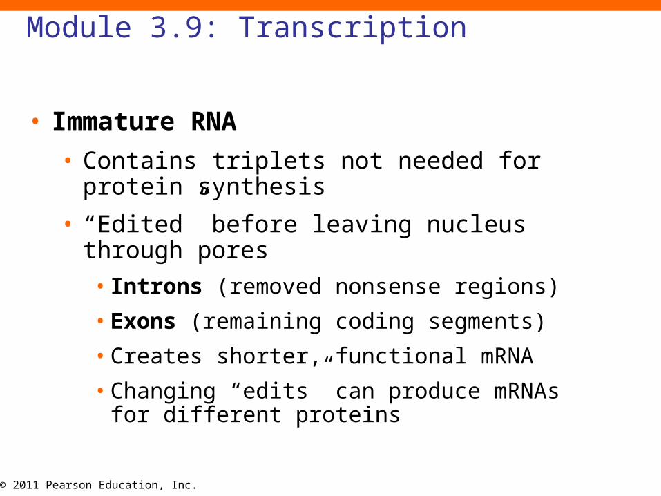
Module 3.9: Transcription
• Immature RNA
• Contains triplets not needed for protein synthesis
• “Edited” before leaving nucleus through pores
• Introns (removed nonsense regions)
• Exons (remaining coding segments)
• Creates shorter, functional mRNA
• Changing “edits” can produce mRNAs for different proteins
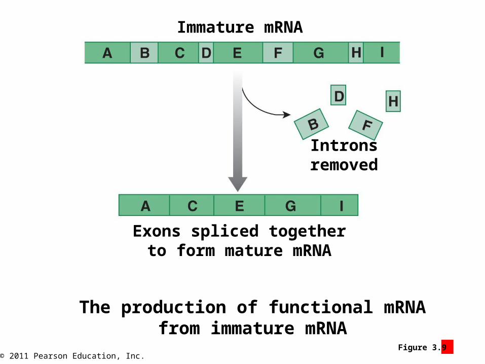
© 2011 Pearson Education, Inc.Figure 3.9 4
The production of functional mRNAfrom immature mRNA
Immature mRNA
Intronsremoved
Exons spliced togetherto form mature mRNA
© 2011 Pearson Education, Inc.
Module 3.9 Review
a. Define DNA template strand.
b. What is transcription?
c. What process would be affected if a cell could not synthesize the enzyme RNA polymerase?
© 2011 Pearson Education, Inc.
Module 3.10: Translation
• Translation (translate nucleic acids to proteins)
• Uses mRNA created in nucleus
• Leaves via nuclear pores
• Occurs in cytoplasm
Animation: Protein Synthesis: Translation Initiation
© 2011 Pearson Education, Inc.
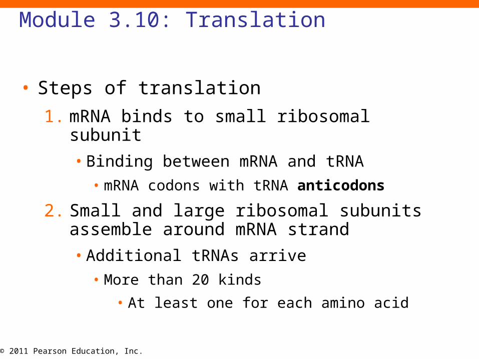
Module 3.10: Translation
• Steps of translation
1. mRNA binds to small ribosomal subunit
• Binding between mRNA and tRNA
• mRNA codons with tRNA anticodons
2. Small and large ribosomal subunits assemble around mRNA strand
• Additional tRNAs arrive
• More than 20 kinds
• At least one for each amino acid
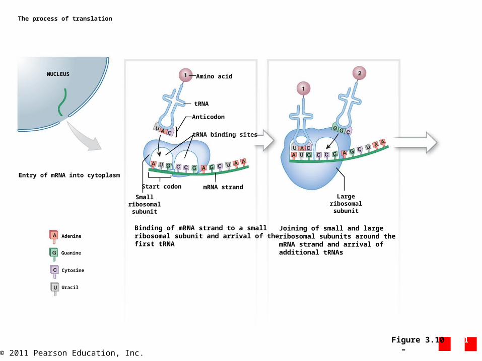
© 2011 Pearson Education, Inc.Figure 3.10 1 – 2
The process of translation
NUCLEUS
Entry of mRNA into cytoplasm
Adenine
Guanine
Cytosine
Uracil
Binding of mRNA strand to a smallribosomal subunit and arrival of thefirst tRNA
Smallribosomal
subunit
Start codon mRNA strand
tRNA binding sites
Anticodon
tRNA
Amino acid
Joining of small and largeribosomal subunits around themRNA strand and arrival ofadditional tRNAs
Largeribosomal
subunit
© 2011 Pearson Education, Inc.
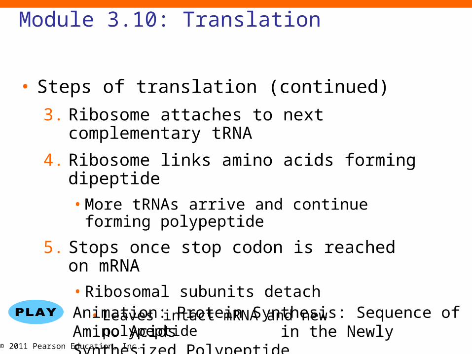
Module 3.10: Translation
• Steps of translation (continued)
3. Ribosome attaches to next complementary tRNA
4. Ribosome links amino acids forming dipeptide
• More tRNAs arrive and continue forming polypeptide
5. Stops once stop codon is reached on mRNA
• Ribosomal subunits detach
• Leaves intact mRNA and new polypeptide
Animation: Protein Synthesis: Sequence of Amino Acids in the Newly Synthesized Polypeptide
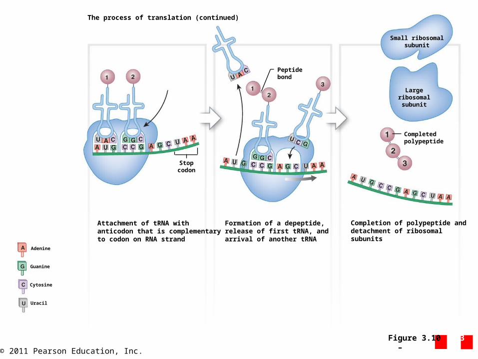
© 2011 Pearson Education, Inc.Figure 3.10 3 – 5
Stopcodon
Attachment of tRNA withanticodon that is complementaryto codon on RNA strand
Formation of a depeptide,release of first tRNA, andarrival of another tRNA
Peptidebond
Completion of polypeptide anddetachment of ribosomalsubunits
Completedpolypeptide
Largeribosomal
subunit
Small ribosomalsubunit
Adenine
Guanine
Cytosine
Uracil
The process of translation (continued)

© 2011 Pearson Education, Inc.
Module 3.10: Translation
• Translation
• Produces a typical protein in ~20 seconds
• mRNA can interact with other ribosomes and produce more proteins
• Multiple ribosomes can attached to a single mRNA strand to quickly produce many proteins
Animation: Transcription Translation
© 2011 Pearson Education, Inc.
Module 3.10 Review
a. What is translation?
b. The nucleotide sequence of three mRNA codons is AUU-GCA-CUA. What is the complementary anticodon sequence for the second codon?
c. During the process of transcription, a nucleotide was deleted from an mRNA sequence that coded for a protein. What effect would this deletion have on the amino acid sequence of the protein?
© 2011 Pearson Education, Inc.
Section 3: Membrane Transport
• Learning Outcomes
• 3.11 Explain the process of diffusion, and identify its significance to the body.
• 3.12 Explain the process of osmosis, and identify its significance to the body.
• 3.13 Describe carrier-mediated transport and its role in the absorption and removal
of specific substances.
• 3.14 Describe vesicular transport as a mechanism for facilitating the
absorption or removal of specific substances from cells.
© 2011 Pearson Education, Inc.
Section 3: Membrane Transport
• Plasma membrane
• Acts as a barrier separating cytosol and ECF
• Must still coordinate cellular activity with extracellular environment
• Permeability (determines which substances can cross membrane)
• Freely permeable (any substances)
• Selectively permeable (some substances cross)
• Impermeable (none can pass)
• No living cell is impermeable

© 2011 Pearson Education, Inc.Figure 3 Section 3 1
Permeability characteristics of membranes
Freely permeable membranes Selectively permeable membranes Impermeable membranes
Freely permeable membranesallow any substance to pass withoutdifficulty.
Selectively permeable membranes,such as plasma membranes, permit thepassage of some materials and preventthe passage of others.
Nothing can pass through impermeablemembranes. Cells may be impermeableto specific substances, but no living cellhas an impermeable membrane.
Protein Protein Protein
Lipids Lipids Lipids
Ions Ions Ions
— — —Water Water Water
Carbohydrates Carbohydrates Carbohydrates
© 2011 Pearson Education, Inc.
Section 3: Membrane Transport
• Selectively permeable membranes
• Selective based on:
1. Characteristics of material to pass
• Size
• Electrical charge
• Molecular shape
• Lipid solubility
• Other factors
2. Characteristics of membrane
• What lipids and proteins present
• How components are arranged
© 2011 Pearson Education, Inc.
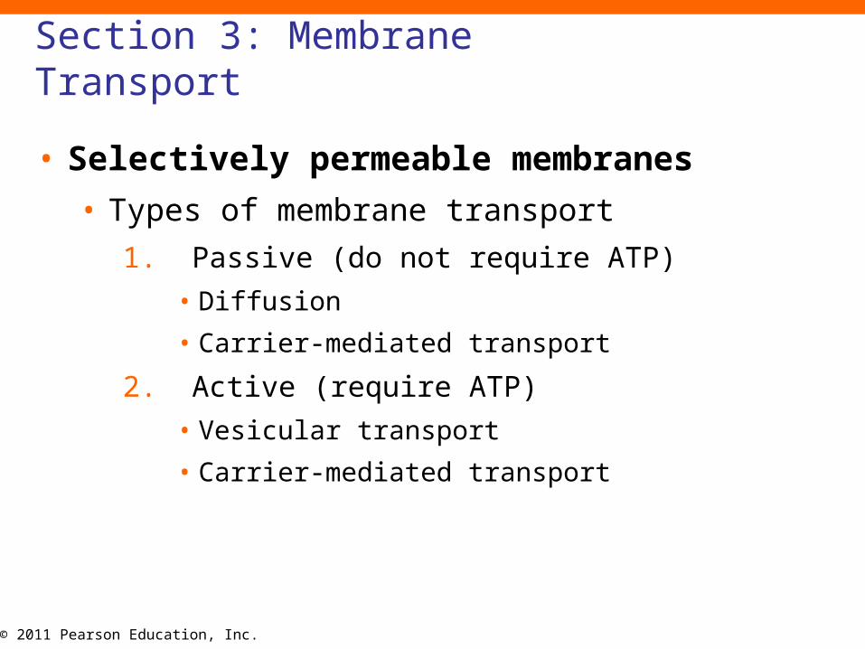
Section 3: Membrane Transport
• Selectively permeable membranes
• Types of membrane transport
1. Passive (do not require ATP)
• Diffusion
• Carrier-mediated transport
2. Active (require ATP)
• Vesicular transport
• Carrier-mediated transport
© 2011 Pearson Education, Inc.Figure 3 Section 3 2
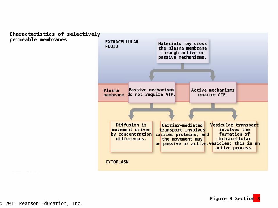
Characteristics of selectivelypermeable membranes EXTRACELLULAR
FLUID
CYTOPLASM
Materials may crossthe plasma membrane
through active orpassive mechanisms.
Passive mechanismsdo not require ATP.
Active mechanismsrequire ATP.
Diffusion ismovement drivenby concentration
differences.
Carrier-mediatedtransport involves
carrier proteins, andthe movement may
be passive or active.
Vesicular transportinvolves theformation ofintracellular
vesicles; this is anactive process.
Plasmamembrane
© 2011 Pearson Education, Inc.
Module 3.11: Diffusion
• Diffusion
• Continuous random movement of ions or molecules in a liquid or gas resulting in even distribution
• Gradient
• Concentration difference or when molecules are not evenly distributed
• At an even distribution, molecular motion continues but no net movement
• Slow in air and water but important over small distances
Animation: Membrane Transport: Diffusion
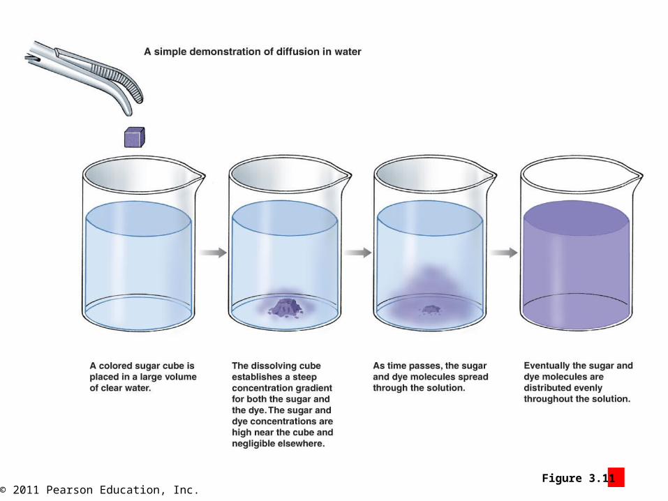
© 2011 Pearson Education, Inc.Figure 3.11 1
© 2011 Pearson Education, Inc.
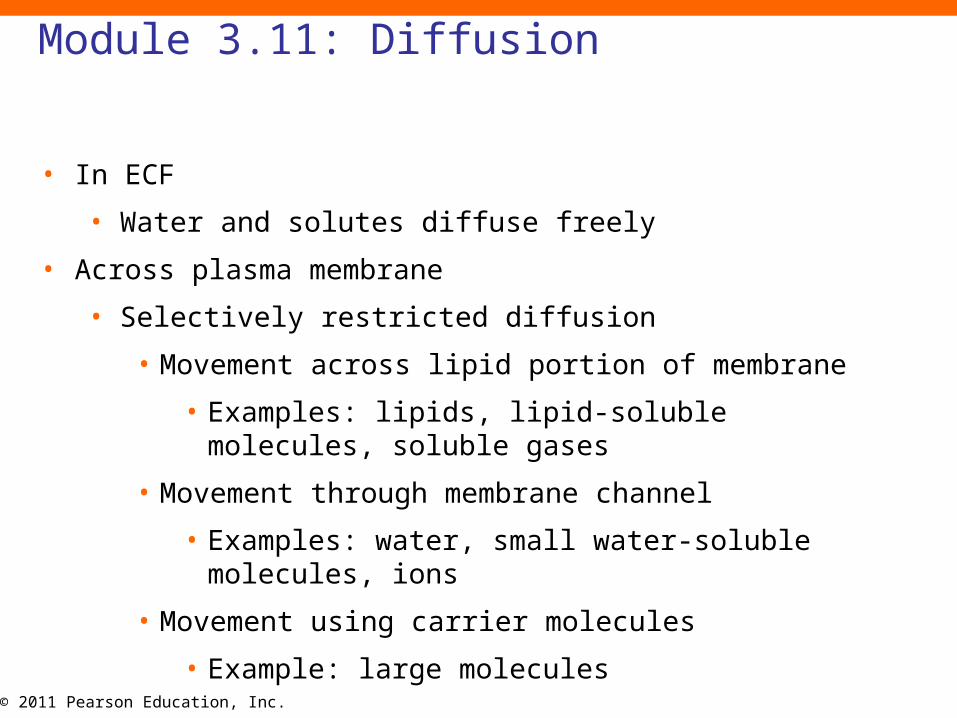
Module 3.11: Diffusion
• In ECF
• Water and solutes diffuse freely
• Across plasma membrane
• Selectively restricted diffusion
• Movement across lipid portion of membrane
• Examples: lipids, lipid-soluble molecules, soluble gases
• Movement through membrane channel
• Examples: water, small water-soluble molecules, ions
• Movement using carrier molecules
• Example: large molecules

© 2011 Pearson Education, Inc.Figure 3.11 2
Large molecules that cannot fitthrough the membrane channelsand cannot diffuse through themembrane lipids can only crossthe plasma membrane whentransported by a carrier mechanism.
Lipids, lipid-soluble molecules,and soluble gases (O2 and CO2)can diffuse across the lipid bilayerof the plasma membrane.
The effects of the plasma membrane, a selectively permeable membrane, onthe diffusion of various substances
Plasma membrane
Channelprotein
EXTRACELLULAR FLUID
CYTOPLASM
Water, small water-soluble molecules,and ions diffuse through membrane channelsthat vary in shape, size, and specificity.
© 2011 Pearson Education, Inc.
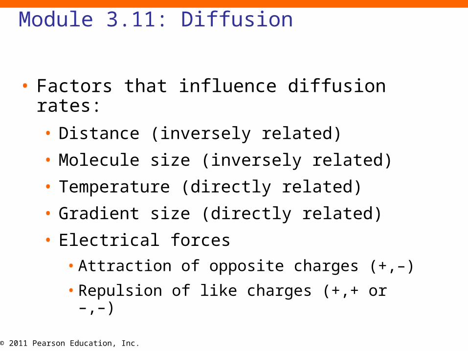
Module 3.11: Diffusion
• Factors that influence diffusion rates:
• Distance (inversely related)
• Molecule size (inversely related)
• Temperature (directly related)
• Gradient size (directly related)
• Electrical forces
• Attraction of opposite charges (+,–)
• Repulsion of like charges (+,+ or –,–)
© 2011 Pearson Education, Inc.
Module 3.11 Review
a. Define diffusion.
b. Identify factors that influence diffusion rates.
c. How would a decrease in the oxygen concentration in the lungs affect the diffusion of oxygen into the blood?

© 2011 Pearson Education, Inc.
Module 3.12: Osmosis
• Osmosis (osmos, a push)
• Net diffusion of water across a membrane
• Maintains similar overall solute concentrations between the cytosol and extracellular fluid
• Osmotic flow
• Movement of water driven by osmosis
• Osmotic pressure
• Indication of force of pure water moving into a solution with higher solute concentration
• Hydrostatic pressure
• Fluid force
• Can be estimate of osmotic pressure when applied to stop osmotic flow
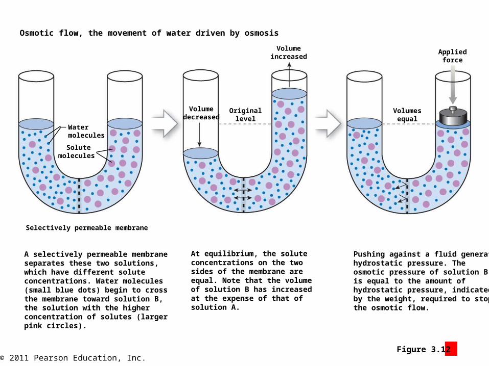
© 2011 Pearson Education, Inc.Figure 3.12 1
Osmotic flow, the movement of water driven by osmosis
Water molecules
Solute molecules
Selectively permeable membrane
A selectively permeable membraneseparates these two solutions,which have different soluteconcentrations. Water molecules(small blue dots) begin to crossthe membrane toward solution B,the solution with the higherconcentration of solutes (largerpink circles).
At equilibrium, the soluteconcentrations on the twosides of the membrane areequal. Note that the volumeof solution B has increasedat the expense of that ofsolution A.
Pushing against a fluid generateshydrostatic pressure. Theosmotic pressure of solution Bis equal to the amount ofhydrostatic pressure, indicatedby the weight, required to stopthe osmotic flow.
Appliedforce
Volumesequal
Volumeincreased
Volumedecreased
Originallevel
© 2011 Pearson Education, Inc.
Module 3.12: Osmosis
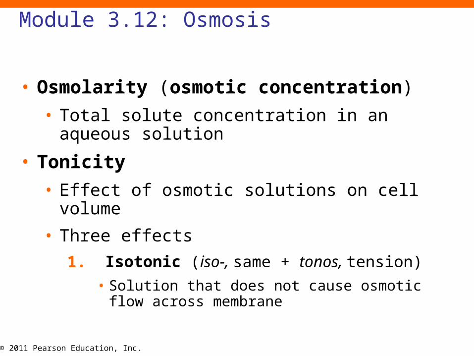
• Osmolarity (osmotic concentration)
• Total solute concentration in an aqueous solution
• Tonicity
• Effect of osmotic solutions on cell volume
• Three effects
1. Isotonic (iso-, same + tonos, tension)
• Solution that does not cause osmotic flow across membrane
© 2011 Pearson Education, Inc.
Module 3.12: Osmosis
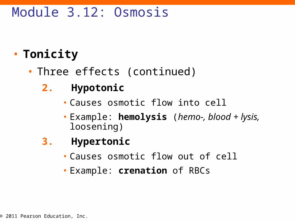
• Tonicity
• Three effects (continued)
2. Hypotonic
• Causes osmotic flow into cell
• Example: hemolysis (hemo-, blood + lysis, loosening)
3. Hypertonic
• Causes osmotic flow out of cell
• Example: crenation of RBCs

© 2011 Pearson Education, Inc.
Module 3.12: Osmosis
• Importance of tonicity vs. osmolarity: Example
• Administering large fluid volumes to patients with blood loss or dehydration
• Administered solution has same osmolarity as ICF but higher concentrations of individual ions/molecules
• Diffusion of solutes may occur across cell membrane
• Water will follow through osmosis
• Cell volume increases
• Normal saline
• 0.9 percent or 0.9 g/dL of NaCl
• Isotonic with blood

© 2011 Pearson Education, Inc.
Module 3.12 Review
a. Describe osmosis.
b. Contrast the effects of a hypotonic solution and a hypertonic solution on a red blood cell.
c. Some pediatricians recommend using a 10 percent salt solution to relieve nasal congestion in infants. Explain the effects this treatment would have on the cells lining the nasal cavity. Would it be effective?

© 2011 Pearson Education, Inc.
Module 3.13: Carrier-mediated transport
• Carrier-mediated transport
• Hydrophilic or large molecules transported across cell membrane by carrier proteins
• Many move specific molecules through the plasma membrane in only one direction
• Cotransport (>1 substance same direction)
• Countertransport (2 substances in opposite directions)
• Carrier called exchange pump

© 2011 Pearson Education, Inc.
Module 3.13: Carrier-mediated transport
• Carrier-mediated transport• Three types
1. Facilitated diffusion
• Requires no ATP (= passive)
• Movement limited by number of available carrier proteins (= can become saturated)
2. Active transport
• Requires energy molecule or ATP (= active)
• Independent of concentration gradient
• Examples:
1.Ion pumps (Na+, K+, Ca2+, and Mg2+)
2.Sodium–potassium ATPase
© 2011 Pearson Education, Inc.
Module 3.13: Carrier-mediated transport
Animation: Membrane Transport: Facilitated Diffusion
Animation: Membrane Transport: Active Transport

© 2011 Pearson Education, Inc.Figure 3.13 1
Facilitated diffusion
EXTRACELLULARFLUID
Glucosemolecule
Carrierprotein
Receptor site
CYTOPLASM
Facilitated diffusion begins when a specificmolecule, such as glucose, binds to a receptorsite on the integral protein.
The shape of the protein then changes, movingthe molecule across the plasma membrane. Thecarrier protein then releases the transportedmolecule into the cytoplasm. Note that this wasaccomplished without ever creating a continuousopen channel between the extracellular fluid andthe cytoplasm.
Glucose releasedinto cytoplasm
© 2011 Pearson Education, Inc.Figure 3.13 2
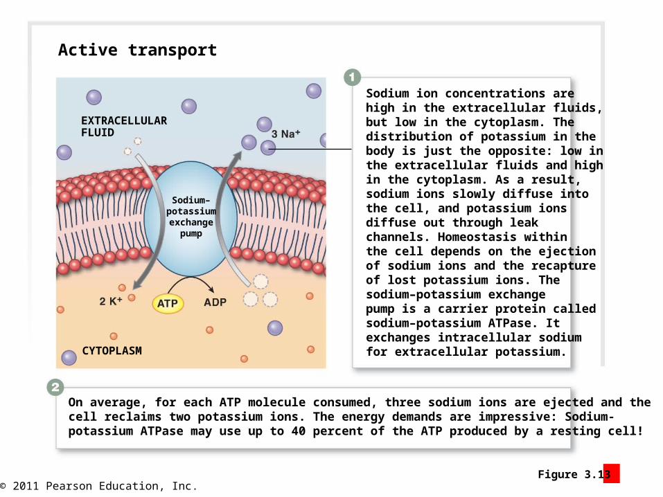
On average, for each ATP molecule consumed, three sodium ions are ejected and thecell reclaims two potassium ions. The energy demands are impressive: Sodium-potassium ATPase may use up to 40 percent of the ATP produced by a resting cell!
Sodium ion concentrations arehigh in the extracellular fluids,but low in the cytoplasm. Thedistribution of potassium in thebody is just the opposite: low inthe extracellular fluids and highin the cytoplasm. As a result,sodium ions slowly diffuse intothe cell, and potassium ionsdiffuse out through leakchannels. Homeostasis withinthe cell depends on the ejectionof sodium ions and the recaptureof lost potassium ions. Thesodium–potassium exchangepump is a carrier protein calledsodium–potassium ATPase. Itexchanges intracellular sodiumfor extracellular potassium.
EXTRACELLULARFLUID
Sodium–potassiumexchange
pump
CYTOPLASM
Active transport
© 2011 Pearson Education, Inc.
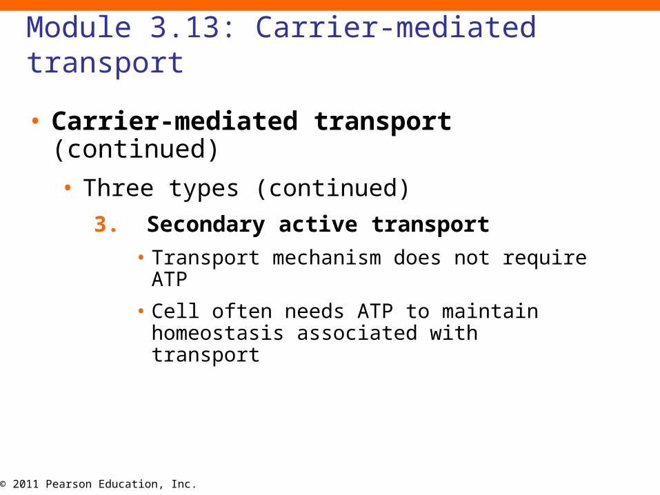
Module 3.13: Carrier-mediated transport
• Carrier-mediated transport (continued)
• Three types (continued)
3. Secondary active transport
• Transport mechanism does not require ATP
• Cell often needs ATP to maintain homeostasis associated with transport

© 2011 Pearson Education, Inc.Figure 3.13 3
A sodium ion and a glucosemolecule bind to receptorsites on the carrier protein.
CYTOPLASM
Glucosemolecule
Sodiumion
Secondary active transport
The carrier protein then changes shape,opening a path to the cytoplasm andreleasing the transported materials. Itthen reassumes its original shape and isready to repeat the process.
To preserve homeostasis, thecell must then expend ATP topump the arriving sodium ionsout of the cell by using thesodium–potassium exchangepump. It thus “costs” the cellone ATP for every threeglucose molecules ittransports into the cell.
Na+–K+
pump
+
+

© 2011 Pearson Education, Inc.
Module 3.13 Review
a. Describe the process of carrier-mediated transport.
b. What do the transport processes of facilitated diffusion and active transport have in common?
c. During digestion, the concentration of hydrogen ions (H+) in the stomach contents increases to many times that in cells lining the stomach. Which transport process could be responsible?
© 2011 Pearson Education, Inc.
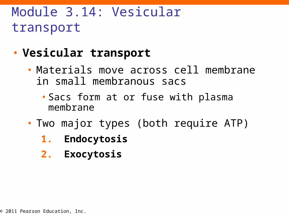
Module 3.14: Vesicular transport
• Vesicular transport
• Materials move across cell membrane in small membranous sacs
• Sacs form at or fuse with plasma membrane
• Two major types (both require ATP)
1. Endocytosis
2. Exocytosis

© 2011 Pearson Education, Inc.
Module 3.14: Vesicular transport
• Vesicular transport (continued)• Two major types (both require ATP)
1. Endocytosis (into cell using endosomes)
a. Receptor-mediated endocytosis
1) Ligand binds to receptor
2) Plasma membrane folds around receptors bound to ligands
3) Coated vesicle forms
4) Vesicle fuses with lysosomes
5) Ligands freed and enter cytosol
6) Lysosome detaches from vesicle
7) Vesicle fuses with plasma membrane again
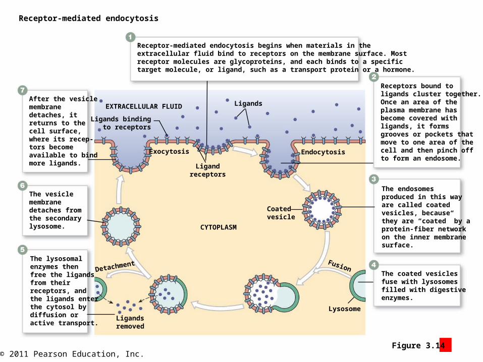
© 2011 Pearson Education, Inc.Figure 3.14 1
Receptor-mediated endocytosis
Receptors bound toligands cluster together.Once an area of theplasma membrane hasbecome covered withligands, it formsgrooves or pockets thatmove to one area of thecell and then pinch offto form an endosome.
The endosomesproduced in this wayare called coatedvesicles, becausethey are “coated” by aprotein-fiber networkon the inner membranesurface.
The coated vesiclesfuse with lysosomesfilled with digestiveenzymes.
The lysosomalenzymes thenfree the ligandsfrom theirreceptors, andthe ligands enterthe cytosol bydiffusion oractive transport.
The vesiclemembranedetaches fromthe secondarylysosome.
After the vesiclemembranedetaches, itreturns to thecell surface,where its recep-tors becomeavailable to bindmore ligands.
EXTRACELLULAR FLUID Ligands
Ligandreceptors
Ligands bindingto receptors
Exocytosis Endocytosis
CYTOPLASM
Coatedvesicle
Ligandsremoved
Lysosome
FusionDetachment
Receptor-mediated endocytosis begins when materials in theextracellular fluid bind to receptors on the membrane surface. Mostreceptor molecules are glycoproteins, and each binds to a specifictarget molecule, or ligand, such as a transport protein or a hormone.

© 2011 Pearson Education, Inc.
Module 3.14: Vesicular transport
• Vesicular transport (continued)• Two major types (both require ATP)
1. Endocytosis (into cell using endosomes) (continued)
b. Pinocytosis (“cell drinking”)
• Formation of endosomes with ECF
• No receptor proteins involved
c. Phagocytosis (“cell eating”)
• Produces phagosomes containing solids
• Phagocytes or macrophages perform phagocytosis
2. Exocytosis
1.Vesicle discharges materials into ECF

© 2011 Pearson Education, Inc.Figure 3.14 2
Endosome
Plasma membrane
Pinocytosis begins with theformation of deep groovesor pockets that then pinchoff and enter the cytoplasm.The steps are similar tothose of receptor-mediatedendocytosis, but they occurin the absence of ligandbinding.
Pinocytosis TEM 20,000
Cytoplasm
Surrounding tissue
Bloodstream

© 2011 Pearson Education, Inc.Figure 3.14 3
Bacterium
Pseudopodium
Phagocytosis
Lysosome
Golgiapparatus
Exocytosis
Phagocytosis begins when cytoplas-mic extensions called pseudopodia(soo-dō-PŌ-dē-ah; pseduo-, falsepodon, foot; singular pseudopodium)surround the object.
The pseudopodia then fuse at theirtips to form a phagosome containingthe targeted material.
This vesicle then fuses with manylysosomes, whereupon its contentsare digested by lysosomal enzymes.
Released nutrients diffuse into thesurrounding cytoplasm.
The residue is then ejected from thecell through exocytosis.
The vesicular events linkingphagocytosis and exocytosis
© 2011 Pearson Education, Inc.
Module 3.14 Review
a. Describe endocytosis.
b. Describe exocytosis.
c. When they encounter bacteria, certain types of white blood cells engulf the bacteria and bring them into the cell. What is this process called?
© 2011 Pearson Education, Inc.
Section 4: Cell Life Cycle
• Learning Outcomes
• 3.15 Describe interphase, and explain its significance.
• 3.16 Describe the process of mitosis, and the cell life cycle.
• 3.17 Discuss the relationship between cell division and cancer.
© 2011 Pearson Education, Inc.
Section 4: Cell Life Cycle
• Cell division
• Production of daughter cells from single cell
• Important in organism development and survival
• Cells have varying life spans and abilities to divide
• Often genetically controlled death occurs (apoptosis)
• Two types
1. Mitosis (2 daughter cells, each with 46 chromosomes)
2. Meiosis (sex cells, each with only 23 chromosomes)
Animation: Cell Life Cycle

© 2011 Pearson Education, Inc.
Section 4: Cell Life Cycle
• Mitosis
• Pair of daughter cells half the size of parent cell
• Grow to size of original cell before dividing
• Identical copies of chromosomes in each
• Ends at complete cell separation (= cytokinesis)
• Followed by nondividing period (= interphase)
• Cell performs normal activities OR
• Prepares to divide again
• Chromosomes duplicated
• Associated proteins synthesized
© 2011 Pearson Education, Inc.Figure 3 Section 4 1
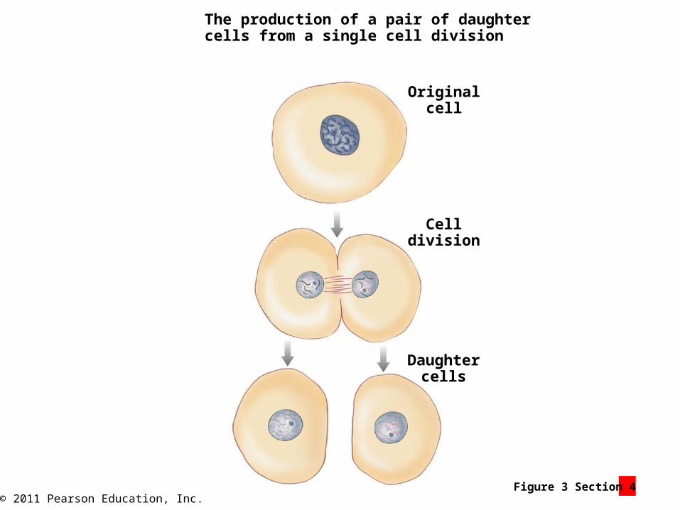
The production of a pair of daughtercells from a single cell division
Originalcell
Celldivision
Daughtercells
© 2011 Pearson Education, Inc.
Module 3.15: Interphase
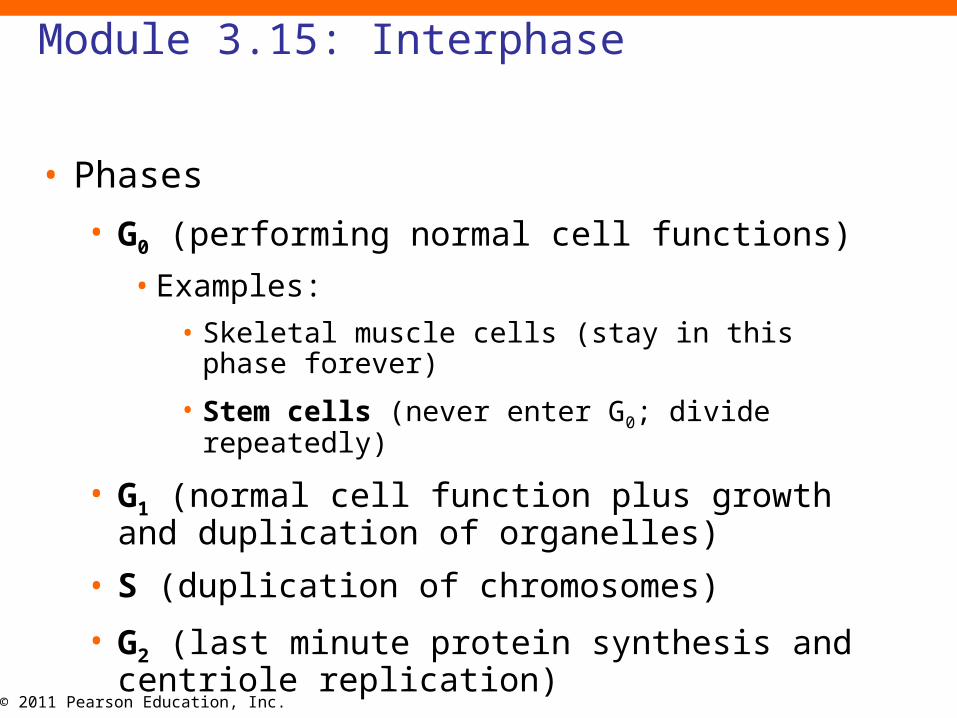
• Phases
• G0 (performing normal cell functions)
• Examples:
• Skeletal muscle cells (stay in this phase forever)
• Stem cells (never enter G0; divide repeatedly)
• G1 (normal cell function plus growth and duplication of organelles)
• S (duplication of chromosomes)
• G2 (last minute protein synthesis and centriole replication)

© 2011 Pearson Education, Inc.
Module 3.15: Interphase
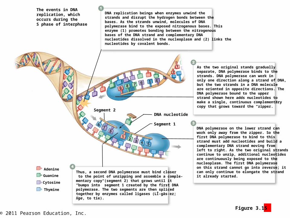
• DNA replication
• Strands unwind
• DNA polymerase binds
• Assembles new DNA strand covalently linking nucleotides
• Works only in one direction
• One polymerase works continuously along one strand toward “zipper”
• One polymerase works away from “zipper”
• As “unzipping” occurs, another polymerase binds closer point of unzipping
• Two new DNA segments bound with ligases
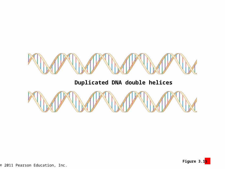
• Two identical DNA strands formed
© 2011 Pearson Education, Inc.Figure 3.15 2
The events in DNAreplication, whichoccurs during theS phase of interphase
DNA replication beings when enzymes unwind thestrands and disrupt the hydrogen bonds between thebases. As the strands unwind, molecules of DNApolymerase bind to the exposed nitrogenous bases. Thisenzyme (1) promotes bonding between the nitrogenousbases of the DNA strand and complementary DNAnucleotides dissolved in the nucleoplasm and (2) links thenucleotides by covalent bonds.
As the two original stands graduallyseparate, DNA polymerase binds to thestrands. DNA polymerase can work inonly one direction along a strand of DNA,but the two strands in a DNA moleculeare oriented in opposite directions. TheDNA polymerase bound to the upperstrand shown here adds nucleotides tomake a single, continuous complementarycopy that grows toward the “zipper.”
DNA polymerase on the lower strand canwork only away from the zipper. So thefirst DNA polymerase to bind to thisstrand must add nucleotides and build acomplementary DNA strand moving fromleft to right. As the two original strandscontinue to unzip, additional nucleotidesare continuously being exposed to thenucleoplasm. The first DNA polymeraseon this strand cannot go into reverse; itcan only continue to elongate the strandit already started.
Thus, a second DNA polymerase must bind closer to the point of unzipping and assemble a comple-mentary copy (segment 2) that grows until it “bumps into” segment 1 created by the first DNA polymerase. The two segments are then spliced together by enzymes called ligases (LĪ-gās-ez; liga, to tie).
Adenine
Guanine
Cytosine
Thymine
Segment 2
Segment 1
DNA nucleotide
© 2011 Pearson Education, Inc.Figure 3.15 3
Duplicated DNA double helices

© 2011 Pearson Education, Inc.
Module 3.15 Review
a. Describe interphase, and identify its stages.
b. What enzymes must be present for DNA replication to proceed normally?
c. A cell is actively manufacturing enough organelles to serve two functional cells. This cell is probably in what phase of interphase?
© 2011 Pearson Education, Inc.
Module 3.16: Mitosis
• Mitosis
• Division and duplication of cell’s nucleus
• Phases
1. Prophase (pro-, before)
• Paired chromosomes tightly coiled
• Chromatid (each copy)
• Connected at centromere with raised area (kinetochore)
• Replicated centrioles move to poles
• Astral rays (extend from centrioles)
• Spindle fibers (interconnect centriole pairs)

© 2011 Pearson Education, Inc.Figure 3.16 1 – 2
The events in mitosis
Centrioles incentrosome
Nucleus
Interphase, whichprecedes mitosis
Prophase, the first phase of mitosis
The nuclearmembranedisintegratesduring this period.
The centrioleshave replicated,and the pairsnow move toopposite sidesof the nucleus.
Microtubules extend outwardfrom each pair of centrioles:astral rays extend into thecytoplasm, whereas spindlefibers interconnect thecentriole pairs.
Chromatids
Kinetochore
The kinetochore ofeach chromatidbecomes attachedto a spindle fiber.
© 2011 Pearson Education, Inc.
Module 3.16: Mitosis
• Mitosis (continued)• Phases (continued)
2. Metaphase (meta, after)
• Chromosomes align at metaphase plate
3. Anaphase (ana-, apart)
• Chromatids separate
• Drawn along spindle apparatus
4. Telophase (telo-, end)
• Cells prepare to enter interphase
• Cytoplasm constricts along metaphase plate (= cleavage furrow)
• Nuclear membranes re-form
• Chromosomes uncoil
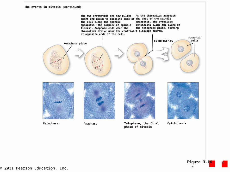
© 2011 Pearson Education, Inc.Figure 3.16 3 – 6
Metaphase Anaphase
Metaphase plate
The two chromatids are now pulledapart and drawn to opposite ends ofthe cell along the spindleapparatus (the complex of spindlefibers). Anaphase ends when thechromatids arrive near the centriolesat opposite ends of the cell.
As the chromatids approachthe ends of the spindleapparatus, the cytoplasmconstricts along the plane ofthe metaphase plate, forminga cleavage furrow.
DaughtercellsCYTOKINESIS
Telophase, the finalphase of mitosis
Cytokinesis
The events in mitosis (continued)
© 2011 Pearson Education, Inc.
Module 3.16: Mitosis
• Cytokinesis (cyto-, cell + kinesis, motion)
• Begins with formation of cleavage furrow
• Continues through telophase
• Completion marks end of cell division

© 2011 Pearson Education, Inc.
Module 3.16 Review
a. Define mitosis, and list its four stages.
b. What is a chromatid, and how many would be present during normal mitosis in a human cell?
c. What would happen if spindle fibers failed to form in a cell during mitosis?
© 2011 Pearson Education, Inc.
Module 3.17: Tumors and cancer
• Cancer
• Illness that disrupts normal rates of cell division
• Characterized by permanent DNA sequence changes (= mutations)
• Most common in tissues with actively dividing cells
• Examples: skin, intestinal lining
• Compete with normal cells for resources

© 2011 Pearson Education, Inc.
Module 3.17: Tumors and cancer
• Cancerous tumor (neoplasm; mass of cells) types
1. Benign
• Remain in original tissue
2. Malignant
• Accelerated growth due to blood vessel growth and supply to the area
• Invasion (cells migrating into surrounding tissues)
• Metastasis (formation of secondary tumors)
© 2011 Pearson Education, Inc.
Module 3.17 Review
a. Define metastasis.
b. What is a benign tumor?
c. Define cancer.