Overview of the Nervous System One of the body’s homeostatic control systems Contains sensors,...
44
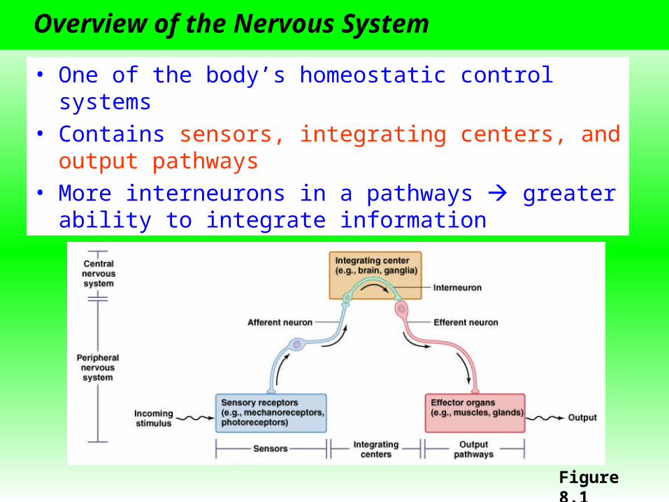
Overview of the Nervous System • One of the body’s homeostatic control systems • Contains sensors, integrating centers, and output pathways • More interneurons in a pathways greater ability to integrate information Figure 8.1
-
date post
22-Dec-2015 -
Category
Documents
-
view
214 -
download
0
Transcript of Overview of the Nervous System One of the body’s homeostatic control systems Contains sensors,...
Overview of the Nervous System
• One of the body’s homeostatic control systems• Contains sensors, integrating centers, and output
pathways• More interneurons in a pathways greater ability to
integrate information
Figure 8.1
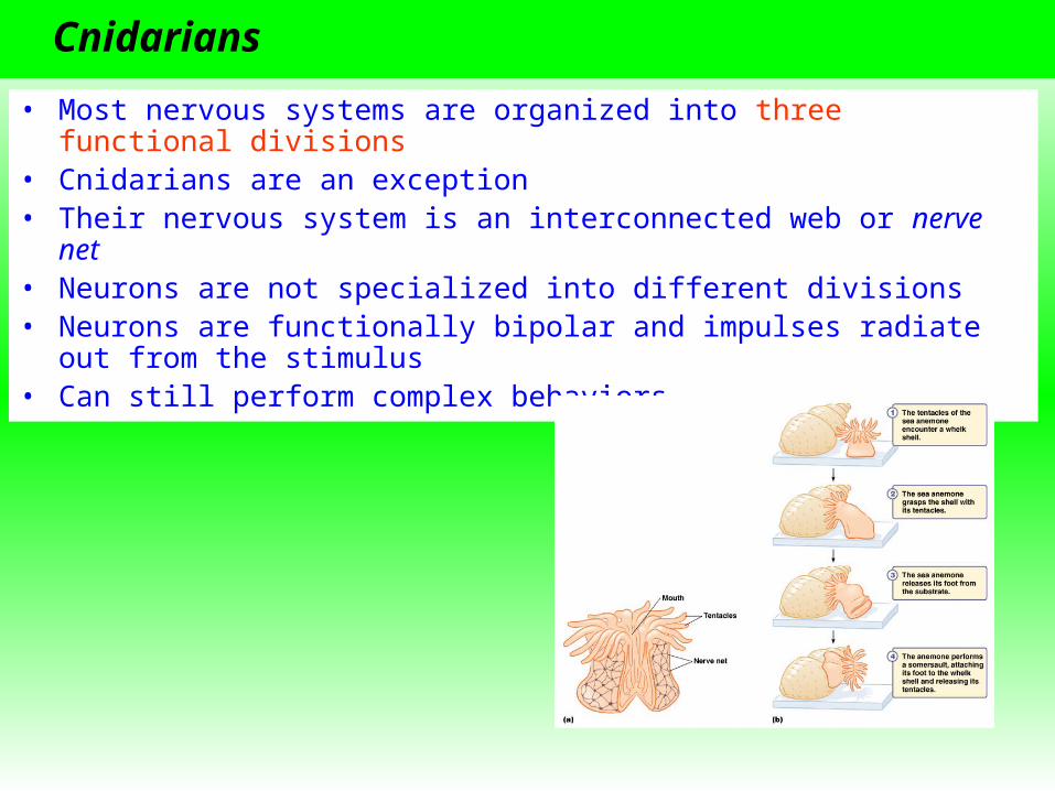
Cnidarians
• Most nervous systems are organized into three functional divisions• Cnidarians are an exception • Their nervous system is an interconnected web or nerve net• Neurons are not specialized into different divisions• Neurons are functionally bipolar and impulses radiate out from the
stimulus• Can still perform complex behaviors

Nervous System Terms
• Bilaterally symmetrical – anterior and posterior end and a right and left side
• Cephalization - sense organs are concentrated at the anterior end
• Brain – a complex integrating center made up of clusters of ganglia
• Ganglia – groupings of neuronal cell bodies• Nuclei – groupings or neuronal cell bodies within the
brain• Tracts – groupings of axons within the brain• Nerves – axons of afferent and efferent neurons
Structure of a Nerve
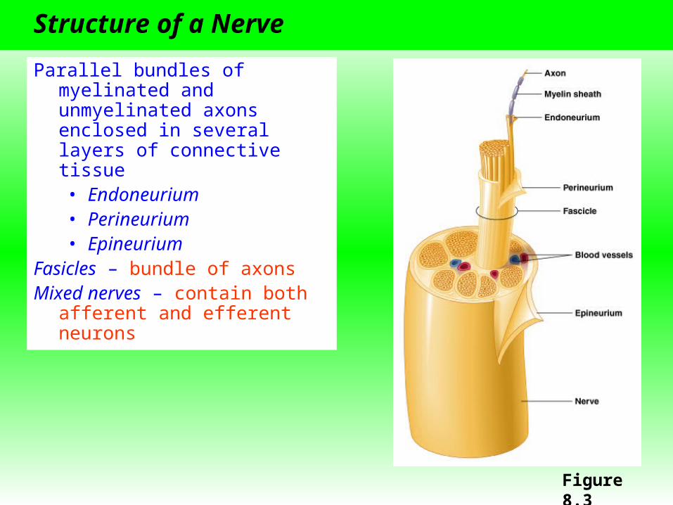
Parallel bundles of myelinated and unmyelinated axons enclosed in several layers of connective tissue• Endoneurium• Perineurium • Epineurium
Fasicles – bundle of axonsMixed nerves – contain both
afferent and efferent neurons
Figure 8.3

Nervous Systems Across Animal Groups
• Cephalization occurs in most animals and becomes more apparent in more complex nervous systems
• Cnidarians and Echinoderms are exceptions• Organisms with more complex nervous systems have more neurons

The Vertebrate Central Nervous System
• Among the most highly cephalized animals
• Unique in having a hollow dorsal nerve cord
• Portion of nervous system is encased within cartilage or bone
• Central nervous system (CNS) – brain and spinal cord
• Peripheral nervous system (PNS) – rest of the nervous system
Figure 8.5a

Cranial and Spinal Nerves
Cranial nerves• Exit directly from the braincase• 13 pairs (labeled with roman numerals)• Some are afferent and some are efferent
Spinal nerves• Emerge from the spinal cord• Named based on the region of the spine where they
originate

Gray and White Matter
Brain and spinal cord contain two types of tissue• Gray matter – neuronal cell bodies• White matter – bundles of axons and their myelin
sheaths
Spinal chord white matter is on the surface and gray matter is inside (opposite for cerebral cortex)
Figure 8.5b

The CNS is Isolated
• Meninges – layers of connective tissue that surround the brain and spinal cord
• Number of layers vary across taxa (fish have one, mammals have three)
• Cerebral spinal fluid (CSF) fills the space within the meninges and acts as a shock absorber
• Blood-brain barrier – tight junctions in brain capillaries prevent material from leaking out of the bloodstream and into the CNS Figure 8.6

The Vertebrate Brain
The brain is an extension of the spinal cord
It is hollow inside and central cavities called ventricles contains CSF
Three main regions• Rhombencephalon (hindbrain)
• Reflexes and involuntary behaviors• Mesencephalon (midbrain)
• Coordination of sensory information• Relay center in mammals
• Prosencephalon (forebrain)• Integration of olfactory information with other
senses• Regulation of body temperature, reproduction,
eating, emotion• Learning and memory in mammals

Brain Size
Most groups have the same major brain structures, although these structures vary in relative size
Figure 8.9

The Parts of the Mammalian Brain
Table 8.2

Hindbrain
Three regions• Pons – located above the medulla
• Pathway between the medulla, the cerebellum, and the forebrain
• Controls alertness and initiates sleep and dreaming
• Cerebellum – two hemispheres at the back of the brain• Responsible for motor coordination• Contains half of the neurons in the brain
• Medulla oblongata – located at the top of the spinal cord• Regulates breathing, heart rate, diameter of
blood vessels, and blood pressure• Contain pathways between the spinal cord and
the brain• Many cross over (e.g., left to right)
Midbrain
• Primary center for coordinating and initiating behavioral responses in fish and amphibians
• Size and function reduced in mammals• Primarily serves as a relay center
• Sometimes grouped with the pons and medulla and termed the brainstem

Forebrain
Involved in processing and integrating sensory information, and in coordinating behavior
Main regions• Cerebrum• Thalamus• Epithalamus• Hypothalamus

Cerebrum
Outer layer is the cortex
Divided into two cerebral hemispheres• Left side controls the right side of the body• Right side controls the left side of the body
Connected by the corpus callosum

Cortex
• Integrates and interprets sensory information and initiates voluntary movements
• Has taken over many of the midbrain functions in lower vertebrates• Six layers• Isocortex (outer layer) is necessary for cognition and higher brain
functions• More folded in more advanced mammals• Gyri – folds• Sulci – grooves

Cortical Lobes
Based on the names of the overlying bones or function
Figure 8.14

Cortical Topology
• Each part of the cortex corresponds to the specific part of the body that it governs
• The areas devoted to various parts of the body are disproportionate
Figure 8.5

Hypothalamus
• Located at the base of the forebrain• Maintains homeostasis• Interacts with the autonomic nervous system• Regulates secretion of pituitary hormones

Limbic System
A network of connected structures that lie between the cortex and the rest of the brain
Influences emotions, motivation, and memory
Sometimes called the “emotional brain”
Includes the hypothalamus and other parts• Amygdala – aggression and
fear responses• Hippocampus – converts
short-term memory to long-term memory
• Olfactory bulbs – sense of smell
Figure 8.11

Thalamus
• Large grouping of gray matter above the hypothalamus• Part of the reticular formation• Receives input from the limbic system and all senses
except olfaction• Relays information to the cortex• Acts as a filter

Epithalamus
Located above the thalamus
Contains • Habenular nuclei – communicates with the
tegmentum of the midbrain• Pineal complex – Establishes circadian rhythms and
secretes melatonin

Peripheral Nervous System Divisions
Figure 8.16

Autonomic Pathways
Involved in homeostasis
“Involuntary nervous system”
Systems • Sympathetic
• Most active during periods of stress or physical activity• “Fight-or-flight” system
• Parasympathetic• Most active during periods of rest• “Resting and digesting” system
• Enteric• Independent of other two systems• Affects digestion by innervating the GI tract, pancreas, and gall
bladder

Maintaining Homeostasis
Balancing of the sympathetic and parasympathetic systems
Three features of maintaining homeostasis• Dual innervation – most internal organs receive input
from both systems• Antagonistic action – one system stimulates while the
other inhibits• Basal tone – Even under resting conditions autonomic
neurons produce APs

Dual Innervation
Figure 8.17

Antagonistic Action
Table 8.3

Similarities in Autonomic Pathways
Pathways contain two neurons in series• Preganglionic – may synapse with many postganglionic neurons
and intrinsic neurons • Postganglionic – release neurotransmitter at the effector from
varicosities
These neurons synapse with each other in the autonomic ganglia
Figure 8.18

Differences in Autonomic Pathways
Differences between the sympathetic (S) and parasympathetic (PS) branches• Preganglionic cell body location
• S: thoracic and lumbar regions of the spinal cord• PS: hindbrain and sacral region of the spinal cord
• Ganglia location• S: chain that runs close to the spinal cord• PS: close to the effector
• Number of postganglionic neurons that synapse with a single preganglionic neuron
• S: 10 or more• P: three or less

Differences in Autonomic Pathways, Cont.Type of neurotransmitter released at the
effector
Figure 8.19

Only Sympathetic Innervation
Some effectors receive only sympathetic innervation• Adrenal medulla – modified postganglionic neuron• Sweat glands• Arrector pili muscles in the skin• Kidneys• Most blood vessels
Figure 8.20

Sympathetic vs. Parasympathetic Systems
Table 8.4

Regulation of the Autonomic System
Figure 8.21
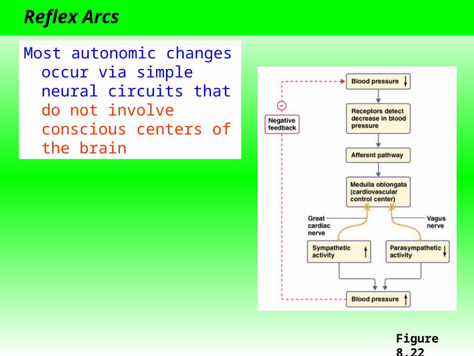
Reflex Arcs
Most autonomic changes occur via simple neural circuits that do not involve conscious centers of the brain
Figure 8.22

Somatic Motor Pathways
• Control skeletal muscle• Usually under conscious control• The “Voluntary nervous system”• Some pathways are not under conscious control, e.g.,
knee-jerk reflex
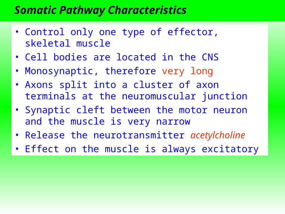
Somatic Pathway Characteristics
• Control only one type of effector, skeletal muscle• Cell bodies are located in the CNS• Monosynaptic, therefore very long• Axons split into a cluster of axon terminals at the
neuromuscular junction• Synaptic cleft between the motor neuron and the muscle
is very narrow• Release the neurotransmitter acetylcholine• Effect on the muscle is always excitatory

Reflex Arcs
• Least complex integrated responses• Can involve as few as two neurons (monosynaptic) or
more than two (polysynaptic)
Figures 8.23 & 8.25

Reflex Arcs, Cont.
Can be arranged in two ways• Convergence – allows spatial summation• Divergence – can amplify signals
Figure 8.24

Learning and Memory
• Most animals can form memories and learn due to the plasticity of the nervous system
• Learning – process of acquiring new information• Memory – retention and retrieval of information• Plasticity – ability to change both synaptic connections
and functional properties of neurons in response to stimuli

Invertebrate Learning and Memory
Well studied in the sea slug, Aplysia (20,000 neurons)
Habituation – decline in response to a stimulus due to repeated exposure• Allows animal to ignore unimportant
stimuli and focus on novel stimuli• Occurs because of changes in the
presynaptic axon terminal• Inactivation of Ca2+ channels
neurotransmitter release
Figure 8.29

Invertebrate Learning and Memory, Cont.Sensitization – increase in the response to a gentle stimulus after exposure to a strong stimulus
• Occurs because of changes in the presynaptic axon terminal Ca2+ entry neurotransmitter release• Involves a secondary circuit
• Releases serotonin binds to G-protein-coupled receptors cascade of reactions inactivation of K+ channels AP duration Ca2+ influx neurotransmitter release

Serotonin Effects
Figure 8.31

Memory in Mammals
The hippocampus is involved in long-term memory, but the memories are stored elsewhere
Long-term potentiation – repetitive stimulation of hippocampal tissue leads to an increase in the response of the postsynaptic neuron