Functional Uncoupling of Inhibitory Interneurons Plays … · Functional Uncoupling of Inhibitory...
10
The Journal of Neuroscience, May 1993, 13(5): 2126-2135 Functional Uncoupling of Inhibitory Interneurons Plays an Important Role in Short-Term Sensitization of Aplysia Gill and Siphon Withdrawal Reflex Louis-Eric Trudeau and Vincent F. Castellucci Laboratoire de Neurobiologie et Comportement, lnstitut de recherches cliniques de Montreal, Montreal, Qukbec H2W lR7 and Centre de recherches en sciences neurologiques, Universitk de Mont&al, Montrkal, Qukbec H3C 3J7, Canada, Attempts to explain learning-associated potentiation of syn- aptic transmission in model systems such as withdrawal reflexes in the mollusk Aplysia or the hippocampus of ver- tebrates have focused on the mechanisms by which trans- mitter release is increased in the principal elements of the circuit. Increased transmission in neuronal networks such as the- gill and siphon withdrawal reflex (GSWR) of Aplysia may, howevet, also be caused by a decrease of transmitter release by inhibitory interneurons. The importance and func- tion of cholinergic inhibitory transmission in the GSWR net- work were investigated. Central application of the nicotinic cholinergic antagonist d-tubocurarine (d-TC) considerably potentiated gill contractions, evoked either by nerve stim- ulation or by tactile stimulation of the siphon. Compound EPSPs evoked in motoneurons upon siphon nerve stimula- tion were also significantly prolonged following application of d-TC, but were unaffected by hexamethonium, a blocker of excitatory ACh receptors in Aplysia. Recordings from ex- citatory interneurons showed that they received excitation followed by powerful inhibitory input upon stimulation of the siphon nerve. Application of d-TC completely blocked this rapid inhibition, thus prolonging the compound EPSPs evoked in the interneurons. These effects were obtained at a con- centration of d-TC (100 PM) that almost totally blocked fast inhibitory cholinergic transmission, but was without effect on monosynaptic connections between sensory neurons and motoneurons of the reflex. Facilitation of (1) compound EPSCs in motoneurons and (2) evoked excitatory interneu- ronal firing was reduced in preparations already disinhibited by pretreatment with d-TC. Facilitation of sensory-motor synapses, however, was not reduced in the presence of d-TC, indicating that facilitatory interneurons are still acti- vated under cholinergic blockade. These data show that transmission through the GSWR neuronal network is gated by a feedback inhibitory mechanism. They also suggest that a reduction of cholinergic inhibition onto excitatory inter- Received July 6, 1992; revised Sept. 22, 1992; accepted Nov. 19, 1992. We thank M. Klein, T. Ouimet, and L. Bemier forcomments on the manuscript, 1. Morin for preparing the illustrations, and N. Guay for typing the manuscript. This work was funded by Grant MA- 10047 from the Medical Research Council of Canada, and by the Richard and Edith Strauss Canada Foundation. L.-E.T. is the recipient of a “I 967” Scholarship from the Conseil de Recherches en Sciences Naturelles et GCnie du Canada. Correspondence should be addressed to Vincent F. Castellucci, Laboratoire de Neurobiologie et Comportement, Institut de recherches cliniques de Montrkal, 110 ouest, avenue des Pins, Montrtal, QuCbec H2W lR7, CAN. Copyright 0 1993 Society for Neuroscience 0270-6474/93/ 132 126-10%05.00/O neurons may be a mechanism through which transmission within the GSWR network is increased during various forms of learning, such as sensitization. These data place new emphasis on the important role of inhibitory interneurons in determining the plastic properties of neuronal networks, in both invertebrates and vertebrates. [Key words: Aplysia, inhibition, ACh, nicotinic receptor, sensitization, neufonal network, learning, reflex] Most of the efforts aimed at understandingthe neuronal mod- ifications underlying short- and long-term behavioral changes associated with learning in Aplysia calijbrnica have been di- rected at the monosynaptic junctions between a subsetof sen- sory neurons and identified motoneurons mediating the reflex behaviors that have been studied. That work has identified, at these critical sensory-motor synapses, cellular and molecular mechanisms that may partially account for the changes in be- havioral output associated with learning and memory in this invertebrate (Castellucci et al., 1970;Brunelli et al., 1976;Hoch- ner et al., 1986a,b; Montarolo et al., 1986; Dash et al., 1990; Bailey et al., 1992; Mayford et al., 1992). We have recently presentedevidence for the importance of the polysynaptic component of the neuronal network in the mediation and plasticity of the gill and siphon withdrawal reflex (GSWR) (Trudeau and Castellucci, 1992). Intemeurons inter- posed between sensoryand motor neurons act to amplify the sensory message reachingthe motoneurons.Only a limited sub- set of interneurons of this type are presently known (Hawkins et al., 198 1 a,b; Byrne, 1983;Frost et al., 1988).The polysynaptic network, consistingof both excitatory and inhibitory intemeu- rons, appearsto be an important site of plastic changes asso- ciated with sensitization of the GSWR. We have found that two neuromodulators of the reflex, 5-HT and small cardioactive peptide B (SCP,), have differential effects on the neuronal net- work, the first preferentially potentiating the monosynaptic pathway, the second preferentially potentiating the polysynaptic pathway (Trudeau and Castellucci, 1992). Sensitization of the reflex by the activation of endogenous facilitator neurons may thus involve, to a large extent, a modification of synaptic trans- mission at the intemeuronal level. The excitatory interneurons may becomemore excitable, they may increase their transmitter release onto motoneurons,or they may receive a decreased input from inhibitory interneurons. An examination of these possi- bilities and an analysis of the mechanisms involved will increase our understanding of the learning-associated modifications in this model system.
-
Upload
nguyentram -
Category
Documents
-
view
227 -
download
0
Transcript of Functional Uncoupling of Inhibitory Interneurons Plays … · Functional Uncoupling of Inhibitory...
The Journal of Neuroscience, May 1993, 13(5): 2126-2135
Functional Uncoupling of Inhibitory Interneurons Plays an Important Role in Short-Term Sensitization of Aplysia Gill and Siphon Withdrawal Reflex
Louis-Eric Trudeau and Vincent F. Castellucci
Laboratoire de Neurobiologie et Comportement, lnstitut de recherches cliniques de Montreal, Montreal, Qukbec H2W lR7 and Centre de recherches en sciences neurologiques, Universitk de Mont&al, Montrkal, Qukbec H3C 3J7, Canada,
Attempts to explain learning-associated potentiation of syn- aptic transmission in model systems such as withdrawal reflexes in the mollusk Aplysia or the hippocampus of ver- tebrates have focused on the mechanisms by which trans- mitter release is increased in the principal elements of the circuit. Increased transmission in neuronal networks such as the- gill and siphon withdrawal reflex (GSWR) of Aplysia may, howevet, also be caused by a decrease of transmitter release by inhibitory interneurons. The importance and func- tion of cholinergic inhibitory transmission in the GSWR net- work were investigated. Central application of the nicotinic cholinergic antagonist d-tubocurarine (d-TC) considerably potentiated gill contractions, evoked either by nerve stim- ulation or by tactile stimulation of the siphon. Compound EPSPs evoked in motoneurons upon siphon nerve stimula- tion were also significantly prolonged following application of d-TC, but were unaffected by hexamethonium, a blocker of excitatory ACh receptors in Aplysia. Recordings from ex- citatory interneurons showed that they received excitation followed by powerful inhibitory input upon stimulation of the siphon nerve. Application of d-TC completely blocked this rapid inhibition, thus prolonging the compound EPSPs evoked in the interneurons. These effects were obtained at a con- centration of d-TC (100 PM) that almost totally blocked fast inhibitory cholinergic transmission, but was without effect on monosynaptic connections between sensory neurons and motoneurons of the reflex. Facilitation of (1) compound EPSCs in motoneurons and (2) evoked excitatory interneu- ronal firing was reduced in preparations already disinhibited by pretreatment with d-TC. Facilitation of sensory-motor synapses, however, was not reduced in the presence of d-TC, indicating that facilitatory interneurons are still acti- vated under cholinergic blockade. These data show that transmission through the GSWR neuronal network is gated by a feedback inhibitory mechanism. They also suggest that a reduction of cholinergic inhibition onto excitatory inter-
Received July 6, 1992; revised Sept. 22, 1992; accepted Nov. 19, 1992. We thank M. Klein, T. Ouimet, and L. Bemier forcomments on the manuscript,
1. Morin for preparing the illustrations, and N. Guay for typing the manuscript. This work was funded by Grant MA- 10047 from the Medical Research Council of Canada, and by the Richard and Edith Strauss Canada Foundation. L.-E.T. is the recipient of a “I 967” Scholarship from the Conseil de Recherches en Sciences Naturelles et GCnie du Canada.
Correspondence should be addressed to Vincent F. Castellucci, Laboratoire de Neurobiologie et Comportement, Institut de recherches cliniques de Montrkal, 110 ouest, avenue des Pins, Montrtal, QuCbec H2W lR7, CAN.
Copyright 0 1993 Society for Neuroscience 0270-6474/93/ 132 126-10%05.00/O
neurons may be a mechanism through which transmission within the GSWR network is increased during various forms of learning, such as sensitization. These data place new emphasis on the important role of inhibitory interneurons in determining the plastic properties of neuronal networks, in both invertebrates and vertebrates.
[Key words: Aplysia, inhibition, ACh, nicotinic receptor, sensitization, neufonal network, learning, reflex]
Most of the efforts aimed at understanding the neuronal mod- ifications underlying short- and long-term behavioral changes associated with learning in Aplysia calijbrnica have been di- rected at the monosynaptic junctions between a subset of sen- sory neurons and identified motoneurons mediating the reflex behaviors that have been studied. That work has identified, at these critical sensory-motor synapses, cellular and molecular mechanisms that may partially account for the changes in be- havioral output associated with learning and memory in this invertebrate (Castellucci et al., 1970; Brunelli et al., 1976; Hoch- ner et al., 1986a,b; Montarolo et al., 1986; Dash et al., 1990; Bailey et al., 1992; Mayford et al., 1992).
We have recently presented evidence for the importance of the polysynaptic component of the neuronal network in the mediation and plasticity of the gill and siphon withdrawal reflex (GSWR) (Trudeau and Castellucci, 1992). Intemeurons inter- posed between sensory and motor neurons act to amplify the sensory message reaching the motoneurons. Only a limited sub- set of interneurons of this type are presently known (Hawkins et al., 198 1 a,b; Byrne, 1983; Frost et al., 1988). The polysynaptic network, consisting of both excitatory and inhibitory intemeu- rons, appears to be an important site of plastic changes asso- ciated with sensitization of the GSWR. We have found that two neuromodulators of the reflex, 5-HT and small cardioactive peptide B (SCP,), have differential effects on the neuronal net- work, the first preferentially potentiating the monosynaptic pathway, the second preferentially potentiating the polysynaptic pathway (Trudeau and Castellucci, 1992). Sensitization of the reflex by the activation of endogenous facilitator neurons may thus involve, to a large extent, a modification of synaptic trans- mission at the intemeuronal level. The excitatory interneurons may become more excitable, they may increase their transmitter release onto motoneurons, or they may receive a decreased input from inhibitory interneurons. An examination of these possi- bilities and an analysis of the mechanisms involved will increase our understanding of the learning-associated modifications in this model system.
The Journal of Neuroscience, May 1993, G’(5) 2127
In other systems, inhibitory interneurons have been suggested to be potentially important sites of modifications associated with learning. It has been proposed that the interplay between ex- citatory and inhibitory interneuronal activity may be a critical determinant of transmission in the neuronal networks control- ling feeding in Pleurobranchaea californica (Davis et al., 1983) and phototaxis in Hermissenda (Lederhendler et al., 1986) in the Mauthner cell system of teleosts (Kom et al., 1992) and in the olfactory bulb network (Elaagouby et al., 1991) as well as in the hippocampal circuitry of the rat (Moot et al., 199 1).
In Aplysia, little is known about the involvement of inhibitory transmission in neuronal network plasticity. In the GSWR net- work, it has been shown that transmission from L30 inhibitory interneurons to L29 facilitatory interneurons is reduced by the stimulation of fibers coursing through the left pleuro-abdominal connective (Frost et al., 1988) a procedure that usually induces sensitization of the reflex. Because the importance of these con- nections in the normal mediation of the reflex has not been evaluated, and because, in these experiments, the effect of con- nective stimulation on the output of the whole network was not monitored in parallel, the question of the involvement of in- hibitory transmission in the plasticity of the GSWR remains unanswered.
In this article we have attempted to characterize further the role of the polysynaptic component of the GSWR neuronal network in the mediation and plasticity of the reflex. We have sought to address two issues. First, we wanted to know the importance of inhibitory transmission in the normal mediation of the GSWR. We have taken advantage of the fact that most described inhibitory synaptic transmission of the fast type in Aplysia nervous system is attributable to a Cll conductance increase induced by ACh binding to nicotinic-type receptors. We were thus able to block this type of transmission by the use of the nicotinic cholinergic receptor blocker d-tubocurarine (d- TC) (Taut and Gerschenfeld, 1962; Blankenship et al., 197 1; Kehoe, 1972) and to assess the effects of this blockade on the GSWR. Because we observed that such cholinergic inhibition was indeed significant and appeared to be involved in a feedback pathway, the second objective was to evaluate the involvement of disinhibitory processes in short-term sensitization of the re- flex. Decreased inhibition upon excitatory interneurons could be responsible for increased excitatory transmission in the net- work. We thus compared facilitation of the reflex in normal versus disinhibited preparations. We found that although facil- itation is still possible in disinhibited networks, it is considerably reduced. We therefore propose that a major mechanism of sen- sitization of the gill and siphon withdrawal reflex may involve a reduction of inhibitory cholinergic transmission.
Materials and Methods Preparations. Aplysiu culijbrnica (100-300 gm) were obtained from Ma- rinus Inc. (Venice, CA) and housed individually in small compartments within a large aquarium containing artificial seawater (ASW; Forty Fath- oms). The temperature was kept at 14°C. Before dissection, animals were anesthetized with an injection of isotonic MgCl, corresponding approximately to a third of their volume. Dissection was then carried out in a solution made with equal parts of isotonic MgCl, and ASW. For the isolated ganglion preparation, the abdominal ganglion was dis- sected out and a long section of both pleuro-abdominal connectives and siphon nerve were kept intact. The ganglion was then exposed for 20 set to 0.5% glutaraldehyde to kill muscle cells, and pinned to the bottom of a small 2 ml Sylgard-coated chamber. The connective tissue covering the ganglion was removed with fine forceps. ASW was then perfused for 1 hr before the beginning of experiments. A semi-intact preparation
was used in some experiments; it consisted of the intact gill connected to the abdominal ganglion by the branchial nerve and the intact siphon connected to the abdominal ganglion through the siphon nerve; in other experiments only the siphon nerve was kept without the siphon. The abdominal ganglion was then isolated from the rest of the preparation with a small chamber that allowed nerves and connectives to exit through Vaseline-sealed apertures. This allowed independent applications of drugs to the ganglion. In these preparations, the gill was cannulated and con- tinuously perfused with ASW. Gill contractions were measured through the output of a photoelectric cell above which the gill was placed. Stimuli were adjusted to evoke a moderate gill contraction (about 30% of max- imum). Tactile stimulations of the siphon were delivered by an elec- tromechanical stimulator. The siphon as well as the base of the gill were held in place by very small insect pins. In such preparations, the gill contractions evoked by tactile stimulation of the siphon may be arti- ficially prolonged because of the involvement of reafference due to the siphon contracting against the pins. Because the same results were ob- tained with direct stimulation of the siphon nerve, this issue does not appear to have been a problem under our experimental conditions. The semi-intact preparations did not include an intact ctenidial (genital) nerve or an intact peripheral pathway, which are known to also play a role in the mediation of evoked gill contractions in the intact animal (Carew et al., 1974; Lukowiak and Peretz, 1977). We have restricted our analysis to the central component of the GSWR network and to its output through the branchial nerve (gill component). All experiments were performed at room temperature (22°C).
Experiments were also carried out on an excitatory cholinergic syn- apse evoked in neuron R15 of the abdominal ganglion by threshold stimulation of an axon coursing in the right pleuro-abdominal connec- tive (Frazier et al., 1967). Additional experiments were performed on identified cholinergic synapses in the buccal ganglion between neurons B4 or B5 and neurons B3 or B6 (Taut and Gerschenfeld, 1962; Gardner, 197 1; Baux et al., 1990). The postsynaptic neurons possess acetylcho- line-gated Cll channels and were voltage clamped at -80 mV. Changes in Cl- reversal potential during experiments were always monitored and responses were expressed in conductance.
Electrophysiology. Intracellular electrodes were filled with either 3 M
KC1 or 2 M K-acetate and had a resistance between 1.5 and 15 Ma. Voltage recording and two-electrode voltage clamping were performed with an Axoclamp-2A (Axon Instruments) amplifier. Extracellular ap- plication of ACh to the cell body of neurons was performed by ionto- phoresis with an Axoprobe-1A (Axon Instruments) amplifier. In the microelectrode, ACh was dissolved in distilled water at a concentration of 1 M. Data were stored directly on hard disk as well as on VHS tape, and were analyzed with the SPIKE software (Hilal Associates). The area of postsynaptic responses was measured from the beginning of the post- synaptic potential/postsynaptic current (PSP/PSC) to the point where the PSP/PSC returned to its initial level. Data in the text are presented as mean t standard error of the mean.
Drugs and solutions. d-Tubocurarine chloride and hexamethonium chloride were obtained from Sigma (St. Louis, MO) and dissolved in ASW. Acetylcholine chloride was obtained from Anachemia (Toronto, Canada). The normal artificial seawater solution (ASW) contained, in mM, NaCl, 460; MgCl,, 30; MgSO,, 25; CaCl,, 11; KCl, 10; and HEPES buffer, 10, pH 7.6. The high-cation medium, containing 2.2 times the normal concentration of Mgz+ and 1.25 times the normal concentration of CaZ+ (2: 1 ASW), consisted of, in mM, NaCl, 368; MgCl,, 10 1; MgSO,, 20; CaCl,, 13.8; KCl, 8; and HEPES buffer, 10, pH 7.6.
Results Efect of d-tubocurarine on monosynaptic cholinergic transmission To establish the importance of cholinergic transmission in the GSWR neuronal network, the effect of the nicotinic cholinergic receptor blocker d-TC was evaluated on evoked gill contractions and evoked compound EPSPs in motoneurons of the reflex. The drug was first tested on identified cholinergic synapses in Aplysia central ganglia to determine the concentration necessary to ob- tain complete blockade of nicotinic-type receptors. In prelimi- nary experiments we confirmed that concentrations higher than 50 PM were necessary to produce almost complete blockade (Taut and Gerschenfeld, 1962; Blankenship et al., 197 1; Kehoe,
2128 Trudeau and Castellucci - Functional Uncoupling of Inhibitory Interneurons
A- BUCCAL SYNAPSE A- SIPHON NERVE STIMULATION
tO0~.1ti d-TC
B- R15 SYNAPSE
-&----- f - 100pM d -TC
C- LE -MOTONEURON SYNAPSE
J
ASW ASW + d-TC
-4 -3 -2 -1 1 2 3 4 TRIAL NO.
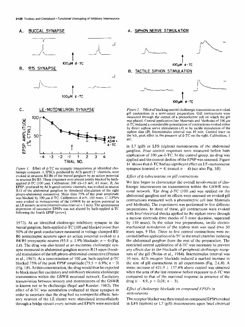
Figure I. Effect of d-TC on synaptic transmission at identified cho- linergic synapses. A, IPSCs, produced by ACh-gated Cl channels, were evoked in neurons B3/B6 of the buccal ganglion by an action potential in neurons B4/B5. These responses were almost totally blocked by bath- applied d-TC (100 PM). Calibration: 200 nS-15 mV, 65 msec. B, An EPSP, produced by ACh-gated cationic channels, was evoked in neuron R15 of the abdominal ganglion by threshold stimulation of the right pleuro-abdominal connective. More than 75% of the peak amplitude was blocked by 100 PM d-TC. Calibration: 4 mV, 100 msec. C, EPSPs were evoked in motoneurons of the GSWR by an action potential in an LE sensory neuron (interstimulus interval = I min). The spontaneous depression of successive EPSPs was not altered by bath-applied d-TC following the fourth EPSP (arrow).
1972). At an identified cholinergic inhibitory synapse in the buccal ganglion, bath-applied d-TC (100 PM) blocked more than 90% of the peak conductance measured in voltage-clamped B3/ B6 postsynaptic neurons upon an action potential evoked in a B4/B5 presynaptic neuron (93.8 f 1.9% blockade; n = 4) (Fig. 1A). The drug was also tested at an excitatory cholinergic syn- apse measured in abdominal ganglion neuron R 15 upon thresh- old stimulation of the left pleuro-abdominal connective (Frazier et al., 1967). At a concentration of 100 PM, bath-applied d-TC blocked 75% of the peak EPSP amplitude (75.5 f 6.9%; n = 3) (Fig. 1B). At this concentration, the drug would thus be expected to block most fast excitatory and inhibitory nicotinic chohnergic transmission within the GSWR neuronal network. Excitatory transmission between sensory and motoneurons of the GSWR is known not to be cholinergic (Segal and Koester, 1982). The effect of d-TC was nonetheless evaluated at these synapses in order to ascertain that the drug had no nonspecific effects. Sen- sory neurons of the LE cluster were stimulated intracellularly through a bridge circuit every minute and EPSPs were recorded
t IOO~M d- TC
B- TACTILE SIPHON STIMULATION
-
t
fi
-
i--
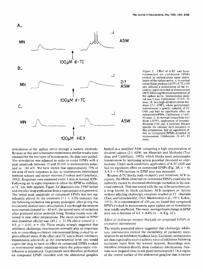
Figure 2. Effect of blocking central cholinergic transmission on evoked gill contraction in a semi-intact preparation. Gill contractions were measured through the output of a photoelectric cell on which the gill was placed. Central application (see Materials and Methods) of 100 PM
d-TC induced a considerable potentiation of contractions evoked either by direct siphon nerve stimulation (A) or by tactile stimulation of the siphon skin (B). Interstimulus interval was 10 min. Control trace on the left, peak effect in the presence of d-TC on the right. Calibration, I sec.
in L7 (gill) or LFS (siphon) motoneurons of the abdominal ganglion. Four control responses were measured before bath application of 100 PM d-TC. In the control group, no drug was applied and the control decline of the EPSP was assessed. Figure 1 Cshows that d-TC had no significant effect on LE-motoneuron synapses (control n = 4; treated n = 4) (see also Fig. 3B).
Efect of d-tubocurarine on gill contractions
We first began to characterize the overall involvement of cho- linergic interneurons on transmission within the GSWR neu- ronal network. The drug d-TC (100 PM) was applied on the abdominal ganglion and its effects were assessed on evoked gill contractions measured with a photoelectric cell (see Materials and Methods). The experiment was performed in five different preparations. In three of these, gill contractions were evoked with brief electrical shocks applied to the siphon nerve through a suction electrode (two shocks of 3 msec duration, separated by 150 msec). In the other two preparations, tactile electro- mechanical stimulation of the siphon skin was used (two 50 msec taps, 9 Hz). Three to five control contractions were re- corded before application of d-TC in the small chamber isolating the abdominal ganglion from the rest of the preparation. The restricted central application of d-TC was necessary to prevent any effects due to the blockade of peripheral cholinergic recep- tors of the gill (Weiss et al., 1984). Interstimulus interval was 10 min. ACh receptor blockade induced a marked increase in the size of gill contractions in all experiments (Fig. 2.4,B). A mean increase of 42 1.9 + 137.4% above control was obtained when the area of the last response before exposure to d-TC was compared to that of the maximal response in presence of the drug (t = 4.9, p < 0.01; rz = 5).
Effect of cholinergic blockade on compound EPSPs in motoneurons
The receptor blocker was then tested on compound EPSPs evoked in LFS (siphon) or L7 (gill) motoneurons upon brief electrical
The Journal of Neuroscience, May 1993, 13(5) 2129
A- J MN x/\_rrc A
T lOO/jM. d -TC
B,
MN
t 100~M d - TC
C-
ML?L-- -A- t
100pM HEX
stimulation of the siphon nerve through a suction electrode. Because in this and subsequent experiments similar results were obtained for the two types of motoneurons, the data were pooled. The stimulation was adjusted in order to evoke EPSPs with a peak amplitude between 15 and 30 mV in motoneurons polar- ized to -80 mV. We have shown that approximately 75% of the area of such responses is due to interneurons intercalated between sensory and motor neurons (Trudeau and Castellucci, 1992). Responses were measured every 5 min in normal ASW. Following six to eight responses to allow the EPSP to stabilize, d-TC was bath applied. Figure 3A illustrates one EPSP before and one after drug application from a representative experiment. The initial peak amplitude of compound EPSPs was not sig- nificantly altered by the treatment (7.1 * 5.0% increase), but the following excitation was greatly prolonged, often giving rise to a second, distinct wave ofexcitation. Even though the neurons were current clamped to ~ 80 mV, the second wave of excitation often produced action potential firing. Similar results were ob- tained in nine other preparations. The mean increase in EPSP area (maximal effects) was 189.1 k 37.9% above control (t = 4.9, p < 0.01; n = 10). These results strongly suggested that inhibitory cholinergic interneurons normally play an important role in controlling excitatory interneuronal firing evoked by si- phon afferent input. I f the effect of d-TC was to block inhibitory transmission selectively at the interneuronal level, one would expect the drug to have no effect on compound EPSPs evoked in motoneurons under conditions where the polysynaptic con- tribution is minimized. Experiments were therefore performed on compound EPSPs recorded with the abdominal ganglion
-
-I
ASW
ASW
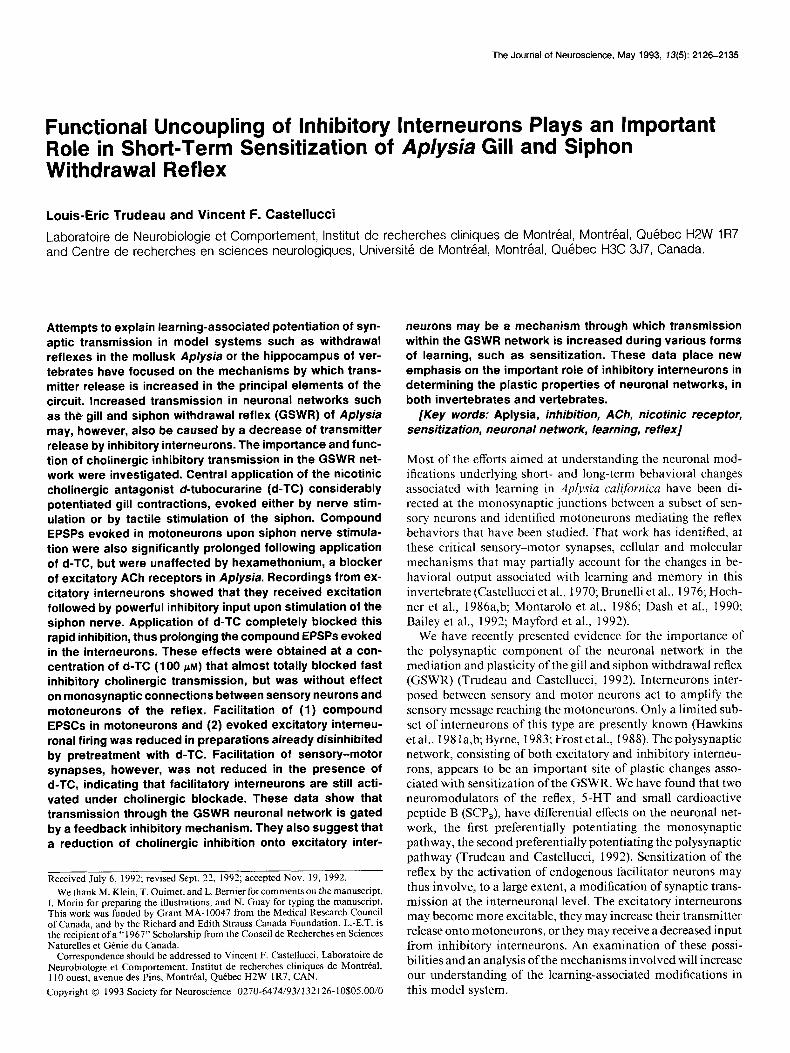
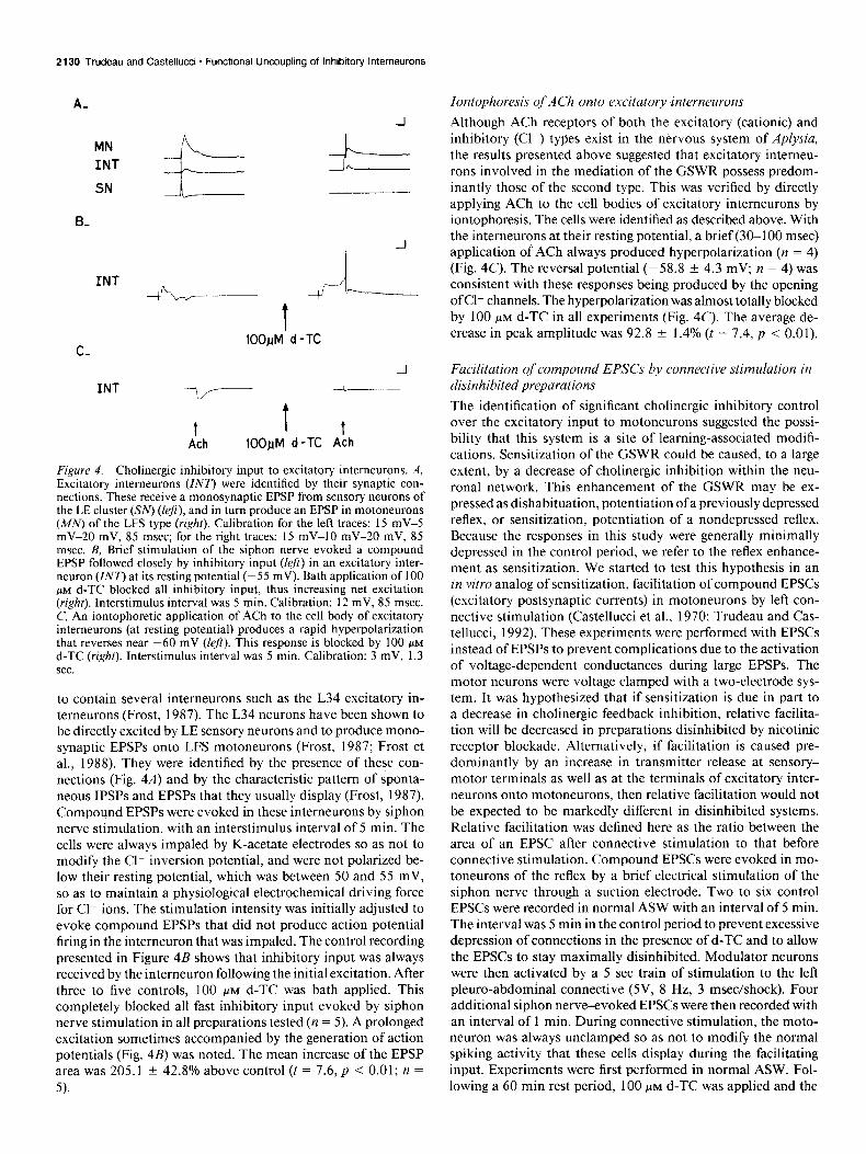
Figure 3. Effect of d-TC and hexa- methonium on compound EPSPs evoked in motoneurons upon stimu- lation of the siphon nerve. A, In normal extracellular medium (ASI@J, d-TC (100 LAM) induced a Dotentiation of the ex-
I
citatory input recorded in motoneurons (MN) following electrical stimulation of the siphon nerve. Interstimulus inter- val was 5 min. Calibration: 15 mV, 70 msec. B, In a high-divalent-cation me- dium (2:1 ASW), where polysynaptic transmission is greatly reduced, d-TC (100 FM) had no significant effect on compound EPSPs. Calibration: I1 mV, 95 msec. C, In normal extracellular me- dium (ASW), application of hexame- thonium (100 KM), a nicotinic blocker specific for cationic ACh receptors in this preparation, had no significant ef- feet on compound EPSPs recorded in motoneurons. Calibration: 10 mV, 95 msec.
bathed in a modified ASW containing a high concentration of divalent cations (2:l ASW; see Materials and Methods) (Tru- deau and Castellucci, 1992), which blocks most polysynaptic transmission by increasing action potential threshold in inter- neurons. Under such conditions, application of d-TC (100 FM)
had no significant effect on compound EPSPs (Fig. 3B; n = 4). A 9.3 +- 6.9% increase in EPSP area was measured.
Because d-TC blocks both excitatory and inhibitory ACh re- ceptors, the effects observed on compound EPSPs could also be indirectly caused by decreased cholinergic excitation in the neu- ronal network. This was tested with the use of hexamethonium, a drug known to block excitatory ACh receptors in Aplysia without affecting cholinergic receptors coupled to Cl channels (Taut and Gerschenfeld, 1962; Blankenship et al., 197 1; Kehoe, 1972). At a concentration of 100 FM, we found that compound EPSPs evoked in motoneurons upon siphon nerve stimulation were totally unaffected. The mean, nonsignificant change in EPSP area was a decrease of 4.6 5 4.4% (n = 4; Fig. 3C).
Effsct of cholinergic receptor blockade on compound EPSPs in excitatory interneurons The results presented above suggested that cholinergic inhibi- tory interneurons control the excitability of excitatory inter- neurons by an inhibitory feedback loop. Excitatory interneurons are thus expected to receive a direct wave of inhibition following excitatory input from the sensory neurons. Recordings were therefore obtained directly from excitatory intemeurons. Neu- rons were impaled close to ink gland motoneurons L14, a region of the ventral surface of the abdominal ganglion that is known
2130 Trudeau and Castellucci - Functional Uncoupling of Inhibitory Interneurons
A- -l
MN \ INT r. SN
B-
INT - J--L t
C- 100~M’ d - TC
INT -7
t t t Ach 100pM d-TC Ach
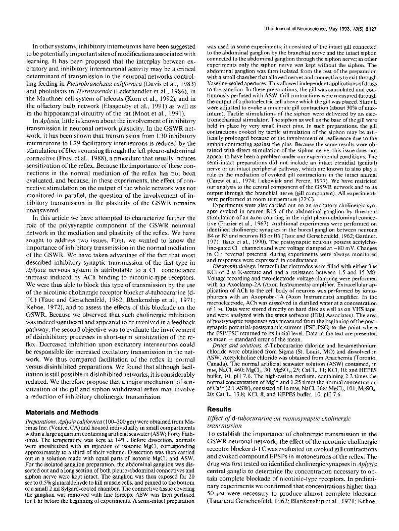
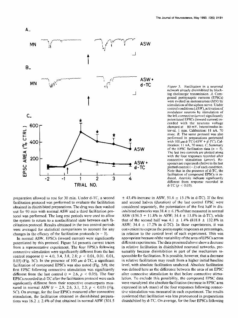
Figure 4. Cholinergic inhibitory input to excitatory intemeurons. A, Excitatory interneurons (ZNT) were identified by their synaptic con- nections. These receive a monosynaptic EPSP from sensory neurons of the LE cluster (XV) (left), and in turn produce an EPSP in motoneurons (MN) of the LFS type (right). Calibration for the left traces: 15 mV-5 mV-20 mV, 85 msec; for the right traces: 15 mV-10 mV-20 mV, 85 msec. B, Brief stimulation of the siphon nerve evoked a compound EPSP followed closely by inhibitory input (left) in an excitatory inter- neuron (ZN7’) at its resting potential (-55 mV). Bath application of 100 PM d-TC blocked all inhibitory input, thus increasing net excitation (right). Interstimulus interval was 5 min. Calibration: 12 mV, 85 msec. C, An iontophoretic application of ACh to the cell body of excitatory intemeurons (at resting potential) produces a rapid hyperpolarization that reverses near -60 mV (left). This response is blocked by 100 PM
d-TC (right). Interstimulus interval was 5 min. Calibration: 3 mV, 1.3 sec.
to contain several intemeurons such as the L34 excitatory in- temeurons (Frost, 1987). The L34 neurons have been shown to be directly excited by LE sensory neurons and to produce mono- synaptic EPSPs onto LFS motoneurons (Frost, 1987; Frost et al., 1988). They were identified by the presence of these con- nections (Fig. 4A) and by the characteristic pattern of sponta- neous IPSPs and EPSPs that they usually display (Frost, 1987). Compound EPSPs were evoked in these intemeurons by siphon nerve stimulation, with an interstimulus interval of 5 min. The cells were always impaled by K-acetate electrodes so as not to modify the Cll inversion potential, and were not polarized be- low their resting potential, which was between 50 and 55 mV, so as to maintain a physiological electrochemical driving force for Cll ions. The stimulation intensity was initially adjusted to evoke compound EPSPs that did not produce action potential firing in the interneuron that was impaled. The control recording presented in Figure 4B shows that inhibitory input was always received by the interneuron following the initial excitation. After three to five controls, 100 PM d-TC was bath applied. This completely blocked all fast inhibitory input evoked by siphon nerve stimulation in all preparations tested (n = 5). A prolonged excitation sometimes accompanied by the generation of action potentials (Fig. 4B) was noted. The mean increase of the EPSP area was 205.1 + 42.8% above control (t = 7.6, p < 0.01; n = 5).
Iontophoresis of ACh onto excitatory interneurons Although ACh receptors of both the excitatory (cationic) and inhibitory (Cll) types exist in the nervous system of Aplysia, the results presented above suggested that excitatory interneu- rons involved in the mediation of the GSWR possess predom- inantly those of the second type. This was verified by directly applying ACh to the cell bodies of excitatory interneurons by iontophoresis. The cells were identified as described above. With the interneurons at their resting potential, a brief (30-100 msec) application of ACh always produced hyperpolarization (n = 4) (Fig. 4C). The reversal potential (-58.8 f 4.3 mV; n = 4) was consistent with these responses being produced by the opening ofCl- channels. The hyperpolarization was almost totally blocked by 100 PM d-TC in all experiments (Fig. 4C). The average de- crease in peak amplitude was 92.8 ? 1.4% (t = 7.4, p < 0.01).
Facilitation of compound EPSCs by connective stimulation in disinhibited preparations The identification of significant cholinergic inhibitory control over the excitatory input to motoneurons suggested the possi- bility that this system is a site of learning-associated modifi- cations. Sensitization of the GSWR could be caused, to a large extent, by a decrease of cholinergic inhibition within the neu- ronal network. This enhancement of the GSWR may be ex- pressed as dishabituation, potentiation of a previously depressed reflex, or sensitization, potentiation of a nondepressed reflex. Because the responses in this study were generally minimally depressed in the control period, we refer to the reflex enhance- ment as sensitization. We started to test this hypothesis in an in vitro analog of sensitization, facilitation of compound EPSCs (excitatory postsynaptic currents) in motoneurons by left con- nective stimulation (Castellucci et al., 1970; Trudeau and Cas- tellucci, 1992). These experiments were performed with EPSCs instead of EPSPs to prevent complications due to the activation of voltage-dependent conductances during large EPSPs. The motor neurons were voltage clamped with a two-electrode sys- tem. It was hypothesized that if sensitization is due in part to a decrease in cholinergic feedback inhibition, relative facilita- tion will be decreased in preparations disinhibited by nicotinic receptor blockade. Alternatively, if facilitation is caused pre- dominantly by an increase in transmitter release at sensory- motor terminals as well as at the terminals of excitatory inter- neurons onto motoneurons, then relative facilitation would not be expected to be markedly different in disinhibited systems. Relative facilitation was defined here as the ratio between the area of an EPSC after connective stimulation to that before connective stimulation. Compound EPSCs were evoked in mo- toneurons of the reflex by a brief electrical stimulation of the siphon nerve through a suction electrode. Two to six control EPSCs were recorded in normal ASW with an interval of 5 min. The interval was 5 min in the control period to prevent excessive depression of connections in the presence of d-TC and to allow the EPSCs to stay maximally disinhibited. Modulator neurons were then activated by a 5 set train of stimulation to the left pleuro-abdominal connective (5V, 8 Hz, 3 msec/shock). Four additional siphon nerve-evoked EPSCs were then recorded with an interval of 1 min. During connective stimulation, the moto- neuron was always unclamped so as not to modify the normal spiking activity that these cells display during the facilitating input. Experiments were first performed in normal ASW. Fol- lowing a 60 min rest period, 100 PM d-TC was applied and the
The Journal of Neuroscience, May 1993, 13(5) 2131
MN
C,
- 600 2 L o\” 400
- I
d z 200
ASW + d-TC Figure 5. Facilitation in a neuronal
-I network already disinhibited by block- ing cholinergic transmission. A, Com- pound postsynaptic currents (EPSCs) were evoked in motoneurons (MN) by stimulation of the siphon nerve. Under control conditions (&‘I+‘), activation of modulator neurons by stimulation of the left connective (arrow) significantly potentiated EPSCs (inward current) re- corded with the neurons voltage clamped at - 80 mV. Interstimulus in- terval. 1 min. Calibration: 11 nA. 70 msec.‘B, The same protocol was also performed in preparations pretreated with 100 PM d-TC (ASW + d-K’). Cal- ibration: 11 nA, 70 msec. C, Summary of the EPSC facilitation data (n = 5). The last two controls are plotted along with the four responses recorded after connective stimulation (arrow). Re- sponses are expressed relative to the last plotted control (- 1) of each condition. Note that in the presence of d-TC, the facilitation of comoound EPSCs is re- duced. Asterisks indicate significantly different from response recorded in d-TC (p < 0.05). TRIAL NO.
4
preparation allowed to rest for 30 min. Under d-TC, a second facilitation protocol was performed to evaluate the facilitation obtained in disinhibited preparations. The drug was then washed out for 90 min with normal ASW and a third facilitation pro- tocol was performed. The long rest periods were used to allow the system to return to a nonfacilitated state between each fa- cilitation protocol. Results obtained in the two control periods were averaged for statistical comparisons to account for any changes in the efficacy of the facilitation protocols (n = 5).
In normal ASW, EPSCs (inward current) were significantly potentiated by this protocol. Figure 54 presents current traces from a representative experiment. The four EPSCs following connective stimulation were significantly different from the last control response (t = 4.0, 3.4, 3.8, 2.8; p < 0.01, 0.01, 0.01, 0.05) (Fig. 5C). In the presence of 100 PM d-TC, a significant facilitation of compound EPSCs was also noted (Fig. 5B); the first EPSC following connective stimulation was significantly different from the last control (t = 2.6, p < 0.05). The four EPSCs recorded in d-TC after the facilitation protocol were each significantly different from their respective counterparts mea- sured in normal ASW (t = 2.9, 2.6, 3.1, 2.3; p < 0.05) (Fig. 5C). On average, for the four EPSCs measured after connective stimulation, the facilitation obtained in disinhibited prepara- tions was 16.2 f 2.4% of that obtained in normal ASW (301.5
-t 43.4% increase in ASW, 5 1.6 f 15.1% in d-TC). If the first and second halves (duration) of the last control EPSC were considered separately, the potentiation of the first half in dis- inhibited networks was 38.4 * 6.1% ofthat measured in normal ASW (136.3 -t 11.8% in ASW; 54.4 i 13.8% in d-TC), while that of the second half was 4.1 f 1.4% (8 18.8 ? 132.8% in ASW; 34.4 * 17.2% in d-TC). In these experiments, it was convenient to express the postsynaptic responses as percentages, in relation to the control level of each experiment. This was appropriate because ofthe variability ofthe area of EPSCs across different experiments. The data presented above show a decrease in relative facilitation in disinhibited neuronal networks, pre- sumably because disinhibition is part of the mechanism re- sponsible for facilitation. It is possible, however, that a decrease in relative facilitation may result from a higher initial baseline level, with absolute facilitation unaltered. Absolute facilitation was defined here as the difference between the area of an EPSC after connective stimulation to that before connective stimu- lation. To exclude this possibility, the compound EPSC data were reanalyzed: the absolute facilitation (increase in EPSC area expressed in nA.msec) of the four responses following connec- tive stimulation was measured in each experiment. The results confirmed that facilitation was less pronounced in preparations disinhibited by d-TC. On average, for the four EPSCs following
2132 Trudeau and Castellucci * Functional Uncoupling of lnhlbitory Interneurons
A-
INT J._._ c ASW
t CONN: STIM.
B-
CONN: STIM.
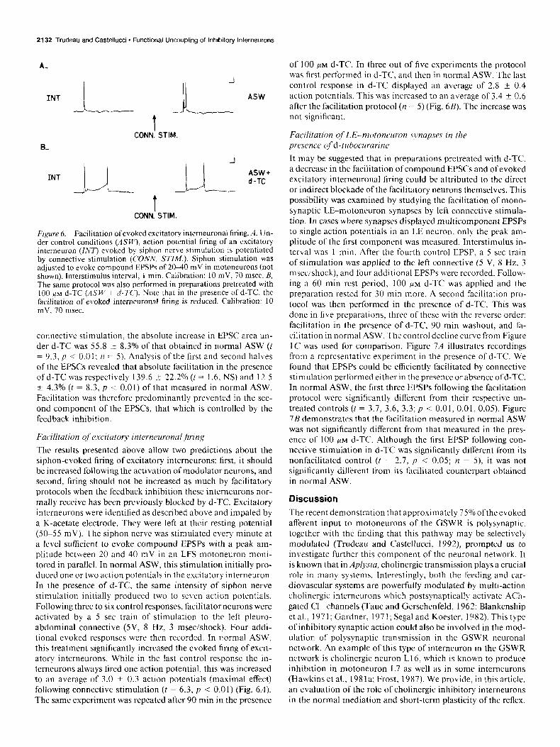
Figure 6. Facilitation of evoked excitatory interneuronal firing. A, IJn- der control conditions (ASW), action potential firing of an excitatory interneuron (INT) evoked by siphon nerve stimulation is potentiated by connective stimulation (CONN. SUM.). Siphon stimulation was adjusted to evoke compound EPSPs of 20-40 mV in motoneurons (not shown). Interstimulus interval, 1 min. Calibration: 10 mV, 70 msec. B, The same protocol was also performed in preparations pretreated with 100 #M d-TC (ASU: -t d-T<‘). Note that in the presence of d-TC. the facilitation of evoked intemeuronal firing is reduced. Calibration: 10 mV. 70 msec.
connective stimulation, the absolute increase in EPSC area un- der d-TC was 55.8 I 8.3”/0 of that obtained in normal ASW (t = 9.3, p < 0.01; n = 5). Analysis of the first and second halves of the EPSCs revealed that absolute facilitation in the presence of d-TC was respectively 139.6 -t 22.2% (t = 1.6, NS) and 12.5 -t 4.3% (t = 8.3, p < 0.01) of that measured in normal ASW. Facilitation was therefore predominantly prevented in the sec- ond component of the EPSCs, that which is controlled by the feedback inhibition.
Facilitation of‘ excitatory interneuronaljiring
The results presented above allow two predictions about the siphon-evoked firing of excitatory intemeurons: first, it should be increased following the activation of modulator neurons, and second, bring should not be increased as much by facilitatory protocols when the feedback inhibition these intcrneurons nor- mally receive has been previously blocked by d-TC. Excitatory intcrneurons were identified as described above and impaled by a K-acetate electrode. They were left at their resting potential (50-55 mV). The siphon nerve was stimulated every minute at a level sufficient to evoke compound EPSPs with a peak am- plitude between 20 and 40 mV in an LFS motoneuron moni- tored in parallel. In normal ASW, this stimulation initially pro- duced one or two action potentials in the excitatory interneuron. In the presence of d-TC, the same intensity of siphon nerve stimulation initially produced two to seven action potentials. Following three to six control responses, facilitator neurons were activated by a 5 set train of stimulation to the left pleuro- abdominal connective (5V, 8 Hz, 3 msec/shock). Four addi- tional evoked responses were then recorded. In normal ASW. this treatment significantly increased the evoked tiring of excit- atory intcrneurons. While in the last control response the in- terneurons always fired one action potential, this was increased to an average of 3.0 + 0.3 action potentials (maximal effect) following connective stimulation (t = 6.3, y < 0.01) (Fig. 611). The same experiment was repeated after 90 min in the presence
of 100 KM d-TC. In three out of five experiments the protocol was first performed in d-TC, and then in normal ASW. The last control response in d-TC displayed an average of 2.8 ? 0.4 action potentials. This was increased to an average of 3.4 +- 0.6 after the facilitation protocol (n = 5) (Fig. 6B). The increase was not significant.
Facilitation of L,E--motoneuron svnapses in the presence cfd-tuhocurarine It may be suggested that in preparations pretreated with d-TC, a decrease in the facilitation of compound EPSCs and of evoked excitatory interneuronal firing could be attributed to the direct or indirect blockade of the facilitatory neurons themselves. This possibility was examined by studying the facilitation of mono- synaptic LE-motoneuron synapses by left connective stimula- tion. In cases where synapses displayed multicomponent EPSPs to single action potentials in an LE neuron, only the peak am- plitude of the first component was measured. Interstimulus in- terval was 1 min. After the fourth control EPSP, a 5 set train of stimulation was applied to the left connective (5 V, 8 Hz, 3 msec/shock), and four additional EPSPs were recorded. Follow- ing a 60 min rest period, 100 PM d-TC was applied and the preparation rested for 30 min more. A second facilitation pro- tocol was then performed in the presence of d-TC. This was done in five preparations, three of these with the reverse order: facilitation in the presence of d-TC, 90 min washout, and fa- cilitation in normal ASW. The control decline curve from Figure IC was used for comparison. Figure 7A illustrates recordings from a representative experiment in the presence of d-TC. We found that EPSPs could be efficiently facilitated by connective stimulation performed either in the presence or absence ofd-TC. In normal ASW, the first three EPSPs following the facilitation protocol were significantly different from their respective un- treated controls (t = 3.7, 3.6, 3.3; p < 0.01, 0.01, 0.05). Figure 7B demonstrates that the facilitation measured in normal ASW was not significantly different from that measured in the pres- ence of 100 FM d-TC‘. Although the first EPSP following con- nective stimulation in d-TC was significantly different from its nonfacilitated control (t = 2.7, p < 0.0s; n = 5), it was not significantly different from its facilitated counterpart obtained in normal ASW.
Discussion
The recent demonstration that approximately 75% oftheevoked afferent input to motoneurons of the GSWR is polysynaptic, together with the finding that this pathway may be selectively modulated (Trudeau and Castellucci, 1992) prompted us to investigate further this component of the neuronal network. It is known that in iiplysia, cholinergic transmission plays a crucial role in many systems. Interestingly, both the feeding and car- diovascular systems are powerfully modulated by multi-action cholinergic interneurons which postsynaptically activate ACh- gated Cll channels (Taut and Gerschenfeld, 1962; Blankenship et al., 197 1; Gardner, 197 1; Segal and Koester, 1982). This type of inhibitory synaptic action could also be involved in the mod- ulation of polysynaptic transmission in the GSWR neuronal network. An example of this type of interneuron in the GSWR network is cholinergic neuron LI 6, which is known to produce inhibition in motoneuron L7 as well as in some intemeurons (Hawkins et al., 198 la: Frost, 1987). We provide, in this article, an evaluation of the role of cholinergic inhibitory interneurons in the normal mediation and short-term plasticity of the reflex.

The Journal of Neuroscience, May 1993, 13(5) 2133
We show that at a concentration that induces a nearly total blockade ofcholinergic transmission (Fig. l), central application of the nicotinic antagonist d-TC markedly potentiates gill con- tractions evoked by siphon stimulation (Fig. 2). The excitatory input received by motoneurons upon siphon nerve stimulation (Fig. 3) is also considerably prolonged by d-TC. The observation that the initial phase of compound EPSPs is not altered by the drug strongly suggests that the excitatory interneurons them- selves are not cholinergic, but that the increased excitation is the result of the blockade of a process that follows the initial excitatory interneuronal firing. This second step is likely to be feedback inhibition upon the excitatory interneurons by cholin- ergic interneurons that produce inhibition through ACh-gated Cll channels. The conclusion that d-TC acted at this level was also supported by the finding that a selective blockade of ACh receptors of the excitatory type by hexamethonium failed to modify compound EPSPs (Fig. 3C). This also confirmed that the excitatory intemeurons recruited by siphon afferent input are not themselves cholinergic. One may therefore describe compound EPSPs as being composed of two phases. In the first, excitatory interneuronal firing is strictly determined by the sen- sory afference and by the intrinsic membrane properties of the excitatory interneurons. In the second phase, an additional in- fluence on excitatory interneuronal firing is the inhibitory input received from inhibitory intemeurons that are part of a feedback loop.
More direct support for the mechanism of the prolonged ex- citation being the uncoupling of feedback inhibition upon ex- citatory intemeurons was obtained by directly recording from excitatory interneurons. Under normal ASW, and at their nor- mal resting potential, it was found that siphon nerve stimulation produced excitation followed closely by strong inhibitory input onto excitatory intemeurons (Fig. 4A). This inhibitory input was totally blocked by d-TC, resulting in an increased excitatory drive. We also showed that excitatory interneurons are directly hyperpolarized by exogenous ACh applied by iontophoresis, and that this response is blocked by d-TC (Fig. 4B). These data provide strong support for the effect of d-TC resulting directly from the blockade of cholinergic inhibitory feedback. They ren- der unlikely the involvement of nonspecific effects of d-TC (Nohmi and Kuba, 1984; Capiod and Ogden, 1989). It should be noted, however, that because d-TC has been previously shown to block Cll channels activated by the iontophoretic application oftransmitters other than ACh in the CNS ofAplysia (Carpenter et al., 1977), one cannot be absolutely sure that ACh is the transmitter at all inhibitory synapses of the GSWR network. It is, however, likely that ACh is the transmitter because (1) most described fast inhibitory synaptic action in the CNS of Aplysia is known to be cholinergic (this is the case, e.g., at all fast in- hibitory synapses that have been characterized in the buccal ganglion), (2) the excitatory intemeurons of the GSWR network respond to the iontophoretic application of ACh by the opening of Cll channels, (3) biochemical evidence (Giller and Schwartz, 1971) has indicated that there are significant levels of ChAT activity in the section of the abdominal ganglion where we have located (L.-E. Trudeau and V. F. Castellucci, unpublished ob- servations) most of the inhibitory intemeurons of the GSWR, and (4) inhibitory intemeuron L16, which is part of the GSWR network, has been demonstrated to be cholinergic (Segal and Koester, 1982). Although a blocker such as a-bungarotoxin could have been used in an attempt to validate our cholinergic hy- pothesis further, it should be pointed out that this blocker is
A- 100~M d-TC
“,I i‘--- f-’ t
CONN.‘STIM.
B-
ASW + d-TC
ASW
f---I---?CONTROL
II I 1
-4 -3 -2 -1 1 2 3 4 TRIAL NO.
Figure 7. Facilitation of sensory-motor synapses under cholinergic blockade. A, EPSPs were evoked in motoneurons (MN) by threshold intracellular stimulation of sensory neurons (SN) of the LE cluster. Interstimulus interval was I min. After the fourth EPSP, modulator neurons were activated by stimulation of the left pleuro-abdominal connective. The protocol was performed either in normal ASW or in the presence of 100 PM d-TC. The sample traces present the last control (left) and first EPSP recorded followina connective stimulation (righf) ofa’representative experiment performed in d-TC. Calibration: s‘ mV- 15 mV, 100 msec. B, Summary of the EPSP facilitation data. Arrow indicates connective stimulation following the fourth control EPSP. Note that facilitation was similar in normal ASW and in the presence of d-TC. The control decline curve of Figure 1 C is also illustrated for comparison.
also not completely specific in Apfysia CNS (Ono and Salvaterra, 198 1; McCaman and Weinreich, 1985).
The importance of such a gating mechanism produced by inhibitory interneurons suggests the possibility that this pathway may be modified during learning in Aplysia. A decreased inhi- bition could be, for example, a major mechanism of reflex en- hancement. This possibility was tested by measuring the facil- itation of compound EPSCs by the activation of facilitatory neurons through stimulation of the left pleuro-abdominal con- nective. This pathway has been previously shown to produce reliable facilitation (Castellucci et al., 1970; Trudeau and Cas- tellucci, 1992). Facilitation was assessed in normal ASW and in the presence of 100 PM d-TC. In the latter condition, excit- atory intemeurons are already released from the inhibitory con- trol of interneurons, and further potentiation of transmission within the GSWR neuronal network cannot result from reduced inhibitory transmission. Relative facilitation of compound EPSC area was found to be smaller in disinhibited preparations (Fig. 5). A more detailed analysis of the facilitation of EPSCs allowed two important observations. First, it was found that facilitation of disinhibited responses was decreased not only in relative
2134 Trudeau and Castellucci - Functional Uncoupling of Inhibitory Interneurons
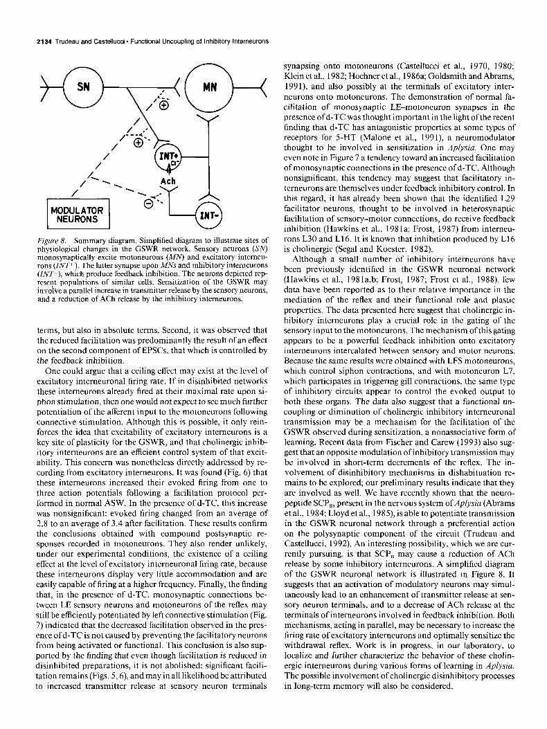
Figure 8. Summary diagram. Simplified diagram to illustrate sites of physiological changes in the GSWR network. Sensory neurons (SN) monosynaptically excite motoneurons (MN) and excitatory intemeu- rons (ZNT+ ). The latter synapse upon MNs and inhibitory interneurons (Z/VP), which produce feedback inhibition. The neurons depicted rep- resent populations of similar cells. Sensitization of the GSWR may invo i- ve a parallel increase in transmitter release by the sensory neurons, and a reduction of ACh release by the inhibitory intemeurons.
terms, but also in absolute terms. Second, it was observed that the reduced facilitation was predominantly the result of an effect on the second component of EPSCs, that which is controlled by the feedback inhibition.
One could argue that a ceiling effect may exist at the level of excitatory intemeuronal firing rate. If in disinhibited networks these interneurons already fired at their maximal rate upon si- phon stimulation, then one would not expect to see much further potentiation of the afferent input to the motoneurons following connective stimulation. Although this is possible, it only rein- forces the idea that excitability of excitatory interneurons is a key site of plasticity for the GSWR, and that cholinergic inhib- itory interneurons are an efficient control system of that excit- ability. This concern was nonetheless directly addressed by re- cording from excitatory interneurons. It was found (Fig. 6) that these interneurons increased their evoked firing from one to three action potentials following a facilitation protocol per- formed in normal ASW. In the presence of d-TC, this increase was nonsignificant: evoked firing changed from an average of 2.8 to an average of 3.4 after facilitation. These results confirm the conclusions obtained with compound postsynaptic re- sponses recorded in motoneurons. They also render unlikely, under our experimental conditions, the existence of a ceiling effect at the level of excitatory intemeuronal firing rate, because these interneurons display very little accommodation and are easily capable of firing at a higher frequency. Finally, the finding that, in the presence of d-TC, monosynaptic connections be- tween LE sensory neurons and motoneurons of the reflex may still be efficiently potentiated by left connective stimulation (Fig. 7) indicated that the decreased facilitation observed in the pres- ence of d-TC is not caused by preventing the facilitatory neurons from being activated or functional. This conclusion is also sup- ported by the finding that even though facilitation is reduced in disinhibited preparations, it is not abolished: significant facili- tation remains (Figs. 5,6), and may in all likelihood be attributed
synapsing onto motoneurons (Castellucci et al., 1970, 1980; Klein et al., 1982; Hochner et al., 1986a; Goldsmith and Abrams, 1991), and also possibly at the terminals of excitatory inter- neurons onto motoneurons. The demonstration of normal fa- cilitation of monosynaptic LE-motoneuron synapses in the presence of d-TC was thought important in the light of the recent finding that d-TC has antagonistic properties at some types of receptors for 5-HT (Malone et al., 1991) a neuromodulator thought to be involved in sensitization in Aplysia. One may even note in Figure 7 a tendency toward an increased facilitation of monosynaptic connections in the presence of d-TC. Although nonsignificant, this tendency may suggest that facilitatory in- temeurons are themselves under feedback inhibitory control. In this regard, it has already been shown that the identified L29 facilitator neurons, thought to be involved in heterosynaptic facilitation of sensory-motor connections, do receive feedback inhibition (Hawkins et al., 198 la; Frost, 1987) from intemeu- rons L30 and L16. It is known that inhibition produced by L16 is cholinergic (Segal and Koester, 1982).
Although a small number of inhibitory interneurons have been previously identified in the GSWR neuronal network (Hawkins et al., 198la,b; Frost, 1987; Frost et al., 1988) few data have been reported as to their relative importance in the mediation of the reflex and their functional role and plastic properties. The data presented here suggest that cholinergic in- hibitory interneurons play a crucial role in the gating of the sensory input to the motoneurons. The mechanism ofthis gating appears to be a powerful feedback inhibition onto excitatory interneurons intercalated between sensory and motor neurons. Because the same results were obtained with LFS motoneurons, which control siphon contractions, and with motoneuron L7, which participates in triggering gill contractions, the same type of inhibitory circuits appear to control the evoked output to both these organs. The data also suggest that a functional un- coupling or diminution of cholinergic inhibitory intemeuronal transmission may be a mechanism for the facilitation of the GSWR observed during sensitization, a nonassociative form of learning. Recent data from Fischer and Carew (1993) also sug- gest that an opposite modulation of inhibitory transmission may
be involved in short-term decrements of the reflex. The in- volvement of disinhibitory mechanisms in dishabituation re- mains to be explored; our preliminary results indicate that they are involved as well. We have recently shown that the neuro- peptide SCP,, present in the nervous system ofAplysia (Abrams et al., 1984; Lloyd et al., 1985), is able to potentiate transmission in the GSWR neuronal network through a preferential action on the polysynaptic component of the circuit (Trudeau and Castellucci, 1992). An interesting possibility, which we are cur- rently pursuing, is that SCP, may cause a reduction of ACh release by some inhibitory interneurons. A simplified diagram of the GSWR neuronal network is illustrated in Figure 8. It suggests that an activation of modulatory neurons may simul- taneously lead to an enhancement of transmitter release at sen- sory neuron terminals, and to a decrease of ACh release at the terminals of interneurons involved in feedback inhibition. Both mechanisms, acting in parallel, may be necessary to increase the firing rate of excitatory interneurons and optimally sensitize the withdrawal reflex. Work is in progress, in our laboratory, to localize and further characterize the behavior of these cholin- ergic interneurons during various forms of learning in Aplysia. The possible involvement of cholinergic disinhibitory processes
to increased transmitter release at sensory neuron terminals in long-term memory will also be considered

The Journal of Neuroscience, May 1993, 13(5) 2135
References Abrams TW, Castellucci VF, Camardo JS, Kandel ER, Lloyd PE (1984)
Two endogenous neuropeptides modulate the gill and siphon with- drawal reflex in Aplysia by presynaptic facilitation involving CAMP- dependent closure of a serotonin-sensitive potassium channel. Proc Nat1 Acad Sci USA 81:7956-7960.
Bailey CH, Chen M, Keller F, Kandel ER (1992) Serotonin-mediated endocytosis of apCAM: an early step of learning-related synaptic growth in Aplysia. Science 256~645-649.
Baux G, Fossier P, Taut L (1990) Histamine and FLRFamide regulate acetylcholine release at an identified synapse in Aplysia in opposite ways. J Physiol (Lond) 429:147-168.
Blankenship JE, Wachtel H, Kandel ER (197 1) Ionic mechanisms of excitatory, inhibitory, and dual synaptic actions mediated by an iden- tified intemeuron in the abdominal ganglion ofApiysia. J Neurophys- iol 34:76-92.
Brunelli M, Castellucci VF, Kandel ER (1976) Synaptic facilitation and behavioral sensitization in Aplysia: possible role of serotonin and CAMP. Science 194: 1178-l 180.
Byrne JH (1983) Identification and initial characterization of a cluster of command and pattern-generating neurons underlying respiratory pumping in Aplysia californica. J Neurophysiol 49149 l-507.
Capiod T, Ogden DC (1989) The properties of calcium-activated po- tassium channels in guinea-pig isolated hepatocytes. J Physiol (Lond) 409:285-295.
Carew TJ, Pinsker H, Rubinson K, Kandel ER (1974) Physiological anM biochemical properties of neuromuscular transmission between identified motoneurons and gill muscle in Aplysia. J Neurophysiol 37:1020-1040.
Carpenter DO, Swann JW, Yarowsky PJ (1977) Effect of curare on responses to different neurotransmitters in Apiysia neurons. J Neu- robiol 8:119-132.
Castellucci VF, Pinsker H, Kupfermann I, Kandel ER (1970) Neuronal mechanisms of habituation and dishabituation of the gill-withdrawal reflex in Aolvsia. Science 167: 1745-1748.
Castellucci VF: Kandel ER, Schwartz JH, Wilson FD, Naim AC, Green- gard P (1980) Intracellular injection of the catalytic subunit of cyclic AMP-dependent protein kinase stimulates facilitation of transmitter release underlying behavioral sensitization in Aplysia. Proc Nat1 Acad Sci USA 7717492-7496.
Dash PK, Hochner B, Kandel ER (1990) Injection of the CAMP- resoonsive element into the nucleus ofAp/ysia sensory neurons blocks long-term facilitation. Nature 345:7 18-721.
Davis WJ. Gillette R. Kovac MP. Croll RP, Matera EM (1983) Or- ganization of synaptic inputs to paracerebral feeding command in- temeurons of Pleurobranchaea californica. III. Modifications induced by experience. J Neurophysio149: 1557-l 572.
Elaagouby A, Ravel N, Gervais R (199 1) Cholinergic modulation of excitability in the rat olfactory bulb: effect of local application of cholinergic agents on evoked field potentials. Neuroscience 45:653- 662.
Fischer TM, Carew TJ (1993) Activity-dependent potentiation of recurrent inhibition: a mechanism for dynamic gain control in the siphon withdrawal reflex of Aplysia. J. Neurosci 13: 1302-l 3 14.
Frazier WT, Kandel ER, Kupfermann I, Waziri R, Coggeshall RE (1967) Morphological and functional properties of identified neurons in the abdominal ganglion of Aplysia californica. J Neurophysiol 30: 1288-1351.
Frost WN (1987) Mechanisms contributing to short- and long-term sensitization in AD/vsia. PhD thesis, Columbia University, New York.
Frost WN, Clark GA; Kandel ER (1988) Parallel processing of short- term memorv for sensitization in Aolvsia. J Neurobiol 19:297-334.
Gardner D (197 1) Bilateral symmetry and intemeuronal organization in the buccal ganglia of Aplysia. Science 173:550-553.
Giller E Jr, Schwartz JH (197 1) Choline acetyltransferase in identified neurons of abdominal ganglion of Aplysia californica. J Neurophysiol 34:93-l 15.
Goldsmith BA, Abrams TW (199 1) Reversal of synaptic depression by serotonin at Aplysia sensory neuron synapses involves activation of adenylyl cyclase. Proc Nat1 Acad Sci USA 88:902 l-9025.
Hawkins RD, Castellucci VF, Kandcl ER (198 la) Intemeurons in- volved in mediation and modulation ofgill-withdrawal reflex in Aply- sia. I. Identification and characterization. J Neurophysiol 45:304- 314.
Hawkins RD, Castellucci VF, Kandel ER (198 1 b) Interneurons in- volved in mediation and modulation of the gill-withdrawal reflex in Aplysia. II. Identified neurons produce heterosynaptic facilitation con- tributing to behavioral sensitization. J Neurophysiol 45:3 15-326.
Hochner B, Klein M, Schacher S, Kandel ER (1986a) Action-potential duration and the modulation of transmitter release from the sensory neurons of Aolvsia in presvnaptic facilitation and behavioral sensi- tization. Pro; Nat1 Acad S&i USA 83:8410-84 14.
Hochner B. Klein M. Schacher S. Kandel ER (1986b) Additional component in the cellular mechanism of presyna‘ptic facilitation con- tributes to behavioral dishabituation in Aplysia. Proc Nat1 Acad Sci USA 83:8794-8798.
Kehoe J (1972) Three acetylcholine receptors in Aplysia neurones. J Physiol (Lond) 225: 115-146.
Klein M, Camardo J, Kandel ER (1982) Serotonin modulates a specific potassium current in the sensory neurons that show presynaptic fa- cilitation in Aplysia. Proc Nat1 Acad Sci USA 79:57 13-57 17.
Kom H, Oda Y, Faber DS (1992) Long-term potentiation ofinhibitory circuits and synapses in the central nervous system. Proc Nat1 Acad Sci USA 89:440-443.
Lederhendler II, Cart S, Alkon DL (1986) Classical conditioning of Hermissenda: origin of a new response. J Neurosci 6: 1325-l 33 1.
Llovd PE. Mahon AC. Kunfermann I. Cohen JL. Scheller RH. Weiss KR (1985) Biochemicaland immunocytological localization of mol- luscan small cardioactive peptides in the nervous system of Aplysia californica. J Neurosci 5: 185 1-l 86 I.
Lukowiak K, Peretz B (1977) The interaction between the central and peripheral nervous systems in the mediation of gill withdrawal reflex behavior in Aplysia. J Comp Physiol 117:2 19-244.
Malone HM, Peters JA, Lambert JJ (1991) Physiological and phar- macological properties of 5-HT3 receptors: a patch-clamp study. Neu- ropeptides 19:25-30.
Mayford M, Barzilai A, Keller F, Schacher S, Kandel ER (1992) Mod- ulation of an NCAM-related adhesion molecule with long-term syn- aptic plasticity in Aplysia. Science 256:638-644.
McCaman RE. Weinreich D (1985) Histamineraic synaptic transmis- sion in the cerebral ganglion of Aplysia. J N&rophysiol 53: 10 16- 1037.
Montarolo PC, Goelet P, Castellucci VF, Morgan J, Kandel ER (1986) A critical time-window for macromolecular synthesis in long-term heterosynaptic facilitation in Aplysia. Science 234: 1249-1254.
Moot DD. Lewis DV (199 1) Facilitation of the induction of long-term potent&ion by GABA, receptors. Science 252: 17 18-l 720. -
Nohmi M, Kuba K (1984) (+)-Tubocuratine blocks the Ca+ + -depen- dent K I channel of the bullfrog ganglion cell. Brain Res 301: 146- 148.
Ono JK, Salvaterra PM (1981) Snake a-toxin effects on cholinergic and noncholinergic responses of Aplysia californica neurons. J Neu- rosci 1~259-270.
Segal MM, Koester J (1982) Convergent cholinergic neurons produce similar postsynaptic actions in Aplysia: implication for neural orga- nization. J Neurouhvsiol 47:742-759.
Taut L, Gerschenfeid HM (1962) A cholinergic mechanism of inhib- itory synaptic transmission in a molluscan nervous system. J Neu- rophysiol 25:236-262.
Trudeau L-E, Castellucci VF (1992) Contribution of polysynaptic pathways in the mediation and plasticity of Apl.vsia gill and siphon withdrawal reflex: evidence for differential modulation. J Neurosci 12:3838-3848.
Weiss S, Goldberg JI, Edstrom JP, Lukowiak K (1984) Cholinergic receptors in the Aplysia gill. J Neurobiol 15:325-332.