Osmoregulation - ELTE Élettani és Neurobiológiai...
26
1 Osmoregulation Ionic and osmotic balance • in multicellular organisms the interstitial fluid is the internal environment • osmoregulatory organs maintain this environment isovolumic, isotonic, isoionic, isohydric, etc. • further task of the osmoregulatory organs: removal of poisonous end products of metabolism (NH 3 from proteins) in the form of urea • through plasma membrane different ionic composition, but equal osmotic pressure • through epithelium fluids are different in both respects • obligate and regulated osmotic exchange • obligate exchange depends on physical factors, regulated exchange compensates for changes 2/24
Transcript of Osmoregulation - ELTE Élettani és Neurobiológiai...
1
Osmoregulation
Ionic and osmotic balance
• in multicellular organisms the interstitial fluid is the internal environment
• osmoregulatory organs maintain this environment isovolumic, isotonic, isoionic, isohydric, etc.
• further task of the osmoregulatory organs: removal of poisonous end products of metabolism (NH3 from proteins) in the form of urea
• through plasma membrane different ioniccomposition, but equal osmotic pressure
• through epithelium fluids are different in both respects
• obligate and regulated osmotic exchange
• obligate exchange depends on physical factors, regulated exchange compensates for changes
2/24
2
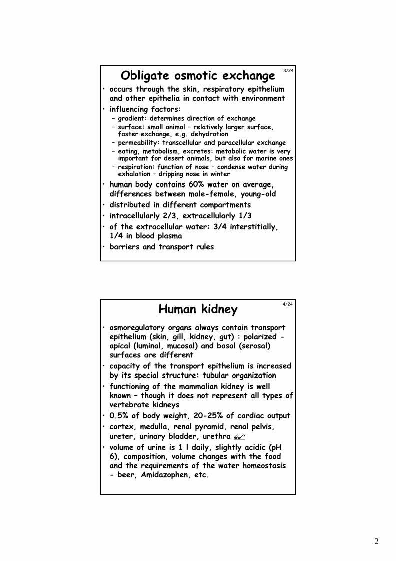
Obligate osmotic exchange• occurs through the skin, respiratory epithelium
and other epithelia in contact with environment
• influencing factors:– gradient: determines direction of exchange– surface: small animal – relatively larger surface,
faster exchange, e.g. dehydration– permeability: transcellular and paracellular exchange– eating, metabolism, excretes: metabolic water is very
important for desert animals, but also for marine ones– respiration: function of nose – condense water during
exhalation – dripping nose in winter
• human body contains 60% water on average, differences between male-female, young-old
• distributed in different compartments
• intracellularly 2/3, extracellularly 1/3
• of the extracellular water: 3/4 interstitially, 1/4 in blood plasma
• barriers and transport rules
3/24
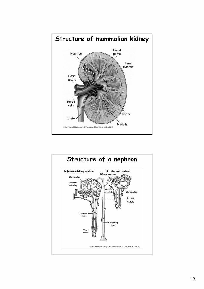
Human kidney
• osmoregulatory organs always contain transport epithelium (skin, gill, kidney, gut) : polarized -apical (luminal, mucosal) and basal (serosal) surfaces are different
• capacity of the transport epithelium is increased by its special structure: tubular organization
• functioning of the mammalian kidney is well known – though it does not represent all types of vertebrate kidneys
• 0.5% of body weight, 20-25% of cardiac output
• cortex, medulla, renal pyramid, renal pelvis, ureter, urinary bladder, urethra ����
• volume of urine is 1 l daily, slightly acidic (pH 6), composition, volume changes with the food and the requirements of the water homeostasis - beer, Amidazophen, etc.
4/24
3
The nephron
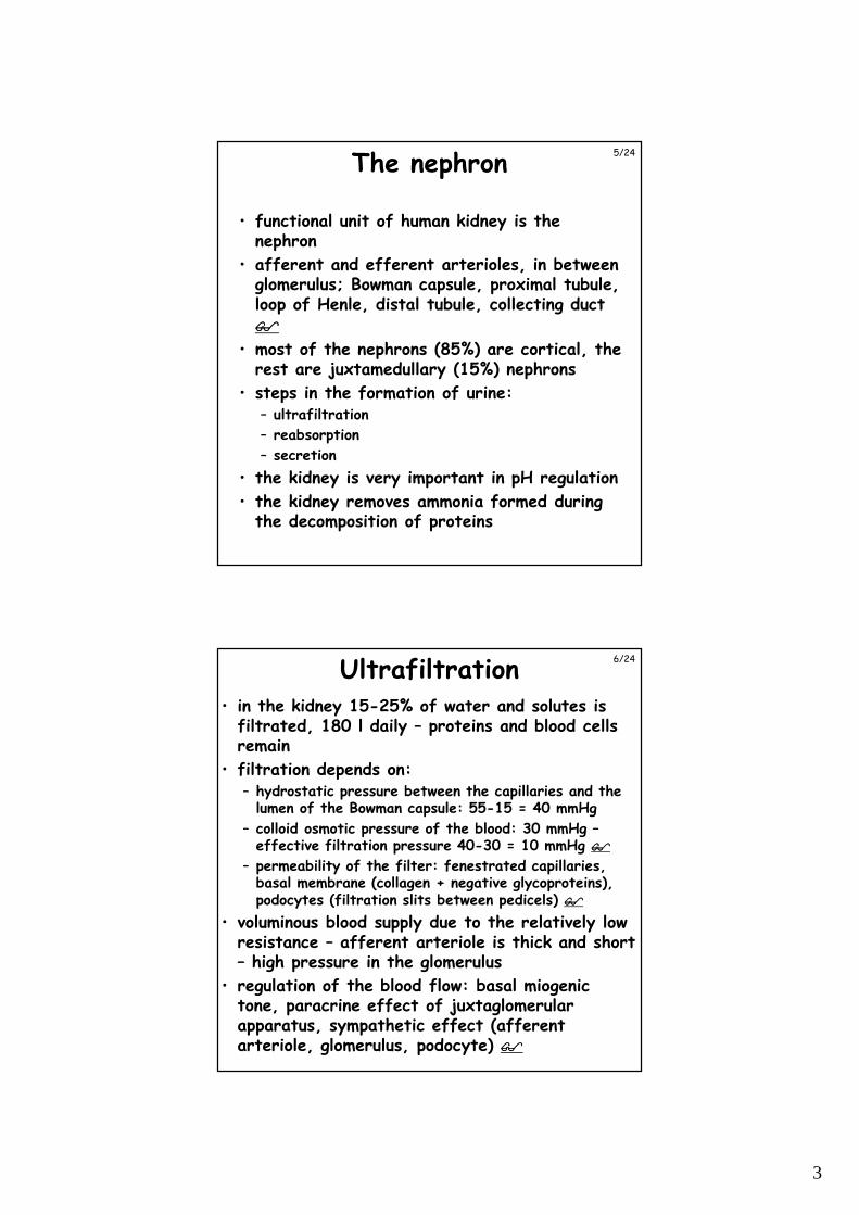
• functional unit of human kidney is the nephron
• afferent and efferent arterioles, in between glomerulus; Bowman capsule, proximal tubule, loop of Henle, distal tubule, collecting duct ����
• most of the nephrons (85%) are cortical, the rest are juxtamedullary (15%) nephrons
• steps in the formation of urine:– ultrafiltration
– reabsorption
– secretion
• the kidney is very important in pH regulation
• the kidney removes ammonia formed during the decomposition of proteins
5/24
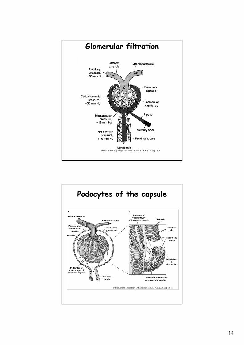
Ultrafiltration• in the kidney 15-25% of water and solutes is
filtrated, 180 l daily – proteins and blood cells remain
• filtration depends on:– hydrostatic pressure between the capillaries and the
lumen of the Bowman capsule: 55-15 = 40 mmHg
– colloid osmotic pressure of the blood: 30 mmHg –effective filtration pressure 40-30 = 10 mmHg ����
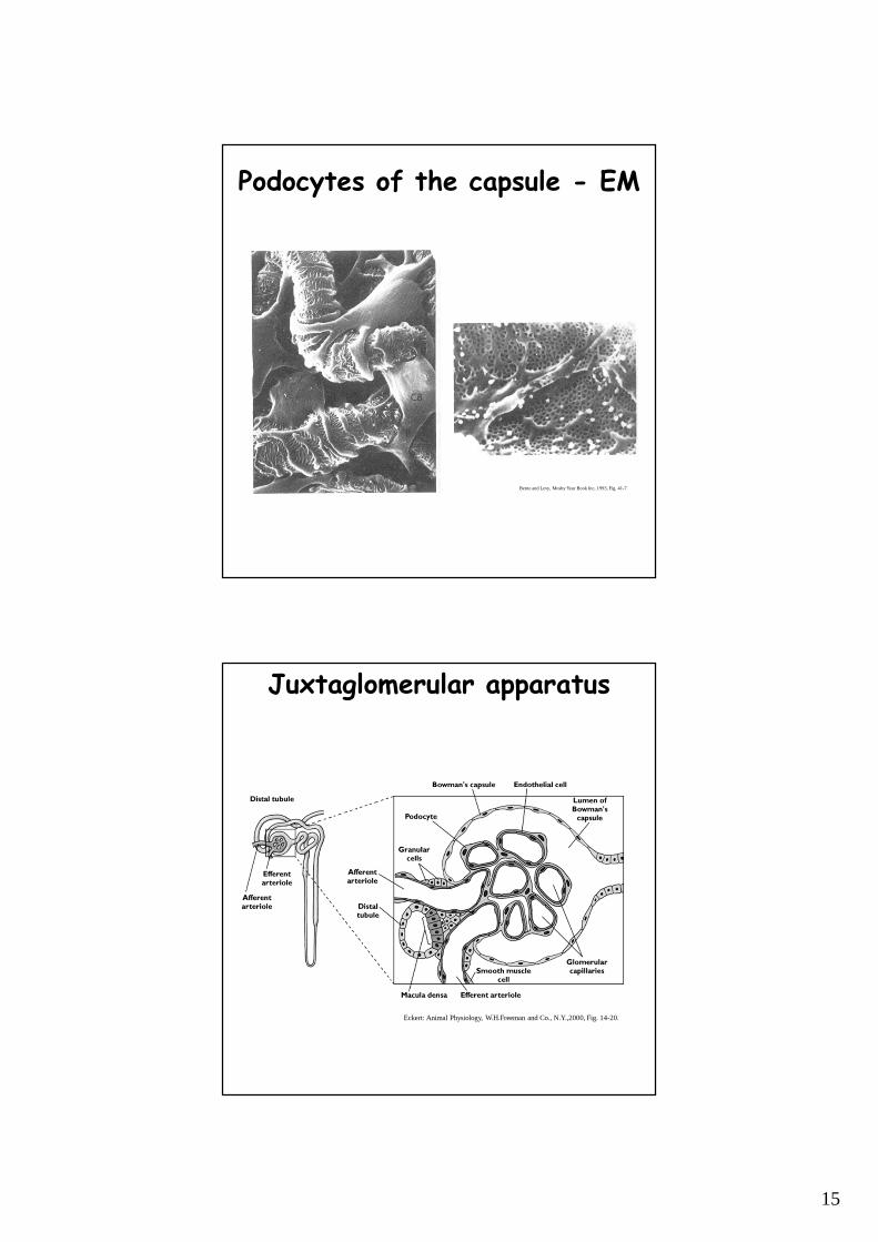
– permeability of the filter: fenestrated capillaries, basal membrane (collagen + negative glycoproteins), podocytes (filtration slits between pedicels) ����
• voluminous blood supply due to the relatively low resistance – afferent arteriole is thick and short – high pressure in the glomerulus
• regulation of the blood flow: basal miogenic tone, paracrine effect of juxtaglomerular apparatus, sympathetic effect (afferent arteriole, glomerulus, podocyte) ����
6/24
4
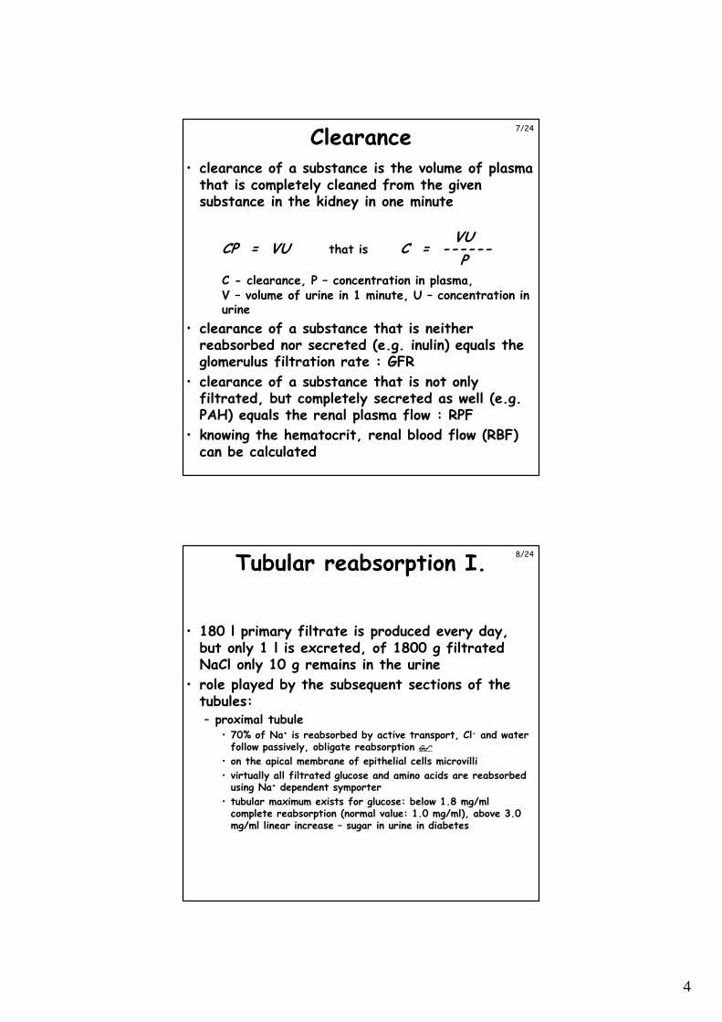
Clearance• clearance of a substance is the volume of plasma
that is completely cleaned from the given substance in the kidney in one minute
VUCP = VU that is C = ------
PC - clearance, P – concentration in plasma,V – volume of urine in 1 minute, U – concentration in urine
• clearance of a substance that is neither reabsorbed nor secreted (e.g. inulin) equals the glomerulus filtration rate : GFR
• clearance of a substance that is not only filtrated, but completely secreted as well (e.g. PAH) equals the renal plasma flow : RPF
• knowing the hematocrit, renal blood flow (RBF) can be calculated
7/24
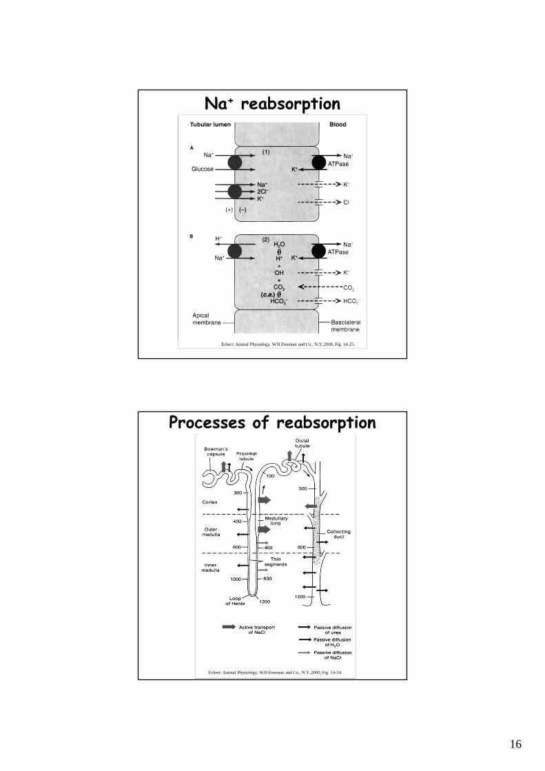
Tubular reabsorption I.
• 180 l primary filtrate is produced every day, but only 1 l is excreted, of 1800 g filtrated NaCl only 10 g remains in the urine
• role played by the subsequent sections of the tubules:– proximal tubule
• 70% of Na+ is reabsorbed by active transport, Cl- and water follow passively, obligate reabsorption ����
• on the apical membrane of epithelial cells microvilli
• virtually all filtrated glucose and amino acids are reabsorbed using Na+ dependent symporter
• tubular maximum exists for glucose: below 1.8 mg/ml complete reabsorption (normal value: 1.0 mg/ml), above 3.0 mg/ml linear increase – sugar in urine in diabetes
8/24
5
Tubular reabsorption II.
– descending part of Henle’s loop• no microvilli, few mitochondria – no active transport• low permeability for NaCl and urea, high for water
– thin ascending part of Henle’s loop• no microvilli, few mitochondria – no active transport• low permeability for water, high for NaCl
– thick ascending part of Henle’s loop• active reabsorption of Na+
• low water permeability
– distal tubule• active reabsorption of Na+, and passive reabsorption
of water• K+, H+ and NH3 transport as needed – see later (pH
regulation)• transport is regulated by hormones – facultative
reabsorption
– collecting duct• active reabsorption of Na+ at the cortical part, high
urea permeability in the internal medullary part• regulated water permeability (ADH) ����
9/24
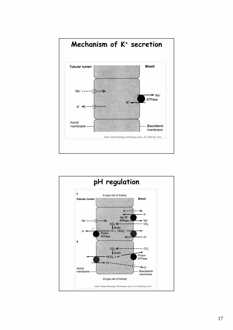
Tubular secretion
• several substances are secreted from the plasma to the tubule in the nephron – best examined: different electrolytes (K+, H+, NH3) organic acids and bases
• active transport – recognizes substances conjugated with glucuronic acid in the liver
• K+ is reabsorbed in the proximal tubule and Henle’s loop (Na/2Cl/K transporter)
• excess K+ is exchanged for Na+ in the distal tubule
• secretion of H+ and NH3 serves pH regulation
10/24
6
pH regulation
• normal pH 7.4 – 7.35 acidosis, 7.45 alkalosis• normal functioning is possible between 7.0-7.8• respiratory alkalosis and acidosis: caused by
hyper-, or hypoventilation• metabolic alkalosis – e.g. vomiting• metabolic acidosis – anaerobic energy production
• buffers: CO2/HCO3-, plasma proteins, phosphate
• respiration: changing respiratory rate• kidney:
– proximal tubule, Henle’s loop: Na+/H+ exchanger, distal tubule, collecting duct: HCO3
- uptake, H+
secretion through A-cells
– in distal tubule and collecting duct: HCO3- secretion,
H+ uptake through B-cells ����
11/24
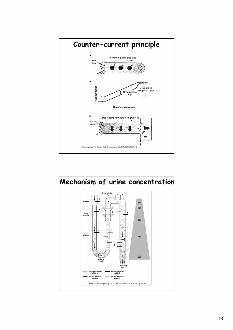
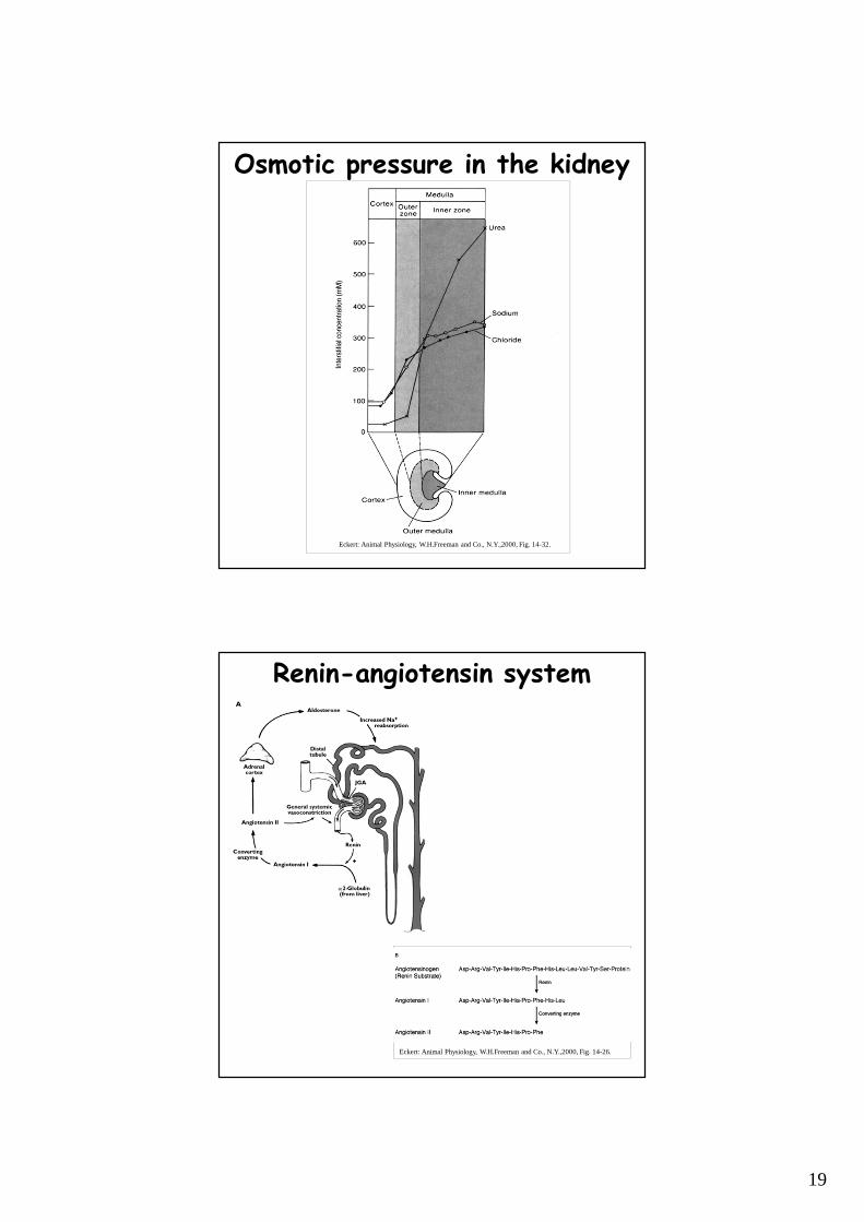
Hyperosmotic urine
• birds and mammals can produce hyperosmotic urine - water reabsorption in the collecting duct due to osmotic pressure differences
• generation of osmotic pressure difference is helped by the counter-current principle ����
• Na+ transport in the ascending part of the Henle’s loop – do not enter the descending part, but attracts water leading to the same result
• in addition, urea present in high concentration because of the reabsorption of water, can only leave the tubule in the internal medulla ����
• osmotic pressure increases from the cortex to the medulla ����
• blood supply to the tubules (vasa recta) is running in parallel to the Henle’s loop, does not decrease the osmotic gradient
12/24
7
Regulation of the kidney• granular cells in the juxtaglomerular apparatus
produce renin in response to a decrease in blood pressure or NaCl delivery to the distal tubule
• renin cuts off angiotensin I (10 amino acids) from angiotensinogen (glycoprotein)
• converting enzyme (mostly in the lung) cuts off 2 amino acids from angiotensin I – angiotensin II
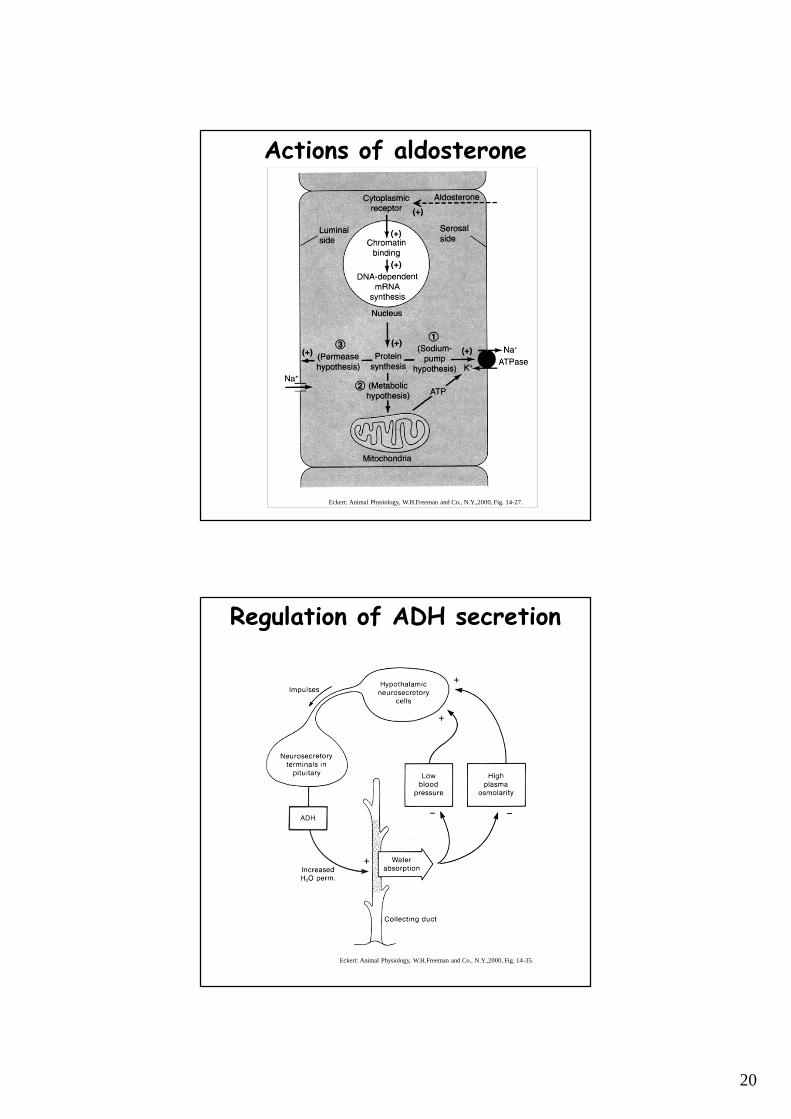
• angiotensin II enhances aldosterone secretion in the adrenal gland, increases blood pressurethrough vasoconstriction and increases ADHproduction ����
• aldosterone increase Na+ reabsorption through 3 different ways: facilitation of the pump, ATP production, increased apical Na+ permeability ����
• ADH producing cells detect blood pressure and osmolality and are sensitive to alcohol ����
• atrial natriuretic peptide (ANP) – released in the atria when venous pressure increases - inhibits renin, aldosterone, ADH production
13/24
Digestion
8
Alimentary canal in vertebrates
• in unicellular and primitive multicellular organisms intracellular digestion
• in more developed multicellular organisms –extracellular digestion
• topologically external to the body
• entrance and exit are protected by sphincters and other devices
• ingested material is subjected to various mechanical, chemical and bacterial effects
• tubular organization allows for functional specialization (i.e. acidic and alkaline environment)
• parts of the alimentary canal: headgut, foregut, midgut, hindgut
15/24
Headgut - Foregut
• headgut
– food enters here – structures related to feeding and swallowing: mouth-parts, buccal (oral) cavity, pharynx, bills, teeth, tongue, salivary glands, additional structures to direct the flow of ingested materials and inspired water or air
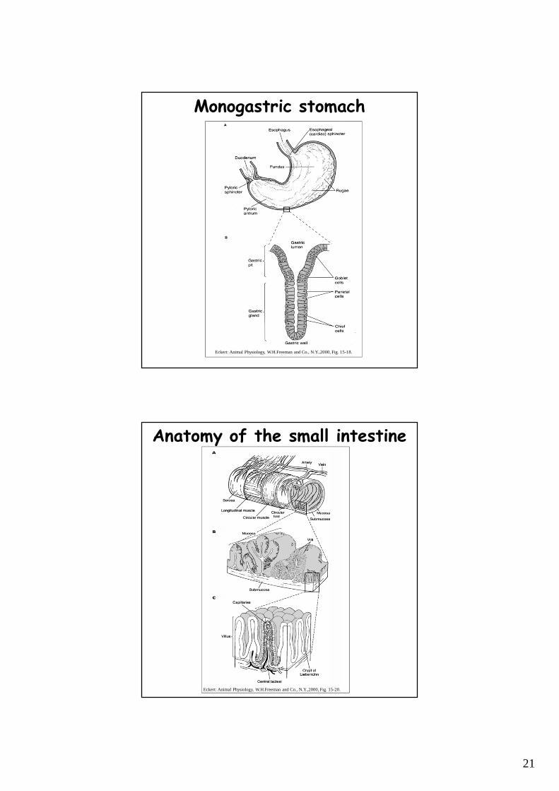
• foregut– in most species: esophagus and stomach– esophagus carries food from headgut to stomach– digestion starts in the stomach– in most vertebrates: pepsinogen and HCl– monogastric stomach in omnivorous and carnivorous
vertebrates– invaginations with gastric pits with gland cells ����
16/24
9
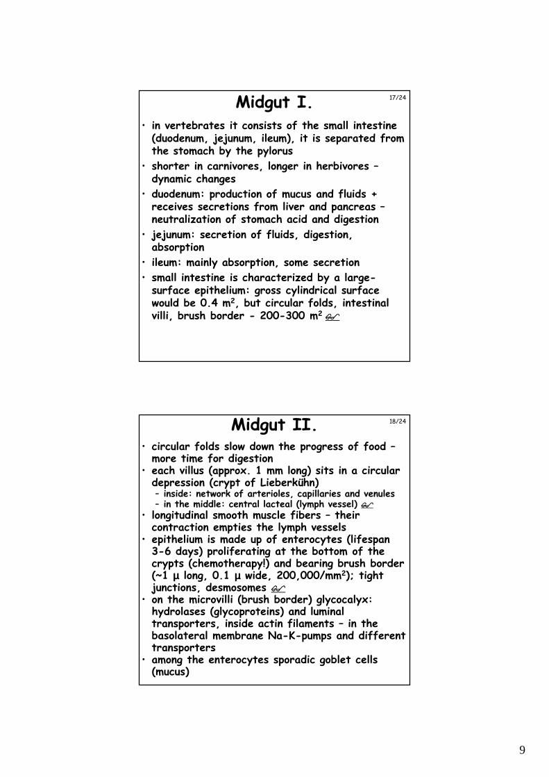
Midgut I.• in vertebrates it consists of the small intestine
(duodenum, jejunum, ileum), it is separated from the stomach by the pylorus
• shorter in carnivores, longer in herbivores –dynamic changes
• duodenum: production of mucus and fluids + receives secretions from liver and pancreas –neutralization of stomach acid and digestion
• jejunum: secretion of fluids, digestion, absorption
• ileum: mainly absorption, some secretion
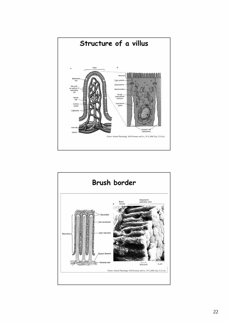
• small intestine is characterized by a large-surface epithelium: gross cylindrical surface would be 0.4 m2, but circular folds, intestinal villi, brush border - 200-300 m2 ����
17/24
Midgut II.• circular folds slow down the progress of food –
more time for digestion• each villus (approx. 1 mm long) sits in a circular
depression (crypt of Lieberkühn)– inside: network of arterioles, capillaries and venules– in the middle: central lacteal (lymph vessel) ����
• longitudinal smooth muscle fibers – their contraction empties the lymph vessels
• epithelium is made up of enterocytes (lifespan 3-6 days) proliferating at the bottom of the crypts (chemotherapy!) and bearing brush border(~1 µµµµ long, 0.1 µµµµ wide, 200,000/mm2); tight junctions, desmosomes ����
• on the microvilli (brush border) glycocalyx: hydrolases (glycoproteins) and luminal transporters, inside actin filaments – in the basolateral membrane Na-K-pumps and different transporters
• among the enterocytes sporadic goblet cells(mucus)
18/24
10
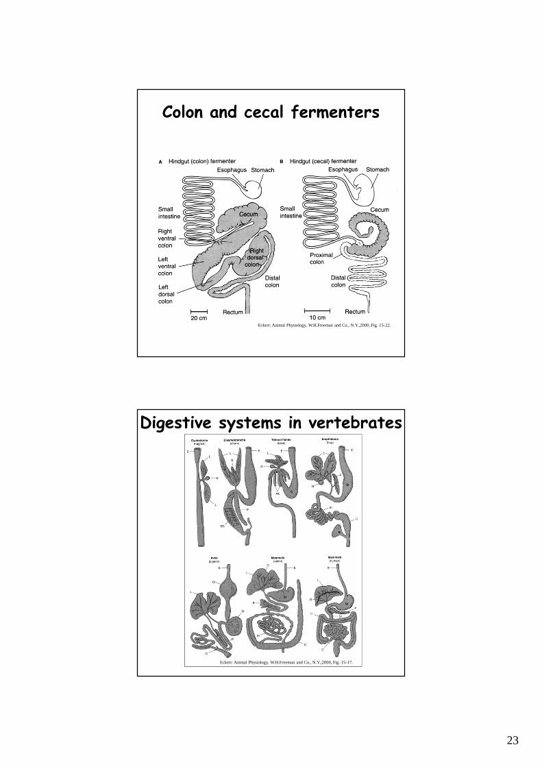
Hindgut
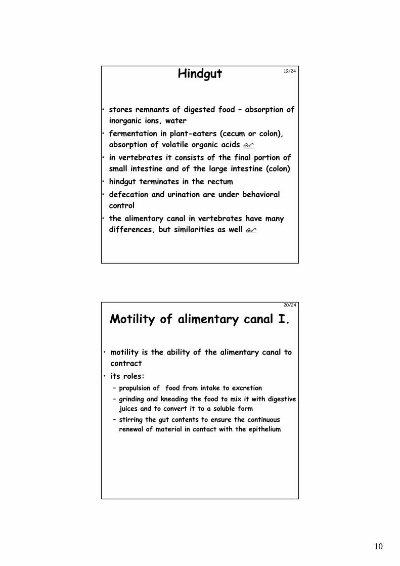
• stores remnants of digested food – absorption of
inorganic ions, water
• fermentation in plant-eaters (cecum or colon),
absorption of volatile organic acids ����
• in vertebrates it consists of the final portion of
small intestine and of the large intestine (colon)
• hindgut terminates in the rectum
• defecation and urination are under behavioral
control
• the alimentary canal in vertebrates have many
differences, but similarities as well ����
19/24
Motility of alimentary canal I.
• motility is the ability of the alimentary canal to
contract
• its roles:
– propulsion of food from intake to excretion
– grinding and kneading the food to mix it with digestive
juices and to convert it to a soluble form
– stirring the gut contents to ensure the continuous
renewal of material in contact with the epithelium
20/24
11
Motility of alimentary canal II.
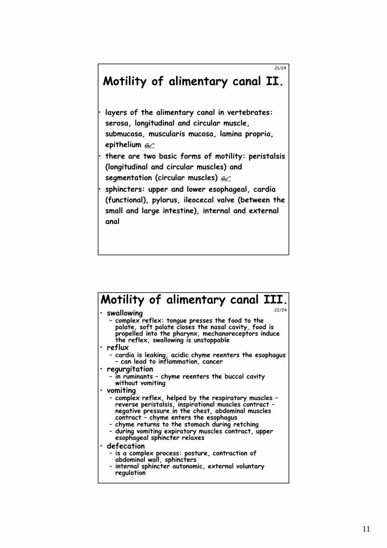
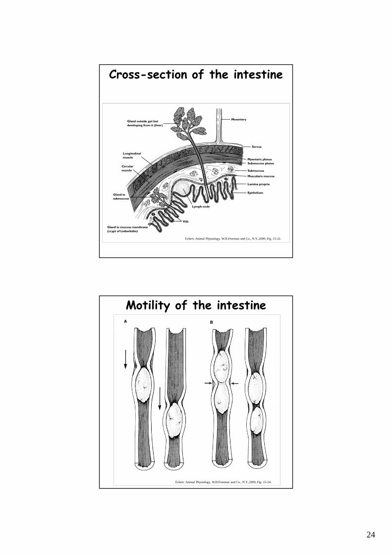
• layers of the alimentary canal in vertebrates:
serosa, longitudinal and circular muscle,
submucosa, muscularis mucosa, lamina propria,
epithelium ����
• there are two basic forms of motility: peristalsis
(longitudinal and circular muscles) and
segmentation (circular muscles) ����
• sphincters: upper and lower esophageal, cardia
(functional), pylorus, ileocecal valve (between the
small and large intestine), internal and external
anal
21/24
Motility of alimentary canal III.• swallowing
– complex reflex: tongue presses the food to the palate, soft palate closes the nasal cavity, food is propelled into the pharynx, mechanoreceptors induce the reflex, swallowing is unstoppable
• reflux– cardia is leaking, acidic chyme reenters the esophagus
– can lead to inflammation, cancer• regurgitation
– in ruminants – chyme reenters the buccal cavity without vomiting
• vomiting– complex reflex, helped by the respiratory muscles –
reverse peristalsis, inspirational muscles contract –negative pressure in the chest, abdominal muscles contract – chyme enters the esophagus
– chyme returns to the stomach during retching– during vomiting expiratory muscles contract, upper
esophageal sphincter relaxes• defecation
– is a complex process: posture, contraction of abdominal wall, sphincters
– internal sphincter autonomic, external voluntary regulation
22/24
12
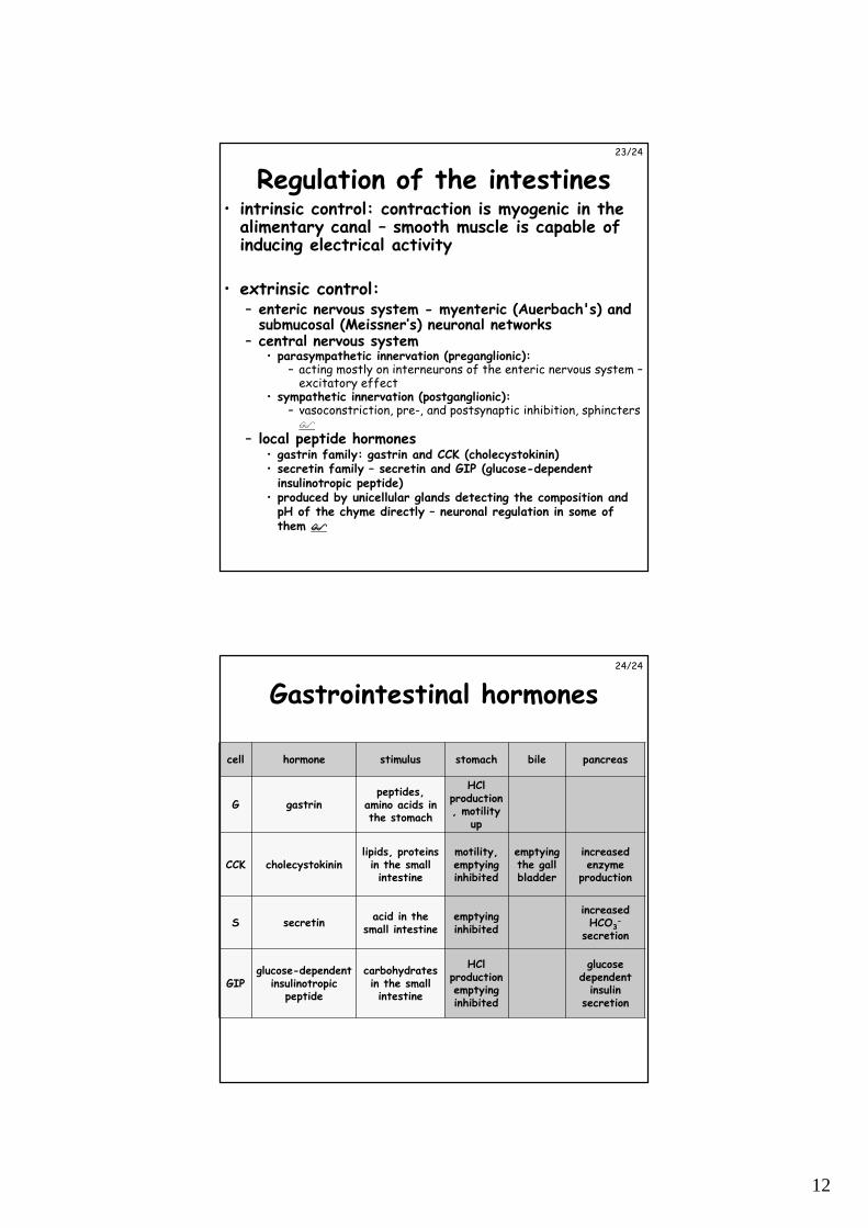
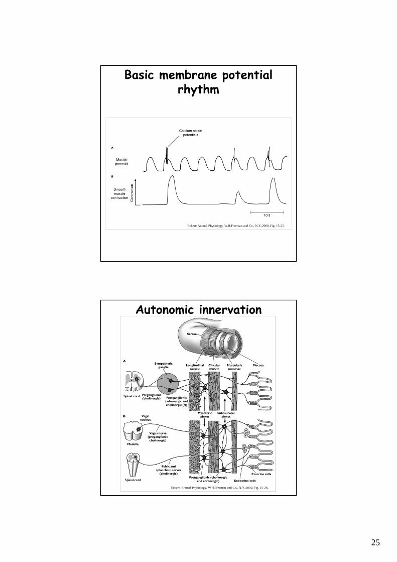
Regulation of the intestines• intrinsic control: contraction is myogenic in the
alimentary canal – smooth muscle is capable of inducing electrical activity
• extrinsic control:– enteric nervous system - myenteric (Auerbach's) and
submucosal (Meissner’s) neuronal networks– central nervous system
• parasympathetic innervation (preganglionic):– acting mostly on interneurons of the enteric nervous system –
excitatory effect• sympathetic innervation (postganglionic):
– vasoconstriction, pre-, and postsynaptic inhibition, sphincters �
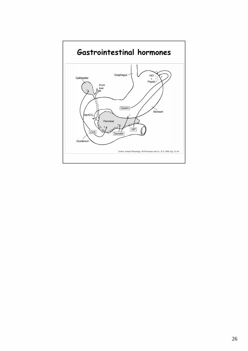
– local peptide hormones• gastrin family: gastrin and CCK (cholecystokinin)• secretin family – secretin and GIP (glucose-dependent
insulinotropic peptide)• produced by unicellular glands detecting the composition and
pH of the chyme directly – neuronal regulation in some of them ����
23/24
Gastrointestinal hormones
cell hormone stimulus stomach bile pancreas
G gastrinpeptides,
amino acids in the stomach
HCl production, motility
up
CCK cholecystokininlipids, proteins in the small intestine
motility, emptying inhibited
emptying the gall bladder
increased enzyme
production
S secretinacid in the
small intestineemptying inhibited
increased HCO3
-
secretion
GIPglucose-dependent
insulinotropic peptide
carbohydrates in the small intestine
HCl productionemptyinginhibited
glucose dependent
insulin secretion
24/24
13
Structure of mammalian kidney
Eckert: Animal Physiology, W.H.Freeman and Co., N.Y.,2000, Fig. 14-13.
Structure of a nephron
Eckert: Animal Physiology, W.H.Freeman and Co., N.Y.,2000, Fig. 14-14.
14
Glomerular filtration
Eckert: Animal Physiology, W.H.Freeman and Co., N.Y.,2000, Fig. 14-18
Podocytes of the capsule
Eckert: Animal Physiology, W.H.Freeman and Co., N.Y.,2000, Fig. 14-19.
15
Podocytes of the capsule - EM
Berne and Levy, Mosby Year Book Inc, 1993, Fig. 41-7
Juxtaglomerular apparatus
Eckert: Animal Physiology, W.H.Freeman and Co., N.Y.,2000, Fig. 14-20.
16
Na+ reabsorption
Eckert: Animal Physiology, W.H.Freeman and Co., N.Y.,2000, Fig. 14-25.
Processes of reabsorption
Eckert: Animal Physiology, W.H.Freeman and Co., N.Y.,2000, Fig. 14-24.
17
Mechanism of K+ secretion
Eckert: Animal Physiology, W.H.Freeman and Co., N.Y.,2000, Fig. 14-28.
pH regulation
Eckert: Animal Physiology, W.H.Freeman and Co., N.Y.,2000, Fig. 14-29.
18
Counter-current principle
Eckert: Animal Physiology, W.H.Freeman and Co., N.Y.,2000, SL. 14-2.
Mechanism of urine concentration
Eckert: Animal Physiology, W.H.Freeman and Co., N.Y.,2000, Fig. 14-32.
19
Osmotic pressure in the kidney
Eckert: Animal Physiology, W.H.Freeman and Co., N.Y.,2000, Fig. 14-32.
Renin-angiotensin system
Eckert: Animal Physiology, W.H.Freeman and Co., N.Y.,2000, Fig. 14-26.
20
Actions of aldosterone
Eckert: Animal Physiology, W.H.Freeman and Co., N.Y.,2000, Fig. 14-27.
Regulation of ADH secretion
Eckert: Animal Physiology, W.H.Freeman and Co., N.Y.,2000, Fig. 14-35.
21
Monogastric stomach
Eckert: Animal Physiology, W.H.Freeman and Co., N.Y.,2000, Fig. 15-18.
Anatomy of the small intestine
Eckert: Animal Physiology, W.H.Freeman and Co., N.Y.,2000, Fig. 15-20.
22
Structure of a villus
Eckert: Animal Physiology, W.H.Freeman and Co., N.Y.,2000, Fig. 15-21a,b.
Brush border
Eckert: Animal Physiology, W.H.Freeman and Co., N.Y.,2000, Fig. 15-21c,d.
23
Colon and cecal fermenters
Eckert: Animal Physiology, W.H.Freeman and Co., N.Y.,2000, Fig. 15-22.
Digestive systems in vertebrates
Eckert: Animal Physiology, W.H.Freeman and Co., N.Y.,2000, Fig. 15-17.
24
Cross-section of the intestine
Eckert: Animal Physiology, W.H.Freeman and Co., N.Y.,2000, Fig. 15-22.
Motility of the intestine
Eckert: Animal Physiology, W.H.Freeman and Co., N.Y.,2000, Fig. 15-24.
25
Basic membrane potentialrhythm
Eckert: Animal Physiology, W.H.Freeman and Co., N.Y.,2000, Fig. 15-25.
Autonomic innervation
Eckert: Animal Physiology, W.H.Freeman and Co., N.Y.,2000, Fig. 15-26.
26
Gastrointestinal hormones
Eckert: Animal Physiology, W.H.Freeman and Co., N.Y.,2000, Fig. 15-34.