ORIGINAL ARTICLE FREQUENCY OF HCV INFECTION IN DIFFERENT DERMATOLOGICAL DISORDERS · 2015-04-18 ·...
4
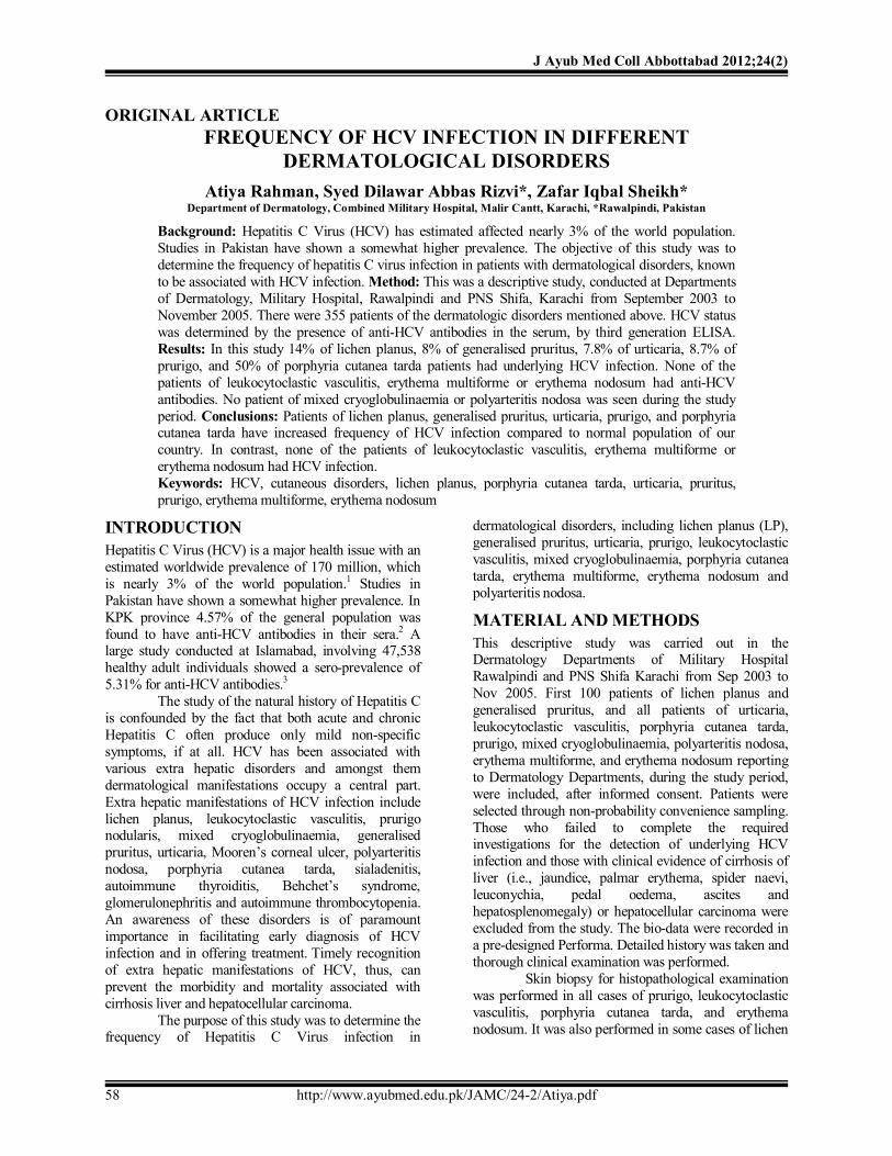
J Ayub Med Coll Abbottabad 2012;24(2) http://www.ayubmed.edu.pk/JAMC/24-2/Atiya.pdf 58 ORIGINAL ARTICLE FREQUENCY OF HCV INFECTION IN DIFFERENT DERMATOLOGICAL DISORDERS Atiya Rahman, Syed Dilawar Abbas Rizvi*, Zafar Iqbal Sheikh* Department of Dermatology, Combined Military Hospital, Malir Cantt, Karachi, *Rawalpindi, Pakistan Background: Hepatitis C Virus (HCV) has estimated affected nearly 3% of the world population. Studies in Pakistan have shown a somewhat higher prevalence. The objective of this study was to determine the frequency of hepatitis C virus infection in patients with dermatological disorders, known to be associated with HCV infection. Method: This was a descriptive study, conducted at Departments of Dermatology, Military Hospital, Rawalpindi and PNS Shifa, Karachi from September 2003 to November 2005. There were 355 patients of the dermatologic disorders mentioned above. HCV status was determined by the presence of anti-HCV antibodies in the serum, by third generation ELISA. Results: In this study 14% of lichen planus, 8% of generalised pruritus, 7.8% of urticaria, 8.7% of prurigo, and 50% of porphyria cutanea tarda patients had underlying HCV infection. None of the patients of leukocytoclastic vasculitis, erythema multiforme or erythema nodosum had anti-HCV antibodies. No patient of mixed cryoglobulinaemia or polyarteritis nodosa was seen during the study period. Conclusions: Patients of lichen planus, generalised pruritus, urticaria, prurigo, and porphyria cutanea tarda have increased frequency of HCV infection compared to normal population of our country. In contrast, none of the patients of leukocytoclastic vasculitis, erythema multiforme or erythema nodosum had HCV infection. Keywords: HCV, cutaneous disorders, lichen planus, porphyria cutanea tarda, urticaria, pruritus, prurigo, erythema multiforme, erythema nodosum INTRODUCTION Hepatitis C Virus (HCV) is a major health issue with an estimated worldwide prevalence of 170 million, which is nearly 3% of the world population. 1 Studies in Pakistan have shown a somewhat higher prevalence. In KPK province 4.57% of the general population was found to have anti-HCV antibodies in their sera. 2 A large study conducted at Islamabad, involving 47,538 healthy adult individuals showed a sero-prevalence of 5.31% for anti-HCV antibodies. 3 The study of the natural history of Hepatitis C is confounded by the fact that both acute and chronic Hepatitis C often produce only mild non-specific symptoms, if at all. HCV has been associated with various extra hepatic disorders and amongst them dermatological manifestations occupy a central part. Extra hepatic manifestations of HCV infection include lichen planus, leukocytoclastic vasculitis, prurigo nodularis, mixed cryoglobulinaemia, generalised pruritus, urticaria, Mooren’s corneal ulcer, polyarteritis nodosa, porphyria cutanea tarda, sialadenitis, autoimmune thyroiditis, Behchet’s syndrome, glomerulonephritis and autoimmune thrombocytopenia. An awareness of these disorders is of paramount importance in facilitating early diagnosis of HCV infection and in offering treatment. Timely recognition of extra hepatic manifestations of HCV, thus, can prevent the morbidity and mortality associated with cirrhosis liver and hepatocellular carcinoma. The purpose of this study was to determine the frequency of Hepatitis C Virus infection in dermatological disorders, including lichen planus (LP), generalised pruritus, urticaria, prurigo, leukocytoclastic vasculitis, mixed cryoglobulinaemia, porphyria cutanea tarda, erythema multiforme, erythema nodosum and polyarteritis nodosa. MATERIAL AND METHODS This descriptive study was carried out in the Dermatology Departments of Military Hospital Rawalpindi and PNS Shifa Karachi from Sep 2003 to Nov 2005. First 100 patients of lichen planus and generalised pruritus, and all patients of urticaria, leukocytoclastic vasculitis, porphyria cutanea tarda, prurigo, mixed cryoglobulinaemia, polyarteritis nodosa, erythema multiforme, and erythema nodosum reporting to Dermatology Departments, during the study period, were included, after informed consent. Patients were selected through non-probability convenience sampling. Those who failed to complete the required investigations for the detection of underlying HCV infection and those with clinical evidence of cirrhosis of liver (i.e., jaundice, palmar erythema, spider naevi, leuconychia, pedal oedema, ascites and hepatosplenomegaly) or hepatocellular carcinoma were excluded from the study. The bio-data were recorded in a pre-designed Performa. Detailed history was taken and thorough clinical examination was performed. Skin biopsy for histopathological examination was performed in all cases of prurigo, leukocytoclastic vasculitis, porphyria cutanea tarda, and erythema nodosum. It was also performed in some cases of lichen
Transcript of ORIGINAL ARTICLE FREQUENCY OF HCV INFECTION IN DIFFERENT DERMATOLOGICAL DISORDERS · 2015-04-18 ·...
J Ayub Med Coll Abbottabad 2012;24(2)
http://www.ayubmed.edu.pk/JAMC/24-2/Atiya.pdf 58
ORIGINAL ARTICLE FREQUENCY OF HCV INFECTION IN DIFFERENT
DERMATOLOGICAL DISORDERS Atiya Rahman, Syed Dilawar Abbas Rizvi*, Zafar Iqbal Sheikh*
Department of Dermatology, Combined Military Hospital, Malir Cantt, Karachi, *Rawalpindi, Pakistan
Background: Hepatitis C Virus (HCV) has estimated affected nearly 3% of the world population. Studies in Pakistan have shown a somewhat higher prevalence. The objective of this study was to determine the frequency of hepatitis C virus infection in patients with dermatological disorders, known to be associated with HCV infection. Method: This was a descriptive study, conducted at Departments of Dermatology, Military Hospital, Rawalpindi and PNS Shifa, Karachi from September 2003 to November 2005. There were 355 patients of the dermatologic disorders mentioned above. HCV status was determined by the presence of anti-HCV antibodies in the serum, by third generation ELISA. Results: In this study 14% of lichen planus, 8% of generalised pruritus, 7.8% of urticaria, 8.7% of prurigo, and 50% of porphyria cutanea tarda patients had underlying HCV infection. None of the patients of leukocytoclastic vasculitis, erythema multiforme or erythema nodosum had anti-HCV antibodies. No patient of mixed cryoglobulinaemia or polyarteritis nodosa was seen during the study period. Conclusions: Patients of lichen planus, generalised pruritus, urticaria, prurigo, and porphyria cutanea tarda have increased frequency of HCV infection compared to normal population of our country. In contrast, none of the patients of leukocytoclastic vasculitis, erythema multiforme or erythema nodosum had HCV infection. Keywords: HCV, cutaneous disorders, lichen planus, porphyria cutanea tarda, urticaria, pruritus, prurigo, erythema multiforme, erythema nodosum
INTRODUCTION Hepatitis C Virus (HCV) is a major health issue with an estimated worldwide prevalence of 170 million, which is nearly 3% of the world population.1 Studies in Pakistan have shown a somewhat higher prevalence. In KPK province 4.57% of the general population was found to have anti-HCV antibodies in their sera.2 A large study conducted at Islamabad, involving 47,538 healthy adult individuals showed a sero-prevalence of 5.31% for anti-HCV antibodies.3
The study of the natural history of Hepatitis C is confounded by the fact that both acute and chronic Hepatitis C often produce only mild non-specific symptoms, if at all. HCV has been associated with various extra hepatic disorders and amongst them dermatological manifestations occupy a central part. Extra hepatic manifestations of HCV infection include lichen planus, leukocytoclastic vasculitis, prurigo nodularis, mixed cryoglobulinaemia, generalised pruritus, urticaria, Mooren’s corneal ulcer, polyarteritis nodosa, porphyria cutanea tarda, sialadenitis, autoimmune thyroiditis, Behchet’s syndrome, glomerulonephritis and autoimmune thrombocytopenia. An awareness of these disorders is of paramount importance in facilitating early diagnosis of HCV infection and in offering treatment. Timely recognition of extra hepatic manifestations of HCV, thus, can prevent the morbidity and mortality associated with cirrhosis liver and hepatocellular carcinoma.
The purpose of this study was to determine the frequency of Hepatitis C Virus infection in
dermatological disorders, including lichen planus (LP), generalised pruritus, urticaria, prurigo, leukocytoclastic vasculitis, mixed cryoglobulinaemia, porphyria cutanea tarda, erythema multiforme, erythema nodosum and polyarteritis nodosa.
MATERIAL AND METHODS This descriptive study was carried out in the Dermatology Departments of Military Hospital Rawalpindi and PNS Shifa Karachi from Sep 2003 to Nov 2005. First 100 patients of lichen planus and generalised pruritus, and all patients of urticaria, leukocytoclastic vasculitis, porphyria cutanea tarda, prurigo, mixed cryoglobulinaemia, polyarteritis nodosa, erythema multiforme, and erythema nodosum reporting to Dermatology Departments, during the study period, were included, after informed consent. Patients were selected through non-probability convenience sampling. Those who failed to complete the required investigations for the detection of underlying HCV infection and those with clinical evidence of cirrhosis of liver (i.e., jaundice, palmar erythema, spider naevi, leuconychia, pedal oedema, ascites and hepatosplenomegaly) or hepatocellular carcinoma were excluded from the study. The bio-data were recorded in a pre-designed Performa. Detailed history was taken and thorough clinical examination was performed.
Skin biopsy for histopathological examination was performed in all cases of prurigo, leukocytoclastic vasculitis, porphyria cutanea tarda, and erythema nodosum. It was also performed in some cases of lichen
J Ayub Med Coll Abbottabad 2012;24(2)
http://www.ayubmed.edu.pk/JAMC/24-2/Atiya.pdf 59
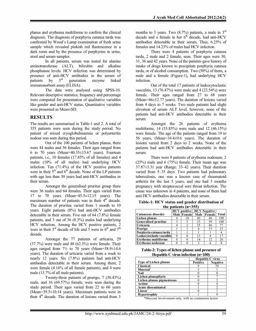
planus and erythema multiforme to confirm the clinical diagnosis. The diagnosis of porphyria cutanea tarda was confirmed by Wood’s Lamp examination of fresh urine sample which revealed pinkish red fluorescence in a dark room and by the presence of porphyrins in urine, stool and serum samples.
In all patients, serum was tested for alanine aminotransferase (ALT), bilirubin and alkaline phosphatase levels. HCV infection was determined by presence of anti-HCV antibodies in the serum of patients by 3rd generation enzyme linked immunosorbent assay (ELISA).
The data were analysed using SPSS-10. Relevant descriptive statistics; frequency and percentage were computed for presentation of qualitative variables like gender and anti-HCV status. Quantitative variables were presented as Mean±SD.
RESULTS The results are summarised in Table-1 and 2. A total of 355 patients were seen during the study period. No patient of mixed cryoglobulinaemia or polyarteritis nodosa was seen during this period.
Out of the 100 patients of lichen planus, there were 44 males and 56 females. Their ages ranged from 6 to 70 years (Mean=40.35±15.67 years). Fourteen patients, i.e., 10 females (17.85% of all females) and 4 males (10% of all males) had underlying HCV infection. Ten (71.4%) of the HCV positive patients were in their 5th and 6th decade. None of the LP patients with age less than 30 years had anti-HCV antibodies in their serum.
Amongst the generalised pruritus group there were 36 males and 64 females. Their ages varied from 17 to 70 years (Mean=45.5±12.15 years). The maximum number of patients was in their 4th decade. The duration of pruritus varied from 1 month to 10 years. Eight patients (8%) had anti-HCV antibodies detectable in their serum. Five out of 64 (7.8%) female patients, and 3 out of 36 (8.3%) males had underlying HCV infection. Among the HCV positive patients, 2 were in their 3rd decade of life and 5 were in 6th and 7th decade.
Amongst the 77 patients of urticaria, 29 (37.7%) were male and 48 (62.3%) were female. Their ages ranged from 7½ to 70 years (Mean=38.9±14.6 years). The duration of urticaria varied from a week to nearly 12 years. Six (7.8%) patients had anti-HCV antibodies detectable in their serum. Among them, 2 were female (4.16% of all female patients), and 4 were male (13.7% of all male patients).
Twenty-three patients of prurigo, 7 (30.43%) male, and 16 (69.57%) female, were seen during the study period. Their ages varied from 22 to 60 years (Mean=39.5±10.14 years). Maximum patients were in their 4th decade. The duration of lesions varied from 3
months to 3 years. Two (8.7%) patients, a male in 3rd decade and a female in her 6th decade, had anti-HCV antibodies detectable in their serum. Thus, 6.25% of females and 14.23% of males had HCV infection.
There were 4 patients of porphyria cutanea tarda, 2 male and 2 female, seen. Their ages were 30, 31, 36 and 42 years. None of the patients gave history of intake of drugs known to precipitate porphyria cutanea tarda, or of alcohol consumption. Two (50%) of them, a male and a female (Figure-1), had underlying HCV infection.
Out of the total 17 patients of leukocytoclastic vasculitis, 13 (76.47%) were male and 4 (23.54%) were female. Their ages ranged from 27 to 68 years (Mean=46±12.77 years). The duration of lesions varied from 4 days to 7 weeks. Two male patients had slight elevation of serum ALT level, however, none of the patients had anti-HCV antibodies detectable in their serum.
Amongst the 26 patients of erythema multiforme, 14 (53.85%) were male and 12 (46.15%) were female. The age of the patients ranged from 19 to 56 years, (Mean=34.4±9.6 years). The duration of lesions varied from 2 days to 2 weeks. None of the patients had anti-HCV antibodies detectable in their serum.
There were 8 patients of erythema nodosum, 2 (25%) male and 6 (75%) female. Their mean age was 37.87±3.31 year (Range: 33–42 years). Their duration varied from 5–35 days. Two patients had pulmonary tuberculosis, one was a known case of rheumatoid arthritis for the last 5 years, and one had 3 months pregnancy with streptococcal sore throat infection. The cause was unknown in 4 patients, and none of them had anti-HCV antibodies detectable in their serum.
Table-1: HCV status and gender distribution of the patients (n=355)
HCV positive HCV Negative Cutaneous disorder Male Female Male Female Total Lichen planus 4 10 40 46 100 Generalised pruritus 3 5 33 59 100 Urticaria 4 2 25 46 77 Prurigo 1 1 6 15 23 Porphyria cutanea tarda 1 1 1 1 4 Leukocytoclastic vasculitis 0 0 13 4 17 Erythema multiforme 0 0 14 12 26 Erythema nodosum 0 0 2 6 8
Table-2: Types of lichen planus and presence of Hepatitis C virus infection (n=100)
Hepatitis C virus Type of Lichen planus Positive Negative Classical 9 58 Mucosal* 3 1 Nail 0 8 Lichen planopilaris 0 7 Lichen planus pigmentosus 0 2 Actinic 0 4 Acute disseminated 1 2 Linear 0 1 Hypertrophic 1 9
*Mucosal involvement only, with no coetaneous lesion

J Ayub Med Coll Abbottabad 2012;24(2)
http://www.ayubmed.edu.pk/JAMC/24-2/Atiya.pdf 60
Figure-2: A 30-year-old lady with porphyria cutanea tarda and Hepatitis C virus infection
DISCUSSION HCV infection has been associated with a number of extra-hepatic manifestations. The evidence about the aetiopathogenic role of HCV ranges from mere epidemiologic associations or few case reports to molecular biological investigations that have identified the virus in the pathologic tissues.4 The association of lichen planus with HCV infection varies from country to country. Mehboob A et al5 found that 23.4% of their lichen planus patients had underlying HCV infection; this is a much higher frequency than seen in our study. Previous studies from Nigeria6, Spain7, and Italy8 found 8.9–28.8% of LP patients having underlying HCV infection. The different results regarding the prevalence of HCV infection in patients of LP in studies from different geographical area could be in relation to the different genetic susceptibility of the hosts. The pathogenic role of HCV in the development of LP is still unclear. Demonstration of HCV RNA in epithelial cells of oral mucosa and skin lesions have led to the theory that direct action of the virus is involved. Reverse transcription-polymerase chain reaction (RT-PCR)9 and in situ hybridisation technique10 have been employed to detect HCV RNA from LP lesions of patients with chronic Hepatitis C.
Pruritus can be considered as one of the most distressing physical sensations we experience. Comparable to chronic pain, chronic pruritus worsens the general condition and may lead to physical and psychological exhaustion. It is well recognised as being associated with Hepatitis C, and persisting even after completion of interferon therapy.11 The leading cutaneous symptom in HCV infected patients can be generalised pruritus, affecting nearly 20% of patients.12 Isolated case reports first indicated an association of urticaria with chronic Hepatitis C infection.13 Kanazawa et al found that out of their 79 patients, 24% had underlying HCV infection. In their control group the rate of antibody carriage was only 1.1%.14 An Iranian study found no association between the two disease entities.15
In addition to the well-known association of alcoholic liver disease with porphyria cutanea tarda, recent work has made it clear that chronic Hepatitis C is also an important risk factor. The most common susceptibility factors for porphyria cutanea tarda are ethanol use, smoking, and chronic Hepatitis C virus infection. In Southern Europe (Italy, Spain, Southern France) 75–90% of patients with porphyria cutanea tarda have CHC.16 Chronic Hepatitis C infection increases oxidative stress within hepatocytes and does so to a greater extent than for other chronic viral infections such as chronic Hepatitis B. This appears to be the major mechanism whereby chronic Hepatitis C infection may act as a trigger for development of porphyria cutanea tarda. A systematic review and metaanalysis17 on the prevalence of Hepatitis C virus infection in porphyria cutanea tarda patients revealed that HCV prevalence by serology was 47%, and 50% with polymerase chain reaction. HCV prevalence markedly varied depending on the country and the type of porphyria cutanea tarda. It was 57% in the sporadic and 26% in the familial form, while it was 0.24% in the control group. In Pakistan no previous study is available to assess the prevalence of HCV infection in patients with porphyria cutanea tarda.
Prurigo nodularis can be considered to be a reaction pattern in which pruritus plays a central role. A variety of systemic conditions have been reported to be associated with prurigo nodularis. PCR has been performed on lesional and non-affected skin for detection of HCV RNA. Viral particles were detected in the biopsy sample taken from the lesions of prurigo only.18 Kanazawa K et al found that 11 of their 28 patients of prurigo (39%) had evidence of underlying HCV infection.19
Recently HCV has been suggested as a trigger of autoimmunity in patients of leukocytoclastic vasculitis. The association of HCV infection, essential mixed cryoglobulinemia and leukocytoclastic vasculitis has been published mainly in case reports. High levels of HCV viremia is significantly associated with leukocytoclastic vasculitis.20 Rahman SB et al21 conducted a study on 30 patients of leukocytoclastic vasculitis to evaluate possible precipitating factors. None of the patients had anti-HCV antibodies in their serum, two had underlying HBV infection.
The association of HCV infection with erythema multiforme and erythema nodosum has been documented mainly in case reports.22,23 None of the patients of erythema nodosum or erythema multiforme in our study had underlying HCV infection. Similarly, there are other studies which have found no association between erythema nodosum and HCV infection.24
CONCLUSION The frequency of HCV infection among LP and porphyria cutanea tarda patients is much higher than that

J Ayub Med Coll Abbottabad 2012;24(2)
http://www.ayubmed.edu.pk/JAMC/24-2/Atiya.pdf 61
seen in the healthy population of our country; whereas patients of generalised pruritus, urticaria and prurigo have marginally higher frequency of HCV infection. Middle and old aged patients with cutaneous disorders have increased likelihood of underlying HCV infection than the young ones. The sample size was relatively small for certain cutaneous disorders to comment on the results with great authority. Further studies, preferably multi-centre, are needed to extend or refute our observations. REFERENCES 1. Higuchi M, Tanaka E, Kiyosawa K. Epidemiology and clinical
aspects on hepatitis C. Jpn J Infect Dis 2002;55:69–77. 2. Muhammad N, Jan A. Frequency of Hepatitis C in Buner, NWFP.
J Coll Physicians Surg Pak 2005;15:11–4. 3. Khokhar N, Gill ML, Malik GJ. General sero-prevalence of
Hepatitis C and Hepatitis B virus infection in population. J Coll Physicians Surg Pak 2004;14:534–6.
4. Rebora A. Skin diseases associated with hepatitis C virus: facts and controversies. Clin Dermatol 2010;28:489–96.
5. Mahboob A, Haroon TS, Iqbal Z, Iqbal F, Butt AK. Frequency of anti-HCV antibodies in patients with lichen planus. J Coll Physicians Surg Pak 2003;13:248–51.
6. Daramola OO, George AO, Ogunbiyi AO. Hepatitis C virus and lichen planus in Nigerians: any relationship? Inter J Dermatol 2002;41:217–9.
7. Gimenez-Garcia R, Perez-Castrillon JL. Lichen planus and hepatitis C virus infection. J Eur Acad Dermatol Venereol 2003;17:291–5.
8. Mignogna MD, Lo Muzio L, Favia G, Mignogna RE, Carbone R, Bucci E. Oral lichen planus and HCV infection: a clinical evaluation of 263 cases. Int J Dermatol 1998;37:575–8.
9. Kurokawa M, Hidaka T, Sasaki H, Nishikata I, Morishita K, Setoyama M. Analysis of hepatitis C virus (HCV) RNA in the lesions of lichen planus in patients with chronic hepatitis C: detection of anti-genomic as well as genomic-strand HCV RNAs in lichen planus lesions. J Dermatol Sci 2003;32:65–70.
10. Arrieta JJ, Rodriquez-Iniqo E, Casqueiro M, Bartolom J, Manzarbeitia F, Herrero M, et al. Detection of hepatitis C virus replication by In situ hybridization in epithelial cells of anti-hepatitis C virus-positive patients with and without oral lichen planus. Hepatology 2000;32:97–103.
11. Maticic M, Poljak M, Lunder T, Rener-Sitar K, Stojanovic L. Lichen planus and other cutaneous manifestations in chronic hepatitis C: pre- and post-interferon-based treatment prevalence vary in a cohort of patients from low hepatitis C virus endemic area. J Eur Acad Dermatol Venereol. 2008;22(7):779–88.
12. Zirwas MJ, Seraly MP. Pruritus of unknown origin: a retrospective study. J Am Acad Dermatol 2001;45:892–6.
13. Raychaudhuri SP, Kaplan M. Chronic urticaria and hepatitis C. Int J Dermatol 1995;34:823–4.
14. Kanazawa K, Yaoita H, Tsuda F, Okamoto H. Hepatitis C virus infection in patients with urticaria. J Am Acad Dermatol 1996;35:195–8.
15. Tousi P, Rahmati M, Khorshid SM. Urticaria and HCV infection: is there a relationship? Int J Dermatol 2002;41:712–5.
16. Jalil S, Grady JJ, Lee C, Anderson KE. Associations among behavior-related susceptibility factors in porphyria cutanea tarda. Clin Gastroenterol Hepatol 2010;8(3):297–302.
17. Gisbert JP, Garcia-Buey L, Pajares JM, Moreno-Otero R. Prevalence of hepatitis C virus infection in porphyria cutanea tarda: systematic review and meta-analysis. J Hepatol 2003;39:620–7.
18. Podányi B, Kiss A, Kaposi-Novák P, Lengyel G, Horányi M, Schaff ZS, Horváth A. Hepatitis C virus RNA in the cutaneous eruption but not in the symptom-free skin from patient with prurigo simplex and chronic C hepatitis. J Eur Acad Dermatol Venereol 2005;19:520–2.
19. Kanazawa K, Yaoita H, Tsuda F, Murata K, Okamoto H. Association of prurigo with hepatitis C virus infection. Arch Dermatol 1995;131:852–3.
20. El-Darouti MA, Mashaly HM, El-Nabarawy E, Eissa AM, Abdel-Halim MR, Fawzi MM, et al. Leukocytoclastic vasculitis and necrolytic acral erythema in patients with hepatitis C infection: do viral load and viral genotype play a role? J Am Acad Dermatol 2010;63(2):259–65.
21. Rahman SB, Sheikh ZI, Hameed A, Khan AA. Leucocytoclastic vasculitis. Pak Armed Forces Med J 1995;45:51–4.
22. Calista D, Landi G. Lichen planus, erythema nodosum, and erythema multiforme in a patient with chronic hepatitis C. Cutis 2001;67:454–6.
23. Geraminejad P, Walling HW, Voigt MD, Stone MS. Severe erythema multiforme responding to interferon alfa. J Am Acad Dermatol 2006;54(2 Suppl):S18–21.
24. Mert A, Ozaras R, Tabak F, Pekmezci S, Demirkesen C, Ozturk R. Erythema nodosum: an experience of 10 years. Scand J Infect Dis 2004;36:424–7.
Address for Correspondence: Atiya Rahman, Department of Dermatology, CMH, Malir Cant., Karachi, Pakistan. Cell: +92-334-5252383 Email: [email protected]