
Oncolytic reovirus as a combined antiviral and anti-tumour ... · HCV cofactor, highlighting a...
12
ORIGINAL ARTICLE Oncolytic reovirus as a combined antiviral and anti-tumour agent for the treatment of liver cancer Adel Samson, 1 Matthew J Bentham, 1 Karen Scott, 1 Gerard Nuovo, 2 Abigail Bloy, 1 Elizabeth Appleton, 1 Robert A Adair, 1 Rajiv Dave, 1 Adam Peckham-Cooper, 1 Giles Toogood, 1 Seishi Nagamori, Matthew Coffey, 3,4 Richard Vile, 1,5,6 Kevin Harrington, 6 Peter Selby, 1 Fiona Errington-Mais, 1 Alan Melcher, 6 Stephen Griffin 1 ▸ Additional material is published online only. To view please visit the journal online (http://dx.doi.org/10.1136/ gutjnl-2016-312009). 1 Leeds Institute of Cancer & Pathology (LICAP) and Leeds Cancer Research UK Clinical Centre, Faculty of Medicine and Health, University of Leeds, St James’ University Hospital, Leeds, UK 2 The Ohio State University, Comprehensive Cancer Centre, Columbus, Ohio, USA 3 Department of Virology II, National Institute of Infectious Diseases 1-23-1 Toyama, Tokyo, Japan 4 Oncolytics Biotech, Calgary, Alberta, Canada 5 Department of Immunology, Mayo Clinic, Rochester, Minnesota, USA 6 Department of Molecular Medicine, The Institute of Cancer Research, London, UK Correspondence to Dr Stephen Griffin and Prof Alan Melcher, Faculty of Medicine and Health, Leeds Institute of Cancer & Pathology, University of Leeds, Wellcome Trust Brenner Building, St James’ University Hospital, Leeds LS7 9TF, UK; s.d.c.griffi[email protected] and [email protected] AM and SG contributed equally. Received 6 April 2016 Revised 26 September 2016 Accepted 13 October 2016 To cite: Samson A, Bentham MJ, Scott K, et al. Gut Published Online First: [ please include Day Month Year] doi:10.1136/gutjnl- 2016-312009 ABSTRACT Objective Oncolytic viruses (OVs) represent promising, proinflammatory cancer treatments. Here, we explored whether OV-induced innate immune responses could simultaneously inhibit HCV while suppressing hepatocellular carcinoma (HCC). Furthermore, we extended this exemplar to other models of virus- associated cancer. Design and results Clinical grade oncolytic orthoreovirus (Reo) elicited innate immune activation within primary human liver tissue in the absence of cytotoxicity and independently of viral genome replication. As well as achieving therapy in preclinical models of HCC through the activation of innate degranulating immune cells, Reo-induced cytokine responses efficiently suppressed HCV replication both in vitro and in vivo. Furthermore, Reo-induced innate responses were also effective against models of HBV- associated HCC, as well as an alternative endogenous model of Epstein–Barr virus-associated lymphoma. Interestingly, Reo appeared superior to the majority of OVs in its ability to elicit innate inflammatory responses from primary liver tissue. Conclusions We propose that Reo and other select proinflammatory OV may be used in the treatment of multiple cancers associated with oncogenic virus infections, simultaneously reducing both virus-associated oncogenic drive and tumour burden. In the case of HCV- associated HCC (HCV-HCC), Reo should be considered as an alternative agent to supplement and support current HCV-HCC therapies, particularly in those countries where access to new HCV antiviral treatments may be limited. INTRODUCTION Hepatocellular carcinoma (HCC) is among the highest causes of cancer-associated mortality and has limited therapeutic options. The majority of HCCs are linked with an underlying oncogenic virus infection, namely HBV (54%) or HCV (31%). 1 HCV-associated HCC (HCV-HCC) has a particularly high risk of recurrence following surgi- cal interventions, compared with either HBV-HCC or other HCC types. 23 Successful antiviral therapy prior to hepatic resection and/or liver transplant sig- nificantly reduces this risk, 4 yet toxicity associated with conventional systemic interferon (IFN)-based HCV therapy has historically excluded many newly diagnosed patients with HCV-HCC from such treatment. New direct-acting antivirals (DAAs) Significance of this study What is already known on this subject? ▸ The majority of hepatocellular carcinomas (HCCs) are linked with an underlying oncogenic virus infection, namely HBV (54%) or HCV (31%), which drives oncogenesis via both indirect inflammatory and direct carcinogenic mechanisms. Accordingly, suppression of HBV/ HCV infections may improve HCC clinical outcomes, but few patients with HCC worldwide are cured of their hepatitis infections due to cost, compliance and treatment toxicity issues. ▸ Therapy for patients with advanced HCC is limited; Sorafenib only improves median survival by 2–3 months, is associated with significant side effects and has not been recommended by the National Institute for Health and Care Excellence in the UK. ▸ Oncolytic viruses (OVs) show potential as cancer therapies, killing malignant cells by direct and immune-mediated mechanisms. What are the new findings? ▸ Clinical grade oncolytic orthoreovirus (Reo) elicited interferon (IFN) secretion and innate immune activation within primary human liver tissue in the absence of cytotoxicity and independently of viral genome replication. ▸ Reo achieved therapy in cell line and murine models of hepatitis virus-positive/hepatitis virus-negative HCC and efficiently suppressed HCV replication in vitro through the effects of repressed replication in vitro, and in vivo, through the effects of type I IFNs. ▸ The antiviral effects of Reo were also applicable to in vitro endogenous models of HBV-HCC and Epstein–Barr virus-associated lymphoma. Of the other OVs tested, only measles virus Edmonston strain recapitulated the potent antiviral effects of Reo. Samson A, et al. Gut 2016;0:1–12. doi:10.1136/gutjnl-2016-312009 1 Hepatology Gut Online First, published on November 15, 2016 as 10.1136/gutjnl-2016-312009 Copyright Article author (or their employer) 2016. Produced by BMJ Publishing Group Ltd (& BSG) under licence.
Transcript of Oncolytic reovirus as a combined antiviral and anti-tumour ... · HCV cofactor, highlighting a...
ORIGINAL ARTICLE
Oncolytic reovirus as a combined antiviral andanti-tumour agent for the treatment of liver cancerAdel Samson,1 Matthew J Bentham,1 Karen Scott,1 Gerard Nuovo,2 Abigail Bloy,1
Elizabeth Appleton,1 Robert A Adair,1 Rajiv Dave,1 Adam Peckham-Cooper,1
Giles Toogood,1 Seishi Nagamori, Matthew Coffey,3,4 Richard Vile,1,5,6
Kevin Harrington,6 Peter Selby,1 Fiona Errington-Mais,1 Alan Melcher,6
Stephen Griffin1
▸ Additional material ispublished online only. To viewplease visit the journal online(http://dx.doi.org/10.1136/gutjnl-2016-312009).
1Leeds Institute of Cancer &Pathology (LICAP) and LeedsCancer Research UK ClinicalCentre, Faculty of Medicineand Health, University ofLeeds, St James’ UniversityHospital, Leeds, UK2The Ohio State University,Comprehensive Cancer Centre,Columbus, Ohio, USA3Department of Virology II,National Institute of InfectiousDiseases 1-23-1 Toyama,Tokyo, Japan4Oncolytics Biotech, Calgary,Alberta, Canada5Department of Immunology,Mayo Clinic, Rochester,Minnesota, USA6Department of MolecularMedicine, The Institute ofCancer Research, London, UK
Correspondence toDr Stephen Griffin and ProfAlan Melcher, Faculty ofMedicine and Health, LeedsInstitute of Cancer &Pathology, University of Leeds,Wellcome Trust BrennerBuilding, St James’ UniversityHospital, Leeds LS7 9TF, UK;[email protected] [email protected]
AM and SG contributedequally.
Received 6 April 2016Revised 26 September 2016Accepted 13 October 2016
To cite: Samson A,Bentham MJ, Scott K, et al.Gut Published Online First:[please include Day MonthYear] doi:10.1136/gutjnl-2016-312009
ABSTRACTObjective Oncolytic viruses (OVs) represent promising,proinflammatory cancer treatments. Here, we exploredwhether OV-induced innate immune responses couldsimultaneously inhibit HCV while suppressinghepatocellular carcinoma (HCC). Furthermore, weextended this exemplar to other models of virus-associated cancer.Design and results Clinical grade oncolyticorthoreovirus (Reo) elicited innate immune activationwithin primary human liver tissue in the absence ofcytotoxicity and independently of viral genomereplication. As well as achieving therapy in preclinicalmodels of HCC through the activation of innatedegranulating immune cells, Reo-induced cytokineresponses efficiently suppressed HCV replication both invitro and in vivo. Furthermore, Reo-induced innateresponses were also effective against models of HBV-associated HCC, as well as an alternative endogenousmodel of Epstein–Barr virus-associated lymphoma.Interestingly, Reo appeared superior to the majority ofOVs in its ability to elicit innate inflammatory responsesfrom primary liver tissue.Conclusions We propose that Reo and other selectproinflammatory OV may be used in the treatment ofmultiple cancers associated with oncogenic virusinfections, simultaneously reducing both virus-associatedoncogenic drive and tumour burden. In the case of HCV-associated HCC (HCV-HCC), Reo should be consideredas an alternative agent to supplement and supportcurrent HCV-HCC therapies, particularly in thosecountries where access to new HCV antiviral treatmentsmay be limited.
INTRODUCTIONHepatocellular carcinoma (HCC) is among thehighest causes of cancer-associated mortality andhas limited therapeutic options. The majority ofHCCs are linked with an underlying oncogenicvirus infection, namely HBV (54%) or HCV(31%).1 HCV-associated HCC (HCV-HCC) has aparticularly high risk of recurrence following surgi-cal interventions, compared with either HBV-HCCor other HCC types.2 3 Successful antiviral therapyprior to hepatic resection and/or liver transplant sig-nificantly reduces this risk,4 yet toxicity associatedwith conventional systemic interferon (IFN)-based
HCV therapy has historically excluded manynewly diagnosed patients with HCV-HCC fromsuch treatment. New direct-acting antivirals (DAAs)
Significance of this study
What is already known on this subject?▸ The majority of hepatocellular carcinomas
(HCCs) are linked with an underlying oncogenicvirus infection, namely HBV (54%) or HCV(31%), which drives oncogenesis via bothindirect inflammatory and direct carcinogenicmechanisms. Accordingly, suppression of HBV/HCV infections may improve HCC clinicaloutcomes, but few patients with HCCworldwide are cured of their hepatitisinfections due to cost, compliance andtreatment toxicity issues.
▸ Therapy for patients with advanced HCC islimited; Sorafenib only improves mediansurvival by 2–3 months, is associated withsignificant side effects and has not beenrecommended by the National Institute forHealth and Care Excellence in the UK.
▸ Oncolytic viruses (OVs) show potential ascancer therapies, killing malignant cells bydirect and immune-mediated mechanisms.
What are the new findings?▸ Clinical grade oncolytic orthoreovirus (Reo)
elicited interferon (IFN) secretion and innateimmune activation within primary human livertissue in the absence of cytotoxicity andindependently of viral genome replication.
▸ Reo achieved therapy in cell line and murinemodels of hepatitis virus-positive/hepatitisvirus-negative HCC and efficiently suppressedHCV replication in vitro through the effects ofrepressed replication in vitro, and in vivo,through the effects of type I IFNs.
▸ The antiviral effects of Reo were also applicableto in vitro endogenous models of HBV-HCC andEpstein–Barr virus-associated lymphoma. Of theother OVs tested, only measles virusEdmonston strain recapitulated the potentantiviral effects of Reo.
Samson A, et al. Gut 2016;0:1–12. doi:10.1136/gutjnl-2016-312009 1
Hepatology Gut Online First, published on November 15, 2016 as 10.1136/gutjnl-2016-312009
Copyright Article author (or their employer) 2016. Produced by BMJ Publishing Group Ltd (& BSG) under licence.

with improved safety profiles are revolutionising HCV therapy,yet compliance issues are common,5 and the high cost of DAAsprofoundly limits their use in poorer countries, where the preva-lence of HCV-HCC is highest.
For patients with advanced HCC, little is known regardingthe impact of antiviral therapy upon existing tumours. Dogmaattributes the emergence and progression of HCV-HCC to abystander phenomenon, driven by chronic hepatic inflamma-tion. However, growing evidence indicates that HCV is alsodirectly oncogenic, further supporting the need for combinedantiviral and anti-tumour therapy. Specifically, antiviral therapyimproves patient outcomes in HCV-associated lymphoma;6
HCC is more common in HCV-positive livers compared withautoimmune hepatitis;7 HCV proteins are directly oncogenic inpreclinical models8 and HCV-HCC is more likely to maintainexpression of the tumour suppressor miRNA122, an essentialHCV cofactor, highlighting a critical role for HCV in oncogen-esis.9 Therapy for patients with advanced HCC is limited;Sorafenib only improves median survival by 2–3 months, is asso-ciated with significant side effects and has not been recommendedby the National Institute for Health and Care Excellence in theUK.10–12 Trans-arterial chemotherapy is routinely used forpatients with locally advanced disease/as a bridging therapy totransplant and has also been tested as a systemic treatment, but isfrequently complicated by reactivation/exacerbation of underlyingHCV/HBV infections.13 14 Thus, given the poor prognosis formost patients with HCC, a combined antiviral and anti-tumourtherapy might yield significant patient benefit.
Oncolytic viruses (OVs) show potential as cancer therapies;classically, tumour selective killing arises from direct cellularlysis expedited by virus-associated traits that favour replicationwithin malignant cells. However, OVs are also potent inducersof host immunity, which is increasingly recognised to be themajor component of their anti-tumour efficacy.15 Encouragingresponses have been observed in late-stage clinical testingwithout significant toxicity, with the first OV recently licensedfor the treatment of melanoma16 (‘T-Vec’, herpes simplex virustype 1 (HSV-1) encoding human Granulocyte macrophagecolony stimulating factor (GM-CSF)). The liver and HCC havebeen the subject of several OV clinical studies,17 includingJX-594, a modified vaccinia virus (VV) encoding GM-CSF,18
and our own investigations using oncolytic Orthoreovirus(Reo) in the treatment of metastatic colorectal cancer (CRC).19
We hypothesised that the proinflammatory nature of OVimmunotherapy may exert concomitant benefit upon both thecancer and the underlying oncogenic virus infection through thestimulation of innate responses, namely IFNs. Accordingly,Reo-induced innate immune responses within primary livertissue simultaneously effected tumour killing and suppression of
HCV replication in vitro and in vivo. These responses did notrequire productive Reo replication and were applicable to othermodels of virus-associated tumours. We propose that this dualmode of action, combined with an excellent safety record, mayfavour the rapid deployment of OVs such as Reo for disadvan-taged patients with advanced, virus-associated HCC.
RESULTSReo-induced innate immune responses occur in normal, aswell as cancerous liverWe previously confirmed that Reo efficiently targets CRC livermetastases following intravenous infusion, despite patient neu-tralising antibodies.19 Cell-mediated virus carriage enabled Reoreplication within tumours and recovery of infectious virus fromtumour explants. However, staining of surrounding normal livertissue was also demonstrable in a number of patients, but infec-tious virus could not be isolated. This led us to re-examinenormal liver patient tissue, confirming Reo protein expressionwithin 5 out of 10 subjects (figure 1A).
Next, we examined Reo infection of normal ex vivo humanliver tissue, generating single-cell suspensions. Mixed liver cellswere fractionated into enriched hepatocyte and liver mono-nuclear cell (LMC) subpopulations (see online supplementaryfigure S1a). Importantly, exposure of enriched primary hepato-cyte populations to Reo did not cause cellular toxicity (figure1B), but did result in limited transcription of viral RNA (figure1C) and protein expression 24 hours post-infection (figure 1D,E) in the absence of de novo infectious virion production (seeonline supplementary figure S1b). Interestingly, RNA andprotein expression decreased between 24 and 48 hours, suggest-ive of the induction of cellular antiviral mechanisms (figure 1D,E). By contrast, Reo replicated efficiently within a broad rangeof HCC lines in vitro, producing new infectious virions (seeonline supplementary figure S1c), and inducing cytotoxicity (seeonline supplementary figure S1d), primarily via apoptosis (seeonline supplementary figure S1e). Importantly, potent cytokineresponses were observed upon ex vivo exposure of mixed livercells, hepatocyte-enriched or LMC fractions to Reo (figure 1F).All cell types expressed IFN-β, whereas IFN-α and IFN-γ segre-gated primarily within the LMC fraction, and IFN-λ (interleukin(IL)-28b/IL-29) was more prominent within hepatocyte-enrichedfractions. Thus, Reo has the potential to stimulate innate immun-ity within a significant proportion of the liver, rather than beingrestricted to tumour cells and/or tumour-associated LMC.
Reo-induced innate immune responses mediate anti-HCCtherapyTo assess the efficacy of Reo against HCC models with/withoutHCV, we employed immunocompromised Severe combinedimmunodeficiency (SCID) mice bearing subcutaneous Huh7, orHuh7-JFH1 cell xenografts, carrying a subgenomic HCV repli-con (genotype 2a, JFH-1 isolate). A single injection of Reo intopalpable tumours (day 5 postimplantation) retarded tumourgrowth over a 3–4 week experiment, yielding small, macroscop-ically less vascular tumours (figure 2A). Histological examin-ation of the tumours confirmed expression of HCVnon-structural protein (NS)5A and association of Reo antigenwith necrosis and cleaved caspase 3 (figure 2B). However,Reo-treated immunodeficient mice developed considerable tox-icity evidenced by weight loss (see online supplementary figureS2a), necessitating premature sacrifice.
To circumvent toxicity issues and to delineate immune-driven,rather than lytic therapy, we revisited previous studies in
Significance of this study
How might it impact on clinical practice in theforeseeable future?▸ The deployment of select IFN-focused OVs in patients with
cancer harbouring underlying oncogenic virus infectionseither as single agents or in combination therapy isanticipated to suppress pathogenic infections, improvingorgan health and extending cancer-specific survival.
2 Samson A, et al. Gut 2016;0:1–12. doi:10.1136/gutjnl-2016-312009
Hepatology

Figure 1 Reo reaches normal liver tissue following intravenous injection and stimulates interferon (IFN) secretion from ex vivo liver cells. (A)Immunohistochemistry (IHC) for Reo σ3 capsid protein (brown) from normal liver tissue derived from a patient treated intravenously with Reo (left)or an untreated control (right). Slides are representative of the clinical trial series or six untreated controls. (B) Viability assay for enriched ex vivohepatocytes treated using Phosphate Buffered Saline (PBS) or 10 PFU/cell Reo for 72 hours and assayed for membrane integrity by Trypan Bluestaining. (C) quantitative reverse transcriptase (qRT)-PCR for Reo σ3. Primary human hepatocytes were treated using 1 PFU/cell Reo or uv-Reo andincubated for 24 or 48 hours prior to RNA extraction. (D) Western blot for Reo σ3 and β-actin. Primary human hepatocytes were treated using 1PFU/cell Reo or uv-Reo and incubated for 24 or 48 hours. Mock (M)-treated hepatocytes and RNA purified from Reolysin stocks (+) served asnegative and positive controls. (E) Human hepatocytes were infected with Reo, or uv-Reo as above, and subjected to immunofluorescence analysisfor Reo σ3 capsid protein, with Hoechst nuclear counterstain. (F) ELISA for IFN-α, IFN-β, interleukin (IL)-28b/IL-29 and IFN-γ derived from ex vivomixed liver cells, enriched hepatocytes and liver mononuclear cells (LMCs) following stimulation with Reo or PBS control. * signifies p<0.005.
Samson A, et al. Gut 2016;0:1–12. doi:10.1136/gutjnl-2016-312009 3
Hepatology

Figure 2 Reo-induced innate immune responses are sufficient for anti-hepatocellular carcinoma (HCC) therapy. (A) Excised subcutaneous flanktumours from SCID mice following a single Itu injection of PBS or 1×10⁶ PFU Reo. (B) Representative IHC staining (brown) for HCV non-structuralprotein (NS)5A, Reo σ3 capsid protein and cleaved caspase 3 in Huh7 and Huh7-JFH1 subcutaneous xenografts in SCID mice treated with a singleItu injection of 1×10⁶ PFU Reo on the 6th day post tumour implantation. (C) Tumour volume of Huh7 (left) and Huh7-JFH1 (right) subcutaneousflank xenografts in SCID mice treated with 3 times weekly Itu injections of PBS or 1×10⁶ PFU uv-Reo for 4 weeks starting the 6th day post tumourimplantation. (D) ELISA for interferon (IFN)-α, IFN-β, IFN-γ and interleukin (IL)-28b/IL-29 derived from ex vivo mixed liver cells following stimulationwith uv-Reo or PBS control. (E) Tumour growth of 1MEA subcutaneous syngeneic tumours in BALB/c mice treated with 3 times weekly Itu PBS,1×106 PFU Reo or 1×10⁶ PFU uv-Reo injections, starting the 6th day post tumour implantation. * signifies p<0.005.
4 Samson A, et al. Gut 2016;0:1–12. doi:10.1136/gutjnl-2016-312009
Hepatology

melanoma models, where uv-inactivated Reo (uv-Reo) (seeonline supplementary figure S2b) primed antitumour immuneresponses comparably with live virus.20 Accordingly, uv-Reocaused significant retardation of both HCV-positive andHCV-negative tumour growth (figure 2C). uv-Reo also elicitedinnate cytokine responses from primary human mixed liver cellsuspensions, although this was more focused to IFN-β(figure 2D). Finally, an immunocompetent HCC subcutaneousmodel (1MEA cells in syngeneic Balb/c mice) revealed similarreductions in tumour growth for Reo and uv-Reo, supportingan immune-driven, rather than an oncolytic mechanism ofaction (figure 2E). Taken together, Reo-induced innate immun-ity appears sufficient for anti-HCC effects in preclinical models,independent of Reo genomic replication, pointing to a majorrole for such a mechanism in humans undergoing treatmentwith live clinical grade virus.
Reo-induced anti-HCC responses require IFN-mediatedactivation of natural killer lymphocytesHistological examination of Reo-treated 1MEA tumours revealedsignificant natural killer (NK) cell infiltration, indicating thatthese may represent an effector component of the antitumourresponse (figure 3A), as seen for other OVs.21 22 Accordingly,uv-Reo treatment was ineffective against Huh7 xenografts withinSCID/Beige mice that lack functional immune cell degranulation,consistent with a therapeutic requirement for NK cell activity(figure 3B). Interestingly, increased tumour growth was notobserved in the SCID/Beige compared with the SCID context,suggesting that degranulating cells could not effectively controltumour growth in the absence of exogenous stimulation.
Consistent with murine models and our previous data, Reoeffectively stimulated human liver-derived NK cells ex vivo via atype 1 IFN-dependent mechanism (figure 3C), thereby activat-ing a major fraction within the LMC population (see onlinesupplementary figure S1a). Likewise, uv-Reo stimulation ofLMC also resulted in NK cell activation (figure 3D), as did Reo,also resulting in enhanced killing of both HCV-positive andHCV-negative Huh7 targets (figure 3E, F). Moreover, depletingNK cells from peripheral blood mononuclear cell (PBMC) pre-vented Reo-stimulated HCC killing (see online supplementaryfigure S3a), further supporting NK cells as major innate effec-tors in OV-mediated therapy. Thus, Reo-induced anti-HCCimmunity is dependent upon type 1 IFN, which in turn activatesNK cells, enabling them to recognise and kill tumour targets.
Reo-induced immunity exerts potent anti-HCV effects invitroGiven the potent induction of inflammatory cytokines uponexposure of ex vivo human liver cells to Reo, we reasoned thatresultant conditioned culture supernatants should exert antiviraleffects. Filtered conditioned media (CM) were prepared follow-ing control, or Reo (reovirus-CM (RCM)) stimulation of mixedliver cells, fractionated hepatocyte-enriched and LMC popula-tions or mixed primary HCC cells. All RCM exerted dose-dependent inhibitory effects upon HCV replication withinHuh7 cells (figure 4A). Comparison with purified, leucocyte-derived IFN-α, IFN-β or IL-29, or combinations thereof,revealed a pattern consistent with a primarily IFN-β-mediatedmechanism (see online supplementary figure S4a); IFN-β wassignificantly more potent in its ability to inhibit HCV replicationcompared with either IFN-α or IL-29, and cytokine combina-tions did not inhibit HCV replication more than IFN-β alone.IFN blockade significantly attenuated the HCV-inhibitory effectof mixed liver cell RCM (figure 4B), with no measurable
cytotoxic or growth-inhibitory effects on Huh7-JFH1 cells (seeonline supplementary figure S4b).
Exposure of multiple HCC lines to Reo resulted in variable pat-terns of cytokine induction, lacking IFN-α, but with significantamounts of IFN-β and IFN-λ (figure 4C). We defined RCMderived from stimulated JHH1, HLE and JHH2 cells as retaininghigh, medium and low IFN-β content, respectively. Antiviral effectsdirectly correlated with RCM IFN-β levels, mirroring the trend lineequation for purified leukocyte-derived cytokine (Spearman’s rankcorrelation of 0.929, p=0.001) (figure 4D, see onlinesupplementary figure S4a). Concomitant decline in HCV proteinexpression was observed by both immunofluorescence (figure 4E)and western blot (see online supplementary figure S4c), again withno discernible cytotoxic effects (see online supplementary figureS4d). Critically, RCM antiviral activity was also applicable to full-length infectious HCV, again with direct correlation to IFN-βlevels, measured by reductions in NS5A-luciferase reporter activity(figure 4F, see online supplementary figure S4e), HCV genomicRNA (see online supplementary figure S5a) and NS5A-GreenFluorescent Protein (GFP) reporter gene fluorescence (see onlinesupplementary figure S5b, c).
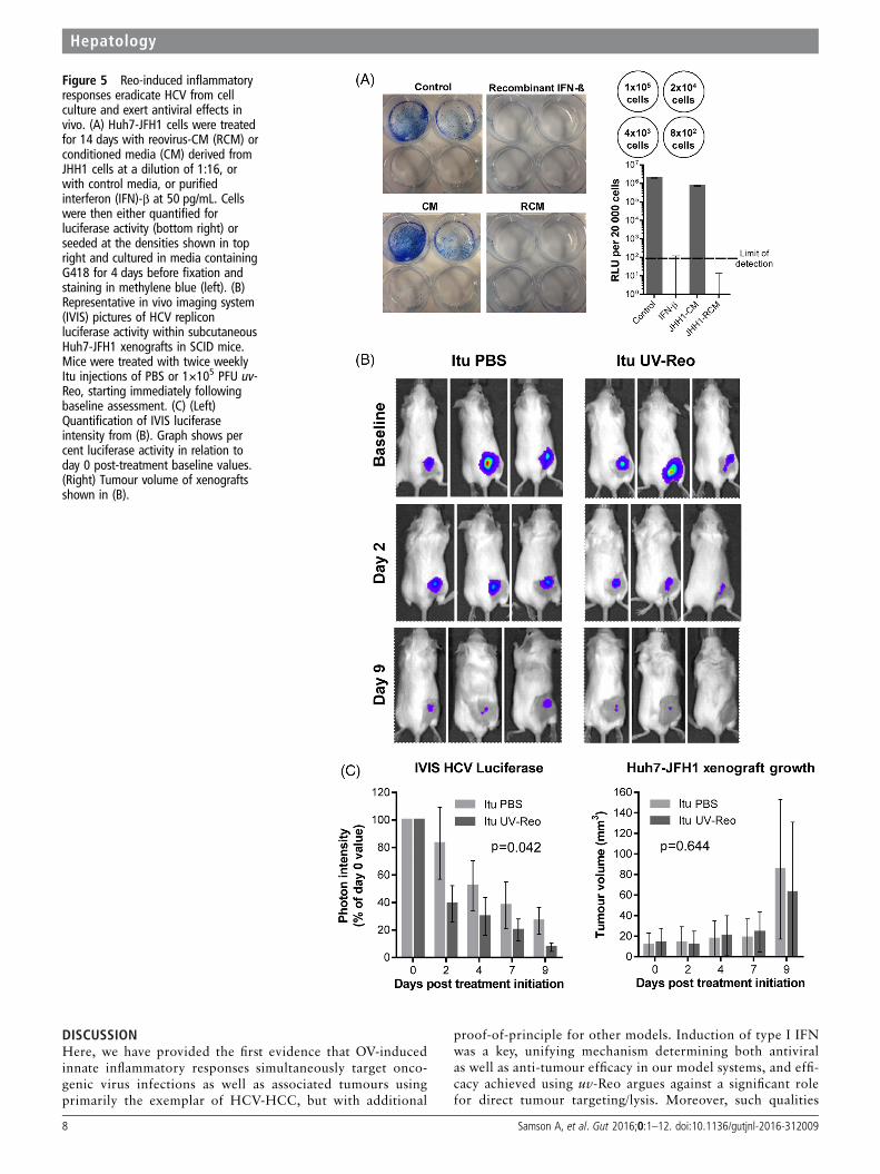
Reo-induced immunity eradicates HCV from cell culture andexerts antiviral effects in vivoWe next examined whether RCM exposure could effectivelycure Huh7-JFH-1 cells. Huh7-JFH-1 were exposed to RCM orcontrol media for 14 days in the absence of selection.Rechallenge with G418 led to a lack of viable colonies in JHH1RCM or IFN-β-treated cells, with undetectable luciferase activ-ity in bulk populations (figure 5A).
Next, we tested whether uv-Reo-stimulated immunity couldexert an antiviral effect in the preclinical SCID Huh7-JFH-1model. Palpable subcutaneous Huh7-JFH-1 xenografts weretreated intratumourally with either low-dose uv-Reo or PBSover a 9-day period, measuring tumour growth, as well as HCVexpressed luciferase within tumours using an in vivo imagingsystem (IVIS). Tumours were normalised and segregated intoequivalent control/treatment groups based upon IVIS readings atday 0. While luciferase readings naturally declined in controlsdue to the lack of G418, treatment with low-dose uv-Reo led toa significant additional reduction in IVIS readings, independentof tumour growth (figure 5B, C). Furthermore, histologicalexamination of additional Huh7-JFH-1 tumours treated with asingle injection of live Reo revealed marked reductions in HCVNS5A protein expression in areas of the tumour distinct fromthose undergoing necrotic changes or staining for Reo antigen,supportive of a trans-acting, cytokine-mediated antiviral effect(see online supplementary figure S6a). Accordingly, RCM gener-ated from uv-Reo exposure of mixed murine liver cells retainedanti-HCV activity in HUH7-JFH1 cells (see onlinesupplementary figure S6b), albeit significantly reduced com-pared with human RCM. Hence, while murine RCM/IFN par-tially cross-stimulates Huh7 IFN receptors, murine xenograftmodels likely underestimate the magnitude of inhibition poten-tially achievable within a human context.
Reo-induced antiviral responses are achievable in othermodels of virus-associated cancerWe reasoned that similar antiviral efficacy may be achieved inother models of virus-associated cancer. We assessed the effectsof Reo-stimulated immunity against PLC/PRF/5 cells containingintegrated HBV, and Daudi cells, which are derived from anEpstein–Barr virus (EBV)-positive Burkitt’s lymphoma, knownto be resistant to Reo-induced cytotoxic effects.23
Samson A, et al. Gut 2016;0:1–12. doi:10.1136/gutjnl-2016-312009 5
Hepatology

PLC/PRF/5 HBV surface antigen (HBsAg) secretion serves asa marker of viral gene expression.24 RCM derived from bothprimary LMC and also JHH-1 cells achieved reductions ofHBsAg secretion comparable with purified IFN-β over a 5-daytime course (figure 6A, see online supplementary figure S7a),with no observable growth-inhibitory effects (see onlinesupplementary figure S7b). We next tested the ability of Reo tosuppress EBV reactivation from tetradecanoylphorbol-13-acetate(TPA)/butyrate-treated Daudi cells. Both direct Reo treatment ofDaudi cells and exposure to RCM derived from PBMC led to asignificant reduction of EBV early antigen (EA)-positive cells(figure 6B, C). As Daudi cells are not permissive hosts, thiseffect was likely attributable to Reo-induced cytokines.
Finally, we assessed whether Reo was unique in its ability togenerate potent anti-HCV cytokine responses from humanprimary mixed liver cells. From a panel of commonly researchedOVs, only CM generated by exposure to measles virusEdmonston strain (MV-Edm) recapitulated the anti-HCV effi-cacy seen for Reo against Huh7-JFH-1 cells (figure 6D).Exposure to very high concentrations of HSV-1 CM generatedonly modest effects, whereas VV did not elicit appreciable anti-viral responses. Moreover, correlation between antiviral effectsand those predicted to be attributable to the induction of IFN-β(see online supplementary figure S7c) was significant (p<0.05)for all OVs except VV; low levels of cytokine were present inVV CM at similar concentrations to that released from mock-
Figure 3 Reo-stimulated interferon (IFN) drives anti-hepatocellular carcinoma (HCC) immune responses via natural killer (NK) cell activation. (A)Representative IHC staining (brown) for NK cells in 1MEA subcutaneous syngeneic tumours in BALB/c mice treated with 3 times weekly Itu injectionsof PBS or 1×10⁶ PFU Reo. (B) Tumour growth of Huh7 subcutaneous xenografts in SCID/Beige mice treated with 3 times weekly Itu injections of PBSor 1×10⁶ PFU uv-Reo. (C) Flow cytometry overlay plot (left) and quantification (right) of median fluorescence intensity (MFI) for NK CD69 expressionwithin liver mononuclear cells (LMCs) treated using PBS, 1 PFU/cell Reo, 1 PFU/cell Reo and type I IFN blocking antibodies or 1 PFU/cell Reo andisotype antibodies. (D) Similar to (C), but LMCs were treated with PBS, 1 PFU/cell Reo or 1 PFU/cell uv-Reo. (E) Degranulation assay showing percent CD107-positive liver NK cells. LMCs were pretreated with PBS or 1 PFU/cell Reo for 24 hours, then coincubated with Huh7 or Huh7-JFH1targets for 4 hours. (F) 51Cr release assay using LMCs pretreated with PBS or 1 PFU/cell Reo for 24 hours, then washed and coincubated with51Cr-labelled Huh7 or Huh7-JFH1 targets for 4 hours. Data are 51Cr release as a percentage of the potential maximum. *signifies p<0.005.
6 Samson A, et al. Gut 2016;0:1–12. doi:10.1136/gutjnl-2016-312009
Hepatology

treated hepatic cells, yet neither exerted antiviral effects uponHuh7-JFH-1 cells. Hence, other factors present in controlmedia may ameliorate the effects of low-level IFN or these lowlevels may not tally with the predictive equation (see online
supplementary figure S7c). Taken together, Reo appears capableof inducing potent, IFN-β-driven antiviral responses that areeffective against both RNA and DNA tumour viruses, yet thisability is not ubiquitous among OVs.
Figure 4 Reo-stimulated interferon (IFN)-β potently inhibits HCV in vitro. (A) Luciferase assay using Huh7-JFH1 cells treated for 24 hours with arange of dilutions of conditioned media (CM) or reovirus-CM (RCM) derived from mixed liver cells, liver mononuclear cells (LMCs), enrichedhepatocytes or mixed primary hepatocellular carcinoma (HCC) cells. Luciferase activity was calculated as a percentage of control media values. (B)Luciferase assay using Huh7-JFH1 cells treated for 4 hours with mixed liver cell RCM (1:16 dilution), mixed liver cell RCM and type I IFN blockingantibodies or mixed liver cell RCM and isotype antibodies. Cells were then incubated in full growth media without treatment for a further 20 hoursprior to analysis. (C) ELISA for IFN-β (left) and interleukin (IL)-28b/IL-29 (right) secretion from a panel of HCC cell lines following treatment for72 hours with PBS or 1 PFU/cell Reo. (D) Luciferase assay using Huh7-JFH1 cells treated for 24 hours with a range of dilutions of CM or RCMderived from JHH1, HLE or JHH2 cells. Dotted red lines represent predicted per cent luciferase activity as a function of RCM IFN-β concentrations, aspredicted from the trend line equation for replicon inhibition using purified IFN-β (see online supplementary figure 4SA). (E) Representativeimmunofluorescence for HCV non-structural protein (NS)5A in Huh7-JFH1 cells treated for 24 hours with CM or RCM at a dilution of 1:16 derivedfrom JHH1, HLE or JHH2 cells. (F) Luciferase assays using HCV-infected Huh7 cells, treated for 24 hours with CM or RCM derived from JHH1, HLE orJHH2 cells at a dilution of 1:16. * signifies p<0.005.
Samson A, et al. Gut 2016;0:1–12. doi:10.1136/gutjnl-2016-312009 7
Hepatology
DISCUSSIONHere, we have provided the first evidence that OV-inducedinnate inflammatory responses simultaneously target onco-genic virus infections as well as associated tumours usingprimarily the exemplar of HCV-HCC, but with additional
proof-of-principle for other models. Induction of type I IFNwas a key, unifying mechanism determining both antiviralas well as anti-tumour efficacy in our model systems, and effi-cacy achieved using uv-Reo argues against a significant rolefor direct tumour targeting/lysis. Moreover, such qualities
Figure 5 Reo-induced inflammatoryresponses eradicate HCV from cellculture and exert antiviral effects invivo. (A) Huh7-JFH1 cells were treatedfor 14 days with reovirus-CM (RCM) orconditioned media (CM) derived fromJHH1 cells at a dilution of 1:16, orwith control media, or purifiedinterferon (IFN)-β at 50 pg/mL. Cellswere then either quantified forluciferase activity (bottom right) orseeded at the densities shown in topright and cultured in media containingG418 for 4 days before fixation andstaining in methylene blue (left). (B)Representative in vivo imaging system(IVIS) pictures of HCV repliconluciferase activity within subcutaneousHuh7-JFH1 xenografts in SCID mice.Mice were treated with twice weeklyItu injections of PBS or 1×10⁵ PFU uv-Reo, starting immediately followingbaseline assessment. (C) (Left)Quantification of IVIS luciferaseintensity from (B). Graph shows percent luciferase activity in relation today 0 post-treatment baseline values.(Right) Tumour volume of xenograftsshown in (B).
8 Samson A, et al. Gut 2016;0:1–12. doi:10.1136/gutjnl-2016-312009
Hepatology

were not ubiquitous among OVs, making the selection ofagent for future applications an important consideration.
Reo-induced IFN both exerted direct antiviral effects uponvirus replication in vitro and anti-tumour effects in vivo via acti-vation of degranulating innate immune cells, most likely NKcells. Hence, SCID/beige mice were unable to respond touv-Reo therapy, yet this was not compromised by the lack ofadaptive immunity within the SCID background. NK cells are
indirectly activated by IFN,25 and they likely also contribute toin vivo antiviral effects via the killing of infected tumour cells/hepatocytes.26
IFN-β-mediated antiviral and anti-tumour effects in preclinicalmodels were not dependent upon Reo replication, as uv-Reoprimed similar responses from human hepatic cells. This is rem-iniscent of other OV studies, where the ability to complete a fullinfectious cycle is dispensable for the priming of IFN-mediated
Figure 6 Reo-induced antiviral responses inhibit HBV and Epstein–Barr virus (EBV) in vitro. (A) ELISA for HBV surface antigen (HBsAg) secreted fromPLC/PRF/5 cells treated for 5 days using a range of dilutions of conditioned media (CM) or reovirus-CM (RCM) derived from liver mononuclear cell(LMCs) (top) or JHH1 (bottom) cells. (B) Representative immunofluorescence (left) for EBV early antigen (EA) and quantification of EA-positive (right)Daudi cells. Cells were treated using PBS for 48 hours, 1 PFU/cell Reo for 48 hours, Histone deacetylase inhibitor (HDI) for 24 hours or 1 PFU/cell Reofor 48 hours followed by HDI for 24 hours. (C) (Left) ELISA for interferon (IFN)-α, IFN-β, IFN-γ and interleukin (IL)-28b/IL-29 derived from peripheralblood MC (PBMC) following stimulation with Reo or PBS control. (Right) Quantification of EA-positive Daudi cells treated for 24 hours using controlmedia, PBMC CM, PBMC RCM, all followed by a further 24 hours treatment using either PBS or HDI. (D) Luciferase assay using Huh7-JFH1 cells treatedfor 24 hours with a range of dilutions of CM or 10 PFU/cell oncolytic virus (OV)-CM derived from mixed liver cells. * signifies p<0.005.
Samson A, et al. Gut 2016;0:1–12. doi:10.1136/gutjnl-2016-312009 9
Hepatology
anti-tumour immunity.20 22 27 Nevertheless, it is likely thatreplication-competent virus provides other clinical advantageswhen treating preimmune human patients, such as potential amp-lification at tumour sites,19 and cell-mediated carriage28 to miti-gate existing antibody responses; whether the latter occurs foruv-inactivated virus is unknown, and this would currently beimpossible to test in vivo given the absence of clinical gradereagent. dsRNA human Orthoreovirus genomes are recognisedby pattern recognition receptors, including RIG-I,29 althoughwhether this is also true for uv-Reo is unclear. Other reoviruses,such as Carp reovirus, are also known to trigger responsesthrough Toll-like-receptor pathways,30 yet the extent to whichthese are activated by uv-Reo in human liver cells is unknown.We cannot rule out a contribution of adaptive immunity totherapy in syngeneic 1MEA models, but this would likely be inaddition to the essential innate anti-tumour response.
This study provides a novel antiviral dimension toOV-induced innate responses, which in turn also mediate muchof their anti-tumour efficacy. This could have significant benefitfor cancers such as HCC, for which therapeutic options arelimited and the majority occur as a result of either HBV orHCV infection. Preclinical models available for the study ofHCV or indeed HBV infection/replication remain severelylimited, and all are set within altered genetic or inducedimmunocompromised backgrounds.31 32 Furthermore, trans-genic murine systems that spontaneously develop HCC in anHCV context necessarily express viral proteins under autolo-gous promoters rather than in a replicative context, makingthese unsuitable for testing OV-mediated therapy. TheHuh7-JFH1 xenograft models employed in the present studyare also limited, but provide the advantage of replicating HCVwithin an HCC context, where responses to innate immune sig-nalling remain intact.
New DAAs targeting HCV have recently been shown to bewell tolerated in patients with HCV-HCC.33 Interestingly, sus-tained DAA responses in patients with chronic HCV wererecently shown to correlate with restoration of hepatic type IIFN signalling homeostasis and increased expression of adefined series of IFN-stimulated genes (ISGs) upon the cessationof treatment.34 OV treatment combined with DAAs could con-ceivably improve the rate at which such intrahepatic ISG expres-sion takes place, effectively improving viral clearance in patientswith HCC. However, the likelihood is that the majority ofpatients with access to DAAs will have their HCV infectioncured prior to the development of HCC, and the accumulatingevidence that HCV is directly oncogenic provides a strongrationale to adopt the same practice for patients with advancedHCC. Thus, Reo could have particular relevance in countriesunable to support widespread administration of DAAs, whereboth HCV and so also HCC prevalence may be higher.Furthermore, Reo anti-HCC effects could conceivably supportor serve as second-line therapy to Sorafenib standard of care orform combinations with trans-arterial chemotherapy. This maybecome increasingly relevant in light of recent controversialstudies supporting that DAA therapy may not be as effective asIFN in preventing the recurrence of HCV-HCC followingsurgery, although this awaits confirmation in much largerpatient cohorts.35 36
We have previously demonstrated that Reo-induced immunityis applicable to a number of varied tumour scenarios, and herewe show that this innate immune activation displays antiviralefficacy. However, this trait was not common to all other OVstested, with only MV-Edm sharing the ability of Reo to stronglyinduce type I IFN expression within primary human hepatic
cells. Perhaps unsurprisingly, this suggests that OVs hailing fromdifferent virus families may vary in the principal mechanism bywhich they exert anti-tumour effects. Interestingly, VV, the plat-form for the modified JX-594, was a poor activator of innateresponses within primary hepatic tissue; it is possible that theconsiderable array of innate immune antagonists encoded by VVcontribute to this outcome.37 Interestingly, JX-594 therapyreduced HBV loads in a small cohort of patients with HCC in atrial substudy, yet this was not directly attributed to hepaticimmune stimulation.38
The clinical potential for Reo to act as a combined antiviraland anti-tumour therapy in the context of HCV-HCC is sup-ported by our finding that the virus can access normal livertissue as well as tumours, following intravenous delivery inpatients. Accordingly, exposure of ex vivo primary hepatic cellsto Reo resulted in both viral RNA transcription and proteinexpression, although this did not culminate in the secretion ofde novo infectious particles. This correlated with potent IFNinduction within normal human liver cells in the absence ofovert toxicity; hence, we infer that such responses limit, ratherthan prevent, viral replication within normal in vivo tissue,whereas this is less likely to occur in a tumour context, therebysubtly modifying the classical model for OV tumour specificity.As such, the favourable safety profile of Reo is well suited to theadvanced hepatic disease state in patients with HCC, the major-ity of whom have underlying cirrhosis. Encouragingly, HCCbiopsies from patients injected with OV in clinical trials showeddiffuse lymphocyte infiltration,18 further supporting the use ofOVs as combined antiviral and anti-HCC immunotherapies.Based upon their excellent safety records, we propose that thedeployment of select OVs in patients with cancer harbouringunderlying oncogenic virus infections be explored in clinicaltrials. Future research should combine IFN-focused OV such asReo with other immunostimulatory agents, including immunecheckpoint inhibitors (CI). Interestingly, the converse applica-tion of CI as antivirals is being explored for chronic infectionssuch as HIV,39 suggesting that both Reo-like OVs and CI mayform effective antiviral/anti-tumour combination therapies.
MATERIALS AND METHODSEthical standardsEx vivo normal and malignant liver tissues were obtained frompatients undergoing routine planned cancer-related surgery atSt. James’ University Hospital, Leeds, UK. Written informedconsent was obtained in accordance with local institutionalethics review and approval.
ImmunohistochemistryEmployed an automated Bond Max system (Leica Biosystems) asdescribed.40 Reovirus σ3 and cleaved caspase 3 antibodies werediluted to 1:1000. Detection of HCV NS5A included antigenretrieval and antibody dilution of 1:100. CD117 represented anNK cell-specific marker.41
Fluorescence microscopySheep anti-NS5A polyclonal serum (1:2000, from Mark Harris,Leeds),42 mouse anti-EBV EA-D clone (0261) (Santa Cruz,1:100), Alexa Fluor-conjugated secondary antibodies(Invitrogen, 1:200) or direct GFP fluorescence was imagedusing an EVOS FL Cell Imaging System or an Incuyte Zoom.4F2 (reovirus σ3 antibody) was deposited to the DSHB byDermody, T.S. (DSHB Hybridoma Product 4F2 (reovirus)).Huh7-JFH1 cells were treated with CM or RCM diluted 1:16
10 Samson A, et al. Gut 2016;0:1–12. doi:10.1136/gutjnl-2016-312009
Hepatology

for 24 hours. Daudi cells were treated with 20 ng/mL TPA and3 mM sodium butyrate.
Preparation and quantification of HCV RNATotal RNA was extracted from Huh7 cells containing HCVusing TRIzol Reagent (Ambion), following the manufacturer’sprotocol. Reverse transcription was undertaken using aSensiFAST cDNA Synthesis Kit (Bioline). Quantification ofHCV transcripts was undertaken using SYBR Green Real-TimePCR Master Mix (Applied Biosystems) on an AppliedBiosystems 7500 Fast Real-Time PCR System, using standardconditions, with an annealing temperature of 63°C. The primersequences were specific to the HCV 50 UTR; Fwd,50-agcgtctagccatggcgt-30 and Rev, 50-ggtgtactcaccggttccg-30,resulting in a 95 bp amplicon.
Generation of (virus-)CMCM and virus-CM (RCM) were derived from mixed liver cellsor enriched hepatocytes treated with oncolytic reovirus,MV-Edm, VV or HSV-1 at 10 PFU/cell, or PBS, for 72 hours.Hepatoma lines, LMCs and PBMCs were treated with Reo at 1PFU/cell for 72 hours. Supernatants were clarified at 400 ×g for5 min, then filtered twice (2 mm (Corning) followed byOptiScale 25 Capsule (Millipore) to remove residual virus) andstored at −80°C.
Type I IFN blockadeHuh7-JFH1 cells were treated with 1.25% (v/v) antihumanIFN-α/β receptor chain 2 or 1.25% (v/v) IgG2a isotype, R&Dsystems, and RCM is mixed with 0.75% (v/v) antihuman IFN-αand 0.75% (v/v) antihuman IFN-β, R&D systems or 1.5% (v/v)heat-inactivated sheep serum control, Sigma and incubated sep-arately for 1 hour prior to mixing. After 4 hours, media wasreplaced without treatment. MTT/luciferase assays were con-ducted after a further 20 hours. LMCs were treated 1 hourprior to Reo stimulation and incubated for 24 hours prior toflow cytometry.
Trypan Blue exclusionPrimary enriched hepatocytes were treated with PBS or10 PFU/cell Reo for 72 hours, diluted 1:1 with 0.4% TrypanBlue (Sigma) and viability was determined from three replicatesof 200 counted cells.
Flow cytometryFlow cytometry was performed using a BD LSRII flow cyt-ometer and data were analysed on FACSDiva software (BDBiosciences). All antibodies comprised fluorescent-conjugatedmouse monoclonal IgG1.
Liver NK cell CD69: washed LMCs were labelled withFluorescein isothiocyanate (FITC)-CD56, PerCP-CD3 andPE-CD69 IgG1 antibodies (all BD Biosciences). Controls werelabelled with PE-conjugated IgG1 isotype.
CD107 a/b degranulation: LMCs were incubated for 24 hourswith 1 PFU/cell reovirus or PBS, washed twice in Hanks'Balanced Salt Solution (HBSS) (Sigma) and coincubated withtarget cells at a ratio of 5:1 for 4 hours. At 1 hour, cells werelabelled with FITC-CD107a, FITC-CD107b antibodies (BDBiosciences), PE-CD56 (AbD Serotec) and PerCP-CD3 (BDBiosciences), with Brefeldin A solution (Biolegend).
ELISAIFN-α (Mabtech), IFN-β (Verikine Human IFN Beta ELISA Kit(PBL)), IFN-γ (BD Biosciences) and IL-28b/IL-29 (R&D
Systems) were detected using matched HBsAg antibody pairs.PLC/PRF/5 cells were RCM treated for 5 days with dailyreplacement, followed by a final 3 hours treatment assessed forHBsAg using the Monolisa HBsAg ULTRA Kit (Bio-Rad), as permanufacturer instructions.
Luciferase assayFirefly (subgenomic HCV) or Renilla (infectious HCV)Luciferase assay systems (Promega) were assessed according tomanufacturer’s protocols using a MITHRAS luminometer(Berthold Technologie).
51Chromium release assay51Chromium release was measured as described.43 Effectorcells (LMCs or PBMCs) were preincubated for 24 hours with1 PFU/cell of reovirus or PBS and dispensed in triplicate at thestated effector to target ratios.
In vivo experimentsCB17-PrkdcSCID (SCID) mice were provided by the St. James’Biomedical Service (SBS). BALB/c mice and CB17 (PrkdcSCID,Lystbg ‘SCID/Beige’) were purchased from Charles RiverLaboratories International. SCID and SCID/Beige mice werehoused in isolator cages, while BALB/c mice were housed in indi-vidually ventilated cages. Cell lines were confirmed free ofmurine pathogens (Charles River Laboratories). Mice were regu-larly examined for signs of deterioration in health or weight loss.
Cell lines were harvested, washed twice in PBS and resus-pended in 100 mL PBS for subcutaneous injection. All treat-ments were in 50 mL of vehicle fluid (PBS for Reo/uv-Reo or forSorafenib by oral gavage in vehicle; PBS, 25% polyethyleneglycol 400 (Sigma), 5% Tween-20, 5% ethanol).
Tumour growth was measured in two dimensions using calli-pers and mice were routinely sacrificed when 15 mm wasreached in any dimension. Tumour volumes were calculatedaccording to the modified ellipsoidal formula.44
For the IVIS (Caliper Life Sciences), SCID mice were anaesthe-tised by 1.5% isofluorane inhalation and injected intraperitone-ally with 80 mL of 150 mg/mL Firefly D-luciferin potassium salt(Caliper Life Sciences) dissolved in PBS. Luciferase activity wasmeasured after 10 min. Data were analysed using Living ImageSoftware for IVIS (Perkin Elmer) by quantifying the total emittedluminescence within a circular area corresponding to the tumourinjection site and subtracting the background emission.
Statisticsp Values were calculated by the two-sided paired t-test for singlepoints or groups, and statistical significance is denoted by*p<0.05. Cells or supernatants were assayed in triplicate whenpossible and data represent the mean and SEM between repeatexperiments or samples from different donors.
Acknowledgements We acknowledge the support of the European ResearchCouncil, the Leeds Experimental Cancer Medicine Centre and the National Instituteof Health Research (NIHR) Leeds Clinical Research Facility. KH acknowledges supportfrom the Institute of Cancer Research (ICR)/RM NIHR Biomedical Research Facility.We are also grateful to Professor Graham Cook (University of Leeds) and Dr AndrewMacdonald (University of Leeds) for useful discussions. We also thank Teklu Egnuni,Martyna Michniewicz, Jan Bilton, and Debra Evans at the St James’ BiomedicalServices for assistance with preclinical models.
Contributors Conceived and planned experiments: SG, AM, FE-M and AS;performed experiments: AS, MJB, KS, GN, AB, EA, RAA, RD and AP-C; provided keyreagents: GT, SN and MC; reviewed manuscript: RV, KH, PS and FE-M; wrotemanuscript: SG, AM and AS.
Funding AS was the recipient of a Cancer Research UK Clinical Fellowship awardedby the Cancer Research UK Leeds Centre to SG/AM.
Samson A, et al. Gut 2016;0:1–12. doi:10.1136/gutjnl-2016-312009 11
Hepatology

Competing interests MC is an employee of Oncolytics Biotech, Calgary, Canada.SG and AM have received research grants from Oncolytics.
Ethics approval North East Leeds ethics committee, St. James’ University Hospital,Leeds, UK. In vivo animal models were approved by the University of Leeds localethics review committee.
Provenance and peer review Not commissioned; externally peer reviewed.
Open Access This is an Open Access article distributed in accordance with theterms of the Creative Commons Attribution (CC BY 4.0) license, which permitsothers to distribute, remix, adapt and build upon this work, for commercial use,provided the original work is properly cited. See: http://creativecommons.org/licenses/by/4.0/
REFERENCES1 Boyle P, Levin B. World cancer report 2008. Lyon: International Agency for Research
on Cancer, 2008.2 Franssen B, Alshebeeb K, Tabrizian P, et al. Differences in surgical outcomes
between hepatitis B- and hepatitis C-related hepatocellular carcinoma: aretrospective analysis of a single North American center. Ann Surg2014;260:650–6; discussion 656–8.
3 Utsunomiya T, Shimada M, Kudo M, et al. A comparison of the surgical outcomesamong patients with HBV-positive, HCV-positive, and non-B non-C hepatocellularcarcinoma: a nationwide study of 11,950 patients. Ann Surg 2015;261:513–20.
4 Hsu CS, Chao YC, Lin HH, et al. Systematic review: impact of interferon-basedtherapy on HCV-related hepatocellular carcinoma. Sci Rep 2015;5:9954.
5 Hagan H, Campbell J, Thiede H, et al. Self-reported hepatitis C virus antibody statusand risk behavior in young injectors. Public Health Rep 2016;121:710–19.
6 Viswanatha DS, Dogan A. Hepatitis C virus and lymphoma. J Clin Pathol2007;60:1378–83.
7 Teufel A, Weinmann A, Centner C, et al. Hepatocellular carcinoma in patients withautoimmune hepatitis. World J Gastroenterol 2009;15:578–82.
8 Wang AG, Lee DS, Moon HB, et al. Non-structural 5A protein of hepatitis C virusinduces a range of liver pathology in transgenic mice. J Pathol 2009;219:253–62.
9 Bandiera S, Pfeffer S, Baumert TF, et al. miR-122—a key factor and therapeutictarget in liver disease. J Hepatol 2015;62:448–57.
10 Llovet JM, Ricci S, Mazzaferro V, et al. Sorafenib in advanced hepatocellularcarcinoma. N Engl J Med 2008;359:378–90.
11 Cheng AL, Kang YK, Chen Z, et al. Efficacy and safety of sorafenib in patients inthe Asia-Pacific region with advanced hepatocellular carcinoma: a phase IIIrandomised, double-blind, placebo-controlled trial. Lancet Oncol 2009;10:25–34.
12 NICE. Sorafenib for the treatment of advanced hepatocellular carcinoma | Guidanceand guidelines | NICE. Published Online First: 2010. http://www.nice.org.uk/guidance/TA189 (accessed 6 Jul 2014).
13 Lao XM, Luo G, Ye LT, et al. Effects of antiviral therapy on hepatitis B virusreactivation and liver function after resection or chemoembolization forhepatocellular carcinoma. Liver Int 2013;33:595–604.
14 Mahale P, Kontoyiannis DP, Chemaly RF, et al. Acute exacerbation and reactivation ofchronic hepatitis C virus infection in cancer patients. J Hepatol 2012;57:1177–85.
15 Melcher A, Parato K, Rooney CM, et al. Thunder and lightning: immunotherapy andoncolytic viruses collide. Mol Ther 2011;19:1008–16.
16 Andtbacka RHI, Kaufman HL, Collichio F, et al. Talimogene laherparepvec improvesdurable response rate in patients with advanced melanoma. J Clin Oncol2015;33:2780–8.
17 Jebar AH, Errington-Mais F, Vile RG, et al. Progress in clinical oncolytic virus-basedtherapy for hepatocellular carcinoma. J Gen Virol 2015;96(Pt 7):1533–50.
18 Heo J, Reid T, Ruo L, et al. Randomized dose-finding clinical trial of oncolyticimmunotherapeutic vaccinia JX-594 in liver cancer. Nat Med 2013;19:329–36.
19 Adair RA, Roulstone V, Scott KJ, et al. Cell carriage, delivery, and selectivereplication of an oncolytic virus in tumor in patients. Sci Transl Med2012;4:138ra77.
20 Prestwich RJ, Ilett EJ, Errington F, et al. Immune-mediated antitumor activity ofreovirus is required for therapy and is independent of direct viral oncolysis andreplication. Clin Cancer Res 2009;15:4374–81.
21 Prestwich RJ, Errington F, Steele LP, et al. Reciprocal human dendritic cell-naturalkiller cell interactions induce antitumor activity following tumor cell infection byoncolytic reovirus. J Immunol 2009;183:4312–21.
22 Zhang J, Tai LH, Ilkow CS, et al. Maraba MG1 virus enhances natural killer cellfunction via conventional dendritic cells to reduce postoperative metastatic disease.Mol Ther 2014;22:1320–32.
23 Alain T, Hirasawa K, Pon KJ, et al. Reovirus therapy of lymphoid malignancies.Blood 2002;100:4146–53.
24 Yamashita Y, Koike K, Takaoki M, et al. Suppression of HBsAg production in PLC/PRF/5 human hepatoma cell line by interferons. Microbiol Immunol1988;32:1119–26.
25 Tu Z, Bozorgzadeh A, Pierce RH, et al. TLR-dependent cross talk between humanKupffer cells and NK cells. J Exp Med 2008;205:233–44.
26 Krämer B, Körner C, Kebschull M, et al. Natural killer p46 high expressiondefines a natural killer cell subset that is potentially involved in control ofhepatitis C virus replication and modulation of liver fibrosis. Hepatology2012;56:1201–13.
27 Wongthida P, Diaz RM, Galivo F, et al. VSV oncolytic virotherapy in the B16 modeldepends upon intact MyD88 signaling. Mol Ther 2011;19:150–8.
28 Ilett E, Kottke T, Donnelly O, et al. Cytokine conditioning enhances systemic deliveryand therapy of an oncolytic virus. Mol Ther 2014;22:1851–63.
29 Loo YM, Fornek J, Crochet N, et al. Distinct RIG-I and MDA5 signaling by RNAviruses in innate immunity. J Virol 2008;82:335–45.
30 Rao Y, Su J. Insights into the antiviral immunity against grass carp(Ctenopharyngodon idella) reovirus (GCRV) in grass carp. J Immunol Res2015;2015:670437.
31 Dorner M, Horwitz JA, Robbins JB, et al. A genetically humanized mouse model forhepatitis C virus infection. Nature 2011;474:208–11.
32 Mercer DF, Schiller DE, Elliott JF, et al. Hepatitis C virus replication in mice withchimeric human livers. Nat Med 2001;7:927–33.
33 Curry MP, Forns X, Chung RT, et al. Sofosbuvir and ribavirin prevent recurrence ofHCV infection after liver transplantation: an open-label study. Gastroenterology2015;148:100–7.e1.
34 Meissner EG, Wu D, Osinusi A, et al. Endogenous intrahepatic IFNs and associationwith IFN-free HCV treatment outcome. J Clin Invest 2014;124:3352–63.
35 Reig M, Mariño Z, Perelló C, et al. Unexpected high rate of early tumor recurrencein patients with HCV-related HCC undergoing interferon-free therapy. J Hepatol2016;65:719–26.
36 Conti F, Buonfiglioli F, Scuteri A, et al. Early occurrence and recurrence ofhepatocellular carcinoma in HCV-related cirrhosis treated with direct-actingantivirals. J Hepatol 2016;65:727–33.
37 Smith GL, Benfield CTO, Maluquer de Motes C, et al. Vaccinia virus immuneevasion: mechanisms, virulence and immunogenicity. J Gen Virol 2013;94:2367–92.
38 Liu TC, Hwang T, Park BH, et al. The targeted oncolytic poxvirus JX-594demonstrates antitumoral, antivascular, and anti-HBV activities in patients withhepatocellular carcinoma. Mol Ther 2008;16:1637–42.
39 Kaufmann DE, Walker BD. PD-1 and CTLA-4 inhibitory cosignaling pathways in HIVinfection and the potential for therapeutic intervention. J Immunol2009;182:5891–7.
40 Nuovo GJ, Garofalo M, Valeri N, et al. Reovirus-associated reduction ofmicroRNA-let-7d is related to the increased apoptotic death of cancer cells inclinical samples. Mod Pathol 2012;25:1333–44.
41 Hughes T, Becknell B, McClory S, et al. Stage 3 immature human natural killer cellsfound in secondary lymphoid tissue constitutively and selectively express the TH 17cytokine interleukin-22. Blood 2009;113:4008–10.
42 Macdonald A, Crowder K, Street A, et al. The hepatitis C virus non-structural NS5Aprotein inhibits activating protein-1 function by perturbing ras-ERK pathwaysignaling. J Biol Chem 2003;278:17775–84.
43 Errington F, Jones J, Merrick A, et al. Fusogenic membrane glycoprotein-mediatedtumour cell fusion activates human dendritic cells for enhanced IL-12 productionand T-cell priming. Gene Ther 2006;13:138–49.
44 Tomayko MM, Reynolds CP. Determination of subcutaneous tumor size in athymic(nude) mice. Cancer Chemother Pharmacol 1989;24:148–54.
12 Samson A, et al. Gut 2016;0:1–12. doi:10.1136/gutjnl-2016-312009
Hepatology


![Elizabeth Sherman, PharmD, AAHIVPhivaidsinstitute.med.miami.edu/documents/...HIV-HCV...• SVR rates similar to HCV monoinfected [1,2] • In HCV/HIV coinfection, treat HCV as though](https://static.fdocuments.net/doc/165x107/5fbc30e57653e03e261e9924/elizabeth-sherman-pharmd-aa-a-svr-rates-similar-to-hcv-monoinfected-12.jpg)