No. 31 1. Anterior Branches of Thoracic Nerves 1. Anterior Branches of Thoracic Nerves 2. Lumbar...
40
No. 31 No. 31 1. Anterior Branches of Thoracic Ne 1. Anterior Branches of Thoracic Ne rves rves 2. Lumbar Plexus 2. Lumbar Plexus 3. Sacral Plexus 3. Sacral Plexus
-
Upload
morris-jefferson -
Category
Documents
-
view
225 -
download
0
Transcript of No. 31 1. Anterior Branches of Thoracic Nerves 1. Anterior Branches of Thoracic Nerves 2. Lumbar...

No. 31No. 31
1. Anterior Branches of Thoracic Nerves 1. Anterior Branches of Thoracic Nerves 2. Lumbar Plexus2. Lumbar Plexus 3. Sacral Plexus3. Sacral Plexus

ⅢⅢ. The Anterior Branches of . The Anterior Branches of Thoracic NervesThoracic Nerves
They are twelve in number on each side.They are twelve in number on each side. The upper eleven lie between the ribs and are called tThe upper eleven lie between the ribs and are called t
he he intercostal nervesintercostal nerves.. The twelfth lies below the last rib, so it is called the The twelfth lies below the last rib, so it is called the susu
bcostal nervebcostal nerve.. A typical intercostal nerve runs, at first, outside the plA typical intercostal nerve runs, at first, outside the pl
eura, across the internal surface of the intercostal meeura, across the internal surface of the intercostal membrane. Close to the costal angle, the nerve enters thmbrane. Close to the costal angle, the nerve enters the fascial space between the intercostales interni and te fascial space between the intercostales interni and the intercostales intimi and continues forward along thhe intercostales intimi and continues forward along the costal groove, where it accompanies the intercostal e costal groove, where it accompanies the intercostal vessels and lies below them.vessels and lies below them.
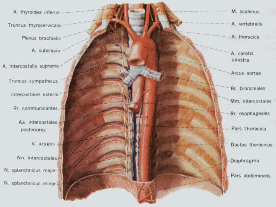

The upper six nerves run toward the sternum and terThe upper six nerves run toward the sternum and terminate as the anterior cutaneous branches that pierce minate as the anterior cutaneous branches that pierce the intercostal muscles and the pectoralis major near the intercostal muscles and the pectoralis major near the sternum.the sternum.
The lower five intercostal nerves and the subcostal neThe lower five intercostal nerves and the subcostal nerve cross the costal arch and continue their course antrve cross the costal arch and continue their course anteriorly between the obliquus internus abdominis and eriorly between the obliquus internus abdominis and transversus abdominis. Then they pierce the sheath of transversus abdominis. Then they pierce the sheath of rectus, penetrate the rectus abdominis and terminate rectus, penetrate the rectus abdominis and terminate as anterior cutaneous branches near the linea alba. as anterior cutaneous branches near the linea alba.
Muscular branches of these nerves supply the intercosMuscular branches of these nerves supply the intercostales and the anterolateral abdominal muscles. The ctales and the anterolateral abdominal muscles. The cutaneous branches are distributed to the skin of the tutaneous branches are distributed to the skin of the thoracic and abdominal wall.horacic and abdominal wall.
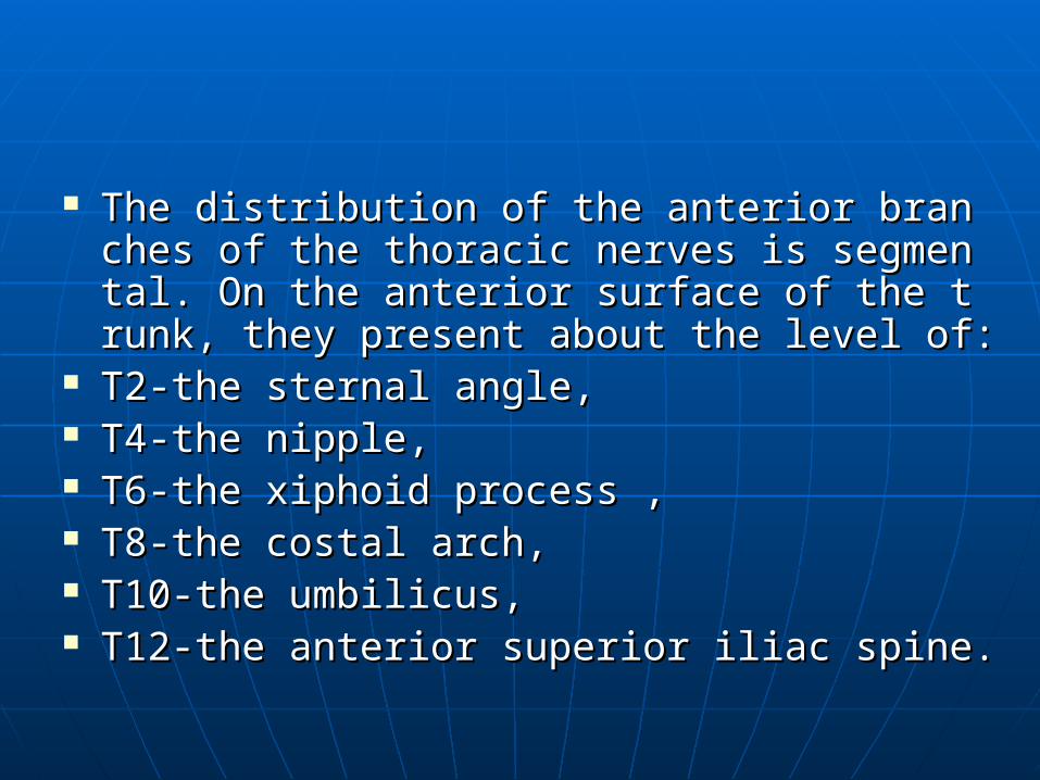
The distribution of the anterior branches of thThe distribution of the anterior branches of the thoracic nerves is segmental. On the anterior e thoracic nerves is segmental. On the anterior surface of the trunk, they present about the lesurface of the trunk, they present about the level of:vel of:
T2-the sternal angle,T2-the sternal angle, T4-the nipple,T4-the nipple, T6-the xiphoid process ,T6-the xiphoid process , T8-the costal arch,T8-the costal arch, T10-the umbilicus,T10-the umbilicus, T12-the anterior superior iliac spine.T12-the anterior superior iliac spine.
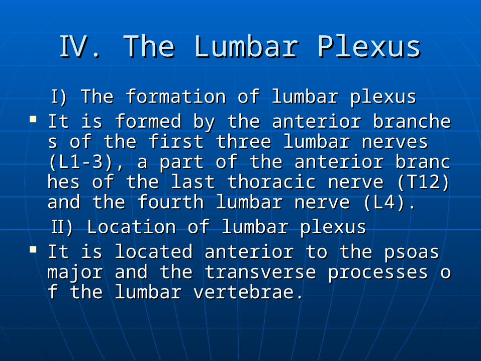
ⅣⅣ. The Lumbar Plexus. The Lumbar Plexus
Ⅰ Ⅰ) The formation of lumbar plexus) The formation of lumbar plexus It is formed by the anterior branches of the firsIt is formed by the anterior branches of the firs
t three lumbar nerves (L1-3), a part of the antet three lumbar nerves (L1-3), a part of the anterior branches of the last thoracic nerve (T12)anrior branches of the last thoracic nerve (T12)and the fourth lumbar nerve (L4).d the fourth lumbar nerve (L4).
Ⅱ Ⅱ) Location of lumbar plexus) Location of lumbar plexus It is located anterior to the psoas major and thIt is located anterior to the psoas major and th
e transverse processes of the lumbar vertebrae transverse processes of the lumbar vertebrae.e.
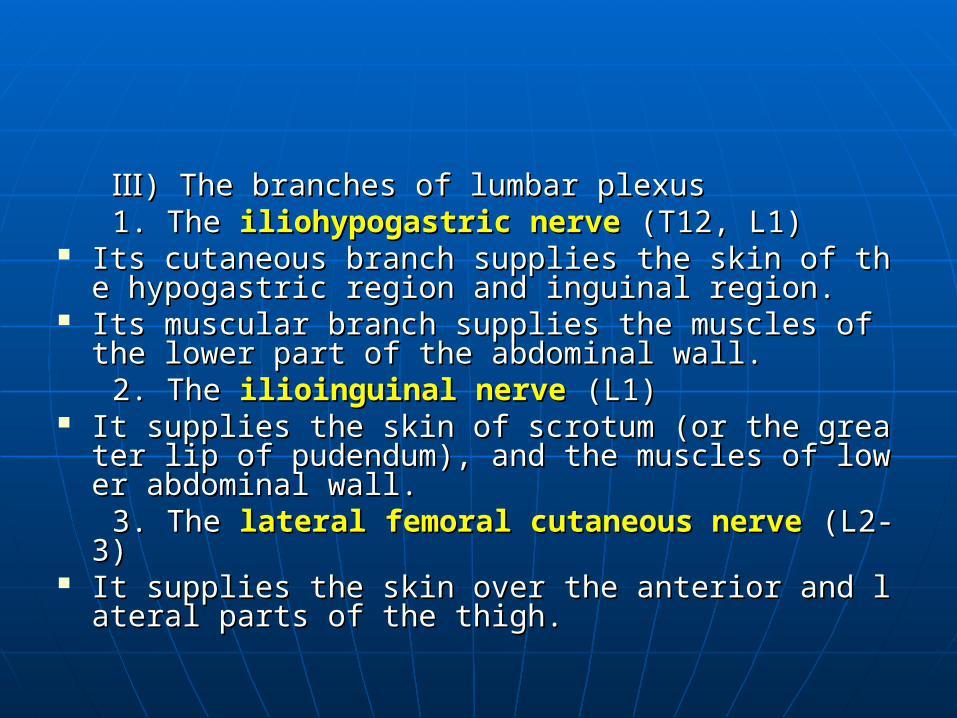
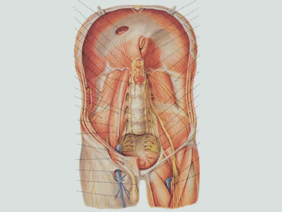
Ⅲ Ⅲ) The branches of lumbar plexus) The branches of lumbar plexus 1. The 1. The iliohypogastric nerveiliohypogastric nerve (T12, L1) (T12, L1) Its cutaneous branch supplies the skin of the hypogastIts cutaneous branch supplies the skin of the hypogast
ric region and inguinal region.ric region and inguinal region. Its muscular branch supplies the muscles of the lower Its muscular branch supplies the muscles of the lower
part of the abdominal wall.part of the abdominal wall. 2. The2. The ilioinguinal nerveilioinguinal nerve (L1) (L1) It supplies the skin of scrotum (or the greater lip of puIt supplies the skin of scrotum (or the greater lip of pu
dendum), and the muscles of lower abdominal wall.dendum), and the muscles of lower abdominal wall. 3. The 3. The lateral femoral cutaneous nervelateral femoral cutaneous nerve (L2-3) (L2-3) It supplies the skin over the anterior and lateral parts It supplies the skin over the anterior and lateral parts
of the thigh.of the thigh.


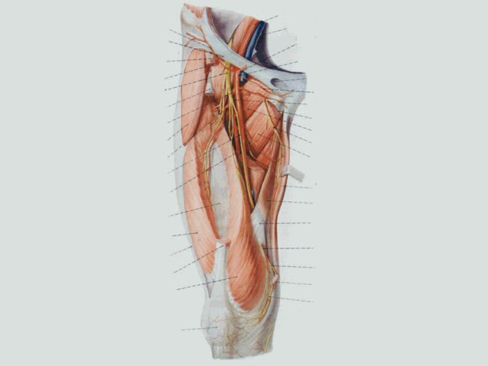
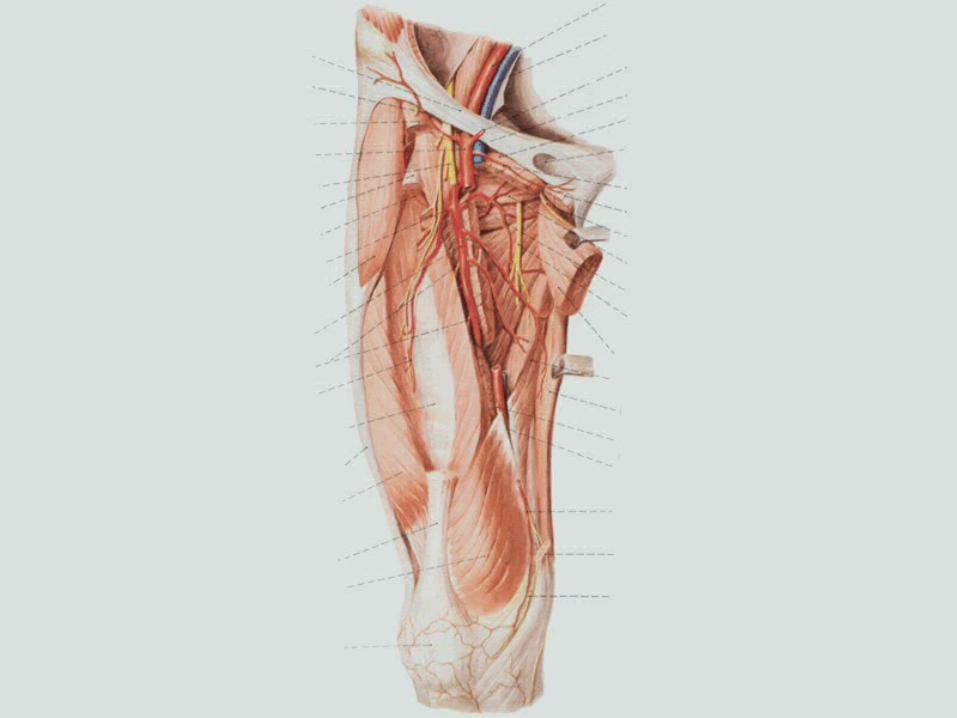
4. The 4. The femoral nervefemoral nerve (L2-4) (L2-4) It descends between the psoas major and the iliacus, tIt descends between the psoas major and the iliacus, t
hen passes down beneath the inguinal ligament into then passes down beneath the inguinal ligament into the femoral triangle, where it is “broken up” into sevhe femoral triangle, where it is “broken up” into several terminal branches.eral terminal branches.
Its Its muscular branchesmuscular branches supply the anterior group of m supply the anterior group of muscles of the thigh---the quadriceps femoris, sartorius uscles of the thigh---the quadriceps femoris, sartorius and pectineus.and pectineus.
Its Its cutaneous branchescutaneous branches to the thigh are the anterior cu to the thigh are the anterior cutaneous branches which are distributed to the anteriotaneous branches which are distributed to the anterior and anteromedial side of the skin of the thigh.r and anteromedial side of the skin of the thigh.
Saphenous nerveSaphenous nerve:: The longest one of its cutaneous branches is tThe longest one of its cutaneous branches is t
he he saphenous nervesaphenous nerve. It accompanies the fem. It accompanies the femoral artery and descends through most of the loral artery and descends through most of the length of the adductor canal, becomes subcutaength of the adductor canal, becomes subcutaneous at the medial side of the knee by emergineous at the medial side of the knee by emerging behind the sartorius. Then it runs downwarng behind the sartorius. Then it runs downward with the great saphenous vein along the med with the great saphenous vein along the medial side of leg as far as the medial side of the fdial side of leg as far as the medial side of the foot. This nerve is distributed to the skin of the oot. This nerve is distributed to the skin of the medial side of the leg and foot.medial side of the leg and foot.


Injury of the femoral nerve results in impaired Injury of the femoral nerve results in impaired flexion of the hip joint. Because the quadricepflexion of the hip joint. Because the quadriceps femoris muscle is paralyzed. It is impossible ts femoris muscle is paralyzed. It is impossible to extend knee and the knee jerk reflex disappeo extend knee and the knee jerk reflex disappears. There would also be a loss of sensation in ars. There would also be a loss of sensation in the skin of the anterior area of the thigh and ththe skin of the anterior area of the thigh and the medial side of the leg and foot.e medial side of the leg and foot.
5. The 5. The obturator nerveobturator nerve (L2-4) (L2-4) It emerges from the medial border of the psoas major, It emerges from the medial border of the psoas major,
whence it passes along the lateral pelvic wall and throwhence it passes along the lateral pelvic wall and through the obturator canal to the medial part of the thigh.ugh the obturator canal to the medial part of the thigh.
The muscular branches of the nerve supply the medial The muscular branches of the nerve supply the medial group of muscles of the thigh, the cutaneous branches group of muscles of the thigh, the cutaneous branches are distributed to the skin of the medial side of the thiare distributed to the skin of the medial side of the thigh.gh.
Injury to the obturator nerve results in impaired adduInjury to the obturator nerve results in impaired adduction of the thigh and a loss of sensation in the skin of ction of the thigh and a loss of sensation in the skin of the medial aspect of the thigh.the medial aspect of the thigh.


6. The 6. The genitofemoral nervegenitofemoral nerve (L1-2) (L1-2) It supplies the skin over the scrotum (or the grIt supplies the skin over the scrotum (or the gr
eater lip of pudendum), and the cremaster.eater lip of pudendum), and the cremaster. The iliohypogastric, ilioinguinal and genitofemThe iliohypogastric, ilioinguinal and genitofem
oral nerves must be protected carefully in the oral nerves must be protected carefully in the operation of the inguinal hernia.operation of the inguinal hernia.

ⅤⅤ. The Sacral Plexus. The Sacral Plexus
Ⅰ Ⅰ) The formation of sacral plexus) The formation of sacral plexus This plexus is formed by the lumbosacral trunThis plexus is formed by the lumbosacral trun
k (a part of L4 and L5), the anterior branches ok (a part of L4 and L5), the anterior branches of the sacral and coccygeal nerves (S1-5, Co1).f the sacral and coccygeal nerves (S1-5, Co1).
Ⅱ Ⅱ) The location of sacral plexus) The location of sacral plexus It is located in the pelvis, where it is closely relIt is located in the pelvis, where it is closely rel
ated to the anterior surface of the piriformis.ated to the anterior surface of the piriformis.
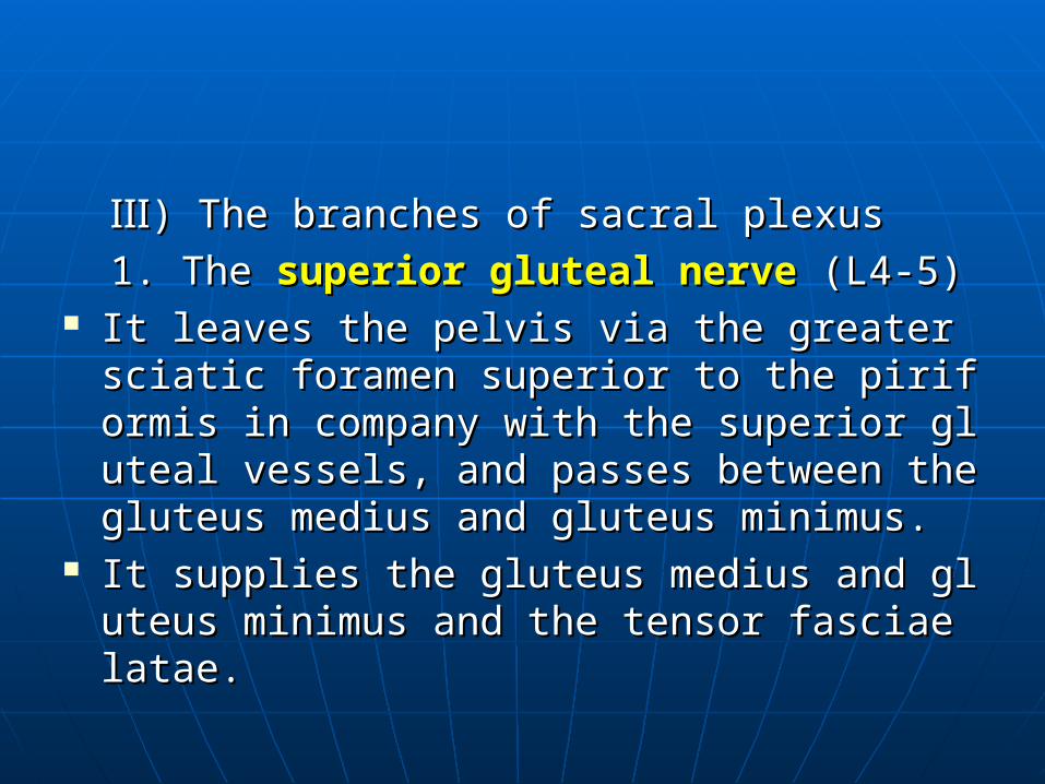
Ⅲ Ⅲ) The branches of sacral plexus) The branches of sacral plexus 1. The 1. The superior gluteal nervesuperior gluteal nerve (L4-5) (L4-5) It leaves the pelvis via the greater sciatic foraIt leaves the pelvis via the greater sciatic fora
men superior to the piriformis in company witmen superior to the piriformis in company with the superior gluteal vessels, and passes betwh the superior gluteal vessels, and passes between the gluteus medius and gluteus minimus.een the gluteus medius and gluteus minimus.
It supplies the gluteus medius and gluteus minIt supplies the gluteus medius and gluteus minimus and the tensor fasciae latae.imus and the tensor fasciae latae.


2. The 2. The inferior glutea nerveinferior glutea nerve (L5-S2) (L5-S2) It leaves the pelvis through the greater sciatic fIt leaves the pelvis through the greater sciatic f
oramen inferior to the piriformis in company oramen inferior to the piriformis in company with the inferior gluteal vessels.with the inferior gluteal vessels.
It supplies the gluteus maximus and the skin oIt supplies the gluteus maximus and the skin over the lower part of the gluteal region.ver the lower part of the gluteal region.
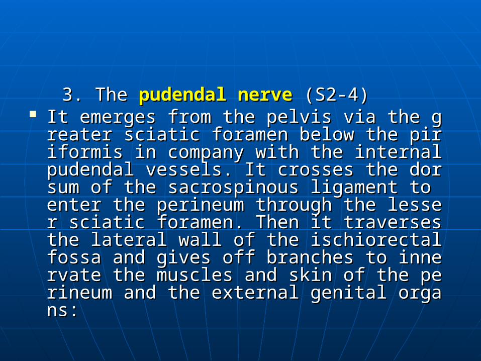
3. The 3. The pudendal nervepudendal nerve (S2-4) (S2-4) It emerges from the pelvis via the greater sciatiIt emerges from the pelvis via the greater sciati
c foramen below the piriformis in company witc foramen below the piriformis in company with the internal pudendal vessels. It crosses the h the internal pudendal vessels. It crosses the dorsum of the sacrospinous ligament to enter dorsum of the sacrospinous ligament to enter the perineum through the lesser sciatic foramthe perineum through the lesser sciatic foramen. Then it traverses the lateral wall of the ischen. Then it traverses the lateral wall of the ischiorectal fossa and gives off branches to innerviorectal fossa and gives off branches to innervate the muscles and skin of the perineum and ate the muscles and skin of the perineum and the external genital organs: the external genital organs:

1) The 1) The anal nerveanal nerve It is distributed to the sphincter ani externus and skin It is distributed to the sphincter ani externus and skin
of the anus.of the anus. 2) The 2) The perineal nerveperineal nerve It is distributed to the muscles of the perineum and thIt is distributed to the muscles of the perineum and th
e skin of the scrotum or the greater lip of pudendum.e skin of the scrotum or the greater lip of pudendum. 3) The 3) The dorsal nerve of penis or clitorisdorsal nerve of penis or clitoris It passes to the dorsum of the penis (or clitoris).It passes to the dorsum of the penis (or clitoris). It supplies the skin of the penis (or clitoris), prepuce aIt supplies the skin of the penis (or clitoris), prepuce a
nd the glans penis (or glans of clitoris).nd the glans penis (or glans of clitoris).
4. The 4. The posterior femoral cutaneous nerveposterior femoral cutaneous nerve (S (S1-3)1-3)
It descends in company with the sciatic nerve It descends in company with the sciatic nerve and is distributed to the skin of the posterior pand is distributed to the skin of the posterior part of the thigh.art of the thigh.
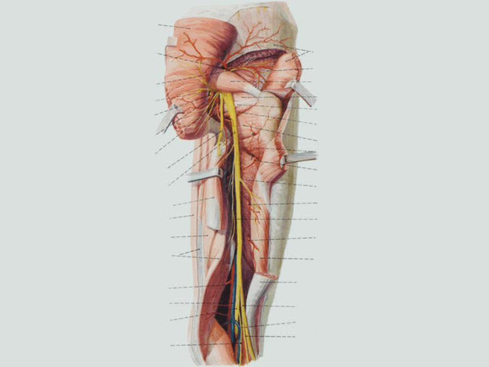
5. The 5. The sciatic nervesciatic nerve (L4-S3) (L4-S3) It is the largest nerve in the body.It is the largest nerve in the body. The nerve leaves the pelvis through the greater sciatic The nerve leaves the pelvis through the greater sciatic
foramen inferior to the piriformis along with the inferiforamen inferior to the piriformis along with the inferior gluteal nerve. Then it runs inferolaterally deep to thor gluteal nerve. Then it runs inferolaterally deep to the gluteus maximus, and descends between the ischial e gluteus maximus, and descends between the ischial tuberosity and the greater trochanter of femur to entetuberosity and the greater trochanter of femur to enter the posterior compartment of the thigh. Here, it passr the posterior compartment of the thigh. Here, it passes downward between the biceps femoris and the sees downward between the biceps femoris and the semimembranosus, semitendinosus to enter the poplitemimembranosus, semitendinosus to enter the popliteal fossa, and terminates by dividing into the tibial and al fossa, and terminates by dividing into the tibial and common peroneal nerves.common peroneal nerves.

The branches of the sciatic nerve supply the mThe branches of the sciatic nerve supply the muscles of the foot, leg and the posterior compauscles of the foot, leg and the posterior compartment of the thigh. It also supplies the skin of rtment of the thigh. It also supplies the skin of the leg and foot.the leg and foot.
The level of division of the sciatic nerve is variaThe level of division of the sciatic nerve is variable. It is usually at the superior angle of the poble. It is usually at the superior angle of the popliteal fossa but these two nerves may be sepapliteal fossa but these two nerves may be separated even at their origins in the pelvis.rated even at their origins in the pelvis.

1) The 1) The tibial nervetibial nerve (L4-S3) (L4-S3) It is the large one of the two terminal branches of the sIt is the large one of the two terminal branches of the s
ciatic nerve.ciatic nerve. At first the nerve descends through the center of the pAt first the nerve descends through the center of the p
opliteal fossa in company with the politeal vessels anopliteal fossa in company with the politeal vessels and posterior to the popliteal vein, it then passes deep td posterior to the popliteal vein, it then passes deep to the triceps surae and posterior to the medial malleolo the triceps surae and posterior to the medial malleolus, where the tibial nerve is divided into the us, where the tibial nerve is divided into the medialmedial an and d lateral plantar nerveslateral plantar nerves to supply the plantar muscle to supply the plantar muscles and the skin over the sole of the foot.s and the skin over the sole of the foot.
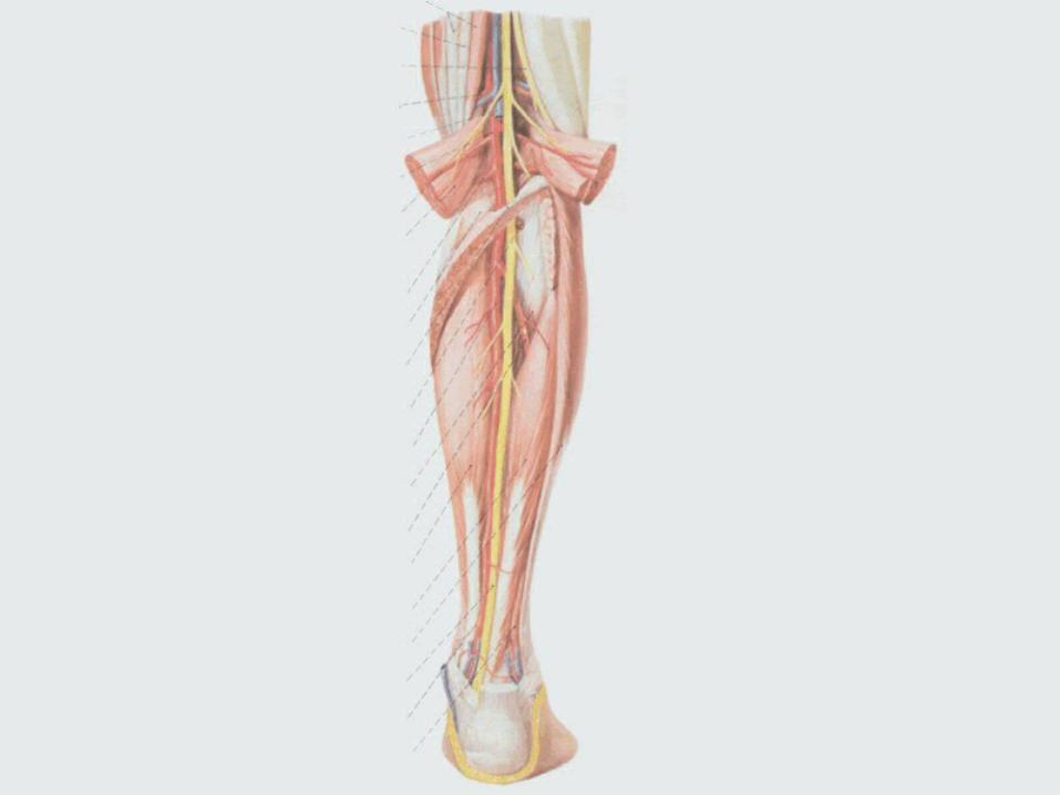
In the popliteal fossa, the tibial nerve gives off branchIn the popliteal fossa, the tibial nerve gives off branches to all the muscles of the posterior compartment of tes to all the muscles of the posterior compartment of the leg. It also gives off a cutaneous branch, the he leg. It also gives off a cutaneous branch, the mediamedial sural cutaneous nervel sural cutaneous nerve, which descends in company , which descends in company with the small saphenous vein. with the small saphenous vein.
At the lower part of the leg, the medial sural cutaneouAt the lower part of the leg, the medial sural cutaneous nerve usually joins the s nerve usually joins the lateral sural cutaneous nervlateral sural cutaneous nervee, coming from the common peroneal nerve, to form t, coming from the common peroneal nerve, to form the he sural nervesural nerve which is distributed to the skin of the p which is distributed to the skin of the posterior and lateral surface of the leg and over the lateosterior and lateral surface of the leg and over the lateral border of the dorsum of the foot.ral border of the dorsum of the foot.
2) The 2) The common peroneal nervecommon peroneal nerve (L4-S2) (L4-S2) It begins at the superior angle of the popliteal fIt begins at the superior angle of the popliteal f
ossa and passes lateroinferiorly along the medossa and passes lateroinferiorly along the medial border of the biceps femoris and its tendon. ial border of the biceps femoris and its tendon. It leaves the fossa by passing superficially to thIt leaves the fossa by passing superficially to the lateral head of the gastrocnemius. The nerve e lateral head of the gastrocnemius. The nerve then passes around the posterolateral surface then passes around the posterolateral surface of the neck of fibula to enter deep to the superof the neck of fibula to enter deep to the superior part of the peroneus longus.ior part of the peroneus longus.
The common peroneal nerve is palpable wherThe common peroneal nerve is palpable where it winds around the neck of fibula, and is divie it winds around the neck of fibula, and is divided into the superficial and deep peroneal nerded into the superficial and deep peroneal nerves.ves.
The common peroneal nerve supplies the musThe common peroneal nerve supplies the muscles of the anterior and lateral compartments cles of the anterior and lateral compartments of the leg. It also gives off branches of the skin of the leg. It also gives off branches of the skin over lateral surface of the leg and dorsum of thover lateral surface of the leg and dorsum of the foot. e foot.
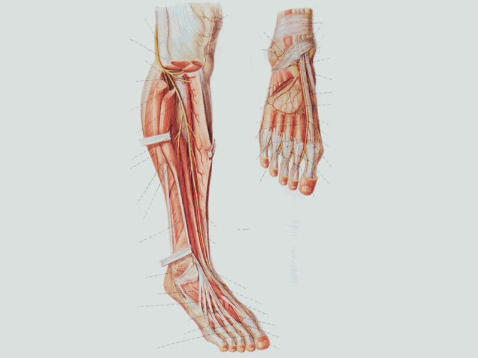
① ① The The superficial peroneal nervesuperficial peroneal nerve It descends between the peroneus longus and It descends between the peroneus longus and
brevis and pierces the deep fascia to become sbrevis and pierces the deep fascia to become superficial in the distal one third of the leg. The uperficial in the distal one third of the leg. The branches of this nerve supply the peroneal mubranches of this nerve supply the peroneal muscles and the skin on the distal part of the antescles and the skin on the distal part of the anterior surface of the leg, the dorsum of the foot arior surface of the leg, the dorsum of the foot and toes.nd toes.
② ② The The deep peroneal nervedeep peroneal nerve It is in company with the anterior tibial artery. At first iIt is in company with the anterior tibial artery. At first i
t lies between the extensor digitorum longus and the tt lies between the extensor digitorum longus and the tibialis anterior and then between the extensor halluciibialis anterior and then between the extensor hallucis longus and the tibialis anterior.s longus and the tibialis anterior.
It supplies the anterior group of muscles of the leg anIt supplies the anterior group of muscles of the leg and a small area of the skin between the first and second d a small area of the skin between the first and second toes.toes.
Functional disturbances in the case of the common peFunctional disturbances in the case of the common peroneal nerve injuries include impaired dorsiflex of the roneal nerve injuries include impaired dorsiflex of the foot, reduced or lost eversion of the foot. This conditiofoot, reduced or lost eversion of the foot. This condition causes the foot to hang down and is known as “foon causes the foot to hang down and is known as “foot-drop” or “talipes equinovarus”. There is also a vat-drop” or “talipes equinovarus”. There is also a variable loss of sensation of the anterolateral aspect of triable loss of sensation of the anterolateral aspect of the leg and the dorsum of the foot.he leg and the dorsum of the foot.