Neuronal Glutamate Transporter EAAT4 Is Expressed in ...whu/HuWH-Publications/EAA4-Glia.pdf ·...
13
Neuronal Glutamate Transporter EAAT4 Is Expressed in Astrocytes WEN-HUI HU, WINSTON M. WALTERS, XIAO-MEI XIA, SHAFFIAT A. KARMALLY, AND JOHN R. BETHEA* The Miami Project to Cure Paralysis, University of Miami School of Medicine, Miami, Florida KEY WORDS excitatory amino acid transporter; immunohistochemistry; Western blot; RT-PCR ABSTRACT High-affinity excitatory amino acid transporters (EAATs) are essential to terminate glutamatergic neurotransmission and to prevent excitotoxicity. To date, five distinct EAATs have been cloned from animal and human tissues: GLAST (EAAT1), GLT-1 (EAAT2), EAAC1 (EAAT3), EAAT4, and EAAT5. EAAT1 and EAAT2 are com- monly known as glial glutamate transporters, whereas EAAT3, EAAT4, and EAAT5 are neuronal. EAAT4 is largely expressed in cerebellar Purkinje cells. In this study, using immunohistochemistry and Western blotting, we found that EAAT4-like immunoreac- tivity (ir) is enriched in the spinal cord and forebrain. Double-labeled fluorescent im- munostaining and confocal image analysis indicated that EAAT4-like ir colocalizes with an astrocytic marker, glial fibrillary acidic protein (GFAP). The astrocytic localization of EAAT4 was further confirmed in astrocyte cultures by double-labeled fluorescent im- munocytochemistry and Western blotting. Reverse transcriptase-polymerase chain re- action analysis demonstrated mRNA expression of EAAT4 in astrocyte cultures. Se- quencing confirmed the specificity of the amplified fragment. These results demonstrate that EAAT4 is expressed in astrocytes. This astrocytic localization of neuronal EAAT4 may reveal a new function of EAAT4 in the central nervous system. © 2003 Wiley-Liss, Inc. INTRODUCTION Glutamate is the predominant excitatory amino acids (EAAs) in the mammalian central nervous sys- tem (CNS) and is involved in many important phys- iological processes and pathological conditions (Ka- nai, 1997; Robinson and Dowd, 1997). High-affinity excitatory amino acid transporters (EAATs) are es- sential to terminate glutamatergic neurotransmis- sion and to prevent excitotoxicity (Danbolt, 2001; O’Shea, 2002). To date, five distinct EAATs have been cloned from animal and human tissues: EAAT1 (GLAST) (Arriza et al., 1994; Kawakami et al., 1994), EAAT2 (GLT-1) (Arriza et al., 1994; Shashidharan et al., 1994), EAAT3 (EAAC1) (Kanai and Hediger, 1992; Arriza et al., 1994), EAAT4 (Fairman et al., 1995), and EAAT5 (Arriza et al., 1997). EAAT family members display 50 –55% amino acid sequence identity. They each contain 10 hydrophobic domains, with both the N- and C-termini in the cytoplasm (Wahle and Stoffel, 1996). The EAAT subtypes differ in regional, cellular, and developmental distribution (Robinson and Dowd, 1997; Schluter et al., 2002). EAAT1 immunoreactivity (ir) is highly abundant in cerebellar Bergmann glia, and EAAT2 ir is concentrated in astrocytes in the hip- pocampus and cerebral cortex (Rothstein et al., 1994; Lehre et al., 1995). EAAT3 is highly expressed in the cortex, hippocampus, caudate–putamen, and periph- eral tissues (Rothstein et al., 1994). EAAT4 is confined to cerebellar Purkinje cells (Yamada et al., 1996; De- hnes et al., 1998), with little expression in other brain regions (Massie et al., 2001). EAAT5 is expressed pre- dominantly in the retina (Arriza et al., 1997). EAAT1 *Correspondence to: Dr. John R. Bethea, The Miami Project to Cure Paralysis, University of Miami School of Medicine, 1095 NW 14th Terrace, Miami, FL 33136. E-mail: [email protected] Received 29 October 2002; Accepted 31 March 2003 DOI 10.1002/glia.10268 GLIA 44:13–25 (2003) © 2003 Wiley-Liss, Inc.
Transcript of Neuronal Glutamate Transporter EAAT4 Is Expressed in ...whu/HuWH-Publications/EAA4-Glia.pdf ·...

Neuronal Glutamate TransporterEAAT4 Is Expressed in Astrocytes
WEN-HUI HU, WINSTON M. WALTERS, XIAO-MEI XIA, SHAFFIAT A. KARMALLY,AND JOHN R. BETHEA*
The Miami Project to Cure Paralysis, University of Miami School of Medicine, Miami, Florida
KEY WORDS excitatory amino acid transporter; immunohistochemistry; Westernblot; RT-PCR
ABSTRACT High-affinity excitatory amino acid transporters (EAATs) are essentialto terminate glutamatergic neurotransmission and to prevent excitotoxicity. To date,five distinct EAATs have been cloned from animal and human tissues: GLAST (EAAT1),GLT-1 (EAAT2), EAAC1 (EAAT3), EAAT4, and EAAT5. EAAT1 and EAAT2 are com-monly known as glial glutamate transporters, whereas EAAT3, EAAT4, and EAAT5 areneuronal. EAAT4 is largely expressed in cerebellar Purkinje cells. In this study, usingimmunohistochemistry and Western blotting, we found that EAAT4-like immunoreac-tivity (ir) is enriched in the spinal cord and forebrain. Double-labeled fluorescent im-munostaining and confocal image analysis indicated that EAAT4-like ir colocalizes withan astrocytic marker, glial fibrillary acidic protein (GFAP). The astrocytic localization ofEAAT4 was further confirmed in astrocyte cultures by double-labeled fluorescent im-munocytochemistry and Western blotting. Reverse transcriptase-polymerase chain re-action analysis demonstrated mRNA expression of EAAT4 in astrocyte cultures. Se-quencing confirmed the specificity of the amplified fragment. These results demonstratethat EAAT4 is expressed in astrocytes. This astrocytic localization of neuronal EAAT4may reveal a new function of EAAT4 in the central nervous system.© 2003 Wiley-Liss, Inc.
INTRODUCTION
Glutamate is the predominant excitatory aminoacids (EAAs) in the mammalian central nervous sys-tem (CNS) and is involved in many important phys-iological processes and pathological conditions (Ka-nai, 1997; Robinson and Dowd, 1997). High-affinityexcitatory amino acid transporters (EAATs) are es-sential to terminate glutamatergic neurotransmis-sion and to prevent excitotoxicity (Danbolt, 2001;O’Shea, 2002). To date, five distinct EAATs havebeen cloned from animal and human tissues: EAAT1(GLAST) (Arriza et al., 1994; Kawakami et al., 1994),EAAT2 (GLT-1) (Arriza et al., 1994; Shashidharan etal., 1994), EAAT3 (EAAC1) (Kanai and Hediger,1992; Arriza et al., 1994), EAAT4 (Fairman et al.,1995), and EAAT5 (Arriza et al., 1997). EAAT familymembers display � 50 –55% amino acid sequenceidentity. They each contain 10 hydrophobic domains,with both the N- and C-termini in the cytoplasm(Wahle and Stoffel, 1996).
The EAAT subtypes differ in regional, cellular, anddevelopmental distribution (Robinson and Dowd, 1997;Schluter et al., 2002). EAAT1 immunoreactivity (ir) ishighly abundant in cerebellar Bergmann glia, andEAAT2 ir is concentrated in astrocytes in the hip-pocampus and cerebral cortex (Rothstein et al., 1994;Lehre et al., 1995). EAAT3 is highly expressed in thecortex, hippocampus, caudate–putamen, and periph-eral tissues (Rothstein et al., 1994). EAAT4 is confinedto cerebellar Purkinje cells (Yamada et al., 1996; De-hnes et al., 1998), with little expression in other brainregions (Massie et al., 2001). EAAT5 is expressed pre-dominantly in the retina (Arriza et al., 1997). EAAT1
*Correspondence to: Dr. John R. Bethea, The Miami Project to Cure Paralysis,University of Miami School of Medicine, 1095 NW 14th Terrace, Miami, FL33136. E-mail: [email protected]
Received 29 October 2002; Accepted 31 March 2003
DOI 10.1002/glia.10268
GLIA 44:13–25 (2003)
© 2003 Wiley-Liss, Inc.

and EAAT2 are commonly known as glial glutamatetransporters, whereas EAAT3, EAAT4, and EAAT5 arethought to be primarily neuronal (Rothstein et al.,1994; Anderson and Swanson, 2000; Maragakis andRothstein, 2001).
The physiological and pathological roles of EAATs inthe brain have been well characterized (Meldrum et al.,1999; Lievens et al., 2000; Maragakis and Rothstein,2001; Trotti et al., 2001; Proper et al., 2002). However,little is known about the characterization of EAATs inthe spinal cord. EAAT2 ir is restricted to and enrichedin the spinal gray matter of rats, mice, and humans(Milton et al., 1997; Fray et al., 1998; Sasaki et al.,2001), whereas EAAT1 ir is primarily localized in su-perficial laminae of the dorsal horn (Rothstein et al.,1995; Vera-Portocarrero et al., 2002). EAAT3 ir ismainly localized in ventral horn motoneurons and dor-sal horn superficial laminae. After spinal cord injury,rapid upregulation of EAAT1, EAAT2, and EAAT3 hasbeen reported (Vera-Portocarrero et al., 2002).Whether EAAT4 is expressed in spinal cord remainselusive, although Western blot studies have indicatedthat EAAT4 is undetectable (Furuta et al., 1997) orweakly expressed (Nagao et al., 1997) in spinal cordhomogenates.
In the course of investigating the role of EAATs inthe spinal cord after injury, we observed that EAAT4 iris enriched throughout the spinal cord of rats and mice.Further studies using double-labeled immunofluores-cent staining, confocal image analysis, and reversetranscriptase-polymerase chain reaction (RT-PCR)demonstrated that EAAT4 is expressed in astrocytes.This astrocytic localization of EAAT4 may reveal a newrole of EAAT4 in the central nervous system.
MATERIALS AND METHODSCortical Astrocyte Cultures
Cortical astrocyte cultures were prepared usingstandard methods. Briefly, the cerebral cortices weredissected from the brain of P7 mouse pups. After re-moval of the meninges, the tissues were dissociated bytrypsinization and trituration. Cells were suspended inDulbecco’s modified Eagle’s medium (DMEM)/F12 sup-plemented with 10% fetal bovine serum and penicillin/streptomycin and plated on uncoated 75 cm2 flasks atan approximate density of 50,000 cells/cm2. The cul-tures were maintained in humidified 5% CO2 incubatorat 37°C. The medium was changed 24 h after platingand then every 3–4 days. The cells were grown andbecame confluent after 10–14 days. At this point, morethan 90% of the adherent cells exhibited the flattened,polygonal appearance typical of type I astrocytes. Non-astrocytes were detached from the flasks by shakingand removed by changing the medium. The remainingcells were dissociated by trypsinization (0.1% trypsin-0.04% EGTA) and plated on uncoated six-well plates ata density of 10,000 cells/cm2. The cells became conflu-ent again 10–14 days after the plating.
RNA Isolation, RT-PCR, T-A Cloningand Sequencing
Total RNA was isolated from the snap-frozen tissuesor cultured astrocytes with TRIzol reagent (Gibco-BRL,Rockville, MD). Two micrograms of RNA, pretreatedwith DNase I for 2 h at 37°C, was used to synthesizecDNA using SuperScript II reverse transcriptase withrandom hexanucleotide as primers. The EAAT4-spe-cific primers were designed according to the knownsequence of mouse EAAT4 (genebank number D83262):upstream primer, 5�-CCGCCAGATCAAGTACTTC-TCCT-3�; downstream primer, 5�-GCAGAGCTGGAAG-AGGTACCC-3� (corresponding to nucleotides 427–450and 1338–1361, respectively). A total of 35 reactioncycles (94°C for 30 s, 55°C for 45 s, 72°C for 1.5 min)was preceded by a hot start at 94°C for 3 min andconcluded with an extension step at 72°C for 10 min. Asnegative controls, RNA without RT was used as tem-plate or the cDNA in the reaction mixture was substi-tuted with water. Amplified products were analyzed byhorizontal agarose gel electrophoresis and visualizedvia ethidium bromide staining. A 1 kb DNA markerwas used as molecular weight reference.
The PCR fragment of expected size was excised fromthe agarose gel, purified using the QIAquick gel extrac-tion kit (Qiagen, Valencia, CA), and ligated into pCR IIvector (Invitrogen, Carlsbad, CA). After transforma-tion of competent E. coli DH5� cells, the plasmid wasextracted with the Miniprep Wizzard kit (Promega,Madison, WI). The cloned fragment was sequencedwith the T7 promotor primer by the Perkin-Elmer ABI373 DNA Sequencer with XL upgrade and Big Dyechemistry.
Mammalian Expression Vector Cloningand Transfection
N-terminally Flag-epitope-tagged pCMV-Tag vector(Stratagene, La Jolla, CA) constructs encoding ratEAAT4 (pCMV-Flag-rEAAT4) were generated by RT-PCR from rat forebrain RNA. N-terminally Myc-epitope-tagged pCMV-Tag vector (Stratagene) con-structs encoding mouse EAAT4 (pCMV-Myc-mEAAT4)were generated by PCR from SuperScript mouse braincDNA library (Invitrogen). All vectors were confirmedby sequencing, immunocytochemistry, and Westernblot analysis.
The human embryonic kidney (HEK) 293T cell lineor monkey kidney COS-7 cell line were maintained inhigh-glucose DMEM containing 10% fetal bovine se-rum and penicillin/streptomycin. For immunofluores-cent microscopy, cells were seeded on eight-well plasticchamber slides. For Western blot and immunoprecipi-tation experiments, cells were seeded on 10 cm dishes.Cells were transfected the following day by the stan-dard calcium phosphate precipitation.
14 HU ET AL.

Western Blot and Immunoprecipitation
Normal healthy adult Sprague-Dawley rats orC57BL/6 mice were anaesthetized in a chamber withhalothane and decapitated. The spinal cords andbrains were removed, frozen instantly in liquid nitro-gen, and stored at �80°C until further processing. Dis-section of gray and white matter of the spinal cord wasperformed as previously described (Hu et al., 1999).
Snap-frozen tissues, cultured astrocytes, or transfectedHEK293T cells were solubilized in Triton X-100-basedlysis buffer containing 20 mM Tris-HCl (pH 7.4), 1%Triton X-100, 1 mM ethylenediaminetetraacetic acid(EDTA), 5 mM dithiothreitol (DTT), 150 mM NaCl, 1 mMphenylmethylsulfonyl fluoride (PMSF), 10 �g/ml aproti-nin, 10 �g/ml leupeptin, and phosphatase inhibitor cock-tail I (Sigma, St. Louis, MO). Lysates were incubatedthrough end-over-end mixing at 4°C for 30 min. The nu-clear and cellular debris were cleared by centrifugation at20,000 g for 20 min at 4°C. The protein concentrations ofthe supernatant were determined with Dc Protein Assaykit from BioRad (Hercules, CA). Proteins were denaturedvia boiling for 5 min in sodium dodecyl sulphate (SDS)sample buffer, fractionated by SDS-polyacrylamide gelelectrophoresis (PAGE) in tris-glycine buffer, and trans-ferred to nitrocellulose membrane (BioRad). The SeeBlueprestained standard (Invitrogen) was used as molecularweight reference. Blots were blocked in 5% nonfat drymilk/tris-buffered saline (pH 7.6) plus 0.1% Tween-20(TBS-T) for 1 h and then incubated overnight at 4°C withaffinity-purified rabbit anti-rEAAT4 polyclonal antibodyagainst a 21 aa synthetic peptide near the rEAAT4 C-terminus (1:1,000; Alpha Diagnostic, San Antonio, TX) in2% milk/TBS-T. After incubation for 1 h with horseradishperoxidase-conjugated antirabbit antiserum (1/2,000;Amersham Biosciences, Piscataway, NJ) in 2% milk/TBS-T, immunoreactive proteins were visualized usingenhanced chemiluminescence (ECL) Western blotting de-tection reagents (Amersham Biosciences). All washingsteps were performed with TBS-T.
For immunoprecipitation, the lysates from HEK293Tcells overexpressing Flag-rEAAT4 were incubated over-night at 4°C with 0.5 �g anti-Flag (Sigma) monoclonalantibody or mouse control IgG. The immune complexeswere precipitated with GammaBind G Sepharose (Amer-sham Pharmacia Biotech, Uppsala, Sweden), washedfour times with 0.6 M NaCl lysis buffer, and then elutedby boiling for 5 min in SDS sample buffer. The elutedproteins were fractionated on SDS-PAGE and detected byWestern blotting with anti-Flag monoclonal antibody (1:2,000) or anti-rEAAT4 polyclonal antibody (1:1,000).
Routine Immunohistochemistry
For immunohistochemical studies, free-floating sec-tions of spinal cord and brain were stained as describedpreviously (Hu et al., 1999; Hausmann et al., 2002).Briefly, animals were transcardially perfused with 4%paraformaldehyde. The spinal cords and brains were
dissected and postfixed overnight in the same fixative.All tissue samples were cryopreserved with buffered25% sucrose. Serial transverse or longitudinal sectionswere cut frozen on the sliding microtome at a thicknessof 40 �m. The sections were permeated with 0.5% Tri-ton X-100 and blocked with 10% normal goat serum for30 min and then incubated with primary anti-rEAAT4polyclonal antibody (1:1,000) overnight at 4°C. The fol-lowing day, sections were washed and incubated withsecondary goat antirabbit biotinated antibody (1:200)for 1 h. The sections were then incubated in ABC com-pound (Vector Laboratories, Burlingame, CA) followedby diaminobenzidine (DAB) staining. Staining specific-ity was determined through omitting the primary an-tibody or preabsorbing it with synthetic rEAAT4 pep-tide (Alpha Diagnostic) for additional sections inparallel experiment.
Immunofluorescent Staining and ConfocalImage Analysis
For sequential double-labeled immunofluorescentstaining, the cultured cells, after fixation with 4% para-formaldehyde for 30 min, or the tissue sections werepretreated with blocking solution of 10% normal goatserum and 0.5% Triton X-100 overnight at 4°C. Thesections were incubated with the primary anti-rEAAT4antibody at 1:400 in blocking solution overnight at 4°C.After washing, the Alexa Fluor 488 (green)-linked sec-ondary goat antirabbit antibody (Molecular Probes,Eugene, OR) 1:400 was added for 1 h. Then differentmonoclonal antibodies against Flag epitope or variouscell markers were used. Neurons were identified withan anti-MAP2 (Sigma) antibody at 1:200. Astrocyteswere stained with an antiglial fibrillary acid protein(GFAP) antibody (Chemicon International, Temecula,CA) 1:400. Oligodendrocytes were stained with an anti-RIP antibody (DSHB, Iowa, IA) 1:40. The Alexa Fluor594 (red) secondary goat antimouse antibody (Molecu-lar Probes) 1:200 in blocking solution was used. Fluo-rescences were analyzed under the confocal Axiovert100 M microscope with the LSM 510 scanning module(Carl Zeiss, Thornwood, NY).
Quantitative Image Analysis
The images from Western blot and RT-PCR werescanned and analyzed with the Quantity One softwarefrom BioRad. The data are presented as adjusted vol-ume (intensity) times total area.
RESULTSCharacterization of Anti-rEAAT4 Antibody
To characterize the commercially available affinity-purified polyclonal antibody against the C-terminus ofrEAAT4, we cloned full-length rEAAT4 into a Flag-
15ASTROCYTES EXPRESSING EAAT4

tagged mammalian expression vector, transfected itinto HEK293T cells, and purified this fusion proteinvia immunoprecipitation with an anti-Flag antibody.Western blot analysis of the purified fusion proteinwith the anti-rEAAT4 polyclonal antibody and anti-Flag monoclonal antibody demonstrates that anti-rEAAT4 antibody specifically recognizes Flag-taggedrEAAT4 fusion protein (Fig. 1A). This antibody alsorecognizes endogenous EAAT4 in both rat spinal cordand brain, and the immunoreactive bands are com-pletely eliminated when the antibody (0.5 �g/ml) waspreabsorbed with 5 �g/ml synthetic rEAAT4 peptide(Fig. 1B). Western blotting of mouse brain and spinalcord tissues as well as Myc-tagged mouse EAAT4 fu-sion protein shows a similar pattern of immunoreactivebands (Fig. 1C). Both endogenous and exogenous ex-pressions of EAAT4 are present largely as fuzzy mul-tiple bands of higher molecular weight, even underreducing conditions (Fig. 1C). These multiple bands areobserved predominantly in cerebellum as evidenced byWestern blotting for different region of brain (Fig. 1D),which is in accord with other previous studies (Bristoland Rothstein, 1996; Yamada et al., 1996, 1997; De-hnes et al., 1998; Lin et al., 1998). However, both en-dogenous (Fig. 1B–D) and exogenous (Fig. 1A and C)EAAT4 appear as a lower-molecular-weight bandroughly at � 60 kDa, which can also be blocked by acompetitive peptide (Fig. 1B). A small difference in themobility of the lower-molecular-weight band appearsamong tissues and transfected cells (Fig. 1C and D). Inthe cerebellum, the small band is not increased inproportion to the multiple bands when compared toother regions (Fig. 1D).
To confirm the specificity of anti-rEAAT4 antibody,double-labeled immunocytochemistry and confocal im-age analysis were performed in HEK293T and COS-7cells overexpressing Flag-tagged rEAAT4 (Fig. 2) orMyc-tagged mEAAT4 (data not shown). As shown inFigure 2, the rEAAT4-like ir colocalizes with the Flag-like ir. The staining is predominantly on the plasmamembrane, which is consistent with the predictedtransmembrane topology of EAAT4 protein (Wahle andStoffel, 1996) and the endoplasmic reticulum/Golgi ap-paratus (Fig. 2).
EAAT4 Protein Expression in Astrocytes ofSpinal Cord and Brain
EAAT4 expression has been widely demonstrated incerebellar Purkinje cells (Yamada et al., 1996; Dehneset al., 1998; Massie et al., 2001). To test if neuronalEAAT4 is expressed in spinal cord, we performed rou-tine immunohistochemical studies. The affinity-puri-fied anti-rEAAT4 antibody stains extensively through-out the spinal cord of rat (Fig. 3) and mouse (data notshown). The staining pattern resembles astrocytes, notneurons. In the white matter, long and enriched net-work processes are immunoreactive for EAAT4 withhighest levels appearing around the peripheral rim. In
the gray matter, the positive processes approach bloodvessels and neurons. Dilution of the primary antibodyreduces the staining and preabsorption of anti-rEAAT4antibody (0.5 �g/ml) with a synthetic rEAAT4 peptide(5 �g/ml) quenches the immunoreactivity (Fig. 3B).
To confirm the cellular distribution of EAAT4 ir,double-labeled immunofluorescent staining and confo-cal image analysis were performed. In the spinal cord,EAAT4-like ir is colocalized with the astrocytic markerGFAP (Fig. 4, top row) but not with a neuronal markerMAP2 (Fig. 4, second row from top) or an oligodendro-cyte marker RIP (Fig. 4, third row from top). As apositive control for EAAT4 ir, the rat cerebellar cortexwas stained (Fig. 4, bottom row). Consistent with pre-vious reports (Yamada et al., 1996; Furuta et al., 1997;Itoh et al., 1997; Nagao et al., 1997; Dehnes et al.,1998), the Purkinje cells are intensively immunoreac-tive for EAAT4, whereas granule cells are not (Fig. 4,bottom row). This antibody also stains astrocytes ex-tensively in forebrain and hindbrain (Fig. 5). LikeGFAP staining, EAAT4 staining, with processes form-ing a dense network, is predominantly in the whitematter, maximally in the subventricular zone and sub-pial layer.
EAAT4 Protein Expression inCultured Astrocytes
To provide direct evidence for the presence of EAAT4protein in astrocytes, mouse cortical astrocyte cultureswere used for immunocytochemistry and Western blot-ting. EAAT4-like ir colocalizes with GFAP-positive as-trocytes (Fig. 6A). Western blotting of cultured astro-cytes shows the same size bands as that in spinal cordtissues (Fig. 6B). To confirm EAAT4 protein expressionin astrocytes, basic fibroblast growth factor (bFGF) andepidermal growth factor (EGF), which have been pre-viously shown to upregulate EAAT expression(Zelenaia et al., 2000; Suzuki et al., 2001), were used totreat the cultured astrocytes. As shown in Figure 6B,treatment with bFGF (10 ng/ml) or EGF (50 ng/ml) for24 h increases EAAT4 expression in cultured astro-cytes.
EAAT4 mRNA Expression in Astrocytes
To confirm the presence of EAAT4 in astrocytes,RT-PCR analysis was performed in cultured astro-cytes, demonstrating a strong band of the predictedsize (Fig. 7). EAAT4 mRNA is also highly expressed inthe mouse spinal cord and brain (Fig. 7). The amplifiedfragments from both astrocytes and spinal cord tissueswere cloned into pCR II TA-cloning vector and se-quenced with the T7 promotor primer. Analysis of thesequence through Genebank database demonstratesthat the RT-PCR-amplified fragment exactly matchesmouse EAAT4 and shares 94% and 89% homology torat and human EAAT4, respectively.
16 HU ET AL.

Fig. 1. Characterization of antirat EAAT4 antibody. A: Exactmatching of rEAAT4 immunoreactivity with Flag-rEAAT4 fusion pro-tein. HEK293T cells were transfected with pCMV-Flag-rEAAT4 vec-tor by standard calcium phosphate precipitation. After 24 h, theoverexpressed Flag-rEAAT4 fusion protein was immunoprecipitated(IP) with anti-Flag monoclonal antibody or control mouse IgG. West-ern blots (WB) were duplicated and analyzed separately with anti-Flag monoclonal antibody or anti-rEAAT4 polyclonal antibody. B:Peptide blockade of rEAAT4 immunoreactivity. The whole homoge-nates of rat spinal cord and brain at amount of 50 �g protein wereresolved in 10% SDS-PAGE and Western blotting was performed with
anti-rEAAT4 antibody (0.5 �g/ml). Preabsorption with rEAAT4 pep-tide (5 �g/ml) completely abolished the specific band of EAAT4 im-munoreactivity. Ns: nonspecific band. C: Comparison of endogenousand exogenous expression of EAAT4. The lysates from mouse brainand spinal cord and HEK293T cells overexpressing Flag-rEAAT4 orMyc-mEAAT4 fusion protein were immunoblotted with anti-rEAAT4antibody. D: Regional distribution of EAAT4 in spinal cord and brain.Proteins are loaded at 50 �g each lane followed by Western blottingwith anti-rEAAT4 antibody. The same blot was reprobed with anti-actin monoclonal antibody after stripping.
17ASTROCYTES EXPRESSING EAAT4

Fig. 2. Colocalization of rEAAT4-like immunoreactivity with Flag-rEAAT4 fusion protein. HEK293T or COS-7 cells were transfectedwith pCMV-Flag-rEAAT4 and 24 h later sequential double-labeledimmunofluorescent staining and confocal image analysis were per-
formed. Note the staining around the plasma membrane (arrow) andendoplasmic reticulum/Golgi apparatus (arrow head). Scale bar, 10�m. [Color figure can be viewed in the online issue, which is availableat www.interscience.wiley.com].

DISCUSSION
The important finding in this study is that the pre-viously described neuronal glutamate transporterEAAT4 is expressed in astrocytes at both protein andmRNA levels. We used a combination of immunostain-
ing, Western blotting, RT-PCR, and sequence analysisto demonstrate EAAT4 expression in astrocytes.
The affinity-purified anti-rEAAT4 antibody used inthis study is a commercially available polyclonal anti-body raised against a synthesized peptide from thecytoplasmic C-terminus of rat EAAT4, which shares
Fig. 3. EAAT4 immunohistochemical staining in rat spinal cord.Affinity-purified anti-rEAAT4 antibody stains extensively throughoutthe spinal cord. The staining pattern resembles astrocytes but notneurons. Preabsorption of anti-rEAAT4 antibody (0.5 �g/ml) with anrEAAT4 peptide (5 �g/ml) quenches the immunoreactivity. C/E and
D/F are amplified from the white matter and gray matter in A,respectively. B: Peptide block. Scale bar, 50 �m. [Color figure can beviewed in the online issue, which is available at www.interscience.wiley.com].
19ASTROCYTES EXPRESSING EAAT4

100% homology to mouse EAAT4 and 95% homology tohuman EAAT4 but no homology to the other subtypesof EAATs (Lin et al., 1998). This antibody recognizes
the purified Flag-tagged rEAAT4 fusion protein asshown by immunoprecipitation and Western blottingwith an anti-Flag antibody. Double-labeled confocal
Fig. 4. Double-labeled immunofluorescent staining of rat spinalcord and confocal image analysis. EAAT4-like immunoreactivity istotally colocalized with GFAP-like immunoreactive astrocytes in ratspinal cord (top row) but not with neuronal marker MAP2 (second row
from top) and oligodendrocyte marker RIP (third row from top). As apositive control in cerebellum, EAAT4 is expressed in Purkinje cells(bottom row). Scale bar, 20 �m. [Color figure can be viewed in theonline issue, which is available at www.interscience.wiley.com].
20 HU ET AL.

Fig. 5. Double-labeled immunofluorescent staining of mouse brainand confocal image analysis of selective regions of white matter inforebrain (A–C) and hindbrain (D and E). EAAT4-like immunoreac-tivity (green) is predominantly in subventricular zone (SVZ) andsubpial region (arrow) and colocalizes with many GFAP-positive cells(red). bas, basilar artery; cc, corpus callosum; D3V, dorsal third ven-
tricles; dhc, dorsal hippocampal commissure; ec, external capsule; 7n,facial nucleus; fi, fimbria of hippocampus; Hp, hippocampus; LV,lateral ventricles; py, pyramidal tract; Rn, raphe nucleus; sp5, spinaltrigeminal tract; vsc, ventral spinocerebellar tract. Scale bar, 50 �m.[Color figure can be viewed in the online issue, which is available atwww.interscience.wiley.com].
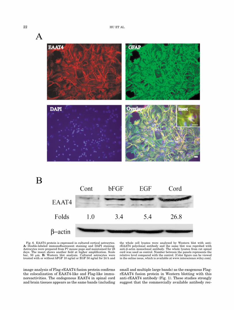
image analysis of Flag-rEAAT4 fusion protein confirmsthe colocalization of EAAT4-like and Flag-like immu-noreactivities. The endogenous EAAT4 in spinal cordand brain tissues appears as the same bands (including
small and multiple large bands) as the exogenous Flag-rEAAT4 fusion protein in Western blotting with thisanti-rEAAT4 antibody (Fig. 1). These studies stronglysuggest that the commercially available antibody rec-
Fig. 6. EAAT4 protein is expressed in cultured cortical astrocytes.A: Double-labeled immunofluorescent staining and DAPI staining.Astrocytes were prepared from P7 mouse pups and maintained for 28days. The insect shows another field at higher amplification. Scalebar, 50 �m. B: Western blot analysis. Cultured astrocytes weretreated with or without bFGF 10 ng/ml or EGF 50 ng/ml for 24 h and
the whole cell lysates were analyzed by Western blot with anti-rEAAT4 polyclonal antibody and the same blot was reprobed withanti-�-actin monoclonal antibody. The whole lysates from rat spinalcord was used as control. Number between the panels represents therelative level compared with the control. [Color figure can be viewedin the online issue, which is available at www.interscience.wiley.com].
22 HU ET AL.

ognizes EAAT4 expressed endogenously and exog-enously.
In the cerebellum and transfected cells, multiple im-munoreactive bands of higher molecular weight wereobserved for EAAT4, consistent with multiple bandsfor EAAT1 and EAAT2 (Haugeto et al., 1996; Dehnes etal., 1998; Danbolt, 2001). These higher bands may beattributed to oxidation of sulf-hydral groups and irre-versible crosslinking to form large molecular mass ag-gregates (Haugeto et al., 1996; Dehnes et al., 1998).However, these higher bands from both the cerebellumand transfected cells could not be separated in ourexperiments using various reducing protocols as previ-ously described (Dehnes et al., 1998; Massie et al.,2001). The chemical properties of the EAAT4 aggre-gates remain to be elucidated.
A smaller band in our Western blotting experimentswas observed for both endogenous and exogenous ex-pression of EAAT4 (Fig. 1). The molecular weight isapproximately � 60 kDa, the predicted size based onthe amino acid sequence (561 residues) of rEAAT4(Maeno-Hikichi et al., 1997; Lin et al., 1998), which isin agreement with several previous reports (Nagao etal., 1997; Schlag et al., 1998). However, other reportshave shown that EAAT4 has a molecular weight of
� 65–70 kDa (Furuta et al., 1997; Dehnes et al., 1998).In the present study, a difference in the mobility of thesmall band was also observed among tissues and trans-fected cells (Fig. 1C and D). It is also reported that asingle band in astrocyte-enriched cultures is 20 kDasmaller than that in cerebellar homogenates (Schlag etal., 1998). These differences, though elusive, might bedue to the variable protein amounts loaded and differ-ent crosslinking status (Haugeto et al., 1996; Schlag etal., 1998). Why the band at the appropriate molecularweight in the cerebellum is not increased in proportionto the EAAT4 multimer band compared with othertissues may be due to the irreversible crosslinking. Toconfirm the specificity of these small bands, peptidesequencing would be required.
To date, there are no detailed studies on the expres-sion of EAAT4 in the spinal cord (Furuta et al., 1997;Nagao et al., 1997). The present study provides the firstevidence that EAAT4 is expressed in spinal cord astro-cytes. The immunohistochemical studies detectEAAT4-like ir in the astrocytes of both white matterand gray matter in the spinal cord. The strongest as-trocytic immunostaining was localized around the rimof white matter, indicating that the white matter maybe more resistant to excitotoxic injury by removing
Fig. 7. EAAT4 mRNA expression in mouse astrocyte cultures and spinal cord tissues. The cDNA wasreverse-transcribed (RT) from equal amount of total RNA pretreated with DNase I for 2 h and amplifiedby PCR with mouse EAAT4 primers. No template represents negative control. The amplified fragmentswere confirmed by sequencing. Number between the panels represents the relative level of mRNAexpression.
23ASTROCYTES EXPRESSING EAAT4
glutamate through EAAT4. This may provide a possi-ble explanation for the peripheral rim of spared tissueand axons in the spinal cord after injury in animals andhuman (Beattie and Bresnahan 2000).
One previous report mentioned in the discussion thatEAAT4-like ir was found at low level in forebrain as-trocytes (Furuta et al., 1997). In the present study, wedemonstrate that EAAT4 ir is extensively expressed inastrocytes of forebrain and hindbrain, especially insubventricular zone and subpial region. The astrocyticexpression of EAAT4 protein is supported by previousobservations in rat astrocyte-enriched cortical cultures(Schlag et al., 1998) and the present study using mousecortical astrocytes. We also show that EAAT4 is ex-pressed at the mRNA level in mouse astrocytes. Al-though previous studies have shown that EAAT4mRNA is expressed in brain tissue (Bristol and Roth-stein, 1996; Yamada et al., 1997; Lin et al., 1998;Massie et al., 2001), the present study is the first dem-onstration that EAAT4 mRNA is expressed in spinalcord tissue. Interestingly, the distribution of EAAT4 irin adult forebrain reveals a noticeable similarity to theexpression of Lewis X, a marker for neural stem cells(Capela and Temple, 2002). The maximal expression ofEAAT4 ir in subventricular zone hints at its correlationto neural stem cells. The potential role of EAAT inneurogenesis deserves further investigation.
It is generally accepted that EAAT1 and EAAT2 areglial while EAAT3, EAAT4, and EAAT5 are neuronal(Rothstein et al., 1994; Anderson and Swanson, 2000;Maragakis and Rothstein, 2001). However, this inter-pretation has been challenged by recent studies. WhileEAAT2 is primarily an astrocytic transporter, a splicevariant of EAAT2 is preferentially expressed in neu-rons and nonastrocytic glial cells in the same cellularand subcellular distribution as EAAT3 (Schmitt et al.,2002). EAAT2 is the predominant nerve terminal glu-tamate transporter (Suchak et al., 2003). Despite theabundance of EAAT2 in the CNS, examples of its ex-pression in vitro as determined in primary astrocytes(Gegelashvili et al., 1997; Swanson et al., 1997), rat C6glioma cell line, and human U373 astrocytoma cell line(Dowd et al., 1996; Palos et al., 1996; Dunlop et al.,1999) cultures are limited. Protein expression ofEAAT2 has been described in cultures of primary hip-pocampal and cortical neurons (Mennerick et al., 1998;Wang et al., 1998) and the NT2 cell line (Dunlop et al.,1998). These cultures exhibit L-glutamate transportactivity sensitive to dihydrokainate, a selective EAAT2inhibitor (Dunlop et al., 1998; Mennerick et al., 1998;Wang et al., 1998). EAAT1 is also detectable in cul-tured hippocampal neurons (Perego et al., 2000; Pla-chez et al., 2000). In contrast, EAAT3 expression hasbeen shown in some astrocytes (Conti et al., 1998), ratC6 glioma cell line (Dowd et al., 1996; Palos et al.,1996), and human U373 astrocytoma cell line (Dunlopet al., 1999). EAAT4 has been found in cultured corticalastrocytes (Schlag et al., 1998). EAAT5 is expressed inboth neurons and glias (Arriza et al., 1997). In thepresent study, we demonstrate that EAAT4 is ex-
pressed in astrocytes of spinal cord and brain, whereasthe neuronal expression of EAAT4 is predominantly inthe cerebellum (Bristol and Rothstein, 1996; Yamadaet al., 1996, 1997; Dehnes et al., 1998; Lin et al., 1998)and, to a less extent, in cortical neurons (Massie et al.,2001). Taken together, the segregation of neuronal andglial EAATs is no longer tenable.
It is currently interpreted that EAAT3 may act pri-marily as a source of metabolic glutamate for neuronsand EAAT2 may mediate most of forebrain glutamatetransport, whereas EAAT1 may be important in thecerebellum during development. Since glutamate hasneurotrophic as well as neurotoxic activities, it is thusof primary importance to understand the role of thedifferent glutamate transporter subtypes in controllingthe extracellular glutamate concentration during de-velopment, neurotransmission, and neurotoxicity. As-trocytes play a key role in removing excess extracellu-lar glutamate. The regional distribution of EAAT4 inthe spinal cord is different from the previously reportedastrocytic transporter EAAT1 and EAAT2, which arepredominantly localized in the gray matter of spinalcord (Rothstein et al., 1995; Milton et al., 1997; Fray etal., 1998; Sasaki et al., 2001; Vera-Portocarrero et al.,2002). Therefore, EAAT4 may be an important regula-tor for the glutamate transmission and excitotoxicity inthe spinal cord, especially in the white matter.
ACKNOWLEDGMENTS
Supported by the National Institutes of HealthGrant NS37130 (to J.R.B.).
REFERENCES
Anderson CM, Swanson RA. 2000. Astrocyte glutamate transport:review of properties, regulation, and physiological functions. Glia32:1–14.
Arriza JL, Fairman WA, Wadiche JI, Murdoch GH, Kavanaugh MP,Amara SG. 1994. Functional comparisons of three glutamate trans-porter subtypes cloned from human motor cortex. J Neurosci 14:5559–5569.
Arriza JL, Eliasof S, Kavanaugh MP, Amara SG. 1997. Excitatoryamino acid transporter 5, a retinal glutamate transporter coupledto a chloride conductance. Proc Natl Acad Sci USA 94:4155–4160.
Beattie MS, Bresnahan JC. 2000. Cell death, repair, and recovery offunction after spinal cord contusion injuries in rats. In: Kalb RG,Strittmatter SM, editors. Neurobiology of spinal cord injury. NewJersey: Human Press. p 1–21.
Bristol LA, Rothstein JD. 1996. Glutamate transporter gene expres-sion in amyotrophic lateral sclerosis motor cortex. Ann Neurol39:676–679.
Capela A, Temple S. 2002. LeX/ssea-1 is expressed by adult mouseCNS stem cells, identifying them as nonependymal. Neuron 35:865–875.
Conti F, DeBiasi S, Minelli A, Rothstein JD, Melone M. 1998. EAAC1,a high-affinity glutamate tranporter, is localized to astrocytes andgabaergic neurons besides pyramidal cells in the rat cerebral cortex.Cereb Cortex 8:108–116.
Danbolt NC. 2001. Glutamate uptake. Prog Neurobiol 65:1–105.Dehnes Y, Chaudhry FA, Ullensvang K, Lehre KP, Storm-Mathisen J,
Danbolt NC. 1998. The glutamate transporter EAAT4 in rat cere-bellar Purkinje cells: a glutamate-gated chloride channel concen-trated near the synapse in parts of the dendritic membrane facingastroglia. J Neurosci 18:3606–3619.
24 HU ET AL.

Dowd LA, Coyle AJ, Rothstein JD, Pritchett DB, Robinson MB. 1996.Comparison of Na�-dependent glutamate transport activity in syn-aptosomes, C6 glioma, and Xenopus oocytes expressing excitatoryamino acid carrier 1 (EAAC1). Mol Pharmacol 49:465–473.
Dunlop J, Beal McIlvain H, Lou Z, Franco R. 1998. The pharmacolog-ical profile of L-glutamate transport in human NT2 neurones isconsistent with excitatory amino acid transporter 2. Eur J Pharma-col 360:249–256.
Dunlop J, Lou Z, McIlvain HB. 1999. Properties of excitatory aminoacid transport in the human U373 astrocytoma cell line. Brain Res839:235–242.
Fairman WA, Vandenberg RJ, Arriza JL, Kavanaugh MP, Amara SG.1995. An excitatory amino-acid transporter with properties of aligand-gated chloride channel. Nature 375:599–603.
Fray AE, Ince PG, Banner SJ, Milton ID, Usher PA, Cookson MR,Shaw PJ. 1998. The expression of the glial glutamate transporterprotein EAAT2 in motor neuron disease: an immunohistochemicalstudy. Eur J Neurosci 10:2481–2489.
Furuta A, Rothstein JD, Martin LJ. 1997. Glutamate transporterprotein subtypes are expressed differentially during rat CNS devel-opment. J Neurosci 17:8363–8375.
Gegelashvili G, Danbolt NC, Schousboe A. 1997. Neuronal solublefactors differentially regulate the expression of the GLT1 andGLAST glutamate transporters in cultured astroglia. J Neurochem69:2612–2615.
Haugeto O, Ullensvang K, Levy LM, Chaudhry FA, Honore T, NielsenM, Lehre KP, Danbolt NC. 1996. Brain glutamate transporter pro-teins form homomultimers. J Biol Chem 271:27715–27722.
Hausmann ON, Hu WH, Keren-Raifman T, Witherow DS, Wang Q,Levay K, Frydel B, Slepak VZ, R Bethea JR. 2002. Spinal cordinjury induces expression of RGS7 in microglia/macrophages inrats. Eur J Neurosci 15:602–612.
Hu WH, Li F, Qiang WA, Liu N, Wang GQ, Xiao J, Liu JS, Liao WH,Jen MF. 1999. Dual role for nitric oxide in dynorphin spinal neu-rotoxicity. J Neurotrauma 16:85–98.
Itoh M, Watanabe Y, Watanabe M, Tanaka K, Wada K, Takashima S.1997. Expression of a glutamate transporter subtype, EAAT4, inthe developing human cerebellum. Brain Res 767:265–271.
Kanai Y, Hediger MA. 1992. Primary structure and functional char-acterization of a high-affinity glutamate transporter. Nature 360:467–471.
Kanai Y. 1997. Family of neutral and acidic amino acid transporters:molecular biology, physiology and medical implications. Curr OpinCell Biol 9:565–572.
Kawakami H, Tanaka K, Nakayama T, Inoue K, Nakamura S. 1994.Cloning and expression of a human glutamate transporter. BiochemBiophys Res Commun 199:171–176.
Lehre KP, Levy LM, Ottersen OP, Storm-Mathisen J, Danbolt NC.1995. Differential expression of two glial glutamate transporters inthe rat brain: quantitative and immunocytochemical observations.J Neurosci 15:1835–1853.
Lievens JC, Bernal F, Forni C, Mahy N, Kerkerian-Le Goff L. 2000.Characterization of striatal lesions produced by glutamate uptakealteration: cell death, reactive gliosis, and changes in GLT1 andGADD45 mRNA expression. Glia 29:222–232.
Lin CL, Tzingounis AV, Jin L, Furuta A, Kavanaugh MP, RothsteinJD. 1998. Molecular cloning and expression of the rat EAAT4 glu-tamate transporter subtype. Brain Res Mol Brain Res 63:174–179.
Maeno-Hikichi Y, Tanaka K, Shibata T, Watanabe M, Inoue Y, Mu-kainaka Y, Wada K. 1997. Structure and functional expression ofthe cloned mouse neuronal high-affinity glutamate transporter.Brain Res Mol Brain Res 48:176–180.
Maragakis NJ, Rothstein JD. 2001. Glutamate transporters in neu-rologic disease. Arch Neurol 58:365–370.
Massie A, Vandesande F, Arckens L. 2001. Expression of the high-affinity glutamate transporter EAAT4 in mammalian cerebral cor-tex. Neuroreport 12:393–397.
Meldrum BS, Akbar MT, Chapman AG. 1999. Glutamate receptorsand transporters in genetic and acquired models of epilepsy. Epi-lepsy Res 36:189–204.
Mennerick S, Dhond RP, Benz A, Xu W, Rothstein JD, Danbolt NC,Isenberg KE, Zorumski CF. 1998. Neuronal expression of the glu-tamate transporter GLT-1 in hippocampal microcultures. J Neuro-sci 18:4490–4499.
Milton ID, Banner SJ, Ince PG, Piggott NH, Fray AE, Thatcher N,Horne CH, Shaw PJ. 1997. Expression of the glial glutamate trans-porter EAAT2 in the human CNS: an immunohistochemical study.Brain Res Mol Brain Res 52:17–31.
Nagao S, Kwak S, Kanazawa I. 1997. EAAT4, a glutamate trans-porter with properties of a chloride channel, is predominantly lo-calized in Purkinje cell dendrites, and forms parasagittal compart-ments in rat cerebellum. Neuroscience 78:929–933.
O’Shea RD. 2002. Roles and regulation of glutamate transporters in thecentral nervous system. Clin Exp Pharmacol Physiol 29:1018–1023.
Palos TP, Ramachandran B, Boado R, Howard BD. 1996. Rat C6 andhuman astrocytic tumor cells express a neuronal type of glutamatetransporter. Brain Res Mol Brain Res 37:297–303.
Perego C, Vanoni C, Bossi M, Massari S, Basudev H, Longhi R,Pietrini G. 2000. The GLT-1 and GLAST glutamate transportersare expressed on morphologically distinct astrocytes and regulatedby neuronal activity in primary hippocampal cocultures. J Neuro-chem 75:1076–1084.
Plachez C, Danbolt NC, Recasens M. 2000. Transient expression ofthe glial glutamate transporters GLAST and GLT in hippocampalneurons in primary culture. J Neurosci Res 59:587–593.
Proper EA, Hoogland G, Kappen SM, Jansen GH, Rensen MG,Schrama LH, van Veelen CW, van Rijen PC, van Nieuwenhuizen O,Gispen WH, de Graan PN. 2002. Distribution of glutamate trans-porters in the hippocampus of patients with pharmaco-resistanttemporal lobe epilepsy. Brain 125:32–43.
Robinson MB, Dowd LA. 1997. Heterogeneity and functional proper-ties of subtypes of sodium-dependent glutamate transporters in themammalian central nervous system. Adv Pharmacol 37:69–115.
Rothstein JD, Martin L, Levey AI, Dykes-Hoberg M, Jin L, Wu D,Nash N, Kuncl RW. 1994. Localization of neuronal and glial gluta-mate transporters. Neuron 13:713–725.
Rothstein JD, Van Kammen M, Levey AI, Martin LJ, Kuncl RW.1995. Selective loss of glial glutamate transporter GLT-1 in amyo-trophic lateral sclerosis. Ann Neurol 38:73–84.
Sasaki S, Warita H, Abe K, Komori T, Iwata M. 2001. EAAT1 andEAAT2 immunoreactivity in transgenic mice with a G93A mutantSOD1 gene. Neuroreport 12:1359–1362.
Schlag BD, Vondrasek JR, Munir M, Kalandadze A, Zelenaia OA,Rothstein JD, Robinson MB. 1998. Regulation of the glial Na�-dependent glutamate transporters by cyclic AMP analogs and neu-rons. Mol Pharmacol 53:355–369.
Schluter K, Figiel M, Rozyczka J, Engele J. 2002. CNS region-specificregulation of glial glutamate transporter expression. Eur J Neuro-sci 16:836–842.
Schmitt A, Asan E, Lesch KP, Kugler P. 2002. A splice variant ofglutamate transporter GLT1/EAAT2 expressed in neurons: cloningand localization in rat nervous system. Neuroscience 109:45–61.
Shashidharan P, Wittenberg I, Plaitakis A. 1994. Molecular cloning ofhuman brain glutamate/aspartate transporter II. Biochim BiophysActa 1191:393–396.
Suchak SK, Baloyianni NV, Perkinton MS, Williams RJ, MeldrumBS, Rattray M. 2003. The “glial” glutamate transporter, EAAT2(Glt-1) accounts for high affinity glutamate uptake into adult rodentnerve endings. J Neurochem 84:522–532.
Suzuki K, Ikegaya Y, Matsuura S, Kanai Y, Endou H, Matsuki N.2001. Transient upregulation of the glial glutamate transporterGLAST in response to fibroblast growth factor, insulin-like growthfactor and epidermal growth factor in cultured astrocytes. J Cell Sci114:3717–3725.
Swanson RA, Liu J, Miller JW, Rothstein JD, Farrell K, Stein BA,Longuemare MC. 1997. Neuronal regulation of glutamate trans-porter subtype expression in astrocytes. J Neurosci 17:932–940.
Trotti D, Aoki M, Pasinelli P, Berger UV, Danbolt NC, Brown RH Jr,Hediger MA. 2001. Amyotrophic lateral sclerosis-linked glutamatetransporter mutant has impaired glutamate clearance capacity.J Biol Chem 276:576–582.
Vera-Portocarrero LP, Mills CD, Ye Z, Fullwood SD, McAdoo DJ,Hulsebosch CE, Westlund KN. 2002. Rapid changes in expression ofglutamate transporters after spinal cord injury. Brain Res 927:104–110.
Wahle S, Stoffel W. 1996. Membrane topology of the high-affinityL-glutamate transporter (GLAST-1) of the central nervous system.J Cell Biol 135:1867–1877.
Wang GJ, Chung HJ, Schnuer J, Pratt K, Zable AC, Kavanaugh MP,Rosenberg PA. 1998. High affinity glutamate transport in rat cor-tical neurons in culture. Mol Pharmacol 53:88–96.
Yamada K, Watanabe M, Shibata T, Tanaka K, Wada K, Inoue Y.1996. EAAT4 is a post-synaptic glutamate transporter at Purkinjecell synapses. Neuroreport 7:2013–2017.
Yamada K, Wada S, Watanabe M, Tanaka K, Wada K, Inoue Y. 1997.Changes in expression and distribution of the glutamate trans-porter EAAT4 in developing mouse Purkinje cells. Neurosci Res27:191–198.
Zelenaia O, Schlag BD, Gochenauer GE, Ganel R, Song W, Beesley JS,Grinspan JB, Rothstein JD, Robinson MB. 2000. Epidermal growthfactor receptor agonists increase expression of glutamate trans-porter GLT-1 in astrocytes through pathways dependent on phos-phatidylinositol 3-kinase and transcription factor NF-kappaB. MolPharmacol 57:667–678.
25ASTROCYTES EXPRESSING EAAT4

![Mitochondria and neuronal glutamate excitotoxicity · 2017-01-03 · At the termination of a transient glutamate expo-sure, [Ca2⁄] c tends to return to baseline as the cation is](https://static.fdocuments.net/doc/165x107/5f4b42c2f42e81321574d49c/mitochondria-and-neuronal-glutamate-excitotoxicity-2017-01-03-at-the-termination.jpg)