Glutamate Receptor Ion Channels: Structure, Regulation ... · pharmacology of glutamate receptors,...
92
ASSOCIATE EDITOR: DAVID SIBLEY Glutamate Receptor Ion Channels: Structure, Regulation, and Function Stephen F. Traynelis, Lonnie P. Wollmuth, Chris J. McBain, Frank S. Menniti, 1 Katie M. Vance, Kevin K. Ogden, Kasper B. Hansen, Hongjie Yuan, Scott J. Myers, and Ray Dingledine Department of Pharmacology, Emory University School of Medicine, Atlanta, Georgia (S.F.T., K.M.V., K.K.O., K.B.H., H.Y., S.J.M., R.D.); Department of Neurobiology and Behavior & Center for Nervous System Disorders, Stony Brook University, Stony Brook, New York (L.P.W.); Eunice Kennedy Shriver National Institute of Child Health and Human Development, Bethesda, Maryland (C.J.M.); and cyclicM LLC, Mystic, Connecticut (F.S.M.) Abstract ................................................................................ 406 I. Introduction and nomenclature ........................................................... 407 II. Structure ............................................................................... 407 A. Subunit organization and quaternary structure ......................................... 407 B. Subunit stoichiometry ................................................................ 409 C. Receptor assembly and trafficking ..................................................... 411 D. The extracellular ligand binding domain ................................................ 412 E. The extracellular amino-terminal domain ............................................... 414 F. The transmembrane domain ........................................................... 415 G. The intracellular carboxyl-terminal domain and protein binding partners .................. 415 H. Transmembrane -amino-3-hydroxy-5-methyl-4-isoxazolepropionic acid receptor regulatory proteins and other auxiliary subunits ........................................ 418 III. Regulation of transcription and translation................................................. 419 A. -Amino-3-hydroxy-5-methyl-4-isoxazolepropionic acid receptors .......................... 419 1. Gria1............................................................................. 419 2. Gria2............................................................................. 420 3. Gria3 and Gria4................................................................... 421 B. Kainate receptors Grik1 to Grik5 ...................................................... 421 C. N-Methyl-D-aspartate receptors ........................................................ 421 1. Grin1 ............................................................................ 421 2. Grin2a ........................................................................... 422 3. Grin2b ........................................................................... 422 4. Grin2c, Grin2d, Grin3a, and Grin3b ................................................. 423 D. Translational control of glutamate receptors ............................................ 424 IV. Post-translational regulation.............................................................. 425 A. -Amino-3-hydroxy-5-methyl-4-isoxazolepropionic acid and kainate receptor phosphorylation ...................................................................... 425 B. N-Methyl-D-aspartate and receptor phosphorylation .................................... 427 C. Other post-translational modifications of glutamate receptors............................. 429 D. Proteolysis of glutamate receptors...................................................... 430 V. Agonist and antagonist pharmacology ..................................................... 431 A. -Amino-3-hydroxy-5-methyl-4-isoxazolepropionic acid, kainate, and receptor agonists ..... 431 B. N-Methyl-D-aspartate receptor agonists ................................................. 434 C. -Amino-3-hydroxy-5-methyl-4-isoxazolepropionic acid and kainate receptor competitive antagonists .......................................................................... 437 D. N-Methyl-D-aspartate receptor competitive antagonists .................................. 439 Address correspondence to: Dr. Stephen Traynelis, Dept Pharmacology, Emory University School of Medicine, Rollins Research Center, 1510 Clifton Road, Atlanta GA 30322-3090. E-mail: [email protected] 1 Current affiliation: Mnemosyne Pharmaceuticals, Inc., Providence, Rhode Island. This article is available online at http://pharmrev.aspetjournals.org. doi:10.1124/pr.109.002451. 0031-6997/10/6203-405– 496$20.00 PHARMACOLOGICAL REVIEWS Vol. 62, No. 3 U.S. Government work not protected by U.S. copyright 2451/3592788 Pharmacol Rev 62:405– 496, 2010 Printed in U.S.A. 405
Transcript of Glutamate Receptor Ion Channels: Structure, Regulation ... · pharmacology of glutamate receptors,...

ASSOCIATE EDITOR: DAVID SIBLEY
Glutamate Receptor Ion Channels:Structure, Regulation, and Function
Stephen F. Traynelis, Lonnie P. Wollmuth, Chris J. McBain, Frank S. Menniti,1 Katie M. Vance, Kevin K. Ogden, Kasper B. Hansen,Hongjie Yuan, Scott J. Myers, and Ray Dingledine
Department of Pharmacology, Emory University School of Medicine, Atlanta, Georgia (S.F.T., K.M.V., K.K.O., K.B.H., H.Y., S.J.M., R.D.);Department of Neurobiology and Behavior & Center for Nervous System Disorders, Stony Brook University, Stony Brook, New York(L.P.W.); Eunice Kennedy Shriver National Institute of Child Health and Human Development, Bethesda, Maryland (C.J.M.); and
cyclicM LLC, Mystic, Connecticut (F.S.M.)
Abstract . . . . . . . . . . . . . . . . . . . . . . . . . . . . . . . . . . . . . . . . . . . . . . . . . . . . . . . . . . . . . . . . . . . . . . . . . . . . . . . . 406I. Introduction and nomenclature . . . . . . . . . . . . . . . . . . . . . . . . . . . . . . . . . . . . . . . . . . . . . . . . . . . . . . . . . . . 407
II. Structure . . . . . . . . . . . . . . . . . . . . . . . . . . . . . . . . . . . . . . . . . . . . . . . . . . . . . . . . . . . . . . . . . . . . . . . . . . . . . . . 407A. Subunit organization and quaternary structure . . . . . . . . . . . . . . . . . . . . . . . . . . . . . . . . . . . . . . . . . 407B. Subunit stoichiometry . . . . . . . . . . . . . . . . . . . . . . . . . . . . . . . . . . . . . . . . . . . . . . . . . . . . . . . . . . . . . . . . 409C. Receptor assembly and trafficking . . . . . . . . . . . . . . . . . . . . . . . . . . . . . . . . . . . . . . . . . . . . . . . . . . . . . 411D. The extracellular ligand binding domain. . . . . . . . . . . . . . . . . . . . . . . . . . . . . . . . . . . . . . . . . . . . . . . . 412E. The extracellular amino-terminal domain . . . . . . . . . . . . . . . . . . . . . . . . . . . . . . . . . . . . . . . . . . . . . . . 414F. The transmembrane domain. . . . . . . . . . . . . . . . . . . . . . . . . . . . . . . . . . . . . . . . . . . . . . . . . . . . . . . . . . . 415G. The intracellular carboxyl-terminal domain and protein binding partners . . . . . . . . . . . . . . . . . . 415H. Transmembrane �-amino-3-hydroxy-5-methyl-4-isoxazolepropionic acid receptor
regulatory proteins and other auxiliary subunits . . . . . . . . . . . . . . . . . . . . . . . . . . . . . . . . . . . . . . . . 418III. Regulation of transcription and translation. . . . . . . . . . . . . . . . . . . . . . . . . . . . . . . . . . . . . . . . . . . . . . . . . 419
A. �-Amino-3-hydroxy-5-methyl-4-isoxazolepropionic acid receptors . . . . . . . . . . . . . . . . . . . . . . . . . . 4191. Gria1. . . . . . . . . . . . . . . . . . . . . . . . . . . . . . . . . . . . . . . . . . . . . . . . . . . . . . . . . . . . . . . . . . . . . . . . . . . . . 4192. Gria2. . . . . . . . . . . . . . . . . . . . . . . . . . . . . . . . . . . . . . . . . . . . . . . . . . . . . . . . . . . . . . . . . . . . . . . . . . . . . 4203. Gria3 and Gria4. . . . . . . . . . . . . . . . . . . . . . . . . . . . . . . . . . . . . . . . . . . . . . . . . . . . . . . . . . . . . . . . . . . 421
B. Kainate receptors Grik1 to Grik5 . . . . . . . . . . . . . . . . . . . . . . . . . . . . . . . . . . . . . . . . . . . . . . . . . . . . . . 421C. N-Methyl-D-aspartate receptors . . . . . . . . . . . . . . . . . . . . . . . . . . . . . . . . . . . . . . . . . . . . . . . . . . . . . . . . 421
1. Grin1 . . . . . . . . . . . . . . . . . . . . . . . . . . . . . . . . . . . . . . . . . . . . . . . . . . . . . . . . . . . . . . . . . . . . . . . . . . . . 4212. Grin2a . . . . . . . . . . . . . . . . . . . . . . . . . . . . . . . . . . . . . . . . . . . . . . . . . . . . . . . . . . . . . . . . . . . . . . . . . . . 4223. Grin2b . . . . . . . . . . . . . . . . . . . . . . . . . . . . . . . . . . . . . . . . . . . . . . . . . . . . . . . . . . . . . . . . . . . . . . . . . . . 4224. Grin2c, Grin2d, Grin3a, and Grin3b . . . . . . . . . . . . . . . . . . . . . . . . . . . . . . . . . . . . . . . . . . . . . . . . . 423
D. Translational control of glutamate receptors . . . . . . . . . . . . . . . . . . . . . . . . . . . . . . . . . . . . . . . . . . . . 424IV. Post-translational regulation. . . . . . . . . . . . . . . . . . . . . . . . . . . . . . . . . . . . . . . . . . . . . . . . . . . . . . . . . . . . . . 425
A. �-Amino-3-hydroxy-5-methyl-4-isoxazolepropionic acid and kainate receptorphosphorylation . . . . . . . . . . . . . . . . . . . . . . . . . . . . . . . . . . . . . . . . . . . . . . . . . . . . . . . . . . . . . . . . . . . . . . 425
B. N-Methyl-D-aspartate and � receptor phosphorylation . . . . . . . . . . . . . . . . . . . . . . . . . . . . . . . . . . . . 427C. Other post-translational modifications of glutamate receptors. . . . . . . . . . . . . . . . . . . . . . . . . . . . . 429D. Proteolysis of glutamate receptors. . . . . . . . . . . . . . . . . . . . . . . . . . . . . . . . . . . . . . . . . . . . . . . . . . . . . . 430
V. Agonist and antagonist pharmacology . . . . . . . . . . . . . . . . . . . . . . . . . . . . . . . . . . . . . . . . . . . . . . . . . . . . . 431A. �-Amino-3-hydroxy-5-methyl-4-isoxazolepropionic acid, kainate, and � receptor agonists . . . . . 431B. N-Methyl-D-aspartate receptor agonists. . . . . . . . . . . . . . . . . . . . . . . . . . . . . . . . . . . . . . . . . . . . . . . . . 434C. �-Amino-3-hydroxy-5-methyl-4-isoxazolepropionic acid and kainate receptor competitive
antagonists . . . . . . . . . . . . . . . . . . . . . . . . . . . . . . . . . . . . . . . . . . . . . . . . . . . . . . . . . . . . . . . . . . . . . . . . . . 437D. N-Methyl-D-aspartate receptor competitive antagonists . . . . . . . . . . . . . . . . . . . . . . . . . . . . . . . . . . 439
Address correspondence to: Dr. Stephen Traynelis, Dept Pharmacology, Emory University School of Medicine, Rollins Research Center,1510 Clifton Road, Atlanta GA 30322-3090. E-mail: [email protected]
1Current affiliation: Mnemosyne Pharmaceuticals, Inc., Providence, Rhode Island.This article is available online at http://pharmrev.aspetjournals.org.doi:10.1124/pr.109.002451.
0031-6997/10/6203-405–496$20.00PHARMACOLOGICAL REVIEWS Vol. 62, No. 3U.S. Government work not protected by U.S. copyright 2451/3592788Pharmacol Rev 62:405–496, 2010 Printed in U.S.A.
405
E. Noncompetitive antagonists . . . . . . . . . . . . . . . . . . . . . . . . . . . . . . . . . . . . . . . . . . . . . . . . . . . . . . . . . . . 440F. Uncompetitive antagonists . . . . . . . . . . . . . . . . . . . . . . . . . . . . . . . . . . . . . . . . . . . . . . . . . . . . . . . . . . . . 443
VI. Allosteric regulation . . . . . . . . . . . . . . . . . . . . . . . . . . . . . . . . . . . . . . . . . . . . . . . . . . . . . . . . . . . . . . . . . . . . . 445A. Positive and negative allosteric modulators . . . . . . . . . . . . . . . . . . . . . . . . . . . . . . . . . . . . . . . . . . . . . 445B. Divalent ions . . . . . . . . . . . . . . . . . . . . . . . . . . . . . . . . . . . . . . . . . . . . . . . . . . . . . . . . . . . . . . . . . . . . . . . . 447C. Monovalent ions. . . . . . . . . . . . . . . . . . . . . . . . . . . . . . . . . . . . . . . . . . . . . . . . . . . . . . . . . . . . . . . . . . . . . . 448D. Protons. . . . . . . . . . . . . . . . . . . . . . . . . . . . . . . . . . . . . . . . . . . . . . . . . . . . . . . . . . . . . . . . . . . . . . . . . . . . . . 449E. Polyamines . . . . . . . . . . . . . . . . . . . . . . . . . . . . . . . . . . . . . . . . . . . . . . . . . . . . . . . . . . . . . . . . . . . . . . . . . . 449F. Neurosteroids . . . . . . . . . . . . . . . . . . . . . . . . . . . . . . . . . . . . . . . . . . . . . . . . . . . . . . . . . . . . . . . . . . . . . . . . 450G. Fatty acids . . . . . . . . . . . . . . . . . . . . . . . . . . . . . . . . . . . . . . . . . . . . . . . . . . . . . . . . . . . . . . . . . . . . . . . . . . 450H. Other allosteric modulators . . . . . . . . . . . . . . . . . . . . . . . . . . . . . . . . . . . . . . . . . . . . . . . . . . . . . . . . . . . 450
VII. Molecular determinants of gating . . . . . . . . . . . . . . . . . . . . . . . . . . . . . . . . . . . . . . . . . . . . . . . . . . . . . . . . . 450A. Time course of glutamate receptor activation and deactivation . . . . . . . . . . . . . . . . . . . . . . . . . . . . 450B. Mechanisms linking agonist binding to channel gating . . . . . . . . . . . . . . . . . . . . . . . . . . . . . . . . . . . 451C. Molecular determinants and mechanisms of partial agonism. . . . . . . . . . . . . . . . . . . . . . . . . . . . . . 453D. Molecular determinants and mechanisms of gating . . . . . . . . . . . . . . . . . . . . . . . . . . . . . . . . . . . . . . 455E. Molecular determinants of desensitization . . . . . . . . . . . . . . . . . . . . . . . . . . . . . . . . . . . . . . . . . . . . . . 457
VIII. Molecular determinants of ion permeation and block . . . . . . . . . . . . . . . . . . . . . . . . . . . . . . . . . . . . . . . . 458A. Nature of the ion permeation pathway . . . . . . . . . . . . . . . . . . . . . . . . . . . . . . . . . . . . . . . . . . . . . . . . . 458B. Mechanisms of ion permeation. . . . . . . . . . . . . . . . . . . . . . . . . . . . . . . . . . . . . . . . . . . . . . . . . . . . . . . . . 460C. Voltage-dependent channel block by endogenous ions . . . . . . . . . . . . . . . . . . . . . . . . . . . . . . . . . . . . 462
IX. Role in synaptic function and plasticity . . . . . . . . . . . . . . . . . . . . . . . . . . . . . . . . . . . . . . . . . . . . . . . . . . . . 463A. Synaptic �-amino-3-hydroxy-5-methyl-4-isoxazolepropionic acid receptors . . . . . . . . . . . . . . . . . . 463B. Synaptic kainate receptors . . . . . . . . . . . . . . . . . . . . . . . . . . . . . . . . . . . . . . . . . . . . . . . . . . . . . . . . . . . . 464C. Synaptic and extrasynaptic N-Methyl-D-aspartate receptors . . . . . . . . . . . . . . . . . . . . . . . . . . . . . . 465D. Synaptic plasticity. . . . . . . . . . . . . . . . . . . . . . . . . . . . . . . . . . . . . . . . . . . . . . . . . . . . . . . . . . . . . . . . . . . . 466
1. N-Methyl-D-aspartate receptors . . . . . . . . . . . . . . . . . . . . . . . . . . . . . . . . . . . . . . . . . . . . . . . . . . . . . 4662. �-Amino-3-hydroxy-5-methyl-4-isoxazolepropionic acid receptors . . . . . . . . . . . . . . . . . . . . . . . 467
X. Therapeutic potential . . . . . . . . . . . . . . . . . . . . . . . . . . . . . . . . . . . . . . . . . . . . . . . . . . . . . . . . . . . . . . . . . . . . 467A. Glutamate receptor antagonists and the prevention of acute neuronal death . . . . . . . . . . . . . . . 467B. The next generation �-amino-3-hydroxy-5-methyl-4-isoxazolepropionic acid and kainate
receptor antagonists . . . . . . . . . . . . . . . . . . . . . . . . . . . . . . . . . . . . . . . . . . . . . . . . . . . . . . . . . . . . . . . . . . 469C. The next generation N-methyl-D-aspartate receptor antagonists. . . . . . . . . . . . . . . . . . . . . . . . . . . 469D. N-Methyl-D-aspartate antagonists. . . . . . . . . . . . . . . . . . . . . . . . . . . . . . . . . . . . . . . . . . . . . . . . . . . . . . 470
1. Neuropathic pain . . . . . . . . . . . . . . . . . . . . . . . . . . . . . . . . . . . . . . . . . . . . . . . . . . . . . . . . . . . . . . . . . . 4702. Major depression . . . . . . . . . . . . . . . . . . . . . . . . . . . . . . . . . . . . . . . . . . . . . . . . . . . . . . . . . . . . . . . . . . 4713. Parkinson’s disease . . . . . . . . . . . . . . . . . . . . . . . . . . . . . . . . . . . . . . . . . . . . . . . . . . . . . . . . . . . . . . . . 471
E. �-Amino-3-hydroxy-5-methyl-4-isoxazolepropionic acid and kainate receptor potentiation . . . . 471F. N-Methyl-D-aspartate receptor potentiation . . . . . . . . . . . . . . . . . . . . . . . . . . . . . . . . . . . . . . . . . . . . . 472
XI. Conclusions . . . . . . . . . . . . . . . . . . . . . . . . . . . . . . . . . . . . . . . . . . . . . . . . . . . . . . . . . . . . . . . . . . . . . . . . . . . . . 473Acknowledgments . . . . . . . . . . . . . . . . . . . . . . . . . . . . . . . . . . . . . . . . . . . . . . . . . . . . . . . . . . . . . . . . . . . . . . . 474References . . . . . . . . . . . . . . . . . . . . . . . . . . . . . . . . . . . . . . . . . . . . . . . . . . . . . . . . . . . . . . . . . . . . . . . . . . . . . . 474
Abstract——The mammalian ionotropic glutamate re-ceptor family encodes 18 gene products that coassemble toform ligand-gated ion channels containing an agonist rec-ognition site, a transmembrane ion permeation pathway,and gating elements that couple agonist-induced confor-mational changes to the opening or closing of the perme-ation pore. Glutamate receptors mediate fast excitatorysynaptic transmission in the central nervous system andare localized on neuronal and non-neuronal cells. Thesereceptors regulate a broad spectrum of processes in thebrain, spinal cord, retina, and peripheral nervous system.Glutamate receptors are postulated to play importantroles in numerous neurological diseases and have at-tracted intense scrutiny. The description of glutamate re-
ceptor structure, including its transmembrane elements,reveals a complex assembly of multiple semiautonomousextracellular domains linked to a pore-forming elementwith striking resemblance to an inverted potassium chan-nel. In this review we discuss International Union of Basicand Clinical Pharmacology glutamate receptor nomencla-ture, structure, assembly, accessory subunits, interactingproteins, gene expression and translation, post-transla-tional modifications, agonist and antagonist pharmacol-ogy, allosteric modulation, mechanisms of gating and per-meation, roles in normal physiological function, as well asthe potential therapeutic use of pharmacological agentsacting at glutamate receptors.
406 TRAYNELIS ET AL.

I. Introduction and Nomenclature
The past decade has revealed both breathtaking ad-vances in our understanding of structure and functionand a growing sophistication at virtually all levels ofexperimental design. The structure of a membrane-spanning tetrameric glutamate receptor has been de-scribed, revealing unprecedented features of channelstructure together with long-awaited details on thepore-forming elements and the channel gate, subunitarrangement, and the nature of linkers connectingmultiple semiautonomous domains that comprise theextracellular portion of the receptor. These compellingdata have set the stage for a predictably explosiveincrease in work on all aspects of function and holdthe promise of catalyzing timely breakthroughs intherapeutic strategies.
Assembling this review was an exciting yet dauntingtask. A staggering volume of literature has been pub-lished over the last 11 years, the period this review mostseeks to summarize. We have focused primarily on thepharmacology of glutamate receptors, the structural ba-sis of receptor function as it relates to neuronal functionand neurological disease, and the regulation of receptorfunction by phosphorylation. We only touch upon theanatomical distribution of glutamate receptors, theirrole in behavior and cognition, their role in nervoussystem development, and the means by which the myr-iad of proteins that bind to glutamate receptors regulatereceptor trafficking. We focus on mammalian receptors,with an emphasis on their relation to potential therapiesnow under development. In selecting the necessarilylimited number of references used to illustrate ad-vances, we have sought to recognize principle, prece-dent, perspective, and (importantly) to acknowledge thefull spectrum of talented individuals and productive lab-oratories engaged in this field. We regret that space doesnot allow a complete listing of relevant work related toeach point made; many fine articles simply could not becited.
After the first report appeared in December 1989 ofthe cloning of a glutamate receptor subunit (Hollmannet al., 1989), the early 1990s witnessed a flurry ofactivity, resulting in reports of more than a dozenglutamate receptor clones in various species withinthe subsequent 6 months. As might be expected, thenomenclature was uncoordinated, with species-or lab-oratory-specific names for the same transcript beingpromoted in the literature. This situation has resolvedslowly. An excellent history of glutamate receptorcloning and nomenclature has appeared (Lodge, 2009).Glutamate receptor nomenclature has recently undergonea needed and systematic revision, the International Unionof Basic and Clinical Pharmacology name replacing thecommon names (Collingridge et al., 2009) (see http://www.iuphar-db.org/LGICNomenclature.jsp). Table 1 summa-
rizes the nomenclature used throughout this review forboth genes and gene products.
II. Structure
A. Subunit Organization and Quaternary Structure
Ionotropic glutamate receptors are integral mem-brane proteins composed of four large subunits (�900residues) that form a central ion channel pore. Sequencesimilarity among all known glutamate receptor sub-units, including the AMPA,1 kainate, NMDA, and � re-
1Abbreviations: 5,7-DCKA, 5,7-dichlorokynurenic acid; AMPA,�-amino-3-hydroxy-5-methyl-4-isoxazolepropionic acid; AP1, activatorprotein-1; ATD, amino-terminal domain; ATPA, 2-amino-3-(5-tert-bu-tyl-3-hydroxyisoxazol-4-yl)propionic acid; ATPO, (R,S)-2-amino-3-[5-tert-butyl-3-(phosphonomethoxy)-4-isoxazolyl] propionic acid; BDNF,brain-derived neurotrophic factor; bp, base pair(s); CA1, cornu ammonis1; CamKII, Ca2�/calmodulin-dependent protein kinase II; CASK, calcium/calmodulin-dependent serine protein kinase; ChIP, chromatin im-munoprecipitation; CI-1041, besonprodil; CNIH, protein cornichon ho-molog; CNQX, 6-cyano-2,3-dihydroxy-7-nitroquinoxaline; CNS, centralnervous system; CNS 5161, N-(2-chloro-5-(methylmercapto)phenyl)-N�-methylguanidine monohydrochloride; con A, concanavalin A; COUP-TF, chicken ovalbumin upstream promoter transcription factor; COX,cytochrome oxidase; CP-101,606, traxoprodil mesylate; CP-465,022,(S)-3-(2-Chlorophenyl)-2-[2-(6-diethylaminomethyl-pyridin-2-yl)-vinyl]-6-fluoro-3H-quinazolin-4-one; CPEB, cytoplasmic polyadenylation ele-ment binding protein; CRE, cAMP response element; CREST, calcium-responsive transactivator; CTD, carboxyl-terminal domain; CX516,ampalex; CX546, 1-(1,4-benzodioxan-6-ylcarbonyl)piperidine; CX614,2H,3H,6aH-pyrrolidino(2�,1�-3�,2�)1,3-oxazino(6�,5�-5,4)benzo(e)1,4-di-oxan-10-one; DAAO, D-amino acid oxidase; DNQX, 6,7-dinitroquinoxa-line-2,3-dione; EPSP, excitatory postsynaptic potential; ER, endoplas-mic reticulum; ERK, extracellular signal-regulated kinases; GRIP,glutamate receptor interacting protein; GV150526, gavestinel; GYKI53773, talampanel; GYKI-52466, benzenamine; GYKI-53655, 1-(4-amino-phenyl)-4-methyl-7,8-methylenedioxy-5H-2,3-benzodiazepine; HDAC, his-tone deacetylase; hERG, human ether-a-go-go–related gene; HEK, humanembryonic kidney; HIBO, homoibotenic acid; IDRA-21, 7-chloro-3-methyl-3,4-dihydro-2H-1,2,4-benzothiadiazine-S,S-dioxide; IEM-1460,1-trimethylammonio-5-(1-adamantane-methylammoniopentane dibro-mide); kb, kilobase(s); LBD, ligand-binding domain; LTD, long-termdepression; LTP, long-term potentiation; LY293558, tezampanel;LY300164, talampanel; LY339434, (2S,4R,6E)-2-amino-4-carboxy-7-(2-naphthyl)hept-6-enoic acid; LY382884, (3S,4aR,6S,8aR)-6-((4-carboxy-phenyl)methyl)-1,2,3,4,4a,5,6,7,8,8a-decahydroisoquinoline-3-carboxylicacid; LY392098, N-(2-(4-(thiophen-3-yl)phenyl)propyl)propane-2-sulfonamide; LY404187, N-[2-(4�-cyanobiphenyl-4-yl)propyl]propane-2-sulfonamide; LY450108, (R)-2-(4-(3,5-difluorobenzoylamino)phenyl)-1-(2-propanesulfonamido)-propane; LY451395, N-((2-(4�-(2-(methylsulfo-nyl)amino)ethyl)(1,1�-biphenyl)-4-yl)propyl)-2-propanesulfonamide;LY466195, 6-((2-carboxy-4,4-difluoro-1-pyrrolidinyl)methyl)decahydro-3-isoquinolinecarboxylic acid; LY503430, (R)-4�-(1-fluoro-1-methyl-2-(pro-pane-2-sulfonylamino)-ethyl)-biphenyl-4-carboxylic acid methylamide;MD, molecular dynamics; mEPSC, miniature excitatory postsynapticcurrent; MK-0657, (3S,4R)-4-methylbenzyl 3-fluoro-4-((pyrimidin-2-ylamino)methyl)piperidine-1-carboxylate; MK-801, dizocilpine maleate;MSVIII-19, 8,9-dideoxyneodysiherbaine; NBQX, 2,3-dihydroxy-6-nitro-7-sulfamoylbenzo(f)quinoxaline; NF�B, nuclear factor �B; NMDA, N-methyl-D-aspartate; NRF-1, nuclear respiratory factor 1; NRSE, neuronrestrictive silencer element; NRSF, neuron restrictive silencing factor;NS1209, 8-methyl-5-(4-(N,N-dimethylsulfamoyl)phenyl)-6,7,8,9,-tetrahydro-1H-pyrrolo(3,2-h)-isoquinoline-2,3-dione-3-O-(4-hydroxybu-tyric acid-2-yl)oxime; NS-3763, 4,6-bis(benzoylamino)-1,3-benzenedi-
GLUTAMATE RECEPTOR ION CHANNELS 407

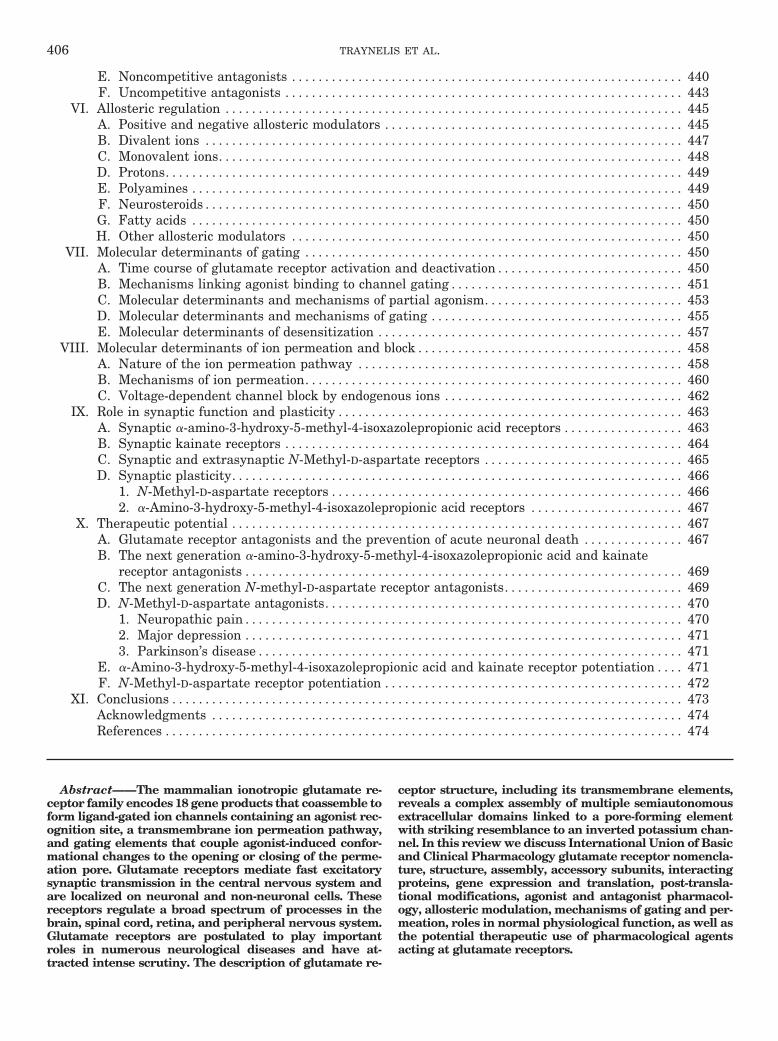
ceptors, suggests they share a similar architecture(Table 2). Glutamate receptor subunits are modularstructures that contain four discrete semiautonomousdomains: the extracellular amino-terminal domain(ATD), the extracellular ligand-binding domain (LBD),the transmembrane domain (TMD), and an intracellularcarboxyl-terminal domain (CTD) (Fig. 1A). Apart fromthe CTD and the M4 segment, each of the individualdomains exhibits low sequence homology to bacterialproteins with known structures and, in some instances,a related function (O’Hara et al., 1993; Wo and Oswald,1995; Wood et al., 1995; Paas, 1998; Kuner et al., 2003).Detailed crystallographic structures have been de-scribed for a membrane-spanning tetrameric glutamate
receptor (Sobolevsky et al., 2009) as well as the isolatedATDs and LBDs in complex with various agonists, an-tagonists, and modulators (discussed in section VI).These data, along with functional and biochemical ex-periments, have begun to define the relationship be-tween receptor structure and function.
The first views of the quaternary glutamate receptorstructure were provided by single particle images ofrecombinant and native AMPA receptors obtained byelectron microscopy (Safferling et al., 2001; Tichelaar etal., 2004; Nakagawa et al., 2005, 2006; Midgett andMadden, 2008). Although these images show the recep-tors at lower resolution (�40–20 Å), some structuralfeatures could be extracted. For example, an internal2-fold rotational symmetry was observed for some ofthese receptor structures (Tichelaar et al., 2004; Midgettand Madden, 2008), consistent with indications that glu-tamate receptors assemble as a dimer of dimers. Thisproposed 2-fold rotational symmetry for glutamate re-ceptors is in contrast to the symmetry observed in struc-tures of other ion channels, such as tetrameric K�-chan-nels and the pentameric nicotinic acetylcholine receptor,in which the quaternary subunit arrangement leads torotational symmetries that correlate with subunit-num-ber (MacKinnon, 2003; Miyazawa et al., 2003; Sobo-levsky et al., 2004; Wollmuth and Sobolevsky, 2004).
Crystallographic studies have provided the first de-tailed structure of a membrane-spanning glutamate re-ceptor (3.6 Å) (Fig. 1B). This structure of an antagonist-bound tetrameric rat GluA2 demonstrates that thereceptor has an overall 2-fold symmetry perpendicularto the membrane plane; the extracellular ATDs andLBDs are organized as dimers of dimers, and the ionchannel domain exhibits a 4-fold symmetry (Sobolevskyet al., 2009). This subunit arrangement relates one ATDdimer to another and one LBD dimer to the second, andhalf of the pore-forming TMDs to the other half. The sym-metry mismatch between the ATDs and LBDs arises be-
carboxylic acid; NVP-AAM077, (R)-[(S)-1-(4-bromo-phenyl)-ethylamino]-(2,3-dioxo-1,2,3,4-tetrahydroquinoxalin-5-yl)-methyl-phosphonic acid;NGX424, tezampanel; Org 25935, N-methyl-N-(6-methoxy-1-phenyl-1,2,3,4-tetrahydronaphthalen-2-ylmethyl)aminomethylcar-boxylic acid; PDZ, postsynaptic density 95/disc-large/zona occludens;PEPA, 4-(2-(phenylsulfonylamino)ethylthio)-2,6-difluoro-phenoxyac-etamide; PF-03463275, 1-methyl-1H-imidazole-4-carboxylic acid (3-chloro-4-fluoro-benzyl)-(3-methyl-3-aza-bicyclo[3.1.0]hex-6-ylm-ethyl)-amide; PF-4778574, N-((3R,4S)-3-(4-(5-cyanothiophen-2-yl)phenyl)-tetrahydro-2H-pyran-4-yl)propane-2-sulfonamide; PhTX,philanthotoxin; PKA, protein kinase A; PKC, protein kinase C; RE1, re-striction element-1; REST, RE-1 silencing transcription factor; Ro 25-6981,�-(4-hydroxyphenyl)-�-methyl-4-(phenylmethyl)-1-piperidine propanol; Ro63-1908, 1-[2-(4-hydroxy-phenoxy)-ethyl]-4-(4-methyl-benzyl)-piperidin-4-ol; S18986, (S)-2,3-dihydro-(3,4)cyclopentano-1,2,4-benzothiadiazine-1,1-dioxide; SCH 900435, N-methyl-N-(6-methoxy-1-phenyl-1,2,3,4-tetrahy-dronaphthalen-2-ylmethyl)aminomethylcarboxylic acid; SN50, Ala-Ala-Val-Ala-Leu-Leu-Pro-Ala-Val-Leu-Leu-Ala-Leu-Leu-Ala-Pro-Val-Gln-Arg-Lys-Arg-Gln-Lys-Leu-Met-Pro; Sp1, specific transcription factor 1;SYM2081, (2S,4R)-4-methylglutamic acid; TARP, transmembraneAMPA receptor regulatory proteins; TBI, traumatic brain injury;Tbr-1, T-brain-1; TMD, transmembrane domain; TTX, tetrodo-toxin; UBP141, (2R,3S)-1-(phenanthrenyl-3-carbonyl)piperazine-2,3-dicarboxylic acid; UBP282, (�S)-�-amino-3-[(4-carboxyphenyl-)methyl]-3,4-dihydro-2,4-dioxo-1(2H)-pyrimidinepropanoic acid;UBP310, (S)-1-(2-amino-2-carboxyethyl)-3-(2-carboxythiophene-3-ylmethyl)-5-methylpyrimidine-2,4-dione; UTR, untranslated region;ZK200775, (1,2,3,4-tetrahydro-7-morpholinyl-2,3-dioxo-6-(trifluorometh-yl)quinoxalin-1-yl)methylphosphonate.
TABLE 1Glutamate receptor subunits
Nonhuman genes are represented by lowercase HUGO symbols (e.g., Gria1).
IUPHAR Name HUGO Symbol Common Names Human Chromosome Amino Acids in Longest Splice Variant
GluA1 GRIA1 GluR1, GluRA 5q31.1 906GluA2 GRIA2 GluR2. GluRB 4q32-q33 901GluA3 GRIA3 GluR3, GluRC Xq25-q26 894GluA4 GRIA4 GluR4, GluRD 11q22 902GluK1 GRIK1 GluR5 21q22.11 918GluK2 GRIK2 GluR6 6q16.3-q21 908GluK3 GRIK3 GluR7 1p34-p33 919GluK4 GRIK4 KA1 11q22.3 956GluK5 GRIK5 KA2 19q13.2 981GluN1 GRIN1 NMDAR1, NR1, GluR�1 9q34.3 938GluN2A GRIN2A NMDAR2A, NR2A, GluR�1 16p13.2 1464GluN2B GRIN2B NMDAR2B, NR2B, GluR�2 12p12 1484GluN2C GRIN2C NMDAR2C, NR2C, GluR�3 17q25 1236GluN2D GRIN2D NMDAR2D, NR2D, GluR�4 19q13.1-qter 1336GluN3A GRIN3A NR3A 9q31.1 1115GluN3B GRIN3B NR3B 19p13.3 1043GluD1 GRID1 �1, GluR delta-1 10q22 1009GluD2 GRID2 �2, GluR delta-2 4q22 1007
408 TRAYNELIS ET AL.
cause the receptor contains two conformationally distinctsubunits, which can be denoted A/C and B/D subunits (Fig.1, C and D). Consequently, the A/C subunits will coupledifferently to the ion channel gate than will the B/D sub-units, which may have important implications for the func-tion of the glutamate receptors.
In the tetrameric structure (Sobolevsky et al., 2009),the ATD forms two distinct types of subunit-subunitcontacts. The most extensive contact is formed betweenA/B and C/D subunits, and this contact is identical tothat observed between subunits in the structure of theisolated GluA2 and GluK2 ATD dimer (Clayton et al.,2009; Jin et al., 2009; Kumar et al., 2009). The othercontact is located on the 2-fold symmetry axis and isformed between B and D subunits of the A/B and C/Ddimers (Fig. 1C). In addition, at the level of the LBD, twodistinct types of subunit-subunit contacts are formed.The LBDs are arranged as A/D and B/C dimers withcontacts between the A and C subunits (Fig. 1C). Thedomain swapping or subunit crossover causes a differentsubunit arrangement at the levels of the ATD and theLBD. As predicted by topology studies (Hollmann et al.,1994; Bennett and Dingledine, 1995) and the homologyto the tetrameric K�-channels (Wo and Oswald, 1995;Wood et al., 1995; Kuner et al., 2003), the glutamatereceptor TMD consists of three transmembrane helices(M1, M3, and M4) and a membrane re-entrant loop (M2)(Sobolevsky et al., 2009). In addition, the subunits havea short helix (pre-M1) that is oriented parallel to themembrane. M1, M2, and M3 form a structure thatclosely resembles that of an inverted K�-channel pore,and M4 primarily makes contacts with the TMD of anadjacent subunit.
The observation that subunits with the same polypep-tide sequence adopt two distinct conformations in thetetrameric receptor complex is without precedent in anion channel (Sobolevsky et al., 2009). The subunit cross-over between the ATD and LBD levels of the tetramer(Fig. 1D) is primarily mediated by the ATD-S1 aminoacid linkers that connect the ATD with the LBD. TheATD-S1 linkers of the A/C subunits adopt a compactconformation, whereas the ATD-S1 linkers of the B/Dsubunits have an extended conformation. This struc-tural role of the ATD-S1 linker is intriguing, because
previous studies have implicated this segment in thecontrol of the open probability of NMDA receptors(Gielen et al., 2009; Yuan et al., 2009a). The symmetrymismatch between the LBD and the TMD levels also ismediated primarily by the linkers connecting the twodomains (S1-M1, M3-S2, and S2-M4 linkers). Alsohere, the linkers adopt two different conformationscorresponding to the A/C subunits and the B/D sub-units. The involvement of the TMD-LBD linkers in thefunction of glutamate receptors has been extensivelystudied (Krupp et al., 1998; Villarroel et al., 1998;Sobolevsky et al., 2002a,b; Watanabe et al., 2002;Yelshansky et al., 2004; Balannik et al., 2005; Schmidet al., 2007), and the tetrameric structure provides anexcellent opportunity to interpret these and other re-sults in a structural context. Whereas tetrameric kai-nate receptors appear to have the same extracellulararchitecture as AMPA receptors (Das et al., 2010), itremains to be shown how well the tetrameric AMPAreceptor structure corresponds to structures for NMDAreceptors.
B. Subunit Stoichiometry
The glutamate receptors assemble as tetrameric com-plexes of subunits (Laube et al., 1998; Mano and Teich-berg, 1998; Rosenmund et al., 1998; Greene, 2001; Mat-suda et al., 2005; Nakagawa et al., 2005; Sobolevsky etal., 2009), and functional receptors are formed exclu-sively by assembly of subunits within the same func-tional receptor class (Partin et al., 1993; Kuusinen et al.,1999; Leuschner and Hoch, 1999; Ayalon and Stern-Bach,2001; Ayalon et al., 2005). Glutamate receptors aregrouped into four distinct classes based on pharmacologyand structural homology, including the AMPA receptors(GluA1–GluA4), the kainate receptors (GluK1–GluK5),the NMDA receptors (GluN1, GluN2A–GluN2D, GluN3A,and GluN3B), and the � receptors (GluD1 and GluD2). TheAMPA receptor subunits GluA1 to GluA4 can form bothhomo- and heteromers. The kainate receptor subunitsGluK1 to GluK3 also form both homo- and heteromers, butGluK4 and GluK5 form functional receptors only whencoexpressed with GluK1 to GluK3. The � receptors GluD1and GluD2 are capable of forming homomeric receptors yetseem incapable of forming heteromers with AMPA, kai-
TABLE 2Sequence identity and conservation of residues in glutamate receptor subunits
Numbers are the percentage of residues in the regions that are identical in all subunits within the group. Numbers in parenthesis are the percentage of residues that areidentical in 50% of the subunits in the group (i.e., conserved). ATD includes the signal peptide, M1M2M3 includes pre-M1 and intracellular loops, LBD is S1 and S2. TMDis M1M2M3 and M4. In GluA2, the regions were defined as amino acids 1–397 (signal peptide and ATD), 398–414 (ATD-S1 linker), 415–527 (S1), 528–534 (S1-M1 linker),535–647 (M1M2M3), 648–652 (M3-S2 linker), 653–794 (S2), 795–809 (S2-M4 linker), 810–838 (M4) and 839–884 (CTD) using the structures of the isolated GluA2 LBD(Armstrong and Gouaux, 2000) and the membrane-spanning tetrameric GluA2 (Sobolevsky et al., 2009) as guides.
Receptor Subunits ATD S1 S2 LBD M1M2M3 M4 TMD CTD All
GluA1–4 35 (89) 74 (99) 84 (100) 80 (100) 84 (97) 93 (100) 87 (98) 9 (60) 54 (90)GluK1–5 16 (67) 54 (89) 53 (94) 53 (92) 60 (96) 41 (97) 56 (96) 0.0 (13) 29 (70)GluA1–4, GluK1–5 6 (36) 37 (81) 33 (77) 34 (79) 45 (78) 28 (62) 42 (77) 0.0 (3) 17 (48)GluN1, GluN2A-D, GluN3A-B 1 (24) 19 (61) 18 (76) 19 (68) 16 (81) 10 (83) 14 (81) 0.0 (2.9) 5 (29)GluN2A-D 19 (76) 60 (94) 66 (99) 63 (96) 75 (99) 69 (100) 73 (99) 2 (47) 25 (70)GluD1–2 60 (60) 67 (67) 57 (57) 62 (62) 51 (51) 62 (62) 54 (54) 34 (34) 54 (54)All subunits 0.2 (15) 7 (49) 6 (48) 6 (48) 10 (55) 10 (52) 10 (55) 0.0 (0.2) 2 (19)
GLUTAMATE RECEPTOR ION CHANNELS 409

nate, and NMDA receptor subunits, both in native cellsand in heterologous expression systems (Partin et al.,1993, 1995; Mayat et al., 1995; Zuo et al., 1997; Kohda etal., 2000; Ikeno et al., 2001; Naur et al., 2007). In addition,GluD1 and GluD2 seem incapable of forming receptorsthat can be activated by any known agonists (see sectionV.A). Whether GluD1 and GluD2 can form heteromericreceptors is unresolved.
Functional NMDA receptors require assembly of twoGluN1 subunits together with either two GluN2 sub-units or a combination of GluN2 and GluN3 subunits(Monyer et al., 1992; Schorge and Colquhoun, 2003;Ulbrich and Isacoff, 2007, 2008). NMDA receptors fur-ther require simultaneous binding of both glutamateand glycine for activation (Johnson and Ascher, 1987;
Kleckner and Dingledine, 1988; Lerma et al., 1990). TheGluN1 and GluN3 subunits provide the glycine bindingsites (Furukawa and Gouaux, 2003; Furukawa et al.,2005; Yao et al., 2008), and the GluN2 subunits form theglutamate binding sites (Furukawa et al., 2005). TheGluN1 subunit expressed alone in Xenopus laevis oo-cytes responded weakly to coapplication of glutamateand glycine (Moriyoshi et al., 1991; Nakanishi et al.,1992; Yamazaki et al., 1992). These responses havebeen proposed to arise because X. laevis oocytes ex-press low levels of endogenous NMDA receptor sub-units (XenGluN1 and XenGluN2) that under somecircumstances functionally assemble with GluN1(Green et al., 2002; Schmidt et al., 2006, 2009;Schmidt and Hollmann, 2008, 2009), which can com-
FIG. 1. Structure and domain organization of glutamate receptors. A, linear representation of the subunit polypeptide chain and schematicillustration of the subunit topology. Glutamate receptor subunits have a modular structure composed of two large extracellular domains [the ATD(green) and the LBD (blue)]; a TMD (orange) that forms part of the ion channel pore; and an intracellular CTD. The LBD is defined by two segmentsof amino acids termed S1 and S2. The TMD contains three membrane-spanning helices (M1, M3, and M4) and a membrane re-entrant loop (M2). Theisolated S1 and S2 segments have been constructed by deleting the ATD along with the TMD and joining S1 and S2 with a hydrophilic linker (dottedline). SP, signal peptide. B, crystal structure at 3.6 Å of the membrane-spanning tetrameric GluA2 AMPA receptor (PDB code 3KG2). C, subunitinterfaces between the ATD, LBD, and TMD of the four subunits in the membrane-spanning tetrameric GluA2 AMPA receptor. The subunits areviewed from top down the 2-fold axis of symmetry. The ATDs and LBDs have a 2-fold axis of symmetry, whereas the TMDs have 4-fold axis ofsymmetry. D, the symmetry mismatch between the TMDs and the extracellular domains (ATDs and LBDs) as well as the subunit crossover (or domainswapping) from the LBD to the ATD give rise to two distinct types of subunits in the homotetrameric GluA2 receptor with two distinct conformations.The subunits are referred to as the A/C and B/D subunits. [Adapted from Sobolevsky AI, Rosconi MP, and Gouaux E (2009) X-ray structure, symmetryand mechanism of an AMPA-subtype glutamate receptor. Nature 462:745–756. Copyright © 2009 Nature Publishing Group. Used with permission.]
410 TRAYNELIS ET AL.

plicate studies on NMDA receptors using the X. laevisexpression system. No responses are observed fromGluN1 expressed alone in mammalian cells.
GluN1 also can combine with two different GluN2 sub-units to form triheteromeric receptors. Numerous studiessupport the formation of GluN1/GluN2A/GluN2B, GluN1/GluN2A/GluN2C, GluN1/GluN2B/GluN2D, GluN1/GluN2A/GluN2D receptors in different brain regions and in specificneuronal subpopulations (Chazot et al., 1994; Sheng etal., 1994; Chazot and Stephenson, 1997; Luo et al., 1997;Sundstrom et al., 1997; Dunah et al., 1998a; Cathala etal., 2000; Green and Gibb, 2001; Pina-Crespo and Gibb,2002; Brickley et al., 2003; Dunah and Standaert, 2003;Fu et al., 2005; Jones and Gibb, 2005; Lu et al., 2006;Brothwell et al., 2008). Few studies have addressed thefunctional implications of the presence of two differentGluN2 subunits in the NMDA receptor complex (Brime-combe et al., 1997; Cheffings and Colquhoun, 2000; Hat-ton and Paoletti, 2005).
The GluN3 subunits bind glycine and do not formfunctional receptors alone (Chatterton et al., 2002; Yaoand Mayer, 2006). When coexpressed with GluN1 in X.laevis oocytes, GluN1/GluN3 receptors can form recep-tors that are activated by glycine alone (Chatterton etal., 2002), but these excitatory glycine receptors havenot yet been observed in GluN3-expressing neurons(Matsuda et al., 2003). At present, surface expression ofglycine-activated GluN1/GluN3A or GluN1/GluN3B re-ceptors in HEK293 cells is unresolved, but GluN1/GluN3A/GluN3B shows some functional expression(Smothers and Woodward, 2007). When GluN3 is coex-pressed with GluN1 and GluN2 in X. laevis oocytes,NMDA- and glutamate-activated current amplitudesare reduced compared with current from GluN1/GluN2,suggesting that either triheteromeric GluN1/GluN2/GluN3 receptors form that have a lower conductance, orGluN3 expression reduces trafficking or assembly ofGluN1/GluN2 (Das et al., 1998; Perez-Otano et al., 2001;Ulbrich and Isacoff, 2007, 2008). Triheteromeric GluN1/GluN2/GluN3 receptors presumably form in corticalneurons based on the observation of single-channel cur-rents with properties that could not be attributed toeither GluN1/GluN2 or GluN1/GluN3 receptors (Sasakiet al., 2002). The subunit stoichiometry and surface ex-pression of GluN3-containing NMDA receptors and thephysiological relevance of triheteromeric GluN1/GluN2/GluN3 receptors are not fully resolved.
C. Receptor Assembly and Trafficking
AMPA receptors assemble as dimers of dimers withATD interactions presumably mediating the initialdimer formation. Subsequent tetramerization (i.e., as-sembly of two subunit dimers) occurs through interac-tions of the LBDs and the TMDs (Ayalon and Stern-Bach, 2001; Mansour et al., 2001; Ayalon et al., 2005).Receptor assembly occurs in the endoplasmic reticulum(ER), where quality control mechanisms ensure correct
subunit folding and assembly. Data suggests that con-formational changes associated with the normal func-tion of glutamate receptors, such as ligand binding, ac-tivation, and desensitization, take place in the ERlumen, and these conformational changes may influencetrafficking (Greger et al., 2002; Fleck et al., 2003; Grun-wald and Kaplan, 2003; Mah et al., 2005; Valluru et al.,2005; Greger et al., 2006; Priel et al., 2006; Penn et al.,2008). Consequently, glutamate receptors may require li-gands or “chemical chaperones” for efficient folding andexport from the ER. This is evident when the conforma-tional changes associated with the normal function aremodified by mutagenesis. Nondesensitizing GluA2(L483Y)mutants exit from the ER inefficiently, whereas GluA2(N754D), which has increased desensitization, exits effi-ciently from the ER (Greger et al., 2006). Block of desen-sitization has been shown to similarly influence kainatereceptor trafficking (Priel et al., 2006; Nayeem et al., 2009).The mechanisms are unclear, but block of desensitizationcould interfere with association and/or dissociation of chap-erones and/or transport proteins, with potential candi-dates being TARPs or CNIHs that are thought to be aux-iliary subunits (see section II.H).
Data suggest that the ATD plays a crucial role inreceptor oligomerization and perhaps trafficking (Kuusi-nen et al., 1999; Leuschner and Hoch, 1999; Ayalon andStern-Bach, 2001; Ayalon et al., 2005; Qiu et al., 2009).The interaction between the ATDs is sufficient to allowisolated ATDs to form stable dimers in solution (Claytonet al., 2009; Jin et al., 2009; Kumar et al., 2009). A keyrole of the AMPA receptor ATD may be to direct assem-bly of the tetrameric receptor and to prevent kainate orNMDA receptor subunits from entering the tetramer(Kuusinen et al., 1999; Leuschner and Hoch, 1999; Aya-lon et al., 2005), and some segments of the AMPA recep-tor ATD have been implicated in subtype-specific assem-bly (Leuschner and Hoch, 1999; Ayalon et al., 2005). Inaddition, AMPA receptor subunit stoichiometry is con-trolled by RNA editing, which precedes mRNA splicingand protein synthesis at two sites that modulate func-tion: the RG site within the GluA2 to GluA4 LBD, andthe QRN site at tip of the reentrant pore loop. Editingswitches the codon at the QRN site from Gln to Arg in amajority of GluA2 RNA. These sites are located withinsubunit interfaces and are thought to affect receptorassembly by favoring heterodimerization over ho-modimerization, which partly explains why GluA2-con-taining AMPA receptors are mostly heteromers (Man-sour et al., 2001; Greger et al., 2002, 2003, 2006). Inaddition, GluA2 subunits edited at the QRN site haveincreased dwell time in the ER compared with otherAMPA receptor subunits, thereby increasing their avail-ability for assembly with other subunits (Greger et al.,2002).
Three models have been suggested for assembly ofNMDA receptors. The first model suggests that GluN1-GluN1 and GluN2-GluN2 homodimers initially form
GLUTAMATE RECEPTOR ION CHANNELS 411

and subsequently coassemble to form the tetramericreceptor (Meddows et al., 2001; Schorge and Colquhoun,2003; Papadakis et al., 2004; Qiu et al., 2005). Thesecond model proposes that a GluN1-GluN1 homodimerforms a correctly folded stable complex to which twoGluN2 monomers are added sequentially to form theNMDA receptor tetramer (Atlason et al., 2007). Thethird model suggests initial GluN1-GluN2 heterodimerformation and subsequent tetramerization (Schuler etal., 2008). At present, there are insufficient data to dis-tinguish between the different models. However, the twoconformationally distinct subunits with two types ofsubunit-subunit contacts observed in the GluA2 AMPAreceptor structure (Sobolevsky et al., 2009) might pro-vide the structural framework needed to design experi-ments to resolve this issue. Similar to AMPA receptors,the ATD is thought to mediate initial dimer formation ofNMDA receptor subunits (Meddows et al., 2001; Pa-padakis et al., 2004).
D. The Extracellular Ligand Binding Domain
The LBD is highly conserved within the different glu-tamate classes (Table 2) and is formed by two extracel-lular stretches of amino acids historically referred to asS1 and S2 (Stern-Bach et al., 1994) (Fig. 1A). The struc-tures of excised S1 and S2 amino acid sequences joinedby an artificial polypeptide linker to form the LBDs havebeen described both with agonist and antagonist bound.All LBD structures adopt a clamshell-like conformation,where the polypeptide segment S1, located on theamino-terminal side of membrane helix M1, forms mostof one half of the clamshell (D1), and the segment S2between the M3 and M4 membrane helices forms most ofthe opposite half of the clamshell (D2) (Fig. 1A). The
agonist binding pocket is located within the cleft be-tween these two lobes. Several lines of experimentalwork have validated that the agonist-binding site in thesoluble LBDs used for crystallization faithfully resem-bles the binding sites in intact receptors (Armstrong andGouaux, 2000; Furukawa and Gouaux, 2003; Du et al.,2005; Gonzalez et al., 2008; Sobolevsky et al., 2009). Inaddition, comparison of UV absorption spectra thatprobe the molecular configuration of the AMPA receptorantagonist CNQX bound to either the isolated GluA2LBD or the full-length GluA2 suggests that the struc-ture of the soluble LBD resembles that within the full-length receptor (Deming et al., 2003).
The initial step in glutamate receptor activation isbinding of the agonist to the LBD. Glycine, D-serine,aspartate, and glutamate analogs are agonists and uni-formly contain moieties that correspond to the �-aminoand �-carboxyl groups. The regions of the binding pocketthat form atomic interactions with the �-carboxyl andthe �-amino groups are similar in all LBD structuresand are composed primarily of residues from D1 (Fig. 2;see also section V.A). Crystallography studies togetherwith homology modeling of the AMPA receptor subunitsGluA1 to GluA4 show that residues that directly inter-act with �-carboxyl and �-amino groups of glutamate,AMPA, and kainate are conserved (Armstrong et al.,1998; Armstrong and Gouaux, 2000; Bjerrum et al.,2003, Pentikainen et al., 2003; Gill et al., 2008) (Fig. 2).Greater variation is observed for the binding mode of the�-positioned groups among AMPA receptor agonists,with a variety of atomic contacts being made for differ-ent agonists. Residues lining the agonist binding cavi-ties of kainate receptor subunits are not fully conserved,providing opportunities for the development of subunit-
FIG. 2. Alignments of agonist-binding residues of glutamate receptor subunits. Residue numbering is according to the total protein including thesignal peptide (initiating methionine is 1). For reference, the predicted size of the signal peptide (SP) is included in parenthesis at the end of thealignment. Amino acid numbering in AMPA and kainate receptor subunits has historically been for the mature protein without the signal peptide,whereas amino acid numbering of NMDA and GluD receptor subunits has started with the initiating methionine as 1. Fully conserved residues areyellow, conserved residues are blue, and similar residues are green. # denotes residues capable of forming hydrogen bonds or electrostatic interactionswith the agonist; � denotes residues capable of forming van der Waals contacts with the agonist.
412 TRAYNELIS ET AL.

selective agonists (Mayer, 2005) (Fig. 2; discussed insection V). Residues that interact with agonists in theNMDA receptor GluN2 subunits are fully conserved(Anson et al., 1998; Laube et al., 2004; Chen et al., 2005;Hansen et al., 2005a; Kinarsky et al., 2005; Erreger etal., 2007). As expected from sequence alignments, theagonist binding pockets of GluN1 and GluN3 are similarto those of GluA2 and GluN2A, but several key differ-ences suggest how these subunits discriminate betweenglutamate and glycine (discussed in section V). Gluta-mate receptor activation involves a conformationalchange of the LBD upon binding of the agonist. Directstructural evidence for this idea arose from comparisonof GluA2 LBD structures with and without agonistbound, as well as structures with bound competitiveantagonists (Armstrong and Gouaux, 2000). In the an-tagonist-bound and the unbound apo structures, D1 andD2 are separated and adopt a more open conformationthan in the agonist-bound structure, where D1 and D2adopt a closed conformation (see also section VII.B formore detail). This mechanism is likely to be conservedin all glutamate receptor subunits, because all ago-nist-bound LBDs examined so far adopt conformationsthat are closed to different degrees relative to the apostructure.
Agonist-induced cleft closure within the LBD dimer,arranged with 2-fold symmetry in a back-to-back fash-ion, is an early conformational event that triggers thesubsequent transition of the ion channel domain into anopen state (see section VII). The intersubunit D1-D1contacts formed across the dimer interface create bothmonovalent and divalent ion binding sites as well as
sites for drug-like allosteric modulators (see section VI).In brief, upon agonist binding, the D2 lobes move andprobably trigger rearrangement of the short segmentsthat link the ion channel-containing TMD to the LBD,which drive rearrangement of M3 and subsequent chan-nel opening (Erreger et al., 2004; Mayer, 2006; Hansenet al., 2007) (Fig. 3 discussed in section VII). The move-ment of D1 and D2 relative to each other results ininstability at the TMD and at the LBD dimer interface.Stability can be restored by LBD reopening, which is thefirst step in the process of agonist dissociation, and weassume that it must be preceded by channel closure (ora change in subconductance state). Alternatively, thereduced stability of the interactions at the LBD dimerinterface upon agonist binding can lead to a rearrange-ment of the dimer interface, allowing the receptor toenter a desensitized state (Sun et al., 2002; Jin et al.,2003., 2005; Horning and Mayer, 2004; Armstrong et al.,2006; Weston et al., 2006b) (Fig. 3; see section VII).
Alternative splicing of the AMPA receptor subunitsgenerates two isoforms of the LBD termed flip and flop(Sommer et al., 1990), which control desensitization anddeactivation as well as sensitivity to allosteric modula-tors (Mosbacher et al., 1994; Partin et al., 1994, 1995).The growing list of structures for LBDs from all subfam-ilies in complex with different agonists provides a firmbasis for understanding agonist selectivity. For severalof these ligands, structural studies in combination withsite-directed mutagenesis and homology modeling haveprovided the structural determinants within the bindingpocket that guide subunit selectivity (see section V).
FIG. 3. Conformational changes in the functioning AMPA receptor. Ribbon diagrams of the crystal structures of the GluA2 LBD dimer inconformations that correspond to the resting state (apo form; PDB code 1FT0), active state (glutamate-bound; PDB code 1FTJ) and desensitized state(glutamate-bound; PDB code 2I3V). In these structures, the LBD exists in a bilobed clamshell-like arrangement with the agonist-binding pocketlocated deep within the cleft between the two lobes referred to as D1 and D2. Binding of glutamate induces a transition of D2 that leads to separationof the linker segments that replace the TMDs in the full-length subunits (represented here by cylinders). The NTD and CTD are omitted for clarity.Distances between the linkers that face the TMD and distances between a glycine residue (Gly739) at the top of the dimer are taken from Armstronget al. (2006). Upon glutamate binding and domain closure, separation of the linkers can result in reorientation of the transmembrane helices andopening of the ion channel. The active, nondesensitized receptor conformation is unstable, and stability can be restored either by reopening of the ABDor by rearrangement at the dimer interface. Rearrangement at the dimer interface results in desensitization by repositioning the transmembranehelices such that the ion channel is closed.
GLUTAMATE RECEPTOR ION CHANNELS 413

E. The Extracellular Amino-Terminal Domain
Beginning at the extracellular ATD, all glutamatereceptors contain a short signal peptide (14–33 residues)that targets the protein to the membrane and is removedby proteolysis after membrane insertion. Subsequent tothe signal sequence, the first �400 to 450 residues in allglutamate receptor subunits (except bacterial GluR0,which lacks the ATD) fold into a semiautonomous do-main. Glutamate receptor ATDs have sequence homol-ogy and are structurally similar to the LBD of themetabotropic glutamate receptor mGluR1a and a groupof soluble bacterial periplasmatic amino acid bindingproteins, such as the leucine/isoleucine/valine bindingprotein (O’Hara et al., 1993; Paas et al., 1996; Paas,1998; Masuko et al., 1999a; Paoletti et al., 2000; Claytonet al., 2009; Jin et al., 2009; Karakas et al., 2009; Kumaret al., 2009). However, the similarity is confounded bynumerous structural differences, such as the differentlocations of disulfide bonds, as well as inserts and dele-tions. Nonetheless, the similarity between the gluta-mate receptor ATD and these proteins suggests that thefunction of the ATD could be to bind endogenous ligands,perhaps within a putative pocket located between thelobes. Numerous mutant subunits have been createdthat lack the entire ATD (Fayyazuddin et al., 2000;Pasternack et al., 2002; Horning and Mayer, 2004; Mat-suda et al., 2005; Rachline et al., 2005; Gielen et al.,2009; Yuan et al., 2009a), and these truncated subunitsseem to assemble into receptors that are functionallysimilar to wild-type receptors. The nonessential natureof the ATD for the core function of the glutamate recep-tors is consistent with a regulatory role for this domain.Truncations of the ATD have been found to influenceopen probability, deactivation, desensitization, and reg-ulation of subunit-specific assembly (Kuusinen et al.,1999; Leuschner and Hoch, 1999; Ayalon and Stern-Bach, 2001; Meddows et al., 2001; Ayalon et al., 2005;Gielen et al., 2009; Yuan et al., 2009a). The ATD alsoharbors binding sites for divalent cations, such as Zn2�,and subunit-selective negative allosteric modulators,such as the phenylethanolamine ifenprodil (see sectionsV and VI). In addition, the ATD may contain bindingsites for extracellular proteins, such as N-cadherin (Sa-glietti et al., 2007) and neuronal pentraxins (NARP andNP1) for AMPA receptors (O’Brien et al., 1999; Sia et al.,2007) the ephrin receptor for NMDA receptors (Dalva etal., 2000; Takasu et al., 2002); cerebellin1 precursorprotein for GluD2 receptors (Matsuda et al., 2010; Ue-mura et al., 2010; see also Uemura and Mishina, 2008;Kakegawa et al., 2009).
The glutamate receptors are glycosylated during theirpassage through the endoplasmic reticulum and Golgi.The consensus sites for N-linked glycosylation primarilyare located in the ATD, but a few are located in the LBD(Hollmann et al., 1994; Standley and Baudry, 2000). It isnot clear how many of these consensus sites are glyco-
sylated, but cell-specific differences in glycosylation ofthe glutamate receptor subunits might contribute to thedifferences in ligand affinities, trafficking, and molecu-lar weights observed between different native receptorsand those expressed in heterologous systems (Chazot etal., 1995; Sydow et al., 1996; Everts et al., 1997; Stand-ley et al., 1998; Standley and Baudry, 2000; Clayton etal., 2009: Kumar et al., 2009). Although the effects ofglycosylation on glutamate receptor function have notbeen studied in detail, glycosylation can affect desensi-tization and maximal currents of AMPA and kainatereceptors (Hollmann et al., 1994; Everts et al., 1997). Inaddition, the lectin concanavalin A (con A) inhibits de-sensitization of kainate receptors in a manner that in-volves association of con A with the N-linked oligosac-charides (Partin et al., 1993; Everts et al., 1997, 1999)(see section VI).
Like the glutamate receptor LBDs, the GluN2B ATDis a clamshell-like structure, roughly composed of twohalves (R1 and R2) tethered together by loops (Karakaset al., 2009). The N terminus is located at the top of R1,and the linker to the LBD is located at the bottom of R2.Overall, the GluN2B ATD structure resembles the li-gand binding domain of the metabotropic glutamate re-ceptor mGluR1 (Kunishima et al., 2000), although theposition of R1 is 50° twisted relative to R2 in GluN2BATD compared with mGluR1. The cleft between R1 andR2 can be divided into three sites: 1) the hydrophilicpocket at the outer end of the cleft, which contains polarresidues involved in Zn2� binding; 2) the hydrophobicpocket deep inside the cleft, which contains residuesthat seem to affect ifenprodil binding; and 3) the ion-binding site that accommodates Na� and Cl� ions withunknown physiological relevance. Binding of ifenprodilto GluN2B and Zn2� to GluN2A or GluN2B has beenproposed to stabilize a closed-cleft conformation of theATD (see section VI; Karakas et al., 2009), althoughstructural data in support of the hypothesized intracleftbinding site is lacking. Nevertheless, the proposed cleft-closure has been speculated to lead to separation of thetwo R2 lobes in the ATD dimer (Gielen et al., 2008,2009).
In contrast to NMDA receptors, no ions or small mol-ecules are known to bind to the AMPA or kainate recep-tor ATD. Crystal structures of the GluA2 and the GluK2ATDs show that these AMPA and kainate receptorATDs adopt an overall structure similar to that ofthe ATD from the NMDA receptor subunit GluN2B, butthe twist between R1 and R2 in GluN2B ATD was lesspronounced in GluA2 and GluK2 ATDs (Clayton et al.,2009; Jin et al., 2009; Kumar et al., 2009). UnlikeGluN2B, the isolated GluA2 and the GluK2 ATDs formdimers in solution and in the crystal lattice. Likewise,the ATDs of GluA1 and GluA4 also form dimers insolution (Kuusinen et al., 1999; Wells et al., 2001b; Jinet al., 2009).
414 TRAYNELIS ET AL.

Comparison of the R1 and R2 lobes of GluA2 andGluK2 ATDs with the corresponding domains ofmGluR1 shows that the GluA2 and GluK2 ATDs adopt aconformation that is intermediate between the canonicalopen-cleft and closed-cleft states of mGluR1. In addition,there are extensive interactions between the two ATDsubunits of the dimer for both GluA2 and GluK2 thatinvolve multiple R1-R1 and R2-R2 domain contacts(Clayton et al., 2009; Jin et al., 2009; Kumar et al.,2009). The extensive interactions between the R2 lobesare mostly hydrophobic contacts situated in a largepatch that is buried after dimerization of the ATD. Theresidues in this hydrophobic patch are conserved or con-servatively substituted between AMPA and kainate re-ceptors. In NMDA receptors, the sequence conservationis lower at the R2-R2 interface, consistent with the ideathat binding of modulators to the NMDA receptor ATDcould stabilize ATD cleft closure and separation at theR2-R2 interface (Gielen et al., 2008, 2009). A separationat the R2-R2 interface in the non-NMDA receptor ATDdimer would expose the large hydrophobic patch on theR2 lobe to the solvent, which would be energeticallyunfavorable. The “weak” R2-R2 interface in the NMDAreceptor could better allow closure of the R1-R2 clam-shell and separation at the R2-R2 interface, therebytriggering allosteric modulation of the ion channel. So-lution of dimeric forms of the ATD will help clarify theseideas.
F. The Transmembrane Domain
In all glutamate receptors, the LBD is connected tothe conserved TMD through three short linkers (Fig.1A). The transmembrane helices M1, M3, and M4 fromeach of the four subunits contribute to formation of thecore of the ion channel and have a small but significantsequence homology with the inverted ion channel do-main of K� channels (Wo and Oswald, 1995; Kuner etal., 2003). This similarity is further highlighted by thebacterial glutamate receptor, GluR0, which sharesstrong functional and structural homology with themammalian glutamate receptors and is a potassium-selective channel with inverted topology compared withthe mammalian glutamate receptors (Chen et al.,1999a). The permeation properties of GluA2-containingAMPA receptors and GluK1 and GluK2 kainate recep-tors are modified post-transcriptionally by RNA editingat the Gln codon that resides at the apex of the re-entrant M2 loop (QRN site). The glutamine within theQRN site is converted to arginine by adenosine deami-nase (Sommer et al., 1991; Bass, 2002). For GluA2, theoverwhelming majority of RNA is edited. AMPA or kai-nate receptors that contain the unedited form of GluA2(Q) have high permeability to Ca2� and are insensitiveto extracellular and intracellular polyamine channelblockers, whereas AMPA receptors containing the editedform of GluA2 (R) have low Ca2� permeability and areinsensitive to polyamine channel blockers (see section
VIII.C). It is noteworthy that the extended region of theM2 loop in the new GluA2 structure that encompassesthe QRN site is disordered. It is unclear whether thisreflects crystallization conditions or a native conforma-tion, which might have significant functional conse-quences for ion permeation and block.
The structure of the antagonist-bound tetrameric ratGluA2 shows that the four subunits arrange their TMDsin a 4-fold axis of symmetry with the core of the ionchannel (M1–M3), strikingly similar to K� channels(Sobolevsky et al., 2009) (Fig. 1C). The M2 loop lines theinner cavity of the pore, whereas the M3 helices line theouter cavity, with positions at the apex tightly opposed,presumably forming the gate that occludes the flux ofions in the closed state (see sections VII and VIII). TheM1 helix is positioned on the exterior of M2 and M3. It isnoteworthy that the M4 segment from one subunit isassociated with the ion channel core (M1-M3) of an ad-jacent subunit. In addition, the linker region precedingM1 (pre-M1) makes a short helix that is oriented paral-lel to the plane of the membrane, making contacts withcarboxyl- and amino-terminal ends of transmembranehelices M3 and M4, respectively. The pre-M1s from thefour subunits resemble a cuff around the external sur-face of the ion channel pore that could be an importantdeterminant for channel gating (see section VII).
G. The Intracellular Carboxyl-Terminal Domain andProtein Binding Partners
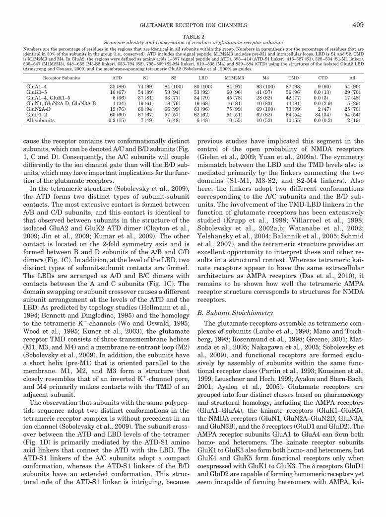
The CTD is the most diverse domain in terms of aminoacid sequence (Table 2), varying in sequence and inlength among the glutamate receptor subunits (Figs.5–7). It shows no sequence homology to any known pro-teins but encodes short docking motifs for intracellularbinding proteins. No structural details exist for thisdomain except for part of the GluN1 CTD with boundCa2�/calmodulin (Ataman et al., 2007). The CTD isthought to influence membrane targeting, stabilization,post-translational modifications (see section IV), andtargeting for degradation. For some glutamate receptorsubunits (e.g., GluN1, GluN2A), deletion of this domaindoes not abolish function but does alter regulation (Kohrand Seeburg, 1996; Ehlers et al., 1998; Krupp et al.,1998; Vissel et al., 2001), because the CTDs containdifferent phosphorylation sites (see section IV) and bind-ing sites for intracellular proteins important for regula-tion of membrane trafficking and receptor function. Sev-eral ER retention signals reside in alternatively splicedexons of GluN1, as well as in GluN2B (Horak andWenthold, 2009). It is noteworthy that there is also ashort span of sequence immediately C-terminal to M4 inGluN2 that also participates in trafficking (Hawkins etal., 2004).
Virtually all members of the glutamate receptor fam-ily bind to a variety of intracellular proteins, which fallinto several classes. Tables 3 and 4 contain noncompre-hensive lists that summarize some of the better known
GLUTAMATE RECEPTOR ION CHANNELS 415

TA
BL
E3
Car
boxy
l-te
rmin
alpr
otei
nbi
nd
ing
part
ner
sfo
rA
MP
Aan
dka
inat
ere
cept
orsu
bun
its
En
trie
sin
dica
teda
tasu
ppor
tin
gdi
rect
inte
ract
ion
sbe
twee
nth
ein
dica
ted
prot
ein
and
glu
tam
ate
rece
ptor
subu
nit
.
Pro
tein
Cla
ssG
luA
1G
luA
2G
luA
3G
luA
4G
luK
1G
luK
2G
luK
5
CA
SK
PD
ZY
2H,c
oIP
,EP
1
GR
IPP
DZ
Y2H
,coI
P,I
HC
2,3
,4,5
Y2H
,coI
P,I
HC
2,3
,4,6
Y2H
,coI
P,I
HC
7Y
2H,c
oIP
,EP
8
GR
IP2
PD
ZY
2H,c
oIP
,IH
C4
,5,7
Y2H
,coI
P,I
HC
4,7
Y2H
,coI
P,I
HC
7Y
2H,c
oIP
,EP
8
mL
IN-1
0P
DZ
coIP
9co
IP9
PIC
K1
PD
ZY
2H,c
oIP
,IH
C2
,6Y
2H,c
oIP
,IH
C2,6
Y2H
,coI
P,I
HC
2,6
Y2H
,coI
P,E
P8
Y2H
,coI
P,E
P8
PS
D95
PD
ZY
2H,c
oIP
,EP
8Y
2H,c
oIP
,IH
C,E
P8,1
0co
IP,I
HC
,EP
10
SA
P97
PD
Zco
IP,I
HC
11,1
2Y
2H,c
oIP
,EP
10
SA
P10
2P
DZ
Y2H
,coI
P,E
P10
Sh
ank3
PD
ZY
2H,c
oIP
,IH
C13
Syn
ten
inP
DZ
Y2H
14
Y2H
14
Y2H
14
Y2H
14
Y2H
,coI
P,E
P8
Y2H
,coI
P,E
P8
RIL
PD
Z/
LIM
Y2H
,coI
P,I
HC
,EP
15
4.1
Cyt
oske
leta
lY
2H,c
oIP
,IH
C16,1
7co
IP,I
HC
18
�-A
ctin
in-1
Cyt
oske
leta
lY
2H,c
oIP
,IH
C1
9
Act
infi
lin
Cyt
oske
leta
lY
2H,c
oIP
,IH
C20
Y2H
,coI
P,I
HC
20
Con
tact
inC
ytos
kele
tal
coIP
21
Dyn
amin
-1C
ytos
kele
tal
coIP
21
Dyn
amit
inC
ytos
kele
tal
coIP
21
Pro
fili
nS
caff
old
coIP
21
Spe
ctri
nS
caff
old
coIP
21
AP
2A
dapt
orco
IP,I
HC
,EP
22
,23
NS
FA
TP
ase
Y2H
,coI
P,I
HC
,EP
24
,25
,26
coIP
21
IQG
AP
1G
TP
ase
Y2H
,coI
P,I
HC
19
Cal
mod
uli
nC
a2�
sen
sor
coIP
21
VIL
IP1
Ca2
�se
nso
rco
IP21
VIL
IP3
Ca2
�se
nso
rco
IP21
14-3
-3O
ther
27
coIP
21
coIP
28
CO
PI
Oth
er27
coIP
28
G-�
(q/1
1)O
ther
27
coIP
,EP
29
SU
MO
Oth
er27
Y2H
,coI
P,I
HC
,EP
30
coIP
,co
imm
un
opre
cipi
tati
onor
pull
-dow
nas
say;
EP
,el
ectr
oph
ysio
logy
;IH
C,
imm
un
ohis
toch
emis
try;
Y2H
,ye
ast
two-
hyb
rid.
1C
ouss
enet
al.(
2002
).2D
evet
al.(
1999
).3D
ong
etal
.(19
97).
4W
yszy
nsk
iet
al.(
1999
).5D
ong
etal
.(19
99).
6X
iaet
al.(
1999
).7S
riva
stav
aet
al.(
1998
).8H
irbe
cet
al.(
2003
).9S
tric
ker
and
Hu
gan
ir(2
003)
.10G
arci
aet
al.(
1998
).11L
eon
ard
etal
.(19
98).
12S
ans
etal
.(20
01).
13U
chin
oet
al.(
2006
).14H
irbe
cet
al.(
2002
).15S
chu
lzet
al.(
2004
).16H
ayas
hi
etal
.(20
05).
17S
hen
etal
.(20
00).
18C
olem
anet
al.(
2003
).1
9N
uri
yaet
al.(
2005
).2
0S
alin
aset
al.(
2006
).21C
ouss
enet
al.(
2005
).22K
astn
ing
etal
.(20
07).
23L
eeet
al.(
2002
).24N
ish
imu
ne
etal
.(19
98).
25O
sten
etal
.(19
98).
26S
ong
etal
.(19
98).
27S
ynap
tic
tran
smem
bran
epr
otei
ns,
sign
alin
gpr
otei
ns,
ubi
quit
in-l
ike
prot
ein
s,or
tran
spor
tre
gula
tory
prot
ein
s.28V
ivit
han
apor
net
al.
(200
6).
29R
uiz
etal
(200
5).
30M
arti
net
al.
(200
7).
416 TRAYNELIS ET AL.
TA
BL
E4
Car
boxy
l-te
rmin
alpr
otei
nbi
nd
ing
part
ner
sfo
rN
MD
Aan
dd
elta
rece
ptor
subu
nit
sE
ntr
ies
indi
cate
data
supp
orti
ng
dire
ctin
tera
ctio
ns
betw
een
the
indi
cate
dpr
otei
nan
dgl
uta
mat
ere
cept
orsu
bun
it.
Pro
tein
Glu
N1
Glu
N1
Glu
N2A
Glu
N2B
Glu
N2C
,DG
luN
3AG
luD
1G
luD
2
Del
phil
inP
DZ
Y2H
,coI
P,I
HC
1
LIN
7P
DZ
coIP
,IH
C2
nP
IST
PD
ZY
2H,c
oIP
,IH
C3
Y2H
,coI
P,I
HC
3
PIC
OIP
K1
PD
ZY
2H,c
oIP
,IH
C4
PS
D93
PD
ZY
2H,c
oIP
5Y
2H,c
oIP
5
PS
D95
PD
ZY
2H6
Y2H
6,7
Y2H
6,7
Y2H
,coI
P5
Y2H
,coI
P5
SA
P97
PD
ZY
2H7
Y2H
7Y
2H6
Y2H
,coI
P5
Y2H
,coI
P5
SA
P10
2P
DZ
coIP
8,9
Sh
ank1
PD
ZY
2H,c
oIP
,IH
C10
Sh
ank2
PD
ZY
2H,c
oIP
,IH
C10
S-S
CA
MP
DZ
Y2H
,coI
P,I
HC
11
Y2H
,coI
P,I
HC
11
Y2H
,coI
P,I
HC
12
�-a
ctin
in-2
Cyt
oske
leta
lY
2H,c
oIP
,IH
C13
Y2H
,coI
P,I
HC
13
EM
AP
Cyt
oske
leta
lY
2H14
Y2H
14
MA
P1S
Cyt
oske
leta
lY
2H,c
oIP
,IH
C15
Ple
ctin
Sca
ffol
dY
2H16
RA
CK
1S
caff
old
coIP
,EP
17
Spe
ctri
nS
caff
old
Y2H
,coI
P,I
HC
18
AP
2A
dapt
orY
2H,I
HC
,EP
19
,20
Y2H
,coI
P,E
P1
9,2
1
AP
4A
dapt
orY
2H,c
oIP
,IH
C22
PA
CS
IN1
Ada
ptor
Y2H
,coI
P,E
P23
Cal
mod
uli
nC
a2�
sen
sor
coIP
,EP
24
CA
RP
1O
ther
25
Y2H
16
CO
PII
Oth
er25
coIP
,IH
C26
GP
S2
Oth
er25
Y2H
16
SA
LM
1O
ther
25
Y2H
,coI
P,I
HC
27
coIP
,co
imm
un
opre
cipi
tati
onor
pull
-dow
nas
say;
EP
,el
ectr
oph
ysio
logy
;IH
C,
imm
un
ohis
toch
emis
try;
Y2H
,ye
ast
two-
hyb
rid.
1M
iyag
iet
al.(
2002
).2Jo
etal
.(19
99).
3Y
ue
etal
.(20
02).
4Y
awat
aet
al.(
2006
).5R
och
eet
al.(
1999
).6K
orn
auet
al.(
1995
).7N
ieth
amm
eret
al.(
1996
).8M
üll
eret
al.(
1996
).9S
ans
etal
.(20
03).
10U
emu
raet
al.(
2004
).1
1H
irao
etal
.(19
98).
12Y
apet
al.(
2003
a).1
3W
yszy
nsk
iet
al.(
1997
).14L
yet
al.(
2002
).15E
riks
son
etal
.(20
07b)
.16E
riks
son
etal
.(20
07a)
.17Y
aka
etal
.(20
02).
18H
irai
and
Mat
suda
(199
9).1
9L
avez
zari
etal
.(20
04).
20V
isse
let
al.(
2001
).21P
ryby
low
ski
etal
.(2
005)
.22Y
apet
al.
(200
3b).
23P
érez
-Ota
ño
etal
.(2
006)
.24E
hle
rset
al.
(199
6).
25S
ynap
tic
tran
smem
bran
epr
otei
ns,
sign
alin
gpr
otei
ns,
ubi
quit
in-l
ike
prot
ein
s,or
tran
spor
tre
gula
tory
prot
ein
s.2
6M
uet
al.
(200
3).
27W
ang
etal
.(2
006)
.
GLUTAMATE RECEPTOR ION CHANNELS 417

interactions between glutamate receptor C-terminaland PDZ, cytoskeletal, scaffolding, adaptor, anchoring,structural, signaling, and other proteins. In addition tothese interactions, several glutamate receptor subunitsbind directly to signaling proteins, including GluA1 andcGMP-dependent protein kinase II (Serulle et al., 2007),GluA4 and PKC (Correia et al., 2003), multiple NMDAreceptor subunits and Ca2�/calmodulin-dependent pro-tein kinase (CamK) II (Gardoni et al., 1998; Strack andColbran, 1998; Leonard et al.,, 1999, 2002), as well astyrosine phosphatase and GluD2 (Hironaka et al., 2000).These interactions allow local signaling to proceed, pro-viding the possibility of spatial and temporal specificityto receptor regulation. Additional localization of signal-ing molecules can be mediated by adjacent proteins, andthe glutamate receptors are embedded into a rich com-plex of signaling molecules that are localized by a myr-iad of adaptor and scaffolding proteins within the postsynaptic density (Husi et al., 2000). Further enhancingthe complexity among different subunits, alternativeRNA splicing of several AMPA and kainate receptorsubunits as well as the NMDA receptor subunit GluN1causes variation in the CTD that also will affect bindingsites for intracellular proteins.
H. Transmembrane �-Amino-3-hydroxy-5-methyl-4-isoxazolepropionic Acid Receptor Regulatory Proteinsand other Auxiliary Subunits
A confound in the study of AMPA receptor biophysicalproperties has been the occasional lack of congruencebetween the properties of recombinant receptors ex-pressed in heterologous systems and those of nativereceptors studied in isolated tissue. This mismatch sug-gests that heterologously expressed receptors lack a mod-ulatory component that can influence essential properties.The discovery of the interaction between AMPA receptorsubunits and the transmembrane AMPA receptor regu-latory proteins (TARPs) has solved many of these dis-crepancies. TARPs are integral membrane proteins withfour transmembrane domains (Letts et al., 1998; Hashi-moto et al., 1999; Chen et al., 2000; Tomita et al., 2003;Coombs and Cull-Candy, 2009) that selectively interactwith AMPA receptors early in synthesis and traffickingand direct proper expression and localization of the re-ceptor at the cell surface (Hashimoto et al., 1999; Chenet al., 2000; Schnell et al., 2002; Tomita et al., 2004,2005a; Vandenberghe et al., 2005). TARPs are present inthe majority of AMPA receptor complexes in the brain,suggesting that TARPs are auxiliary subunits for nativeAMPA receptors (Fukata et al., 2005; Nakagawa et al.,2005, 2006; Vandenberghe et al., 2005). It has beensuggested that two or four TARPs can associate with theAMPA receptor tetramer, depending on availability(Vandenberghe et al., 2005; Milstein et al., 2007; Shi etal., 2009). The interaction sites between TARPs andAMPA receptors involve intracellular, transmembrane,and extracellular regions of both proteins (Tomita et al.,
2005a, 2007; Bedoukian et al., 2006; Milstein and Nicoll,2009; Sager et al., 2009).
The classic members of TARPs, �-2, �-3, �-4, and �-8,interact with all four AMPA receptor subunits. The pro-totypical TARP (�-2 or stargazin) originally was identi-fied in the cerebellum as a protein essential for deliveryof AMPA receptors to the plasma membrane (Letts etal., 1998; Chen et al., 2000). The functional properties ofAMPA receptors associated with the TARP subtypes �-2,�-3, �-4, and �-8 are different from those of AMPA re-ceptors devoid of auxiliary subunits. In addition to theirtrafficking capabilities, TARPs increase single channelconductance, increase open probability, increase the ac-tivation rate, slow the deactivation time course, andreduce desensitization (Yamazaki et al., 2004; Priel etal., 2005; Tomita et al., 2005a; Turetsky et al., 2005;Zhang et al., 2006b; Kato et al., 2007; Soto et al., 2007,2009). Prolonged activation of AMPA receptors triggersa form of desensitization that results from dissociationof the TARP, potentially providing a novel mechanismfor receptor tuning (Morimoto-Tomita et al., 2009). Fi-nally, �-2 reduces GluA2-lacking AMPA receptor affinityfor polyamine block, resulting in receptors with weakinward-rectification (Soto et al., 2007).
The TARP �-5 increases glutamate potency and glu-tamate-evoked peak currents, reduces steady-state cur-rents, and accelerates the time course of deactivationand desensitization only in GluA2-containing receptors,but this modulation does not involve regulation of GluA2surface expression (Kato et al., 2008). The TARP �-7shares many of these properties but is not selective forreceptors comprising GluA2-containing subunits, en-hancing peak currents in channels containing GluA1 orGluA2 (Kato et al., 2007). In contrast, other studiesshow that �-5 interacts with all AMPA receptor subunitsand modifies their behavior (Soto et al., 2009).
Proteins with homology to TARP (STG-1 and STG-2)and with similar functional roles have been discoveredin Caenorhabditis elegans, Apis mellifera, and Dro-sophila melanogaster (Walker et al., 2006a; Wang etal., 2008). However, an important difference betweenTARPs and STG-1 and STG-2 is the obligatory require-ment of an additional transmembrane auxiliary subunit(SOL-1) that is structurally unrelated to TARPs andinteracts directly with invertebrate AMPA receptor sub-units (GLR-1 and GLR-2) to slow and reduce the extentof receptor desensitization (Zheng et al., 2006; Walker etal., 2006a,b).
Another distinct class of transmembrane proteins hasbeen shown to assemble with and regulate AMPA recep-tors (Schwenk et al., 2009). These proteins, CNIH-2 andCNIH-3, are members of the mammalian CNIH familyand are homologous to the cornichon proteins from fliesand yeast (Roth et al., 1995; Bokel et al., 2006; Castroet al., 2007). CNIH proteins are necessary for the exportof a number of proteins from the endoplasmic reticulum,including the epidermal growth factor receptor ligands.
418 TRAYNELIS ET AL.

The role of the CNIH proteins in AMPA receptor regu-lation is not yet fully understood.
Accessory proteins for NMDA and kainate receptorshave also recently been described. Neto1 possesses asingle transmembrane domain containing two comple-ment C1r/C1s, Uegf, Bmp1 domains and is a componentof the NMDA receptor complex (Ng et al., 2009). Neto1interacts with an extracellular domain of GluN2 as wellas through an intracellular interaction with PSD95.Loss of Neto1 in transgenic mice preferentially results ina loss of synaptic GluN2A expression, with only a mod-est impact on GluN2B expression, which leads to im-paired hippocampal LTP and hippocampal-dependentlearning and memory (Ng et al., 2009). A second C1r/C1s, Uegf, Bmp1 domain-containing protein, Neto2, in-teracts with GluK2 to increase peak amplitude and openprobability and to slow the decay time course of bothGluK2 recombinant receptors and kainate receptor-me-diated mEPSCs in cerebellar granule cells (Zhang et al.,2009c). In recombinant expression systems, Neto2 hadno impact on GluK2 surface expression.
III. Regulation of Transcription and Translation
The level of expressed glutamate receptors reflects abalance of transcription, translation, mRNA level, pro-tein stability, receptor assembly, and presentation at thecell surface, all of which are integrated through numer-ous environmental stimuli. Therefore, the particularsubunits that each neuron chooses to express are strongdeterminants of synaptic phenotype, and this is the ra-tionale for understanding how the genetic cis elementsand trans factors regulate gene transcription in neuralcells. Over the past decade steady work toward under-standing the control of ionotropic glutamate expressionin neuronal and non-neuronal cells has occurred,roughly doubling both the number of subunits studiedand the identification of promoter elements controllingexpression in neuronal cells. Furthermore, how chroma-tin remodeling affects glutamate receptor expression inboth neurons and non-neuronal cells has been identifiedafter, for example, status epilepticus or transient isch-emia. Most studies have employed a combination of pro-tein-DNA binding assays with functional analysis of na-tive and mutant promoter constructs driving a reportergene, overexpression of candidate transcription factorsin cultured cells or in vivo, occupancy of cis elements bytranscription factors in vivo using chromatin immuno-precipitation (ChIP) assays, and the use of real-timequantitative polymerase chain reaction experiments.The use of ChIP assays and real-time quantitative poly-merase chain reaction on endogenous gene transcripts(and also on exogenously expressed constructs) havebeen particularly fruitful in helping to advance our un-derstanding of how an acute stimulus causes a change intranscript level dependent on candidate promoter ele-ments and trans-acting factors. Studies such as these
have begun to tie neuronal activity, energy metabolism,and glutamate receptor expression together more coher-ently. Furthermore, an appreciation for the role of epi-genetic modifications and chromatin remodeling at glu-tamate receptor promoters is also emerging and holdspromise for new understanding of the neurobiology ofglutamate receptors. Despite this progress, vexingquestions still remain regarding the mechanisms thatcontrol cell-specific and developmental expression forglutamate receptor subunits.
Glutamate receptor genes have a number of featuresin common, such as multiple transcriptional start sitesin a TATAA-less promoter with high GC content. The5�UTR ranges between 200 bp (Gria1) to over 1200 bp(Grin2a) and, in the case of Gria4, Grik5, Grin2a,Grin2b, and Grin2c, is formed from multiple exons. Fi-nally, one or more Sp1 elements reside near the majortranscriptional start site of all genes studied, severalglutamate receptor promoters contain NF�B, CRE,AP1/2, Tbr-1, NRF-1, and RE1/NRSE sites, and geneexpression of many is responsive to neuronal activity.The schematic organization of the promoter regions ispresented in Fig. 4. It also should be noted that NMDA,AMPA, and kainate receptors are key mediators of sig-nal transduction events that convert environmentalstimuli into genetic changes through regulation of genetranscription and epigenetic chromatin remodeling inneural cells, an area of emerging interest (Carrasco andHidalgo, 2006; Wang et al., 2007; Cohen and Greenberg,2008; Lubin et al., 2008).
A. �-Amino-3-hydroxy-5-methyl-4-isoxazolepropionicAcid Receptors
1. Gria1. The rat Gria1 gene has been evaluated fortranscription initiation and regulation of promoter func-tion by transfection of constructs into primary mixedneuronal cultures. Gria1 promoter activity was strongerin neurons, neuronal specificity being primarily depen-dent on sequences lying within the regions �1395 to�743 and �253 to �48. Although five CRE sites wereable to bind recombinant CRE binding-bZIP proteins,conditions under which these CRE sites come into playin native neurons are unknown. GluA1 receptors (likeall AMPA receptor subunits) are expressed by neuronsand glial cells in vivo and in vitro (Gallo and Ghiani,2000), but the density of functional receptors is muchlower in astrocytes than neurons. In oligodendrocyteprogenitor O-2A cells, the transcriptional rate of GluA1is increased by platelet-derived growth factor and basicfibroblast growth factor (Chew et al., 1997). Regulationof Gria1 transcription occurs via acid sphingomyelinaseand NF�B sites in the Gria1 promoter (Borges andDingledine, 2001), which was found to both account forthe elevation of GluA1 by tumor necrosis factor-� (Yu etal., 2002) and contribute to the sensitization by tumornecrosis factor-� of NT2-N cells to kainate-induced celldeath.
GLUTAMATE RECEPTOR ION CHANNELS 419
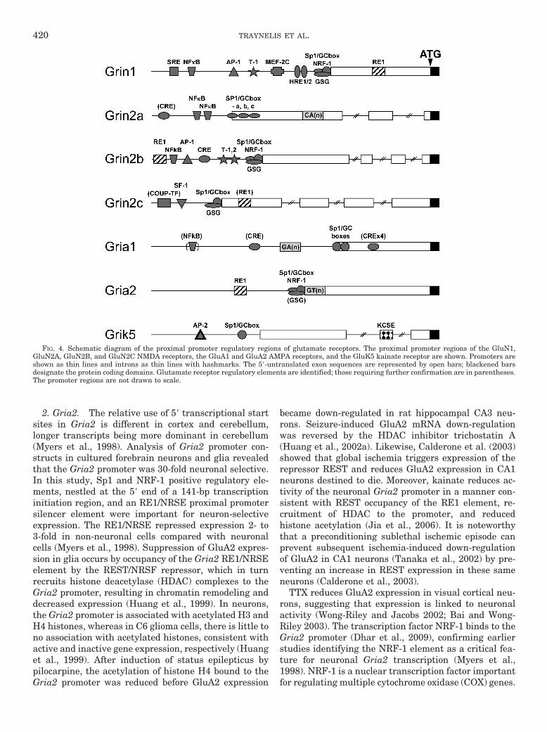
2. Gria2. The relative use of 5� transcriptional startsites in Gria2 is different in cortex and cerebellum,longer transcripts being more dominant in cerebellum(Myers et al., 1998). Analysis of Gria2 promoter con-structs in cultured forebrain neurons and glia revealedthat the Gria2 promoter was 30-fold neuronal selective.In this study, Sp1 and NRF-1 positive regulatory ele-ments, nestled at the 5� end of a 141-bp transcriptioninitiation region, and an RE1/NRSE proximal promotersilencer element were important for neuron-selectiveexpression. The RE1/NRSE repressed expression 2- to3-fold in non-neuronal cells compared with neuronalcells (Myers et al., 1998). Suppression of GluA2 expres-sion in glia occurs by occupancy of the Gria2 RE1/NRSEelement by the REST/NRSF repressor, which in turnrecruits histone deacetylase (HDAC) complexes to theGria2 promoter, resulting in chromatin remodeling anddecreased expression (Huang et al., 1999). In neurons,the Gria2 promoter is associated with acetylated H3 andH4 histones, whereas in C6 glioma cells, there is little tono association with acetylated histones, consistent withactive and inactive gene expression, respectively (Huanget al., 1999). After induction of status epilepticus bypilocarpine, the acetylation of histone H4 bound to theGria2 promoter was reduced before GluA2 expression
became down-regulated in rat hippocampal CA3 neu-rons. Seizure-induced GluA2 mRNA down-regulationwas reversed by the HDAC inhibitor trichostatin A(Huang et al., 2002a). Likewise, Calderone et al. (2003)showed that global ischemia triggers expression of therepressor REST and reduces GluA2 expression in CA1neurons destined to die. Moreover, kainate reduces ac-tivity of the neuronal Gria2 promoter in a manner con-sistent with REST occupancy of the RE1 element, re-cruitment of HDAC to the promoter, and reducedhistone acetylation (Jia et al., 2006). It is noteworthythat a preconditioning sublethal ischemic episode canprevent subsequent ischemia-induced down-regulationof GluA2 in CA1 neurons (Tanaka et al., 2002) by pre-venting an increase in REST expression in these sameneurons (Calderone et al., 2003).
TTX reduces GluA2 expression in visual cortical neu-rons, suggesting that expression is linked to neuronalactivity (Wong-Riley and Jacobs 2002; Bai and Wong-Riley 2003). The transcription factor NRF-1 binds to theGria2 promoter (Dhar et al., 2009), confirming earlierstudies identifying the NRF-1 element as a critical fea-ture for neuronal Gria2 transcription (Myers et al.,1998). NRF-1 is a nuclear transcription factor importantfor regulating multiple cytochrome oxidase (COX) genes.
FIG. 4. Schematic diagram of the proximal promoter regulatory regions of glutamate receptors. The proximal promoter regions of the GluN1,GluN2A, GluN2B, and GluN2C NMDA receptors, the GluA1 and GluA2 AMPA receptors, and the GluK5 kainate receptor are shown. Promoters areshown as thin lines and introns as thin lines with hashmarks. The 5�-untranslated exon sequences are represented by open bars; blackened barsdesignate the protein coding domains. Glutamate receptor regulatory elements are identified; those requiring further confirmation are in parentheses.The promoter regions are not drawn to scale.
420 TRAYNELIS ET AL.

Reduction of NRF-1 with small hairpin RNA preventedthe depolarization-stimulated increase of GluA2 expres-sion, whereas overexpression of NRF-1 restored GluA2expression in the face of TTX treatment. Changes inGluA1, GluA3, and GluA4 expression were not observed,rendering NRF-1 control specific to GluA2 (Dhar et al.,2009). Thus, neuronal activity is tightly coupled at themolecular level to GluA2 expression by a process thatinvolves NRF-1.
3. Gria3 and Gria4. The human GRIA3 gene is onthe long arm of the X chromosome and is subject to Xinactivation through methylation (Gecz et al., 1999).The Gria3 transcript contains a TC repeat in the 5�-UTRthat is polymorphic and is also present in rodent tran-scripts. The 5�-UTR and ATG codon are contained in alarge 1102-bp exon 1 and are conserved in rodent andhuman sequences. Neither an RE1 repressor elementnor an NRF-1 site was identified.
GluA4 is widely expressed in brain; however, its abun-dance is less than GluA1–3 (Petralia and Wenthold,1992). In transfected mixed cortical cultures, Gria4 pro-moter constructs drove luciferase expression predomi-nantly in neurons, indicating a 6- to 12-fold neuronalpreference (Borges et al., 2003). Deletion of the Gria4transcriptional initiation region decreased luciferase ac-tivity in neurons, but increased activity in C6 cells,suggesting that neuronal regulatory elements reside inthis region. Sp1, Ikaros, and basic helix-loop-helix bind-ing element sites are conserved in rat, mouse, and hu-man genes within �150 bp of transcription initiationsites; however, specific evaluation of these elements re-quires further investigation. A distal region of Gria4,�4427 to �4885, is in a long interspersed element se-quence that has been suggested to recruit chromatinremodeling enzymes to the Gria4 gene (Borges et al.,2003).
B. Kainate Receptors Grik1 to Grik5
Regulatory cis elements have been computationallypredicted in promoter regions of human GRIK1 andGRIK2 genes but not functionally evaluated (Barbonand Barlati, 2000; Barbon et al., 2001). However, GRIK2was identified as a novel epigenetic target in gastriccancer as a potential tumor suppressor gene (Wu et al.,2010). There are no reports on the functional evaluationof transcriptional regulation of the Grik3 and Grik4genes.
Initial studies of the rat Grik5 gene identified a neg-ative regulatory sequence in the first intron that bindsnuclear orphan receptors such as chicken ovalbuminupstream promoter transcription factor I in both neuraland non-neural cells (Huang and Gallo, 1997; Chew etal., 1999). Transgenic mouse lines carrying 4 kb of the5�-flanking sequence showed lacZ reporter expressionpredominantly in the nervous system. Reporter assaysin central glial (CG-4) and non-neural cells indicatedthat a 1200-bp 5�-flanking region could sustain neural
cell-specific promoter activity (Chew et al., 2001). Sp1binding suggests a functional role for Sp1 in initiator-mediated activation of Grik5 transcription that involvestranscription factor II D-mediated basal activity (Chewet al., 2001). Removal of two putative AP2 sequencesreduced promoter activity in both neural and non-neuralcells, suggesting that these sites are also important forbasal transcription. Furthermore, a 77-bp sequencetermed the kainate cell-specific enhancer region, in-volved in cell-specific expression, includes a functionalSp1 site that when placed downstream of the Grik5promoter, silenced reporter expression in NIH3T3 fibro-blasts and attenuated activity in CG-4 cells. These stud-ies show that elements contributing to tissue-specificexpression are contained within the first exon (Chew etal., 2001).
C. N-Methyl-D-aspartate Receptors
1. Grin1. Rat, human, and chicken Grin1 promoterregions have been cloned and characterized (Bai andKusiak, 1993, 1995; Zimmer et al., 1995; Moreno-Gonza-lez et al., 2008). Transcription of the Grin1 gene is con-trolled by both positive and negative regulatory ele-ments. A consensus RE1/NRSE silencer in exon1contributes to neuronal-specific expression (Bai et al.,1998; Okamoto et al., 1999). Ablation of the RE1/NRSEsite elevated GluN1 expression in non-neural cell linesand undifferentiated P19 cells (Bai et al., 1998, 2003;Okamoto et al., 1999, 2002). Likewise, during differen-tiation of P19 cells, REST/NRSF is down-regulated, re-sulting in de-repression of the Grin1 promoter (Okamotoet al., 1999). De-repression of the Grin1 promoter byabsence of REST/NRSF occurs before subsequent ex-pression of positive acting trans factors required for fullGrin1 promoter activity (Bai et al., 2003). A 27-bp GC-rich region (GC-box) proximal to the transcription startsites has been identified that controls induction of theGrin1 gene upon differentiation of P19 cells, and thissite is recognized by Sp1 and myc-associated zinc fingerprotein transcription factors (Okamoto et al., 2002).These sites previously were known to respond to Sp1, -3,and -4 transcription factors (Bai and Kusiak, 1995, 1997;Bai et al., 1998; Liu et al., 2001) and interact with anelement further 5� in the promoter (�520/�529) that isrecognized by myocyte enhancer factor 2C (Krainc et al.,1998). Studies with Grin1 promoter constructs in PC12cells suggest that NGF uses both the Ras/extracellularsignal-regulated kinase (ERK) and phosphatidylinositol3-kinase pathways to up-regulate Grin1 promoter activ-ity through Sp1 (Liu et al., 2001). Activation of serumglucocorticoid kinase 1, a downstream target of phos-phatidylinositol 3-kinase, increases Grin1 promoter ac-tivity in PC12 cells and hippocampal neurons in anNF�B-independent manner (Tai et al., 2009). This find-ing is consistent with a previous report that the Sp-related factors regulate Grin1 promoter activity throughoccupancy of a putative NF�B consensus element �3 kb
GLUTAMATE RECEPTOR ION CHANNELS 421

upstream of the GC box in neurons and in cell lines (Liuet al., 2004a).
AP1 protein complexes containing �FosB bind the ratGrin1 promoter at the AP1 consensus element, and AP1binding is up-regulated after electroconvulsive shocks.Furthermore, an increase in Fos-like immunoreactivitywas observed in the same cortical neurons that showedan increase in GluN1 immunoreactivity. Accordingly,up-regulation of GluN1 did not occur after seizures infosB(�/�) mice (Hiroi et al., 1998).
GluN1 expression might be coupled to energy metab-olism based on evidence that both the Grin1 and mito-chondrial COX genes are under control of the NRF-1transcription factor via binding elements in their respec-tive proximal promoter regions (Dhar et al., 2008, Dharand Wong-Riley, 2009). Grin1 and Grin2b, but notGrin2a or Grin3a, are positively regulated by the NRF-1transcription factor through NRF-1 promoter elements.Furthermore, control of Grin1 and Grin2b by NRF-1 wasactivity-dependent. KCl up-regulated and TTX down-regulated expression in cultured rat cortical neurons,and NRF-1 itself is up-regulated at both protein andmRNA levels by depolarization (Yang et al., 2006; Dharand Wong-Riley, 2009). Thus, NRF-1 is an essentialtranscription factor in the coregulation of Grin1, Grin2b,Gria2, and COX genes, coupling coordinated expressionof glutamate receptors and energy metabolism at thetranscriptional level (Wong-Riley et al., 1998a,b; Dhar etal., 2008, 2009; Dhar and Wong-Riley, 2009).
A nonpalindromic T-box element in the Grin1 pro-moter is likely to be recognized and regulated by Tbr-1/CASK protein complexes in vivo because GluN1 expres-sion was reduced in Tbr-1(�/�) (T-brain-1) mice by �50% (Wang et al., 2004b). Tbr-1 is a neuron-specificT-box factor (Hsueh et al., 2000) that may play a role inneurogenesis and induction of GluN1. GluN1 expressionis subject to control by hypoxia-inducible factors thatfunction under stress conditions, especially during hyp-oxia (Yeh et al., 2008). Based on GluN1 up-regulationafter lipopolysaccharide injection into the prefrontal cor-tex and in cultured neurons, the predicted cis hypoxiaresponse elements were localized within the Grin1 pro-moter. However, 1 h of ischemia produced by middlecerebral artery occlusion decreased GluN1 (Gascon etal., 2005), possibly as a result of activation of the RE1silencer (Fig. 4).
2. Grin2a. Regulation of the rat Grin2a promoterwas explored with a series of 3�- and 5�-truncated con-structs in primary neurons, primary glia, and non-neu-ronal cell lines (Desai et al., 2002; Richter et al., 2002;Liu et al., 2003). The core promoter resides in exon 1,prefers neurons but also requires downstream se-quences for full activity, and does not use a consensusTATA box. On the basis of overexpression studies andgel shift assays in stable cell lines, three GC-boxes (A, B,and C) seem to regulate Sp1 and Sp4 but not Sp3 trans-activation (Liu et al., 2003).
Two regions of the mouse Grin2a promoter, from�9.2kb/�210 or �1253/�210, were able to confer ner-vous system expression of a transgene reporter. Basedon primary cultures prepared from a �9.2kb/�210Grin2a luciferase mouse, there was �700-fold selectiveexpression in neuronal enriched cultures compared withglial cultures. Two RE1/NRSE-like sequences that con-tain key mismatches in the consensus sequences wereidentified at �989 and �427 and do not seem to act assilencers of the Grin2a gene. Thus, neuronal specificityfor GluN2A expression seems to result from transcrip-tional activation selectively in neurons rather than bynon-neuronal silencing. Furthermore, three regionswere identified (�1253/�1079, �486/�447, 8 kb 5� of�1253) that are important for maximal neuron-selectiveexpression. Sequences between �9.2 kb and �1253 bpcontribute to the maturational increase of Grin2a ex-pression in cultured neurons and elements residing be-tween �1253 and �1180 bp are crucial for this up-regulation (Desai et al., 2002).
Two NF�B sites were identified in the Grin2a pro-moter, which, when removed by mutation, resulted inloss of modification of transactivation by constitutivelyactive SGK (SGK-S422D) that activates NF�B (Taiet al., 2009). Furthermore, the transactivation of aGrin2a construct was sensitive to the NF�B inhibitorpeptide SN50 (Lin et al., 1995). A putative CRE elementvariant found in numerous promoters was identified at�1195 in mouse and �1215 in rat and raises interest inactivity-dependent elevation of Grin2a in vivo. The pu-tative CRE site resides in a region important for positiveneuronal expression in both rat and mouse promoters(Desai et al., 2002; Richter et al., 2002; Liu et al., 2003).
3. Grin2b. Initial promoter analysis using transgeneconstructs in mice (Sasner and Buonanno, 1996) re-vealed that the proximal promoter region and exon 1(�550/�255 relative to the 5�-most transcription site)were sufficient to restrict tissue specificity to brain.However, inclusion of intron 1 and exon 2 in the trans-gene (�550/�1627) were required both to restrict ex-pression to brain and to recapitulate the proper devel-opmental profile of GluN2B expression in cerebellargranule cells. The presence of an RE1/NRSE-like ele-ment at the end of exon 1 was not responsible for con-ferring neural-selective expression in the mouse trans-genes (Sasner and Buonanno, 1996). Of several putativeRE1/NRSE elements in the more distal Grin2b pro-moter, the �2029/�2049 NRSE element bound REST/NRSF and repressed expression of Grin2b reporter con-structs transfected into cultured neurons. Moreover,ethanol treatment of cortical cultures reduced REST/NRSF expression, resulting in GluN2B derepression(Qiang et al., 2005). Analysis of the Grin2b promoteridentified Sp1 and CRE elements (Klein et al., 1998),and the CRE site was later confirmed to bind to phos-pho-CREB in a gel-shift assay. Mutation of the CREmotif in the Grin2b promoter region significantly de-
422 TRAYNELIS ET AL.

creased promoter activity in transfected cortical cellsand also abolished ethanol-induced increase in promoteractivity (Rani et al., 2005). Likewise, an AP-1 site wasactive in cultured neurons and responsive to ethanoltreatment (Qiang and Ticku, 2005). Furthermore, it wasfound that long-term ethanol exposure promoted de-methylation of CpG islands in the Grin2b promoter re-gion that could result in up-regulation of the gene inmouse cortical neurons (Ravindran and Ticku, 2005).
Two nonpalindromic T-box elements in the Grin2bpromoter were identified that are conserved across rat,human, and mouse (Wang et al., 2004b), and these ele-ments are recognized by the Tbr-1 protein (Wang et al.,2004b). Functional and mutational studies in rat hip-pocampal cultures showed that overexpression of Tbr-1alone and in combination with CASK elevated Grin2bpromoter-driven luciferase activity by up to 120-fold de-pendent on each T-box element with the upstream T-element dominant. Recognition of the Grin2b T-boxelements by a Tbr-1–CASK complex in vivo was demon-strated by ChIP analysis using rat hippocampal cul-tured neurons (Wang et al., 2004a). In Tbr-1–null mice,GluN2B expression was decreased up to 60%, andGluN2B expression was down-regulated in brain regionswhere Tbr-1 immunoreactivity was lost in the mutantmice. These two T-box elements reside within the min-imal transgene construct sufficient for neuron-specificexpression of the Grin2b gene (Sasner and Buonanno,1996). A point mutation in CASK that disrupts CASK–Tbr-1–CASK-interacting nucleosome assembly proteincomplexes down-regulates Grin2b promoter activity(Huang and Hsueh, 2009). CASK interacts with tran-scription factor Tbr-1 and CASK-interacting nucleosomeassembly protein–cell division autoantigen-1–differen-tially expressed nucleolar transforming growth fac-tor-�1 target in the nuclei of neurons, which may re-model the chromatin structure flanking Tbr-1 bindingsites (Hsueh et al., 2000; Wang et al., 2004a).
As shown for the Grin2a promoter, activation of se-rum glucocorticoid kinase 1 pathway elevates Grin2bgene expression in hippocampal neurons and Neuro2Acells in an NF�B-dependent manner. The site of NF�Bbinding in the Grin2b promoter was not specificallyidentified but was proposed to reside between �1480and �2020 bp from the rat transcription start site (Taiet al., 2009).
Stimulation of cortical cultures with bicuculline ele-vated GluN2B expression in a transcription- and cal-cineurin-dependent manner (Qiu and Ghosh, 2008) andrevealed association of the Grin2b promoter withCREST, brahma-related gene 1, CRE binding protein,and HDAC-1. Bicuculline stimulation increased CREbinding protein, decreased HDAC1, and increased theassociation of the Grin2b promoter with acetylated his-tones. The increase in GluN2B expression also requiredNMDA receptor activation and was shown to depend onCREST in vivo because a bicuculline-induced increase in
GluN2B expression was absent in CREST-null neurons.These findings suggest that the activity-dependent in-crease in GluN2B expression involves a switch from arepressor to activator complex and requires CRESTfunction that may involve CRE and Sp1 sites in thepromoter in a manner similar to regulation of the im-mediate early gene c-fos.
Evidence for the coupling of GluN2B expression toenergy metabolism has also been described through aseries of electrophoretic mobility shift assay, supershift,and ChIP assays and promoter mutations (Yang et al.,2006; Dhar et al., 2009). GluN2B was shown to be reg-ulated in cultured neurons by NRF-1 transcription fac-tor via an NRF-1 element in the proximal promoterregion. GluN1, GluN2B, and NRF-1 transcripts are up-regulated by KCl and down-regulated by TTX in cul-tured primary neurons. Thus, NRF-1 coordinates thecoregulation of Grin1, Grin2b, and COX genes (Dhar etal., 2008; Dhar and Wong-Riley, 2009).
4. Grin2c, Grin2d, Grin3a, and Grin3b. Elementswithin Grin2c exon 1 and intron 1 in transgenic mouselines selectively drive expression of �-galactosidase incerebellar granule cells (Suchanek et al., 1995, 1997).This region contains a consensus RE1/NRSE silencer,but the role of this element has not been fully evaluated.Furthermore, Sp1 and COUP-TF consensus elements(Nagasawa et al., 1996; Pieri et al., 1999) bound Sp1 andfushi tarazu factor 1/COUP-TF protein in gel shift as-says, but mutations did not modify promoter function ina transfected neuronal cell line (Pieri et al., 1999). TheCOUP-TF site seems to require other elements for func-tion. Coexpression of steroidogenic factor-1 elevatedGrin2c promoter activity modestly in a neuronal cell linedependent on a promoter region centered �250 fromtranscription � 1 site (Pieri et al., 1999); however, directexpression in neurons was not examined.
The developmental up-regulation of GluN2C in theadult cerebellum upon innervation of mossy fibers ontogranule cells has been reported to be due to neuregulin-�(Ozaki et al., 1997). Neuregulin-� potently up-regulatedGluN2C with no change in GluN2B expression in cul-tured mouse cerebellar slices; up-regulation was sensi-tive to block by TTX or the NMDA antagonist 2-amino-5-phosphonopentanoate, suggesting that synapticallyactivated NMDA receptors are involved (Ozaki et al.,1997). Activity regulates GluN2B expression in cerebel-lar granule cells (Vallano et al., 1996), and both GluN2Band GluN2C in organotypic cultures (Audinat et al.,1994), although the stimulatory effect of neuregulin-� onGluN2C was not recapitulated in cultures of dissociatedgranule cells (Rieff et al., 1999). For granule cells cul-tured in low (5 mM) KCl, BDNF up-regulated GluN2CmRNA via the tyrosine kinase receptor ERK1/2 cascade,whereas under 25 mM KCl, depolarization stimulatedCa2� entry through voltage-sensitive Ca2� channels andactivated Ca2�/calmodulin-dependent calcineurin phos-phatase, which opposed GluN2C mRNA up-regulation
GLUTAMATE RECEPTOR ION CHANNELS 423
(Suzuki et al., 2005). However, the depolarization-induced Ca2� increases simultaneously up-regulatedBDNF mRNA via CaMK. Thus, convergent mechanismsof the BDNF and Ca2� signaling cascades are importantfor GluN2C induction in granule cells during develop-ment (Suzuki et al., 2005). NMDA receptor activationwas shown to coordinate both the up-regulation ofGluN2C and the down-regulation of GluN2B mRNA,including a switch of GluN2 subunit associated with cellsurface NMDA receptors in cultured mouse granule cells(Iijima et al., 2008). Although much has been learnedabout the signal transduction pathways leading to re-ceptor subunit changes in granule cells, the promotercontrol elements responsible for transcription subunitswitching remain to be identified.
In the human GRIN2D gene, the 3�-UTR contains fourhalf-palindromic estrogen responsive elements within a0.2-kb region that are highly preserved in the rat, sug-gesting that the GluN2D subunit may be up-regulatedin vivo via neuroendocrine control. In ovariectomizedrats, up-regulation of GluN2D mRNA in the hypothala-mus upon 17�-estradiol treatment was observed (Wa-tanabe et al., 1999), and the Grin2d half-palindromicestrogen responsive elements, placed in a 5� or 3�-UTRposition in a chloramphenicol acetyltransferase pro-moter construct, were responsive to estrogen and thy-roid hormone exposure in an orientation- and hormonereceptor-dependent manner (Watanabe et al., 1999; Va-sudevan et al., 2002).
The amino acid sequences and expression profile forGrin3a and Grin3b have been reported (Ciabarra et al.,1995; Sucher et al., 1995; Andersson et al., 2001; Nishiet al., 2001; Chatterton et al., 2002; Eriksson et al.,2002; Matsuda et al., 2002; Bendel et al., 2005). Studiesdescribing control of transcription with respect to cisregulatory elements and trans factors have not beenreported.
D. Translational Control of Glutamate Receptors
The translation of mRNA to protein is regulated bymechanisms that control 5� capping, 3� polyadenylation,splicing, RNA editing, mRNA transport, stability, andinitiation and elongation (VanDongen and VanDongen,2004; Coyle, 2009). The 5�-UTR of most glutamate re-ceptor mRNAs is unusually long. These long 5�-UTRsoften exhibit stretches of high GC content and some-times contain multiple out-of-frame AUG codons thatcould act as decoys for scanning ribosomes, reducing orpreventing translation initiation at the true glutamatereceptor AUG (Myers et al., 1999; VanDongen and Van-Dongen, 2004). Translational suppression has been in-ferred for GluN1 mRNA natively expressed in PC12 cellsbecause no GluN1 protein can be detected despite amoderately high mRNA level (Sucher et al., 1993). Onthe basis of that study (Sucher et al., 1993), it wasproposed that translation of GluN1 message may besuppressed, perhaps by an unidentified motif in the 5�-
or 3�-UTR. Two pools of GluN1 mRNA with differenttranslational activities have been identified in neonateand adult brain, further implicating potential transla-tion control mechanisms at work in neurons (Awobuluyiet al., 2003). Whether GluN1 translation rate was deter-mined by alternative 3�UTRs was not explored. Visualdeprivation in juvenile mice reduced the GluN2A/GluN2B ratio in the deprived cortex with the findingthat translation of GluN2B is probably a major regula-tory mechanism (Chen and Bear, 2007).
The efficiency of translation of another subunit,GluN2A, also depends upon features of the 5�-UTR.Here, in both in vitro translation in rabbit reticulocytelysate and the X. laevis oocyte expression system, re-moval of most of the 282 bases of the 5�-UTR from thecDNA increased GluN1/GluN2A protein expression bymore than 100-fold. Mutation of three of the five up-stream AUG codons modestly increased translation;however, disruption of a proposed GC-rich stem-loopstructure 170 bases upstream of the AUG increasedGluN2A translation by 40-fold (Wood et al., 1996).Translation suppression of GluN2A transcripts in vivo isone interpretation of the finding that high GluN2AmRNA levels were measured in the inferior and superiorcolliculus and striatum of adult rats (Goebel and Poosch,1999), whereas immunocytochemical studies showedstaining for the GluN2A protein to be light in theseareas (Petralia et al., 1994).
The translation efficiencies of several GluA2 5�-UTRtranscripts in rabbit reticulocyte lysates, X. laevis oo-cytes, and primary cultured neurons has been investi-gated (Myers et al., 2004). Transcripts containing long 5�leaders were translated poorly compared with thosewith shorter leaders, and short transcripts were prefer-entially associated with polyribosomes in vivo. Suppres-sion of GluA2 translation was dominated by a 34- to42-nucleotide imperfect GU repeat sequence in the 5�-UTR predicted to form a secondary structure. It is note-worthy that the GU repeat domain is polymorphic inman and is included in a subset of rat and human GluA2transcripts based on the site of transcription initiation(Myers et al., 1998, 2004). GluA2 translation was notmodified significantly by deletion of any or all of the fiveupstream AUG codons. An interpretation of both theGluN2A and GluA2 studies is that a scanning ribosomeencounters the proposed stem-loop and stalls because ofan inability to “melt” the stem-loop structure; alterna-tively, a ribosome may encounter a blocking proteinbound at the stem-loop motif. Either case could result indissociation of the ribosome from the mRNA.
GluA2 transcripts are processed to form either a shortor a long 3�-UTR giving rise to two pools of GluA2mRNAs of 4 and 6 kb in length in brain. In the hip-pocampus, long 3�-UTR GluA2 transcripts are retainedprimarily in translationally dormant complexes of ribo-some-free messenger ribonucleoprotein, whereas GluA2transcripts bearing the short 3�UTR are associated
424 TRAYNELIS ET AL.

mostly with actively translating ribosomes (Irier et al.,2009b). After pilocarpine-induced status epilepticus, se-lective translational derepression of GluA2 mRNA me-diated by the long 3�-UTR transcripts was observed,suggesting that the long 3�-UTR of GluA2 mRNA aloneis sufficient to suppress translation and that an activity-dependent regulatory signaling mechanism exists thatdifferentially targets GluA2 transcripts with alternative3�-UTRs (Irier et al., 2009b). Differential effects of anti-biotics that target translational initiation and elonga-tion suggest that the long 3�-UTR suppresses GluA2 atthe initiation step, implying a loop-back mechanism(Irier et al., 2009a). The mechanism of translation is notknown but could involve binding of a cytoplasmic poly-adenylation element binding protein (CPEB). Amongthe CPEB proteins, CPEB3 is expressed specifically inneurons (Theis et al., 2003) and seems to bind to GluA2long 3�-UTR. RNA interference knockdown of CPEB3mRNA induces GluA2 protein expression in culturedhippocampal neurons (Huang et al., 2006). CPEB pro-tein regulates translation initiation (Richter and Sonen-berg, 2005), facilitates targeting of mRNAs to dendrites(Huang et al., 2003), and has been implicated in controlof GluN1 translation (Wells et al., 2001a). A relatedreport identified a deletion allele in the Grik4 gene 3�-UTR that was negatively associated with bipolar disor-der, and it was proposed, on the basis of expression data,that RNA secondary structure modified mRNA stabilityto enhance protein expression (Pickard et al., 2008).
The localization of translation machinery nearpostsynaptic sites (Steward and Levy, 1982; Stewardand Reeves, 1988) and differential distribution ofmRNAs to dendrites, including those for GluA1,GluA2, and GluN1, have been investigated (Stewardand Schuman, 2001; Schratt et al., 2004; Grooms etal., 2006). It is becoming clear that these dendriticmRNAs may form a pool poised for translation tomodify neuronal plasticity. Other studies have dem-onstrated that protein synthesis in dendrites is criti-cal for long-term potentiation (LTP) and long-termdepression (LTD) (Kang and Schuman, 1996; Huber etal., 2000; Tang and Schuman, 2002; Bradshaw et al.,2003; Cracco et al., 2005; Mameli et al., 2007). Theinduction of protein synthesis is, not unexpectedly,dependent upon NMDA receptor activation (Scheetzet al., 2000; Huang et al., 2002b; Gong et al., 2006;Tran et al., 2007).
IV. Post-Translational Regulation
A. �-Amino-3-hydroxy-5-methyl-4-isoxazolepropionicAcid and Kainate Receptor Phosphorylation
Considerable advances have been made to identifydynamically regulated post-translational phosphoryla-tion sites on the C-terminal domains of most of theglutamate receptor subunits and to understand thefunctions of these modifications. Phosphorylation has
been shown to regulate glutamate receptor traffickingfrom the ER, insertion into the plasma membrane, en-docytosis, synaptic localization, and binding to otherproteins (Malinow and Malenka, 2002). In a few cases,phosphorylation seems to regulate the relative fre-quency of opening for different subconductance levels ofthe channels in a manner independent of trafficking.However, more work is needed to understand the mech-anisms by which phosphorylation can control glutamatereceptor function. In some cases, the strategic combina-tion of specific phosphoprotein antibodies, site-directedmutagenesis, and chromophore-tagged receptors hasmade it possible to associate the functional conse-quences of kinase activation with phosphorylation ofspecific amino acid residues. However, the specific func-tional roles of many phosphorylation sites have simplynot yet been explored, and in nearly all cases, the mo-lecular mechanisms by which phosphorylation changesreceptor function (membrane insertion, open probabil-ity, etc.) are unknown. These topics represent excellentopportunities for future progress.
The C-terminal domains are shown for each of thesubunits in Figs. 5–7, with residues that have beenshown to be post-translationally modified in living cellsin red; residues identified only in cell-free kinase assaysare omitted. Authors have variably included the signalsequence in the numbering schemes and studied variousC-terminal splice variants. Here, sites will be referred toby residue position in the literature. To minimize confu-sion with numbering schemes, we simply include thesurrounding sequences in the text together with themost commonly used residue number. This number di-verges from the numbering in Figs. 5 to 7 when authorsindexed the predicted signal peptide cleavage site as 1.
The GluA1 C terminus has multiple phosphorylationsites, including four for PKC, one for PKA, and one forCAMKII. Phosphorylation of the two membrane-proxi-mal PKC sites (SRS816ES818KR) enhances interactionbetween the actin-binding 4.1N protein and the GluA1C-terminal domain, which facilitates insertion of thissubunit into the plasma membrane (Boehm et al., 2006;Lin et al., 2009), a mechanism involved in long-termpotentiation. Phosphorylation of TST840LPR by PKChas been suggested to influence synaptic transmissionin an age-dependent fashion (Lee et al., 2007b). Twoother GluA1 phosphorylation sites control functionalproperties of AMPA receptor channels. Phosphorylationof RNS845GA by PKA (Roche et al., 1996) increases theopen probability of homomeric GluA1 channels studiedin outside-out patches (Banke et al., 2000), which hasbeen proposed to reflect a change in the equilibrium ofGluA1 with an inactive state, perhaps relating to phos-phodependent binding of intracellular regulatory pro-teins. PKA phosphorylation additionally drives GluA1subunits into synaptic membranes (Esteban et al., 2003;Man et al., 2007). Phosphorylation of QQS831IN byCAMKII or PKC (Barria et al., 1997; Mammen et al.,
GLUTAMATE RECEPTOR ION CHANNELS 425
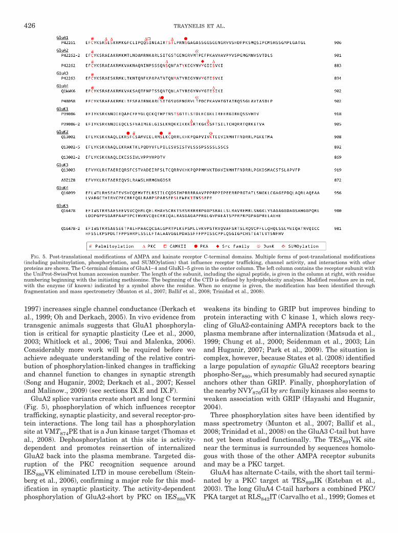
1997) increases single channel conductance (Derkach etal., 1999; Oh and Derkach, 2005). In vivo evidence fromtransgenic animals suggests that GluA1 phosphoryla-tion is critical for synaptic plasticity (Lee et al., 2000,2003; Whitlock et al., 2006; Tsui and Malenka, 2006).Considerably more work will be required before weachieve adequate understanding of the relative contri-bution of phosphorylation-linked changes in traffickingand channel function to changes in synaptic strength(Song and Huganir, 2002; Derkach et al., 2007; Kesseland Malinow., 2009) (see sections IX.E and IX.F).
GluA2 splice variants create short and long C termini(Fig. 5), phosphorylation of which influences receptortrafficking, synaptic plasticity, and several receptor-pro-tein interactions. The long tail has a phosphorylationsite at VMT874PE that is a Jun kinase target (Thomas etal., 2008). Dephosphorylation at this site is activity-dependent and promotes reinsertion of internalizedGluA2 back into the plasma membrane. Targeted dis-ruption of the PKC recognition sequence aroundIES880VK eliminated LTD in mouse cerebellum (Stein-berg et al., 2006), confirming a major role for this mod-ification in synaptic plasticity. The activity-dependentphosphorylation of GluA2-short by PKC on IES880VK
weakens its binding to GRIP but improves binding toprotein interacting with C kinase 1, which slows recy-cling of GluA2-containing AMPA receptors back to theplasma membrane after internalization (Matsuda et al.,1999; Chung et al., 2000; Seidenman et al., 2003; Linand Huganir, 2007; Park et al., 2009). The situation iscomplex, however, because States et al. (2008) identifieda large population of synaptic GluA2 receptors bearingphospho-Ser880, which presumably had secured synapticanchors other than GRIP. Finally, phosphorylation ofthe nearby NVY876GI by src family kinases also seems toweaken association with GRIP (Hayashi and Huganir,2004).
Three phosphorylation sites have been identified bymass spectrometry (Munton et al., 2007; Ballif et al.,2008; Trinidad et al., 2008) on the GluA3 C-tail but havenot yet been studied functionally. The TES891VK sitenear the terminus is surrounded by sequences homolo-gous with those of the other AMPA receptor subunitsand may be a PKC target.
GluA4 has alternate C-tails, with the short tail termi-nated by a PKC target at TES899IK (Esteban et al.,2003). The long GluA4 C-tail harbors a combined PKC/PKA target at RLS842IT (Carvalho et al., 1999; Gomes et
FIG. 5. Post-translational modifications of AMPA and kainate receptor C-terminal domains. Multiple forms of post-translational modifications(including palmitoylation, phosphorylation, and SUMOylation) that influence receptor trafficking, channel activity, and interactions with otherproteins are shown. The C-terminal domains of GluA1–4 and GluK1–5 given in the center column. The left column contains the receptor subunit withthe UniProt-SwissProt human accession number. The length of the subunit, including the signal peptide, is given in the column at right, with residuenumbering beginning with the initiating methionine. The beginning of the CTD is defined by hydrophobicity analyses. Modified residues are in red,with the enzyme (if known) indicated by a symbol above the residue. When no enzyme is given, the modification has been identified throughfragmentation and mass spectrometry (Munton et al., 2007; Ballif et al., 2008; Trinidad et al., 2008).
426 TRAYNELIS ET AL.

al., 2007) and may bind directly to some PKC isoforms(Correia et al., 2003). Phosphorylation is enhanced bysynaptic activity and promotes surface expression ofGluA4 by disrupting its association with �-actinin-1(Nuriya et al., 2005). Similar to GluA2, the Jun kinasesite on GluA4 (VLT855PD) is phosphorylated at rest butrapidly dephosphorylated within minutes of synapticactivity and presumably functions, as in GluA2-long, toenhance surface expression of GluA4 (Thomas et al.,2008). The functional consequence of GluA4 phosphory-lation by c-Jun NH2-terminal kinase has not yet beenexamined.
Most studies have suggested a trafficking function forphosphorylation of specific residues in AMPA receptor Ctermini, and a similar pattern seems to exist for kainatereceptor phosphorylation. Serine residues KKS879RTand GKS885SF of GluK1 are phosphorylated by PKC,resulting in internalization (Rivera et al., 2007). Thesesites, previously identified by Hirbec et al. (2003) on thebasis of in vitro kinase assays, may be involved in auto-regulation by kainate receptor activation (Rivera et al.,2007). The long C-tail of GluK2 is phosphorylated byPKA on serine residues KFS825FC and RMS837LK,which potentiates receptor activation in whole-cell patchstudies (Kornreich et al., 2007), apparently through anincrease in receptor open probability (Traynelis andWahl, 1997). There are no reported modification sites inthe C-tail of GluK3 or GluK5, whereas GluK4 has fourphosphorylation sites identified by mass spectrometry.
B. N-Methyl-D-aspartate and � ReceptorPhosphorylation
Given the function of NMDA receptors in synapticplasticity (see section IX), a wealth of studies exist de-scribing the consequences of modification of specific res-idues on its C termini. GluN1 has four different C-terminal tails created by alternative splicing (Fig. 6),only the longest of which has been shown to be influ-enced by phosphorylation. Disrupting a ring of tyrosineresidues adjacent to the M4 domain by site-directedmutagenesis of IAY837KR on GluN1 and LFY842WK onGluN2A prevented use-dependent desensitization ofGluN1/GluN2A receptors (Vissel et al., 2001). It is note-worthy that tyrosine phosphorylation of GluN2A but notGluN1 was detected with phosphotyrosine antibodies(Lau and Huganir, 1995). This same ring of tyrosines ispresent in all NMDA receptor subunits as well as allAMPA and kainate receptor subunits except GluK4 andGluK5 (Figs. 5 to 7), but whether each of these tyrosinesis subject to phosphorylation by src family kinases withresulting functional consequences has yet to be explored.PKC targets serine residues ASS890FK and RRS896SKon GluN1, and PKA targets the adjacent RSS897KD.Within minutes of PKC activation, phosphorylation ofASS890FK disrupts surface clusters of NMDA receptors(Tingley et al., 1997). The dual PKC-PKA phosphoryla-tion of RRS896S897K, on the other hand, promotes exit ofthe subunit from the endoplasmic reticulum and transit
FIG. 6. Post-translational modifications of GluN1 and GluN2A NMDA receptor C-terminal domains. The GluN1 and GluN2A NMDA receptorsubunits undergo the indicated post-translational modification. The left column is the NMDA receptor subunit with the UniProt-SwissProt humanaccession number. The C-terminal domains of the GluN1 and GluN2A subunits are listed in the center column. The length of the receptor subunit isgiven in the right column, the numbering beginning with the initiating methionine. The beginning of the CTD is defined by hydrophobicity analyses.Modified residues are in red, with the enzyme (when known) indicated by a symbol above.
GLUTAMATE RECEPTOR ION CHANNELS 427

to the surface membrane (Scott et al., 2001, 2003). Thephosphorylation of GluN1 ASS890FK and RRS896SK isachieved by PKC� and PKC�, respectively (Sanchez-Perez and Felipo, 2005), potentially contributing to theselective modulation of NMDA receptor function and/orintracellular localization. Prolonged synaptic activationduring status epilepticus initially causes dephosphory-lation of ASS890FK, then hyperphosphorylation that de-velops over hours and subsides within a day (Niimura etal., 2005). The PKA site RSS897KD behaves similarly,but the adjacent PKC site RRS896SK seems untouchedby status epilepticus, demonstrating a remarkablespecificity.
The GluN2A subunit also occurs with alternative C-tails (Fig. 6) (444 and 626 amino acids), and each hasphosphorylation sites that can modify function. The con-sequence of phosphorylation of LFY842WK, which ispresent on both C-tails, was described above. Both C-tails also harbor MRS1232PF; phosphorylation of thisserine by CDK5 is associated with increased NMDA-evoked currents (Li et al., 2001), and excitotoxic death ofhippocampal CA1 pyramidal cells (Wang et al., 2003).Krupp et al. (2002) demonstrated that glycine-indepen-dent desensitization of GluN1/GluN2A receptors couldbe eliminated by mutation of either of two serine resi-
dues proximal to the fourth transmembrane domain,LRS900AK and RGS932LI, but the kinases acting onthese serines have not been identified. Nine additionalserines or tyrosines have been reported by mass spec-troscopy as phosphorylated on both long and short C-tails, but phosphorylation of exclusive residues near theend of the longer C-tail regulates NMDA receptor mod-ulation. For example, src kinase weakens high-affinityzinc inhibition of recombinant GluN1/GluN2A receptors,an effect that is eliminated by mutation of DPY1387KH(Zheng et al., 1998; but see Xiong et al., 1999); this sametyrosine was shown to account for approximately 30% ofthe src-induced phosphorylation of GluN2A (Yang andLeonard, 2001). Src also phosphorylated HSY1292DN(Yang and Leonard, 2001), but this had no effect on zincinhibition (Zheng et al., 1998). By contrast, mutation ofQVY1267QQ blocked the src effect on zinc inhibition, butthis residue does not seem to be directly phosphorylatedby src (Yang and Leonard, 2001). These results, consid-ered together, raise the possibility that the electrophys-iological consequences of mutating Tyr1267 are due toan allosteric effect on receptor function.
Insulin potentiates the activation of GluN2A-contain-ing NMDA receptors, an effect traced to two serinesphosphorylated by PKC, QHS1291YD, and SIS1312LK
FIG. 7. Post-translational modifications of the GluN2B-D NMDA and GluD1–2 � receptor C-terminal domains. The post-translational modifica-tions of the NMDA and � receptors are shown in red. The enzymes mediating the modifications are identified (when known) by a symbol above. Whenno enzyme is designated, the modification has been identified by fragmentation and mass spectrometry. The left column is the receptor subunit andthe UniProt-SwissProt human accession number. The C-terminal domains are shown in the center column, and the length of the subunit, beginningwith the initiating methionine, is in the right column. The beginning of the CTD is defined by hydrophobicity analyses.
428 TRAYNELIS ET AL.

(Jones and Leonard, 2005). It is noteworthy thatSer1291 is immediately adjacent to the src targetTyr1292, yielding high potential for cross-talk betweenthese kinases; one wonders, for example, whether thelack of effect of Tyr1292 on zinc inhibition (Zheng et al.,1998) was due to obstructive phosphorylation of theadjacent Ser1291 by PKC.
The GluN2B C-tail (Fig. 7) contains a prominent srctarget very near the C terminus on HVY1472EK, whichmay regulate endocytosis in some conditions (Cheungand Gurd, 2001; Nakazawa et al., 2001; Snyder et al.,2005). Phosphorylation of this tyrosine is eliminated in afyn knockout (Abe et al., 2005) and is increased in hip-pocampal CA1 during LTP (Nakazawa et al., 2001) aswell as in a chronic neuropathic pain model (Abe et al.,2005). Although regulation of this tyrosine by fyn kinaseis well understood, the immediate functional conse-quence of phosphorylation has not been studied. Thistyrosine is also phosphorylated in an activity-dependentmanner by the transmembrane tyrosine kinase Eph-rinB2 (Nateri et al., 2007), which is activated by up-stream ERK pathways. Therefore, multiple kinases tar-get Tyr1472 in an activity-dependent manner.
Another major modification of GluN2B in the postsyn-aptic density fraction of the forebrain occurs by phos-phorylation of QHS1303YD by CaMKII (Omkumar et al.,1996). The CaMKII system provides a mechanism bywhich GluN2B-containing NMDA receptors can be mod-ified rapidly upon Ca2� influx associated with NMDAreceptor activation and in this sense may represent theprototype of a kinase activated by its target (Bayer et al.,2001), but the functional consequences of this modifica-tion have not been directly addressed. Finally, the C-terminal PDZ domain of GluN2B contains a site,IES1480DV, that, when phosphorylated by casein kinaseII, disrupts the interaction of GluN2B with PSD-95 andSAP102, thereby decreasing surface expression of thisreceptor (Chung et al., 2004). This represents anotheractivity-dependent phosphorylation that regulates traf-ficking of GluN2B in the plasma membrane. The asso-ciation between active CaMKII and GluN2B seems to berequired for LTP, although the phosphorylation targethas not been identified (Barria and Malinow, 2005).
PKC and PKA both phosphorylate GluN2C RIS1230SL(Fig. 7) near the extreme carboxyl terminus but exert noapparent effect on surface expression of GluN2C or on itsinteraction with PDZ family proteins on the adjacent PDZdomain. However, a phosphomimetic GluN2C(S1230E)mutant exhibited faster activation and inactivation ki-netics in outside-out patches (Chen et al., 2006). Thus,unlike other glutamate receptor subunits, phosphoryla-tion of this serine near the PDZ domain does not affecttrafficking but instead alters channel properties. A sitein the C-terminal domain of GluN2C, HAS1096LP, is aunique (among the glutamate receptors) target for PKB/Akt; interestingly, phosphorylation of Ser1096 creates abinding site for 14-3-3 (Chen and Roche, 2009), which
apparently assists in the trafficking of GluN2C to thesurface membrane. Phosphorylation of Ser1096 is en-hanced by insulin-like growth factor-1 stimulation orNMDA receptor activation, providing a link betweenhormonal state and NMDA receptor function. Threephosphorylated serines in GluN2D have been identifiedby mass spectroscopy (Fig. 7) but have not yet beenstudied functionally. Likewise, a phosphotyrosine anti-body labels GluN2D immunoprecipitated from rat thal-amus (Dunah et al., 1998b), but the targeted tyrosineshave not been identified.
No modifications of GluN3A, GluN3B, or GluD1 havebeen described yet, but GluD2 has four phosphoserines,one of which (TLS945AK) was shown to be a PKC target(Fig. 7) (Kondo et al., 2005). The GluD2 subunit is ex-pressed mainly by cerebellar Purkinje cells, and both itand PKC seem to be essential for long-term depressionat the parallel fiber-Purkinje cell synapse (Kashiwabu-chi et al., 1995; Kondo et al., 2005). However, LTD couldbe rescued in a GluD2-null mouse by a GluD2(S945A)transgene, demonstrating that phosphorylation ofSer945 plays no role in LTD (Nakagami et al., 2008).Indeed, a transgene lacking the transmembrane do-mains could rescue LTD (Kakegawa et al., 2007b), sug-gesting that GluD2 might function as a scaffolding pro-tein rather than an ionotropic receptor.
C. Other Post-Translational Modifications ofGlutamate Receptors
Additional post-translational modifications can affectglutamate receptor localization or activity. All ionotropicglutamate receptor subunits seem to be glycosylated (seesection II), which seems to be involved in proper foldingof the subunit during synthesis (Everts et al., 1997; Mahet al., 2005; Nanao et al., 2005; Gill et al., 2009). Inaddition, multiple glutamate receptor subunits undergodynamic regulation by palmitoylation (Figs. 5–7). TheAMPA receptors have two known palmitoylation sites inthe membrane domain 2 and C-tail. All AMPA receptorshave a conserved cysteine residue proximal to M4 thatundergoes palmitoylation (GluA1-EFC811YK, GluA2-C836, GluA3-C841, GluA4-C817) (Hayashi et al., 2005).In GluA1 and GluA2, palmitoylation of the C-tail resi-due reduces insertion rate (Hayashi et al., 2005) andregulates phosphorylation of the two serines on the C-terminal tail by PKC (Lin et al., 2009). Thus, the inter-play between palmitoylation and phosphorylation ofresidues on this membrane-proximal region of GluA1 in-fluences membrane insertion and thus synaptic availabil-ity of this subunit. All AMPA receptors have anotherconserved cysteine residue just downstream of the QRNsite (GluA1-QGC585DI, GluA2-C610, GluA3-C615, GluA4-C611) that, when palmitoylated, increases AMPA receptorsurface expression (Hayashi et al., 2005). The homomerickainate receptor GluK2 undergoes palmitoylation of thecysteines SFC858SA and LKC871QR, both of which have noapparent effect on basal receptor function. In addition to
GLUTAMATE RECEPTOR ION CHANNELS 429
palmitoylation, GluK2 has been shown to be SUMOylatedat lysine IVK896TE, a process that facilitates endocytosisafter kainate receptor activation (Martin et al., 2007).GluK3 has been shown to be SUMOylated, although thesite has not been identified (Wilkinson et al., 2008).
Like AMPA and kainate receptors, the GluN2A andGluN2B subunits also undergo palmitoylation at their Ctermini (Hayashi et al., 2009). Both of these subunits havetwo separate clusters of cysteine residues that are palmi-toylated with distinct consequences on receptor expressionand internalization. The first cluster of palmitoylated cys-teine residues within GluN2A (RFC848FT, GVC853SD,YSC870IH) and the homologous cysteines in GluN2B(Fig. 7) increase nearby tyrosine phosphorylation of theC-tails by src family kinases, regulating surface expres-sion and receptor internalization. Tyrosine phosphory-lation of the C-tails eliminated receptor interaction withactivator protein-2, decreasing clathrin-mediated endo-cytosis. Palmitoylation of the second cluster of cysteineresidues within GluN2A (THC1214RSC1217LS, FKC1236DAC1239LR) and the homologous residues in GluN2Bplus EAC1245KK leads to accumulation of the NMDAreceptors in the Golgi apparatus and decreased surfaceexpression (Hayashi et al., 2009). Thus, palmitoylationof GluN2A and GluN2B provides a dual mechanism bywhich post-translational modification controls NMDAreceptor surface expression. It is unknown if GluN2C orGluN2D subunits undergo palmitoylation, but both havecysteine residues homologous to GluN2A and GluN2Bwithin their C-terminal tails, leading to the possibilitythat palmitoylation also regulates their surface expres-sion. Studies so far suggest that the GluN1 subunit doesnot undergo palmitoylation (Hayashi et al., 2005).
Both GluN1 and GluN2 subunits undergo S-nitrosy-lation on cysteine thiol groups by endogenous nitric ox-ide (NO) and exogenously applied S-nitrosothiols. TheGluN1 subunit has two cysteine groups within theM3–M4 extracellular linker, QKC744DL and QEC798DS,and the GluN2A subunit has three cysteine groupswithin the ATD, HVC87DL, ASC320YG, and SDC399EP,that can be S-nitrosylated (Choi et al., 2000). The S-nitrosylated cysteine residues within the GluN1 andGluN2 subunits mainly fit the S-nitrosylation consensusmotif of a cysteine residue preceded by an acidic or basicresidue and followed by an acidic residue (Stamler et al.,1997). S-Nitrosylation of any of these cysteine residueswithin GluN1/GluN2A leads to moderate NMDA recep-tor inhibition, decreasing the response to agonist-evokedcurrents by approximately 20%, but a majority of theinhibition is due to Cys399 on the GluN2A subunit ATD(Choi et al., 2000). Inhibition of GluN2A current ampli-tude through S-nitrosylation of Cys399 is due to de-creased channel opening, which may be caused by theincreased affinity of the receptor for Zn2� and gluta-mate, leading to receptor desensitization (Paoletti et al.,2000; Zheng et al., 2001; Lipton et al., 2002). TheGluN2A subunit is sensitized to S-nitrosylation when
Cys744 and Cys798 of the GluN1 subunit are S-nitrosy-lated (Takahashi et al., 2007). It is not known whetherthe remaining GluN2 subunits are S-nitrosylated or in-hibited by this modification. The ability of NO to regu-late NMDA receptors may provide a feedback mecha-nism to prevent excessive receptor activity, becauseNMDA receptor-catalyzed transmembrane currents in-crease the local Ca2�concentration, which activates neu-ronal nitric-oxide synthase through their mutual asso-ciation with PSD-95 (Sattler et al., 1999; Rameau et al.,2003).
NMDA receptors are regulated by the extracellularredox state; sulfhydryl reducing agents such as dithio-threitol and dihydrolipoic acid potentiate NMDA-evokedcurrents through the formation of free thiol groups,whereas oxidizing agents such as 5,5�-dithio-bis(2-nitro-benzoic acid) and oxidized glutathione inhibit currentsthrough the formation of disulfide bonds (Aizenman etal., 1989; Sucher and Lipton, 1991; Kohr et al., 1994;Choi and Lipton, 2000). Both GluN1 and GluN2 sub-units are responsible for redox modulation. Disulfidebonds formed between two pairs of cysteine residueswithin GluN1 (SVC79ED and RGC308VG within the ATDand Cys744 and Cys798 in the LBD) cause the interme-diate and slow components of redox modulation for allGluN2-containing NMDA receptors (Sullivan et al.,1994; Choi et al., 2001). A disulfide bond has been pro-posed to form in the GluN2A subunit between Cys87 andCys320 of the GluN2A ATD and has been shown to formbetween Cys86 and Cys321 in the crystal structure ofGluN2B ATD (Karakas and Furukawa, 2009). This di-sulfide bond has been suggested to mediate a fast-com-ponent of redox modulation, although this may also re-flect modification of the Zn2� binding site (Kohr et al.,1994; Choi et al., 2001). Reducing agents can chelatezinc, and transient potentiation that occurs only in thepresence of agents such as dithiothreitol reflects in partthe relief of zinc inhibition through zinc chelation (Pa-oletti et al., 1997). In addition, reduction of cysteineresidues within GluN1/GluN2A NMDA receptors alsoreduces high-affinity voltage-independent Zn2� inhibi-tion of NMDA receptors (Choi et al., 2001).
D. Proteolysis of Glutamate Receptors
A number of proteases can cleave glutamate recep-tors. Principal among these are serine proteases, whichcan act on GluN1, GluN2A, and GluN2B. Tissue plas-minogen activator can cleave GluN1 at ALR260YA,whereas plasmin and thrombin cleave at multiple siteswithin GluN1, presumably with functional conse-quences (Gingrich et al., 2000; Fernandez-Monreal etal., 2004; Samson et al., 2008). A specific plasmin cleav-age site (EAK317AS) has been described in GluN2A,proteolysis of which leads to removal of the ATD and thehigh-affinity Zn2� binding site contained therein (Yuanet al., 2009b). Thrombin cleaves the analogous residue(Lys318) in GluN2B (Leung et al., 2007). In addition, the
430 TRAYNELIS ET AL.

calcium-dependent nonlysosomal protease family cal-pains can cleave the C termini of GluA1, GluN2A,GluN2B, and GluN2C, resulting in receptor degradationand reduced synaptic activity (Bi et al., 1998a,b; Gutt-mann et al., 2001, 2002; Rong et al., 2001; Simpkins etal., 2003; Araujo et al., 2005). It is noteworthy thatspecific proteolytic cleavage has been proposed to occurin pathological situations such as ischemia (Yuen et al.,2007), blood-brain barrier breakdown (Yuan et al.,2009b), and status epilepticus (Araujo et al., 2005). Inaddition, matrix metalloproteinase-7 has been shown tocleave the LBDs of the GluN1 and GluN2A subunits,decreasing the NMDA receptor-mediated calcium influxand increasing the ratio of AMPA to NMDA receptors incortical slices (Szklarczyk et al., 2008). Among theAMPA receptors, GluA3 has been reported to serve as asubstrate for proteolysis by gamma secretase and gran-zyme B (Gahring et al., 2001; Meyer et al., 2002); glyco-sylation of ISN388DS protects GluA3 from cleavage bygranzyme B during breakdown of the blood-brain bar-rier. GluA1 can be cleaved at SHD865FP through activa-tion of calpain and caspase8-like activity (Bi et al., 1996;Meyer et al., 2002). AMPA receptor proteolysis may becommon in neuropathological conditions (Bi et al.,1998a; Chan et al., 1999).
V. Agonist and Antagonist Pharmacology
Amino acid numbering in AMPA and kainate receptorsubunits has historically been for the mature proteinwithout the signal peptide, whereas amino acid number-ing of NMDA and GluD2 receptor subunits has startedwith the initiating methionine as 1. To simplify compar-ison with the published literature, we will maintain thisinformal convention here.
A. �-Amino-3-hydroxy-5-methyl-4-isoxazolepropionicAcid, Kainate, and � Receptor Agonists
Glutamate activates all AMPA and kainate receptorsby binding within the cleft between domains D1 and D2of the LBD to induce domain closure. Neither NMDA norL-aspartate can activate the non-NMDA receptors, andD-aspartate acts as a low-affinity competitive antagonistfor native AMPA receptors (Gong et al., 2005). In thestructure of the glutamate-bound GluA2 LBD (Fig. 8A)(Armstrong and Gouaux, 2000), the �-amino group ofglutamate forms a tetrahedral network of interactionswith the backbone carbonyl oxygen of Pro478, the sidechain hydroxyl of Thr480, and the carboxylate group ofGlu705. The �-carboxyl group of glutamate forms a bi-dentate interaction with Arg485 and receives hydrogen
FIG. 8. Agonist binding pockets of glutamate receptors. A, binding of glutamate (yellow) in the agonist binding pocket of GluA2 (PDB code 1FTJ).Only side chains of interacting residues are shown. Not all residues are labeled. B, binding of glutamate in GluK2 (PDB code 1S7Y). Compared withthe glutamate-bound ligand binding pocket of GluA2, there is a loss of a direct hydrogen bond to the �-amino group of glutamate at position Ala487in GluK2, which is the site equivalent to Thr480 in GluA2. An additional water molecule forms a hydrogen bond to the �-amino group of glutamatein GluK2. C, binding of glutamate in GluN2A (PDB code 2A5T). Compared with glutamate bound in GluA2, the salt bridge between Asp731 and thepositively charged �-amino group of glutamate is absent. Instead, the �-amino group of glutamate forms water-mediated hydrogen bonds to Glu413and Tyr761. D, binding of glycine in GluN1 (PDB code 2A5T). Specificity of GluN1 for glycine can be explained by the hydrophobic environment createdby Val689 and the steric barrier formed by Trp731. E, binding of glycine in GluN3A (PDB code 2RC7). Trp731 of GluN1 is replaced by M844, allowingroom for a water molecule in the pocket. F, binding of D-serine in GluD2 (PDB code 2V3U).
GLUTAMATE RECEPTOR ION CHANNELS 431

bonds from the backbone amide nitrogens of Thr480 andSer654. The glutamate �-carboxyl group interacts withthe hydroxyl group and backbone amide nitrogen ofThr655 (Fig. 8A), whereas the isoxazole hydroxyl groupof AMPA interacts with the amide nitrogen of Thr655via a water molecule. Furthermore, the side chain ofTyr450 forms an electron-dense ring structure above theglutamate �- and �-carbon atoms resembling a lid abovethe agonist binding pocket (Fig. 8A). Structures of thekainate receptor subunits GluK1 and GluK2 LBDsshow similar atomic contacts for the �-carboxyl group,�-amino group, and �-carboxyl groups of glutamate, al-though residues lining the GluK1 agonist binding pocketare smaller than GluK2 (Mayer, 2005; Nanao et al.,2005; Naur et al., 2005; Mayer et al., 2006) (Fig. 8B).There are several noteworthy differences among the kai-nate receptor LBDs, such as the loss of a hydrogen bondto the agonist �-amino group at GluK2 Ala487, which isequivalent to Thr480 in GluA2 and Thr503 in GluK1.This may contribute to the higher glutamate EC50 forGluK2 compared with GluK1 (Mayer, 2005). A centralfeature of AMPA and kainate receptor agonist binding isclosure of the cleft in which the agonist binds, a confor-mational change mediated by movement of D2 relativeto D1 within the bilobed LBD (Armstrong and Gouaux,2000; Mayer, 2005) (see sections II.D, VII.B).
In addition to glutamate, a number of naturally occur-ring molecules, such as ibotenic acid and willardiine,plus an array of AMPA, ibotenic acid, and willardiineanalogs, activate AMPA and kainate receptors (Table 5).It has been difficult to identify naturally occurring orsynthetic agonists that discriminate well between allAMPA and kainate receptors. AMPA acts as a partialagonist at some kainate receptor subunit combinations(Herb et al., 1992; Swanson et al., 1996; Schiffer et al.,1997), and kainate can induce very rapid desensitizationof neuronal AMPA receptors (Patneau et al., 1993). Nev-ertheless, there are some examples of kainate receptor-selective agonists, such as (2S,4R)-4-methylglutamicacid (SYM2081) and perhaps ATPA, a tert-butyl analogof AMPA (see Tables 5 and 6). Crystal structures of theGluK1 and GluK2 LBDs have revealed differences be-tween kainate and AMPA receptors that partly explainthese kainate receptor-selective actions. The agonist-binding cavities of GluK1 and GluK2 are 40 and 16%larger, respectively, than GluA2, allowing GluK1 andGluK2 to accommodate larger ligands (Mayer, 2005;Naur et al., 2005). Indeed, steric occlusion between the4-methyl group of SYM2081 and GluA2 Leu650 contrib-utes to its selectivity for GluK1 and GluK2, both ofwhich have a smaller valine in the corresponding posi-tions (GluK1, Val670; GluK2, Val654) (Armstrong et al.,1998, 2003; Mayer, 2005). Likewise, the isoprenyl groupof kainate shows reduced steric occlusion in GluK1 andGluK2 compared with GluA2 because of the smallervaline residues. Steric occlusion also may explain selec-tivity of ATPA for GluK1, because its bulky tert-butyl
group interacts with Leu650, Thr686, and Met708 inGluA2, which are replaced by the smaller amino acidsVal670, Ser706, and Ser726, respectively, in GluK1(Lunn et al., 2003). The availability of crystallographicdata for multiple kainate receptor subunits emphasizeshow useful structural information can be across eachsubunit family. Thus, there remains the need for newcrystallographic data for additional members of eachglutamate receptor subtype, as opposed to the relianceon homology modeling and molecular dynamics simula-tions. Although useful, molecular dynamics simulationsof homology models carry significant caveats, includinguncertainty associated with representation of aminoacid insertions, placement of new ligands, approxima-tions of force fields for membrane-spanning elements,and computational limitations in simulating movementof large multisubunit protein assemblies. At the sametime, crystals are rarely formed on demand, low resolu-tion can create ambiguities in protein threading anddetails of ligand binding pose, and dynamic aspectsranging from ligand interactions to domain coupling arenot revealed by a static X-ray structure. Early in aproject, in the absence of a three-dimensional structure,a homology model can offer unique insights, whereas inlate phases of a project, possession of a crystal structurecan benefit significantly from dynamic refinement and
TABLE 5AMPA receptor agonist EC50 values in micromolar
EC50 values for GluA1, GluA3, or GluA4 coexpressed with GluA2 can be found inStein et al. (1992), Coquelle et al. (2000), Nakanishi et al. (1990), and Vogensenet al. (2000).
Agonist GluA1 GluA2 GluA3 GluA4
�M
L-Glutamate 3.4–22a-d 6.2–296a,e,f 1.3–35a-c 560g
AMPA 1.3–8.7c,h,i 66f 1.4–130c,h,i 1.3i
Kainate 32–34d,h 130–170j 31–36c,h
Willardiine 11.5k 6.3l
F-Willardiine 0.47k 0.2–0.5l,m 20.9m 11.9m
Br-Willardiine 2.8k 0.84l
I-Willardiine 33.6k 1.5l
Br-HIBO 14a 5.4a 202a 39a
Cl-HIBO 4.7n 1.7n 2700n 1300n
(S)-CPW399 24.9o 13.9o 224o 34.3o
(S)-ATPA 22p 7.9p 7.6p
ACPA 1.1–11c,q 15q 0.1–5c,q 1.1q
(S)-4-AHCP 4.5r 7.2r 15r
(S)-Thio-ATPA 5.2s 13–40s 32s 20s
2-Et-Tet-AMPA 42t 52t 18t 4t
(S)-2-Me-Tet-AMPA 0.16i 3.4f 0.014i 0.009i
SYM2081 132b 453b
Domoic Acid 1.3d 0.97b 21b
ACPA, (R,S)-2-amino-3-(3-carboxy-5-methyl-4-isoxazolyl)propionic acid; (S)-4-AHCP, (R,S)-2-amino-3-(3-hydroxy-7,8-dihydro-6H-cycloheptadisoxazol-4-yl)propi-onic acid; 2-Et-Tet-AMPA, (R,S)-2-amino-3-3-hydroxy-5-(2-ethyl-2H-5-tetrazolyl)-4-isoxazolylpropionic acid; (S)-2-Me-Tet-AMPA, (S)-2-amino-3-3-hydroxy-5-(2-methyl-2H-tetrazol-5-yl)isoxazol-4-ylpropionic acid; (S)-ATPA, (S)-2-amino-3-(5-tert-butyl-3-hydroxyisoxazol-4-yl)propionic acid; (S)-thio-ATPA, (S)-2-amino-3-(5-tert-butyl-3-hydroxy-4-isothiazolyl)propionic acid; (S)-CPW399, (S)-1-(2-amino-2-carboxyethyl)-6,7-dihydro-1H-cyclopentapyrimidine-2,4(1H,3H)dione; Br-HIBO, (R,S)-4-bromo-HIBO; Cl-HIBO, (R,S)-4-chloro-HIBO.
aCoquelle et al. (2000). (S)-Br-HIBO is active; however, the racemic mixture wasused for the determination of EC50. bDonevan et al. (1998). cBanke et al. (1997).dDawson et al. (1990). eJin et al. (2002). fZhang et al. (2006b). gSchiffer et al. (1997).hNakanishi et al. (1990). iVogensen et al. (2000). jHolm et al. (2005). kKizelsztein etal. (2000). lJin et al. (2003). mGreenwood et al. (2006). nBjerrum et al. (2003).oCampiani et al. (2001). pStensbøl et al. (1999). qStrange et al. (2006). rBrehm et al.(2003). sStensbol et al. (2001). tJensen et al. (2007).
432 TRAYNELIS ET AL.
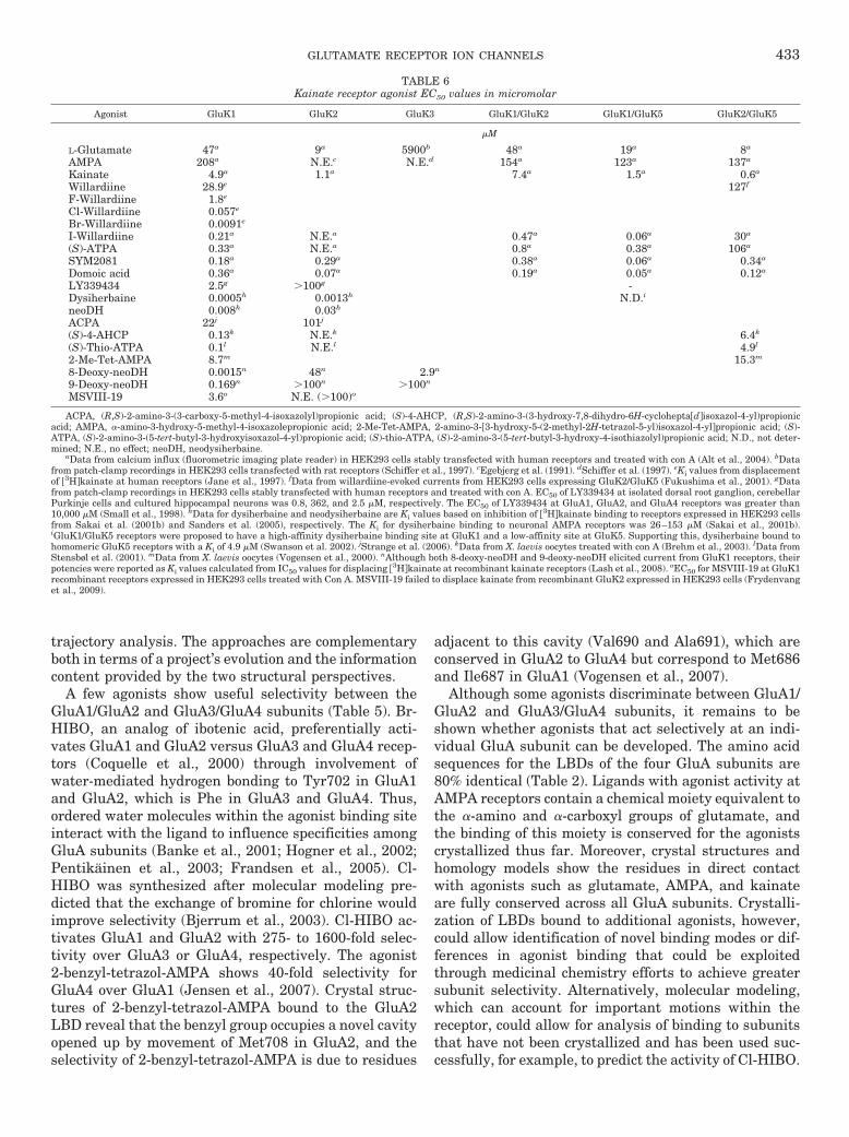
trajectory analysis. The approaches are complementaryboth in terms of a project’s evolution and the informationcontent provided by the two structural perspectives.
A few agonists show useful selectivity between theGluA1/GluA2 and GluA3/GluA4 subunits (Table 5). Br-HIBO, an analog of ibotenic acid, preferentially acti-vates GluA1 and GluA2 versus GluA3 and GluA4 recep-tors (Coquelle et al., 2000) through involvement ofwater-mediated hydrogen bonding to Tyr702 in GluA1and GluA2, which is Phe in GluA3 and GluA4. Thus,ordered water molecules within the agonist binding siteinteract with the ligand to influence specificities amongGluA subunits (Banke et al., 2001; Hogner et al., 2002;Pentikainen et al., 2003; Frandsen et al., 2005). Cl-HIBO was synthesized after molecular modeling pre-dicted that the exchange of bromine for chlorine wouldimprove selectivity (Bjerrum et al., 2003). Cl-HIBO ac-tivates GluA1 and GluA2 with 275- to 1600-fold selec-tivity over GluA3 or GluA4, respectively. The agonist2-benzyl-tetrazol-AMPA shows 40-fold selectivity forGluA4 over GluA1 (Jensen et al., 2007). Crystal struc-tures of 2-benzyl-tetrazol-AMPA bound to the GluA2LBD reveal that the benzyl group occupies a novel cavityopened up by movement of Met708 in GluA2, and theselectivity of 2-benzyl-tetrazol-AMPA is due to residues
adjacent to this cavity (Val690 and Ala691), which areconserved in GluA2 to GluA4 but correspond to Met686and Ile687 in GluA1 (Vogensen et al., 2007).
Although some agonists discriminate between GluA1/GluA2 and GluA3/GluA4 subunits, it remains to beshown whether agonists that act selectively at an indi-vidual GluA subunit can be developed. The amino acidsequences for the LBDs of the four GluA subunits are80% identical (Table 2). Ligands with agonist activity atAMPA receptors contain a chemical moiety equivalent tothe �-amino and �-carboxyl groups of glutamate, andthe binding of this moiety is conserved for the agonistscrystallized thus far. Moreover, crystal structures andhomology models show the residues in direct contactwith agonists such as glutamate, AMPA, and kainateare fully conserved across all GluA subunits. Crystalli-zation of LBDs bound to additional agonists, however,could allow identification of novel binding modes or dif-ferences in agonist binding that could be exploitedthrough medicinal chemistry efforts to achieve greatersubunit selectivity. Alternatively, molecular modeling,which can account for important motions within thereceptor, could allow for analysis of binding to subunitsthat have not been crystallized and has been used suc-cessfully, for example, to predict the activity of Cl-HIBO.
TABLE 6Kainate receptor agonist EC50 values in micromolar
Agonist GluK1 GluK2 GluK3 GluK1/GluK2 GluK1/GluK5 GluK2/GluK5
�M
L-Glutamate 47a 9a 5900b 48a 19a 8a
AMPA 208a N.E.c N.E.d 154a 123a 137a
Kainate 4.9a 1.1a 7.4a 1.5a 0.6a
Willardiine 28.9e 127f
F-Willardiine 1.8e
Cl-Willardiine 0.057e
Br-Willardiine 0.0091e
I-Willardiine 0.21a N.E.a 0.47a 0.06a 30a
(S)-ATPA 0.33a N.E.a 0.8a 0.38a 106a
SYM2081 0.18a 0.29a 0.38a 0.06a 0.34a
Domoic acid 0.36a 0.07a 0.19a 0.05a 0.12a
LY339434 2.5g �100g -Dysiherbaine 0.0005h 0.0013h N.D.i
neoDH 0.008h 0.03h
ACPA 22j 101j
(S)-4-AHCP 0.13k N.E.k 6.4k
(S)-Thio-ATPA 0.1l N.E.l 4.9l
2-Me-Tet-AMPA 8.7m 15.3m
8-Deoxy-neoDH 0.0015n 48n 2.9n
9-Deoxy-neoDH 0.169n �100n �100n
MSVIII-19 3.6o N.E. (�100)o
ACPA, (R,S)-2-amino-3-(3-carboxy-5-methyl-4-isoxazolyl)propionic acid; (S)-4-AHCP, (R,S)-2-amino-3-(3-hydroxy-7,8-dihydro-6H-cycloheptadisoxazol-4-yl)propionicacid; AMPA, �-amino-3-hydroxy-5-methyl-4-isoxazolepropionic acid; 2-Me-Tet-AMPA, 2-amino-3-3-hydroxy-5-(2-methyl-2H-tetrazol-5-yl)isoxazol-4-ylpropionic acid; (S)-ATPA, (S)-2-amino-3-(5-tert-butyl-3-hydroxyisoxazol-4-yl)propionic acid; (S)-thio-ATPA, (S)-2-amino-3-(5-tert-butyl-3-hydroxy-4-isothiazolyl)propionic acid; N.D., not deter-mined; N.E., no effect; neoDH, neodysiherbaine.
aData from calcium influx (fluorometric imaging plate reader) in HEK293 cells stably transfected with human receptors and treated with con A (Alt et al., 2004). bDatafrom patch-clamp recordings in HEK293 cells transfected with rat receptors (Schiffer et al., 1997). cEgebjerg et al. (1991). dSchiffer et al. (1997). eKi values from displacementof 3Hkainate at human receptors (Jane et al., 1997). fData from willardiine-evoked currents from HEK293 cells expressing GluK2/GluK5 (Fukushima et al., 2001). gDatafrom patch-clamp recordings in HEK293 cells stably transfected with human receptors and treated with con A. EC50 of LY339434 at isolated dorsal root ganglion, cerebellarPurkinje cells and cultured hippocampal neurons was 0.8, 362, and 2.5 �M, respectively. The EC50 of LY339434 at GluA1, GluA2, and GluA4 receptors was greater than10,000 �M (Small et al., 1998). hData for dysiherbaine and neodysiherbaine are Ki values based on inhibition of 3Hkainate binding to receptors expressed in HEK293 cellsfrom Sakai et al. (2001b) and Sanders et al. (2005), respectively. The Ki for dysiherbaine binding to neuronal AMPA receptors was 26–153 �M (Sakai et al., 2001b).iGluK1/GluK5 receptors were proposed to have a high-affinity dysiherbaine binding site at GluK1 and a low-affinity site at GluK5. Supporting this, dysiherbaine bound tohomomeric GluK5 receptors with a Ki of 4.9 �M (Swanson et al. 2002). jStrange et al. (2006). kData from X. laevis oocytes treated with con A (Brehm et al., 2003). lData fromStensbøl et al. (2001). mData from X. laevis oocytes (Vogensen et al., 2000). nAlthough both 8-deoxy-neoDH and 9-deoxy-neoDH elicited current from GluK1 receptors, theirpotencies were reported as Ki values calculated from IC50 values for displacing 3Hkainate at recombinant kainate receptors (Lash et al., 2008). oEC50 for MSVIII-19 at GluK1recombinant receptors expressed in HEK293 cells treated with Con A. MSVIII-19 failed to displace kainate from recombinant GluK2 expressed in HEK293 cells (Frydenvanget al., 2009).
GLUTAMATE RECEPTOR ION CHANNELS 433

The development of useful subunit-selective com-pounds is further complicated by the formation of het-eromeric receptors in native tissue, which contain twodifferent GluA subunits, and by the association of AMPAreceptors with interacting partners. Association of theTARP auxiliary subunits with AMPA receptors (see sec-tion II.H) increases efficacy and affinity for a range ofAMPA receptor agonists, including kainate (Turetsky etal., 2005; Kott et al., 2007). Because agonist pharmacol-ogy has been studied extensively in recombinant sys-tems without coexpression of TARPs, the caveat existsas to whether agonist properties (Table 5) will remainthe same in the presence of TARPs.
Kainate has a 2-carboxypyrrolidine-3-acetic acid back-bone, and analogs containing this backbone, known askainoids (Sonnenberg et al., 1996; Hodgson et al., 2005;Sagot et al., 2008; Bunch and Krogsgaard-Larsen, 2009),include domoic acid (Hampson et al., 1992; Alt et al.,2004) and acromelic acid (Kwak et al., 1992; Smith andMcIlhinney, 1992). Agonist potency and efficacy are sub-unit-specific, because kainate and domoic acid are po-tent agonists of GluK1 and GluK2 but show low potencyat GluK3 receptors (Table 6; Jane et al., 2009). Agonistsacting preferentially at kainate receptors over AMPAreceptors include the glutamate analogs SYM2081(Zhou et al., 1997), dysiherbaine (Sakai et al., 1997), andneodysiherbaine (Sakai et al., 2001a). SYM2081 hassimilar potencies for GluK1 and GluK2 and causes pro-nounced desensitization (Jane et al., 2009). Dysi-herbaine has nanomolar affinity for GluK1 and GluK2and micromolar affinity for GluK5 (Sakai et al., 2001b;Swanson et al., 2002; Sanders et al., 2005). Because ofits high affinity for GluK1, dysiherbaine promotes adesensitized state of the receptor that persists for atleast 20 to 45 min after removal. This unique activity ofdysiherbaine was used to block GluK1 subunits inGluK1/GluK5 diheteromeric receptors, which revealedthat glutamate evokes a desensitizing response from theremaining GluK5 subunits (Swanson et al., 2002). Thisfinding suggests that kainate receptors undergo sub-unit-specific gating similar to AMPA receptors (Jin etal., 2003).
Multiple agonists act selectively at GluK1 over the otherkainate receptor subunits (Table 6). The neodysiherbaineanalogs 8-deoxy-neodysiherbaine, 9-deoxy-neodysiherbaine,and MSVIII-19 show nanomolar affinity for GluK1 and�1000-fold selectivity over GluK2, GluK3, and GluK5,with 8- and 9-deoxy-neodysiherbaine acting as a par-tial and full agonists, respectively (Lash et al., 2008).MSVIII-19 was originally reported as a GluK1 antag-onist (Sanders et al., 2005), but crystallographic studiesrevealed that it induces full domain closure of the GluK1LBD, prompting further functional studies that showedit to be an agonist of extremely low efficacy (Frydenvanget al., 2009). (2S,4R,6E)-2-Amino-4-carboxy-7-(2-naph-thyl)hept-6-enoic acid (LY339434), ATPA, (S)-2-amino-3-(3-hydroxy-7,8-dihydro-6H-cyclohepta[d]isoxazol-4-yl)
propionic acid, (S)-5-iodowillardiine, and (4R)-isopentylglutamate are potent agonists at homomeric GluK1 re-ceptors but have little to no activity at GluK2 receptors(Clarke et al., 1997; Jane et al., 1997; Zhou et al., 1997;Small et al., 1998; Brehm et al., 2003; Bunch et al.,2009). GluK1 selectivity arises from the larger GluK1binding cavity, which relieves steric occlusion of thebulky tert-butyl group of ATPA and halogen atom of(S)-5-iodowillardiine (Mayer, 2005). Likewise, steric oc-clusion explains why AMPA binds GluK1 but not GluK2,because the isoxazole ring cannot make key contactswith D2 (Mayer, 2005). Mutagenesis studies have con-firmed the importance of cavity size, and the exchange ofSer706 in GluK1 to the larger Asn690 in GluK2 predict-ably alters potency of AMPA, iodowillardiine, and ATPAtoward GluK1 (Swanson et al., 1997a, 1998; Nielsen etal., 2003).
The LBD of the GluD2 (�2) family of glutamate recep-tor subunits binds D-serine, sharing some but not allfeatures of D-serine binding to GluN1 (Fig. 8F) (Naur etal., 2007). However, GluD2 receptor function remainspoorly understood. Transgenic experiments show thatinsertion of a mutant GluD2 into GluD2(�/�) mice canrescue these mice from neurological deficits even whenthe inserted GluD2 has a mutation in the ion channelpore that either disrupts Ca2� permeability (Kakegawaet al., 2007a) or abolishes current flow through the ionchannel (Kakegawa et al., 2007b). In addition, GluD2can induce presynaptic terminal differentiation evenwithout its LBD, which contains the D-serine bindingsite (Kuroyanagi et al., 2009; Torashima et al., 2009).These data suggest that GluD2 does not influence cere-bellar function through actions as a ligand-gated ionchannel. No data has yet shown functional ionic cur-rents in wild-type GluD2 receptors. However, a muta-tion in M3, GluD2(A654T), within the highly conservedSYTANLAAF gating motif causes spontaneously activereceptors (Zuo et al., 1997; Kohda et al., 2000), and thesereceptors are inhibited by the binding of D-serine, per-haps through destabilization of the dimer interface anddesensitization (Naur et al., 2007; Hansen et al., 2009).It remains to be determined whether D-serine has func-tional effects on neuronal GluD2.
B. N-Methyl-D-aspartate Receptor Agonists
NMDA receptors are unique among the glutamatereceptor family in that the simultaneous binding of gly-cine to GluN1 and glutamate to GluN2 is required foractivation (Kleckner and Dingledine, 1988). Crystalstructures of the bilobed GluN1 LBD show that glycineand related agonists (Table 7) bind within the cleft be-tween the D1 and D2 domains. The �-carboxyl group ofglycine forms hydrogen bonds within the binding pocketwith Arg522, Thr518, and Ser688, whereas the aminogroup of glycine interacts with the carbonyl oxygen ofPro516, the hydroxyl group of Thr518, and the carboxy-late oxygen of Asp732 (Fig. 8D) (Furukawa and Gouaux,
434 TRAYNELIS ET AL.

2003). Trp731 within the GluN1 binding pocket has beenproposed to hinder glutamate binding because of stericclash between Trp731 and the glutamate �-carboxylgroup. In GluN2A, the smaller side chain of Tyr730 is invan der Waals contact with the �-carboxylate of gluta-mate (Fig. 8C). GluN1 has Val689 corresponding toGluN2A Thr690, which leads to loss of a hydrogen bonddonor that stabilizes the glutamate �-carboxyl group inGluN2A (Fig. 8, C and D). Comparison between crystal-lographic structures of the GluN1 subunit bound to fulland partial agonists indicates that the degree of closureof the LBDs’ D1 and D2 domains is not correlated withrelative agonist efficacy, as has been demonstrated withthe GluA2 LBD. Partial agonists 1-aminocyclobutane-1-carboxylic acid and 1-aminocyclopropane-1-carboxylicacid (Priestley et al., 1995) induce a similar degree ofdomain closure as glycine, differing by less than 0.5°(Inanobe et al., 2005).
In addition to glycine, the D- and L-isomers of serineand alanine are agonists at the GluN1 subunit (Pullanet al., 1987; McBain et al., 1989) (Table 7). D-Serine ismore potent than L-serine and may be the primary li-gand for GluN1 in regions such as the supraoptic nu-cleus (e.g., Panatier et al., 2006). D-Serine is synthesizedfrom L-serine by serine racemase in both astrocytes(Wolosker et al., 1999) and neurons (Mustafa et al.,2004; Miya et al., 2008; Wolosker et al., 2008). It isnoteworthy that serine racemase is regulated by NMDAreceptor activity, mGluR5 activation, nitrosylation, di-valent cations, and nucleotides (Shoji et al., 2006; Baum-gart and Rodríguez-Crespo, 2008; Balan et al., 2009;Mustafa et al., 2009), and deletion of serine racemasealters glutamatergic synaptic transmission and pro-duces behavioral phenotypes (Basu et al., 2009).
Cyclic and halogenated analogs of glycine, includingD-cycloserine, act as GluN1 partial agonists (Hood et al.,1989; Priestley and Kemp, 1994; Sheinin et al., 2001;Dravid et al., 2010) (Table 7). Although D-cycloserine isa partial agonist of GluN2A-, GluN2B-, and GluN2D-containing NMDA receptors, the responses of GluN2C-
containing NMDA receptors are greater in D-cycloserinethan those evoked by glycine (Sheinin et al., 2001;Dravid et al., 2010). This raises the possibility thatpotentiation of GluN2C-containing NMDA receptorscould underlie the positive effects of D-cycloserine oncognition, fear extinction, and motor dysfunction (Kaliaet al., 2008; Norberg et al., 2008) through action onGluN2C-expressing neurons (Monyer et al., 1994). It isnoteworthy that the identity of the GluN2 subunitwithin the NMDA receptor determines the potencies ofGluN1 agonists, which are least potent (highest EC50)for GluN1/GluN2A and most potent (lowest EC50) forGluN1/GluN2D (Kuryatov et al., 1994; Wafford et al.,1995; Furukawa and Gouaux, 2003; Chen et al., 2008)(Table 7).
Glutamate binding to GluN2A involves interactions ofthe agonist �-carboxylate group with Arg518 and theagonist �-carboxylate group with Tyr730 within thebinding pocket, together with an interdomain hydrogenbond formed between Tyr730 and Glu413 (Fig. 8C; Fu-rukawa et al., 2005). The crystal structure of the ago-nist-bound GluN1-GluN2A LBD heterodimer suggests amechanism for selectivity for NMDA over AMPA recep-tors (Furukawa et al., 2005). The GluN2A subunit hasAsp731 within the binding pocket, whereas the GluA2,GluK1, and GluK2 receptor subunits have a glutamateresidue at the corresponding position that interacts withthe agonist amino group (Fig. 8, A–C). Because the as-partate residue within the GluN2A subunit is a methyl-ene group shorter than the glutamate residue found inGluA and GluK subunits, it cannot interact with theagonist �-amino group of glutamate, which instead formswater-mediated hydrogen bonds to GluN2A Glu413 andTyr761. Surprisingly, the charge-conserving substitutionGluN2A(D731E) and GluN2B(D732E) renders the recep-tor nonfunctional (Williams et al., 1996; Laube et al.,2004; Chen et al., 2005), perhaps a result of interferencewith the water-mediated interactions at the �-aminogroup of glutamate and/or a disruption of the bindingpocket. The reduced side-chain length of Asp731 also
TABLE 7EC50 values in micromolar for agonists binding to the GluN1 subunit of the NMDA receptor
Percentage relative efficacy (in parentheses) is the current response to a maximally effective concentration of agonist relative to the response to a maximally effectiveconcentration of glycine. All values are from recombinant rat NMDA receptors expressed in X. laevis oocytes and coactivated by glutamate (Chen et al., 2008; Dravid et al.,2010) except HA 966 and ACBC (Priestley et al., 1995). Values are given to two significant digits.
Glycine-Site Agonist GluN2A GluN2B GluN2C GluN2D
�M (%)
Glycine 1.1 (100) 0.72 (100) 0.34 (100) 0.13 (100)L-Serine 212 (95) 77 (98) 27 (110) 15 (98)D-Serine 1.3 (98) 0.65 (96) 0.32 (110) 0.16 (93)L-Alanine 96 (79) 36 (65) 28 (92) 13 (97)D-Alanine 3.1 (96) 0.89 (84) 0.56 (96) 0.22 (99)D-Cycloserine 19 (90) 8.2 (65) 3.3 (190) 2.9 (94)HA 966 12 (12) 4.6 (14)�-Cl-D-Alanine 21 (84) 9.9 (88) 3.7 (79) 1.7 (81)�-F-DL-Alanine 11 (92) 0.98 (88) 0.40 (84) 0.40 (91)tri-F-DL-Alanine 1.3 (130) 0.65 (64) 0.32 (110) 0.16 (93)ACPC 1.3 (79) 0.65 (89) 0.35 (88) 0.083 (89)ACBC 45 (13) 6.6 (33)
ACBC, 1-aminocyclobutane-1-carboxylic acid; ACPC, 1-aminocyclopropane-1-carboxylic acid; HA 966, (�)-(1-hydroxy-3-aminopyrrolidine-2-one).
GLUTAMATE RECEPTOR ION CHANNELS 435

creates space for NMDA to fit within the GluN2A bind-ing pocket. Modeling of NMDA into the GluN2A LBDsuggests that the N-methyl group of NMDA is accom-modated in the binding pocket by displacement of thewater molecule that binds the �-amino group of gluta-mate (Furukawa et al., 2005). Other studies using mu-tagenesis and homology modeling of GluN2A andGluN2B LBDs have suggested that NMDA cannot bindto GluA subunits because of steric clash between theN-methyl group of NMDA and Met708 in GluA2, whichis conserved among all AMPA receptors (Laube et al.,2004; Chen et al., 2005).
GluN2 endogenous agonists include glutamate, D- andL-aspartate (Benveniste, 1989; Nicholls, 1989; Fleck etal., 1993; Schell et al., 1997; Wang and Nadler, 2007;Errico et al., 2008; Zhang and Nadler, 2009), homocyste-ate, and cysteinesulfinate (Do et al., 1986, 1988; Olneyet al., 1987; Yuzaki and Connor, 1999) (Table 8). Cyclicanalogs with conformationally constrained rings also actas potent GluN2 agonists, in some cases with EC50 val-ues lower than glutamate (Shinozaki et al., 1989; Scho-epp et al., 1991; Erreger et al., 2007) (Table 8). Thepotencies and relative agonist efficacies of GluN2 li-gands generally display a graded variation amongGluN2A- through GluN2D-containing NMDA receptors,with the lowest potency at GluN2A-containing NMDAreceptors and the highest potency at GluN2D-containingreceptors (Kutsuwada et al., 1992; Monyer et al., 1992;
Erreger et al., 2007) (Table 8). Selective agonists thatdistinguish between the GluN2 subunits have not beendeveloped, although series such as the N-hydroxypyra-zol-5-yl glycine derivatives show promising selectivityfor some GluN2 subunits (Clausen et al., 2008) (Table 8).In addition, SYM2081 has a 46-fold lower EC50 value forGluN2D-containing receptors compared with GluN2A-containing NMDA receptors (Table 8). The modest sub-unit selectivity of SYM2081 may be due to steric clash ofthe 4-methyl substituent with the Tyr730, which in mo-lecular dynamics simulations resides within the cleft ofGluN2A but makes a hydrogen bond with the �-carboxylof glutamate in GluN2D (Erreger et al., 2007).
GluN3, like GluN1, binds glycine and can coassemblewith other NMDA receptor subunits. Several differencesexist between the manner by which GluN3 and GluN1subunits bind glycine (Yao et al., 2008). Glycine andD-serine bind within the GluN3 cleft of the clamshellformed by domains D1 and D2, which causes a degree ofclosure similar to that caused by glycine when bound toGluN1. In GluN3A, the �-carboxyl group interacts withArg638, Ser633, and Ser801, and the �-amino groupinteracts with Asp845, Ser631, and Ser633 (Fig. 8E)(Yao et al., 2008). Although both GluN1 and GluN3subunits bind glycine, their LBDs are only �30% homol-ogous, and glycine affinity for the isolated GluN3 LBD ismore than 600-fold higher than that for the isolatedGluN1 LBD (Yao and Mayer, 2006). The ligand binding
TABLE 8EC50 values in micromolar for agonists binding to the GluN2 subunit of the NMDA receptor
Percentage relative efficacy (in parentheses) is the current response to a maximally effective concentration of agonist relative to the response to a maximally effectiveconcentration of glutamate. All values are from recombinant rat NMDA (GluN1 plus the indicated GluN2) receptors expressed in X. laevis oocytes activated by agonist plusmaximally effective concentration of glycine. NHP5G, ethyl-NHP5G, and propyl-NHP5G values are from Clausen et al. (2008). L-trans-ADC values are from Sivaprakasamet al. (2009). cis-2,3-Piperidinedicarboxylic acid are from Priestley et al. (1995). All remaining values are from Erreger et al. (2007). Values are given to two significant digits.
Glutamate-Site Agonist GluN2A GluN2B GluN2C GluN2D
�M (%)
L-Glutamate 3.3 (100) 2.9 (100) 1.7 (100) 0.51 (100)D-Glutamate 250 (99) 160 (120) 110 (100) 42 (110)L-Aspartate 48 (99) 14 (78) 41 (110) 12 (91)D-Aspartate 30 (103) 10 (91) 9.3 (99) 2.1 (90)N-Methyl-L-aspartate 580 (99) 130 (69) 150 (66) 40 (75)N-Methyl-D-aspartate 94 (93) 30 (78) 22 (86) 7.3 (92)SYM2081 140 (72) 25 (89) 18 (71) 3.2 (75)L-Homocysteinsulfinate 73 (91) 18 (94) 14 (70) 6.2 (84)D-Homocysteinsulfinate 36 (89) 14 (99) 7.9 (92) 3.0 (100)L-Homocysteate 34 (86) 8.1 (90) 12 (53) 3.4 (69)D-Homocysteate 180 (92) 86 (94) 110 (74) 22 (87)L-Cysteinesulfinate 140 (110) 100 (81) 22 (100) 9.2 (98)L-Cysteate 560 (95) 180 (77) 80 (82) 30 (83)D-Cysteate 220 (31) 210 (67) 580 (110) 100 (97)Homoquinolinate 22 (75) 16 (96) 81 (51) 32 (88)Ibotenate 160 (99) 26 (89) 40 (72) 13 (78)(R,S)-(Tetrazol-5-yl)glycine 1.7 (98) 0.52 (97) 0.49 (89) 0.099 (78)L-CCG-IV 0.26 (99) 0.083 (120) 0.11 (90) 0.036 (110)trans-ACBD 3.1 (91) 0.99 (81) 1.2 (67) 0.51 (81)cis-ADA 890 (100) 220 (95) 80 (81) 32 (140)trans-ADC 470 (38) 170 (48) 95 (73) 50 (80)cis-ACPD 61 (76) 21 (75) 22 (49) 11 (77)cis-2,3-Piperidinedicarboxylic acid 21 (3) 38 (7)(R)-NHP4G 150 (33) 61 (76) 120 (54) 48 (77)(R,S)-Ethyl-NHP5G 47 (5) 68 (45) 91 (52) 43 (70)(R)-Propyl-NHP5G N.E. 105 (6) 429 (22) 153 (37)
trans-ACBD, trans-1-aminocyclobutane-1,3-dicarboxylate; cis-ACPD, (1R,3R)-aminocyclopentane-cis-dicarboxylate; ADC, azetidine-2,4-dicarboxylic acid; cis-ADA, cis-azetidine-2,4-dicarboxylic acid; L-CCG-IV, (2S,3R,4S)-2-(carboxycyclopropyl)glycine; N.E., no effect; NHP4G, 2-(N-hydroxylpyrazol-4-yl)glycine; NHP5G, 2-(N-hydroxypyra-zol-5-yl)glycine.
436 TRAYNELIS ET AL.

site of GluN3A is capped by Tyr605, which hydrogenbonds with the side chains of Ser631 and Glu522. Theseinteractions are not present in GluN1 because all threeamino acids differ. The differences in GluN1 and GluN3residues within the binding pocket markedly alter thewater molecule organization. Eight interdomain inter-actions between domains D1 and D2 of the GluN3 sub-unit are unique for the GluN3 subunit when glycine isbound and may contribute to the differences in ligandaffinity (Yao and Mayer, 2006; Yao et al., 2008).
The GluN3 subunit forms functional diheteromericcation-permeable GluN1/GluN3 receptors in X. laevisoocytes, and GluN3 may be incorporated into GluN1/GluN2/GluN3 receptors (Cavara and Hollmann, 2008;Schuler et al., 2008; Ulbrich and Isacoff, 2008). Theexact nature and extent of GluN3 involvement in neu-ronal and glial receptors is an area of active study (Stysand Lipton, 2007). Within recombinant GluN1/GluN3receptors expressed in heterologous systems, binding ofglycine to GluN3 alone seems permissive for channelactivation, in contrast to GluN1/GluN2 receptors, whichrequire simultaneous binding of glycine to GluN1 andglutamate to GluN2. Occupancy of the GluN1 subunit byligand within GluN1/GluN3 receptors seems to facilitatedesensitization. In GluN1/GluN3 receptors, GluN3 isactivated fully by glycine and partially by D-serine and
1-aminocyclopropane-1-carboxylic acid (Chatterton etal., 2002; Smothers and Woodward, 2007), which mayaccount for observations that D-serine antagonizedGluN1/GluN3 receptors (Awobuluyi et al., 2007).
C. �-Amino-3-hydroxy-5-methyl-4-isoxazolepropionicAcid and Kainate Receptor Competitive Antagonists
AMPA and kainate antagonists typically possess an�-amino group connected by a heterocyclic ring systemto an acidic moiety (Szymanska et al., 2009). The firstwidely used competitive AMPA receptor antagonistswere quinoxalinediones (CNQX, DNQX, NBQX), whichwere highly selective over NMDA receptors but antago-nized kainate receptors. NBQX seems to be more selec-tive for AMPA receptors (Wilding and Huettner, 1996)and is thus commonly used to block AMPA receptor-mediated currents (Table 9). Surprisingly, association ofAMPA receptors with TARPs converts CNQX andDNQX, but not NBQX, from antagonists to weak partialagonists. CNQX and DNQX induce partial domain clo-sure, consistent with the activity of a partial agonist(Armstrong and Gouaux, 2000). Although it is notknown how interaction with TARPs results in the par-tial agonist activity of CNQX and DNQX, it is possiblethat the auxiliary subunits alter the extent of domain
TABLE 9Equilibrium dissociation constants in micromolar for AMPA receptor competitive antagonists
For all antagonist tables, Ki is the equilibrium dissociation constant calculated from radioligand binding studies, and KB is the equilibrium dissociation constant calculatedfrom functional assays using either a Schild analysis or a Cheng-Prusoff correction of IC50 values. IC50 values are the concentration of drug required to produce half-maximalinhibition. Data for CNQX and NBQX inhibition of GluA1 or GluA4 coexpressed with GluA2 can be found in Stein et al. (1992).
Antagonist GluA1 GluA2 GluA3 GluA4
�M
CNQX 0.6a 0.18b 2.11c
DNQX 0.25d 0.45b 1.66c 0.19–0.49d
NBQX 0.4e 0.59f 0.31–0.63c,f 0.1e
ATPO 38f 65f 110f 150f
YM90K 1.96c
NS102 N.E.c
NS1209 0.12g 0.13g 0.11g 0.06g
Kynurenic acid 1900h
LY293558 9.2i 0.4–3.2i,j 32k 51i
LY377770 �100j
LY382884 N.E.l N.E.j,l N.E.l N.E.l
LY466195 75m 270m N.E.j 312m 432m
UBP296 N.E.n
UBP310 �100o
ACET �100o
ACET, (S)-1-(2-amino-2-carboxyethyl)-3-(2-carboxy-5-phenylthiophene-3-yl-methyl)-5-methylpyrimidine-2,4-dione; LY377770, (3S,4aR,6S,8aR)-6-(((1H- tetrazol-5-ylm-ethyl)oxy)methyl)-1,2,3,4,4a,5,6,7,8,8a-decahydroisoquinoline-3-carboxylic acid; N.E., no effect; NS102, 5-nitro-6,7,8,9-tetrahydrobenzogindole-2,3-dione-3-oxime; UBP296,(R,S)-1-(2-amino-2-carboxyethyl)-3-(2-carboxybenzyl)pyrimidine-2,4-dione; YM90K, 6-(1H-imidazol-1-yl)-7-nitro-2,3-(1H,4H)-quinoxalinedione.
aKB values are from Schild analysis of responses from rat receptors expressed in X. laevis oocytes and activated by kainate (Dawson et al., 1990). bKi values are fordisplacing 3HAMPA binding to BHK cells transfected with GluA2(Q) (Tygesen et al., 1995). cKB values are from the Cheng-Prusoff correction of IC50 values for inhibitionof Ca2� influx evoked by 30 �M glutamate in HEK293 cells transfected with human GluA3 (Varney et al., 1998). dKi values are for displacing 3HAMPA binding to BHKcells transfected with rat cDNA (Andersen et al., 1996). eKB values are from Schild analysis of responses from recombinant receptors expressed in X. laevis oocytes activatedby glutamate (Stein et al., 1992). fKB values are from the Cheng-Prusoff correction of IC50 values for inhibition of glutamate-activated Ca2� influx in HEK293 cells stablytransfected with rat recombinant receptors (Strange et al., 2006). gData for GluA1 and GluA4 are Ki values for displacement of 3HAMPA from Sf9 cells. GluA4 exhibitedlow- and high-affinity binding; the high-affinity Ki is reported here. Data for GluA2 and GluA3 are KB values from the Cheng-Prusoff correction of IC50 values for inhibitionof Ca2� influx stimulated by glutamate (250 �M) in HEK293 cells stably expressing GluA2 or GluA3 (Kasper et al., 2006). hIC50 values are for inhibition of GluA2 expressedin X. laevis oocytes and activated by 100 �M glutamate (Prescott et al., 2006). iKi values are for displacing 3HAMPA at human receptors expressed in HEK293 cells(Simmons et al., 1998). jKB values are from the Cheng-Prusoff correction of IC50 values from inhibition of glutamate (200 �M)-evoked Ca2� influx at HEK293 cells expressingGluA2. LY377770 caused a 35% inhibition at 100 �M, whereas LY382884 and LY466195 (called compound 5 in this reference) had no effect at 100 �M (Jones et al., 2006).kKi values are for displacing 3HAMPA at human GluA3 expressed in HEK293 cells (Bleakman et al., 1999). lKi values are for displacing 3HAMPA at human receptorsexpressed in HEK293 cells. N.E. indicates LY382884 displaced less than 30% at 100 �M (Bortolotto et al., 1999). mKi values are for displacing 3HAMPA at human receptorsexpressed in HEK293 cells (Weiss et al., 2006). nKB values are from the Cheng-Prusoff corrected IC50 values for inhibition of Ca2� influx evoked by glutamate (100 �M) athuman receptors expressed in HEK293 cells (Dolman et al., 2005). oIC50 values are for inhibition of Ca2� influx activated by 100 �M glutamate at HEK293 cells expressinghuman GluA2 (Dolman et al., 2007).
GLUTAMATE RECEPTOR ION CHANNELS 437

closure necessary for channel opening (Menuz et al.,2007; Kott et al., 2009).
Crystal structures of DNQX and the structurally dis-tinct competitive AMPA receptor antagonist ATPO in com-plex with the GluA2 LBD showed that both ligands induceonly a small degree of domain closure and that their bind-ing to the receptor is different. DNQX interacts with resi-dues primarily within domain D1 of the clamshell, therebydepriving agonist of its initial contact sites with the opencleft of the binding pocket (Armstrong and Gouaux, 2000).By contrast, ATPO binds in a manner similar to agonists,contacting residues in domains D1 and D2, but the bulky�-substituents prevent cleft closure (Hogner et al., 2003;Hald et al., 2007). In contrast to ATPO and DNQX, theAMPA receptor competitive antagonists 8-methyl-5-(4-(N,N-dimethylsulfamoyl)phenyl)-6,7,8,9-tetrahydro-1H-pyrrolo(3,2-h)-isoquinoline-2,3-dione-3-O-(4-hydroxybutyricacid-2-yl)oxime (NS1209) and (�S)-�-amino-3-[(4-carboxy-phenyl)methyl]-3,4-dihydro-2,4-dioxo-1(2H)-pyrimidine-propanoic acid (UBP282) stabilize the LBD in a hy-perextended conformation compared with the apo
state (Kasper et al., 2006; Ahmed et al., 2009), sug-gesting that the LBD has more flexibility than previ-ously thought.
Several classes of GluK1-selective antagonists designedfrom different templates include the decahydroisoquino-lines (3S,4aR,6S,8aR)-6-((4-carboxyphenyl)methyl)-1,2,3,4,4a,5,6,7,8,8a-decahydroisoquinoline-3-carboxylic acid(LY382884) (Bortolotto et al., 1999) and 6-((2-carboxy-4,4-difluoro-1-pyrrolidinyl)methyl)decahydro-3-isoquino-linecarboxylic acid (LY466195) (Weiss et al., 2006),and the 3-substituted phenylalanine analogs (Szymanskaet al., 2009) (Table 10). LY382884 is a competitive antag-onist of heteromeric kainate receptors, including GluK1/GluK2 and GluK1/GluK5, with similar potencies as athomomeric GluK1 (Bortolotto et al., 1999; Alt et al., 2004).The willardiine analogs UBP302 (More et al., 2004), (S)-1-(2-amino-2-carboxyethyl)-3-(2-carboxythiophene-3-yl-methyl)-5-methylpyrimidine-2,4-dione (UBP310) (Mayer etal., 2006), and (S)-1-(2-amino-2-carboxyethyl)-3-(2-carboxy-5-phenylthiophene-3-yl-methyl)-5-methylpyrimidine-2,4-dione(Dolman et al., 2007; Dargan et al., 2009) also are selective for
TABLE 10Equilibrium dissociation constants in micromolar for kainate receptor competitive antagonists
Antagonist GluK1 GluK2 GluK3 GluK1/GluK2 GluK1/GluK5 GluK2/GluK5
�M
CNQXa 2.6 1.5 1.3 1.6 5.3DNQX 0.35b
NBQXa 8.0 1.7 3c 6.2 4.2 6.4ATPO 54d �340d
NS102 1.4e
NS1209 0.62f 13f
LU97175g 0.088 0.31 0.022LU115455g 1.3 0.37 0.21LU136541g 1.1 1.2 0.26Kynurenic acida 130 33 160 99 N.E.LY293558a 0.22 N.E. N.E. 0.65 0.80 N.E.LY377770a 0.064 N.E. 0.13 0.16 N.E.LY382884a 0.64 N.E. N.E. 1.3 0.64 N.E.LY466195h 0.024 N.E. 8.9 0.024 0.054UBP296 0.6i N.E.i 374j 0.8i 1.0i N.E.i
UBP302k 0.6l N.E.m 4.0m 0.8l 1.0l N.E.l
UBP304 0.12n N.E.n 111o 0.12n 0.18n N.E.n
UBP310 0.010l N.E.l 0.023m 0.008l N.E.l
ACETp 0.007 N.E. 0.092m 0.005 N.E.2,4-Epi-neoDHq 7.5 (2.4) 74 (7.7)
2,4-Epi-neoDH, 2,4-epi-neodysiherbaine; ACET, (S)-1-(2-amino-2-carboxyethyl)-3-(2-carboxy-5-phenylthiophene-3-yl-methyl)-5-methylpyrimidine-2,4-dione; CNQX, 6-cyano-7-nit-roquinoxaline-2,3-dione; LU115455, N-(1-(1-carboxymethyl-5,6,7,8-tetrahydro-benzofquinoxaline-2,3-(1H,4H)-dion-9-yl)pyrrol-3-yl)methyl-N0-(4-carboxyphenyl)-urea; LU136541,N�-(4-carboxyphenyl)-N-(1-(1-hydroxy-5,6,7,8-tetrahydrobenzofquinoxaline-2,3-(1H,4H)-dion-9-yl)pyrrol-3-yl)methyl-urea; LU97175, 1-benzamido-7-pyrrol-1-yl-6-trifluorometh-ylquinoxaline-2,3-(1H,4H)-dione; LY377770, (3S,4aR,6S,8aR)-6-(((1H-tetrazol-5-ylmethyl)oxy)methyl)-1,2,3,4,4a,5,6,7,8,8a-decahydroisoquinoline-3-carboxylic acid; N.E., no effect;NS102, 5-nitro-6,7,8,9-tetrahydrobenzogindole-2,3-dione-3-oxime; UBP296, (R,S)-1-(2-amino-2-carboxyethyl)-3-(2-carboxybenzyl)pyrimidine-2,4-dione; UBP302, 1-(2-amino-2-car-boxyethyl)-3-(2-carboxybenzyl)pyrimidine-2,4-dione; UBP304, 1-(2-amino-2-carboxyethyl)-3-(2-carboxythiophene-3-ylmethyl)pyrimidine-2,4-dione.
aKB values for NBQX, CNQX, kynurenic acid, LY293558, LY377770, and LY382884 are calculated using the Cheng-Prusoff correction with the IC50 values reported inAlt et al. (2004). bKi values are for displacing 3HAMPA binding to BHK cells stably transfected with GluK2 (Tygesen et al., 1995). cKi values are calculated from theCheng-Prusoff correction using IC50 values for displacement of 3Hkainate in GluK3 receptors expressed in HEK (Löscher et al., 1999). dKB values are from theCheng-Prusoff correction of IC50 values for inhibition of glutamate-activated Ca2� influx in HEK293 cells stably transfected with rat recombinant receptors (Strange et al.,2006). eKi values are from inhibition of 3Hkainate binding to homomeric GluK2 expressed in HEK293 cells (Verdoorn et al, 1994). In functional studies, however, 10 �MNS102 caused only 50% inhibition of currents evoked from GluK2 by 300 �M glutamate and inhibited GluA2/GluA4 receptors 20%. In functional studies at rat cerebralcortical neurons or dorsal root ganglion neurons, taken to be AMPA receptors and kainate receptors, respectively, NS102 had a KB of 114 �M and 6 �M, respectively (Wildingand Huettner, 1996). fKi values are for inhibition of 3HAMPA and 3Hkainate binding at recombinant human GluK1 and GluK2, respectively, expressed in HEK293 cells(Christensen et al., 2004b). gKi for displacement of [3H]kainate to recombinant receptors expressed in HEK293 cells. LU 97175 had a Ki of 1.3 �M and LU 115455 had a Kiof 0.018 �M for displacement of [3H]AMPA at rat brain membranes (Löscher et al., 1999). hKi values are for displacement of 3Hkainate at human receptors expressed inHEK293 cells (Weiss et al., 2006). iKB values are from the Cheng-Prusoff corrected IC50 values for inhibition of Ca2� influx evoked by glutamate (100 �M) at recombinanthuman receptors expressed in HEK293 cells (More et al., 2004). jKi value are for displacement of 3Hkainate from human GluK3 expressed in HEK293 cells (Dolman et al.,2005). kUBP302 is the active enantiomer of UBP296. lKB values are for inhibition of Ca2� influx evoked by glutamate (100 �M) at human recombinant receptors expressedin HEK293 cells (Dolman et al., 2007). mIC50 values are for inhibition of currents activated by glutamate (30 mM) at HEK293 cells expressing recombinant receptors (Perraiset al., 2009). nKB values were calculated using the Cheng-Prusoff correction for inhibition of Ca2� influx in HEK293 cells expressing human recombinant receptors (Dolmanet al., 2006). oKi values were calculated from displacement of 3Hkainate from human GluK3 expressed in HEK293 cells (Dolman et al., 2006). pData for ACET at GluK1,GluK2, GluK1/GluK5, and GluK2/GluK5 are from Dolman et al. (2007). qIC50 values are for inhibition of currents activated by glutamate (10 mM) from HEK293-T/17 cellsexpressing recombinant receptors (Lash et al., 2008). In the same assay, 300 �M 2,4-epi-neoDH failed to inhibit GluA4 receptors. Ki values for displacement of 3Hkainatefrom HEK293-T/17 cells expressing recombinant receptors are given in parentheses.
438 TRAYNELIS ET AL.
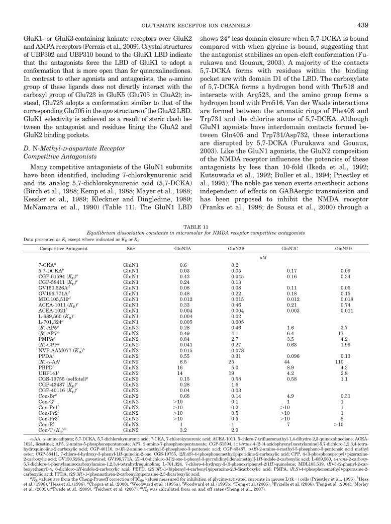
GluK1- or GluK3-containing kainate receptors over GluK2and AMPA receptors (Perrais et al., 2009). Crystal structuresof UBP302 and UBP310 bound to the GluK1 LBD indicatethat the antagonists force the LBD of GluK1 to adopt aconformation that is more open than for quinoxalinediones.In contrast to other agonists and antagonists, the �-aminogroup of these ligands does not directly interact with thecarboxyl group of Glu723 in GluK5 (Glu705 in GluA2); in-stead, Glu723 adopts a conformation similar to that of thecorresponding Glu705 in the apo structure of the GluA2 LBD.GluK1 selectivity is achieved as a result of steric clash be-tween the antagonist and residues lining the GluA2 andGluK2 binding pockets.
D. N-Methyl-D-aspartate ReceptorCompetitive Antagonists
Many competitive antagonists of the GluN1 subunitshave been identified, including 7-chlorokynurenic acidand its analog 5,7-dichlorokynurenic acid (5,7-DCKA)(Birch et al., 1988; Kemp et al., 1988; Mayer et al., 1988;Kessler et al., 1989; Kleckner and Dingledine, 1989;McNamara et al., 1990) (Table 11). The GluN1 LBD
shows 24° less domain closure when 5,7-DCKA is boundcompared with when glycine is bound, suggesting thatthe antagonist stabilizes an open-cleft conformation (Fu-rukawa and Gouaux, 2003). A majority of the contacts5,7-DCKA forms with residues within the bindingpocket are with domain D1 of the LBD. The carboxylateof 5,7-DCKA forms a hydrogen bond with Thr518 andinteracts with Arg523, and the amino group forms ahydrogen bond with Pro516. Van der Waals interactionsare formed between the aromatic rings of Phe408 andTrp731 and the chlorine atoms of 5,7-DCKA. AlthoughGluN1 agonists have interdomain contacts formed be-tween Gln405 and Trp731/Asp732, these interactionsare disrupted by 5,7-DCKA (Furukawa and Gouaux,2003). Like the GluN1 agonists, the GluN2 compositionof the NMDA receptor influences the potencies of theseantagonists by less than 10-fold (Ikeda et al., 1992;Kutsuwada et al., 1992; Buller et al., 1994; Priestley etal., 1995). The noble gas xenon exerts anesthetic actionsindependent of effects on GABAergic transmission andhas been proposed to inhibit the NMDA receptor(Franks et al., 1998; de Sousa et al., 2000) through a
TABLE 11Equilibrium dissociation constants in micromolar for NMDA receptor competitive antagonists
Data presented as Ki except where indicated as KB or Kd.
Competitive Antagonist Site GluN2A GluN2B GluN2C GluN2D
�M
7-CKAa GluN1 0.6 0.25,7-DCKAb GluN1 0.03 0.05 0.17 0.09CGP-61594 (KB)b GluN1 0.43 0.045 0.16 0.34CGP-58411 (KB)c GluN1 0.24 0.13GV150,526Ad GluN1 0.08 0.08 0.11 0.05GV196,771Ad GluN1 0.48 0.22 0.18 0.15MDL105,519d GluN1 0.012 0.015 0.012 0.018ACEA-1011 (KB)e GluN1 0.33 0.46 0.21 0.74ACEA-1021f GluN1 0.004 0.004 0.003 0.011L-689,560 (KB)c GluN1 0.004 0.02L-701,324a GluN1 0.005 0.005(R)-AP5g GluN2 0.28 0.46 1.6 3.7(R)-AP7g GluN2 0.49 4.1 6.4 17PMPAg GluN2 0.84 2.7 3.5 4.2(R)-CPPg GluN2 0.041 0.27 0.63 1.99NVP-AAM077 (KB)h GluN2 0.015 0.078PPDAi GluN2 0.55 0.31 0.096 0.13(R)-�-AAi GluN2 6.5 25 44 110PBPDi GluN2 16 5.0 8.9 4.3UBP141j GluN2 14 19 4.2 2.8CGS-19755 (selfotel)g GluN2 0.15 0.58 0.58 1.1CGP-43487 (KB)c GluN2 0.28 1.6CGP-40116 (KB)c GluN2 0.04 0.03Con-Brk GluN2 0.68 0.14 4.9 0.31Con-Gl GluN2 �10 0.1 1 1Con-Pr1l GluN2 �10 0.2 �10 1Con-Pr2l GluN2 �10 0.5 �10 1Con-Pr3l GluN2 �10 0.5 �10 8Con-Rl GluN2 1 1 7 �10Con-T (Kd)m GluN2 3.2 2.9
�-AA, �-aminoadipate; 5,7-DCKA, 5,7-dichlorokynurenic acid; 7-CKA, 7-chlorokynurenic acid; ACEA-1011, 5-chloro-7-trifluoromethyl-1,4-dihydro-2,3-quinoxalinedione; ACEA-1021, licostinel; AP5, 2-amino-5-phosphonopentanoate; AP7, 2-amino-7-phosphonopentanoate; CGP-61594, (�)-trans-4-2-(4-azidophyenyl)acetylamino-5,7-dichloro-1,2,3,4-tetra-hydroquinoline-2-carboxylic acid; CGP-40116, D-(E)-2-amino-4-methyl-5-phosphono-3-pentenoic acid; CGP-43487, D-(E)-2-amino-4-methyl-5-phosphono-3-pentenoic acid methylester; CGP-58411, 7-chloro-4-hydroxy-3-phenyl-1H-quinolin-2-one. CGS-19755, (2R,4S)-4-(phosphonomethyl)piperidine-2-carboxylic acid; CPP, 4-(3-phosphonopropyl) pizerazine-2-carboxylic acid; GV150,526A, gavestinel; GV196,771A, (E)-4,6-dichloro-3-(2-oxo-1-phenyl-3-pyrrolidinylidene)methyl-1H-indole-2-carboxylic acid; L-689,560, 4-trans-2-carboxy-5,7-dichloro-4-phenylaminocarbonylamino-1,2,3,4-tetrahydroquinoline; L-701,324, 7-chloro-4-hydroxy-3-(3-phenoxy)phenyl-2(1H)-quinolone; MDL105,519, (E)-3-(2-phenyl-2-car-boxyethenyl)-4, 6-dichloro-1H-indole-2-carboxylic acid; PBPD, (2S,3R)-1-(biphenyl-4-carbonyl)piperazine-2,3-dicarboxylic acid; PMPA, (R,S)-4-(phosphonomethyl)-piperazine-2-carboxylic acid; PPDA, (2S,3R)-1-(phenanthren-2-carbonyl)piperazine-2,3-dicarboxylic acid.
aKB values are from the Cheng-Prusoff correction of IC50 values measured for inhibition of glycine-activated currents in mouse L(tk�) cells (Priestley et al., 1995). bHesset al. (1998). cHess et al. (1996). dChopra et al. (2000). eWoodward et al. (1995a). fWoodward et al. (1995b). gFeng et al. (2005). hFrizelle et al. (2006). iFeng et al. (2004). jMorleyet al. (2005). kTwede et al. (2009). lTeichert et al. (2007). mKd was calculated from on and off rates (Sheng et al., 2007).
GLUTAMATE RECEPTOR ION CHANNELS 439

direct interaction within the glycine binding site (Dick-inson et al., 2007). Thus, xenon, like ketamine, maytarget the glutamatergic system to provide anestheticeffects.
The GluN2 competitive antagonist (R)-2-amino-5-phos-phonopentanoate and its analogs are used widely as phar-macological tools to distinguish NMDA receptor-mediatedactivity from AMPA and kainate receptor activity (Davieset al., 1981, 1986; Evans et al., 1981; Lester et al., 1990)(Table 11). Unfortunately, GluN2 subunit selectivity hasbeen difficult to achieve for competitive antagonists. Forexample, the antagonist 3-((R)-2-carboxypiperazin-4-yl)-propyl-1-phosphonic acid shows �50-fold preference forGluN2A over GluN2D but has intermediate affinities atGluN2B and GluN2C (Ikeda et al., 1992; Kutsuwada et al.,1992; Feng et al., 2005) (Table 11). The competitive antag-onist (R)-[(S)-1-(4-bromo-phenyl)-ethylamino]-(2,3-dioxo-1,2,3,4-tetrahydroquinoxalin-5-yl)-methyl-phosphonic acid(NVP-AAM077), originally reported to have more than100-fold selectivity for GluN2A-containing receptors overGluN2B-containing receptors, was used to evaluate phys-iological roles of GluN2A and GluN2B in rodent models ofsynaptic plasticity and neurotoxicity before a full pharma-cological characterization was completed (Liu et al., 2004b;Massey et al., 2004; Zhou and Baudry, 2006). Subsequentdetermination of KB from Schild analysis suggested thatthe selectivity was only �5-fold, precluding its use as aselective tool (Frizelle et al., 2006; Neyton and Paoletti,2006). Antagonists with bulky, hydrophobic substituents,such as phenanthrene-piperazine dicarboxylic acid ana-logs (2S,3R)-1-(phenanthren-2-carbonyl)piperazine-2,3-di-carboxylic acid and (2R,3S)-1-(phenanthrenyl-3-carbon-yl)piperazine-2,3-dicarboxylic acid (UBP141), show onlymodest 10-fold higher affinity for GluN2C- and GluN2D-containing receptors over GluN2A and GluN2B (Feng etal., 2004, 2005; Morley et al., 2005; Costa et al., 2009)(Table 11). Subunit selectivity for competitive antagonistsmay be difficult to achieve because of high homologyamong GluN2 subunit LBDs. Of the 39 residues lining thebinding pocket, only 8 are divergent in GluN2A toGluN2D. All 10 residues that come into direct contact with
glutamate are conserved between GluN2 subunits (Fu-rukawa et al., 2005; Kinarsky et al., 2005).
Conantokins, peptides of 17 to 27 amino acids that lackdisulfide bonds and are rich in �-carboxyglutamate resi-dues, have both competitive and noncompetitive antago-nist activity on NMDA receptors (Prorok and Castellino,2007). Conantokin-G is 20-fold more potent on GluN2Aand GluN2B-containing NMDA receptors than on GluN2Cand GluN2D-containing receptors (Wittekindt et al.,2001; Sheng et al., 2007; Teichert et al., 2007), andconantokin-Br is more potent for the GluN2D-contain-ing NMDA receptors than other conantokins (Table 11)(Twede et al., 2009). Although conantokin-G bindswithin the GluN2 ligand binding pocket, one moleculardeterminant of specificity of conantokin-G for GluN2Bhas been identified as a Met739 residue outside of thebinding pocket within the D2 domain of the LBD. Thisresidue is not conserved in GluN2A receptors but ispresent in GluN2C and GluN2D subunits (Teichert etal., 2007). Conantokin-R and conantokin-T are antago-nists of NMDA receptors, but antagonism is not GluN2subunit-specific (Klein et al., 2001; Sheng et al., 2007;Teichert et al., 2007). Conantokins R and G have beenstudied preclinically for their effectiveness in the treat-ment of chronic pain, stroke, and seizure (White et al.,2000; Layer et al., 2004; Hama and Sagen, 2009).
E. Noncompetitive Antagonists
Several classes of noncompetitive antagonists at AMPAreceptors have been used to selectively block AMPA but notkainate receptors, including 2,3-benzodiazepines and 1,2-di-hydrophthalazines, as well as tetrahydroisoquinolines (Gittoet al., 2003). However, the most potent of the 2,3-benzodiaz-epines, 1-(4-aminophenyl)-4-methyl-7,8-methylenedioxy-5H-2,3-benzodiazepine (GYKI-53655) (Table 12), blocks GluK2/GluK3 heteromeric and GluK3 homomeric receptors with anIC50 that is approximately 10-fold higher than at AMPAreceptors (Bleakman et al., 1996; Perrais et al., 2009). Incontrast to the competitive antagonists CNQX and DNQX,GYKI-53655 remains an effective AMPA receptor antagonistin the presence of TARPs, with the potency of the antagonist
TABLE 12IC50 values in micromolar for noncompetitive AMPA and kainate receptor antagonists
Noncompetitive Antagonist GluA1 GluA2 GluA3 GluA4 GluK1 GluK2 GluK3 GluK2/GluK5
�M
GYKI 52466 18–117a,b 34c 22–66a,b �100a �100a
GYKI 53405 (LY 293606) 24a 28a �100a �100a
GYKI 53773 (LY 300164)d 21e 18e 19e 18e �100a
GYKI 53655 (LY 300168) 6a 5a �100a 198f 63f �100a
GYKI 53784 (LY 303070)d 3a 3a �100a �100a �100a
CP-465,022 0.5g 0.5g 0.3g �100g �1h
NS-3763 1.6i �30i N.E.j
GYKI 52466, 1-(4-aminophenyl)-4-methyl-7,8-methylenedioxy-5H-2,3-benzodiazepine; GYKI 53405, 1-(4-aminophenyl)-3-acetyl-4-methyl-3,4-dihydro-7,8-methylenedioxy-5H-2,3-benzodiazepine; GYKI 53655, 1-(4-aminophenyl)-3-methylcarbamoyl-4-methyl-3,4-dihydro-7,8-methylenedioxy-5H-2,3 benzo-diazepine; GYKI 53784, (�)-1-(4-aminophenyl)-4-methyl-7,8-methylenedioxy-4,5-dihydro-3-methylcarbamoyl-2,3-benzodiazepine.
aBleakman et al. (1996). bJohansen et al. (1995). cIC50 value was determined from inhibition of Ca2� influx activated by 30 �M glutamate at humanGluA3 expressed in HEK69–8 cells (Varney et al., 1998). dThis compound is the active enantiomer of the compound directly above it. eCotton andPartin (2000). fPerrais et al. (2009). gBalannik et al. (2005). hLazzaro et al. (2002). iValues are for inhibition of Ca2� influx evoked by domoate (2 �Mfor GluK1 and 0.2 �M for GluK2) in HEK293 cells expressing recombinant human receptors (Christensen et al., 2004b). jValues are for inhibition ofglutamate-evoked currents at HEK293 cells expressing recombinant receptors (Christensen et al., 2004b).
440 TRAYNELIS ET AL.

increased (Menuz et al., 2007; Cokic and Stein, 2008). (S)-3-(2-Chlorophenyl)-2-[2-(6-diethylaminomethyl-pyridin-2-yl)-vinyl]-6-fluoro-3H-quinazolin-4-one (CP-465,022) (Menniti etal., 2000), an analog of quinazolinone, is approximately 100-fold more potent than GYKI-53655 at neuronal AMPA recep-tors (Bleakman et al., 1996; Menniti et al., 2000; Lazzaro etal., 2002) and seems 100-fold selective for AMPA over kainatereceptors (Lazzaro et al., 2002). Mutagenesis studies suggestthat GYKI-53655 and CP-465,022 share overlapping molec-ular determinants of action, requiring both S2–M4 and theS1–M1 linkers (Fig. 9; Balannik et al., 2005), the latter ofwhich may be a critical element in gating (Sobolevsky et al.,2009) (see section VII.B and VII.D). The mechanism of blockremains unclear but seems not to involve desensitization(Donevan and Rogawski, 1998).
A major advance in kainate receptor pharmacologywas made with the identification of arylureidobenzoicacids as noncompetitive GluK1 receptor antagonists(Valgeirsson et al., 2003, 2004). The most potent of these2-arylureidobenzoic acids is 4,6-bis(benzoylamino)-1,3-benzenedicarboxylic acid (NS-3763), a selective antago-nist of homomeric GluK1 (Table 12) that has no activityat heteromeric GluK1/GluK2 or GluK1/GluK5, AMPAreceptors, or NMDA receptors. NS-3763 exhibits 10-foldgreater potency in inhibiting glutamate-evoked currentsfrom GluK1–2b versus GluK1–1a, two splice variants
differing in 15 extracellular amino acid residues in theATD and a cytoplasmic domain (Christensen et al.,2004b). Because NS-3763 inhibits only homomericGluK1 receptors, it has been useful in exploring na-tive kainate receptor stoichiometry (Christensen etal., 2004a).
The first subunit-selective NMDA receptor antagonistwas the phenylethanolamine ifenprodil, which defined anew class of noncompetitive, voltage-independent par-tial antagonists (maximal inhibition �90%) of GluN2B-containing NMDA receptors (Table 13). Ifenprodil inhib-its GluN2B-containing receptors with high affinity andis 200 to 400-fold more potent for GluN1/GluN2B recep-tors than for GluN1/GluN2A, GluN1/GluN2C, or GluN1/GluN2D receptors (Williams, 1993; Hess et al., 1998;IC50 �150 nM). GluN1 splice variants do not influencethe ability of ifenprodil to inhibit GluN2B-containingreceptors (Gallagher et al., 1996), but triheteromericreceptors containing GluN1/GluN2A/GluN2B are pro-posed to be less sensitive to ifenprodil (Hatton and Pa-oletti, 2005), and insensitive to CP-101,606 (Brimecombeet al., 1997; Chazot et al., 2002). The kinetic properties ofifenprodil also are altered in triheteromeric receptors; themacroscopic association rate of ifenprodil in GluN1/GluN2A/GluN2B receptors was slower, whereas the disso-ciation rate was faster than in GluN1/GluN2B receptors
FIG. 9. Binding sites for the agonists, antagonists, and modulators described in sections V and VI are shown for the glutamate receptor. Thereceptor targets of ligands selective for one or several subunits are listed in parenthesis. AMPA and kainate indicates that the ligand selectivelytargets GluA or GluK receptor subunits, respectively. The ATDs, LBDs, TMDs, and linkers are shown in purple, orange, green, and gray, respectively.[Adapted from Sobolevsky AI, Rosconi MP, and Gouaux E (2009) X-ray structure, symmetry and mechanism of an AMPA-subtype glutamate receptor.Nature 462:745–756. Copyright © 2009 Nature Publishing Group. Used with permission.]
GLUTAMATE RECEPTOR ION CHANNELS 441

(Hatton and Paoletti, 2005). Ifenprodil and its more potentderivatives, including �-(4-hydroxyphenyl)-�-methyl-4-(phenylmethyl)-1-piperidine propanol (Ro 25-6981), 1-[2-(4-hydroxy-phenoxy)-ethyl]-4-(4-methyl-benzyl)-piperidin-4-ol (Ro 63-1908), besonprodil (CI-1041), and traxoprodilmesylate (CP-101,606) (Gotti et al., 1988; Williams,1993; Chenard et al., 1995; Fischer et al., 1997; Mott etal., 1998; Gill et al., 2002; Barton and White, 2004)(Table 13) are thought to interact with the GluN2B ATDand have been proposed to act by stabilizing an agonist-bound state in which the receptor has a low open prob-ability (Kew et al., 1996, 1998; Fischer et al., 1997;Perin-Dureau et al., 2002) (Fig. 9). Ifenprodil inhibitionis incomplete at saturating concentrations, and ifen-prodil binds to a known modulatory domain, suggestingits actions might also be considered negative allostericmodulation, although it is widely referred to in the lit-erature as a noncompetitive antagonist. Similar to Zn2�
binding to the ATD of GluN2A (see section VI.E), ifen-prodil increases the potency of proton inhibition ofNMDA receptors (Pahk and Williams, 1997; Mott et al.,1998). Rich pharmacology exists for this site, withnearly a dozen structural classes described, includingoxamides (Barta-Szalai et al., 2004), 4-(3,4-dihydro-1H-isoquinolin-2-yl)-quinolines (Buttelmann et al., 2003),benzamidines (Claiborne et al., 2003), 5-substituted ben-zimidazoles (McCauley et al., 2004), indole-2-carboxam-ides (Borza et al., 2006, 2007), benzyl cinnamamidines(Tamiz et al., 1999; Curtis et al., 2003), and other biarylanalogs (Tamiz et al., 1998; Wright et al., 2000; Tahi-rovic et al., 2008; Mosley et al., 2009).
GluN2B-selective antagonists have been implicated inthe treatment of a variety of diseases and neurologicaldisorders (see section X). However, development ofGluN2B-selective antagonists has been hindered bytheir activity at �1-adrenergic receptors, serotonin re-ceptors, calcium channels, and hERG potassium chan-nels (Lynch and Gallagher, 1996). The problem of targetselectivity has been partially overcome by potent ifen-prodil analogs such as Ro 25-6981 and CP-101,606(Fischer et al., 1997; Taniguchi et al., 1997; Tahirovic et
al., 2008). Off-target activity at hERG channels byGluN2B antagonists is decreased by eliminating basicnitrogen atoms; increasing the number of oxygen at-oms within the linker of GluN2B antagonists alsodecreases affinity for hERG channels and �1-adrener-gic receptors (Kawai et al., 2007; Mosley et al., 2009).The GluN2B selective antagonist (3S,4R)-4-methyl-benzyl 3-fluoro-4-((pyrimidin-2-ylamino)methyl)pip-eridine-1-carboxylate (MK-0657) unexpectedly in-creased both systolic and diastolic blood pressure. It isunclear whether these effects occur peripherally orcentrally (Addy et al., 2009; Mony et al., 2009).
Certain quinazolin-4-one analogs of the noncompeti-tive AMPA receptor antagonist CP-465,022 can also in-hibit recombinant NMDA receptors in a voltage-inde-pendent and noncompetitive fashion (Mosley et al.,2010). These data suggest that a new site for noncom-petitive antagonism may exist on recombinant NMDAreceptors. Some quinazolin-4-ones are greater than 50-fold more potent at recombinant NMDA receptors thatcontain GluN2C/GluN2D subunits over NMDA recep-tors that contain GluN2A/GluN2B subunits or AMPA/kainate receptors. These compounds could provide astarting point for the development of new classes ofsubunit-selective antagonists for NMDA receptors thatcontain GluN2C or GluN2D subunits.
Ethanol has been proposed to be a noncompetitiveantagonist of NMDA receptors, with sensitivity depen-dent upon the GluN2 subunit (Lovinger et al., 1989,1990; Kuner et al., 1993; Masood et al., 1994; Mirshahiand Woodward, 1995; Kash et al., 2008; Nagy, 2008) butindependent of GluN1 splice variant, pH, Zn2�, or redoxstate (Kuner et al., 1993; Chu et al., 1995; Peoples andWeight, 1999). A wide range of studies has suggestedthat actions at native NMDA receptors contribute to thecentral effects of alcohol, which may involve perturba-tion of the interaction between glutamate and dopamine(Lovinger, 2002; Maldve et al., 2002). However, recentstudies suggest that recombinant NMDA receptor inhi-bition is modest at the U.S./U.K. drink-drive limit aswell as at intoxicating levels (Otton et al., 2009). Dynor-phin peptides have been reported to have a variety ofactions on NMDA receptors, acting as noncompetitiveantagonists (Chen and Huang, 1998; Kanemitsu et al.,2003; Oz et al., 2004) as well as potentiators under somecircumstances (Zhang et al., 1997; Caudle and Dubner,1998; Lai et al., 1998). Dynorphin A (1–17) and (2–17)can displace glycine at GluN1 (Voorn et al., 2007) andcan bind to an anionic intracellular epitope on GluN1that may interact with dopamine receptors (Jackson etal., 2006; Woods et al., 2006). Potency for Dynorphin A(1–13) is dependent upon GluN2 subunit, with the pep-tide being most potent for GluN2A-containing NMDAreceptors (Brauneis et al., 1996). Longer dynorphin pep-tides are more potent inhibitors [e.g., Dynorphin A (1–32); Chen and Huang, 1998]. Inhibition by dynorphin A
TABLE 13IC50 values in micromolar for noncompetitive GluN2B-selective NMDA
receptor antagonistsData are for rat recombinant receptors, except for ifenprodil, which was character-ized on human recombinant receptors. Data for ifenprodil are from Hess et al. (1998).Values for Ro 25-6981 are from Fischer et al. (1997) and values for CP 101,606 arefrom Mott et al. (1998). Values for eliprodil are from Avenet et al. (1997), and valuesfor felbamate are from Harty and Rogawski (2000). Values for haloperidol are fromIlyin et al. (1996).
NoncompetitiveAntagonist GluN2A GluN2B GluN2C GluN2D
�M
Ifenprodil 39 0.15 29 76Ro 25-6981 52 0.0090CP-101,606 �100 0.039 �100 �100Eliprodil �100 3.02Felbamate 2600 520 2400Haloperidol N.E. 3 N.E. N.E.
CP-101,606, traxoprodil mesylate; N.E., �10% inhibition at 300 �M.
442 TRAYNELIS ET AL.

is pH sensitive in that inhibition increases at lower pH(Kanemitsu et al., 2003; Oz et al., 2004).
F. Uncompetitive Antagonists
Open channel blockers require that the receptor porebe open to allow access to the blockers’ binding site tocause subsequent block of receptor activity (Neely andLingle, 1986; Huettner and Bean, 1988; Kiskin et al.,1989) (Fig. 9). Because of this requirement, the onset ofinhibition is use-dependent and increases with increas-ing channel open probability. After channel closure,some blockers can become trapped in the pore, and theseantagonists are called trapping blockers [PCP, dizo-cilpine maleate (MK-801), ketamine at NMDA receptors,1-trimethylammonio-5-(1-adamantane-methylammonio-pentane dibromide) (IEM-1460) at AMPA receptors] andpartially trapping blockers (memantine at NMDA recep-tors). Channel block by trapping blockers is slow toreverse and requires channel reactivation by agonistsbefore the blocker can dissociate (Brackley et al., 1993;Parsons et al., 1995; Blanpied et al., 1997; Magazanik etal., 1997).
A large number of naturally occurring AMPA andkainate receptor channel blockers, as well as a host ofsynthetic analogs, have been identified (Table 14), in-cluding argiotoxin-636 (Herlitze et al., 1993), Joro spidertoxin (Blaschke et al., 1993), Ageltoxin-489 (Washburnand Dingledine, 1996), philanthotoxin-433 (Jones et al.,1990), IEM-1460 (Magazanik et al., 1997), and N1-naph-thylacetylspermine (Koike et al., 1997), which alsoblocks mutant Lurcher GluD2 channels. Some of thesecompounds have nonspecific actions at other ion chan-nels (Welch et al., 2008). All of these uncompetitiveantagonists have structural similarity (i.e., a polyaminemoiety) and, when applied extracellularly, exhibit volt-age-dependent block. These compounds act primarily onGluA2-lacking Ca2�-permeable AMPA receptors, al-though Joro spider toxin and philanthotoxin also blockunedited GluK2 channels (Blaschke et al., 1993; Bahr-ing and Mayer, 1998). The QRN site at the apex of the
M2 reentrant pore-lining loop is a key structural deter-minant of polyamine block (see sections II.E and VIII),with receptors lacking the edited GluA2(R) subunitsshowing strong block by polyamines and toxins. Thus,these channel blockers have been useful pharmacologi-cal tools to probe the subunit composition of AMPAreceptors (Laezza et al., 1999; Liu and Cull-Candy, 2000;Plant et al., 2006), although many also show actions atkainate receptors. The amino groups of these compoundsinteract with residues that reside deeper in the porethan the QRN site, including the main-chain oxygenatom from the QRN � 2 site (Tikhonov et al., 2002), andat least two amino groups are required for potent antag-onism at AMPA receptors (Bolshakov et al., 2005). Somecompounds (e.g., phenylcyclohexyl derivative IEM-1925)can permeate the channel, allowing closed channels toescape from block (Tikhonova et al., 2008). Other block-ers [e.g., adamantane derivative IEM-1676 (Tikhonovaet al., 2008)] produce a voltage-dependent closed chan-nel block from the intracellular compartment in additionto open channel block from the extracellular compart-ment (Tikhonova et al., 2009). Association of AMPAreceptors with TARPs �2, �3, and �8 reduces channelblock by N1-naphthylacetylspermine (Kott et al., 2009),an intriguing finding because TARPs also increase chan-nel opening frequency (Tomita et al., 2005a) (see sectionII.H).
Structure-activity relationships of philanthotoxinshave highlighted the importance of the polyamine moi-ety and led to potent and selective AMPA receptor block-ers. Shortening the polyamine chain of PhTX-343 causeda marked decrease in potency at AMPA receptors (Mel-lor et al., 2003). Moreover, replacing the two secondaryamines in the polyamine moiety with either oxygen ormethylene resulted in a complete loss of activity,whereas replacing only one with methylene improvedpotency 15-fold and increased selectivity for AMPA ver-sus NMDA receptors to 100-fold (Mellor et al., 2003).Further modification of the polyamine tail of PhTX-343resulted in PhTX-56 and PhTX-74, which differ in the
TABLE 14IC50 values in micromolar for uncompetitive AMPA receptor antagonists
All data from GluA2 are from the edited form GluA2(R).
Uncompetitive Antagonist GluA1 GluA2 GluA3 GluA4 GluA1/GluA2
�M
Argiotoxin 636 0.35–3.4a,b N.E.a 0.23a 0.43a 300b
Joro spider toxin 0.04c N.E.c 0.03c N.E.c
Philanthotoxin 433d 0.8 N.E.Philanthotoxin 343 2.8b 270b
Philanthotoxin 56 3.3 pMe 5.2e
Philanthotoxin 74 0.17e 1.6e
IEM-1460 1.6f N.E.g 1.6f
IEM-1754 6.0f 6.0f
HPP-spermined 0.5 0.08 0.5 N.E.
HPP-spermine, N-(4-hydroxyphenylpropanoyl)spermine trihydrochloride; IEM-1754, 1-ammonio-5-(1-adamantane-methylammoniopentane dibromide); N.E., no effect.aHerlitze et al. (1993); holding membrane potential was �70 mV. bBrackley et al. (1993); holding membrane potential, �80 mV. cBlaschke et al. (1993); holding membrane
potential, �100 mV. dWashburn and Dingledine (1996); holding membrane potential, �70 mV. Although IC50 values were not calculated, initial experiments suggested thatphilanthotoxin-433 had a lower affinity for GluA1 and GluA4 receptors compared with GluA3. ePhilanthotoxin 56 has an IC50 of 3.3 pM for recombinant GluA1 (Kromannet al., 2002); holding membrane potential, �80 mV. fMagazanik et al. (1997); holding membrane potential, �80 mV. gSchlesinger et al. (2005); holding membrane potential,�60 mV, recombinant human GluA2(R).
GLUTAMATE RECEPTOR ION CHANNELS 443

number of amines and intervening methylenes (Kro-mann et al., 2002). PhTX-56 is highly selective for Ca2�-permeable AMPA receptors, being 1000-fold more potentat GluA2-lacking receptors. PhTX-56 is 500-fold selec-tive for AMPA over kainate receptors (Kromann et al.,2002). PhTX-74 inhibits GluA1/GluA2 but not GluA2/GluA3 receptors (Nilsen and England, 2007).
Uncompetitive NMDA receptor antagonists includeMg2�, polyamines (see section VIII.C), dissociative an-esthetics phencyclidine and ketamine, MK-801, amino-adamantane derivatives memantine and amantadine,pentamidine, 9-tetrahydroaminoacridine, dextrometho-rphan, and its metabolite dextrorphan (Table 15). It isnoteworthy that pentamidine and 9-tetrahydroamino-acridine also inhibit currents of the mutant LurcherGluD2 receptors (Williams et al., 2003). The structure-activity relationship underlying the trapping nature ofblockers is unrelated to lipophilicity, and thus blockersare not capable of escaping through the membrane(Mealing et al., 2001; Bolshakov et al., 2005). Partiallytrapping blockers, such as memantine and amantadine,bind after channel opening. However, these drugs alsounbind rapidly (Blanpied et al., 1997, 2005; Chen andLipton, 1997; Mealing et al., 1999), which has been pro-posed to be therapeutically beneficial, because normalsynaptic transmission may not be influenced by thedrug, but overactivation of NMDA receptors should bedecreased (Chen and Lipton, 2006) (see section X.G).Memantine may access multiple binding sites, one ofwhich has been proposed to reside superficially near theextracellular end of the pore in GluN1/GluN2A. Occu-pancy of this superficial site may prevent full channelclosure (Sobolevsky and Koshelev, 1998; Sobolevsky etal., 1998; Bolshakov et al., 2003); however, more work isneeded to determine the exact location with respect to
the gate of this superficial site. The presence of thesuperficial binding site also could be relevant for im-proved side effect profile for memantine (Kotermanski etal., 2009).
Most NMDA receptor channel blockers are eithernonselective or at best weakly selective (�10-fold) forspecific GluN2 subtypes (Yamakura et al., 1993). Forexample, MK-801 is �10-fold more potent for GluN2A-and GluN2B-containing receptors than GluN2C- andGluN2D-containing receptors (Bresink et al., 1996;Dravid et al., 2007). However, aryl-polyamine deriva-tives show high potency as well as slightly better sub-unit selectivity, with N1-dansyl-spermine and thetribenzyltriamine TB-3-4 showing approximately 40-fold lower IC50 values at GluN2A- than GluN2D-con-taining NMDA receptors (Chao et al., 1997; Igarashi etal., 1997; Jin et al., 2007). The molecular determinantsof activity of channel blockers are not identical but over-lap, with dependence on residues within the M2 reen-trant pore region and residues in other pore-formingelements as well as the pre-M1 region (Yamakura et al.,1993; Yamakura and Shimoji, 1999; Kashiwagi et al.,2002; LePage et al., 2005; Jin et al., 2007). The sequencesimilarity within the pore-forming elements of NMDAreceptors may constitute a challenge in the future de-velopment of subunit-selective compounds. It is note-worthy that the potencies of channel blocking com-pounds are sensitive to pH. Although acidic pH reducesNMDA receptor open probability (Traynelis and Cull-Candy, 1990), acidic pH paradoxically increases the as-sociation rate of the trapping blocker MK-801, whichmay reflect an interaction of MK-801 with the structuralelements forming the gate of the receptor (Dravid et al.,2007). Several reports raise the possibility that trappingblockers do not passively reside in the pore when their
TABLE 15IC50 values in micromolar for uncompetitive NMDA receptor antagonists
All values were measured in 0 Mg2�, unless otherwise indicated. Values for memantine and (�)-ketamine are from Kotermanski and Johnson (2009) with membranepotential held at �66 mV. All remaining values are from Dravid et al. (2007) with membrane potential held at �40 mV.
Uncompetitive Antagonist GluN2A GluN2B GluN2C GluN2D
�M
(�)-MK-801 0.015 0.009 0.024 0.038(�)-MK-801 0.35 0.32 0.038 0.17(�)-Ketamine 16 1.6 1.1 1.5(�)-Norketamine 51 8.7 5.6 7.5Dextromethorphan 11 3.7 1.1 5.4Levomethorphan 13 2.2 1.1 2.6Dextrorphan 1.3 0.33 0.15 0.74Levorphanol 1.8 1.2 0.58 2.1Phencyclidine 0.82 0.16 0.16 0.22PCA 19 3.9 1.6 1.7CNS-1102 0.13 0.068 0.087 0.14Amantadine 130 70 35 38Remacemide 81 35 92 63Pentamidine 0.72 1.5 10 9.19-aminoacridine 7.8 7.5 29 38Memantine 0.80 0.57 0.52 0.54Memantine–1 mM Mg2� 13 10 1.6 1.8(�)-Ketamine 0.33 0.31 0.51 0.83(�)-Ketamine–1 mM Mg2� 5.4 5.08 1.2 2.9
CNS-1102, aptiganel; PCA, 1-phenylcyclohexylamine.
444 TRAYNELIS ET AL.

exit path is removed as a result of channel closure, butrather stabilize the closed state, thereby promotingchannel closure and their own subsequent trapping(Blanpied et al., 2005; Yuan et al., 2005). Recent workconducted on ketamine and memantine indicate thatthese antagonists may be more selective at physiologicalconditions than previously reported, because 1 mMMg2� produced a 5- to 10-fold selectivity for GluN2C-and GluN2D-containing receptors over GluN2A andGluN2B (Table 15; Kotermanski and Johnson, 2009).These findings may be clinically and functionally signif-icant, depending on concentrations of memantinereached in brain. These results also emphasize the needto study pharmacology under physiological conditions.
VI. Allosteric Regulation
Allosteric modulators for glutamate receptors haveattracted attention because of their ability to fine-tunenormal receptor function as well as for their potentialutility as therapeutic agents. Drug-like positive or neg-ative allosteric modulators have distinct therapeutic ad-vantages over agonists and competitive antagonists, in-cluding higher potential for receptor subtype selectivity,because they often target less conserved regions thanthe agonist binding site (Figs. 2 and 9). Clinically, mod-ulators are generally thought to be better tolerated be-cause they modify existing levels and patterns of recep-tor activation, rather than constitutively blocking (aswith antagonists) or overactivating (as with agonists) allreceptors.
A. Positive and Negative Allosteric Modulators
A growing range of compounds potentiate AMPA re-ceptor activity through modification of the deactivationand/or desensitization time course (Arai et al., 1994;Staubli et al., 1994; Lauterborn et al., 2000). Binding ofAMPA receptor agonists within the cleft of the LBDinduces closure around the ligand, which has been pro-posed to create strain within the receptor complex thatcan be relieved by channel opening or relaxation of theLBD dimer interface to a desensitized state, in whichagonist remains bound but the channel is closed (Sun etal., 2002; Sobolevsky et al., 2009) (Fig. 3). The receptordeactivates as the channel closes and the ligand fullyunbinds, exiting the binding cleft. Positive AMPA recep-tor modulators bind at the LBD dimer interface, makinga number of intraprotomer atomic contacts that stabilizethe dimerized configuration and prevent transition tothe relaxed state after agonist binding, thus preventingdesensitization (Figs. 3 and 10B). Some of these com-pounds also can slow the rate of agonist exit, thus pre-venting deactivation.
At present, there are three structural classes of positiveAMPA receptor modulators: 1) pyrrolidinone and relatedpiperidine compounds [e.g., aniracetam, piracetam, oxirac-etam, and the ampakines 2H,3H,6aH-pyrrolidino(2�,1�-3�,
2�)1,3-oxazino(6�,5�-5,4)benzo(e)1,4-dioxan-10-one (CX614),ampalex (CX516), and 1-(1,4-benzodioxan-6-ylcarbonyl)piperidine (CX546)], 2) benzothiadiazide compounds[e.g., cyclothiazide, diazoxide, (S)-2,3-dihydro-(3,4)cyclo-pentano-1,2,4-benzothiadiazine-1,1-dioxide (S18986), and7-chloro-3-methyl-3,4-dihydro-2H-1,2,4-benzothiadiazine-S,S-dioxide (IDRA-21)], and 3) biarylpropylsulfonamidecompounds [e.g., PEPA, N-[2-(4�-cyanobiphenyl-4-yl)pro-pyl]propane-2-sulfonamide (LY404187), N-(2-(4-(thiophen-3-yl)phenyl)propyl)propane-2-sulfonamide (LY392098), (R)-2-(4-(3,5-difluorobenzoylamino)phenyl)-1-(2-propanesulfon-amido)-propane (LY450108), LY451395, and LY503430] (Itoet al., 1990; Copani et al., 1992; Arai et al., 1996, 2000;Baumbarger et al., 2001a,b). Although positive AMPA recep-tor modulators fall into three structural classes, the mostimportant difference between them is their mechanisms ofaction.
Aniracetam primarily slows the deactivation ratewithout changing agonist potency. In contrast, PEPApotentiates AMPA receptor function by attenuating theextent of receptor desensitization without any effect ondeactivation (Sekiguchi et al., 2002). Cyclothiazideseems to slow the onset of desensitization and indirectlyslow the channel closure rate by increasing agonist po-tency (Yamada and Tang, 1993; Partin et al., 1994, 1996;Sekiguchi et al., 2002; Arai and Kessler, 2007).LY404187 stabilizes the open state of AMPA receptorsin the presence of agonist without altering the rate atwhich channels desensitize, thereby permitting desensi-tized AMPA receptors to make a transition to an openstate either directly or through intermediate desensi-tized and/or closed states (Baumbarger et al., 2001b).Some compounds (e.g., CX614) inhibit both desensitiza-tion and deactivation; the mechanism underlying this isnot well understood (Arai et al., 2000).
Further complexity arises from differences in the po-tencies of positive allosteric modulators for RNA splicevariants of the AMPA receptors, particularly the flip/flop region within the LBD. Cyclothiazide almost com-pletely eliminates desensitization of flip splice variantsbut only slows the entry into desensitized state for theflop splice variant (Johansen et al., 1995; Partin et al.,1996; Hennegriff et al., 1997; Sekiguchi et al., 2002). Thespecificity of cyclothiazide for flip splice variants arisesfrom its interaction with Ser754, which is a larger Asn inflop variants and precludes tight cyclothiazide binding(Sun et al., 2002). Aniracetam has a similar potency atflip and flop splice forms, but the efficacy is greater forthe flop form (Johansen et al., 1995). PEPA and CX614are selective for the flop variants (Hennegriff et al.,1997; Sekiguchi et al., 1997, 1998). LY404187 andLY503430 suppress receptor desensitization with a dis-tinct time-dependence in the presence of agonist; thesecompounds show the highest potency for flip variants ofGluA2 and GluA4 (Miu et al., 2001; Murray et al., 2003).
The functional effects of the different positive alloste-ric modulators are complex because multiple partially
GLUTAMATE RECEPTOR ION CHANNELS 445
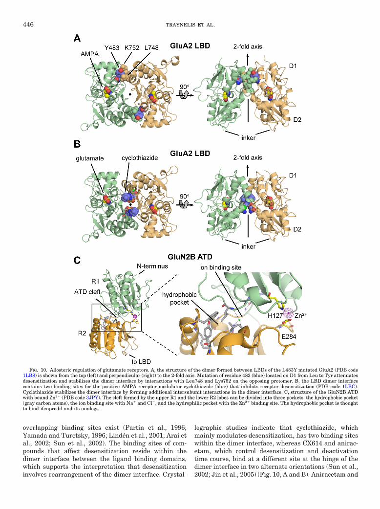
overlapping binding sites exist (Partin et al., 1996;Yamada and Turetsky, 1996; Linden et al., 2001; Arai etal., 2002; Sun et al., 2002). The binding sites of com-pounds that affect desensitization reside within thedimer interface between the ligand binding domains,which supports the interpretation that desensitizationinvolves rearrangement of the dimer interface. Crystal-
lographic studies indicate that cyclothiazide, whichmainly modulates desensitization, has two binding siteswithin the dimer interface, whereas CX614 and anirac-etam, which control desensitization and deactivationtime course, bind at a different site at the hinge of thedimer interface in two alternate orientations (Sun et al.,2002; Jin et al., 2005) (Fig. 10, A and B). Aniracetam and
FIG. 10. Allosteric regulation of glutamate receptors. A, the structure of the dimer formed between LBDs of the L483Y mutated GluA2 (PDB code1LB8) is shown from the top (left) and perpendicular (right) to the 2-fold axis. Mutation of residue 483 (blue) located on D1 from Leu to Tyr attenuatesdesensitization and stabilizes the dimer interface by interactions with Leu748 and Lys752 on the opposing protomer. B, the LBD dimer interfacecontains two binding sites for the positive AMPA receptor modulator cyclothiazide (blue) that inhibits receptor desensitization (PDB code 1LBC).Cyclothiazide stabilizes the dimer interface by forming additional intersubunit interactions in the dimer interface. C, structure of the GluN2B ATDwith bound Zn2� (PDB code 3JPY). The cleft formed by the upper R1 and the lower R2 lobes can be divided into three pockets: the hydrophobic pocket(gray carbon atoms), the ion binding site with Na� and Cl�, and the hydrophilic pocket with the Zn2� binding site. The hydrophobic pocket is thoughtto bind ifenprodil and its analogs.
446 TRAYNELIS ET AL.
CX614 bind to the center of the dimer interface and donot penetrate the ligand binding domain clamshell. Thebinding sites for aniracetam/CX614 and cyclothiazide dooverlap, with the endocyclic sulfonamide group fromcyclothiazide overlapping with the site recognizing thefive-member rings of CX614/aniracetam. Aniracetamand CX614 stabilize the closed-cleft conformation of theligand binding domain, slowing the deactivation timecourse. Cyclothiazide, which does not significantly sta-bilize the closed-cleft conformation of the LBD, insteadstabilizes the dimer assembly, leading to an attenuationof desensitization. These data indicate that the molecu-lar and structural determinants of deactivation and de-sensitization are separable (Sun et al., 2002; Jin et al.,2005). The challenge is now to understand how thesemechanistically distinct compounds differentially affectbrain circuitry to produce effects on behavior that aretherapeutically meaningful (Lynch, 2006).
Con A, a plant lectin from Canavalia ensiformis, irre-versibly potentiates agonist-evoked currents from mostkainate receptors by apparently reducing receptor de-sensitization and increasing agonist affinity (Huettner,1990; Partin et al., 1993; Wong et al., 1994; Bleakman etal., 2002). Lectins seem to bind to N-linked glycosylationwithin the ATD (Everts et al., 1997, 1999). The action ofcon A is state-dependent, because agonist-induced de-sensitization before application of con A eliminates po-tentiation (Fay and Bowie, 2006). Con A has been sug-gested to keep the activated channel in the open stateand inhibit conformational changes leading to the de-sensitized state (Partin et al., 1993; Wong and Mayer1993; Yue et al., 1995). Alternatively, con A may shiftthe contribution of different kainate receptor open states(Bowie et al., 2003). Although con A has proven experi-mentally useful for characterizing kainate receptorpharmacology, it does not alter synaptic kainate recep-tor currents and only weakly potentiates whole-cell cur-rents from cultured hippocampal neurons (Wilding andHuettner, 1997). Other lectins, however, includingwheat germ agglutinin, soybean agglutinin, and succi-nyl-concanavalin A, potentiate native kainate receptorfunction (Thio et al., 1993; Yue et al., 1995).
B. Divalent Ions
Multiple divalent cations influence glutamate recep-tor inactivation, voltage-dependent channel block, andagonist dissociation rates, in addition to potentiatingand inhibiting receptor responses. Among divalent cat-ions, extracellular Zn2� most potently inhibits native(Peters et al., 1987; Westbrook and Mayer, 1987) andrecombinant NMDA receptors (Williams et al., 1996; Chenet al., 1997; Paoletti et al., 1997; Traynelis et al., 1998). Theendogenous ion zinc is packaged into synaptic vesicles inaxons terminating in the hippocampus, striatum, amyg-dala, neocortex, and cortex (Perez-Clausell and Danscher,1985; Frederickson, 1989; Valente et al., 2002; Danscherand Stoltenberg, 2005; Paoletti et al., 2009) and may be
coreleased with glutamate from the vesicles into the syn-aptic cleft during neuronal activity. The expression ofGluN2A- and GluN2B-containing NMDA receptors mayallow zinc to act as an endogenous modulator of the NMDAreceptor, depending on its synaptic concentration. Zn2�
inhibition curves for GluN1/GluN2A receptors are bipha-sic, revealing high-affinity voltage-independent inhibition(IC50 10–30 nM) and low-affinity voltage-dependent chan-nel block (IC50 20–100 �M). In the absence of voltage-dependent channel block, Zn2� inhibition is incompleteeven at saturating concentrations. The high-affinity Zn2�
binding site resides within the cleft of the GluN2A bilobedATD and most likely involves Zn2�coordination by a seriesof histidine residues (Choi and Lipton, 1999; Fayyazuddinet al., 2000; Low et al., 2000). Removal of the GluN2A ATDby mutagenesis or alteration of this domain after cleavageby the endogenous serine protease plasmin at Lys317 elim-inates or reduces voltage-independent high-affinity Zn2�
inhibition, respectively (Gielen et al., 2009; Yuan et al.,2009a,b). Low-affinity voltage-dependent Zn2� inhibitioninvolves residues inside the reentrant M2 pore loop similarto the Mg2� blocking site (Legendre and Westbrook, 1990;Paoletti et al., 2000).
Micromolar concentrations of Zn2� inhibit GluN2B-containing receptors in both a voltage-independent andvoltage-dependent manner (Williams, 1996; Traynelis etal., 1998; Choi and Lipton, 1999; Rachline et al., 2005).Crystallographic coordinates of the GluN2B ATD showthat a Zn2� binding site exists within the cleft of theclamshell of the ATD (Fig. 9C) (Karakas et al., 2009),and mutagenesis suggests that this site can account forvoltage-independent inhibition. As hypothesized forhigh-affinity binding of Zn2� to the GluN2A ATD (Pa-oletti et al., 2000), crystallographic studies of GluN2Bshow Zn2� binding stabilizes a closed-cleft conformationwithin the ATD through direct contact with residuesHis127 and Glu284. Mutation of these residues does notinfluence inhibition by the GluN2B-selective antagonistifenprodil, suggesting that Zn2� and ifenprodil bind atunique sites within the GluN2B ATD. Residues Glu47and Asp265, although not directly involved in Zn2� bind-ing, influence Zn2� sensitivity, perhaps through coordi-nation of water molecules that can interact with Zn2�
(Karakas et al., 2009). It is noteworthy that mutations atGluN2B His127, one of the residues in contact withZn2�, also block potentiation of GluN2B receptors byNi2� (Marchetti and Gavazzo, 2005; Gavazzo et al.,2009).
Studies with covalent modification of mutant GluN2Areceptors that harbor a cysteine residue within the ATDcleft raise the idea that the extent of ATD closure aroundthe Zn2� binding cleft negatively correlates with openprobability, perhaps through actions at the LBD dimerinterface (Gielen et al., 2009). Specifically, high-affinityZn2� inhibition of NMDA receptors has been proposed toinvolve domain closure within the bilobed ATD aroundZn2�, and this has been suggested to destabilize the
GLUTAMATE RECEPTOR ION CHANNELS 447

dimer interface of the LBD (Mayer, 2006; Gielen et al.,2008) analogous to desensitization of AMPA and kainatereceptors (Armstrong et al., 2006; Weston et al., 2006b;Plested and Mayer, 2007) (see sections II.D and VII.E).In support of this idea, mutations that destabilize theLBD dimer interface enhance Zn2� sensitivity, whereascysteine cross-linking of the dimer interface of mutantGluN1 and GluN2A receptors reduce Zn2� inhibition(Gielen et al., 2008). In addition to effects at the dimerinterface, Zn2� shifts the proton inhibition curve left-ward, suggesting that Zn2� binding to the ATD in-creases proton sensitivity (discussed below in sectionVI.D) and consequently increases the proportion of pro-tonated, nonfunctional receptors at physiological pH(Choi and Lipton, 1999; Low et al., 2000). Fitting bothmacroscopic and single-channel data with kinetic mod-els containing explicit Zn2� and proton binding stepssuggests that association of Zn2� with its GluN2A bind-ing site enhances proton binding (Erreger and Traynelis,2005, 2008). Mutations at the same LBD dimer interfaceresidues that alter Zn2� inhibition also strongly influ-ence proton inhibition (Gielen et al., 2008), supporting afunctional link between these two forms of modulationand confirming the hypothesis that Zn2� binding en-hances proton inhibition. The potential link betweenproton inhibition and NMDA receptor LBD dimer inter-face stability fits well with previous observations thatprotons modify GluA receptor desensitization (Ihle andPatneau, 2000; Lei et al., 2001), a process unequivocallyshown to involve rearrangement at the LBD dimer in-terface (see sections II.D and VII.E). Chen et al. (1997)first described a rapid component of desensitization inthe presence of extracellular Zn2� and postulated thatZn2� accelerated receptor desensitization. Subsequentstudies, drawing from the mechanism of glycine-depen-dent desensitization (see section VII.E), showed that apositive intrasubunit interaction occurs between gluta-mate binding to the LBD and Zn2� binding to the ATD(Zheng et al., 2001). The working hypothesis, supportedby multiple lines of evidence, suggests that binding ofglutamate enhances Zn2� binding, which at subsaturat-ing concentrations of Zn2� causes a relaxation to a newequilibrium as Zn2� binds to the receptor in a concen-tration-dependent fashion (Zheng et al., 2001; Erregerand Traynelis, 2005). Thus, the Zn2�-dependent desen-sitization time course reflects the time course for Zn2�
association with GluN2A receptors after a shift of theZn2� binding site into a high-affinity state.
Calcium, barium, magnesium, and zinc ions can in-hibit or potentiate current responses of recombinant andnative AMPA receptors (Rassendren et al., 1990; Bres-ink et al., 1996; Dreixler and Leonard, 1997; Shen andYang, 1999; Zhang et al., 2002; Blakemore and Tromb-ley, 2004; Kreitzer et al., 2009) and kainate receptors(Hoo et al., 1994; Fukushima et al., 2003; Mott et al.,2008). Zn2� and Ca2� can permeate through AMPA/kainate receptors (Sensi et al., 1999; Weiss and Sensi,
2000) (see section VIII.B). Of these various actions, kai-nate receptors are most sensitive to inhibition by Zn2�,which exerts voltage-independent inhibition, suggestingit acts as a noncompetitive antagonist (Fukushima et al.,2003). Zinc inhibition of kainate receptors is subunit-dependent, because GluK4- and GluK5-containing re-ceptors have lower IC50 values (1–2 �M) than GluK1–3receptors (�70 �M). Inhibition by Zn2� is also depen-dent upon pH, because increasing the proton concentra-tion decreases Zn2� potency by 2- to 9-fold for recombi-nant receptors and 15-fold for synaptic receptors (Mottet al., 2008). One possible mechanism for proton-depen-dent zinc inhibition of kainate receptors is that receptorprotonation diminishes the ability of Zn2� to bind thereceptor, but this has not been tested.
Although the GluD2 glutamate receptor has noknown agonists or modulators, the spontaneously ac-tive Lurcher mutant GluD2 receptor is positively mod-ulated by Ca2� (Wollmuth et al., 2000). The currentamplitude of Lurcher GluD2 more than doubles in thepresence of Ca2� in a voltage-independent manner un-affected by editing of the QRN site within the pore(Wollmuth et al., 2000). Potentiation by Ca2� reflectsstabilization of the LBD dimer interface, which de-creases desensitization of the active receptor (Hansen etal., 2009). Ca2� decreases the potency of D-serine, aligand that inhibits Lurcher GluD2 currents, and crystal-lographic data of the GluD2 LBD show that Ca2� binds atthe interface and is coordinated by residues Glu531,Asp535, and Asp782 (Naur et al., 2007; Hansen et al.,2009). Taken together, these results suggest that Ca2�
binding stabilizes the LBD dimer interface, thereby reduc-ing the ability of D-serine to cause dimer interface break-down and desensitization (Hansen et al., 2009).
C. Monovalent Ions
External monovalent ions, such as Na� and Cl�, reg-ulate the gating of kainate receptors, but not AMPA orNMDA receptors, through an allosteric mechanism in-volving both anions and cations (Bowie, 2002; Wong etal., 2007). Increased concentrations of external Na� andCl� potentiate the amplitude and prolong deactivationof kainate receptor responses. Structural studies indi-cate that two Na� ions flank a single Cl� ion and bind ina charged pocket of the LBD dimer interface betweentwo subunits, leading to a 50-fold increase in dimeraffinity and a decrease in the rate of receptor desensiti-zation (Plested and Mayer, 2007; Chaudhry et al., 2009).Other monovalent cations, including Li�, K�, Rb�, andCs�, are capable of binding to kainate receptors, but theseions bind at lower affinity and are less efficacious thanNa� (Plested et al., 2008). Mutation of a Met770 in the D1region of the GluK2 LBD dimer interface, although notdirectly involved in cation binding, can perturb the site(Paternain et al., 2003; Plested and Mayer, 2009).
Neuronal NMDA receptor function is potentiated upto 2-fold by increases in intracellular Na� to 40 mM,
448 TRAYNELIS ET AL.

with Na� sensitivity set by the nonreceptor tyrosinekinase src (Yu and Salter, 1998, 1999). The enhance-ment of action by intracellular Na� interacts with Ca2�-dependent inactivation (Xin et al., 2005). Increases inintracellular Na� concentration peak during high fre-quency firing coincident with src activation (Yu andSalter, 1999), suggesting that this effect could be rele-vant to synaptic plasticity and cell death caused byoveractivation of NMDA receptors (Yu, 2006).
D. Protons
Protons inhibit all glutamate receptors withoutchanging ionization of the agonist (Christensen andHida, 1990; Giffard et al., 1990; Tang et al., 1990;Traynelis and Cull-Candy, 1990; Vyklicky et al., 1990;Ihle and Patneau, 2000; Lei et al., 2001; Mott et al.,2003). Among NMDA receptors, proton IC50 for inhibi-tion varies with the GluN2 subunit, with IC50 valuesnear physiological pH for GluN2A, GluN2B, andGluN2D (7.0–7.4), leading to the idea that these recep-tors are under tonic inhibition (Traynelis et al., 1995;Gielen et al., 2009). Proton inhibition depends on alter-native RNA splicing of the GluN1 subunit within theATD (Traynelis et al., 1995), a region that controls pro-ton sensitivity on its own (Gielen et al., 2009) andthrough association with Zn2� (see section VI.B) or ifen-prodil (Mott et al., 1998) (see section V.E). Proton inhi-bition is independent of voltage and ligand binding(Banke et al., 2005). At the single channel level, protonsreduce open probability of GluN2B subunit-containingNMDA receptors, with modest effects on open durationand single channel conductance (Traynelis and Cull-Candy, 1991; Banke et al., 2005). Protons inhibit GluN1/GluN2A receptors somewhat differently than GluN1/GluN2B, reducing mean channel open time and openprobability (Dravid et al., 2007; Erreger and Traynelis,2008). Mutagenesis data show a cluster of residues thatmediate pH sensitivity located near the gate and theLBD dimer interface (Low et al., 2003; Gielen et al.,2008; Sobolevsky et al., 2009), and it seems likely thatthe NMDA receptor gating elements are tightly coupledto the proton sensor. Evidence supporting tight couplingbetween protons and gating includes the ability of chan-nel blockers to sense the protonation state of the recep-tor while entering the pore (see section V.F), and theobservation that the proton sensor is a common down-stream substrate for modulators binding to the ATD.
Extracellular protons inhibit AMPA receptors in avoltage-independent manner by enhancing receptor de-sensitization and lowering channel open probability(Christensen and Hida, 1990; Ihle and Patneau, 2000;Lei et al., 2001). Proton inhibition varies with receptorsubunit and flip/flop isoform, because GluA4 flop recep-tors are most sensitive to proton inhibition (Ihle andPatneau, 2000). Extracellular protons also inhibit re-combinant and native kainate receptors in a voltage-independent manner without altering the desensitiza-
tion time course (Mott et al., 2003). The IC50 values forGluK1 and GluK2 correspond to pH 6.9, indicating thatreceptors may be partially inhibited by protons at phys-iological pH. Like AMPA receptors, inhibition of kainatereceptors by protons is subunit-dependent, because het-eromeric receptors containing GluK5 are less sensitiveto inhibition by protons, and GluK2/GluK4 heteromericreceptors are potentiated by protons (Mott et al., 2003).Mutant Lurcher GluD2 receptors are incompletely in-hibited by protons in a voltage-independent manner butwith an IC50 corresponding to pH �7.5 (Williams et al.,2003). Thus, pH sensitivity is a shared feature acrossthe entire glutamate receptor family and could be im-portant during processes that alter extracellular pHsuch as normal synaptic activity, glutamate release, glu-tamate uptake (Siesjo, 1985; Chesler and Kaila, 1992),and neuropathological conditions including stroke andseizure (Balestrino and Somjen, 1988; Giffard et al.,1990; Nedergaard et al., 1991; Kaku et al., 1993; Hiranoet al., 2003).
E. Polyamines
Voltage-dependent block of glutamate receptors bypolyamines and their analogs is described in sectionVIII.C. In addition, extracellular polyamines such asspermine and spermidine enhance NMDA receptorresponses in a voltage-independent manner by both gly-cine-dependent and -independent mechanisms. Glycine-dependent potentiation occurs in GluN2A- and GluN2B-containing NMDA receptors through enhancement ofglycine binding (Ransom and Deschenes, 1990; Ransom,1991; Williams et al., 1994; Zheng et al., 1994; Dingle-dine et al., 1999) as a result of an allosteric interactionbetween polyamine binding to the ATD and glycine bind-ing to the LBD (Masuko et al., 1999a; Han et al., 2008).Glycine-independent polyamine potentiation of NMDAreceptor function occurs in saturating glycine concentra-tions only for GluN2B-containing NMDA receptors (forreview, see Johnson 1996; Williams, 1997; Dingledine etal., 1999). Polyamine binding shifts the pKa of the protonsensor to reduce tonic inhibition at physiological pH inNMDA receptors that lack the highly charged GluN1exon5 (Durand et al., 1993; Traynelis et al., 1995; Wil-liams et al., 1995; Kashiwagi et al., 1996, 1997; Kum-amoto, 1996; Masuko et al., 1999a).
Similar to its action on NMDA receptors, the endoge-nous polyamine spermine potentiates edited GluK2(R)receptor current response, apparently by relieving pro-ton inhibition (Mott et al., 2003). Spermine potentiationis voltage-independent and affects neither the timecourse of desensitization nor agonist EC50 (Mott et al.,2003). However, unedited GluK2(Q) is inhibited byspermine and spermidine in a manner similar to AMPAreceptors (see section VIII.C). Polyamines accelerate thedeactivation of GluK2(Q), possibly through increasedclosing rate and stabilization of closed states (Bowie andMayer, 1995; Bowie et al., 1998). Studies on the effects of
GLUTAMATE RECEPTOR ION CHANNELS 449

polyamines on the Lurcher GluD2 receptors indicatethat they undergo voltage-dependent channel block byendogenous and synthetic polyamines (Wollmuth et al.,2000; Williams et al., 2003).
F. Neurosteroids
NMDA receptors can be positively or negatively mod-ulated by endogenous sulfated neurosteroids, the sulfateor negatively charged group at the C3 carbon beingessential for activity (Wu et al., 1991; Park-Chung et al.,1997; Weaver et al., 2000). Unsaturated sulfated neuro-steroids act as potentiators of NMDA receptors, whereasplanar, saturated neurosteroids act as inhibitors (Weaveret al., 2000). Neurosteroid potentiation is subunit-depen-dent, because pregnenolone sulfate significantly potenti-ates GluN2A- and GluN2B-containing NMDA receptorsbut has much lower efficacy at GluN2C- and GluN2D-containing NMDA receptors (Malayev et al., 2002; Jang etal., 2004; Horak et al., 2006). This subunit-selectivity wasused to identify the molecular determinants of potentiationfor pregnenolone sulfate, which reside on the D2 domain ofthe GluN2 LBD (Jang et al., 2004; Horak et al., 2006; Stollet al., 2007). Pregnenolone sulfate causes potentiation byincreasing channel open probability, but no consensus hasdeveloped regarding the underlying mechanism (Bowlby,1993; Ceccon et al., 2001; Malayev et al., 2002). Othersaturated sulfated steroids, including pregnanolone sul-fate, are use-dependent yet voltage-independent NMDAreceptor inhibitors (Park-Chung et al., 1997; Petrovic etal., 2005). Pregnanolone sulfate reduces open probability,reduces channel open time, and increases receptor desen-sitization (Park-Chung et al., 1997; Petrovic et al., 2005;Kussius et al., 2009). Inhibition by pregnanolone sulfate isweakly subunit-dependent, being 2-fold more potent atGluN2C- and GluN2D-containing NMDA receptors thanGluN2A- and GluN2B-containing receptors (Petrovic et al.,2005).
Although less work has been conducted on neuroste-roid activity at AMPA and kainate receptors comparedwith NMDA receptors, studies have shown that sulfatedsteroids, such as pregnenolone sulfate, pregnanolonesulfate, and pregnenolone hemisuccinate inhibit GluA1and GluA3 AMPA receptors and GluK2 kainate recep-tors (Yaghoubi et al., 1998; Shirakawa et al., 2005; Sed-lacek et al., 2008). Sulfated steroid inhibition is voltage-independent and noncompetitive, reducing agonistefficacy but not potency. Steroids may bind AMPA re-ceptors at the LBD (Spivak et al., 2004).
G. Fatty Acids
NMDA receptors are positively modulated through adirect interaction with polyunsaturated fatty acids, in-cluding arachidonic acid, oleic acid, and docosahexae-noic acid (Miller et al., 1992; Nishikawa et al., 1994).Potentiation by arachidonic acid occurs through an in-crease in open probability with no change in channelconductance (Nishikawa et al., 1994; Tabuchi et al.,
1997; Casado and Ascher, 1998). The fatty acid bindingsite is not known. It is noteworthy that lysophospholip-ids reversibly inhibited the NMDA receptor in a voltage-independent manner (Casado and Ascher, 1998).
Although saturated fatty acids, such as myristic,palmitic, and stearic acids have no effect on kainatereceptors, unsaturated fatty acids inhibit AMPA andkainate receptors in a voltage-independent and use-in-dependent manner (Kovalchuk et al., 1994; Wilding etal., 1998). Inhibition of kainate receptors by fatty acidsdepends on the residue at the QRN site within the re-entrant M2 loop (Wilding et al., 2008). GluK2(R) ho-momers and GluK1(R)/GluK2(R) heteromers are inhib-ited by docosahexaenoic acid, arachidonic acid, andlinolenic acid (Wilding et al., 1998, 2005). However,GluK1(Q), GluK2(Q), and GluK1(Q)/GluK2(Q) hetero-mers containing at least one unedited subunit with Q atthe QRN site are less sensitive to unsaturated fattyacids (Wilding et al., 2005).
H. Other Allosteric Modulators
In addition to the modulators discussed above, NMDAreceptor function also is sensitive to osmotic pressure,with open probability reduced by compression and in-creased by stretch (Paoletti and Ascher, 1994; Casadoand Ascher, 1998). Pb2� is a voltage-independent antag-onist of NMDA receptors that has been proposed toshare a partially overlapping binding site with Zn2�
(Guilarte and Miceli, 1992; Busselberg et al., 1994; Gui-larte and McGlothan, 2003; Marchetti and Gavazzo,2005; Gavazzo et al., 2008). The neuropeptide N-acety-laspartylglutamate is an inhibitor of NMDA receptorfunction, although some studies suggest that it may alsoact as a potentiator (Westbrook et al., 1986; Coyle, 1997;Greene, 2001; Bergeron et al., 2005, 2007; Fricker et al.,2009). Another peptide, the pituitary adenylate cyclase-activating polypeptide, increases NMDA receptor open-ing frequency (Wu and Dun, 1997; Liu and Madsen,1998; Yaka et al., 2003; Yang et al., 2009). In addition,ATP may have actions on NMDA receptors (Kloda et al.,2004). It is noteworthy that aminoglycoside antibiotics(Masuko et al., 1999b) and histamine (Bekkers, 1993;Vorobjev et al., 1993; Williams, 1994; Burban et al.,2010) have been suggested to selectively potentiate thefunction of GluN2B-containing NMDA receptors. Bycontrast, some H3-histamine receptor competitive an-tagonists also bind to the GluN2B ATD to inhibit recep-tor function, perhaps at a site overlapping the histaminebinding site (Hansen et al., 2010).
VII. Molecular Determinants of Gating
A. Time Course of Glutamate Receptor Activationand Deactivation
One of the most prominent features of glutamate re-ceptors is their diversity in gating kinetics, which de-fines the time course of synaptic currents (Lester et al.,
450 TRAYNELIS ET AL.

1990) and their role in synaptic physiology and plasticity.The response time course varies among receptor subtypes,specific subunits within each subtype, alternative RNAsplicing, posttranslational modifications, and accessorysubunits. Recombinant AMPA receptor subtypes show fastactivation and deactivation rates in addition to both rapidand strong desensitization (Table 16; Fig. 11), which re-strict signaling to the millisecond time scale (Mosbacheret al., 1994; Edmonds et al., 1995; Erreger et al., 2004).In contrast, NMDA receptors show much slower gatingkinetics, activating in milliseconds, deactivating be-tween tens and thousands of milliseconds, with rela-tively weak or no desensitization (Monyer et al., 1992;Vicini et al., 1998; Wyllie et al., 1998) (Table 16; Fig. 11).One idea advanced to explain the slow deactivation timecourse of the NMDA receptor was derived from struc-tural data showing that GluN1 Tyr535 occupies a posi-tion in the GluN1/GluN2A LBD heterodimer analogousto that of the AMPA receptor potentiator aniracetam inGluA2 LBD homodimer (see section VI.A). BecauseTyr535 is complexed at the protomer interface in a man-ner analogous to that of aniracetam, it may similarlymodulate deactivation of NMDA receptors, assumingthe two receptors share similar mechanisms underlyingdeactivation (Furukawa et al., 2005). The time course ofdeactivation of virtually all GluN1/GluN2 receptors canbe described by multiple exponential components, whichfor GluN2A may reflect complex channel behavior (e.g.,
Erreger et al., 2005b; Zhang et al., 2008b). Heterolo-gously expressed recombinant kainate receptors, likeAMPA receptors, can show relatively fast gating kineticsand strong desensitization (Table 16). At synapses, thetime course of a single evoked excitatory postsynapticcurrent mediated by AMPA receptors is faster than thatmediated by NMDA receptors (Fig. 12). Native kainatereceptors mediate a slower synaptic current than AMPAreceptors (Castillo et al., 1997; Kidd and Isaac, 1999,2001) (Fig. 12A).
B. Mechanisms Linking Agonist Binding toChannel Gating
Many studies using varied approaches have addressedhow ligand binding can lead to opening of the glutamatereceptor pore. Glutamate receptors have, as core struc-tural/functional elements, an LBD and an ion channel(see section II). Our current understanding of how ago-nist binding leads to channel opening has so far largelybeen driven by structural (e.g., crystallography or NMR)and functional (e.g., UV and IR spectrometric measure-ments) studies of the water-soluble LBD (S1S2 con-struct) combined with functional studies of the intactreceptor. Structures of the LBD are available for repre-sentatives of all major GluR subtypes, including theunbound or apo state, the agonist-bound state, andwith various competitive antagonists (Madden, 2002;Gouaux, 2004; Mayer, 2006; Oswald et al., 2007). The
TABLE 16Kinetic parameters describing glutamate activation of AMPA, kainate and NMDA receptors
EC501 -Deactivation2 -Desensitization -Recovery3,4 SS/Peak Ratio
�M ms ms ms
GluA1-flip 500–7005,6 0.7–1.27–9 2.5–4.17–10 111–1476,7 0.002–0.0327,11,12
GluA1-flop 45013 0.86–1.37–9,14 3.2–4.27–10,14 147–1557,14 0.023–0.0807,14,15
GluA2-flipQ16 139017 0.62–1.19,17 5.9–9.99,10,17 11.717 0.06817
GluA2-flopQ16 1140–138013,17 0.54–0.99,17 1.2–1.99,10,17 31.317 0.01117
GluA3-flip 1000–197015,18,19 0.5615 3.0–5.18,10,15,20 15–7015,21 0.024–0.05415,21
GluA3-flop 1100–178015,18,19 0.63–1.0514,15 1.1–2.88–10,14,15,20 55–14214,15,22 0.0115
GluA4-flip 181012,18 0.68 3.6–5.18,10 6–216,21 0.006–0.0421
GluA4-flop 44.323–25 0.68 0.98,10 31–4321 0.00321
GluA1-flip/GluA2-flip 5.126 28–6726 0.00926
GluA3-flip/GluA2-flip 4.926 15–2626 0.015–0.02226
GluK1a Q16 63027 4.1–8.9,6927,28 50,510029,30 0.0129
GluK2 Q16 427–104031–34 1.6–2.529,32,33 3.4–5.331–36 1900–302031–33,35,37 0.008–0.0431,35,36
GluK3a38 590039 8.4–939 0.0439
GluK1 Q/GluK540 1941 2.1–1542
GluK2 Q/GluK5 15–3134,41 0.5, 4634 4.734 2400–270034
GluK3a/GluK4 7.639 0.0339
GluK3a/GluK5 5.339 0.01839
GluN1/GluN2A 1.8–7.743,44 22–23045–47 386–750, 200045,47 61845 0.28–4248,49
GluN1/GluN2B 0.9–443,44,50 71–95, 538–61745,50 100, 49550 1014–210045,50 0.027–0.5318,50,51
GluN1/GluN2C 1.024,52,53 260–38245–47 59–71954 1.047
GluN1/GluN2D 0.424,53,55 1700–440845,56 N.A. N.A. 1.056
N.A., not applicable; i.e., GluN1/GluN2C and GluN1/GluN2D receptors show no or minimal desensitization in the continued presence of agonist.1Determined from the peak response to rapid glutamate application. 2Measurements are from outside out-patches; two time constants can be detected for many receptors.
3See Lomeli et al. (1994) for RNA editing control of recovery. 4The rate of recovery from desensitization is more complex; see Bowie (2002), Robert and Howe (2003). 5Wahlet al. (1998). 6Robert and Howe (2003). 7Partin et al. (1995, 1996). 8Mosbacher et al. (1994). 9Krampfl et al. (2001). 10Quirk et al. (2004). 11Banke et al. (2000). 12Robert etal. (2001). 13Pei et al. (2009). 14Banke et al. (2001). 15Sekiguchi et al. (2002). 16Edited receptors or mutant receptors had a glutamine at the Q/R/N site. 17Koike et al. (2000).18Values predicted from simulations using rate constants. 19Pei et al. (2007). 20Grosskreutz et al. (2003). 21Lomeli et al. (1994). 22Schlesinger et al (2005). 23Gallo et al. (1992).24Determined in X. laevis oocytes. 25Determined for activation by kainate. 26Mosbacher et al. (1994). 27Sommer et al. (1992). 28Swanson et al. (1997a). 29Swanson andHeinemann (1998). 30Onset and recovery from desensitization is variable from cell to cell. 31Traynelis and Wahl (1997). 32Weston et al. (2006a). 33Kistler and Fleck (2007).34Barberis et al. (2008). 35Heckmann et al. (1996). 36Zhang et al. (2008c). 37Bowie (2002). 38Splice variants 7a and 7b have similar rates. 39Schiffer et al. (1997). 40Determinedby rapid application of glutamate; kainate-evoked currents desensitized with a dual exponential time course, with the fastest time constant being 15 ms (Herb et al., 1992).41Alt et al. (2004). 42Swanson et al. (2002). 43Varney et al. (1996). 44Chen et al. (2001). 45Vicini et al. (1998). 46Monyer et al. (1992). 47Villarroel et al. (1998). 48Wyllie et al.(1996). 49Krupp et al. (1998). 50Banke and Traynelis (2003). 51SS/Peak current ratio is typically higher in whole-cell recordings. 52Ishii et al. (1993). 53Yuan et al. (2009a).54Dravid et al. (2008). 55Ikeda et al. (1992). 56Wyllie et al. (1998).
GLUTAMATE RECEPTOR ION CHANNELS 451

principles of agonist binding and channel gating seem tobe common across all glutamate receptor subtypes andinvolve at least three sequential steps: 1) initial agonistassociation or binding, 2) a conformational change—so-called clam shell closure—that prevents agonist dissoci-ation (Armstrong et al., 1998; Abele et al., 2000; Arm-strong and Gouaux, 2000; Cheng et al., 2005), and 3) aconformational change in the ion channel that is tightlycoupled to that in the LBD (Jin et al., 2003; Zhang et al.,2008a). The slower activation kinetics of NMDA receptorsallows additional kinetically distinct conformationalchanges to be observed that have yet to be clearly linked tostructural elements (see section VII.D).
Crystallographic studies unequivocally indicate thatthe agonist binding site is in the cleft of a clamshell-likestructure (Armstrong et al., 1998; Armstrong andGouaux, 2000; Sobolevsky et al., 2009). The D1 portionof the LBD clamshell (Figs. 1 and 3) forms an interfacebetween the LBD of adjacent dimerized subunits, andthe second lobe (D2) moves as �-carboxylate atomic in-teractions are satisfied to “close the clamshell” and pre-vent glutamate dissociation during subsequent gatingsteps (Fig. 13). Considerable evidence (with a few excep-tions) supports the idea that movement of D2, the lobeproximal to the ion channel with connections to M1 and
M3, enhances the probability of channel opening. Thiscombination of agonist binding and clamshell closureprovides the energy to drive channel opening in NMDA,AMPA, and kainate receptors.
Evidence supporting the idea that binding proceedsvia a two-step process comes in part from time-resolvedmonitoring of agonist binding to LBD in solution usingUV and IR spectrometric measurements with ultrafastapplication of agonists (Abele et al., 2000; Cheng et al.,2005). These results show that the ligand initiallymakes contact with residues on D1, inducing a relativelyslower second step during which D2 undergoes a tran-sition to form further ligand-protein and D1-D2 interac-tions. This leads to closure of the clamshell and lockingof the ligand into the agonist-bound conformation (Arm-strong and Gouaux, 2000). Similar dock-and-lock mech-anisms are well described for the LBD of the homologousG-protein coupled mGluRs (Lampinen et al., 1998; Salo-pek-Sondi et al., 2003).
Dimerization of the LBD of adjacent subunits is a keystructural/functional feature that underlies coupling ofcleft closure to conformational changes in the ion chan-nel (Sun et al., 2002). Within the receptor, the four LBDsare arranged as 2-fold symmetric pairs, each pair com-posed of a back-to-back dimer interface that includes
FIG. 11. Desensitization of recombinant AMPA, kainate, and NMDA receptors expressed in the absence of accessory proteins, which can alterresponse time course (section II). AMPA and kainate receptors activated by L-glutamate undergo pronounced and rapid desensitization that occurswithin milliseconds after activation and results in steady-state currents less than 5% of the peak response. A and B, voltage-clamp recordings areshown from outside-out patches excised from human embryonic kidney 293 cells expressing recombinant rat GluA1 AMPA receptors (A) orrecombinant rat GluK2 kainate receptors (B). Receptors are activated by saturating glutamate (10 mM) for 75 ms. C, voltage-clamp recordings fromexcised outside-out patches for GluN1/GluN2A-C and a whole-cell voltage-clamp recording of GluN1/GluN2D are given in which the receptors areactivated for 1 s by saturating L-glutamate and glycine. The degree and time course of desensitization is subunit-dependent. GluN2A-containingreceptors desensitize rapidly, GluN2B-containg receptors show slower desensitization, and GluN2C- and GluN2D-containing receptors undergo littleto no desensitization. All traces are shown with the peak amplitude normalized to 1. Bottom, steady-state single-channel recordings of GluA1, GluK2,GluN1/GluN2A and GluN1/GluN2D are shown beneath appropriate panels, and illustrate qualitative differences in unitary currents exhibited byAMPA, kainate, and NMDA receptors. Unpublished data for GluK2, GluN2A, GluN2C, and GluN2D, from S. M. Dravid, K. M. Vance, and S. F.Traynelis. Data for GluA1 single-channel recordings were from Banke et al. (2000), GluK2 single channel recordings were from Zhang et al. (2009c),and GluN2B macroscopic current recordings were from Banke et al. (2005).
452 TRAYNELIS ET AL.

both hydrophobic and nonhydrophobic surface regionson the D1 domain (Armstrong and Gouaux, 2000; Sun etal., 2002; Sobolevsky et al., 2009) (see Fig. 3). The inter-subunit D1-D1 contacts formed across the dimer inter-face constrain D1 movement (Horning and Mayer, 2004;Furukawa et al., 2005; Mayer, 2005; Naur et al., 2007;Sobolevsky et al., 2009). In contrast, the D2 domainsappear relatively free to move, which is of particularinterest because this part of the LBD contains the an-chor points that, in full-length receptors, extend to theion channel-containing transmembrane segments (Fig.1). Superposition of apo and agonist-bound structuresreveals a striking difference in the relative position ofresidues that in the intact receptor would be connectedto the transmembrane segments. These structures sug-
gest that the distance between the linkers on the bottomof D2 in the LBD dimer is increased after agonist bind-ing, consistent with a structural model for AMPA recep-tor activation in which cleft closure involves movementof D2, whereas D1 and the dimer interface remain rel-atively fixed. The D2 transition leads to displacement ofthe linker regions, which would reposition transmem-brane segments (such as M3) in the intact subunit anddrive channel opening (Erreger et al., 2004; Mayer,2006; Hansen et al., 2007) (discussed further in sectionVII.D).
Crystal structures, although valuable, are inherentlystatic in nature and do not provide details of permittedmotions that underlie the protein transitions underphysiological conditions—a problem highlighted by find-ings that some functionally distinct partial agonists in-duce a similar degree of cleft closure in GluA2 (e.g.,Holm et al., 2005) and by functional and crystallo-graphic studies of GluN1 and GluN3 (Inanobe et al.,2005; Yao et al., 2008) (see section V.D). Computer-aidedmolecular dynamics (MD) simulations that are based onthe growing number of LBD structures can be a usefultool to estimate protein fluctuations at physiologicaltemperatures, provided the appropriate caveats are rec-ognized (see section V.A). Such studies have providedhints about permissible intraprotein motions within theLBD during the cleft closure transition as well as inter-actions at the domain interface (Arinaminpathy et al.,2002; Kaye et al., 2006; Lau and Roux, 2007; Dravid etal., 2010). MD simulations can provide estimates of thestability of the ligand-protein interactions in the bindingpocket, adding further detail to our understanding ofligand selectivity and efficacy (Mendieta et al., 2001,2005; Erreger et al., 2005b; Kaye et al., 2006; Penti-kainen et al., 2006; Erreger et al., 2007). Additionalexperimental techniques that can test predictions ob-tained from MD simulations or crystal structures in-clude NMR, ultraviolet or infrared spectroscopy, andfluorescence resonance energy transfer (McFeetersand Oswald, 2002, 2004; Cheng et al., 2005; Du et al.,2005; Madden et al., 2005; Valentine and Palmer,2005).
C. Molecular Determinants and Mechanisms ofPartial Agonism
Structural data have stimulated new work addressingthe mechanism by which different ligands can occupythe same binding site yet have different relative effica-cies. Partial agonists exist for all glutamate receptorsubtypes (see sections V.A and V.B) and show differenteffects on the LBDs. A series of AMPA receptor partialagonists, the 5-substituted willardiines (Table 5), varyby a single atom and induce a differential degree of cleftclosure that correlates with their relative efficacy (Par-tin et al., 1994; Jin et al., 2003). Single-channel studiesshow that willardiines with different relative efficaciesactivate different proportions of the same set of conduc-
FIG. 12. Contribution of glutamate receptor subtypes to synaptic ac-tivity. A, a recording of spontaneous mEPSCs in the presence of 100 �M(R)-2-amino-5-phosphonopentanoate (D-APV), 100 �M bicuculline, and 1�M tetrodotoxin from a CA3 pyramidal cell shows the contribution ofAMPA and kainate receptors to synaptic activity. Fast mEPSCs mediatedby synaptic AMPA receptors and slower mEPSCs mediated by synaptickainate receptors were both present before application of 100 �M ben-zenamine (GYKI-52466), but only the slower kainate receptor-mediatedmEPSC persisted in the presence of GYKI-52466. CNQX blocks both fastAMPA-mediated and slow kainate receptor-mediated mEPSCs. B, AMPAand NMDA receptor-mediated EPSCs at the pyramidal to multipolarinterneuron synapse in the visual cortex. The contributions of AMPA andNMDA receptors to synaptic time course are contrasted using evokedresponses from a synaptically connected pair of neurons (pyramidal-to-interneuron). In the presence of the AMPA receptor antagonist CNQXand the absence of Mg2�, the NMDA receptor activity is isolated, high-lighting the slow rise time and deactivation time course. There is nosignificant kainate receptor component at this synapse. Data in A is fromMott et al., 2008; unpublished data in B is from L. P. Wollmuth.
GLUTAMATE RECEPTOR ION CHANNELS 453

tance levels. Building on previous studies (Rosenmundet al., 1998; Smith and Howe, 2000), AMPA receptorshave been proposed to ratchet open their channel as afunction of the agonist-induced conformational changeswithin each subunit, with the probability that each sub-unit can partially open the gate increasing with thedegree of agonist-induced cleft closure (Jin and Gouaux,2003; Jin et al., 2003). This finding provides a structuralmechanism underlying partial agonism at AMPA recep-tors in which the relative effectiveness or efficiency withwhich each agonist can activate a single subunit ac-counts for observed changes in single channel conduc-tances (Jin et al., 2003). This mechanism has recentlybeen extended to show that AMPA receptor subunitgating displays cooperativity, a property most notableat low agonist concentrations (Prieto and Wollmuth,2010). Comparison of the structures of the GluK2kainate receptor subunit in complex with full agonists(glutamate, SYM2081, quisqualate) and a partial ag-
onist (kainate) reveals that the latter induces a lesserdegree of cleft closure, consistent with the idea thatthe degree of cleft closure can influence agonist effi-cacy for GluK2 (Mayer, 2005).
The first structures available for the LBD of GluN1complexed with ligands exhibited cleft closure that par-alleled agonist- or antagonist-bound GluA2 structures(Furukawa and Gouaux, 2003; Furukawa et al., 2005;Inanobe et al., 2005). However, in contrast to GluA2, nosubstantial difference in the degree of cleft closure existsbetween the full agonist glycine, the partial agonist D-cycloserine, and a series of structurally related partialagonists. Thus, the isolated LBD of the GluN1 subunitshows no relationship between the degree of agonist-induced cleft closure and agonist efficacy. This findingsuggests that important differences exist betweenAMPA and NMDA receptor subunits with respect to howintraprotein conformational changes are transmittedfrom the LBD to the channel gate.
FIG. 13. A, the structure of GluA2 with two subunits (A and C) transparent. Red dashed line indicates interface between LBD and TMD. B,expanded view of the LBD-TMD regions of subunits B and D. The structure of the water-soluble GluA2 LBD (S1S2) crystallized in complex withglutamate has been superimposed, using the D1 domain, on the corresponding region of GluA2cryst and is shown in green. Helical regions of the ionchannel as well as parts of LBD that are proposed to move upon activation are shown as cylinders. Purple and green spheres indicate positions of the�-carbons for the residues Lys393 and Pro632. Stick models of ZK200775 and glutamate are shown in purple and green, respectively. Red arrowsindicate proposed movement during receptor activation. C, the transmembrane domain architecture is shown for subunit A parallel to the channel poreas a ribbon structure (left). The transmembrane domains for all four subunits are shown viewed from the intracellular side down the axis of the pore(center), and as a surface representation for subunits B, C, and D with subunit A membrane-associated helices shown as green cylinders (right).[Adapted from Sobolevsky AI, Rosconi MP, and Gouaux E (2009) X-ray structure, symmetry and mechanism of an AMPA-subtype glutamate receptor.Nature 462:745–756. Copyright © 2009 Nature Publishing Group. Used with permission.]
454 TRAYNELIS ET AL.

D. Molecular Determinants and Mechanisms of Gating
The three transmembrane helical segments (M1, M3,M4) are directly coupled to the LBD in all glutamatereceptor subunits (Fig. 1). Not surprisingly, point muta-tions in all of these transmembrane helices, in the link-ers that couple them to the LBD, and in the pore liningreentrant M2 loop can affect gating (Schneggenburgerand Ascher, 1997; Zuo et al., 1997; Krupp et al., 1998;Villarroel et al., 1998; Ren et al., 2003). Hence, multiplestructural elements contribute to the free energy of anystate-dependent conformation. Understanding how mul-tiple structural determinants of closed and open confor-mations relate to atomic structure presents a dauntingchallenge.
The M3 segment is a key determinant of gating inglutamate receptors. The initial evidence for this camefrom the Lurcher M3 mutant GluD2(A654T), which pro-duced constitutively active channels (Zuo et al., 1997).This led to a number of mutagenesis studies that de-scribed striking effects of M3 mutations on glutamatereceptor function (Kohda et al., 2000; Jones et al., 2002;Yuan et al., 2005). This region in M3 contains the mosthighly conserved motif (SYTANLAAF) among the mam-malian glutamate receptor subunits (Kuner et al., 2003).Several activity-dependent changes in the accessibilityand reactivity of substituted cysteines have been iden-tified in this region (Sobolevsky et al., 2002a; Chang andKuo, 2008). The M3 segment in glutamate receptors ishomologous to the major gating domain (the inner helix)in K� channels (Doyle et al., 1998; Jiang et al., 2002;Yellen, 2002; Swartz, 2004).
An essential determinant of function in all channels isthe activation gate—the structure that occludes the fluxof ions in the closed state. Numerous mechanisms can beenvisioned to account for an activation gate, includinglocal potential changes, electrostatic repulsion, andsteric hindrance (Hille, 2001). In certain K� channelsubtypes, the activation gate arises from tight stericclosure (del Camino and Yellen, 2001) at the bundlehelical crossing of the inner helix located at the end ofthe internal cavity (Swartz, 2004). In other K� channelsand the related cyclic nucleotide-gated channels, theactivation gate is formed at the P-loop (Flynn andZagotta, 2001; Bruening-Wright et al., 2002). The AMPAreceptor structure, obtained in the antagonist-bound(closed) state, showed that positions (underlined resi-dues) in the highly conserved gating motif as well asthose located C-terminal to it in tetramer subunits A/C(SYTANLAAFLTVERM) and tetramer subunits B/D(SYTANLAAFLTVERM) are located near each other(Fig. 14). Given this finding, the high overall structuralsimilarity to inverted K� channels, and existing func-tional data, Sobolevsky et al., (2009) persuasively arguethat this region represents the activation gate in theglutamate receptor family.
The idea that the activation gate was located at theexternal entrance to the pore was originally based onexperiments, as in K� channels, describing the state-dependent block by organic blockers and Mg2� (Qianand Johnson, 2002). Most experiments have been car-ried out in NMDA receptors (but see Tikhonova et al.,2008) because of the diversity of channel blockers both interms of size and kinetic properties. UncompetitiveNMDA receptor antagonists such as memantine andamantadine act as “trapping” blockers (see section V.F)(Blanpied et al., 1997). That is, the channel gate canclose and agonist can unbind with the blocking moleculebound within the pore. The most likely explanation forthis result is that the blocker is trapped in a cavitylocated internal to the activation gate but external to theapex of the reentrant M2 loop, an idea supported by thefull-length GluA2 structure (Sobolevsky et al., 2009).Additional support for this model is provided by sequen-tial blockers such as 9-aminoacridine (Benveniste andMayer, 1995), larger amantadine derivatives (Antonov andJohnson, 1996; Antonov et al., 1998), and tetrapentylam-monium (Sobolevsky et al., 1999). These molecules appar-ently bind in the same cavity as trapping blockers, buttheir large size interferes with the gating machinerylocated more externally, thus preventing channelclosure.
Experiments evaluating accessibility of substitutedcysteines to covalent modifying reagents found that theexternal entrance of the pore undergoes considerablerearrangements, becoming more restricted with channelclosure (Beck et al., 1999; Sobolevsky et al., 2002a,2003). Experiments by Chang and Kuo (2008) takingadvantage of substituted cysteines, charge substitu-tions, and mutant cycle analysis with the GluN1 andGluN2B receptor subunits demonstrated that the acti-vation gate was located within the SYTANLAAF motif,the underlined alanine representing the point of thebundle helical crossing.
At present, the mechanism by which the conformationchange induced by ligand binding leads to opening of theactivation gate is unknown, in part because of the ab-sence of a structure of the intact receptor in the presenceof glutamate. Nevertheless, given the structural similar-ity of GluA2 to known structures of open and closedK�-selective and nonselective cation channels (Doyle etal., 1998; Jiang et al., 2002; Alam and Jiang, 2009) andthe available structures of the water-soluble LBDs in theantagonist- (closed) and agonist-bound states (see above),Sobolevsky et al. (2009) propose that the gate opens via arotation of the M3 helices away from the central axis of thepore, comparable with that proposed for K� channels(Doyle et al., 1998; Jiang et al., 2002). However, given thatglutamate receptors lack a glycine hinge within their M3transmembrane helix, a structural element proposed to beessential for K� channel opening (Doyle et al., 1998; Jianget al., 2002), there will be differences in detail of how theconformational changes in M3 occur.
GLUTAMATE RECEPTOR ION CHANNELS 455
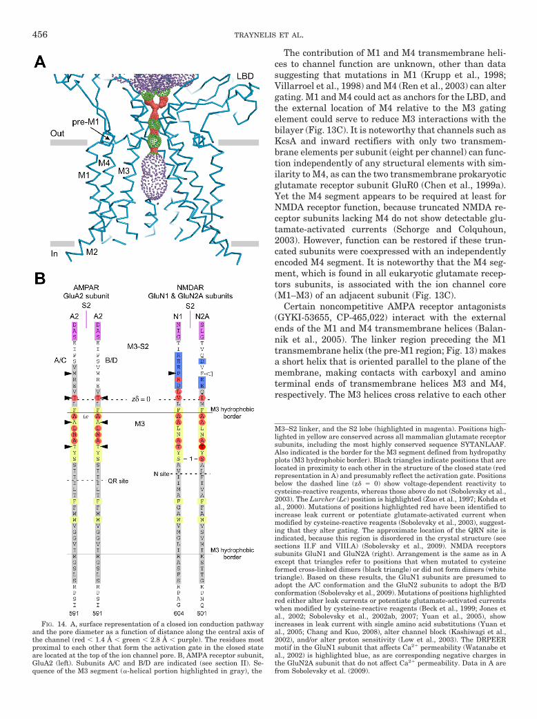
The contribution of M1 and M4 transmembrane heli-ces to channel function are unknown, other than datasuggesting that mutations in M1 (Krupp et al., 1998;Villarroel et al., 1998) and M4 (Ren et al., 2003) can altergating. M1 and M4 could act as anchors for the LBD, andthe external location of M4 relative to the M3 gatingelement could serve to reduce M3 interactions with thebilayer (Fig. 13C). It is noteworthy that channels such asKcsA and inward rectifiers with only two transmem-brane elements per subunit (eight per channel) can func-tion independently of any structural elements with sim-ilarity to M4, as can the two transmembrane prokaryoticglutamate receptor subunit GluR0 (Chen et al., 1999a).Yet the M4 segment appears to be required at least forNMDA receptor function, because truncated NMDA re-ceptor subunits lacking M4 do not show detectable glu-tamate-activated currents (Schorge and Colquhoun,2003). However, function can be restored if these trun-cated subunits were coexpressed with an independentlyencoded M4 segment. It is noteworthy that the M4 seg-ment, which is found in all eukaryotic glutamate recep-tors subunits, is associated with the ion channel core(M1–M3) of an adjacent subunit (Fig. 13C).
Certain noncompetitive AMPA receptor antagonists(GYKI-53655, CP-465,022) interact with the externalends of the M1 and M4 transmembrane helices (Balan-nik et al., 2005). The linker region preceding the M1transmembrane helix (the pre-M1 region; Fig. 13) makesa short helix that is oriented parallel to the plane of themembrane, making contacts with carboxyl and aminoterminal ends of transmembrane helices M3 and M4,respectively. The M3 helices cross relative to each other
FIG. 14. A, surface representation of a closed ion conduction pathwayand the pore diameter as a function of distance along the central axis ofthe channel (red � 1.4 Å � green � 2.8 Å � purple). The residues mostproximal to each other that form the activation gate in the closed stateare located at the top of the ion channel pore. B, AMPA receptor subunit,GluA2 (left). Subunits A/C and B/D are indicated (see section II). Se-quence of the M3 segment (�-helical portion highlighted in gray), the
M3–S2 linker, and the S2 lobe (highlighted in magenta). Positions high-lighted in yellow are conserved across all mammalian glutamate receptorsubunits, including the most highly conserved sequence SYTANLAAF.Also indicated is the border for the M3 segment defined from hydropathyplots (M3 hydrophobic border). Black triangles indicate positions that arelocated in proximity to each other in the structure of the closed state (redrepresentation in A) and presumably reflect the activation gate. Positionsbelow the dashed line (z� � 0) show voltage-dependent reactivity tocysteine-reactive reagents, whereas those above do not (Sobolevsky et al.,2003). The Lurcher (Lc) position is highlighted (Zuo et al., 1997; Kohda etal., 2000). Mutations of positions highlighted red have been identified toincrease leak current or potentiate glutamate-activated current whenmodified by cysteine-reactive reagents (Sobolevsky et al., 2003), suggest-ing that they alter gating. The approximate location of the QRN site isindicated, because this region is disordered in the crystal structure (seesections II.F and VIII.A) (Sobolevsky et al., 2009). NMDA receptorssubunits GluN1 and GluN2A (right). Arrangement is the same as in Aexcept that triangles refer to positions that when mutated to cysteineformed cross-linked dimers (black triangle) or did not form dimers (whitetriangle). Based on these results, the GluN1 subunits are presumed toadopt the A/C conformation and the GluN2 subunits to adopt the B/Dconformation (Sobolevsky et al., 2009). Mutations of positions highlightedred either alter leak currents or potentiate glutamate-activated currentswhen modified by cysteine-reactive reagents (Beck et al., 1999; Jones etal., 2002; Sobolevsky et al., 2002ab, 2007; Yuan et al., 2005), showincreases in leak current with single amino acid substitutions (Yuan etal., 2005; Chang and Kuo, 2008), alter channel block (Kashiwagi et al.,2002), and/or alter proton sensitivity (Low et al., 2003). The DRPEERmotif in the GluN1 subunit that affects Ca2� permeability (Watanabe etal., 2002) is highlighted blue, as are corresponding negative charges inthe GluN2A subunit that do not affect Ca2� permeability. Data in A arefrom Sobolevsky et al. (2009).
456 TRAYNELIS ET AL.

at this level along the permeation path for the antago-nist-bound closed GluA2 structure, raising the intrigu-ing possibility that this pre-M1 helix may restrain M3mobility in the closed state, yet with domain closure,promote channel opening in the ligand-bound state(Sobolevsky et al., 2009). If this structural feature wereshared by NMDA receptors, and four pre-M1 heliceswere to move independently in the agonist-bound recep-tor, one might imagine these conformational changescould constitute rate limiting and kinetically distinctsteps that precede concerted channel opening involvingsimultaneous rearrangement of all four M3 helices. Sin-gle channel studies of agonist-bound NMDA receptorshave identified multiple kinetically distinguishablesteps that precede channel opening via a concerted poredilation involving all or no repositioning of M3 trans-membrane helices (Banke and Traynelis, 2003; Popescuand Auerbach, 2003; Auerbach and Zhou, 2005; Erregeret al., 2005a; Schorge et al., 2005; Dravid et al., 2008).These rate-limiting and kinetically distinct steps mustreflect rate limiting conformational changes that occurbefore rapid pore dilation (Schorge et al., 2005). It isnoteworthy that the role for the pre-M1 linker proposedby Sobolevsky et al. (2009) provides an example of apotential structural rearrangement that could occur in asubunit-dependent fashion (Banke and Traynelis, 2003)and necessarily precede simultaneous rearrangement ofall four M3 helices as the pore opens. Moreover, themechanisms underlying multiple conductance levels forAMPA receptors and gating to the subconductance andmain conductance states for NMDA receptors could liein how this pre-M1 region interacts with the each M3helix that contributes to the gate. Perhaps M3 rear-rangement within individual subunits is permitted for
AMPA receptors, giving rise to as many as four detect-able conductance levels (Rosenmund et al., 1998; Bankeet al., 2000; Smith and Howe, 2000; Jin et al., 2003;Prieto and Wollmuth, 2010), in contrast to GluN2A/B-containing NMDA receptors, for which all four M3 heli-ces might move at once, giving rise primarily to a singleconductance level (Table 17).
E. Molecular Determinants of Desensitization
All glutamate receptors undergo desensitization,which by definition is a reduction in response in thepresence of a sustained stimulus. This process is fast inAMPA and kainate receptors, occurring within 20 msand generating greater than 90% decrease in currentamplitudes at steady state (Table 16). However, theonset of desensitization is slower and less extensive inNMDA receptors and is virtually absent for GluN2C-and GluN2D-containing NMDA receptors (Monyer et al.,1994; Krupp et al., 1996; Wyllie et al., 1998; Dravid etal., 2008). Our understanding of the structural mecha-nisms underlying rapid desensitization, particularly forAMPA and kainate receptors, has been dramaticallyaccelerated through a broad range of structural studieson the isolated AMPA and kainate receptor LBDs incombination with extensive functional studies on thefull-length receptor (Sun et al., 2002; Robert and Howe,2003; Horning and Mayer, 2004; Klein and Howe, 2004;Robert et al., 2005; Armstrong et al., 2006; Weston et al.,2006a; Zhang et al., 2006a; Nayeem et al., 2009).
For GluA2, the initial step of desensitization is a re-arrangement of the D1-D1 dimer interface betweenLBDs of adjacent subunits (Sun et al., 2002; Horningand Mayer, 2004). This idea was driven by LBD struc-tures of wild type GluA2 and GluA2 containing a leucine-
TABLE 17Single channel properties of AMPA, kainate, and NMDA receptors
POPEN Open Time Conductance
ms pS
GluA1-flip 0.4–1.01–3 0.2–0.91 8, 15, 23, 311,4–6
GluA2-flip Q7 0.618 0.32, 1.479 7, 15, 24, 369,10
GluA3-flip 0.8211,12
GluA4-flip 0.772,12 0.14, 3.313 9, 20, 31, 4513,14
GluK1 Q7 0.3, 0.615 5, 9, 1415
GluK2 Q7 0.5–1.03,16 0.6, 2.315 8, 15, 2515
GluK1 Q/GluK5 0.315 5, 9, 1715
GluK2 Q/GluK5 0.4, 2.115 7, 13, 2015
GluN1/GluN2A 0.36–0.5017,18,19 0.06, 1.3, 3.620–22 51, 3822
GluN1/GluN2B 0.07–0.1717,19,23 0.6, 2–3.223 51, 3922
GluN1/GluN2C 0.024 0.622 36, 1922
GluN1/GluN2D 0.01–0.0420,25 0.1, 0.9, 2.620 35, 1726
GluN1/GluN2A/GluN2D 0.08–0.2427 0.07, 1.25, 3.027 30, 40, 5027
GluN1/GluN2B/GluN2D 0.02–0.0328,29 2.4–2.828,29 18, 30, 41, 5428,29
GluN1/GluN3B 12, 3730
GluN1/GluN2A/GluN3B 4.431 26, 4831
GluN1/GluN2A/GluN3A 0.1, 4.732 35, 7533/29, 4732
1Banke et al. (2000). 2Robert et al. (2001). 3POPEN depends on PKA phosphorylation of Ser845. 4Measurements in cell-attached patches were similar to those in outside-outpatches. 5Derkach et al. (1999). 6Prieto and Wollmuth (2010). 7Edited receptors or mutant receptors have a Gln at the QRN site; editing to Arg decreases conductance (Howe,1996; Swanson et al., 1996). 8Koike et al. (2000). 9Jin et al. (2003). 10Zhang et al. (2008a). 11Sekiguchi et al. (2002). 12Predicted from simulations using rate constants;GluA3-flop was predicted to have a POPEN value of 0.42. 13Swanson et al. (1997b). 14Tomita et al. (2005a). 15Swanson et al. (1996). 16Traynelis and Wahl (1997). 17Chen etal. (1999b). 18Popescu et al. (2004). 19Erreger et al. (2005a). 20Wyllie et al. (1998). 21Popescu and Auerbach (2003). 22Stern et al. (1992, 1994). 23Banke and Traynelis (2003).24Dravid et al. (2008). 25Yuan et al. (2009a). 26Wyllie et al. (1996). 27Cheffings and Colquhoun (2000). 28Jones and Gibb (2005). 29Brickley et al. (2003). 30Chatterton et al.(2002). 31Sasaki et al. (2002). 32Perez-Otano et al. (2001). 33Das et al. (1998).
GLUTAMATE RECEPTOR ION CHANNELS 457
to-tyrosine substitution GluA2(L483Y), a mutation that inthe intact receptor blocks desensitization (Stern-Bach etal., 1998), and structures of GluA2 bound to cyclothiazide,an inhibitor of AMPA receptor desensitization (Partin etal., 1995; Sun et al., 2002). Ultracentrifugation studies,which showed that both the mutation and cyclothiazideincreased dimer stability (Sun et al., 2002), suggested arole for the dimer interface in desensitization (see sec-tion VI.A). The mutation GluA2(L483Y) or cyclothiazidestabilized the dimer formation, further suggesting thatstabilization of the LBD dimer interface in full-lengthfunctional AMPA receptors can reduce desensitization(Sun et al., 2002). On the atomic level, Leu483 is locatedin the dimer interface, and mutation to tyrosine pro-motes formation of additional interactions with residueslocated on the opposite LBD (Fig. 10A).
The rate at which cysteine residues introduced at theGluA2 dimer interface are modified by (2-sulfonatoeth-yl)methane thiosulfonate (MTSES) depends on whetherthe receptor is resting, active or desensitized (Armstronget al., 2006). The mutation GluA2(E486C) displays pro-nounced state-dependent modification by (2-sulfonato-ethyl)methane thiosulfonate, being more accessible inthe desensitized state than in the resting or activestates. Crystallographic data show that GluA2 Glu486participates in interactions at the dimer interface and islargely inaccessible to solvent when the receptor is inthe active state. However, in the desensitized state,Glu486 becomes solvent accessible, providing direct ev-idence that desensitization in full-length AMPA recep-tors is accompanied by a rearrangement at the dimerinterface (Armstrong et al., 2006). The same study alsoused variable length cross-linkers as molecular rulers torestrict motion at the dimer interface and thus report onthe magnitude of the rearrangement. The structure ofan additional cross-linked mutant, GluA2(S729C), pos-sessed a conformation with the dimer interface sepa-rated by distances that agreed well with those estimatedfrom cross-linking experiments at different residues.These data suggest that the structure of cross-linkedGluA2(S729C) reveals a conformation similar to full-length desensitized AMPA receptors (Armstrong et al.,2006). Furthermore, imaging studies suggest that theseparation of the dimer interface in the desensitizedstate is not accompanied by significant changes in thedegree of domain closure in the isolated LBD (Du et al.,2005). Thus, during receptor desensitization, the sepa-ration of the dimer interface enables the linkers thatreplace the transmembrane segments to move closertogether by 10 Å relative to the agonist-bound nonde-sensitized conformation, thereby preventing opening ofthe ion channel (Fig. 3).
Mutations in the AMPA receptor binding pocket thatreduce steric collision between the agonist and the LBDcan increase agonist efficacy and desensitization (Mad-den et al., 2004; Holm et al., 2005). Furthermore, thereis a direct correlation between the degree of domain
closure with full and partial AMPA receptor agonistsand the degree of desensitization produced by those ago-nists (Jin and Gouaux, 2003; Jin et al., 2003; Frandsenet al., 2005). Thus, the degree of agonist-induced LBDclosure seems to be the primary determinant of tensionat the LBD dimer interface and thus governs transitioninto both the open and desensitized state.
Using the LBD crystal structure of the nondesensitizingGluA2(L483Y) as a guide, Weston et al. (2006b) introducedcysteine residues in kainate receptor subunits at positionswhere cross-linking should stabilize the dimer interface.This yielded nondesensitizing GluK1, GluK2, and GluK3responses. The AMPA receptor subunit GluA2 was ren-dered nondesensitizing when cross-linked at homologouspositions, thereby establishing that the dimer interfacerearrangement is necessary for kainate receptor desensi-tization (Weston et al., 2006b). However, not all interac-tions predicted on the basis of AMPA receptor studies arenecessary for kainate receptor desensitization (Fleck et al.,2003; Nanao et al., 2005).
Desensitization of NMDA receptors has been studiedextensively (reviewed by Dingledine et al., 1999) andinvolves a number of pathways, including glycine-de-pendent desensitization (Mayer et al., 1989; Benvenisteet al., 1990; Lester et al., 1993; Nahum-Levy et al., 2001;Regalado et al., 2001), Ca2�-dependent inactivation(Clark et al., 1990; Legendre et al., 1993, Vyklicky, 1993;Rosenmund and Westbrook, 1993; Medina et al., 1995;Vissel et al., 2002), Zn2�-dependent desensitization (seesection VI), and glycine/Ca2�-independent desensitiza-tion (Chen et al., 2004; Hu and Zheng, 2005; Sessoms-Sikes et al., 2005). The molecular mechanisms for theseforms of desensitization are poorly understood.
VIII. Molecular Determinants of Ion Permeationand Block
A. Nature of the Ion Permeation Pathway
The ion channel associated with all glutamate recep-tors consists of a water-filled pore divided into externaland internal cavities separated by a narrow constrictionat which the pore reaches its smallest dimension (seesection II). The residues lining the pore, including thosepositioned at the external and internal entrances, formthe permeation pathway and are the primary determi-nants of ion selectivity and unitary conductance. Withonly rare exceptions, the conduction pathway is cation-selective. Glutamate receptors show a wide range ofsingle channel conductances that vary from �1 to �30pS for AMPA and kainate receptors and from 20 to 60 pSfor NMDA receptors (Table 17). AMPA receptors showup to four different conductance levels, the presence ofwhich has been explained by their mechanism of activa-tion (see section VII). NMDA receptors show a pair ofconductance states, the amplitude and relative fre-quency of which depends on the GluN2 subunit presentand ionic conditions.
458 TRAYNELIS ET AL.

The pore of the glutamate receptor family is formed bythree transmembrane helices and a reentrant loop, ar-ranged with striking similarity to an inverted potassiumchannel (Doyle et al., 1998; Sobolevsky et al., 2009); thissimilarity had been predicted to exist on the basis ofsequence similarity and from shared functional proper-ties of a bacterial glutamate receptor and potassiumchannels (Wo and Oswald, 1995; Wood et al., 1995; Chenet al., 1999a; Panchenko et al., 2001; Kuner et al., 2003).Functional and biophysical experiments corroborate theavailable structure. For example, studies examining thechannel block of NMDA receptors by organic cations andMg2� (Villarroel et al., 1995; Zarei and Dani, 1995;Antonov and Johnson, 1999), and the voltage-depen-dence of the reactivity rate for substituted cysteines(Sobolevsky et al., 2002a, 2003) suggested that the nar-row constriction is located approximately halfway acrossthe transmembrane electric field. Scanning mutagenesisstudies correctly predicted that the re-entrant M2 poreloop lines the inner cavity with the QRN site located atthe tip of this loop (Kuner et al., 1996, 2001; Wollmuth etal., 1996).
Immediately external to the tip of the reentrant poreloop lies a central cavity (Fig. 14A) (Sobolevsky et al.,2009), similar to that of potassium channels, lined pri-marily by M3 (Beck et al., 1999; Kashiwagi et al., 2002;Sobolevsky et al., 2003). All glutamate receptor subtypesshare a common transmembrane arrangement, butthere are differences in permeation and block propertiesbetween subtypes. These differences imply some degreeof molecular heterogeneity within the pore, most likelyinvolving the narrowest constriction that sets the max-imum rate of ion permeation either through steric orelectrostatic effects.
A key determinant of the conductance and permeationproperties of all glutamate receptors is the identity ofthe residue occupying a functionally critical position atthe apex of the M2 loop, the QRN site. This site harborseither Gln (Q, unedited) or Arg (R, edited) in AMPA andkainate receptors (Hume et al., 1991; Verdoorn et al.,1991), Asn (N) in GluN1 and GluN2 receptors (Burna-shev et al., 1992b; Mori et al., 1992), Gly (G) in GluN3subunits, and Gln (Q) in GluD2 subunits. Not surpris-ingly, the key structural positioning of the QRN siteallows this residue to influence single-channel conduc-tance (Swanson et al., 1996, 1997b; Traynelis and Wahl,1997), Ca2� permeability (Burnashev et al., 1992a,b;Egebjerg and Heinemann, 1993), channel block by poly-amines (Bowie and Mayer, 1995; Kamboj et al., 1995;Washburn et al., 1997), Mg2� sensitivity (Burnashev etal., 1992b; Dingledine et al., 1992; Mori et al., 1992),channel block by organic compounds (Kashiwagi et al.,2002; Chen and Lipton, 2005), and assembly into het-eromeric complexes (Greger et al., 2003). In addition,homomeric edited R-forms of kainate receptor channelsare no longer purely cation-selective, but are also per-meable to Cl� (Burnashev et al., 1996) (Table 18). Re-placement of the QRN Asn with Gln in GluN1 orGluN2B yields channels with multiple conductance lev-els (Premkumar et al., 1997; Schneggenburger and As-cher, 1997). The functional effects of TARPs on AMPAreceptors (Korber et al., 2007) and of membrane lipids onkainate receptors (Wilding et al., 2005) are influenced byediting at the QRN site. It is noteworthy that Sobolevskyet al. (2009) observed gaps between the transmembranehelices, which they predict may be filled by residuesprojecting from TARPs (see section II.H). This potentialassociation with the pore could provide a means by
TABLE 18Pore properties of AMPA, kainate, and NMDA receptors
The relative permeability of Ca2� to monovalent ions (PCa/PX) and the fractional Ca2� current (Pf) values shown are not necessarily those given in the original manuscriptbut rather have been standardized to common parameters for direct comparison. The values shown are not corrected for activities. Pore dimensions are derived from thepermeability of differently sized organic cations. Only values derived from recombinant channels are shown; see Dingledine et al. (1999) for more details and examples ofnative channels. PCa/PX values are derived from the Lewis equation (see Jatzke et al., 2002) based on changes in reversal potentials given in articles typically measured underbionic conditions where a reference solution (X, usually Cs� or Na�) is replaced with a solution containing high Ca2� (typically 100 mM). PCa/PX (from Pf) is derived fromPf measurements using the GHK equation (Burnashev et al., 1995). Fractional Ca2� currents (Pf) are the percentage of the total current carried by Ca2� and are measuredusing whole-cell currents and high concentrations of fura-2 to ensure that all incoming Ca2� is captured by dye. Pf values are concentration- and voltage-dependent with themagnitude of these effects dependent on specific subtypes (Burnashev et al., 1995; Jatzke et al., 2002). The values shown were either recorded at �60 mV and 1.8 mM Ca2�
or adjusted to these conditions assuming GHK (see Jatzke et al., 2002).
Subunit Combination Pore Diameter PCa/PX PCa/PX (from Pf) Pf PCl/PCs
nm %
GluA1a,b 0.78c 1.6 0.7 3.2–3.6GluA2 (Arg)c 0.14GluA1/GluA2 (Arg)c 0.7 0.03 0.11 0.54 0GluA4 3.9GluK2 (Gln)a,b,d 0.75g 0.14–0.17 0.30–0.48 1.55–2.4 0GluK2 (Arg)a,c,d 0.76 �0.04 �0.2 0.74GluK2 (Gln)/GluK2 (Arg) a,c,d 0.74 0.58 0GluN1-GluN2Aa,b,e,f,g 0.55h,i 2.8–4.5 3.0–3.6 11.0–15.9GluN1-GluN2Bf 3.6 3.6 15.9GluN1-GluN2Ca 1.8 1.73 8.2GluN1-GluN3Aj 0.6GluD2Lc 2.6k,l
aBurnashev et al. (1995). bJatzke et al. (2002). cBurnashev et al. (1996). dThe kainate receptor GluK2 is edited in three different locations: two in M1 (Ile, Tyr fullyunedited and Val, Cys fully edited) and one in M2 (Gln in unedited, Arg in edited). All values shown are for receptors that are fully edited in M1. eSharma and Stevens (1996).fSchneggenburger (1996). gSchneggenburger (1998). hVillarroel et al. (1995). iWollmuth et al. (1996). jPerez-Otano et al. (2001). kGluD2Lc is a constitutively active form(Lurcher) of the GluD2 subunit. lWollmuth et al. (2000).
GLUTAMATE RECEPTOR ION CHANNELS 459
which TARPs influence ion permeation, conductance,and polyamine block.
NMDA receptors are obligate heteromultimers likelycomposed of 2 GluN1 and 2 GluN2 subunits in a 1-2-1-2arrangement (Sobolevsky et al., 2009). The narrow con-striction in NMDA receptors is formed by, at minimum,the QRN site asparagine of GluN1 and an asparagineadjacent to the QRN site (i.e., QRN � 1 site) in GluN2(Fig. 14A) (Wollmuth et al., 1996). It is noteworthy thatthere is also a functional asymmetry between QRN Asnresidues in the various NMDA receptor subunits: sub-stitutions at the GluN1 QRN site have strong effects onCa2� permeability but only weak effects on Mg2� block,and equivalent substitutions of the QRN site in GluN2produce opposite effects (Burnashev et al., 1992b). Thisfunctional and presumed structural asymmetry betweensubunits most likely reflects modest differences at theatomic scale. A highly conserved GluN2B tryptophanresidue in the M2 loop also has been proposed to con-tribute to the narrow constriction (Williams et al., 1998),and the homologous tryptophan in GluN1 can influencechannel block (e.g., Jin et al., 2007).
B. Mechanisms of Ion Permeation
A major determinant of ion selectivity arises from thephysical or electrical interactions of permeating ionswith the pore wall within the narrow region of a channelin which ions and side chains or main-chain atoms ofresidues come close enough to create a permeation bar-rier. The dimensions of the smallest diameters of vari-ous glutamate receptor channels have been estimatedfrom the permeability of differently sized organic cations(Table 18). The finding that channels containing one ormore arginine residues at the QRN site have roughly thesame dimension as channels containing the smaller glu-tamine residues suggests that the side chain of the QRNsite does not define the dimensions of the narrow con-striction and that its effect on permeation may be influ-enced by electrostatics (Kuner et al., 2001).
All mammalian glutamate receptor subunits are cat-ion-selective and lack the highly conserved TVGYG se-quence that defines selectivity in K� channels. The pro-karyotic GluR0 does have the TVGYG sequence and isK�-selective (Chen et al., 1999a), but introduction of thissequence into mammalian glutamate receptor subunitsdoes not confer K� selectivity (Hoffmann et al., 2006a),probably because of different interactions of this regionwithin the mammalian glutamate receptor comparedwith K� channels. Indeed, in the membrane-spanningGluA2 structure, the extended region in the M2 loop isdisordered, and this may reflect the absence of TVGYGand stabilizing interactions that underlie K� selectivityin K� channels. Moreover, under certain nonphysiologi-cal conditions, edited (R) forms of homomeric GluA2 andGluK2 subunits are permeable to Cl� ions (Table 18).The central location of the QRN site and the fact thatthese homomeric receptor channels show mixed cation/
anion permeability suggests other structural elementssuch as M2 loop dipole, carbonyl side chains in M2,and/or elements in the outer cavity may contribute tocation selectivity. Indeed, in K� channels, the pore loopdipole alters the environment in the inner cavity (corre-sponding to the outer cavity in glutamate receptors) toallow a high flux while maintaining ion selectivity (Morais-Cabral et al., 2001). It is noteworthy that the GluA2 struc-ture reveals various charged residues positioned near theapex of the M3 segment that could influence conductance.Consistent with this idea, mutations of homologous posi-tions in GluN1 M3 reduce single channel conductance andCa2� permeability (Watanabe et al., 2002).
Selectivity, unitary conductance, and channel blockcan all be influenced by the number of ions that reside inthe pore at any one time. Potassium channels can ac-commodate multiple permeating ions, and this is a keycomponent of their high selectivity and transport rates(Doyle et al., 1998; Morais-Cabral et al., 2001; Zhou etal., 2001). Studies of NMDA receptors suggest that thepore contains only a single permeating ion (Zarei andDani, 1994; Schneggenburger, 1996; Iino et al., 1997),which may reflect the nonselective nature of the pore. InK� channels as well as in other highly selective chan-nels, the interaction between the narrow constrictionand permeating ions is tight and this reduces their freeenergy in this region. Allowing multiple permeating ionscreates ion-ion interactions that are critical to facilitat-ing high conductance rates (Morais-Cabral et al., 2001).In contrast, in the larger glutamate receptor channel,the interaction between the pore walls and permeatingions may be weaker (i.e., fewer waters of hydration areremoved), which could prevent occupancy by multipleions. The pore of NMDA receptors can energetically ac-commodate a blocking particle such as Mg2� and addi-tional permeant ions (Antonov et al., 1998; Antonov andJohnson, 1999), and these additional permeant ion-bind-ing sites may make a significant contribution to ionselectivity and conductance in addition to effects onchannel block.
Glutamate receptors show limited selectivity for al-kali metal cations, which for GluN1/GluN2A is Cs� �Rb� K� � Na� � Li�, for GluA1 is K� Rb� � Cs� �Na� Li�, and for GluK2(Q) is K� Cs� Rb� �Na� Li� (Tsuzuki et al., 1994; Jatzke et al., 2002). Interms of Eisenman sequences (Hille, 2001), NMDA re-ceptors display type I weak-field-strength sites, whereasnon-NMDA receptors display type IV sites. In terms ofselectivity at a synapse (K� versus Na�), the differencesare minimal (PK/PNa 1.14 for NMDA receptors andPK/PNa 1.25 for AMPA and kainate receptors). Thus,the current carried through most glutamate receptorchannels is a mixture of monovalent cations (K� andNa�) plus Ca2� (MacDermott et al., 1986; Mayer andWestbrook, 1987). Receptors containing the edited formof GluA2, GluK1, or GluK2 (Hume et al., 1991; Burna-shev et al., 1992a), however, are impermeable to Ca2�
460 TRAYNELIS ET AL.

(Table 18). The flux of Ca2� through NMDA and Ca2�-permeable AMPA receptor channels is a key factor con-tributing to various forms of synaptic plasticity (Gu etal., 1996; Bloodgood and Sabatini, 2007; Citri andMalenka, 2008) (sections IV and IX), gene regulation(section III), neuropathology (Choi, 1995; Kalia et al.,2008) (section X), and fast transmitter release (see sec-tion IX) (Chavez et al., 2006).
Three general approaches have been used to study themagnitude and mechanism of Ca2� permeation in glu-tamate receptors, and include measurement of 1) rela-tive Ca2� permeability (PCa/PMonovalent), 2) channelblock by Ca2�, and 3) fractional Ca2� currents (Pf),which are determined from whole-cell currents recordedin the presence of high concentrations of intracellularFura-2 (Burnashev et al., 1995; Premkumar and Auer-bach, 1996; Schneggenburger, 1996; Sharma and Stevens,1996). The use of fractional Ca2� currents is advantageousover the other methods for channels with a mixed Ca2�
and monovalent permeability because it has fewer as-sumptions about flux properties, directly quantifies theCa2� flux under physiological conditions, and allows thefraction of the total current carried by Ca2� to be quanti-fied over a wide voltage range (Neher, 1995).
NMDA receptors are approximately 3 to 4 times morepermeable to Ca2� than unedited Ca2�-permeableAMPA or kainate receptors (Table 18). For NMDA re-ceptors, there are subunit-specific differences, dictatedby the GluN2 and GluN3 subunits, with receptors con-taining GluN2A/GluN2B showing the highest Ca2� per-meability, GluN2C showing a somewhat reduced Ca2�
permeability, and GluN3 showing the lowest permeabil-ity. Ca2�-permeable AMPA receptors show a slightlygreater Ca2� permeability than kainate receptors. Mu-tant Lurcher GluD2 channels are Ca2�-permeable (Ta-ble 18). The magnitude of Ca2� influx through at leastNMDA receptors can be regulated by phosphorylation(Skeberdis et al., 2006) and synaptic activity (Sobczykand Svoboda, 2007).
It is noteworthy that NMDA receptors are highly per-meable to Ca2� yet show the peculiar property of beingblocked by external Ca2� in a largely voltage-indepen-dent manner (Ascher and Nowak, 1988; Premkumar andAuerbach, 1996; Sharma and Stevens, 1996), whichmanifests as a reduction in single channel conductance(Gibb and Colquhoun, 1991; Stern et al., 1994; Premku-mar and Auerbach, 1996). These results lead to thesuggestion of multiple Ca2� binding sites within thepore (Premkumar and Auerbach, 1996; Sharma andStevens, 1996), including the QRN site as well as anexternal site for Ca2�. Moreover, NMDA receptors showan interaction between Ca2� and monovalent ions in thepore, concentration-dependent PCa/PMonovalent (Woll-muth and Sakmann, 1998), and block by Ca2�. However,the PCa/PMonovalent value for NMDA receptors derivedfrom the Goldman-Hodgkin-Katz equation, which as-sumes no ion-ion interactions in the pore (Hille, 2001),
describes fractional Ca2� currents over a wide voltageand concentration range (Burnashev et al., 1995;Schneggenburger, 1996, 1998; Jatzke et al., 2002).These differences could be reconciled if the pore, whichis occupied by a single ion, largely existed in two states,both of which individually follow Goldman-Hodgkin-Katz, one with the pore occupied by Ca2� and the otheroccupied by a monovalent ion. In this model, a competi-tion for the pore occurs between Ca2� and monovalentcations (Schneggenburger, 1998).
The process of Ca2� influx in Ca2�-permeable AMPAreceptors might be less complex than that in NMDAreceptors, because Ca2�-permeable AMPA receptors donot select for or against divalent cations. PCa/PMonovalentin Ca2�-permeable AMPA receptors is approximatelyunity (not taking into account ion activities; Table 18).Ca2�-permeable AMPA receptors are not blocked byCa2�, and the predicted fractional Ca2� current at �60mV in �150 mM monovalent ions and 1.8 mM Ca2� is�4%, in agreement with a channel that is nonselectivefor Ca2� (PCa/PMonovalent � 1; Table 18; see Jatzke et al.,2002).
Ca2� permeability is influenced by residues occupyingthe QRN site residing at the channel’s narrow constric-tion (see section VIII.A). NMDA receptors in which theQRN site asparagine is mutated to various other resi-dues, including the glutamine found in AMPA receptors,show reduced Ca2� permeability (Burnashev et al.,1992b) (Table 18). In addition to residues at or near theQRN sites within the narrow constriction, other ele-ments in the inner cavity may also influence Ca2� per-meability, either directly or by altering the structure ofthe M2 loop (Ferrer-Montiel et al., 1996). One such res-idue is a negatively charged Glu in the M2 loop, locatedfive positions C-terminal to the QRN site in GluN1.Mutation of this Glu to the positively charged Lys re-duces Ca2� permeability (Schneggenburger, 1998). Inaddition, in the GluN2A subunit, an adjacent position isoccupied by the polar glutamine, and replacing it with aGlu (as in GluN2C subunits that have reduced Ca2�
permeability; Table 18) yields channels that show a re-duced Ca2� permeability (Vissel et al., 2002), suggestingthat this position may underlie GluN2 subunit-specificdifferences in Ca2� permeability. Nevertheless, it is un-clear whether Ca2� directly interacts with the side chainat this position or whether mutations at this positionalter the structure of the QRN site, because this positionis not water accessible (Kuner et al., 1996).
The high Ca2� influx in NMDA receptors has beenproposed to be due, at least in part, to an external Ca2�
binding site (Premkumar and Auerbach, 1996; Sharmaand Stevens, 1996). The most likely determinant of thisCa2� binding site is a cluster of charged residues, theDRPEER motif (Fig. 14), located C-terminal to the M3segment in GluN1 but absent in all GluN2, GluA, GluD,and GluK subunits (Watanabe et al., 2002). This uniquemotif is positioned at the external entrance to the chan-
GLUTAMATE RECEPTOR ION CHANNELS 461

nel (Beck et al., 1999), and possesses a net negativecharge, predicted from the three potentially (dependingon local pKa values) negative and one positive water-accessible residues. In mutant channels with the nega-tive charges in DRPEER neutralized (ARPAAR), Pf val-ues are reduced by approximately half at �60 mV(Watanabe et al., 2002), suggesting that the negativelycharged side chains represent an important determi-nant of high fractional Ca2� currents in NMDA recep-tors. It is noteworthy that the GluN2A subunit has a netnegative charge at positions homologous to the DRPEERmotif, but mutation of these residues has no effect onCa2� permeability (Watanabe et al., 2002). One poten-tial explanation that is consistent with structural dataplacing elements of the GluN1 M3-S2 linker (tetramerA/C subunits) in the vicinity of each other (Sobolevsky etal., 2009) is that the DRPEER motif lies closer to thepermeation pathway than homologous positions inGluN2 (tetramer B/D subunits). It is noteworthy thatsubstituting an Asn at the QRN site of AMPA receptors,which should make the narrow constriction more NMDAreceptor-like, results in only a modest increase in frac-tional Ca2� current from 4% to 5% (Wollmuth and Sak-mann, 1998). Part of the difference in Ca2� permeabilitybetween AMPA and NMDA receptors may reflect theabsence of the DRPEER motif in non-NMDA receptors(Fig. 14; Jatzke et al., 2003).
For NMDA receptors, coexpression of GluN3 subunits(which contain a Gly at the QRN site) with GluN1 orGluN1/GluN2 results in channels with reduced singlechannel conductance, decreased Ca2� permeability, andreduced Mg2� block (Perez-Otano et al., 2001; Matsudaet al., 2003) (Table 18), suggesting that GluN3 directlyinfluences the structure of the permeation path. Addi-tional evidence for this idea was provided by Wada et al.(2006), who showed that cysteine-substitutions in theM3 segment of GluN3A were modified by extracellularcysteine-reactive reagents and suggested that some res-idues lining the pore may differ from GluN1 and GluN2subunits. Nevertheless, molecular mechanisms regulat-ing conductance and permeability in GluN3-containingreceptors are poorly understood.
C. Voltage-Dependent Channel Block byEndogenous Ions
A number of endogenous ions can regulate glutamatereceptor function, including protons, Zn2�, Mg2�, andpolyamines (see section VI). Of these, the multivalentions Zn2�, Mg2�, and polyamines have voltage-depen-dent block mechanisms that have important implica-tions for neuronal function independent of the dynamicregulation of their concentrations. We will consider themechanism and implications of block of the pore byMg2� and polyamines, both of which endow receptorswith the ability to detect previous neuronal activity, andthus have roles in short- and long-term plasticity. Al-though channel block by Zn2� may be important in some
situations (Vogt et al., 2000), Zn2� channel block isrelatively low affinity, rapidly reversing, and will not beconsidered further.
Mg2� block represents perhaps the most distinctivefeature of the NMDA receptor and was first described ina now-classic series of studies from multiple laboratories(Mayer et al., 1984; Nowak et al., 1984; Ascher et al.,1988; Dingledine et al., 1999). Because of the strongvoltage-dependence of block, NMDA receptors act ascoincident detectors, sensing postsynaptic depolariza-tion at the same time as or shortly after release ofglutamate or other excitatory amino acids. This blockallows NMDA receptors to mediate cellular mechanismsof learning and memory (see section IX). The most prom-inent biophysical feature of extracellular Mg2� block isits strong voltage-dependence. Indeed, the major block-ing site for Mg2� is at or near the narrow constriction(for review, see Dingledine et al., 1999), located approx-imately 50% across the channel. Part of the additionalvoltage-dependence is due to the presence of permeantion-binding sites in the external and internal cavities(Antonov and Johnson, 1999; Zhu and Auerbach,2001a,b; Qian et al., 2002). Occupancy of these ion bind-ing sites by Na� or K� alters the association and disso-ciation rates of Mg2� from its blocking site. The natureof these permeant ion-binding sites differs betweenGluN2 subunits (Qian and Johnson, 2006) and hencemay underlie some GluN2-specific effects on Mg2� block(Monyer et al., 1994; Kuner and Schoepfer, 1996).
An advance in our mechanistic understanding of ex-tracellular Mg2� block has come from studies of the rateof Mg2� block and unblock, which provides a physiolog-ical view of the actions of Mg2�. As a first approxima-tion, the block and unblock by extracellular Mg2� wasconsidered instantaneous; however, this simplifying as-sumption would limit the role of NMDA receptors in den-dritic integration. A variety of studies show that the rate ofunblock and reblock of the channel is not instantaneous,showing both fast and slow components (Nowak et al.,1984; Vargas-Caballero and Robinson, 2003, 2004; Kampaet al., 2004; Clarke and Johnson, 2006) with rates depend-ing on the GluN2 subunit (Clarke and Johnson, 2006). ForGluN2B, the slow component of Mg2� unblock arises frominherent voltage dependence of NMDA receptor gating(Clarke and Johnson, 2008).
These rates for Mg2� unblock and reblock at the syn-apse will alter the response time and can lengthen thewindow of opportunity for back-propagating action po-tentials and local synaptic activity to modulate currentflux (including Ca2�) through NMDA receptors. Itis noteworthy that receptors containing GluN2C orGluN2D unblock more rapidly—almost instantaneo-usly—than those containing GluN2A or GluN2B, thelatter of which shows the slowest unblock (Clarke andJohnson, 2006). Given the possible regulation of Mg2�
block by membrane lipids (Parnas et al., 2009), pertur-bations in ion concentrations during activity, and GluN2
462 TRAYNELIS ET AL.

subunit-specific differences in rates of unblock and re-block, mechanisms regulating NMDA receptor block byMg2� at a synapse are likely to be diverse.
Regulation of NMDA receptor activity by extracellularMg2� is well known for its contribution to synaptic plas-ticity and associative learning. Less well known is thepotential role in short-term synaptic plasticity played bythe block of Ca2�-permeable non-NMDA receptors byinternal polyamines such as spermine, spermidine, andputrescine. Polyamine block of AMPA and kainate re-ceptors is dependent on membrane potential, which mayreflect interaction in the pore between monovalent ionsand polyamines, rather than membrane voltage itself(Bowie et al., 1998). Polyamines interact with the pore atvarious sites in the M2 loop including the QRN site anda negatively charged residue (Asp in AMPA receptorsand Glu in kainate receptors) located C-terminal to theQRN site (for review, see Dingledine et al., 1999; Pan-chenko et al., 1999). Polyamines can dissociate fromopen channels to either the internal or external solutionand thus permeate the channel (Bahring et al., 1997).Under physiological conditions, unedited AMPA andkainate receptors have doubly rectifying current-voltagerelations reflecting block by internal polyamines atdepolarized membrane potentials and permeation of in-ternal polyamines at even more positive membrane po-tentials. Surprisingly, polyamine block is also state-de-pendent. The affinity for polyamines is higher in closedthan in open channels, but the kinetics of polyamineblock are slower in closed channels (Bowie et al., 1998;Rozov et al., 1998). As a result, synaptic Ca2�-permeableAMPA receptors can be blocked by polyamines in restingsynapses. Synaptic activity partially relieves this poly-amine block, and block recovers slowly, so that the re-sponse to subsequent synaptic events can be potentiated(Rozov and Burnashev, 1999; Shin et al., 2005). Thedegree of potentiation depends on the frequency of syn-aptic events. If the frequency is too low, block of closedchannels returns to its equilibrium level between thefirst and second synaptic events, and no potentiationoccurs. Alternatively, if the frequency is sufficientlyhigh, block does not re-equilibrate during the brief in-terval between synaptic events, and the response to thesecond synaptic event is potentiated. Thus, state-depen-dent polyamine block of postsynaptic AMPA receptorscould act as a high-pass filter for synaptic activity andmay be an important component of circuit function inwhich information is encoded by synaptic timing (Abbottand Regehr, 2004).
IX. Role in Synaptic Function and Plasticity
A. Synaptic �-Amino-3-hydroxy-5-methyl-4-isoxazolepropionic Acid Receptors
Within the mammalian CNS, the vast majority of fastexcitatory synaptic transmission is mediated by hetero-meric AMPA receptors assembled from differing combi-
nations of the four subunits, GluA1 to GluA4. Expres-sion of all subunits is developmentally regulated, regionand cell-type specific, and activity-dependent (Ashby etal., 2008). Within adult forebrain principal neurons, in-cluding hippocampus and cortex, the predominant GluAsubtype comprises GluA1 and GluA2 with a secondaryminor role played by GluA2/3 receptors (Lu et al., 2009).By virtue of its edited QRN site, the presence or absenceof GluA2 is a critical determinant of GluA function anddictates the single channel conductance and Ca2�-per-meability of the native AMPA receptor (see sectionVIII.B). An absence of GluA2 also renders the channelpore sensitive to extra- and intracellular polyamineblock, endowing GluA receptors with an inwardly recti-fying current-voltage relationship and a novel postsyn-aptic mechanism of short-term plasticity involving theuse-dependent unblock of polyamines (Isaac et al., 2007)(see section VIII.C). In many neurons, GluA2 expressionis developmentally regulated, with low early postnatalexpression levels, which, coupled with the transient highexpression of GluA4, renders many neonatal forebrainreceptors Ca2�-permeable (Pickard et al., 2000; Zhu etal., 2000). This Ca2�-permeability may play a role insynapse maturation and circuit development (Aizenmanet al., 2002; Kumar et al., 2002; Eybalin et al., 2004; Hoet al., 2007; Migues et al., 2007). In the forebrain, asGluA2 expression increases throughout the early post-natal period, GluA4 is down-regulated. In adult, GluA4expression remains highly enriched primarily in cerebel-lar tissue, where it exists as heteromeric GluA2/GluA4in granule cells and in non-neuronal Bergmann glia ashomomeric GluA4 (Petralia and Wenthold, 1992; Martinet al., 1993).
At a prototypical synapse, binding of glutamate tosynaptic AMPA receptors triggers a brief, rapidly risingconductance that decays rapidly (1–2 ms; Fig. 12) as aresult of the deactivation of the agonist-receptor com-plex. The kinetics and amplitude of the excitatory syn-aptic response are determined by the biophysical prop-erties of the receptor subunit combination (Table 17; Fig.12; see section VII.A) and the density of receptor expres-sion, convolved with the time course of glutamate re-lease and uptake. Receptor subunit composition and thekinetics of the synaptic response are tailored to the roleplayed by the synapse and cell type in the circuit inwhich they are embedded. For example, many corticallocal circuit inhibitory interneurons express AMPA re-ceptors that comprise homomeric GluA1 (Isaac et al.,2007). These receptors have rapid kinetics such that theoverall synaptic conductance time course is complete inless than a millisecond. The subsequent excitatorypostsynaptic potential (EPSP) is large, rises and decaysrapidly, and is capable of triggering action potentialswithin a narrow temporal window (Geiger et al., 1997).Such receptor assemblies are typical in cells involved inthe timing of oscillatory activity (Lawrence and McBain,2003; Jonas et al., 2004; Isaac et al., 2007).
GLUTAMATE RECEPTOR ION CHANNELS 463

Although the subunit composition is a major determi-nant of synaptic conductance kinetics, synapses typi-cally are distributed across often elaborate dendritictrees. The resulting EPSP can be subject to extensivecable attenuation as it travels from its source (e.g.,spines or smooth portions of narrow dendrites) to thesite of action potential initiation. Consequently, in manyneurons, synaptic potentials are often capable of trigger-ing appreciable intrinsic voltage-dependent sodium, cal-cium, and/or potassium conductances that can normal-ize the impact of dendritic location or alter the temporalvoltage window initiated by the synaptic conductance(Bloodgood and Sabatini, 2008).
Systematic mRNA and immunohistochemical profil-ing of GluA subunit expression in individual neuronshas provided a detailed picture showing how receptortypes are linked to the functional properties of the cell.However, such profiling is complicated by the observa-tion that single neurons often manufacture AMPA re-ceptors of different subunit compositions and targetthem to different synaptic inputs across the somatoden-dritic axis (Toth and McBain, 2000). How individualneurons differentially target such receptor assemblies tooften overlapping afferent inputs remains to be deter-mined. However, in neurons of the lateral geniculatenucleus, Ras and Rap2 drive bidirectional trafficking ofGluA1 between the deliverable and synaptic pools atretinogeniculate synapses (GluA1 dominant) but not atcorticogeniculate synapses (GluA4 dominant), despitethe existence of intracellular pools of GluA1 at bothsynapses (Kielland et al., 2009). This suggests that in-trinsic activity within the vision-dependent pathwaypreferentially drives GluA1 between deliverable andsynaptic pools at retinogeniculate synapses. Further-more, in cortical principal cells, where the dominantAMPA receptor configuration is composed of GluA1 andGluA2, significant intracellular pools of GluA2-lackingCa2�-permeable receptors exist that can reach the sur-face under certain conditions (Ju et al., 2004; Clem andBarth, 2006; Plant et al., 2006; Sutton et al., 2006),suggesting that AMPA receptor expression is a dynamicand highly regulated process.
B. Synaptic Kainate Receptors
Since the binding studies of Monaghan and Cotman(1982), it has been clear that high-affinity kainate re-ceptor binding sites are prevalent throughout the CNS.Translating early binding studies into a function for allkainate receptor subunits in synaptic transmission hasproved harder than would have been expected. In recom-binant systems, GluK1 to GluK3 form functional homo-meric receptors, whereas GluK4 and GluK5 do not.These latter two subunits are believed to play a support-ing role by modifying both the pharmacological and bio-physical receptor properties (Contractor and Swanson,2008). Unlike AMPA and NMDA receptors (althoughexceptions exist), kainate receptors can play prominent
roles at both pre- and postsynaptic sites. The rules ofsubunit assembly and combination remain unclear, andkainate receptors show strong developmental and re-gional regulation (Contractor and Swanson, 2008). Dur-ing development, synaptic transmission at thalamocor-tical synapses is initially mediated by slow kainatereceptors but switches during the first postnatal week toAMPA-receptor mediated transmission, representing anovel developmental form of LTP (Kidd and Isaac, 1999).This switch from kainate receptor to AMPA receptorsubunits as the primary mediators of synaptic transmis-sion shortens the window for coincidence detection andoutput timing in this circuit (Bannister et al., 2005; Dawet al., 2006). A similar activity-dependent switch fromkainate to AMPA receptor-mediated transmission alsohas been observed at synapses onto perirhinal cortexLayer I/II neurons (Park et al., 2006).
Unlike currents through recombinant kainate recep-tors (see section VII.A), synaptic kainate receptors oftenpossess a slower time course (decay time constants often�100 ms) and typically are activated only after shortbursts of presynaptic activity (Contractor and Swanson,2008). The best studied site of kainate receptor-medi-ated synaptic transmission is at the mossy fiber-CA3pyramidal cell synapse. There, heterotetramers formedby GluK2 and GluK5 are found postsynaptically, whereasreceptors formed from combinations of GluK1 to GluK3are located presynaptically. At mossy fiber synapses,kainate receptors act in a concerted manner to amplifysynaptic integration and frequency-dependent spiketransmission (but see Kwon and Castillo, 2008; Sa-chidhanandam et al., 2009). Long-term depression of thekainate receptor component of the mossy fiber EPSCwas traced to internalization of kainate receptors, me-diated by a protein interacting with C kinase/synapto-some-associated protein 25/PKC complex (Selak et al.,2009). In contrast, kainate receptors are absent at asso-ciational/commissural synapses also made onto CA3 py-ramidal cells, indicating another example of target-spe-cific localization of glutamate receptors (Toth andMcBain, 2000). Elsewhere in cortical circuits, GluK2 iswidely expressed at synapses on local circuit inhibitoryinterneurons (Cossart et al., 1998, 2002; Frerking et al.,1998; Mulle et al., 2000), where they have been impli-cated in the generation of and � oscillatory activity(Fisahn et al., 2004; Goldin et al., 2007). GluK1-con-taining receptors have been described on inhibitoryaxons presynaptic to principal cells and inhibitoryinterneurons, where they function to set the inhibi-tory tone of the hippocampal network (Cossart et al.,1998; Semyanov and Kullmann, 2001; Fisahn et al.,2004).
In addition to their role as ionotropic receptors, kainatereceptors also signal via G-protein-coupled second-messen-ger cascades to downstream effectors (Rodríguez-Morenoand Lerma, 1998). By triggering a PKC-signaling cascade,GluK2-containing receptors modulate the slow- and
464 TRAYNELIS ET AL.

medium-duration afterhyperpolarization conductance inCA1 and CA3 pyramidal cells (Melyan et al., 2002; Fisahnet al., 2005). Moreover, different subunits within the re-ceptor complex have been proposed to independently con-trol the ionotropic (GluK2) and metabotropic (GluK5) ac-tions of kainate (Ruiz et al., 2005). However, knockout ofGluK4 and GluK5 eliminated kainate-mediated EPSCs atmossy fiber-CA3 synapses but failed to eliminate the kain-ate-mediated modulation of the slow afterhyperpolariza-tion, demonstrating that GluK4 and GluK5 are essentialfor normal ionotropic function but are not linked to theproposed metabotropic function of native kainate receptors(Fernandes et al., 2009).
C. Synaptic and ExtrasynapticN-Methyl-D-aspartate Receptors
At virtually all central synapses, NMDA receptorscolocalize with AMPA receptors to form the functionalsynaptic unit, such that presynaptically released gluta-mate typically coactivates both NMDA and AMPA re-ceptors (Petralia and Wenthold, 2008). The ratio ofAMPA receptor- to NMDA receptor-mediated synapticcurrent varies across a wide range. Synaptic NMDAreceptors are thought to be heterotetramers comprisingtwo GluN1 subunits and two GluN2 or GluN3 subunits(Wenthold et al., 2003); evidence also exists for nativetriheteromeric receptors composed of GluN1/GluN2A/GluN2B, GluN1/GluN2A/GluN2C, and GluN1/GluN2B/GluN2D (see section II.B). Like AMPA receptor sub-units, GluN subunit expression is under developmentaland regional control during the early postnatal life. Be-cause GluN1 is an obligate receptor subunit for channelfunction, development is targeted primarily towardGluN2 and GluN3 expression. The best example of thisdevelopmental expression is the switch from GluN1/GluN2B receptors in cortex, hippocampus, and cerebel-lum, which predominate in the first weeks of postnatallife, to GluN1/GluN2A-containing receptors (vanZundert et al., 2004). GluN1/GluN2B receptors are im-portant for circuit formation and development, and thisswitch to GluN1/GluN2A results in NMDA receptor-mediated EPSCs with a more rapid decay. In hippocam-pal principal cells, this subunit switch is triggered byagonist binding (Barria and Malinow, 2002) and can bedriven by stimulus protocols that produce long-lastingpotentiation (Bellone and Nicoll, 2007). Likewise, sen-sory experience drives this subunit switch at thalamo-cortical synapses in primary sensory neocortex (Quin-lan et al., 1999; Barth and Malenka, 2001; Lu et al.,2001). In mossy fiber-cerebellar granule cell synapses, afurther subunit switch to GluN1/GluN2C is observedaround P40 and is accompanied by a slowing of currentkinetics and a reduction in Mg2� sensitivity (Cathala etal., 2000). NMDA receptors containing GluN2D are mostcommon in early postnatal life in neurons of the dien-cephalon, brainstem, and cerebellum; mRNA expressionlevels persist in subpopulations of interneurons in, for
example, the hippocampus (Monyer et al., 1994). Thesignaling mechanism(s) underlying these regulatorysteps are unknown. GluN3A is expressed primarily inearly postnatal life, whereas GluN3B is found in adultbrainstem and spinal cord (Petralia and Wenthold,2008). GluN3A expression seems to be a critical step insynaptogenesis and spine formation, and it maintainsreceptors in an immature state (Perez-Otano et al.,2006), perhaps with enhanced neuroprotection (Nakan-ishi et al., 2009). Subsequent down-regulation ofGluN3A expression is important for synapse maturationand the emergence of a competent state for synapticplasticity (Roberts et al., 2009). The potential roles forthe myriad of glutamate receptor compositions havebeen discussed elsewhere (Petralia and Wenthold,2008). Although exceptions exist, GluN2B-, GluN2D-,and GluN3A-containing receptors generally predomi-nate early in postnatal life, whereas GluN2A- andGluN2C-containing receptors become more abundant inadult brain (Watanabe et al., 1992, 1993, 1994a,b,c).
At some synapses, there may be a correlation betweenthe types of AMPA- and NMDA-receptors expressed.Synapses between mossy fiber axons of the dentate gy-rus granule cells and stratum lucidum interneuronsare composed of two AMPA receptor types. At GluA2-lacking AMPA receptor synapses, the NMDA to AMPAreceptor ratio is low compared with that observed atGluA2-containing AMPA receptor synapses (Lei andMcBain, 2002). At GluA2-lacking AMPA receptor syn-apses, NMDA receptors comprise GluN2B subunits thathave a low open probability and contribute currents ofsmall amplitude and long duration. By contrast, syn-apses with GluA2-containing AMPA receptors typicallyuse GluN2A-containing NMDA receptors (Bischofbergerand Jonas, 2002; Lei and McBain, 2002). This coregula-tion may reflect gene expression, receptor assembly, ortrafficking (Barria and Malinow, 2002). Like AMPA re-ceptor subunits, examples exist of cells targeting NMDAreceptors of distinct subunit composition to differentlocations across their somatodendritic axis (Kohr, 2006).Thus, NMDA receptor subunit composition and locationare major determinants of glutamatergic postsynapticproperties. Moreover, NMDA receptors spatially inter-act with voltage-dependent conductances in spines ordendrites to shape the synaptic signal (Bloodgood andSabatini, 2008) (see section VIII.C).
Although the synaptic composition of NMDA recep-tors varies throughout the brain, most mature corticalsynapses contain GluN2A, whereas GluN2B-containingreceptors are often extrasynaptic. Although there is phar-macological evidence that extrasynaptic receptors com-posed of GluN2B can participate in the induction of long-term depression, data suggesting synaptically locatedreceptors comprising GluN2A can induce long-lasting po-tentiation (Liu et al., 2004b; Massey et al., 2004) have beencompromised by poor selectivity of the competitive an-tagonist NVP-AAM077 (see section V.D). Moreover,
GLUTAMATE RECEPTOR ION CHANNELS 465

GluN2A-containing subunits also have been observedextrasynaptically (Rumbaugh and Vicini, 1999; Thomaset al., 2006), in addition to evidence for GluN2B at adultsynapses (Petralia et al., 2005). Recent studies suggestthat astrocytic release of glutamate can influence non-synaptic NMDA receptors, which can alter function ofpostsynaptic NMDA receptors (Lee et al., 2007a; Her-mann et al., 2009).
D. Synaptic Plasticity
1. N-Methyl-D-aspartate Receptors. The enormousvolume of work exploring mechanisms for postsynapticexpression of NMDA-dependent LTP and LTD broughtan unexpected bonus. The combination of electrophysi-ological, biochemical, and cell biological approaches usedto dissect potential presynaptic and postsynaptic mech-anisms involved in LTP and LTD also advanced ourunderstanding of receptor assembly and trafficking, pro-viding the basis for a careful dissection of the myriadproteins that interact with AMPA and NMDA receptors(see section II.G). Several excellent reviews on theseprotein-protein interactions exist (Collingridge et al.,2004; Malenka and Bear, 2004; Shepherd and Huganir,2007; Ashby et al., 2008; Pelkey and McBain, 2008).
Within the hippocampus, a brief high-frequency trainof stimuli delivered to the Schaffer collateral input toCA1 pyramidal cells initiates a long-lasting strengthen-ing of synaptic transmission. Induction of this form ofLTP is unambiguously NMDA receptor-dependent atmost synapses onto principal neurons. When NMDAreceptors embedded in the postsynaptic grid are acti-vated, the subsequent increase in intracellular Ca2� (seesection VIII.B) triggers a long-lasting change in AMPAreceptor-mediated synaptic transmission. This plasticityhas been the most widely studied form of LTP and hasbeen posited as a cellular correlate underlying learningand memory (Kessels and Malinow, 2009). NMDA recep-tors, by virtue of their voltage-dependent Mg2� block,function as coincidence detectors whose participationsatisfies the requirement for synapse specificity, asso-ciativity, and cooperativity. Within hippocampal CA1pyramidal cells, NMDA receptors are assemblies ofGluN1 in combination with GluN2A or GluN2B, withGluN2A-containing receptors being predominant in theadult. Overexpression of GluN2B enhances LTP andshifts the frequency-dependence of induction to lowerfrequencies, consistent with the longer synaptic conduc-tance time course mediated by GluN2B (Tang et al.,1999; Erreger et al., 2005a).
Downstream of the NMDA receptor-associated in-crease in intracellular Ca2�, a number of effector mech-anisms, such as CaMKII, PKC, and PKA are thought tobe involved in LTP induction. CaMKII is the principalresident of the postsynaptic density, optimally posi-tioned to sense the transient Ca2� elevation. PKA seemsto be a key mediator of LTP induction in young animals,whereas CaMKII is necessary and sufficient for early-
phase NMDA receptor-dependent LTP in older animals.Roles for the atypical PKC isoform PKM�, the nonrecep-tor tyrosine kinase src, and nitric-oxide synthase havebeen described previously (Pelkey and McBain, 2008).
The original observation of “silent” synapses (i.e., syn-apses that contain functional NMDA receptors but lackfunctional AMPA receptors) paved the way for the sub-sequent deluge of studies that demonstrated AMPA re-ceptor delivery and retrieval as a mechanism underlyinglong-lasting plasticity (Kerchner and Nicoll, 2008). Inthe original studies (Isaac et al., 1995; Liao et al., 1995),silent synapses were subjected to an LTP-induction pro-tocol, which then triggered the appearance of AMPAreceptor-mediated currents with no change in NMDAreceptor-mediated events. This emergence of AMPA re-ceptor-mediated currents suggested that the Ca2� entryafter NMDA receptor activation triggered an insertion ofnascent AMPA receptors into the postsynaptic density, ahypothesis that has received strong experimental sup-port (Collingridge et al., 2004; Ashby et al., 2008; Pelkeyand McBain, 2008). To summarize, this model suggeststhat LTP arises from the interplay between the traffick-ing of two distinct AMPA receptor species in hippocam-pal principal neurons. GluA1/GluA2 heteromers are typ-ically excluded from synaptic sites in the absence ofappropriate LTP conditioning stimuli. After LTP induc-tion, GluA1/GluA2 receptors are rapidly incorporatedinto either silent or active synapses. In the originalmodel, GluA2/GluA3 receptors, which were active inconstitutive cycling, then replaced GluA1/GluA2 recep-tors at recently potentiated or unsilenced synapses.However, emerging evidence suggests only a minor rolefor GluA2/GluA3 receptors at these synapses (Lu et al.,2009). The original evidence that GluA1/GluA2 wereinserted was based on the observation that the vastmajority of AMPA receptors in pyramidal cells containGluA2. However, homomeric GluA1 receptors are pref-erentially incorporated into synapses after LTP induc-tion protocols or by active CaMKII (Shi et al., 2001). Inaddition, principal neurons contain substantial reservepools of GluA2-lacking AMPA receptors, and evidencesuggests that GluA2-lacking Ca2�-permeable AMPA re-ceptors, presumably GluA1 homomers, are inserted im-mediately after LTP induction, only to be replaced byGluA2-containing receptors 20 min after induction(Plant et al., 2006; but see Adesnik and Nicoll, 2007).The role for GluA1 in LTP is strengthened by the ob-served defects of conventional high frequency-inducedNMDA receptor-dependent LTP in adult mice lackingthe GluA1 subunit; however, a slowly developing form of burst-induced LTP remains (Zamanillo et al., 1999;Mack et al., 2001). In contrast, LTP is enhanced inGluA2 and GluA2/GluA3 knockout mice, which may re-flect the increased Ca2� influx through the remainingCa2�-permeable AMPA receptors (Jia et al., 1996; Menget al., 2003).
466 TRAYNELIS ET AL.

2. �-Amino-3-hydroxy-5-methyl-4-isoxazolepropionicAcid Receptors. Native GluA1 subunits undergo regu-lated phosphorylation at two sites (Ser831 and Ser845)in their C-terminal tails (see section IV.A). Ser831 is asubstrate for both CaMKII- and PKC-dependent phos-phorylation, whereas Ser845 is a substrate for PKAphosphorylation (Roche et al., 1996; Mammen et al.,1997). LTP is markedly diminished in adult mice bear-ing a GluA1 knock-in phosphomutant that lacks bothserine residues (Lee et al., 2003). However, the incom-plete block of LTP, especially at the earliest time pointsafter induction, argues against a role for either of thesesites in high-frequency stimulation-induced NMDA re-ceptor-dependent LTP. Moreover, recombinant mutantGluA1(S831A) receptors are still driven into synapsesafter an LTP induction protocol or active CaMKII treat-ment (Esteban et al., 2003). In contrast, the mutantGluA1(S845A) cannot be driven into synapses by consti-tutively active CaMKII. In addition, PKA phosphoryla-tion of Ser845 is insufficient to drive GluA1 homomersinto synapses, suggesting that the Ser845 site may beimportant for anchoring newly inserted receptors beforetheir turnover to the stable population (Lee et al., 2003).Given that neither of these sites is a substrate forCaMKII-mediated receptor insertion suggests that othersignaling cascades are also subject to CaMKII phosphor-ylation, such as the Ras family of GTPases and mitogen-activated protein kinases (Pelkey and McBain, 2008).
CaMKII- and PKA-dependent phosphorylation of re-combinant GluA1 Ser831 and Ser845 also increaseschannel open probability and single-channel conduc-tance (see section IV.A). Although evidence for a changein open probability after LTP is lacking, evidence sup-ports an increase in AMPA receptor conductance as amechanism for LTP expression (Benke et al., 1998;Luthi et al., 2004; Palmer et al., 2004). CaMKII can stillphosphorylate GluA1 Ser831 in recombinant GluA1/GluA2, but this does not increase the single-channelconductance, as observed in homomeric GluA1, unless aTARP such as stargazin is coexpressed (Oh andDerkach, 2005; Jenkins et al., 2010). Moreover, the po-tential insertion of GluA2-lacking receptors (which pos-sess a higher conductance than GluA2-containing recep-tors) into the synapse after LTP could account in part forthe conductance change (Plant et al., 2006). It is clearthat more work is needed to untangle the overlappingroles of CaMKII and PKA phosphorylation.
A second mechanism of plasticity comes from theemerging role of TARPs in AMPA receptor trafficking(Tomita et al., 2005a,b). The original observation thatoverexpression of �-2 could rescue AMPA receptor-mediated transmission in the stargazer mutant mousesuggested a role for TARPs in the biosynthetic and traf-ficking pathway of the AMPA receptors (Chen et al.,2000, 2003). Indeed, TARP association in the endoplas-mic reticulum is essential for export to the Golgi appa-ratus and ultimate surface expression. An absence of
TARP association renders receptors unstable and ulti-mately targets them for degradation (Tomita et al.,2003). TARP-associated AMPA receptors are insertedinto the plasma membrane at extrasynaptic sites andsubsequently incorporated at synapses via an interac-tion of the TARP C-terminal tail and PSD-95. This syn-aptic incorporation requires CaMKII/PKC-dependentserine phosphorylation of the TARP C-terminal tail, andthis active incorporation differs from the constitutiveinsertion of AMPA receptors at extrasynaptic sites (Mil-stein and Nicoll, 2009). A critical role for TARP C-ter-minal phosphorylation in synaptic incorporation, butnot delivery to the plasma membrane, was demon-strated using a truncated form of �-2 in which substan-tial extrasynaptic receptors accumulated in the absenceof significant synaptic incorporation. In this model,AMPA receptors ready for delivery to the plasma mem-brane are held in an extrasynaptic pool before beingincorporated into synapses in an activity-dependentmanner (Chetkovich et al., 2002; Bats et al., 2007). Therelative simplicity of the TARP two-step model of AMPAreceptor incorporation into synapses is an attractive hy-pothesis to explain many features of synaptic plasticity.
X. Therapeutic Potential
A. Glutamate Receptor Antagonists and the Preventionof Acute Neuronal Death
Cerebrovascular accident is projected to become thefourth most prevalent cause of disability by 2020(Michaud et al., 2001) and remains a leading cause ofdeath, which continues to fuel investigation of gluta-mate receptor ligands for therapeutic use in preventingacute neuronal death caused by cerebral ischemia andtraumatic brain injury (TBI) (Kalia et al., 2008). TheNMDA receptor is strongly implicated in this acute neu-rotoxicity, and NMDA receptor antagonists targeted atthe glutamate binding site, the glycine binding site, thechannel pore, and an allosteric site on the GluN2B sub-unit are all neuroprotective in multiple preclinical mod-els (Green, 2002; Parsons et al., 2002; Lo et al., 2003;Hoyte et al., 2004; Small and Tauskela, 2005; Wang andShuaib, 2005; Muir, 2006). These data supported theclinical investigation for utility in preventing death andlong term disability after stroke and TBI in man. Sev-eral large clinical trials have been undertaken (Dyker etal., 1999; Lees et al., 2000; Albers et al., 2001; Sacco etal., 2001; Yurkewicz et al., 2005). Unfortunately, allclinical tests of glutamate antagonists for neuroprotec-tion have failed.
The reasons for the failure of the NMDA antagonistsin stroke and TBI trials have been extensively discussed(Dawson et al., 2001; Danysz and Parsons, 2002; Glad-stone et al., 2002; Muir and Lees, 2003; Doppenberg etal., 2004; Hoyte et al., 2004; Small and Tauskela, 2005;Muir, 2006). In some cases, failure is attributable toclass-specific factors. For the channel blockers and the
GLUTAMATE RECEPTOR ION CHANNELS 467

glutamate site antagonists, the levels of occupancyneeded for neuroprotection can also alter cardiovascularfunction and disrupt cognition, leading to psychotomi-metic effects (Small and Tauskela, 2005; Muir, 2006).For the channel blocker aptiganel (Cerestat; CNS 1102),these side effects limited plasma concentrations testedin phase III trials to the minimum predicted for neuro-protection, thereby limiting chances for efficacy (Dykeret al., 1999; Albers et al., 2001). In contrast, the glycinesite antagonist gavestinel (GV150526) was well toler-ated and administered to plasma concentration and du-ration significantly above those predicted to be neuro-protective, yet gavestinel also failed to improve outcomein stroke (Lees et al., 2000; Sacco et al., 2001). In thiscase, the relative lack of CNS side effects raised thequestion of whether adequate brain exposure wasachieved. The uniform clinical failures of the NMDAantagonists must also call into question the hypothesisthat blockade of NMDA receptors alone is sufficient toyield improved clinical outcome. Stroke and TBI triggera number of potentially neurotoxic cascades in additionto NMDA receptor-mediated toxicity (Lo et al., 2003,2005; Rogalewski et al., 2006; Doyle et al., 2008; Green,2008). In addition to neuron loss, white matter damagemay be particularly significant to outcome in humans(Dewar et al., 1999). Unique NMDA receptors have beenidentified on oligodendrocytes (Karadottir et al., 2005;Salter and Fern, 2005; Micu et al., 2006; Stys and Lip-ton, 2007), and it is unclear whether inhibition of thesereceptors is sufficient to prevent ischemia-induced whitematter loss (Baltan et al., 2008; Baltan, 2009). There isalso strong evidence indicating that NMDA receptor in-hibition can impede recovery of function of neurons dam-aged but not killed during ischemia (Yu et al., 2001;Ikonomidou and Turski, 2002). Thus, the duration ofNMDA receptor inhibition in relation to the phase ofinjury may be a critical variable (Ikonomidou and Tur-ski, 2002). Another aspect of this dual role is that NMDAreceptors in different subcellular compartments maymediate beneficial or deleterious effects (Hardingham,2006; Papadia and Hardingham, 2007; Leveille et al.,2008). In particular, deleterious effects can be mediatedby extrasynaptic receptors containing GluN2B subunits(Tu et al., 2010). However, the GluN2B-selective antag-onist CP-101,606 was apparently insufficient to achieveefficacy in severe TBI (Yurkewicz et al., 2005).
Many of the issues raised above are perhaps sur-mountable with different pharmacologies, pharmaceu-tics, and durations of treatment. However, a factor noteasily addressed is the time between onset of brain in-jury and initiation of drug treatment. The preclinicalliterature indicates that efficacy with NMDA receptorantagonists can be realized with treatment initiated upto �2 h after injury (Dirnagl et al., 1999), although theoptimal timing may be shorter (Hoyte et al., 2004). How-ever, a 2-h time window is challenging in typical criticalcare units. Thus, out of necessity, treatment windows in
previous clinical trials were extended to hours after theonset of injury, reducing potential neuroprotective ef-fects of NMDA receptor antagonists. Despite the ex-tended window, patients still were enrolled before theextent of injury was characterized, exacerbating heter-ogeneity in sampled populations. In sum, the narrowtreatment window meant that trials had to capturesmall effects from a highly variable patient sample. Thiswas probably a significant, if not principal, factor in theacross-the-board failure of the NMDA antagonists asneuroprotectants. Nevertheless, NMDA receptor antag-onists remain an attractive target for treatment of acutebrain injury, and clinical trials such as the FAST-MAGtrial have demonstrated the feasibility of initiating treat-ment within 2 h of the onset of injury (Saver et al., 2004),which should allow a more rigorous evaluation of the neu-roprotective potential of NMDA receptor antagonists.
An alternative to preventing glutamate-induced neu-rotoxicity is to target overactivation of AMPA and kai-nate receptors. Interest in this mechanism arose fromthe neuroprotective efficacy of NBQX and analogs(Sheardown et al., 1990; Catarzi et al., 2007), as well asprotective effects of AMPA receptor antagonists onwhite matter injury (McDonald et al., 1998; Follett et al.,2000, 2004). These compounds are competitive AMPAreceptor antagonists that also inhibit NMDA and kai-nate receptors to varying degrees (Bleakman and Lodge,1998). The efficacy of these compounds in a variety ofpreclinical models of neuronal injury is more robustthan that of the NMDA receptor antagonists (Gill, 1994),perhaps with a longer therapeutic window (Xue et al.,1994; Hoyte et al., 2004). These compounds also protectagainst ischemia-induced white matter damage (Follettet al., 2000). However, the poor solubility of NBQX andanother early clinical candidate [6-(1H-imidazol-1-yl)-7-nitro-2,3(1H,4H)-quinoxalinedione] prevented success-ful development (Akins and Atkinson, 2002). A secondgeneration compound, (1,2,3,4-tetrahydro-7-morpholi-nyl-2,3-dioxo-6-(trifluoromethyl)quinoxalin-1-yl)meth-ylphosphonate (ZK200775) (Turski et al., 1998), had im-proved pharmaceutical properties, but development washalted in phase II trials because of CNS depression anda resultant transient worsening in National Institutes ofHealth stroke scale score during treatment (Walters etal., 2005). Results of phase II trials with another secondgeneration compound, [2,3-dioxo-7-(1H-imidazol-1-yl)-6-nitro-1,2,3,4-tetrahydroquinoxalin-1-yl]-acetic acid mo-nohydrate (YM872) (Takahashi et al., 2002), in ischemicstroke are not yet available.
The CNS-depressant effects of AMPA receptor antag-onists that led to the halting of the ZK20075 trial are notunexpected, given that AMPA receptors mediate fastexcitatory synaptic transmission. However, it is unclearwhether the remarkable neuroprotective efficacy of thequinoxalinediones in preclinical models actually re-quires AMPA receptor inhibition. Two findings promptthis question. First, a series of decahydroisoquinoline
468 TRAYNELIS ET AL.

competitive antagonists that varied in specificity forAMPA and kainate receptors were evaluated in a globalischemia model and found to differ in neuroprotectiveefficacy (O’Neill et al., 1998). Significantly, efficacy wasnot correlated with either AMPA or kainate receptorinhibition. Second, the quinazolinone CP-465,022, anoncompetitive antagonist highly specific for AMPA re-ceptors (Lazzaro et al., 2002; Balannik et al., 2005),failed to demonstrate efficacy in ischemia models underconditions identical to those in which NBQX was highlyefficacious (Menniti et al., 2003). These findings chal-lenge whether AMPA receptor inhibition per se can ac-count for the efficacy of NBQX and related molecules.This line of inquiry may help identify new glutamatereceptor targets to prevent neuronal death, perhapswhile circumventing the CNS-depressant liability ofAMPA receptor inhibition. There are a wealth of struc-turally diverse non-NMDA receptor antagonists thatcould be explored (Gill, 1994; Bleakman and Lodge,1998; O’Neill et al., 1998; Elger et al., 2006; Catarziet al., 2007).
Clinical study over the last decade has brought thefuture of glutamate receptor antagonists as neuropro-tectants to an impasse. The Stroke Therapy AcademicIndustry Roundtable (STAIR) initiative continues tolead an effort to integrate preclinical and clinical re-search in stroke to guide development of new neuropro-tective stroke therapies. Particularly relevant is the ideaof a multimodal neuroprotective approach (Rogalewskiet al., 2006; Fisher et al., 2007), which may includeglutamate receptor antagonists. Extension of the treat-ment initiation time window also is a relevant concept(Saver et al., 2009). Success in these efforts, togetherwith clinical strategies to administer treatment withintwo hours (Saver et al., 2004), may reinvigorate thestudy of glutamate receptor antagonists for acute braininjury.
B. The Next Generation �-Amino-3-hydroxy-5-methyl-4-isoxazolepropionic Acid and KainateReceptor Antagonists
There are two AMPA receptor antagonists in latestage clinical development (Swanson, 2009). Talam-panel (GYKI 53773, LY300164; Howes and Bell, 2007)and perampanel (E2007) are noncompetitive antago-nists that are highly selective for AMPA over otherglutamate receptors. Talampanel demonstrated efficacyin phase II trials in patients with refractory partialseizures (Chappell et al., 2002), and perampanel is un-der study. There remains an interest in AMPA receptorinhibition as a neuroprotective strategy, and talampanelis currently in a phase II clinical trial for amyotrophiclateral sclerosis (Duncan, 2009). It has also been re-ported that parampanel had efficacy against neuro-pathic pain (Swanson, 2009).
Although kainate receptors are widely expressed inthe brain, much less is known about their physiological
function compared with AMPA and NMDA receptors.This is due in large part to the slow development ofspecific pharmacological tools (reviewed by Jane et al.,2009). To date, the most advanced of these are a series ofdecahydroisoquinolines that are competitive inhibitorsof GluK1, with varying degrees of selectivity for compet-itive inhibition of AMPA receptors (Bortolotto et al.,1999; Bleakman et al., 2002). Localization of GluK1 todorsal root ganglion neurons and the results of studiesin preclinical models of neuropathic pain suggested thatinhibitors of GluK1 may have therapeutic analgesic ac-tivity. This hypothesis received preliminary support inhumans with the activity of LY293558 (tezampanel, pro-drug NGX424) in a clinical model of dental pain (Gilronet al., 2000). The results of preclinical studies also sug-gest a utility in migraine, and again this hypothesisreceived preliminary support in a small study with thissame compound in patients suffering acute migraine(Sang et al., 2004). Preclinical data suggest additionalindications for GluK1 inhibitors, including in epilepsyand neuropsychiatric conditions (Jane et al., 2009).
C. The Next Generation N-methyl-D-aspartateReceptor Antagonists
The side effect liabilities of NMDA antagonists, al-though potentially manageable in life-threatening situ-ations, pose significant problems in less acute neuropsy-chiatric conditions. However, the complex nature ofNMDA receptor modulation and block offers the oppor-tunity for pharmacological manipulations that maynonetheless provide an advantageous therapeutic bene-fit-to-side effect ratio. For example, the NMDA receptorantagonist memantine has achieved approval for treat-ment of moderate to severe Alzheimer’s disease (Reis-berg et al., 2003; Tariot, 2006; Winblad et al., 2007).Memantine also benefits patients with moderate to se-vere vascular dementia (Mobius and Stoffler, 2003) andthe dementia of Parkinson’s disease (Aarsland et al.,2009), although it is not yet approved for such use.Memantine primarily improves activities of daily livingand reduces caregiver burden in these severely impairedpatients (Doody et al., 2004). Significantly, memantineis well tolerated at clinically used doses in healthy per-sons and in patients with dementia.
The tolerability profile of memantine seems paradox-ical, given the cognitive disruption and poor tolerabilityof other NMDA receptor channel blockers such as ket-amine, phencyclidine, and MK-801 (Schmitt, 2005;Muir, 2006; Wolff and Winstock, 2006). However, me-mantine differs mechanistically from potent first gener-ation channel blockers in its lower affinity, faster disso-ciation kinetics, the distinct mechanism by which itbecomes trapped in the channel (Parsons et al., 1999;Danysz et al., 2000; Chen and Lipton, 2006; Koterman-ski et al., 2009; Kotermanski and Johnson, 2009) (seesection V.F), and its potent blocking action of �7-nico-tinic receptors (Aracava et al., 2005). These properties
GLUTAMATE RECEPTOR ION CHANNELS 469

may allow memantine to effectively re-establish activi-ty-dependent NMDA receptor channel block disruptedby the pathological conditions of the Alzheimer brain(Danysz et al., 2000). It has been suggested that theaffinity, kinetics, and mechanism of memantine block atthe NMDA channel are such that the compound prefer-entially blocks aberrantly activated NMDA receptorsthat contribute to pathology but spares physiologicallevels of activity essential for tolerability (Chen andLipton, 2006). Memantine may preferentially inhibit ex-trasynaptic NMDA receptors involved in triggering toxicsignaling cascades (Leveille et al., 2008). Emerging lit-erature suggests NMDA receptor inhibition paradoxi-cally augments the activity of glutamatergic pyramidalneurons through inhibition of GABAergic interneurons(Krystal et al., 2002). Whereas this “hyperglutamater-gic” state results in cognitive and behavioral disruptionin healthy persons who are administered high-affinitychannel blockers, it possible that such an effect is ben-eficial in boosting the activity of underactive circuitry inmid- to late-stage Alzheimer’s disease. It remains to beestablished whether memantine has longer-term effectson the neurodegenerative process (Chen and Lipton,2006).
Memantine demonstrates that clinical success can beachieved with NMDA antagonists of unique pharmaco-logical characteristics. Another class of compounds thathas gained attention as having such potential is theGluN2B subunit-selective NMDA receptor antagonists(Chazot, 2004; Gogas, 2006; Mony et al., 2009), for whicha rich pharmacology exists (Chenard and Menniti, 1999;Nikam and Meltzer, 2002; McCauley, 2005; Layton etal., 2006). By blocking only one NMDA receptor subtype,these compounds could have therapeutic benefits butminimize on-target side effects. GluN2B-selective antag-onists act as negative modulators, and a small degree(�10%) of receptor function remains even at saturatingconcentrations, which may also limit undesirable ef-fects. GluN2B-selective antagonists have efficacy in awide variety of preclinical models (Chizh et al., 2001;Chazot, 2004; Gogas, 2006; Mony et al., 2009; Wu andZhuo, 2009). It is noteworthy that GluN2B antagonistsdo not cause behavioral disruption in preclinical species,suggesting that specifically targeting the GluN2B sub-unit may capture the efficacy afforded by pan-NMDAreceptor inhibition and retain good tolerability. Clinicaldata in support of this hypothesis has emerged fromrecent studies with CP-101,606 (see section X.D). Al-though the clinical study of CP-101,606 has ended, sev-eral new GluN2B antagonists, including MK-0657 andradiprodil, are under consideration for clinical develop-ment.
D. N-Methyl-D-aspartate Antagonists
1. Neuropathic Pain. Extensive preclinical literatureindicates that NMDA receptor inhibition reduces or pre-vents the development of neuropathic pain (Petrenko et
al., 2003). These data suggest that hyperalgesic statesarise from NMDA receptor-dependent maladaptive plas-ticity in neuronal pain pathways (Woolf and Thompson,1991; Woolf and Salter, 2000). Clinical support for thishypothesis is most clearly demonstrated for the channelblocker ketamine in short-term treatment of pain result-ing from surgery, cancer, peripheral nerve disease, andspinal cord injury (Hocking and Cousins, 2003; Visserand Schug, 2006; Bell, 2009). However, psychotomimeticeffects and a relatively short half-life relegate ketamineto second- or third-tier treatment (Hocking and Cousins,2003). Cardiovascular side effects also have impeded thedevelopment of a newer high-affinity channel blocker,N-(2-chloro-5-(methylmercapto)phenyl)-N�-methylgua-nidine monohydrochloride (CNS 5161) (Forst et al.,2007). In search of better tolerability, lower affinitychannel blockers have been investigated but with mixedresults. Clinical studies of dextromethorphan, which ismetabolized to the more potent antagonist dextrorphan(Wong et al., 1988), suggest efficacy for short-term treat-ment of postoperative pain, phantom limb pain, anddiabetic neuropathy but not postherpetic neuralgia(Ben-Abraham and Weinbroum, 2000; Weinbroum et al.,2000, 2001, 2002, 2003, 2004; Sang et al., 2002; BenAbraham et al., 2003; Siu and Drachtman, 2007). Me-mantine has modest or no efficacy for diabetic neuropa-thy and postherpetic neuralgia (Sang et al., 2002), painafter surgery (Nikolajsen et al., 2000), and phantomlimb pain (Maier et al., 2003). Although positive resultshave been reported for complex regional pain syndrome(Sinis et al., 2007), more studies are needed (Rogers etal., 2009). Taken together, these clinical data suggestthat NMDA receptor channel block may provide efficacyfor the short-term treatment of neuropathic pain andreduce the development of hyperalgesic states, althoughhigher affinity channel block seems to correlate withclinical efficacy, and treatments with channel blockersmay be limited by side effects.
Preclinical pharmacological (Taniguchi et al., 1997;Boyce et al., 1999; Suetake-Koga et al., 2006) and ge-netic (Wei et al., 2001; Tan et al., 2005) studies indicatethat GluN2B subunit-containing NMDA receptors maybe specifically targeted to treat neuropathic pain(Chizh et al., 2001; Wu and Zhuo, 2009). Prompted bythese data, the analgesic efficacy of a single dose ofCP-101,606 was tested in a small number of patientssuffering from pain due to spinal cord injury andmonoradiculopathy, and a clinically meaningful reduc-tion in reported pain was observed (Sang et al., 2003).CP-101,606 infusion was associated with cognitive ad-verse events, although these were insufficiently severeto halt infusions. Further study of dose response rela-tionships for GluN2B antagonists in a variety of neuro-pathic pain conditions is needed to determine whetheran acceptable therapeutic index can be achieved.
There also has been long-standing interest in the useof NMDA antagonists to modify the analgesic response
470 TRAYNELIS ET AL.
to opiates and to block tolerance (Wiesenfeld-Hallin,1998; Mao, 1999; Ko et al., 2008). Clinical support forsuch effects was evident in a meta-analysis of 40 smallclinical trials (McCartney et al., 2004). Large scale clin-ical study of this concept has been conducted aroundMorphidex, a combination of morphine and dextro-methorphan (Caruso and Goldblum, 2000). However,this combination did not prove to be advantageous overmorphine in a series of late-stage clinical trials (Galer etal., 2005). Further work is needed to realize the benefitin this concept.
2. Major Depression. Major depression is the mostprevalent neuropsychiatric disorder and a major causeof disability (Kessler et al., 2003). Multiple classes ofcompounds are used to treat this disorder (Hansen et al.,2005b; Gartlehner et al., 2008; Cipriani et al., 2009).However, all treatments show poor efficacy in as many ashalf of the patients (Insel, 2006; Trivedi et al., 2006), andrequire multiple weeks of treatment before a substantialbenefit is realized (Anderson et al., 2000; Mitchell, 2006).Recent clinical data suggest that short-term NMDA recep-tor inhibition may address these shortcomings.
Skolnick and coworkers (Paul and Skolnick, 2003;Skolnick et al., 2009) first suggested that NMDA antag-onists may have antidepressant effects. Clinical proof-of-concept was established by Berman et al. (2000) in asmall study using ketamine. Forty-minute infusion ofketamine produced clear antidepressant efficacy within2 h, and this effect was maintained for 3 days. A similareffect was reported in treatment-resistant patients(Zarate et al., 2006a; Mathew et al., 2009). Furthermore,the prolonged antidepressant effect of brief ketamineinfusion was sustained with repeated dosing when thedrug was administered every 2 days over a 12-day period(aan het Rot et al., 2010). In these studies, ketamineinfusion was accompanied by psychotomimetic effects.However, these were confined to the infusion period, incontrast to the antidepressant effects, which endured fordays. By contrast, memantine failed to demonstrate an-tidepressant activity when administered at the well tol-erated doses used to treat Alzheimer’s disease (Zarate etal., 2006b).
More recently, the antidepressant effects of CP-101,606were investigated in treatment-resistant patients (Pres-korn et al., 2008). In that trial, a small number of pa-tients received a high dose of drug, and several suffereddissociative effects. When the dose was reduced, so werethe incidence and severity of dissociative effects. Analy-sis of the combined dose groups indicated an antidepres-sant effect that was maintained in some patients for aslong as 30 days. It is noteworthy that approximately halfof the patients that responded to treatment experiencedno dissociative episodes during drug infusion.
These clinical studies suggest that NMDA antagonistsmay provide antidepressant efficacy that is robust and ofrapid onset in treatment-resistant patients. On the basisof the Preskorn et al. (2008) study with CP-101,606, it
seems that antidepressant effects can be realized in theabsence of significant psychotomimetic effects, at leastwith GluN2B antagonists. These results also indicatethat episodic dosing is sufficient to provide long-lastingefficacy, which in some ways mirrors effects of electro-convulsive shock therapy (Fink and Taylor, 2007) andsleep deprivation (Giedke and Schwarzler, 2002). Thisraises the interesting possibility that the antidepressanteffects observed with these latter treatments may stemfrom brief inhibition of NMDA receptors. Clinical trials indepression with the glycine-site partial agonists and themixed GluN2A and GluN2B antagonists are ongoing.
3. Parkinson’s Disease. The glutamatergic systemhas long been considered a target for the treatment ofParkinson’s disease (Greenamyre and O’Brien, 1991).NMDA receptor antagonists have multiple advanta-geous effects in animal models of Parkinson’s disease,including on primary parkinsonian symptoms and ondopamine agonist-induced side effects (Hallett andStandaert, 2004). However, the clinical translation ofthese findings has been mixed. The low-affinity NMDAantagonist amantadine (Table 15) has been used as anadjunct to L-DOPA therapy, producing a modest poten-tiation of the antiparkinsonian effects and reducing dys-kinesias (Crosby et al., 2003a,b). Memantine seems tohave similar effects against primary symptoms but notfor dyskinesias (Rabey et al., 1992). However, a meta-analysis found insufficient clinical data to definitivelyestablish the therapeutic value of either compound(Lang and Lees, 2002). Remacemide, another low-affin-ity channel blocker (Table 15), demonstrated a trendtoward improvement in primary motor symptoms in pa-tients treated with L-DOPA in a 279 patient phase IItrial (Shoulson et al., 2001). However, the results wereinsufficiently robust to underwrite further development.Recent phase II studies of GluN2B antagonists also pro-vide mixed results. MK-0657, administered as a singledose in the absence of a dopamine agonist, failed toimprove primary motor symptoms in Parkinson’s pa-tients (Addy et al., 2009). Infusion of CP-101,606 aloneat two different rates (before initiating a coinfusion withL-DOPA) provided no effect on primary motor symptoms(Nutt et al., 2008), but was associated with a decrease inL-DOPA-induced dyskinesias. It is noteworthy that al-though both infusion rates had equivalent efficacyagainst dyskinesias, there was a clear dose responsewith regard to dissociative effects, suggesting that itmay be possible to separate efficacy from side effects.
E. �-Amino-3-hydroxy-5-methyl-4-isoxazolepropionicAcid and Kainate Receptor Potentiation
Considerable therapeutic potential is associated withaugmentation of synaptic AMPA receptor activity(Lynch and Gall, 2006; Lynch, 2006), and a diverse classof compounds that modulate AMPA receptor deactiva-tion and desensitization have been discovered (see sec-tion VI.A). Extensive preclinical work with these com-
GLUTAMATE RECEPTOR ION CHANNELS 471

pounds suggests that therapeutic benefit may be derivedfrom effects on synaptic network dysfunction, synapticplasticity, and activity-dependent production of the neu-rotrophin BDNF (Lynch and Gall, 2006). All of thesefunctional effects may influence learning and memory(Lynch, 2002; O’Neill and Dix, 2007). In addition, thepotential up-regulation of BDNF suggests utility in thetreatment of depression, Parkinson’s disease, Hunting-ton’s disease (O’Neill and Witkin, 2007; Simmons et al.,2009), and possibly neurodegenerative conditions (Des-tot-Wong et al., 2009).
The compound for which there is the most clinicaldata is CX516 (Danysz, 2002; Lynch, 2006), a low-affin-ity AMPA receptor modulator that primarily slows re-ceptor deactivation (see section VI). Initial clinical stud-ies in aged healthy volunteers found CX516 facilitatesrecognition memory performance (Ingvar et al., 1997;Lynch et al., 1997). This compound also facilitated at-tention and reduced memory deficits in schizophrenicpatients taking clozapine (Goff et al., 2001), although itwas without dramatic effect when used as sole treat-ment (Marenco et al., 2002). Unfortunately, the resultsof larger studies of patients with schizophrenia showedno effect of CX516 on cognitive dysfunction (Goff et al.,2008b). CX516 was without effect on cognitive dysfunc-tion in patients with Fragile X syndrome (Berry-Kraviset al., 2006). CX717 is a second generation AMPA recep-tor potentiator with improved potency and pharmaceu-tical properties. Nonhuman primate and human studiessuggest a beneficial effect on decreased vigilance andcognitive function caused by sleep deprivation. The com-pound failed to have an effect in a phase II study simu-lating shift work (Wesensten et al., 2007). AnotherAMPA potentiator of the same class, farampator,showed acute effects on short term memory in a prelim-inary study in aged healthy human volunteers (Wezen-berg et al., 2007).
Another class of AMPA receptor modulators is repre-sented by LY451395 (Chappell et al., 2007), which slowsboth desensitization and deactivation and has the poten-tial to more robustly potentiate AMPA receptor re-sponses. To date, LY451395 has been tested in a phase IItrial for efficacy in patients with mild to moderate Alz-heimer’s disease but failed to improve cognitive functionassessed by ADAS-Cog (Chappell et al., 2007). S18986(Desos et al., 1996) represents a class of compoundswith impact on AMPA receptor function intermediate be-tween that of CX516 and LY451395. S18986, andN-((3R,4S)-3-(4-(5-cyanothiophen-2-yl)phenyl)-tetrahydro-2H-pyran-4-yl)propane-2-sulfonamide (PF-4778574) areall under consideration for clinical development.
F. N-Methyl-D-aspartate Receptor Potentiation
The majority of pharmaceutical development relatedto NMDA receptors has focused on antagonists. How-ever, there is substantial therapeutic potential in aug-mentation of NMDA receptor activity (Lisman et al.,
2008). Indeed, overexpression of some NMDA receptorsubunits (GluN2B) can enhance learning and memory inmodel systems (Tang et al., 1999, 2001; Cao et al., 2007).As with antagonists, the complexity of these receptorsoffers opportunities for pharmacological manipulationin ways that may provide a therapeutic benefit-to-sideeffect ratio. The recognition that NMDA inhibitor-in-duced behavioral effects closely mimic the symptoms ofschizophrenia (Luby et al., 1959; Javitt and Zukin, 1991)engendered the hypothesis that NMDA receptor dys-function may be a causative factor in the disease (Olneyet al., 1999; Krystal et al., 2002; Tsai and Coyle, 2002;Yamada et al., 2005; Javitt, 2007; Morita et al., 2007).This hypothesis directly led to the idea that NMDAreceptor potentiation may have therapeutic benefit(Heresco-Levy, 2000), a strategy explored in clinical tri-als of agonists at the glycine site on the NMDA receptor(Coyle and Tsai, 2004; Shim et al., 2008; Labrie andRoder, 2010). A meta-analysis of seven small studies ofglycine and D-serine as adjuncts to first-line therapyantipsychotics found evidence for a moderate reductionof negative symptoms and a trend toward a decrease incognitive symptoms but no evidence for a beneficial ef-fect on positive symptoms (Tuominen et al., 2005). Arecent clinical trial indicated a beneficial effect of theglycine site agonist D-alanine on positive and negativesymptoms (Tsai et al., 2006). The effect of glycine wasexamined in a larger patient population in the CON-SIST trial (Buchanan et al., 2007). Surprisingly, therewas no efficacy against any symptom domain. The ma-jority of patients enrolled in CONSIST were taking sec-ond-generation antipsychotic medications, and this mayaccount for the failure to replicate the earlier positivefindings.
D-Cycloserine, an antibiotic and glycine site ligand,was the first synthetic compound examined for augmen-tation of primary antipsychotic therapy. This compoundis a partial agonist at the glycine site and may prefer-entially activate NMDA receptors containing theGluN2C subunit (Sheinin et al., 2001; Dravid et al.,2010) expressed on interneurons and cerebellar granulecells (Monyer et al., 1994). Initial clinical studies ofD-cycloserine indicated a beneficial effect on negativesymptoms over a narrow dose range (Goff et al., 1999).However, in meta-analysis of five trials, evidence of ben-efit was not supported (Tuominen et al., 2005). D-Cy-closerine was also examined in the CONSIST trial, andno evidence of efficacy was found (Buchanan et al.,2007). However, the preclinical data suggest the possi-bility of tachyphylaxis to glycine site ligands, leading toexamination of the effects of D-cycloserine intermittentlydosed, which improved negative symptoms comparedwith placebo in patients suffering schizophrenia (Goff etal., 2008a).
D-Cycloserine has been studied as an adjunct to be-havioral therapy to promote the extinction of maladap-tive associations. This therapy is based on the hypothe-
472 TRAYNELIS ET AL.

sis that D-cycloserine will augment therapy-directedlearning through potentiation of NMDA receptor-depen-dent learning. In clinical trials, D-cycloserine increasedthe efficacy of behavioral therapy in patients sufferingacrophobia (Ressler et al., 2004), social anxiety disorder(Hofmann et al., 2006b), and obsessive compulsive disorder(Kushner et al., 2007; Wilhelm et al., 2008). In a meta-analysis of the preclinical and clinical data, Norberg et al.(2008) stress that the timing of D-cycloserine treatmentrelative to the period of learning consolidation is a keyfactor in attaining efficacy, in terms of both promotinglearning consolidation and avoiding tachyphylaxis.
The work summarized above evidences both consider-able progress and remaining hurdles in developing thetherapeutic potential of NMDA receptor augmentation.The first and most obvious hurdle is the need for im-proved pharmaceutical agents. Problems inherent in theuse of glycine, including the very high dose (60 g/day),poor brain penetration, and poor tolerability, make itdifficult to use for detailed hypothesis testing. D-Serineand D-alanine suffer the same issues, albeit to a lesserdegree. Fortunately, there are two new molecular tar-gets being pursued that offer unique pharmacology andthe promise of more attractive small molecules. Themore advanced target is the inhibition of glycine re-uptake by GlyT1 in the perisynaptic region (Javitt, 2008,2009). Preclinical studies indicate GlyT1 inhibitors aug-ment NMDA receptor-dependent processes in vitro andin vivo (Sur and Kinney, 2007; Bridges et al., 2008; Yangand Svensson, 2008). Clinical proof of concept has beenobtained with sarcosine, a naturally occurring interme-diate in glycine metabolism that is a low-affinity GlyT1inhibitor (Zhang et al., 2009a,b). Sarcosine improvespositive and negative symptoms in schizophrenic pa-tients stabilized on antipsychotic medication (Tsai et al.,2004, see also Lane et al., 2006) or those suffering anacute exacerbation of symptoms (Lane et al., 2005), al-though it is only weakly efficacious in the absence ofantipsychotic medication (Lane et al., 2008). There arenow several synthetic GlyT1 inhibitors in early clinicaldevelopment, including N-methyl-N-(6-methoxy-1-phe-nyl-1,2,3,4-tetrahydronaphthalen-2-ylmethyl)aminomethylcarboxylic acid (SCH 900435 or Org 25935), and1-methyl-1H-imidazole-4-carboxylic acid (3-chloro-4-fluoro-benzyl)-(3-methyl-3-aza-bicyclo[3.1.0]hex-6-ylmethyl)-amide (PF-03463275). A second new approach toaugmenting NMDA receptor activity targets D-serine me-tabolism. In the last decade, D-serine has been recognizedas a principal physiological coagonist at the glycine bind-ing site of the NMDA receptor (Mothet et al., 2000; Wolo-sker, 2007). In brain, D-serine is degraded by D-amino acidoxidase (DAAO) (Mothet et al., 2000; Wang and Zhu,2003), and there is genetic evidence for an involvement ofDAAO in schizophrenia (Chumakov et al., 2002, but seeWilliams, 2009). DAAO inhibitors may increase levels ofendogenous D-amino acids or block the metabolism of ex-
ogenously administered compounds (Horio et al., 2009;Smith et al., 2009).
Despite the positive findings noted above, the factremains that efficacy with glycine and related analogs inschizophrenia has been modest and primarily confinedto negative symptoms. This is in contrast to the dra-matic efficacy by which NMDA antagonists recapitulatepositive, negative, and cognitive symptom domains. Im-proved efficacy may come with better pharmaceuticaltools and better understanding of optimal dosing inter-vals. Nonetheless, the clinical data may also be read tosuggest limited efficacy through targeting the glycinesite. The physiological underpinnings of this limit maystem from the relative affinities of different GluN2-con-taining receptors for glycine. Specifically, the potency forglycine is higher for GluN2B-, GluN2C-, and GluN2D-compared with the GluN2A-containing receptors (seesection V). Thus, if GluN2A were a key target, glycinetherapy might be expected to yield only modest results.It is noteworthy that the clinical finding that theGluN2B selective antagonist CP-101,606 produces dis-sociative effects, at least at high doses, suggests thatGluN2B subunit-containing receptors could be an im-portant target. Several preclinical studies of mice over-expressing the GluN2B subunit suggest that GluN2B-selective potentiators may indeed have procognitiveefficacy (Tang et al., 2001; Cao et al., 2007). More dataon GluN2-selective potentiation could provide neededclarity on the therapeutic potential for NMDA receptoraugmentation in schizophrenia.
XI. Conclusions
Without question, the field of glutamate receptors hasentered a new, structural era. Crystallographic data setshave created new opportunities to design functionalstudies from a perspective that previously was largelyconjecture. Such studies promise to achieve a new levelof understanding on how glutamate receptors link ago-nist binding to channel gating and will aide in defininghow channel activation is controlled by receptor sub-types, stoichiometry, post-translational modifications,and protein-protein/protein-lipid interactions. Thesestudies are essential because the gating machinery at allglutamate receptors represents a promising target formodulating neuronal and glial function for therapeuticgain in an unusually wide range of neurological dis-eases. Emerging clarity in how cells regulate synapticglutamate receptor function and control localization pro-vide further understanding that aids in elucidating therole of glutamate receptors in normal functions such aslearning and memory as well as in disease. Progresstoward the initial promise of new glutamate receptor-based clinical agents is being made despite early set-backs. This progress is accompanied by the very realprospect of breakthrough medicines that expand treat-ment options for many patients. Indeed, rather than
GLUTAMATE RECEPTOR ION CHANNELS 473

winding down as newer topics compete for investigatorinterest, glutamate receptor biology is instead poised foran increase in interest and activity in the coming de-cade, driven by emerging structural concepts.
Acknowledgments. This work was supported by the National In-stitutes of Health National Institute of Neurological Disorders andStroke [Grants NS065371, NS036654, NS068464 (all to S.F.T.),NS036604 (to R.D.)]; the National Institutes of Health NationalInstitute of Mental Health [Grant MH066892 (to L.P.W.); the Na-tional Institutes of Health National Eye Institute [GrantEY01697905 (to L.P.W.); the National Institutes of Health NationalInstitute of Child Health & Human Development [Intramural Re-search Award (to C.J.M.)]; the National Institutes of Health NationalInstitute of General Medical Sciences [Grant T32-GM008602 (toK.K.O.)]; the National Institutes of Health National Institute onDrug Abuse [Grant T32-DA01504006 (to K.M.V.)]; the National In-stitutes of Health National Institute of Environmental Health Sci-ences [Grant T32-ES012870 (to K.M.V.)]; the Michael J. Fox Foun-dation (to S.F.T.); the Lundbeck Foundation (to K.B.H.); and theVillum Kann Rasmussen Foundation (to K.B.H.). We are grateful toStefka Gyoneva, Randy Hall, Richard Huganir, Meag Jenkins, JonJohnson, John Isaac, Mark Mayer, and Ken Pelkey for providing criticalcomments on portions of the manuscript, as well as the reviewers fortheir painstaking and careful reading of the manuscript. We also thankDrs. Howe, Furukawa, Mott, Rosconi, Sobolevsky, Gouaux, and othersfor sharing unpublished data and Polina Lyuboslavsky and KimberlyVellano for excellent technical assistance.
REFERENCES
aan het Rot M, Collins KA, Murrough JW, Perez AM, Reich DL, Charney DS, andMathew SJ (2010) Safety and efficacy of repeated-dose intravenous ketamine fortreatment-resistant depression. Biol Psychiatry 67:139–145.
Aarsland D, Ballard C, Walker Z, Bostrom F, Alves G, Kossakowski K, Leroi I,Pozo-Rodriguez F, Minthon L, and Londos E (2009) Memantine in patients withParkinson’s disease dementia or dementia with Lewy bodies: a double-blind,placebo-controlled, multicentre trial. Lancet Neurol 8:613–618.
Abbott LF and Regehr WG (2004) Synaptic computation. Nature 431:796–803.Abe T, Matsumura S, Katano T, Mabuchi T, Takagi K, Xu L, Yamamoto A, Hattori
K, Yagi T, Watanabe M, et al. (2005) Fyn kinase-mediated phosphorylation ofNMDA receptor NR2B subunit at Tyr1472 is essential for maintenance of neuro-pathic pain. Eur J Neurosci 22:1445–1454.
Abele R, Keinanen K, and Madden DR (2000) Agonist-induced isomerization in aglutamate receptor ligand-binding domain. A kinetic and mutagenetic analysis.J Biol Chem 275:21355–21363.
Addy C, Assaid C, Hreniuk D, Stroh M, Xu Y, Herring WJ, Ellenbogen A, Jinnah HA,Kirby L, Leibowitz MT, et al. (2009) Single-dose administration of MK-0657, anNR2B-selective NMDA antagonist, does not result in clinically meaningful im-provement in motor function in patients with moderate Parkinson’s disease. J ClinPharmacol 49:856–864.
Adesnik H and Nicoll RA (2007) Conservation of glutamate receptor 2-containingAMPA receptors during long-term potentiation. J Neurosci 27:4598–4602.
Ahmed AH, Thompson MD, Fenwick MK, Romero B, Loh AP, Jane DE, SondermannH, and Oswald RE (2009) Mechanisms of antagonism of the GluR2 AMPA receptor:structure and dynamics of the complex of two willardiine antagonists with theglutamate binding domain. Biochemistry 48:3894–3903.
Aizenman CD, Munoz-Elías G, and Cline HT (2002) Visually driven modulation ofglutamatergic synaptic transmission is mediated by the regulation of intracellularpolyamines. Neuron 34:623–634.
Aizenman E, Lipton SA, and Loring RH (1989) Selective modulation of NMDAresponses by reduction and oxidation. Neuron 2:1257–1263.
Akins PT and Atkinson RP (2002) Glutamate AMPA receptor antagonist treatmentfor ischaemic stroke. Curr Med Res Opin 18:s9–s13.
Alam A and Jiang Y (2009) High-resolution structure of the open NaK channel. NatStruct Mol Biol 16:30–34.
Albers GW, Goldstein LB, Hall D, Lesko LM, and Aptiganel Acute Stroke Investi-gators (2001) Aptiganel hydrochloride in acute ischemic stroke: a randomizedcontrolled trial. JAMA 286:2673–2682.
Alt A, Weiss B, Ogden AM, Knauss JL, Oler J, Ho K, Large TH, and Bleakman D(2004) Pharmacological characterization of glutamatergic agonists and antago-nists at recombinant human homomeric and heteromeric kainate receptors invitro. Neuropharmacology 46:793–806.
Andersen PH, Tygesen CK, Rasmussen JS, Søegaard-Nielsen L, Hansen A, HansenK, Kiemer A, and Stidsen CE (1996) Stable expression of homomeric AMPA-selective glutamate receptors in BHK cells. Eur J Pharmacol 311:95–100.
Anderson IM, Nutt DJ, and Deakin JF (2000) Evidence-based guidelines for treatingdepressive disorders with antidepressants: a revision of the 1993 British Associ-ation for Psychopharmacology guidelines. British Association for Psychopharma-cology. J Psychopharmacol 14:3–20.
Andersson O, Stenqvist A, Attersand A, and von Euler G (2001) Nucleotide sequence,
genomic organization, and chromosomal localization of genes encoding the humanNMDA receptor subunits NR3A and NR3B. Genomics 78:178–184.
Anson LC, Chen PE, Wyllie DJ, Colquhoun D, and Schoepfer R (1998) Identificationof amino acid residues of the NR2A subunit that control glutamate potency inrecombinant NR1/NR2A NMDA receptors. J Neurosci 18:581–589.
Antonov SM and Johnson JW (1996) Voltage-dependent interaction of open-channelblocking molecules with gating of NMDA receptors in rat cortical neurons.J Physiol (Lond) 493:425–445.
Antonov SM and Johnson JW (1999) Permeant ion regulation of N-methyl-D-aspartate receptor channel block by Mg2�. Proc Natl Acad Sci USA 96:14571–14576.
Antonov SM, Gmiro VE, and Johnson JW (1998) Binding sites for permeant ions inthe channel of NMDA receptors and their effects on channel block. Nat Neurosci1:451–461.
Aracava Y, Pereira EF, Maelicke A, and Albuquerque EX (2005) Memantine blocks�7* nicotinic acetylcholine receptors more potently than N-methyl-D-aspartatereceptors in rat hippocampal neurons. J Pharmacol Exp Ther 312:1195–1205.
Arai A, Kessler M, Ambros-Ingerson J, Quan A, Yigiter E, Rogers G, and Lynch G(1996) Effects of a centrally active benzoylpyrrolidine drug on AMPA receptorkinetics. Neuroscience 75:573–585.
Arai A, Kessler M, Xiao P, Ambros-Ingerson J, Rogers G, and Lynch G (1994) Acentrally active drug that modulates AMPA receptor gated currents. Brain Res638:343–346.
Arai AC and Kessler M (2007) Pharmacology of ampakine modulators: from AMPAreceptors to synapses and behavior. Curr Drug Targets 8:583–602.
Arai AC, Kessler M, Rogers G, and Lynch G (2000) Effects of the potent ampakineCX614 on hippocampal and recombinant AMPA receptors: interactions with cy-clothiazide and GYKI 52466. Mol Pharmacol 58:802–813.
Arai AC, Xia YF, Rogers G, Lynch G, and Kessler M (2002) Benzamide-type AMPAreceptor modulators form two subfamilies with distinct modes of action. J Phar-macol Exp Ther 303:1075–1085.
Araujo IM, Xapelli S, Gil JM, Mohapel P, Petersen A, Pinheiro PS, Malva JO, BahrBA, Brundin P, and Carvalho CM (2005) Proteolysis of NR2B by calpain in thehippocampus of epileptic rats. Neuroreport 16:393–396.
Arinaminpathy Y, Sansom MS, and Biggin PC (2002) Molecular dynamics simula-tions of the ligand-binding domain of the ionotropic glutamate receptor GluR2.Biophys J 82:676–683.
Armstrong N and Gouaux E (2000) Mechanisms for activation and antagonism of anAMPA-sensitive glutamate receptor: crystal structures of the GluR2 ligand bind-ing core. Neuron 28:165–181.
Armstrong N, Jasti J, Beich-Frandsen M, and Gouaux E (2006) Measurement ofconformational changes accompanying desensitization in an ionotropic glutamatereceptor. Cell 127:85–97.
Armstrong N, Mayer M, and Gouaux E (2003) Tuning activation of the AMPA-sensitive GluR2 ion channel by genetic adjustment of agonist-induced conforma-tional changes. Proc Natl Acad Sci USA 100:5736–5741.
Armstrong N, Sun Y, Chen GQ, and Gouaux E (1998) Structure of a glutamate-receptor ligand-binding core in complex with kainate. Nature 395:913–917.
Ascher P, Bregestovski P, and Nowak L (1988) N-methyl-D-aspartate-activated chan-nels of mouse central neurones in magnesium-free solutions. J Physiol 399:207–226.
Ascher P and Nowak L (1988) The role of divalent cations in the N-methyl-D-aspartate responses of mouse central neurones in culture. J Physiol (Lond) 399:247–266.
Ashby MC, Daw MI, and Isaac JTR (2008) AMPA receptors, in The GlutamateReceptors (Gereau RW and Swanson GT eds) pp 1–44, Humana Press, Totowa, NJ.
Ataman ZA, Gakhar L, Sorensen BR, Hell JW, and Shea MA (2007) The NMDAreceptor NR1 C1 region bound to calmodulin: structural insights into functionaldifferences between homologous domains. Structure 15:1603–1617.
Atlason PT, Garside ML, Meddows E, Whiting P, and McIlhinney RA (2007) N-methyl-D-aspartate (NMDA) receptor subunit NR1 forms the substrate for oligo-meric assembly of the NMDA receptor. J Biol Chem 282:25299–25307.
Audinat E, Lambolez B, Rossier J, and Crepel F (1994) Activity-dependent regula-tion of N-methyl-D-aspartate receptor subunit expression in rat cerebellar granulecells. Eur J Neurosci 6:1792–1800.
Auerbach A and Zhou Y (2005) Gating reaction mechanisms for NMDA receptorchannels. J Neurosci 25:7914–7923.
Avenet P, Leonardon J, Besnard F, Graham D, Depoortere H, and Scatton B (1997)Antagonist properties of eliprodil and other NMDA receptor antagonists at ratNR1A/NR2A and NR1A/NR2B receptors expressed in Xenopus oocytes. NeurosciLett 223:133–136.
Awobuluyi M, Lipton SA, and Sucher NJ (2003) Translationally distinct populationsof NMDA receptor subunit NR1 mRNA in the developing rat brain. J Neurochem87:1066–1075.
Awobuluyi M, Yang J, Ye Y, Chatterton JE, Godzik A, Lipton SA, and Zhang D(2007) Subunit-specific roles of glycine-binding domains in activation of NR1/NR3N-methyl-D-aspartate receptors. Mol Pharmacol 71:112–122.
Ayalon G and Stern-Bach Y (2001) Functional assembly of AMPA and kainatereceptors is mediated by several discrete protein-protein interactions. Neuron31:103–113.
Ayalon G, Segev E, Elgavish S, and Stern-Bach Y (2005) Two regions in the N-terminal domain of ionotropic glutamate receptor 3 form the subunit oligomeriza-tion interfaces that control subtype-specific receptor assembly. J Biol Chem 280:15053–15060.
Bahring R, Bowie D, Benveniste M, and Mayer ML (1997) Permeation and block ofrat GluR6 glutamate receptor channels by internal and external polyamines.J Physiol (Lond) 502:575–589.
Bahring R and Mayer ML (1998) An analysis of philanthotoxin block for recombinantrat GluR6(Q) glutamate receptor channels. J Physiol (Lond) 509:635–650.
Bai G and Kusiak JW (1993) Cloning and analysis of the 5�-flanking sequence of the
474 TRAYNELIS ET AL.

rat N-methyl-D-aspartate receptor 1 (NMDAR1) gene. Biochim Biophys Acta 1152:197–200.
Bai G and Kusiak JW (1995) Functional analysis of the proximal 5�-flanking regionof the N-methyl-D-aspartate receptor subunit gene, NMDAR1. J Biol Chem 270:7737–7744.
Bai G and Kusiak JW (1997) Nerve growth factor up-regulates the N-methyl-D-aspartate receptor subunit promoter in PC12 cells. J Biol Chem 272:5936–5942.
Bai G, Norton DD, Prenger MS, and Kusiak JW (1998) Single-stranded DNA-bindingproteins and neuron-restrictive silencer factor participate in cell-specific transcrip-tional control of the NMDAR1 gene. J Biol Chem 273:1086–1091.
Bai G, Zhuang Z, Liu A, Chai Y, and Hoffman PW (2003) The role of the RE1 elementin activation of the NR1 promoter during neuronal differentiation. J Neurochem86:992–1005.
Bai X and Wong-Riley MT (2003) Neuronal activity regulates protein and geneexpressions of GluR2 in postnatal rat visual cortical neurons in culture. J Neuro-cytol 32:71–78.
Balan L, Foltyn VN, Zehl M, Dumin E, Dikopoltsev E, Knoh D, Ohno Y, Kihara A,Jensen ON, Radzishevsky IS, et al. (2009) Feedback inactivation of D-serinesynthesis by NMDA receptor-elicited translocation of serine racemase to the mem-brane. Proc Natl Acad Sci USA 106:7589–7594.
Balannik V, Menniti FS, Paternain AV, Lerma J, and Stern-Bach Y (2005) Molecularmechanism of AMPA receptor noncompetitive antagonism. Neuron 48:279–288.
Balestrino M and Somjen GG (1988) Concentration of carbon dioxide, interstitial pHand synaptic transmission in hippocampal formation of the rat. J Physiol (Lond)396:247–266.
Ballif BA, Carey GR, Sunyaev SR, and Gygi SP (2008) Large-scale identification andevolution indexing of tyrosine phosphorylation sites from murine brain. J Pro-teome Res 7:311–318.
Baltan S (2009) Ischemic injury to white matter: an age-dependent process. Neuro-scientist 15:126–133.
Baltan S, Besancon EF, Mbow B, Ye Z, Hamner MA, and Ransom BR (2008) Whitematter vulnerability to ischemic injury increases with age because of enhancedexcitotoxicity. J Neurosci 28:1479–1489.
Banke TG, Bowie D, Lee H, Huganir RL, Schousboe A, and Traynelis SF (2000)Control of GluR1 AMPA receptor function by cAMP-dependent protein kinase.J Neurosci 20:89–102.
Banke TG, Dravid SM, and Traynelis SF (2005) Protons trap NR1/NR2B NMDAreceptors in a nonconducting state. J Neurosci 25:42–51.
Banke TG, Greenwood JR, Christensen JK, Liljefors T, Traynelis SF, Schousboe A,and Pickering DS (2001) Identification of amino acid residues in GluR1 responsiblefor ligand binding and desensitization. J Neurosci 21:3052–3062.
Banke TG, Schousboe A, and Pickering DS (1997) Comparison of the agonist bindingsite of homomeric, heteromeric, and chimeric GluR1(o) and GluR3(o) AMPA re-ceptors. J Neurosci Res 49:176–185.
Banke TG and Traynelis SF (2003) Activation of NR1/NR2B NMDA receptors. NatNeurosci 6:144–152.
Bannister NJ, Benke TA, Mellor J, Scott H, Gurdal E, Crabtree JW, and Isaac JT(2005) Developmental changes in AMPA and kainate receptor-mediated quantaltransmission at thalamocortical synapses in the barrel cortex. J Neurosci 25:5259–5271.
Barberis A, Sachidhanandam S, and Mulle C (2008) GluR6/KA2 kainate receptorsmediate slow-deactivating currents. J Neurosci 28:6402–6406.
Barbon A and Barlati S (2000) Genomic organization, proposed alternative splicingmechanisms, and RNA editing structure of GRIK1. Cytogenet Cell Genet 88:236–239.
Barbon A, Vallini I, and Barlati S (2001) Genomic organization of the human GRIK2gene and evidence for multiple splicing variants. Gene 274:187–197.
Barria A, Derkach V, and Soderling T (1997) Identification of the Ca2�/calmodulin-dependent protein kinase II regulatory phosphorylation site in the alpha-amino-3-hydroxyl-5-methyl-4-isoxazole-propionate-type glutamate receptor. J Biol Chem272:32727–32730.
Barria A and Malinow R (2002) Subunit-specific NMDA receptor trafficking tosynapses. Neuron 35:345–353.
Barria A and Malinow R (2005) NMDA receptor subunit composition controls syn-aptic plasticity by regulating binding to CaMKII. Neuron 48:289–301.
Barta-Szalai G, Borza I, Bozo E, Kiss C, Agai B, Proszenyak A, Keseru GM, Gere A,Kolok S, Galgoczy K, et al. (2004) Oxamides as novel NR2B selective NMDAreceptor antagonists. Bioorg Med Chem Lett 14:3953–3956.
Barth AL and Malenka RC (2001) NMDAR EPSC kinetics do not regulate the criticalperiod for LTP at thalamocortical synapses. Nat Neurosci 4:235–236.
Barton ME and White HS (2004) The effect of CGX-1007 and CI-1041, novel NMDAreceptor antagonists, on kindling acquisition and expression. Epilepsy Res 59:1–12.
Bass BL (2002) RNA editing by adenosine deaminases that act on RNA. Annu RevBiochem 71:817–846.
Basu AC, Tsai GE, Ma CL, Ehmsen JT, Mustafa AK, Han L, Jiang ZI, BenneyworthMA, Froimowitz MP, Lange N, et al. (2009) Targeted disruption of serine racemaseaffects glutamatergic neurotransmission and behavior. Mol Psychiatry 14:719–727.
Bats C, Groc L, and Choquet D (2007) The interaction between Stargazin andPSD-95 regulates AMPA receptor surface trafficking. Neuron 53:719–734.
Baumbarger P, Muhlhauser M, Yang CR, and Nisenbaum ES (2001a) LY392098, anovel AMPA receptor potentiator: electrophysiological studies in prefrontal corti-cal neurons. Neuropharmacology 40:992–1002.
Baumbarger PJ, Muhlhauser M, Zhai J, Yang CR, and Nisenbaum ES (2001b)Positive modulation of alpha-amino-3-hydroxy-5-methyl-4-isoxazole propionic acid(AMPA) receptors in prefrontal cortical pyramidal neurons by a novel allostericpotentiator. J Pharmacol Exp Ther 298:86–102.
Baumgart F and Rodríguez-Crespo I (2008) D-amino acids in the brain: the biochem-istry of brain serine racemase. FEBS J 275:3538–3545.
Bayer KU, De Koninck P, Leonard AS, Hell JW, and Schulman H (2001) Interaction
with the NMDA receptor locks CaMKII in an active conformation. Nature 411:801–805.
Beck C, Wollmuth LP, Seeburg PH, Sakmann B, and Kuner T (1999) NMDARchannel segments forming the extracellular vestibule inferred from the accessibil-ity of substituted cysteines. Neuron 22:559–570.
Bedoukian MA, Weeks AM, and Partin KM (2006) Different domains of the AMPAreceptor direct stargazin-mediated trafficking and stargazin-mediated modulationof kinetics. J Biol Chem 281:23908–23921.
Bekkers JM (1993) Enhancement by histamine of NMDA-mediated synaptic trans-mission in the hippocampus. Science 261(5117):104–106.
Bell RF (2009) Ketamine for chronic non-cancer pain. Pain 141:210–214.Bellone C and Nicoll RA (2007) Rapid bidirectional switching of synaptic NMDA
receptors. Neuron 55:779–785.Ben Abraham R, Marouani N, and Weinbroum AA (2003) Dextromethorphan miti-
gates phantom pain in cancer amputees. Ann Surg Oncol 10:268–274.Ben-Abraham R and Weinbroum AA (2000) Dextromethorphan in chronic pain: a
disappointing update. Isr Med Assoc J 2:708–710.Bendel O, Meijer B, Hurd Y, and von Euler G (2005) Cloning and expression of the
human NMDA receptor subunit NR3B in the adult human hippocampus. NeurosciLett 377:31–36.
Benke TA, Luthi A, Isaac JT, and Collingridge GL (1998) Modulation of AMPAreceptor unitary conductance by synaptic activity. Nature 393:793–797.
Bennett JA and Dingledine R (1995) Topology profile for a glutamate receptor: threetransmembrane domains and a channel-lining reentrant membrane loop. Neuron14:373–384.
Benveniste H (1989) Brain microdialysis. J Neurochem 52:1667–1679.Benveniste M, Clements J, Vyklicky L Jr, and Mayer ML (1990) A kinetic analysis
of the modulation of N-methyl-D-aspartic acid receptors by glycine in mousecultured hippocampal neurones. J Physiol 428:333–357.
Benveniste M and Mayer ML (1995) Trapping of glutamate and glycine during openchannel block of rat hippocampal neuron NMDA receptors by 9-aminoacridine.J Physiol 483:367–384.
Bergeron R, Coyle JT, Tsai G, and Greene RW (2005) NAAG reduces NMDA receptorcurrent in CA1 hippocampal pyramidal neurons of acute slices and dissociatedneurons. Neuropsychopharmacology 30:7–16.
Bergeron R, Imamura Y, Frangioni JV, Greene RW, and Coyle JT (2007) EndogenousN-acetylaspartylglutamate reduced NMDA receptor-dependent current neuro-transmission in the CA1 area of the hippocampus. J Neurochem 100:346–357.
Berman RM, Cappiello A, Anand A, Oren DA, Heninger GR, Charney DS, andKrystal JH (2000) Antidepressant effects of ketamine in depressed patients. BiolPsychiatry 47:351–354.
Berry-Kravis E, Krause SE, Block SS, Guter S, Wuu J, Leurgans S, Decle P, PotanosK, Cook E, Salt J, et al. (2006) Effect of CX516, an AMPA-modulating compound,on cognition and behavior in fragile X syndrome: a controlled trial. J Child AdolescPsychopharmacol 16:525–540.
Bi X, Chang V, Molnar E, McIlhinney RA, and Baudry M (1996) The C-terminaldomain of glutamate receptor subunit 1 is a target for calpain-mediated proteol-ysis. Neuroscience 73:903–906.
Bi X, Chen J, and Baudry M (1998a) Calpain-mediated proteolysis of GluR1 subunitsin organotypic hippocampal cultures following kainic acid treatment. Brain Res781:355–357.
Bi X, Rong Y, Chen J, Dang S, Wang Z, and Baudry M (1998b) Calpain-mediatedregulation of NMDA receptor structure and function. Brain Res 790:245–253.
Birch PJ, Grossman CJ, and Hayes AG (1988) Kynurenic acid antagonises responsesto NMDA via an action at the strychnine-insensitive glycine receptor. Eur J Phar-macol 154:85–87.
Bischofberger J and Jonas P (2002) TwoB or not twoB: differential transmission atglutamatergic mossy fiber-interneuron synapses in the hippocampus. Trends Neu-rosci 25:600–603.
Bjerrum EJ, Kristensen AS, Pickering DS, Greenwood JR, Nielsen B, Liljefors T,Schousboe A, Brauner-Osborne H, and Madsen U (2003) Design, synthesis, andpharmacology of a highly subtype-selective GluR1/2 agonist, (RS)-2-amino-3-(4-chloro-3-hydroxy-5-isoxazolyl)propionic acid (Cl-HIBO). J Med Chem 46:2246–2249.
Blakemore LJ and Trombley PQ (2004) Diverse modulation of olfactory bulb AMPAreceptors by zinc. Neuroreport 15:919–923.
Blanpied TA, Boeckman FA, Aizenman E, and Johnson JW (1997) Trapping channelblock of NMDA-activated responses by amantadine and memantine. J Neuro-physiol 77:309–323.
Blanpied TA, Clarke RJ, and Johnson JW (2005) Amantadine inhibits NMDA re-ceptors by accelerating channel closure during channel block. J Neurosci 25:3312–3322.
Blaschke M, Keller BU, Rivosecchi R, Hollmann M, Heinemann S, and Konnerth A(1993) A single amino acid determines the subunit-specific spider toxin block ofalpha-amino-3-hydroxy-5-methylisoxazole-4-propionate/kainate receptor chan-nels. Proc Natl Acad Sci USA 90:6528–6532.
Bleakman D, Ballyk BA, Schoepp DD, Palmer AJ, Bath CP, Sharpe EF, Woolley ML,Bufton HR, Kamboj RK, Tarnawa I, et al. (1996) Activity of 2,3-benzodiazepines atnative rat and recombinant human glutamate receptors in vitro: stereospecificityand selectivity profiles. Neuropharmacology 35:1689–1702.
Bleakman D, Gates MR, Ogden AM, and Mackowiak M (2002) Kainate receptoragonists, antagonists and allosteric modulators. Curr Pharm Des 8:873–885.
Bleakman D and Lodge D (1998) Neuropharmacology of AMPA and kainate recep-tors. Neuropharmacology 37:1187–1204.
Bleakman D, Ogden AM, Ornstein PL, and Hoo K (1999) Pharmacological charac-terization of a GluR6 kainate receptor in cultured hippocampal neurons. EurJ Pharmacol 378:331–337.
Bloodgood BL and Sabatini BL (2007) Ca2� signaling in dendritic spines. Curr OpinNeurobiol 17:345–351.
Bloodgood BL and Sabatini BL (2008) Regulation of synaptic signalling by postsyn-aptic, non-glutamate receptor ion channels. J Physiol 586:1475–1480.
GLUTAMATE RECEPTOR ION CHANNELS 475

Boehm J, Kang MG, Johnson RC, Esteban J, Huganir RL, and Malinow R (2006)Synaptic incorporation of AMPA receptors during LTP is controlled by a PKCphosphorylation site on GluR1. Neuron 51:213–225.
Bokel C, Dass S, Wilsch-Brauninger M, and Roth S (2006) Drosophila Cornichon actsas cargo receptor for ER export of the TGFalpha-like growth factor Gurken.Development 133:459–470.
Bolshakov KV, Gmiro VE, Tikhonov DB, and Magazanik LG (2003) Determinants oftrapping block of N-methyl-d-aspartate receptor channels. J Neurochem 87:56–65.
Bolshakov KV, Kim KH, Potapjeva NN, Gmiro VE, Tikhonov DB, Usherwood PN,Mellor IR, and Magazanik LG (2005) Design of antagonists for NMDA and AMPAreceptors. Neuropharmacology 49:144–155.
Borges K and Dingledine R (2001) Functional organization of the GluR1 glutamatereceptor promoter. J Biol Chem 276:25929–25938.
Borges K, Myers SJ, Zhang S, and Dingledine R (2003) Activity of the rat GluR4promoter in transfected cortical neurons and glia. J Neurochem 86:1162–1173.
Bortolotto ZA, Clarke VR, Delany CM, Parry MC, Smolders I, Vignes M, Ho KH, MiuP, Brinton BT, Fantaske R, et al. (1999) Kainate receptors are involved in synapticplasticity. Nature 402:297–301.
Borza I, Bozo E, Barta-Szalai G, Kiss C, Tarkanyi G, Demeter A, Gati T, Hada V,Kolok S, Gere A, et al. (2007) Selective NR1/2B N-methyl-D-aspartate receptorantagonists among indole-2-carboxamides and benzimidazole-2-carboxamides.J Med Chem 50:901–914.
Borza I, Kolok S, Gere A, Nagy J, Fodor L, Galgoczy K, Fetter J, Bertha F, Agai B,Horvath C, et al. (2006) Benzimidazole-2-carboxamides as novel NR2B selectiveNMDA receptor antagonists. Bioorg Med Chem Lett 16:4638–4640.
Bowie D (2002) External anions and cations distinguish between AMPA and kainatereceptor gating mechanisms. J Physiol (Lond) 539:725–733.
Bowie D, Garcia EP, Marshall J, Traynelis SF, and Lange GD (2003) Allostericregulation and spatial distribution of kainate receptors bound to ancillary pro-teins. J Physiol 547:373–385.
Bowie D, Lange GD, and Mayer ML (1998) Activity-dependent modulation of gluta-mate receptors by polyamines. J Neurosci 18:8175–8185.
Bowie D and Mayer ML (1995) Inward rectification of both AMPA and kainatesubtype glutamate receptors generated by polyamine-mediated ion channel block.Neuron 15:453–462.
Bowlby MR (1993) Pregnenolone sulfate potentiation of N-methyl-D-aspartate recep-tor channels in hippocampal neurons. Mol Pharmacol 43:813–819.
Boyce S, Wyatt A, Webb JK, O’Donnell R, Mason G, Rigby M, Sirinathsinghji D, HillRG, and Rupniak NM (1999) Selective NMDA NR2B antagonists induce antino-ciception without motor dysfunction: correlation with restricted localisation ofNR2B subunit in dorsal horn. Neuropharmacology 38:611–623.
Brackley PT, Bell DR, Choi SK, Nakanishi K, and Usherwood PN (1993) Selectiveantagonism of native and cloned kainate and NMDA receptors by polyamine-containing toxins. J Pharmacol Exp Ther 266:1573–1580.
Bradshaw KD, Emptage NJ, and Bliss TV (2003) A role for dendritic protein syn-thesis in hippocampal late LTP. Eur J Neurosci 18:3150–3152.
Brauneis U, Oz M, Peoples RW, Weight FF, and Zhang L (1996) Differential sensi-tivity of recombinant N-methyl-D-aspartate receptor subunits to inhibition bydynorphin. J Pharmacol Exp Ther 279:1063–1068.
Brehm L, Greenwood JR, Hansen KB, Nielsen B, Egebjerg J, Stensbøl TB, Brauner-Osborne H, Sløk FA, Kronborg TT, and Krogsgaard-Larsen P (2003) (S)-2-Amino-3-(3-hydroxy-7,8-dihydro-6H-cyclohepta[d]isoxazol-4-yl)propion ic acid, a potentand selective agonist at the GluR5 subtype of ionotropic glutamate receptors.Synthesis, modeling, and molecular pharmacology. J Med Chem 46:1350–1358.
Bresink I, Benke TA, Collett VJ, Seal AJ, Parsons CG, Henley JM, and CollingridgeGL (1996) Effects of memantine on recombinant rat NMDA receptors expressed inHEK 293 cells. Br J Pharmacol 119:195–204.
Brickley SG, Misra C, Mok MH, Mishina M, and Cull-Candy SG (2003) NR2B andNR2D subunits coassemble in cerebellar Golgi cells to form a distinct NMDAreceptor subtype restricted to extrasynaptic sites. J Neurosci 23:4958–4966.
Bridges TM, Williams R, and Lindsley CW (2008) Design of potent GlyT1 inhibitors:in vitro and in vivo profiles. Curr Opin Mol Ther 10:591–601.
Brimecombe JC, Boeckman FA, and Aizenman E (1997) Functional consequences ofNR2 subunit composition in single recombinant N-methyl-D-aspartate receptors.Proc Natl Acad Sci USA 94:11019–11024.
Brothwell SL, Barber JL, Monaghan DT, Jane DE, Gibb AJ, and Jones S (2008)NR2B- and NR2D-containing synaptic NMDA receptors in developing rat substan-tia nigra pars compacta dopaminergic neurones. J Physiol 586:739–750.
Bruening-Wright A, Schumacher MA, Adelman JP, and Maylie J (2002) Localizationof the activation gate for small conductance Ca2�-activated K� channels. J Neu-rosci 22:6499–6506.
Buchanan RW, Javitt DC, Marder SR, Schooler NR, Gold JM, McMahon RP,Heresco-Levy U, and Carpenter WT (2007) The Cognitive and Negative Symptomsin Schizophrenia Trial (CONSIST): the efficacy of glutamatergic agents for nega-tive symptoms and cognitive impairments. Am J Psychiatry 164:1593–1602.
Buller AL, Larson HC, Schneider BE, Beaton JA, Morrisett RA, and Monaghan DT(1994) The molecular basis of NMDA receptor subtypes: native receptor diversityis predicted by subunit composition. J Neurosci 14:5471–5484.
Bunch L, Gefflaut T, Alaux S, Sagot E, Nielsen B, and Pickering DS (2009) Phar-macological characterization of (4R)-alkyl glutamate analogues at the ionotropicglutamate receptors—focus on subtypes iGlu(5–7). Eur J Pharmacol 609:1–4.
Bunch L and Krogsgaard-Larsen P (2009) Subtype selective kainic acid receptoragonists: discovery and approaches to rational design. Med Res Rev 29:3–28.
Burban A, Faucard R, Armand V, Bayard C, Vorobjev V, and Arrang JM (2010)Histamine potentiates N-methyl-D-aspartate receptors by interacting with anallosteric site distinct from the polyamine binding site. J Pharmacol Exp Ther332:912–921.
Burnashev N, Monyer H, Seeburg PH, and Sakmann B (1992a) Divalent ion perme-ability of AMPA receptor channels is dominated by the edited form of a singlesubunit. Neuron 8:189–198.
Burnashev N, Schoepfer R, Monyer H, Ruppersberg JP, Gunther W, Seeburg PH,
and Sakmann B (1992b) Control by asparagine residues of calcium permeabilityand magnesium blockade in the NMDA receptor. Science 257:1415–1419.
Burnashev N, Villarroel A, and Sakmann B (1996) Dimensions and ion selectivity ofrecombinant AMPA and kainate receptor channels and their dependence on Q/Rsite residues. J Physiol 496:165–173.
Burnashev N, Zhou Z, Neher E, and Sakmann B (1995) Fractional calcium currentsthrough recombinant GluR channels of the NMDA, AMPA and kainate receptorsubtypes. J Physiol 485:403–418.
Busselberg D, Michael D, and Platt B (1994) Pb2� reduces voltage-activated andN-methyl-D-aspartate (NMDA)-activated calcium channel currents. Cell Mol Neu-robiol 14:711–722.
Buttelmann B, Alanine A, Bourson A, Gill R, Heitz MP, Mutel V, Pinard E, Trube G,and Wyler R (2003) 4-(3,4-Dihydro-1H-isoquinolin-2yl)-pyridines and 4-(3,4-dihydro-1H-isoquinolin-2-yl)-quinolines as potent NR1/2B subtype selectiveNMDA receptor antagonists. Bioorg Med Chem Lett 13:1759–1762.
Calderone A, Jover T, Noh KM, Tanaka H, Yokota H, Lin Y, Grooms SY, Regis R,Bennett MV, and Zukin RS (2003) Ischemic insults derepress the gene silencerREST in neurons destined to die. J Neurosci 23:2112–2121.
Campiani G, Morelli E, Nacci V, Fattorusso C, Ramunno A, Novellino E, GreenwoodJ, Liljefors T, Griffiths R, Sinclair C, et al. (2001) Characterization of the 1H-cyclopentapyrimidine-2,4(1H,3H)-dione derivative (S)-CPW399 as a novel, potent,and subtype-selective AMPA receptor full agonist with partial desensitizationproperties. J Med Chem 44:4501–4504.
Cao X, Cui Z, Feng R, Tang YP, Qin Z, Mei B, and Tsien JZ (2007) Maintenance ofsuperior learning and memory function in NR2B transgenic mice during ageing.Eur J Neurosci 25:1815–1822.
Carrasco MA and Hidalgo C (2006) Calcium microdomains and gene expression inneurons and skeletal muscle cells. Cell Calcium 40:575–583.
Caruso FS and Goldblum R (2000) Dextromethorphan, an NMDA-receptor antago-nist, enhances the analgesic properties of morphine. Inflammopharmacology8:161–173.
Carvalho AL, Kameyama K, and Huganir RL (1999) Characterization of phosphor-ylation sites on the glutamate receptor 4 subunit of the AMPA receptors. J Neu-rosci 19:4748–4754.
Casado M and Ascher P (1998) Opposite modulation of NMDA receptors by lyso-phospholipids and arachidonic acid: common features with mechanosensitivity.J Physiol (Lond) 513:317–330.
Castillo PE, Malenka RC, and Nicoll RA (1997) Kainate receptors mediate a slowpostsynaptic current in hippocampal CA3 neurons. Nature 388:182–186.
Castro CP, Piscopo D, Nakagawa T, and Derynck R (2007) Cornichon regulatestransport and secretion of TGFalpha-related proteins in metazoan cells. J Cell Sci120:2454–2466.
Catarzi D, Colotta V, and Varano F (2007) Competitive AMPA receptor antagonists.Med Res Rev 27:239–278.
Cathala L, Misra C, and Cull-Candy S (2000) Developmental profile of the changingproperties of NMDA receptors at cerebellar mossy fiber-granule cell synapses.J Neurosci 20:5899–5905.
Caudle RM and Dubner R (1998) Ifenprodil blocks the excitatory effects of the opioidpeptide dynorphin 1–17 on NMDA receptor-mediated currents in the CA3 regionof the guinea pig hippocampus. Neuropeptides 32:87–95.
Cavara NA and Hollmann M (2008) Shuffling the deck anew: how NR3 tweaksNMDA receptor function. Mol Neurobiol 38:16–26.
Ceccon M, Rumbaugh G, and Vicini S (2001) Distinct effect of pregnenolone sulfateon NMDA receptor subtypes. Neuropharmacology 40:491–500.
Chan SL, Griffin WS, and Mattson MP (1999) Evidence for caspase-mediated cleav-age of AMPA receptor subunits in neuronal apoptosis and Alzheimer’s disease.J Neurosci Res 57:315–323.
Chang HR and Kuo CC (2008) The activation gate and gating mechanism of theNMDA receptor. J Neurosci 28:1546–1556.
Chao J, Seiler N, Renault J, Kashiwagi K, Masuko T, Igarashi K, and Williams K(1997) N1-dansyl-spermine and N1-(n-octanesulfonyl)-spermine, novel glutamatereceptor antagonists: block and permeation of N-methyl-D-aspartate receptors.Mol Pharmacol 51:861–871.
Chappell AS, Gonzales C, Williams J, Witte MM, Mohs RC, and Sperling R (2007)AMPA potentiator treatment of cognitive deficits in Alzheimer disease. Neurology68:1008–1012.
Chappell AS, Sander JW, Brodie MJ, Chadwick D, Lledo A, Zhang D, Bjerke J,Kiesler GM, and Arroyo S (2002) A crossover, add-on trial of talampanel inpatients with refractory partial seizures. Neurology 58:1680–1682.
Chatterton JE, Awobuluyi M, Premkumar LS, Takahashi H, Talantova M, Shin Y,Cui J, Tu S, Sevarino KA, Nakanishi N, et al. (2002) Excitatory glycine receptorscontaining the NR3 family of NMDA receptor subunits. Nature 415:793–798.
Chaudhry C, Plested AJ, Schuck P, and Mayer ML (2009) Energetics of glutamatereceptor ligand binding domain dimer assembly are modulated by allosteric ions.Proc Natl Acad Sci USA 106:12329–12334.
Chavez AE, Singer JH, and Diamond JS (2006) Fast neurotransmitter releasetriggered by Ca influx through AMPA-type glutamate receptors. Nature 443:705–708.
Chazot PL (2004) The NMDA receptor NR2B subunit: a valid therapeutic target formultiple CNS pathologies. Curr Med Chem 11:389–396.
Chazot PL, Cik M, and Stephenson FA (1995) An investigation into the role ofN-glycosylation in the functional expression of a recombinant heteromeric NMDAreceptor. Mol Membr Biol 12:331–337.
Chazot PL, Coleman SK, Cik M, and Stephenson FA (1994) Molecular characteriza-tion of N-methyl-D-aspartate receptors expressed in mammalian cells yields evi-dence for the coexistence of three subunit types within a discrete receptor mole-cule. J Biol Chem 269:24403–24409.
Chazot PL, Lawrence S, Thompson CL (2002) Studies on the subtype selectivity ofCP-101,606: evidence for two classes of NR2B-selective NMDA receptor antago-nists. Neuropharmacology 42:319–324.
Chazot PL and Stephenson FA (1997) Molecular dissection of native mammalian
476 TRAYNELIS ET AL.

forebrain NMDA receptors containing the NR1 C2 exon: direct demonstration ofNMDA receptors comprising NR1, NR2A, and NR2B subunits within the samecomplex. J Neurochem 69:2138–2144.
Cheffings CM and Colquhoun D (2000) Single channel analysis of a novel NMDAchannel from Xenopus oocytes expressing recombinant NR1a, NR2A and NR2Dsubunits. J Physiol 526:481–491.
Chen BS, Braud S, Badger JD 2nd, Isaac JT, and Roche KW (2006) Regulation ofNR1/NR2C N-methyl-D-aspartate (NMDA) receptors by phosphorylation. J BiolChem 281:16583–16590.
Chen BS and Roche KW (2009) Growth factor-dependent trafficking of cerebellarNMDA receptors via protein kinase B/Akt phosphorylation of NR2C. Neuron62:471–478.
Chen GQ, Cui C, Mayer ML, and Gouaux E (1999a) Functional characterization of apotassium-selective prokaryotic glutamate receptor. Nature 402:817–821.
Chen HS and Lipton SA (1997) Mechanism of memantine block of NMDA-activatedchannels in rat retinal ganglion cells: uncompetitive antagonism. J Physiol 499:27–46.
Chen HS and Lipton SA (2005) Pharmacological implications of two distinct mech-anisms of interaction of memantine with N-methyl-D-aspartate-gated channels.J Pharmacol Exp Ther 314:961–971.
Chen HS and Lipton SA (2006) The chemical biology of clinically tolerated NMDAreceptor antagonists. J Neurochem 97:1611–1626.
Chen L, Chetkovich DM, Petralia RS, Sweeney NT, Kawasaki Y, Wenthold RJ, BredtDS, and Nicoll RA (2000) Stargazin regulates synaptic targeting of AMPA recep-tors by two distinct mechanisms. Nature 408:936–943.
Chen L, El-Husseini A, Tomita S, Bredt DS, and Nicoll RA (2003) Stargazin differ-entially controls the trafficking of alpha-amino-3-hydroxyl-5-methyl-4-isox-azolepropionate and kainate receptors. Mol Pharmacol 64:703–706.
Chen L and Huang LY (1998) Dynorphin block of N-methyl-D-aspartate channelsincreases with the peptide length. J Pharmacol Exp Ther 284:826–831.
Chen N, Li B, Murphy TH, and Raymond LA (2004) Site within N-methyl-D-aspartate receptor pore modulates channel gating. Mol Pharmacol 65:157–164.
Chen N, Luo T, and Raymond LA (1999b) Subtype-dependence of NMDA receptorchannel open probability. J Neurosci 19:6844–6854.
Chen N, Moshaver A, and Raymond LA (1997) Differential sensitivity of recombi-nant N-methyl-D-aspartate receptor subtypes to zinc inhibition. Mol Pharmacol51:1015–1023.
Chen N, Ren J, Raymond LA, and Murphy TH (2001) Changes in agonist concen-tration dependence that are a function of duration of exposure suggest N-methyl-D-aspartate receptor nonsaturation during synaptic stimulation. Mol Pharmacol59:212–219.
Chen PE, Geballe MT, Katz E, Erreger K, Livesey MR, O’Toole KK, Le P, Lee CJ,Snyder JP, Traynelis SF, et al. (2008) Modulation of glycine potency in rat recom-binant NMDA receptors containing chimeric NR2A/2D subunits expressed inXenopus laevis oocytes. J Physiol 586:227–245.
Chen PE, Geballe MT, Stansfeld PJ, Johnston AR, Yuan H, Jacob AL, Snyder JP,Traynelis SF, and Wyllie DJ (2005) Structural features of the glutamate bindingsite in recombinant NR1/NR2A N-methyl-D-Aspartate receptors determined bysite-directed mutagenesis and molecular modeling. Mol Pharmacol 67:1470–1484.
Chen WS and Bear MF (2007) Activity-dependent regulation of NR2B translationcontributes to metaplasticity in mouse visual cortex. Neuropharmacology 52:200–214.
Chenard BL, Bordner J, Butler TW, Chambers LK, Collins MA, De Costa DL, DucatMF, Dumont ML, Fox CB, and Mena EE (1995) (1S,2S)-1-(4-hydroxyphenyl)-2-(4-hydroxy-4-phenylpiperidino)-1-propanol: a potent new neuroprotectant whichblocks N-methyl-D-aspartate responses. J Med Chem 38:3138–3145.
Chenard BL and Menniti FS (1999) Antagonists selective for NMDA receptorscontaining the NR2B subunit. Curr Pharm Des 5:381–404.
Cheng Q, Du M, Ramanoudjame G, and Jayaraman V (2005) Evolution of glutamateinteractions during binding to a glutamate receptor. Nat Chem Biol 1:329–332.
Chesler M and Kaila K (1992) Modulation of pH by neuronal activity. TrendsNeurosci 15:396–402.
Chetkovich DM, Chen L, Stocker TJ, Nicoll RA, and Bredt DS (2002) Phosphoryla-tion of the postsynaptic density-95 (PSD-95)/discs large/zona occludens-1 bindingsite of stargazin regulates binding to PSD-95 and synaptic targeting of AMPAreceptors. J Neurosci 22:5791–5796.
Cheung HH and Gurd JW (2001) Tyrosine phosphorylation of the N-methyl-D-aspartate receptor by exogenous and postsynaptic density-associated Src-familykinases. J Neurochem 78:524–534.
Chew LJ, Fleck MW, Wright P, Scherer SE, Mayer ML, and Gallo V (1997) Growthfactor-induced transcription of GluR1 increases functional AMPA receptor densityin glial progenitor cells. J Neurosci 17:227–240.
Chew LJ, Huang F, Boutin JM, and Gallo V (1999) Identification of nuclear orphanreceptors as regulators of expression of a neurotransmitter receptor gene. J BiolChem 274:29366–29375.
Chew LJ, Yuan X, Scherer SE, Qie L, Huang F, Hayes WP, and Gallo V (2001)Characterization of the rat GRIK5 kainate receptor subunit gene promoter and itsintragenic regions involved in neural cell specificity. J Biol Chem 276:42162–42171.
Chizh BA, Headley PM, and Tzschentke TM (2001) NMDA receptor antagonists asanalgesics: focus on the NR2B subtype. Trends Pharmacol Sci 22:636–642.
Choi DW (1995) Calcium: still center-stage in hypoxic-ischemic neuronal death.Trends Neurosci 18:58–60.
Choi Y, Chen HV, and Lipton SA (2001) Three pairs of cysteine residues mediateboth redox and Zn2� modulation of the NMDA receptor. J Neurosci 21:392–400.
Choi YB and Lipton SA (1999) Identification and mechanism of action of twohistidine residues underlying high-affinity Zn2� inhibition of the NMDA receptor.Neuron 23:171–180.
Choi YB and Lipton SA (2000) Redox modulation of the NMDA receptor. Cell MolLife Sci 57:1535–1541.
Choi YB, Tenneti L, Le DA, Ortiz J, Bai G, Chen HS, and Lipton SA (2000) Molecular
basis of NMDA receptor-coupled ion channel modulation by S-nitrosylation. NatNeurosci 3:15–21.
Chopra B, Chazot PL, and Stephenson FA (2000) Characterization of the binding oftwo novel glycine site antagonists to cloned NMDA receptors: evidence for twopharmacological classes of antagonists. Br J Pharmacol 130:65–72.
Christensen BN and Hida E (1990) Protonation of histidine groups inhibits gating ofthe quisqualate kainate channel protein in isolated catfish cone horizontal cells.Neuron 5:471–478.
Christensen JK, Paternain AV, Selak S, Ahring PK, and Lerma J (2004a) A mosaicof functional kainate receptors in hippocampal interneurons. J Neurosci 24:8986–8993.
Christensen JK, Varming T, Ahring PK, Jørgensen TD, and Nielsen EØ (2004b) Invitro characterization of 5-carboxyl-2,4-di-benzamidobenzoic acid (NS3763), a non-competitive antagonist of GLUK5 receptors. J Pharmacol Exp Ther 309:1003–1010.
Chu B, Anantharam V, and Treistman SN (1995) Ethanol inhibition of recombinantheteromeric NMDA channels in the presence and absence of modulators. J Neu-rochem 65:140–148.
Chumakov I, Blumenfeld M, Guerassimenko O, Cavarec L, Palicio M, Abderrahim H,Bougueleret L, Barry C, Tanaka H, La Rosa P, et al. (2002) Genetic and physio-logical data implicating the new human gene G72 and the gene for D-amino acidoxidase in schizophrenia. Proc Natl Acad Sci USA 99:13675–13680.
Chung HJ, Huang YH, Lau LF, and Huganir RL (2004) Regulation of the NMDAreceptor complex and trafficking by activity-dependent phosphorylation of theNR2B subunit PDZ ligand. J Neurosci 24:10248–10259.
Chung HJ, Xia J, Scannevin RH, Zhang X, and Huganir RL (2000) Phosphorylationof the AMPA receptor subunit GluR2 differentially regulates its interaction withPDZ domain-containing proteins. J Neurosci 20:7258–7267.
Ciabarra AM, Sullivan JM, Gahn LG, Pecht G, Heinemann S, and Sevarino KA(1995) Cloning and characterization of �-1: a developmentally regulated member ofa novel class of the ionotropic glutamate receptor family. J Neurosci 15:6498–6508.
Cipriani A, Furukawa TA, Salanti G, Geddes JR, Higgins JP, Churchill R, WatanabeN, Nakagawa A, Omori IM, McGuire H, et al. (2009) Comparative efficacy andacceptability of 12 new-generation antidepressants: a multiple-treatments meta-analysis. Lancet 373:746–758.
Citri A and Malenka RC (2008) Synaptic plasticity: multiple forms, functions, andmechanisms. Neuropsychopharmacology 33:18–41.
Claiborne CF, McCauley JA, Libby BE, Curtis NR, Diggle HJ, Kulagowski JJ,Michelson SR, Anderson KD, Claremon DA, Freidinger RM, et al. (2003) Orallyefficacious NR2B-selective NMDA receptor antagonists. Bioorg Med Chem Lett13:697–700.
Clark GD, Clifford DB, and Zorumski CF (1990) The effect of agonist concentration,membrane voltage and calcium on N-methyl-D-aspartate receptor desensitization.Neuroscience 39:787–797.
Clarke VR, Ballyk BA, Hoo KH, Mandelzys A, Pellizzari A, Bath CP, Thomas J,Sharpe EF, Davies CH, Ornstein PL, et al. (1997) A hippocampal GluR5 kainatereceptor regulating inhibitory synaptic transmission. Nature 389:599–603.
Clarke RJ and Johnson JW (2006) NMDA receptor NR2 subunit dependence of theslow component of magnesium unblock. J Neurosci 26:5825–5834.
Clarke RJ and Johnson JW (2008) Voltage dependent gating of NR1/2B NMDAreceptors. J Physiol 586:5727–5741.
Clausen RP, Christensen C, Hansen KB, Greenwood JR, Jørgensen L, Micale N,Madsen JC, Nielsen B, Egebjerg J, Brauner-Osborne H, et al. (2008) N-Hydroxypyrazolyl glycine derivatives as selective N-methyl-D-aspartic acid recep-tor ligands. J Med Chem 51:4179–4187.
Clayton A, Siebold C, Gilbert RJ, Sutton GC, Harlos K, McIlhinney RA, Jones EY,and Aricescu AR (2009) Crystal structure of the GluR2 amino-terminal domainprovides insights into the architecture and assembly of ionotropic glutamatereceptors. J Mol Biol 392:1125–1132.
Clem RL and Barth A (2006) Pathway-specific trafficking of native AMPARs by invivo experience. Neuron 49:663–670.
Cohen S and Greenberg ME (2008) Communication between the synapse and thenucleus in neuronal development, plasticity, and disease. Annu Rev Cell Dev Biol24:183–209.
Cokic B and Stein V (2008) Stargazin modulates AMPA receptor antagonism. Neu-ropharmacology 54:1062–1070.
Coleman SK, Cai C, Mottershead DG, Haapalahti JP, and Keinanen K (2003)Surface expression of GluR-D AMPA receptor is dependent on an interactionbetween its C-terminal domain and a 4.1 protein. J Neurosci 23:798–806.
Collingridge GL, Isaac JT, and Wang YT (2004) Receptor trafficking and synapticplasticity. Nat Rev Neurosci 5:952–962.
Collingridge GL, Olsen RW, Peters J, and Spedding M (2009) A nomenclature forligand-gated ion channels. Neuropharmacology 56:2–5.
Contractor A and Swanson GT (2008) Kainate receptors, in The Glutamate Receptors(Gereau RW and Swanson GT eds) pp 99–158, Humana Press, Totowa, NJ.
Coombs ID and Cull-Candy SG (2009) Transmembrane AMPA receptor regulatoryproteins and AMPA receptor function in the cerebellum. Neuroscience 162:656–665.
Copani A, Genazzani AA, Aleppo G, Casabona G, Canonico PL, Scapagnini U, andNicoletti F (1992) Nootropic drugs positively modulate alpha-amino-3-hydroxy-5-methyl-4-isoxazolepropionic acid-sensitive glutamate receptors in neuronal cul-tures. J Neurochem 58:1199–1204.
Coquelle T, Christensen JK, Banke TG, Madsen U, Schousboe A, and Pickering DS(2000) Agonist discrimination between AMPA receptor subtypes. Neuroreport 11:2643–2648.
Correia SS, Duarte CB, Faro CJ, Pires EV, and Carvalho AL (2003) Protein kinaseC gamma associates directly with the GluR4 alpha-amino-3-hydroxy-5-methyl-4-isoxazole propionate receptor subunit. Effect on receptor phosphorylation. J BiolChem 278:6307–6313.
Cossart R, Epsztein J, Tyzio R, Becq H, Hirsch J, Ben-Ari Y, and Crepel V (2002)
GLUTAMATE RECEPTOR ION CHANNELS 477
Quantal release of glutamate generates pure kainate and mixed AMPA/kainateEPSCs in hippocampal neurons. Neuron 35:147–159.
Cossart R, Esclapez M, Hirsch JC, Bernard C, and Ben-Ari Y (1998) GluR5 kainatereceptor activation in interneurons increases tonic inhibition of pyramidal cells.Nat Neurosci 1:470–478.
Costa BM, Feng B, Tsintsadze TS, Morley RM, Irvine MW, Tsintsadze V, LozovayaNA, Jane DE, and Monaghan DT (2009) N-methyl-D-aspartate (NMDA) receptorNR2 subunit selectivity of a series of novel piperazine-2,3-dicarboxylate deriva-tives: preferential blockade of extrasynaptic NMDA receptors in the rat hippocam-pal CA3-CA1 synapse. J Pharmacol Exp Ther 331:618–626.
Cotton JL and Partin KM (2000) The contributions of GluR2 to allosteric modulationof AMPA receptors. Neuropharmacology 39:21–31.
Coussen F, Normand E, Marchal C, Costet P, Choquet D, Lambert M, Mege RM, andMulle C (2002) Recruitment of the kainate receptor subunit glutamate receptor 6by cadherin/catenin complexes. J Neurosci 22:6426–6436.
Coussen F, Perrais D, Jaskolski F, Sachidhanandam S, Normand E, Bockaert J,Marin P, and Mulle C (2005) Co-assembly of two GluR6 kainate receptor splicevariants within a functional protein complex. Neuron 47:555–566.
Coyle JT (1997) The nagging question of the function of N-acetylaspartylglutamate.Neurobiol Dis 4:231–238.
Coyle JT (2009) MicroRNAs suggest a new mechanism for altered brain gene ex-pression in schizophrenia. Proc Natl Acad Sci USA 106:2975–2976.
Coyle JT and Tsai G (2004) The NMDA receptor glycine modulatory site: a thera-peutic target for improving cognition and reducing negative symptoms in schizo-phrenia. Psychopharmacology (Berl) 174:32–38.
Cracco JB, Serrano P, Moskowitz SI, Bergold PJ, and Sacktor TC (2005) Proteinsynthesis-dependent LTP in isolated dendrites of CA1 pyramidal cells. Hippocam-pus 15:551–556.
Crosby NJ, Deane KH, and Clarke CE (2003a) Amantadine in Parkinson’s disease.Cochrane Database Syst Rev 2003:CD003468.
Crosby NJ, Deane KH, and Clarke CE (2003b) Amantadine for dyskinesia in Par-kinson’s disease. Cochrane Database Syst Rev 2003:CD003467.
Curtis NR, Diggle HJ, Kulagowski JJ, London C, Grimwood S, Hutson PH, MurrayF, Richards P, Macaulay A, and Wafford KA (2003) Novel N1-(benzyl)cinnamami-dine derived NR2B subtype-selective NMDA receptor antagonists. Bioorg MedChem Lett 13:693–696.
Dalva MB, Takasu MA, Lin MZ, Shamah SM, Hu L, Gale NW, and Greenberg ME(2000) EphB receptors interact with NMDA receptors and regulate excitatorysynapse formation. Cell 103:945–956.
Danscher G and Stoltenberg M (2005) Zinc-specific autometallographic in vivo sele-nium methods: tracing of zinc-enriched (ZEN) terminals, ZEN pathways, and poolsof zinc ions in a multitude of other ZEN cells. J Histochem Cytochem 53:141–153.
Danysz W (2002) CX-516 Cortex pharmaceuticals. Curr Opin Investig Drugs 3:1081–1088.
Danysz W and Parsons CG (2002) Neuroprotective potential of ionotropic glutamatereceptor antagonists. Neurotox Res 4:119–126.
Danysz W, Parsons CG, Mobius HJ, Stoffler A, and Quack G (2000) Neuroprotectiveand symptomatological action of memantine relevant for Alzheimer’s disease—aunified glutamatergic hypothesis on the mechanism of action. Neurotox Res 2:85–97.
Dargan SL, Clarke VR, Alushin GM, Sherwood JL, Nistico R, Bortolotto ZA, OgdenAM, Bleakman D, Doherty AJ, Lodge D, et al. (2009) ACET is a highly potent andspecific kainate receptor antagonist: characterisation and effects on hippocampalmossy fibre function. Neuropharmacology 56:121–130.
Das U, Kumar J, Mayer ML, and Plested AJ (2010) Domain organization andfunction in GluK2 subtype kainate receptors. Proc Natl Acad Sci USA 107:8463–8468.
Das S, Sasaki YF, Rothe T, Premkumar LS, Takasu M, Crandall JE, Dikkes P,Conner DA, Rayudu PV, Cheung W, et al. (1998) Increased NMDA current andspine density in mice lacking the NMDA receptor subunit NR3A. Nature 393:377–381.
Davies J, Evans RH, Francis AA, Jones AW, and Watkins JC (1981) Antagonism ofexcitatory amino acid-induced and synaptic excitation of spinal neurones by cis-2,3-piperidine dicarboxylate. J Neurochem 36:1305–1307.
Davies J, Evans RH, Herrling PL, Jones AW, Olverman HJ, Pook P, and Watkins JC(1986) CPP, a new potent and selective NMDA antagonist. Depression of centralneuron responses, affinity for [3H]D-AP5 binding sites on brain membranes andanticonvulsant activity. Brain Res 382:169–173.
Daw MI, Bannister NV, and Isaac JT (2006) Rapid, activity-dependent plasticity intiming precision in neonatal barrel cortex. J Neurosci 26:4178–4187.
Dawson DA, Wadsworth G, and Palmer AM (2001) A comparative assessment of theefficacy and side-effect liability of neuroprotective compounds in experimentalstroke. Brain Res 892:344–350.
Dawson TL, Nicholas RA, and Dingledine R (1990) Homomeric GluR1 excitatoryamino acid receptors expressed in Xenopus oocytes. Mol Pharmacol 38:779–784.
de Sousa SL, Dickinson R, Lieb WR, and Franks NP (2000) Contrasting synapticactions of the inhalational general anesthetics isoflurane and xenon. Anesthesiol-ogy 92:1055–1066.
del Camino D and Yellen G (2001) Tight steric closure at the intracellular activationgate of a voltage-gated K� channel. Neuron 32:649–656.
Deming D, Cheng Q, and Jayaraman V (2003) Is the isolated ligand binding domaina good model of the domain in the native receptor? J Biol Chem 278:17589–17592.
Derkach V, Barria A, and Soderling TR (1999) Ca2�/calmodulin-kinase II enhanceschannel conductance of alpha-amino-3-hydroxy-5-methyl-4-isoxazolepropionatetype glutamate receptors. Proc Natl Acad Sci USA 96:3269–3274.
Derkach VA, Oh MC, Guire ES, and Soderling TR (2007) Regulatory mechanisms ofAMPA receptors in synaptic plasticity. Nat Rev Neurosci 8:101–113.
Desai A, Turetsky D, Vasudevan K, and Buonanno A (2002) Analysis of transcrip-tional regulatory sequences of the N-methyl-D-aspartate receptor 2A subunit genein cultured cortical neurons and transgenic mice. J Biol Chem 277:46374–46384.
Desos P, Serkiz B, Morain P, Lepagnol J, and Cordi A (1996) Enantioselective
synthesis of a pyrrolo-benzothiadiazine derivative S 18986, a new AMPA receptorpositive modulator. Bioorg Med Chem Lett 6:3003–3008.
Destot-Wong KD, Liang K, Gupta SK, Favrais G, Schwendimann L, Pansiot J, BaudO, Spedding M, Lelievre V, Mani S, et al. (2009) The AMPA receptor positiveallosteric modulator, S18986, is neuroprotective against neonatal excitotoxic andinflammatory brain damage through BDNF synthesis. Neuropharmacology 57:277–286.
Dev KK, Nishimune A, Henley JM, and Nakanishi S (1999) The protein kinase Calpha binding protein PICK1 interacts with short but not long form alternativesplice variants of AMPA receptor subunits. Neuropharmacology 38:635–644.
Dewar D, Yam P, and McCulloch J (1999) Drug development for stroke: importanceof protecting cerebral white matter. Eur J Pharmacol 375:41–50.
Dhar SS, Liang HL, and Wong-Riley MT (2009) Nuclear respiratory factor 1 co-regulates AMPA glutamate receptor 2 and cytochrome c oxidase: tight coupling ofglutamatergic transmission and energy metabolism in neurons. J Neurochem108:1595–1606.
Dhar SS, Ongwijitwat S, and Wong-Riley MT (2008) Nuclear respiratory factor 1regulates all ten nuclear-encoded subunits of cytochrome c oxidase in neurons.J Biol Chem 283:3120–3129.
Dhar SS and Wong-Riley MT (2009) Coupling of energy metabolism and synaptictransmission at the transcriptional level: role of nuclear respiratory factor 1 inregulating both cytochrome c oxidase and NMDA glutamate receptor subunitgenes. J Neurosci 29:483–492.
Dickinson R, Peterson BK, Banks P, Simillis C, Martin JC, Valenzuela CA, Maze M,and Franks NP (2007) Competitive inhibition at the glycine site of the N-methyl-D-aspartate receptor by the anesthetics xenon and isoflurane: evidence from mo-lecular modeling and electrophysiology. Anesthesiology 107:756–767.
Dingledine R, Borges K, Bowie D, and Traynelis SF (1999) The glutamate receptorion channels. Pharmacol Rev 51:7–61.
Dingledine R, Hume RI, and Heinemann SF (1992) Structural determinants ofbarium permeation and rectification in non-NMDA glutamate receptor channels.J Neurosci 12:4080–4087.
Dirnagl U, Iadecola C, and Moskowitz MA (1999) Pathobiology of ischaemic stroke:an integrated view. Trends Neurosci 22:391–397.
Do KQ, Herrling PL, Streit P, and Cuenod M (1988) Release of neuroactive sub-stances: homocysteic acid as an endogenous agonist of the NMDA receptor. J Neu-ral Transm 72:185–190.
Do KQ, Herrling PL, Streit P, Turski WA, and Cuenod M (1986) In vitro release andelectrophysiological effects in situ of homocysteic acid, an endogenous N-methyl-(D)-aspartic acid agonist, in the mammalian striatum. J Neurosci 6:2226–2234.
Dolman NP, More JC, Alt A, Knauss JL, Pentikainen OT, Glasser CR, Bleakman D,Mayer ML, Collingridge GL, and Jane DE (2007) Synthesis and pharmacologicalcharacterization of N3-substituted willardiine derivatives: role of the substituentat the 5-position of the uracil ring in the development of highly potent and selectiveGLUK5 kainate receptor antagonists. J Med Chem 50:1558–1570.
Dolman NP, More JC, Alt A, Knauss JL, Troop HM, Bleakman D, Collingridge GL,and Jane DE (2006) Structure-activity relationship studies on N3-substitutedwillardiine derivatives acting as AMPA or kainate receptor antagonists. J MedChem 49:2579–2592.
Dolman NP, Troop HM, More JC, Alt A, Knauss JL, Nistico R, Jack S, Morley RM,Bortolotto ZA, Roberts PJ, et al. (2005) Synthesis and pharmacology of willardiinederivatives acting as antagonists of kainate receptors. J Med Chem 48:7867–7881.
Donevan SD, Beg A, Gunther JM, and Twyman RE (1998) The methylglutamate,SYM 2081, is a potent and highly selective agonist at kainate receptors. J Phar-macol Exp Ther 285:539–545.
Donevan SD and Rogawski MA (1998) Allosteric regulation of alpha-amino-3-hydroxy-5-methyl-4-isoxazole-propionate receptors by thiocyanate and cyclothia-zide at a common modulatory site distinct from that of 2,3-benzodiazepines.Neuroscience 87:615–629.
Dong H, O’Brien RJ, Fung ET, Lanahan AA, Worley PF, and Huganir RL (1997)GRIP: a synaptic PDZ domain-containing protein that interacts with AMPA re-ceptors. Nature 386:279–284.
Dong H, Zhang P, Song I, Petralia RS, Liao D, and Huganir RL (1999) Character-ization of the glutamate receptor-interacting proteins GRIP1 and GRIP2. J Neu-rosci 19:6930–6941.
Doody R, Wirth Y, Schmitt F, and Mobius HJ (2004) Specific functional effects ofmemantine treatment in patients with moderate to severe Alzheimer’s disease.Dement Geriatr Cogn Disord 18:227–232.
Doppenberg EM, Choi SC, and Bullock R (2004) Clinical trials in traumatic braininjury: lessons for the future. J Neurosurg Anesthesiol 16:87–94.
Doyle DA, Morais Cabral J, Pfuetzner RA, Kuo A, Gulbis JM, Cohen SL, Chait BT,and MacKinnon R (1998) The structure of the potassium channel: molecular basisof K� conduction and selectivity. Science 280:69–77.
Doyle KP, Simon RP, and Stenzel-Poore MP (2008) Mechanisms of ischemic braindamage. Neuropharmacology 55:310–318.
Dravid SM, Burger PB, Prakash A, Geballe MT, Yadav R, Le P, Vellano K, SnyderJP, and Traynelis SF (2010) Structural determinants of D-cycloserine efficacy atthe NR1/NR2C NMDA receptor. J Neurosci 30:2741–2754.
Dravid SM, Erreger K, Yuan H, Nicholson K, Le P, Lyuboslavsky P, Almonte A,Murray E, Mosely C, Barber J, et al. (2007) Subunit-specific mechanisms andproton sensitivity of NMDA receptor channel block. J Physiol 581:107–128.
Dravid SM, Prakash A, and Traynelis SF (2008) Activation of recombinant NR1/NR2C NMDA receptors. J Physiol 586:4425–4439.
Dreixler JC and Leonard JP (1997) Effects of external calcium on zinc modulation ofAMPA receptors. Brain Res 752:170–174.
Du M, Reid SA, and Jayaraman V (2005) Conformational changes in the ligand-binding domain of a functional ionotropic glutamate receptor. J Biol Chem 280:8633–8636.
Dunah AW, Luo J, Wang YH, Yasuda RP, and Wolfe BB (1998a) Subunit compositionof N-methyl-D-aspartate receptors in the central nervous system that contain theNR2D subunit. Mol Pharmacol 53:429–437.
478 TRAYNELIS ET AL.
Dunah AW and Standaert DG (2003) Subcellular segregation of distinct heteromericNMDA glutamate receptors in the striatum. J Neurochem 85:935–943.
Dunah AW, Yasuda RP, and Wolfe BB (1998b) Developmental regulation of tyrosinephosphorylation of the NR2D NMDA glutamate receptor subunit in rat centralnervous system. J Neurochem 71:1926–1934.
Duncan K (2009) The role of AMPA receptor-mediated excitotoxicity in ALS: isdeficient RNA editing to blame? Curr Anaesth Crit Care 20:227–235.
Durand GM, Bennett MV, and Zukin RS (1993) Splice variants of the N-methyl-D-aspartate receptor NR1 identify domains involved in regulation by polyamines andprotein kinase C. Proc Natl Acad Sci USA 90:6731–6735.
Dyker AG, Edwards KR, Fayad PB, Hormes JT, and Lees KR (1999) Safety andtolerability study of aptiganel hydrochloride in patients with an acute ischemicstroke. Stroke 30:2038–2042.
Edmonds B, Gibb AJ, and Colquhoun D (1995) Mechanisms of activation of gluta-mate receptors and the time course of excitatory synaptic currents. Annu RevPhysiol 57:495–519.
Egebjerg J, Bettler B, Hermans-Borgmeyer I, and Heinemann S (1991) Cloning of acDNA for a glutamate receptor subunit activated by kainate but not AMPA.Nature 351:745–748.
Egebjerg J and Heinemann SF (1993) Ca2� permeability of unedited and editedversions of the kainate selective glutamate receptor GluR6. Proc Nat Acad SciUSA 90:755–759.
Ehlers MD, Fung ET, O’Brien RJ, and Huganir RL (1998) Splice variant-specificinteraction of the NMDA receptor subunit NR1 with neuronal intermediate fila-ments. J Neurosci 18:720–730.
Ehlers MD, Zhang S, Bernhadt JP, and Huganir RL (1996) Inactivation of NMDAreceptors by direct interaction of calmodulin with the NR1 subunit. Cell 84:745–755.
Elger B, Gieseler M, Schmuecker O, Schumann I, Seltz A, and Huth A (2006)Extended therapeutic time window after focal cerebral ischemia by non-competitive inhibition of AMPA receptors. Brain Res 1085:189–194.
Eriksson M, Nilsson A, Froelich-Fabre S, Akesson E, Dunker J, Seiger A, FolkessonR, Benedikz E, and Sundstrom E (2002) Cloning and expression of the humanN-methyl-D-aspartate receptor subunit NR3A. Neurosci Lett 321:177–181.
Eriksson M, Nilsson A, Samuelsson H, Samuelsson EB, Mo L, Akesson E, BenedikzE, and Sundstrom E (2007a) On the role of NR3A in human NMDA receptors.Physiol Behav 92:54–59.
Eriksson M, Samuelsson H, Samuelsson EB, Liu L, McKeehan WL, Benedikz E, andSundstrom E (2007b) The NMDAR subunit NR3A interacts with microtubule-associated protein 1S in the brain. Biochem Biophys Res Commun 361:127–132.
Erreger K, Chen PE, Wyllie DJ, and Traynelis SF (2004) Glutamate receptor gating.Crit Rev Neurobiol 16:187–224.
Erreger K, Dravid SM, Banke TG, Wyllie DJ, and Traynelis SF (2005a) Subunit-specific gating controls rat NR1/NR2A and NR1/NR2B NMDA channel kineticsand synaptic signalling profiles. J Physiol 563:345–358.
Erreger K, Geballe MT, Dravid SM, Snyder JP, Wyllie DJ, and Traynelis SF (2005b)Mechanism of partial agonism at NMDA receptors for a conformationally re-stricted glutamate analog. J Neurosci 25:7858–7866.
Erreger K, Geballe MT, Kristensen A, Chen PE, Hansen KB, Lee CJ, Yuan H, Le P,Lyuboslavsky PN, Micale N, et al. (2007) Subunit-specific agonist activity atNR2A-, NR2B-, NR2C-, and NR2D-containing N-methyl-D-aspartate glutamatereceptors. Mol Pharmacol 72:907–920.
Erreger K and Traynelis SF (2005) Allosteric interaction between zinc and glutamatebinding domains on NR2A causes desensitization of NMDA receptors. J Physiol569:381–393.
Erreger K and Traynelis SF (2008) Zinc inhibition of rat NR1/NR2A N-methyl-D-aspartate receptors. J Physiol 586:763–778.
Errico F, Rossi S, Napolitano F, Catuogno V, Topo E, Fisone G, D’Aniello A, CentonzeD, and Usiello A (2008) D-aspartate prevents corticostriatal long-term depressionand attenuates schizophrenia-like symptoms induced by amphetamine and MK-801. J Neurosci 28:10404–10414.
Esteban JA, Shi SH, Wilson C, Nuriya M, Huganir RL, and Malinow R (2003) PKAphosphorylation of AMPA receptor subunits controls synaptic trafficking under-lying plasticity. Nat Neurosci 6:136–143.
Evans RH, Francis AA, Jones AW, Smith DA, and Watkins JC (1981) The effects ofa series of omega-phosphonic alpha-carboxylic amino acids on electrically evokedand excitant amino acid-induced responses in isolated spinal cord preparations.Br J Pharmacol 75:65–75.
Everts I, Petroski R, Kizelsztein P, Teichberg VI, Heinemann SF, and Hollmann M(1999) Lectin-induced inhibition of desensitization of the kainate receptor GluR6depends on the activation state and can be mediated by a single native or ectopicN-linked carbohydrate side chain. J Neurosci 19:916–927.
Everts I, Villmann C, and Hollmann M (1997) N-Glycosylation is not a prerequisitefor glutamate receptor function but is essential for lectin modulation. Mol Phar-macol 52:861–873.
Eybalin M, Caicedo A, Renard N, Ruel J, and Puel JL (2004) Transient Ca2�-permeable AMPA receptors in postnatal rat primary auditory neurons. Eur J Neu-rosci 20:2981–2989.
Fay AM and Bowie D (2006) Concanavalin-A reports agonist-induced conformationalchanges in the intact GluR6 kainate receptor. J Physiol 572:201–213.
Fayyazuddin A, Villarroel A, Le Goff A, Lerma J, and Neyton J (2000) Four residuesof the extracellular N-terminal domain of the NR2A subunit control high-affinityZn2� binding to NMDA receptors. Neuron 25:683–694.
Feng B, Morley RM, Jane DE, and Monaghan DT (2005) The effect of competitiveantagonist chain length on NMDA receptor subunit selectivity. Neuropharmacol-ogy 48:354–359.
Feng B, Tse HW, Skifter DA, Morley R, Jane DE, and Monaghan DT (2004) Struc-ture-activity analysis of a novel NR2C/NR2D-preferring NMDA receptor antago-nist: 1-(phenanthrene-2-carbonyl) piperazine-2,3-dicarboxylic acid. Br J Pharma-col 141:508–516.
Fernandes HB, Catches JS, Petralia RS, Copits BA, Xu J, Russell TA, Swanson GT,
and Contractor A (2009) High-affinity kainate receptor subunits are necessary forionotropic but not metabotropic signaling. Neuron 63:818–829.
Fernandez-Monreal M, Lopez-Atalaya JP, Benchenane K, Cacquevel M, Dulin F, LeCaer JP, Rossier J, Jarrige AC, Mackenzie ET, Colloc’h N, et al. (2004) Arginine260 of the amino-terminal domain of NR1 subunit is critical for tissue-type plas-minogen activator-mediated enhancement of N-methyl-D-aspartate receptor sig-naling. J Biol Chem 279:50850–50856.
Ferrer-Montiel AV, Sun W, and Montal M (1996) A single tryptophan on M2 ofglutamate receptor channels confers high permeability to divalent cations. Bio-phys J 71:749–758.
Fink M and Taylor MA (2007) Electroconvulsive therapy: evidence and challenges.JAMA 298:330–332.
Fisahn A, Contractor A, Traub RD, Buhl EH, Heinemann SF, and McBain CJ (2004)Distinct roles for the kainate receptor subunits GluR5 and GluR6 in kainate-induced hippocampal gamma oscillations. J Neurosci 24:9658–9668.
Fisahn A, Heinemann SF, and McBain CJ (2005) The kainate receptor subunitGluR6 mediates metabotropic regulation of the slow and medium AHP currents inmouse hippocampal neurones. J Physiol 562:199–203.
Fischer G, Mutel V, Trube G, Malherbe P, Kew JN, Mohacsi E, Heitz MP, and KempJA (1997) Ro 25-6981, a highly potent and selective blocker of N-methyl-D-aspartate receptors containing the NR2B subunit. J Pharmacol Exp Ther 283:1285–1292.
Fisher M, Hanley DF, Howard G, Jauch EC, Warach S, and STAIR Group (2007)Recommendations from the STAIR V Meeting on acute stroke trials, technologyand outcomes. Stroke 38:245–248.
Fleck MW, Cornell E, and Mah SJ (2003) Amino-acid residues involved in glutamatereceptor 6 kainate receptor gating and desensitization. J Neurosci 23:1219–1227.
Fleck MW, Henze DA, Barrionuevo G, and Palmer AM (1993) Aspartate and gluta-mate mediate excitatory synaptic transmission in area CA1 of the hippocampus.J Neurosci 13:3944–3955.
Flynn GE and Zagotta WN (2001) Conformational changes in S6 coupled to theopening of cyclic nucleotide-gated channels. Neuron 30:689–698.
Follett PL, Deng W, Dai W, Talos DM, Massillon LJ, Rosenberg PA, Volpe JJ, andJensen FE (2004) Glutamate receptor-mediated oligodendrocyte toxicity inperiventricular leukomalacia: a protective role for topiramate. J Neurosci 24:4412–4420.
Follett PL, Rosenberg PA, Volpe JJ, and Jensen FE (2000) NBQX attenuates exci-totoxic injury in developing white matter. J Neurosci 20:9235–9241.
Forst T, Smith T, Schutte K, Marcus P, Pfutzner A, and CNS 5161 Study Group(2007) Dose escalating safety study of CNS 5161 HCl, a new neuronal glutamatereceptor antagonist (NMDA) for the treatment of neuropathic pain. Br J ClinPharmacol 64:75–82.
Frandsen A, Pickering DS, Vestergaard B, Kasper C, Nielsen BB, Greenwood JR,Campiani G, Fattorusso C, Gajhede M, Schousboe A, et al. (2005) Tyr702 is animportant determinant of agonist binding and domain closure of the ligand-binding core of GluR2. Mol Pharmacol 67:703–713.
Franks NP, Dickinson R, de Sousa SL, Hall AC, and Lieb WR (1998) How does xenonproduce anaesthesia? Nature 396:324.
Frederickson CJ (1989) Neurobiology of zinc and zinc-containing neurons. Int RevNeurobiol 31:145–238.
Frerking M, Malenka RC, and Nicoll RA (1998) Synaptic activation of kainatereceptors on hippocampal interneurons. Nat Neurosci 1:479–486.
Fricker AC, Mok MH, de la Flor R, Shah AJ, Woolley M, Dawson LA, and Kew JN(2009) Effects of N-acetylaspartylglutamate (NAAG) at group II mGluRs andNMDAR. Neuropharmacology 56:1060–1067.
Frizelle PA, Chen PE, and Wyllie DJ (2006) Equilibrium Constants for (R)-[(S)-1-(4-Bromo-phenyl)-ethylamino]-(2,3-dioxo-1,2,3,4-tetrahydroquinoxalin-5-yl)-methyl]-phosphonic acid (NVP-AAM077) Acting at recombinant NR1/NR2A andNR1/NR2B N-methyl-D-aspartate receptors: implications for studies of synaptictransmission. Mol Pharmacol 70:1022–1032.
Frydenvang K, Lash LL, Naur P, Postila PA, Pickering DS, Smith CM, Gajhede M,Sasaki M, Sakai R, Pentikaïnen OT, et al. (2009) Full domain closure of theligand-binding core of the ionotropic glutamate receptor iGluR5 induced by thehigh affinity agonist dysiherbaine and the functional antagonist 8,9-dideoxyneody-siherbaine. J Biol Chem 284:14219–14229.
Fu Z, Logan SM, and Vicini S (2005) Deletion of the NR2A subunit preventsdevelopmental changes of NMDA-mEPSCs in cultured mouse cerebellar granuleneurones. J Physiol 563:867–881.
Fukata Y, Tzingounis AV, Trinidad JC, Fukata M, Burlingame AL, Nicoll RA, andBredt DS (2005) Molecular constituents of neuronal AMPA receptors. J Cell Biol169:399–404.
Fukushima T, Marshall J, Sakata K, and Shingai R (2001) Calcium inhibits willar-diine-induced responses in kainate receptor GluR6(Q)/KA-2. Neuroreport 12:163–167.
Fukushima T, Shingai R, Ogurusu T, and Ichinose M (2003) Inhibition of willardiine-induced currents through rat GluR6/KA-2 kainate receptor channels by Zinc andother divalent cations. Neurosci Lett 349:107–110.
Furukawa H and Gouaux E (2003) Mechanisms of activation, inhibition and speci-ficity: crystal structures of the NMDA receptor NR1 ligand-binding core. EMBO J22:2873–2885.
Furukawa H, Singh SK, Mancusso R, and Gouaux E (2005) Subunit arrangementand function in NMDA receptors. Nature 438:185–192.
Gahring L, Carlson NG, Meyer EL, and Rogers SW (2001) Granzyme B proteolysisof a neuronal glutamate receptor generates an autoantigen and is modulated byglycosylation. J Immunol 166:1433–1438.
Galer BS, Lee D, Ma T, Nagle B, and Schlagheck TG (2005) MorphiDex (morphinesulfate/dextromethorphan hydrobromide combination) in the treatment of chronicpain: three multicenter, randomized, double-blind, controlled clinical trials fail todemonstrate enhanced opioid analgesia or reduction in tolerance. Pain 115:284–295.
Gallagher MJ, Huang H, Pritchett DB, and Lynch DR (1996) Interactions between
GLUTAMATE RECEPTOR ION CHANNELS 479

Ifenprodil and the NR2B subunit of the N-methyl-D-aspartate receptor. J BiolChem 271:9603–9611.
Gallo V, Upson LM, Hayes WP, Vyklicky L Jr, Winters CA, and Buonanno A (1992)Molecular cloning and development analysis of a new glutamate receptor subunitisoform in cerebellum. J Neurosci 12:1010–1023.
Gallo V and Ghiani CA (2000) Glutamate receptors in glia: new cells, new inputs andnew functions. Trends Pharmacol Sci 21:252–258.
Garcia EP, Mehta S, Blair LA, Wells DG, Shang J, Fukushima T, Fallon JR, GarnerCC, and Marshall J (1998) SAP90 binds and clusters kainate receptors causingincomplete desensitization. Neuron 21:727–739.
Gardoni F, Caputi A, Cimino M, Pastorino L, Cattabeni F, and Di Luca M (1998)Calcium/calmodulin-dependent protein kinase II is associated with NR2A/B sub-units of NMDA receptor in postsynaptic densities. J Neurochem 71:1733–1741.
Gartlehner G, Gaynes BN, Hansen RA, Thieda P, DeVeaugh-Geiss A, Krebs EE,Moore CG, Morgan L, and Lohr KN (2008) Comparative benefits and harms ofsecond-generation antidepressants: background paper for the American College ofPhysicians. Ann Intern Med 149:734–750.
Gascon S, Deogracias R, Sobrado M, Roda JM, Renart J, Rodríguez-Pena A, andDíaz-Guerra M (2005) Transcription of the NR1 subunit of the N-methyl-D-aspartate receptor is down-regulated by excitotoxic stimulation and cerebral isch-emia. J Biol Chem 280:35018–35027.
Gavazzo P, Guida P, Zanardi I, and Marchetti C (2009) Molecular determinants ofmultiple effects of nickel on NMDA receptor channels. Neurotox Res 15:38–48.
Gavazzo P, Zanardi I, Baranowska-Bosiacka I, and Marchetti C (2008) Moleculardeterminants of Pb2� interaction with NMDA receptor channels. Neurochem Int52:329–337.
Gecz J, Barnett S, Liu J, Hollway G, Donnelly A, Eyre H, Eshkevari HS, Baltazar R,Grunn A, Nagaraja R, et al. (1999) Characterization of the human glutamatereceptor subunit 3 gene (GRIA3), a candidate for bipolar disorder and nonspecificX-linked mental retardation. Genomics 62:356–368.
Geiger JR, Lubke J, Roth A, Frotscher M, and Jonas P (1997) Submillisecond AMPAreceptor-mediated signaling at a principal neuron-interneuron synapse. Neuron18:1009–1023.
Gibb AJ and Colquhoun D (1991) Glutamate activation of a single NMDA receptor-channel produces a cluster of channel openings. Proc Biol Sci 243:39–45.
Giedke H and Schwarzler F (2002) Therapeutic use of sleep deprivation in depres-sion. Sleep Med Rev 6:361–377.
Gielen M, Le Goff A, Stroebel D, Johnson JW, Neyton J, and Paoletti P (2008)Structural rearrangements of NR1/NR2A NMDA receptors during allosteric inhi-bition. Neuron 57:80–93.
Gielen M, Siegler Retchless B, Mony L, Johnson JW, and Paoletti P (2009) Mecha-nism of differential control of NMDA receptor activity by NR2 subunits. Nature459:703–707.
Giffard RG, Monyer H, Christine CW, and Choi DW (1990) Acidosis reduces NMDAreceptor activation, glutamate neurotoxicity, and oxygen-glucose deprivation neu-ronal injury in cortical cultures. Brain Res 506:339–342.
Gill A, Birdsey-Benson A, Jones BL, Henderson LP, and Madden DR (2008) Corre-lating AMPA receptor activation and cleft closure across subunits: crystal struc-tures of the GluR4 ligand-binding domain in complex with full and partial ago-nists. Biochemistry 47:13831–13841.
Gill MB, Vivithanaporn P, and Swanson GT (2009) Glutamate binding and confor-mational flexibility of ligand-binding domains are critical early determinants ofefficient kainate receptor biogenesis. J Biol Chem 284:14503–14512.
Gill R (1994) The pharmacology of alpha-amino-3-hydroxy-5-methyl-4-isoxazole pro-pionate (AMPA)/kainate antagonists and their role in cerebral ischaemia. Cere-brovasc Brain Metab Rev 6:225–256.
Gill R, Alanine A, Bourson A, Buttelmann B, Fischer G, Heitz MP, Kew JN, Levet-Trafit B, Lorez HP, Malherbe P, et al. (2002) Pharmacological characterization ofRo 63–1908 (1-[2-(4-hydroxy-phenoxy)-ethyl]-4-(4-methyl-benzyl)-piperidin-4-ol),a novel subtype-selective N-methyl-D-aspartate antagonist. J Pharmacol Exp Ther302:940–948.
Gilron I, Max MB, Lee G, Booher SL, Sang CN, Chappell AS, and Dionne RA (2000)Effects of the 2-amino-3-hydroxy-5-methyl-4-isoxazole-proprionic acid/kainate an-tagonist LY293558 on spontaneous and evoked postoperative pain. Clin Pharma-col Ther 68:320–327.
Gingrich MB, Junge CE, Lyuboslavsky P, and Traynelis SF (2000) Potentiation ofNMDA receptor function by the serine protease thrombin. J Neurosci 20:4582–4595.
Gitto R, Barreca ML, De Luca L, De Sarro G, Ferreri G, Quartarone S, Russo E,Constanti A, and Chimirri A (2003) Discovery of a novel and highly potent non-competitive AMPA receptor antagonist. J Med Chem 46:197–200.
Gladstone DJ, Black SE, Hakim AM, and Heart and Stroke Foundation of OntarioCentre of Excellence in Stroke Recovery (2002) Toward wisdom from failure:lessons from neuroprotective stroke trials and new therapeutic directions. Stroke33:2123–2136.
Goebel DJ and Poosch MS (1999) NMDA receptor subunit gene expression in the ratbrain: a quantitative analysis of endogenous mRNA levels of NR1com, NR2A,NR2B, NR2C, NR2D, and NR3A. Mol Brain Res 69:164–170.
Goff DC, Cather C, Gottlieb JD, Evins AE, Walsh J, Raeke L, Otto MW, SchoenfeldD, and Green MF (2008a) Once-weekly D-cycloserine effects on negative symptomsand cognition in schizophrenia: an exploratory study. Schizophr Res 106:320–327.
Goff DC, Lamberti JS, Leon AC, Green MF, Miller AL, Patel J, Manschreck T,Freudenreich O, and Johnson SA (2008b) A placebo-controlled add-on trial of theAmpakine, CX516, for cognitive deficits in schizophrenia. Neuropsychopharmacol33:465–472.
Goff DC, Leahy L, Berman I, Posever T, Herz L, Leon AC, Johnson SA, and LynchG (2001) A placebo-controlled pilot study of the ampakine CX516 added to cloza-pine in schizophrenia. J Clin Psychopharmacol 21:484–487.
Goff DC, Tsai G, Levitt J, Amico E, Manoach D, Schoenfeld DA, Hayden DL,McCarley R, and Coyle JT (1999) A placebo-controlled trial of D-cycloserine added
to conventional neuroleptics in patients with schizophrenia. Arch Gen Psychiatry56:21–27.
Gogas KR (2006) Glutamate-based therapeutic approaches: NR2B receptor antago-nists. Curr Opin Pharmacol 6:68–74.
Goldin M, Epsztein J, Jorquera I, Represa A, Ben-Ari Y, Crepel V, and Cossart R(2007) Synaptic kainate receptors tune oriens-lacunosum moleculare interneuronsto operate at theta frequency. J Neurosci 27:9560–9572.
Gomes AR, Correia SS, Esteban JA, Duarte CB, and Carvalho AL (2007) PKCanchoring to GluR4 AMPA receptor subunit modulates PKC-driven receptor phos-phorylation and surface expression. Traffic 8:259–269.
Gong R, Park CS, Abbassi NR, and Tang SJ (2006) Roles of glutamate receptors andthe mammalian target of rapamycin (mTOR) signaling pathway in activity-dependent dendritic protein synthesis in hippocampal neurons. J Biol Chem281:18802–18815.
Gong XQ, Frandsen A, Lu WY, Wan Y, Zabek RL, Pickering DS, and Bai D (2005)D-aspartate and NMDA, but not L-aspartate, block AMPA receptors in rat hip-pocampal neurons. Br J Pharmacol 145:449–459.
Gonzalez J, Rambhadran A, Du M, and Jayaraman V (2008) VLRET investigationsof conformational changes in the ligand binding domain of a functional AMPAreceptor. Biochemistry 47:10027–10032.
Gotti B, Duverger D, Bertin J, Carter C, Dupont R, Frost J, Gaudilliere B, MacK-enzie ET, Rousseau J, and Scatton B (1988) Ifenprodil and SL 82.0715 as cerebralanti-ischemic agents. I. Evidence for efficacy in models of focal cerebral ischemia.J Pharmacol Exp Ther 247:1211–1221.
Gouaux E (2004) Structure and function of AMPA receptors. J Physiol 554:249–253.Green AR (2002) Why do neuroprotective drugs that are so promising in animals fail
in the clinic? An industry perspective. Clin Exp Pharmacol Physiol 29:1030–1034.Green AR (2008) Pharmacological approaches to acute ischaemic stroke: reperfusion
certainly, neuroprotection possibly. Br J Pharmacol 153 (Suppl 1):S325–338.Green GM and Gibb AJ (2001) Characterization of the single-channel properties of
NMDA receptors in laminae I and II of the dorsal horn of neonatal rat spinal cord.Eur J Neurosci 14:1590–1602.
Green T, Rogers CA, Contractor A, and Heinemann SF (2002) NMDA receptorsformed by NR1 in Xenopus laevis oocytes do not contain the endogenous subunitXenU1. Mol Pharmacol 61:326–333.
Greenamyre JT and O’Brien CF (1991) N-methyl-D-aspartate antagonists in thetreatment of Parkinson’s disease. Arch Neurol 48:977–981.
Greene R (2001) Circuit analysis of NMDAR hypofunction in the hippocampus, invitro, and psychosis of schizophrenia. Hippocampus 11:569–577.
Greenwood JR, Mewett KN, Allan RD, Martín BO, and Pickering DS (2006) 3-Hy-droxypyridazine 1-oxides as carboxylate bioisosteres: a new series of subtype-selective AMPA receptor agonists. Neuropharmacology 51:52–59.
Greger IH, Akamine P, Khatri L, and Ziff EB (2006) Developmentally regulated,combinatorial RNA processing modulates AMPA receptor biogenesis. Neuron 51:85–97.
Greger IH, Khatri L, and Ziff EB (2002) RNA editing at arg607 controls AMPAreceptor exit from the endoplasmic reticulum. Neuron 34:759–772.
Greger IH, Khatri L, Kong X, and Ziff EB (2003) AMPA receptor tetramerization ismediated by Q/R editing. Neuron 40:763–774.
Grooms SY, Noh KM, Regis R, Bassell GJ, Bryan MK, Carroll RC, and Zukin RS(2006) Activity bidirectionally regulates AMPA receptor mRNA abundance indendrites of hippocampal neurons. J Neurosci 26:8339–8351.
Grosskreutz J, Zoerner A, Schlesinger F, Krampfl K, Dengler R, and Bufler J (2003)Kinetic properties of human AMPA-type glutamate receptors expressed inHEK293 cells. Eur J Neurosci 17:1173–1178.
Grunwald ME and Kaplan JM (2003) Mutations in the ligand-binding and poredomains control exit of glutamate receptors from the endoplasmic reticulum in C.elegans. Neuropharmacology 45:768–776.
Gu JG, Albuquerque C, Lee CJ, and MacDermott AB (1996) Synaptic strengtheningthrough activation of Ca2�-permeable AMPA receptors. Nature 381:793–796.
Guilarte TR and McGlothan JL (2003) Selective decrease in NR1 subunit splicevariant mRNA in the hippocampus of Pb2�-exposed rats: implications for synaptictargeting and cell surface expression of NMDAR complexes. Mol Brain Res 113:37–43.
Guilarte TR and Miceli RC (1992) Age-dependent effects of lead on [3H]MK-801binding to the NMDA receptor-gated ionophore: in vitro and in vivo studies.Neurosci Lett 148:27–30.
Guttmann RP, Baker DL, Seifert KM, Cohen AS, Coulter DA, and Lynch DR (2001)Specific proteolysis of the NR2 subunit at multiple sites by calpain. J Neurochem78:1083–1093.
Guttmann RP, Sokol S, Baker DL, Simpkins KL, Dong Y, and Lynch DR (2002)Proteolysis of the N-methyl-d-aspartate receptor by calpain in situ. J PharmacolExp Ther 302:1023–1030.
Hald H, Naur P, Pickering DS, Sprogøe D, Madsen U, Timmermann DB, Ahring PK,Liljefors T, Schousboe A, Egebjerg J, et al. (2007) Partial agonism and antagonismof the ionotropic glutamate receptor iGLuR5: structures of the ligand-binding corein complex with domoic acid and 2-amino-3-[5-tert-butyl-3-(phosphonomethoxy)-4-isoxazolyl]propionic acid. J Biol Chem 282:25726–25736.
Hallett PJ and Standaert DG (2004) Rationale for and use of NMDA receptorantagonists in Parkinson’s disease. Pharmacol Ther 102:155–174.
Hama A and Sagen J (2009) Antinociceptive effects of the marine snail peptidesconantokin-G and conotoxin MVIIA alone and in combination in rat models ofpain. Neuropharmacology 56:556–563.
Hampson DR, Huang XP, Wells JW, Walter JA, and Wright JL (1992) Interaction ofdomoic acid and several derivatives with kainic acid and AMPA binding sites in ratbrain. Eur J Pharmacol 218:1–8.
Han X, Tomitori H, Mizuno S, Higashi K, Full C, Fukiwake T, Terui Y, LeewanichP, Nishimura K, Toida T, et al. (2008) Binding of spermine and ifenprodil to apurified, soluble regulatory domain of the N-methyl-D-aspartate receptor. J Neu-rochem 107:1566–1577.
Hansen KB, Clausen RP, Bjerrum EJ, Bechmann C, Greenwood JR, Christensen C,
480 TRAYNELIS ET AL.

Kristensen JL, Egebjerg J, and Brauner-Osborne H (2005a) Tweaking agonistefficacy at N-methyl-D-aspartate receptors by site-directed mutagenesis. MolPharmacol 68:1510–1523.
Hansen KB, Mullasseril P, Dawit S, Kurtkaya NL, Yuan H, Vance KM, Orr AG,Kvist T, Ogden KK, Le P, et al. (2010) Implementation of a fluorescence-basedscreening assay identifies histamine H3 receptor antagonists clobenpropit andiodophenpropit as subunit-selective N-methyl-D-aspartate receptor antagonists.J Pharmacol Exp Ther 333:650–662.
Hansen KB, Naur P, Kurtkaya NL, Kristensen AS, Gajhede M, Kastrup JS, andTraynelis SF (2009) Modulation of the dimer interface at ionotropic glutamate-likereceptor delta2 by D-serine and extracellular calcium. J Neurosci 29:907–917.
Hansen KB, Yuan H, and Traynelis SF (2007) Structural aspects of AMPA receptoractivation, desensitization and deactivation. Curr Opin Neurobiol 17:281–288.
Hansen RA, Gartlehner G, Lohr KN, Gaynes BN, and Carey TS (2005b) Efficacy andsafety of second-generation antidepressants in the treatment of major depressivedisorder. Ann Intern Med 143:415–426.
Hardingham GE (2006) 2B synaptic or extrasynaptic determines signalling from theNMDA receptor. J Physiol 572:614–615.
Harty TP and Rogawski MA (2000) Felbamate block of recombinant N-methyl-D-aspartate receptors: selectivity for the NR2B subunit. Epilepsy Res 39:47–55.
Hashimoto K, Fukaya M, Qiao X, Sakimura K, Watanabe M, and Kano M (1999)Impairment of AMPA receptor function in cerebellar granule cells of ataxic mutantmouse stargazer. J Neurosci 19:6027–6036.
Hatton CJ and Paoletti P (2005) Modulation of triheteromeric NMDA receptors byN-terminal domain ligands. Neuron 46:261–274.
Hawkins LM, Prybylowski K, Chang K, Moussan C, Stephenson FA, and WentholdRJ (2004) Export from the endoplasmic reticulum of assembled N-methyl-d-aspartic acid receptors is controlled by a motif in the c terminus of the NR2subunit. J Biol Chem 279:28903–28910.
Hayashi T and Huganir RL (2004) Tyrosine phosphorylation and regulation of theAMPA receptor by SRC family tyrosine kinases. J Neurosci 24:6152–6160.
Hayashi T, Rumbaugh G, and Huganir RL (2005) Differential regulation of AMPAreceptor subunit trafficking by palmitoylation of two distinct sites. Neuron 47:709–723.
Hayashi T, Thomas GM, and Huganir RL (2009) Dual palmitoylation of NR2 sub-units regulates NMDA receptor trafficking. Neuron 64:213–226.
Heckmann M, Bufler J, Franke C, and Dudel J (1996) Kinetics of homomeric GluR6glutamate receptor channels. Biophys J 71:1743–1750.
Hennegriff M, Arai A, Kessler M, Vanderklish P, Mutneja MS, Rogers G, Neve RL,and Lynch G (1997) Stable expression of recombinant AMPA receptor subunits:binding affinities and effects of allosteric modulators. J Neurochem 68:2424–2434.
Herb A, Burnashev N, Werner P, Sakmann B, Wisden W, and Seeburg PH (1992)The KA-2 subunit of excitatory amino acid receptors shows widespread expressionin brain and forms ion channels with distantly related subunits. Neuron 8:775–785.
Heresco-Levy U (2000) N-Methyl-D-aspartate (NMDA) receptor-based treatmentapproaches in schizophrenia: the first decade. Int J Neuropsychopharmacol 3:243–258.
Herlitze S, Raditsch M, Ruppersberg JP, Jahn W, Monyer H, Schoepfer R, andWitzemann V (1993) Argiotoxin detects molecular differences in AMPA receptorchannels. Neuron 10:1131–1140.
Hermann GE, Van Meter MJ, Rood JC, and Rogers RC (2009) Proteinase-activatedreceptors in the nucleus of the solitary tract: evidence for glial-neural interactionsin autonomic control of the stomach. J Neurosci 29:9292–9300.
Hess SD, Daggett LP, Crona J, Deal C, Lu CC, Urrutia A, Chavez-Noriega L, EllisSB, Johnson EC, and Velicelebi G (1996) Cloning and functional characterizationof human heteromeric N-methyl-D-aspartate receptors. J Pharmacol Exp Ther278:808–816.
Hess SD, Daggett LP, Deal C, Lu CC, Johnson EC, and Velicelebi G (1998) Func-tional characterization of human N-methyl-D-aspartate subtype 1A/2D receptors.J Neurochem 70:1269–1279.
Hille B (2001) Ion Channels of Excitable Membranes, 3rd ed. Sinauer Associates,Inc., Sunderland, MA.
Hirai H and Matsuda S (1999) Interaction of the C-terminal domain of delta gluta-mate receptor with spectrin in the dendritic spines of cultured Purkinje cells.Neurosci Res 34:281–287.
Hirano A, Moridera N, Akashi M, Saito M, and Sugawara M (2003) Imaging ofL-glutamate fluxes in mouse brain slices based on an enzyme-based membranecombined with a difference-image analysis. Anal Chem 75:3775–3783.
Hirao K, Hata Y, Ide N, Takeuchi M, Irie M, Yao I, Deguchi M, Toyoda A, Sudhof TC,and Takai Y (1998) A novel multiple PDZ domain-containing molecule interactingwith N-methyl-D-aspartate receptors and neuronal cell adhesion proteins. J BiolChem 273:21105–21110.
Hirbec H, Francis JC, Lauri SE, Braithwaite SP, Coussen F, Mulle C, Dev KK,Coutinho V, Meyer G, Isaac JT, et al. (2003) Rapid and differential regulation ofAMPA and kainate receptors at hippocampal mossy fibre synapses by PICK1 andGRIP. Neuron 37:625–638.
Hirbec H, Perestenko O, Nishimune A, Meyer G, Nakanishi S, Henley JM, and DevKK (2002) The PDZ proteins PICK1, GRIP, and syntenin bind multiple glutamatereceptor subtypes. Analysis of PDZ binding motifs. J Biol Chem 277:15221–15224.
Hiroi N, Marek GJ, Brown JR, Ye H, Saudou F, Vaidya VA, Duman RS, GreenbergME, and Nestler EJ (1998) Essential role of the fosB gene in molecular, cellular,and behavioral actions of chronic electroconvulsive seizures. J Neurosci 18:6952–6962.
Hironaka K, Umemori H, Tezuka T, Mishina M, and Yamamoto T (2000) Theprotein-tyrosine phosphatase PTPMEG interacts with glutamate receptor delta 2and epsilon subunits. J Biol Chem 275:16167–16173.
Ho MT, Pelkey KA, Topolnik L, Petralia RS, Takamiya K, Xia J, Huganir RL,Lacaille JC, and McBain CJ (2007) Developmental expression of Ca2�-permeableAMPA receptors underlies depolarization-induced long term depression at mossyfiber CA3 pyramid synapses. J Neurosci 27:11651–11662.
Hocking G and Cousins MJ (2003) Ketamine in chronic pain management: anevidence-based review. Anesth Analg 97:1730–1739.
Hodgson DM, Hachisu S, and Andrews MD (2005) Stereocontrolled syntheses ofkainoid amino acids from 7-azabicyclo[2.2.1]heptadienes using tandem radicaladdition-homoallylic radical rearrangement. J Org Chem 70:8866–8876.
Hoffmann J, Gorodetskaia A, and Hollmann M (2006a) Ion pore properties of iono-tropic glutamate receptors are modulated by a transplanted potassium channelselectivity filter. Mol Cell Neurosci 33:335–343.
Hofmann SG, Meuret AE, Smits JA, Simon NM, Pollack MH, Eisenmenger K,Shiekh M, and Otto MW (2006b) Augmentation of exposure therapy with D-cycloserine for social anxiety disorder. Arch Gen Psychiatry 63:298–304.
Hogner A, Greenwood JR, Liljefors T, Lunn ML, Egebjerg J, Larsen IK, Gouaux E,and Kastrup JS (2003) Competitive antagonism of AMPA receptors by ligands ofdifferent classes: crystal structure of ATPO bound to the GluR2 ligand-bindingcore, in comparison with DNQX. J Med Chem 46:214–221.
Hogner A, Kastrup JS, Jin R, Liljefors T, Mayer ML, Egebjerg J, Larsen IK, andGouaux E (2002) Structural basis for AMPA receptor activation and ligand selec-tivity: crystal structures of five agonist complexes with the GluR2 ligand-bindingcore. J Mol Biol 322:93–109.
Hollmann M, O’Shea-Greenfield A, Rogers SW, and Heinemann S (1989) Cloning byfunctional expression of a member of the glutamate receptor family. Nature 342:643–648.
Hollmann M, Maron C, and Heinemann S (1994) N-glycosylation site tagging sug-gests a three transmembrane domain topology for the glutamate receptor GluRI.Neuron 13:1331–1343.
Holm MM, Lunn ML, Traynelis SF, Kastrup JS, and Egebjerg J (2005) Structuraldeterminants of agonist-specific kinetics at the ionotropic glutamate receptor 2.Proc Natl Acad Sci USA 102:12053–12058.
Hoo KH, Nutt SL, Fletcher EJ, Elliott CE, Korczak B, Deverill RM, Rampersad V,Fantaske RP, and Kamboj RK (1994) Functional expression and pharmacologicalcharacterization of the human EAA4 (GluR6) glutamate receptor: a kainate selec-tive channel subunit. Recept Channels 2:327–337.
Hood WF, Compton RP, and Monahan JB (1989) D-Cycloserine: a ligand for theN-methyl-D-aspartate coupled glycine receptor has partial agonist characteristics.Neurosci Lett 98:91–95.
Horak M, Vlcek K, Chodounska H, and Vyklicky L Jr (2006) Subtype-dependence ofN-methyl-D-aspartate receptor modulation by pregnenolone sulfate. Neuroscience137:93–102.
Horak M and Wenthold RJ (2009) Different roles of C-terminal cassettes in thetrafficking of full-length NR1 subunits to the cell surface. J Biol Chem 284:9683–9691.
Horio M, Fujita Y, Ishima T, Iyo M, Ferraris D, Tsukamoto T, and Hashimoto K(2009) Effects of D-amino acid oxidase inhibitor on the extracellular D-alaninelevels and the efficacy of D-alanine on dizocilpine-induced prepulse inhibitiondeficits in mice. Open Clin Chem J 2:16–21.
Horning MS and Mayer ML (2004) Regulation of AMPA receptor gating by ligandbinding core dimers. Neuron 41:379–388.
Howe JR (1996) Homomeric and heteromeric ion channels formed from the kainate-type subunits GluR6 and KA2 have very small, but different, unitary conduc-tances. J Neurophysiol 76:510–519.
Howes JF and Bell C (2007) Talampanel. Neurotherapeutics 4:126–129.Hoyte L, Barber PA, Buchan AM, and Hill MD (2004) The rise and fall of NMDA
antagonists for ischemic stroke. Curr Mol Med 4:131–136.Hsueh YP, Wang TF, Yang FC, and Sheng M (2000) Nuclear translocation and
transcription regulation by the membrane-associated guanylate kinase CASK/LIN-2. Nature 404:298–302.
Hu B and Zheng F (2005) Molecular determinants of glycine-independent desensi-tization of NR1/NR2A receptors. J Pharmacol Exp Ther 313:563–569.
Huang F and Gallo V (1997) Gene structure of the rat kainate receptor subunit KA2and characterization of an intronic negative regulatory region. J Biol Chem 272:8618–8627.
Huang TN and Hsueh YP (2009) CASK point mutation regulates protein–proteininteractions and NR2b promoter activity. Biochem Biophys Res Com 382:219–222.
Huang Y, Doherty JJ, and Dingledine R (2002a) Altered histone acetylation atglutamate receptor 2 and brain-derived neurotrophic factor genes is an early eventtriggered by status epilepticus. J Neurosci 22:8422–8428.
Huang Y, Myers SJ, and Dingledine R (1999) Transcriptional repression by REST:recruitment of Sin3A and histone deacetylase to neuronal genes. Nat Neurosci2:867–872.
Huang YS, Carson JH, Barbarese E, and Richter JD (2003) Facilitation of dendriticmRNA transport by CPEB. Genes Dev 17:638–653.
Huang YS, Jung MY, Sarkissian M, and Richter JD (2002b) N-methyl-D-aspartatereceptor signaling results in Aurora kinase-catalyzed CPEB phosphorylation andalpha CaMKII mRNA polyadenylation at synapses. EMBO J 21:2139–2148.
Huang YS, Kan MC, Lin CL, and Richter JD (2006) CPEB3 and CPEB4 in neurons:analysis of RNA-binding specificity and translational control of AMPA receptorGluR2 mRNA. EMBO J 25:4865–4876.
Huber KM, Kayser MS, and Bear MF (2000) Role for rapid dendritic protein syn-thesis in hippocampal mGluR-dependent long-term depression. Science 288:1254–1257.
Huettner JE (1990) Glutamate receptor channels in rat DRG neurons: activation bykainate and quisqualate and blockade of desensitization by Con A. Neuron 5:255–266.
Huettner JE and Bean BP (1988) Block of N-methyl-D-aspartate-activated current bythe anticonvulsant MK-801: selective binding to open channels. Proc Natl Acad SciUSA 85:1307–1311.
Hume RI, Dingledine R, and Heinemann SF (1991) Identification of a site in gluta-mate receptor subunits that controls calcium permeability. Science 253:1028–1031.
Husi H, Ward MA, Choudhary JS, Blackstock WP, and Grant SG (2000) Proteomic
GLUTAMATE RECEPTOR ION CHANNELS 481
analysis of NMDA receptor-adhesion protein signaling complexes. Nat Neurosci3:661–669.
Igarashi K, Shirahata A, Pahk AJ, Kashiwagi K, and Williams K (1997) Benzyl-polyamines: novel, potent N-methyl-D-aspartate receptor antagonists. J Pharma-col Exp Ther 283:533–540.
Ihle EC and Patneau DK (2000) Modulation of alpha-amino-3-hydroxy-5-methyl-4-isoxazolepropionic acid receptor desensitization by extracellular protons. MolPharmacol 58:1204–1212.
Iijima K, Abe H, Okazawa M, Moriyoshi K, and Nakanishi S (2008) Dual regulationof NR2B and NR2C expression by NMDA receptor activation in mouse cerebellargranule cell cultures. Proc Natl Acad Sci USA 105:12010–12015.
Iino M, Ciani S, Tsuzuki K, Ozawa S, and Kidokoro Y (1997) Permeation propertiesof Na� and Ca2� ions through the mouse 2/�1 NMDA receptor channel expressedin Xenopus oocytes. J Membr Biol 155:143–156.
Ikeda K, Nagasawa M, Mori H, Araki K, Sakimura K, Watanabe M, Inoue Y, andMishina M (1992) Cloning and expression of the epsilon 4 subunit of the NMDAreceptor channel. FEBS Lett 313:34–38.
Ikeno K, Yamakura T, Yamazaki M, and Sakimura K (2001) The Lurcher mutationreveals Ca2� permeability and PKC modification of the GluRdelta channels. Neu-rosci Res 41:193–200.
Ikonomidou C and Turski L (2002) Why did NMDA receptor antagonists fail clinicaltrials for stroke and traumatic brain injury? Lancet Neurol 1:383–386.
Ilyin VI, Whittemore ER, Guastella J, Weber E, and Woodward RM (1996) Subtype-selective inhibition of N-methyl-D-aspartate receptors by haloperidol. Mol Phar-macol 50:1541–1550.
Inanobe A, Furukawa H, and Gouaux E (2005) Mechanism of partial agonist actionat the NR1 subunit of NMDA receptors. Neuron 47:71–84.
Ingvar M, Ambros-Ingerson J, Davis M, Granger R, Kessler M, Rogers GA, SchehrRS, and Lynch G (1997) Enhancement by an ampakine of memory encoding inhumans. Exp Neurol 146:553–559.
Insel TR (2006) Beyond efficacy: the STAR*D trial. Am J Psychiatry 163:5–7.Irier HA, Quan Y, Yoo J, and Dingledine R (2009a) Control of glutamate receptor 2
(GluR2) translational initiation by its alternative 3� untranslated regions. MolPharmacol 76:1145–1149.
Irier HA, Shaw R, Lau A, Feng Y, and Dingledine R (2009b) Translational regulationof GluR2 mRNAs in rat hippocampus by alternative 3� untranslated regions.J Neurochem 109:584–594.
Isaac JT, Nicoll RA, and Malenka RC (1995) Evidence for silent synapses: implica-tions for the expression of LTP. Neuron 15:427–434.
Isaac JT, Ashby M, and McBain CJ (2007) The role of the GluR2 subunit in AMPAreceptor function and synaptic plasticity. Neuron 54:859–871.
Ishii T, Moriyoshi K, Sugihara H, Sakurada K, Kadotani H, Yokoi M, Akazawa C,Shigemoto R, Mizuno N, and Masu M (1993) Molecular characterization of thefamily of the N-methyl-D-aspartate receptor subunits. J Biol Chem 268:2836–2843.
Ito I, Tanabe S, Kohda A, and Sugiyama H (1990) Allosteric potentiation of quis-qualate receptors by a nootropic drug aniracetam. J Physiol 424:533–543.
Jackson SN, Wang HY, Yergey A, and Woods AS (2006) Phosphate stabilization ofintermolecular interactions. J Proteome Res 5:122–126.
Jane DE, Hoo K, Kamboj R, Deverill M, Bleakman D, and Mandelzys A (1997)Synthesis of willardiine and 6-azawillardiine analogs: pharmacological character-ization on cloned homomeric human AMPA and kainate receptor subtypes. J MedChem 40:3645–3650.
Jane DE, Lodge D, and Collingridge GL (2009) Kainate receptors: pharmacology,function and therapeutic potential. Neuropharmacology 56:90–113.
Jang MK, Mierke DF, Russek SJ, and Farb DH (2004) A steroid modulatory domainon NR2B controls N-methyl-D-aspartate receptor proton sensitivity. Proc NatlAcad Sci USA 101:8198–8203.
Jatzke C, Hernandez M, and Wollmuth LP (2003) Extracellular vestibule determi-nants of Ca2� influx in Ca2�-permeable AMPA receptor channels. J Physiol549:439–452.
Jatzke C, Watanabe J, and Wollmuth LP (2002) Voltage and concentration depen-dence of Ca2� permeability in recombinant glutamate receptor subtypes. J Physiol538:25–39.
Javitt DC (2007) Glutamate and schizophrenia: phencyclidine, N-methyl-D-aspartate receptors, and dopamine-glutamate interactions. Int Rev Neurobiol 78:69–108.
Javitt DC (2008) Glycine transport inhibitors and the treatment of schizophrenia.Biol Psychiatry 63:6–8.
Javitt DC (2009) Glycine transport inhibitors for the treatment of schizophrenia:symptom and disease modification. Curr Opin Drug Discov Devel 12:468–478.
Javitt DC and Zukin SR (1991) Recent advances in the phencyclidine model ofschizophrenia. Am J Psychiatry 148:1301–1308.
Jenkins MA, Kristensen AS, and Traynelis SF (2010) Stargazin and the GluA1C-terminus influence the effects of CamKII-mediated phosphorylation on AMPA-Rconductance, at the 54th Annual Meeting of the Biophysical Society; 2010 Feb20–24; San Francisco, CA. Poster L-220. Biophysical Society, Bethesda, MD.
Jensen AA, Christesen T, Bølcho U, Greenwood JR, Postorino G, Vogensen SB,Johansen TN, Egebjerg J, Brauner-Osborne H, and Clausen RP (2007) Functionalcharacterization of Tet-AMPA [tetrazolyl-2-amino-3-(3-hydroxy-5-methyl- 4-isox-azolyl)propionic acid] analogues at ionotropic glutamate receptors GluR1-GluR4.The molecular basis for the functional selectivity profile of 2-Bn-Tet-AMPA. J MedChem 50:4177–4185.
Jia YH, Zhu X, Li SY, Ni JH, and Jia HT (2006) Kainate exposure suppressesactivation of GluR2 subunit promoter in primary cultured cerebral cortical neu-rons through induction of RE1-silencing transcription factor. Neurosci Lett 403:103–108.
Jia Z, Agopyan N, Miu P, Xiong Z, Henderson J, Gerlai R, Taverna FA, Velumian A,MacDonald J, Carlen P, et al. (1996) Enhanced LTP in mice deficient in the AMPAreceptor GluR2. Neuron 17:945–956.
Jiang Y, Lee A, Chen J, Cadene M, Chait BT, and MacKinnon R (2002) The open poreconformation of potassium channels. Nature 417:523–526.
Jin L, Sugiyama H, Takigawa M, Katagiri D, Tomitori H, Nishimura K, Kaur N,Phanstiel O 4th, Kitajima M, Takayama H, et al. (2007) Comparative studies ofanthraquinone- and anthracene-tetraamines as blockers of N-methyl-D-aspartatereceptors. J Pharmacol Exp Ther 320:47–55.
Jin R, Banke TG, Mayer ML, Traynelis SF, and Gouaux E (2003) Structural basis forpartial agonist action at ionotropic glutamate receptors. Nat Neurosci 6:803–810.
Jin R, Clark S, Weeks AM, Dudman JT, Gouaux E, and Partin KM (2005) Mecha-nism of positive allosteric modulators acting on AMPA receptors. J Neurosci25:9027–9036.
Jin R and Gouaux E (2003) Probing the function, conformational plasticity, anddimer-dimer contacts of the GluR2 ligand-binding core: studies of 5-substitutedwillardiines and GluR2 S1S2 in the crystal. Biochemistry 42:5201–5213.
Jin R, Horning M, Mayer ML, and Gouaux E (2002) Mechanism of activation andselectivity in a ligand-gated ion channel: structural and functional studies ofGluR2 and quisqualate. Biochemistry 41:15635–15643.
Jin R, Singh SK, Gu S, Furukawa H, Sobolevsky AI, Zhou J, Jin Y, and Gouaux E(2009) Crystal structure and association behaviour of the GluR2 amino-terminaldomain. EMBO J 28:1812–1823.
Jo K, Derin R, Li M, and Bredt DS (1999) Characterization of MALS/Velis-1, -2, and-3: a family of mammalian LIN-7 homologs enriched at brain synapses in associ-ation with the postsynaptic density-95/NMDA receptor postsynaptic complex.J Neurosci 19:4189–4199.
Johansen TH, Chaudhary A, and Verdoorn TA (1995) Interactions among GYKI-52466, cyclothiazide, and aniracetam at recombinant AMPA and kainate recep-tors. Mol Pharmacol 48:946–955.
Johnson JW and Ascher P (1987) Glycine potentiates the NMDA response in cul-tured mouse brain neurons. Nature 325:529–531.
Johnson TD (1996) Modulation of channel function by polyamines. Trends Pharma-col Sci 17:22–27.
Jonas P, Bischofberger J, Fricker D, and Miles R (2004) Interneuron Diversity series:fast in, fast out—temporal and spatial signal processing in hippocampal interneu-rons. Trends Neurosci 27:30–40.
Jones CK, Alt A, Ogden AM, Bleakman D, Simmons RM, Iyengar S, Dominguez E,Ornstein PL, and Shannon HE (2006) Antiallodynic and antihyperalgesic effects ofselective competitive GLUK5 (GluR5) ionotropic glutamate receptor antagonists inthe capsaicin and carrageenan models in rats. J Pharmacol Exp Ther 319:396–404.
Jones KS, VanDongen HM, and VanDongen AM (2002) The NMDA receptor M3segment is a conserved transduction element coupling ligand binding to channelopening. J Neurosci 22:2044–2053.
Jones MG, Anis NA, and Lodge D (1990) Philanthotoxin blocks quisqualate-, AMPA-and kainate-, but not NMDA-, induced excitation of rat brainstem neurones invivo. Br J Pharmacol 101:968–970.
Jones ML and Leonard JP (2005) PKC site mutations reveal differential modulationby insulin of NMDA receptors containing NR2A or NR2B subunits. J Neurochem92:1431–1438.
Jones S and Gibb AJ (2005) Functional NR2B- and NR2D-containing NMDA recep-tor channels in rat substantia nigra dopaminergic neurons. J Physiol 569:209–221.
Ju W, Morishita W, Tsui J, Gaietta G, Deerinck TJ, Adams SR, Garner CC, Tsien RY,Ellisman MH, and Malenka RC (2004) Activity-dependent regulation of dendriticsynthesis and trafficking of AMPA receptors. Nat Neurosci 7:244–253.
Kakegawa W, Kohda K, and Yuzaki M (2007b) The delta2 ‘ionotropic’ glutamatereceptor functions as a non-ionotropic receptor to control cerebellar synaptic plas-ticity. J Physiol 584:89–96.
Kakegawa W, Miyazaki T, Hirai H, Motohashi J, Mishina M, Watanabe M, andYuzaki M (2007a) Ca2� permeability of the channel pore is not essential for thedelta2 glutamate receptor to regulate synaptic plasticity and motor coordination.J Physiol 579:729–735.
Kakegawa W, Miyazaki T, Kohda K, Matsuda K, Emi K, Motohashi J, Watanabe M,and Yuzaki M (2009) The N-terminal domain of GluD2 (GluRdelta2) recruitspresynaptic terminals and regulates synaptogenesis in the cerebellum in vivo.J Neurosci 29:5738–5748.
Kaku DA, Giffard RG, and Choi DW (1993) Neuroprotective effects of glutamateantagonists and extracellular acidity. Science 260:1516–1518.
Kalia LV, Kalia SK, and Salter MW (2008) NMDA receptors in clinical neurology:excitatory times ahead. Lancet Neurol 7:742–755.
Kamboj SK, Swanson GT, and Cull-Candy SG (1995) Intracellular spermine confersrectification on rat calcium-permeable AMPA and kainate receptors. J Physiol486:297–303.
Kampa BM, Clements J, Jonas P, and Stuart GJ (2004) Kinetics of Mg2� unblock ofNMDA receptors: implications for spike-timing dependent synaptic plasticity.J Physiol 556:337–345.
Kanemitsu Y, Hosoi M, Zhu PJ, Weight FF, Peoples RW, McLaughlin JS, and ZhangL (2003) Dynorphin A inhibits NMDA receptors through a pH-dependent mecha-nism. Mol Cell Neurosci 24:525–537.
Kang H and Schuman EM (1996) A requirement for local protein synthesis inneurotrophin-induced hippocampal synaptic plasticity. Science 273:1402–1406.
Karadottir R, Cavelier P, Bergersen LH, and Attwell D (2005) NMDA receptors areexpressed in oligodendrocytes and activated in ischaemia. Nature 438:1162–1166.
Karakas E, Simorowski N, and Furukawa H (2009) Structure of the zinc-boundamino-terminal domain of the NMDA receptor NR2B subunit. EMBO J 28:3910–3920.
Kash TL, Matthews RT, and Winder DG (2008) Alcohol inhibits NR2B-containingNMDA receptors in the ventral bed nucleus of the stria terminalis. Neuropsycho-pharmacology 33:1379–1390.
Kashiwabuchi N, Ikeda K, Araki K, Hirano T, Shibuki K, Takayama C, Inoue Y,Kutsuwada T, Yagi T, and Kang Y (1995) Impairment of motor coordination,
482 TRAYNELIS ET AL.

Purkinje cell synapse formation, and cerebellar long-term depression in GluRdelta 2 mutant mice. Cell 81:245–252.
Kashiwagi K, Fukuchi J, Chao J, Igarashi K, and Williams K (1996) An aspartateresidue in the extracellular loop of the N-methyl-D-aspartate receptor controlssensitivity to spermine and protons. Mol Pharmacol 49:1131–1141.
Kashiwagi K, Masuko T, Nguyen CD, Kuno T, Tanaka I, Igarashi K, and WilliamsK (2002) Channel blockers acting at N-methyl-D-aspartate receptors: differentialeffects of mutations in the vestibule and ion channel pore. Mol Pharmacol 61:533–545.
Kashiwagi K, Pahk AJ, Masuko T, Igarashi K, and Williams K (1997) Block andmodulation of N-methyl-D-aspartate receptors by polyamines and protons: role ofamino acid residues in the transmembrane and pore-forming regions of NR1 andNR2 subunits. Mol Pharmacol 52:701–713.
Kasper C, Pickering DS, Mirza O, Olsen L, Kristensen AS, Greenwood JR, LiljeforsT, Schousboe A, Watjen F, Gajhede M, et al. (2006) The structure of a mixed GluR2ligand-binding core dimer in complex with (S)-glutamate and the antagonist (S)-NS1209. J Mol Biol 357:1184–1201.
Kastning K, Kukhtina V, Kittler JT, Chen G, Pechstein A, Enders S, Lee SH, ShengM, Yan Z, and Haucke V (2007) Molecular determinants for the interactionbetween AMPA receptors and the clathrin adaptor complex AP-2. Proc Natl AcadSci USA 104:2991–2996.
Kato AS, Siuda ER, Nisenbaum ES, and Bredt DS (2008) AMPA receptor subunit-specific regulation by a distinct family of type II TARPs. Neuron 59:986–996.
Kato AS, Zhou W, Milstein AD, Knierman MD, Siuda ER, Dotzlaf JE, Yu H, Hale JE,Nisenbaum ES, Nicoll RA, et al. (2007) New transmembrane AMPA receptorregulatory protein isoform, gamma-7, differentially regulates AMPA receptors.J Neurosci 27:4969–4977.
Kawai M, Ando K, Matsumoto Y, Sakurada I, Hirota M, Nakamura H, Ohta A, SudoM, Hattori K, Takashima T, et al. (2007) Discovery of (�)-6-[2-[4-(3-fluorophenyl)-4-hydroxy-l-piperidinyl]-1-hydroxyethyl]-3, 4-dihydro-2(1H)-quinolinone—a po-tent NR2B-selective N-methyl D-aspartate (NMDA) antagonist for the treatmentof pain. Bioorg Med Chem Lett 17:5558–5562.
Kaye SL, Sansom MS, and Biggin PC (2006) Molecular dynamics simulations of theligand-binding domain of an N-methyl-D-aspartate receptor. J Biol Chem 281:12736–12742.
Kemp JA, Foster AC, Leeson PD, Priestley T, Tridgett R, Iversen LL, and WoodruffGN (1988) 7-Chlorokynurenic acid is a selective antagonist at the glycine modu-latory site of the N-methyl-D-aspartate receptor complex. Proc Natl Acad Sci USA85:6547–6550.
Kerchner GA and Nicoll RA (2008) Silent synapses and the emergence of a postsyn-aptic mechanism for LTP. Nat Rev Neurosci 9:813–825.
Kessels HW and Malinow R (2009) Synaptic AMPA receptor plasticity and behavior.Neuron 61:340–350.
Kessler M, Terramani T, Lynch G, and Baudry M (1989) A glycine site associatedwith N-methyl-D-aspartic acid receptors: characterization and identification of anew class of antagonists. J Neurochem 52:1319–1328.
Kessler RC, Berglund P, Demler O, Jin R, Koretz D, Merikangas KR, Rush AJ,Walters EE, Wang PS, and National Comorbidity Survey Replication (2003) Theepidemiology of major depressive disorder: results from the National ComorbiditySurvey Replication (NCS-R). JAMA 289:3095–3105.
Kew JN, Trube G, and Kemp JA (1996) A novel mechanism of activity-dependentNMDA receptor antagonism describes the effect of ifenprodil in rat culturedcortical neurones. J Physiol 497:761–772.
Kew JN, Trube G, and Kemp JA (1998) State-dependent NMDA receptor antagonismby Ro 8–4304, a novel NR2B selective, non-competitive, voltage-independentantagonist. Br J Pharmacol 123:463–472.
Kidd FL and Isaac JT (1999) Developmental and activity-dependent regulation ofkainate receptors at thalamocortical synapses. Nature 400:569–573.
Kidd FL and Isaac JT (2001) Kinetics and activation of postsynaptic kainate recep-tors at thalamocortical synapses: role of glutamate clearance. J Neurophysiol86:1139–1148.
Kielland A, Bochorishvili G, Corson J, Zhang L, Rosin DL, Heggelund P, and Zhu JJ(2009) Activity patterns govern synapse-specific AMPA receptor trafficking be-tween deliverable and synaptic pools. Neuron 62:84–101.
Kinarsky L, Feng B, Skifter DA, Morley RM, Sherman S, Jane DE, and MonaghanDT (2005) Identification of subunit- and antagonist-specific amino acid residues inthe N-methyl-D-aspartate receptor glutamate-binding pocket. J Pharmacol ExpTher 313:1066–1074.
Kiskin NI, Kryshtal’ OA, Tsyndrenko AIa, Volkova TM, and Grishin EV (1989)[Argiopine, argiopinines and pseudoargiopinines—blockers of the glutamate re-ceptors in hippocampal neurons]. Neirofiziologiia 21:748–756.
Kistler T and Fleck MW (2007) Functional consequences of natural substitutions inthe GluR6 kainate receptor subunit ligand-binding site. Channels (Austin) 1:417–428.
Kizelsztein P, Eisenstein M, Strutz N, Hollmann M, and Teichberg VI (2000) Mutantcycle analysis of the active and desensitized states of an AMPA receptor inducedby wllardiines. Biochemistry 39:12819–12827.
Kleckner NW and Dingledine R (1988) Requirement for glycine in activation ofNMDA-receptors expressed in Xenopus oocytes. Science 241:835–837.
Kleckner NW and Dingledine R (1989) Selectivity of quinoxalines and kynureninesas antagonists of the glycine site on N-methyl-D-aspartate receptors. Mol Phar-macol 36:430–436.
Klein M, Pieri I, Uhlmann F, Pfizenmaier K, and Eisel U (1998) Cloning andcharacterization of promoter and 5�-UTR of the NMDA receptor subunit epsilon 2:evidence for alternative splicing of 5�-non-coding exon. Gene 208:259–269.
Klein RC, Prorok M, Galdzicki Z, and Castellino FJ (2001) The amino acid residue atsequence position 5 in the conantokin peptides partially governs subunit-selectiveantagonism of recombinant N-methyl-D-aspartate receptors. J Biol Chem 276:26860–26867.
Klein RM and Howe JR (2004) Effects of the lurcher mutation on GluR1 desensiti-zation and activation kinetics. J Neurosci 24:4941–4951.
Kloda A, Clements JD, Lewis RJ, and Adams DJ (2004) Adenosine triphosphate actsas both a competitive antagonist and a positive allosteric modulator at recombi-nant N-methyl-D-aspartate receptors. Mol Pharmacol 65:1386–1396.
Ko SW, Wu LJ, Shum F, Quan J, and Zhuo M (2008) Cingulate NMDA NR2Breceptors contribute to morphine-induced analgesic tolerance. Mol Brain 1:2.
Kohda K, Wang Y, and Yuzaki M (2000) Mutation of a glutamate receptor motifreveals its role in gating and delta2 receptor channel properties. Nat Neurosci3:315–322.
Kohr G (2006) NMDA receptor function: subunit composition versus spatial distri-bution. Cell Tissue Res 326:439–446.
Kohr G, Eckardt S, Luddens H, Monyer H, and Seeburg PH (1994) NMDA receptorchannels: subunit-specific potentiation by reducing agents. Neuron 12:1031–1040.
Kohr G and Seeburg PH (1996) Subtype-specific regulation of recombinant NMDAreceptor-channels by protein tyrosine kinases of the src family. J Physiol 492:445–452.
Koike M, Iino M, and Ozawa S (1997) Blocking effect of 1-naphthyl acetyl spermineon Ca2�-permeable AMPA receptors in cultured rat hippocampal neurons. Neuro-sci Res 29:27–36.
Koike M, Tsukada S, Tsuzuki K, Kijima H, and Ozawa S (2000) Regulation of kineticproperties of GluR2 AMPA receptor channels by alternative splicing. J Neurosci20:2166–2174.
Kondo T, Kakegawa W, and Yuzaki M (2005) Induction of long-term depression andphosphorylation of the delta2 glutamate receptor by protein kinase C in cerebellarslices. Eur J Neurosci 22:1817–1820.
Korber C, Werner M, Hoffmann J, Sager C, Tietze M, Schmid SM, Kott S, andHollmann M (2007) Stargazin interaction with alpha-amino-3-hydroxy-5-methyl-4-isoxazole propionate (AMPA) receptors is critically dependent on the amino acidat the narrow constriction of the ion channel. J Biol Chem 282:18758–18766.
Kornau HC, Schenker LT, Kennedy MB, and Seeburg PH (1995) Domain interactionbetween NMDA receptor subunits and the postsynaptic density protein PSD-95.Science 269:1737–1740.
Kornreich BG, Niu L, Roberson MS, and Oswald RE (2007) Identification of C-terminal domain residues involved in protein kinase A-mediated potentiation ofkainate receptor subtype 6. Neuroscience 146:1158–1168.
Kotermanski SE and Johnson JW (2009) Mg2� imparts NMDA receptor subtypeselectivity to the Alzheimer’s drug memantine. J Neurosci 29:2774–2779.
Kotermanski SE, Wood JT, and Johnson JW (2009) Memantine binding to a super-ficial site on NMDA receptors contributes to partial trapping. J Physiol 587:4589–4604.
Kott S, Sager C, Tapken D, Werner M, and Hollmann M (2009) Comparative analysisof the pharmacology of GluR1 in complex with transmembrane AMPA receptorregulatory proteins gamma2, gamma3, gamma4, and gamma8. Neuroscience 158:78–88.
Kott S, Werner M, Korber C, and Hollmann M (2007) Electrophysiological propertiesof AMPA receptors are differentially modulated depending on the associatedmember of the TARP family. J Neurosci 27:3780–3789.
Kovalchuk Y, Miller B, Sarantis M, and Attwell D (1994) Arachidonic acid depressesnon-NMDA receptor currents. Brain Res 643:287–295.
Krainc D, Bai G, Okamoto S, Carles M, Kusiak JW, Brent RN, and Lipton SA (1998)Synergistic activation of the N-methyl-D-aspartate receptor subunit 1 promoter bymyocyte enhancer factor 2C and Sp1. J Biol Chem 273:26218–26224.
Krampfl K, Schlesinger F, Wolfes H, Dengler R, and Bufler J (2001) Functionaldiversity of recombinant human AMPA type glutamate receptors: possible impli-cations for selective vulnerability of motor neurons. J Neurol Sci 191:19–23.
Kreitzer MA, Birnbaum AD, Qian H, and Malchow RP (2009) Pharmacologicalcharacterization, localization, and regulation of ionotropic glutamate receptors inskate horizontal cells. Vis Neurosci 26:375–387.
Kromann H, Krikstolaityte S, Andersen AJ, Andersen K, Krogsgaard-Larsen P,Jaroszewski JW, Egebjerg J, and Strømgaard K (2002) Solid-phase synthesis ofpolyamine toxin analogues: potent and selective antagonists of Ca2�-permeableAMPA receptors. J Med Chem 45:5745–5754.
Krupp JJ, Vissel B, Heinemann SF, and Westbrook GL (1996) Calcium-dependentinactivation of recombinant N-methyl-D-aspartate receptors is NR2 subunit spe-cific. Mol Pharmacol 50:1680–1688.
Krupp JJ, Vissel B, Heinemann SF, and Westbrook GL (1998) N-terminal domainsin the NR2 subunit control desensitization of NMDA receptors. Neuron 20:317–327.
Krupp JJ, Vissel B, Thomas CG, Heinemann SF, and Westbrook GL (2002) Cal-cineurin acts via the C-terminus of NR2A to modulate desensitization of NMDAreceptors. Neuropharmacology 42:593–602.
Krystal JH, Anand A, and Moghaddam B (2002) Effects of NMDA receptor antago-nists: implications for the pathophysiology of schizophrenia. Arch Gen Psychiatry59:663–664.
Kumamoto E (1996) Neuromodulation by Mg2� and polyamines of excitatory aminoacid currents in rodent neurones in culture. Magnes Res 9:317–327.
Kumar J, Schuck P, Jin R, and Mayer ML (2009) The N-terminal domain of GluR6-subtype glutamate receptor ion channels. Nat Struct Mol Biol 16:631–638.
Kumar SS, Bacci A, Kharazia V, and Huguenard JR (2002) A developmental switchof AMPA receptor subunits in neocortical pyramidal neurons. J Neurosci 22:3005–3015.
Kuner T, Beck C, Sakmann B, and Seeburg PH (2001) Channel-lining residues of theAMPA receptor M2 segment: structural environment of the Q/R site and identifi-cation of the selectivity filter. J Neurosci 21:4162–4172.
Kuner T and Schoepfer R (1996) Multiple structural elements determine subunitspecificity of Mg2� block in NMDA receptor channels. J Neurosci 16:3549–3558.
Kuner T, Schoepfer R, and Korpi ER (1993) Ethanol inhibits glutamate-inducedcurrents in heteromeric NMDA receptor subtypes. Neuroreport 5:297–300.
Kuner T, Seeburg PH, and Guy HR (2003) A common architecture for K� channelsand ionotropic glutamate receptors? Trends Neurosci 26:27–32.
Kuner T, Wollmuth LP, Karlin A, Seeburg PH, and Sakmann B (1996) Structure of
GLUTAMATE RECEPTOR ION CHANNELS 483

the NMDA receptor channel M2 segment inferred from the accessibility of substi-tuted cysteines. Neuron 17:343–352.
Kunishima N, Shimada Y, Tsuji Y, Sato T, Yamamoto M, Kumasaka T, Nakanishi S,Jingami H, and Morikawa K (2000) Structural basis of glutamate recognition by adimeric metabotropic glutamate receptor. Nature 407:971–977.
Kuroyanagi T, Yokoyama M, and Hirano T (2009) Postsynaptic glutamate receptordelta family contributes to presynaptic terminal differentiation and establishmentof synaptic transmission. Proc Natl Acad Sci USA 106:4912–4916.
Kuryatov A, Laube B, Betz H, and Kuhse J (1994) Mutational analysis of theglycine-binding site of the NMDA receptor: structural similarity with bacterialamino acid-binding proteins. Neuron 12:1291–1300.
Kushner MG, Kim SW, Donahue C, Thuras P, Adson D, Kotlyar M, McCabe J,Peterson J, and Foa EB (2007) D-cycloserine augmented exposure therapy forobsessive-compulsive disorder. Biol Psychiatry 62:835–838.
Kussius CL, Kaur N, and Popescu GK (2009) Pregnanolone sulfate promotes desen-sitization of activated NMDA receptors. J Neurosci 29:6819–6827.
Kutsuwada T, Kashiwabuchi N, Mori H, Sakimura K, Kushiya E, Araki K, MeguroH, Masaki H, Kumanishi T, and Arakawa M (1992) Molecular diversity of theNMDA receptor channel. Nature 358:36–41.
Kuusinen A, Abele R, Madden DR, and Keinanen K (1999) Oligomerization andligand-binding properties of the ectodomain of the alpha-amino-3-hydroxy-5-methyl-4-isoxazole propionic acid receptor subunit GluRD. J Biol Chem 274:28937–28943.
Kwak S, Aizawa H, Ishida M, and Shinozaki H (1992) New, potent kainate deriva-tives: comparison of their affinity for [3H]kainate and [3H]AMPA binding sites.Neurosci Lett 139:114–117.
Kwon HB and Castillo PE (2008) Role of glutamate autoreceptors at hippocampalmossy fiber synapses. Neuron 60:1082–1094.
Labrie V and Roder JC (2010) The involvement of the NMDA receptor D-serine/glycine site in the pathophysiology and treatment of schizophrenia. NeurosciBiobehav Rev 34:351–372.
Laezza F, Doherty JJ, and Dingledine R (1999) Long-term depression in hippocam-pal interneurons: joint requirement for pre- and postsynaptic events. Science285:1411–1414.
Lai SL, Gu Y, and Huang LY (1998) Dynorphin uses a non-opioid mechanism topotentiate N-methyl-D-aspartate currents in single rat periaqueductal gray neu-rons. Neurosci Lett 247:115–118.
Lampinen M, Pentikainen O, Johnson MS, and Keinanen K (1998) AMPA receptorsand bacterial periplasmic amino acid-binding proteins share the ionic mechanismof ligand recognition. EMBO J 17:4704–4711.
Lane HY, Chang YC, Liu YC, Chiu CC, and Tsai GE (2005) Sarcosine or D-serineadd-on treatment for acute exacerbation of schizophrenia: a randomized, double-blind, placebo-controlled study. Arch Gen Psychiatry 62:1196–1204.
Lane HY, Huang CL, Wu PL, Liu YC, Chang YC, Lin PY, Chen PW, and Tsai G(2006) Glycine transporter I inhibitor, N-methylglycine (sarcosine), added to clo-zapine for the treatment of schizophrenia. Biol Psychiatry 60:645–649.
Lane HY, Liu YC, Huang CL, Chang YC, Liau CH, Perng CH, and Tsai GE (2008)Sarcosine (N-methylglycine) treatment for acute schizophrenia: a randomized,double-blind study. Biol Psychiatry 63:9–12.
Lang AE and Lees A (2002) Management of Parkinson’s disease: An evidence-basedreview. Mov Disord 17:S1–S166.
Lash LL, Sanders JM, Akiyama N, Shoji M, Postila P, Pentikainen OT, Sasaki M,Sakai R, and Swanson GT (2008) Novel analogs and stereoisomers of the marinetoxin neodysiherbaine with specificity for kainate receptors. J Pharmacol ExpTher 324:484–496.
Lau AY and Roux B (2007) The free energy landscapes governing conformationalchanges in a glutamate receptor ligand-binding domain. Structure 15:1203–1214.
Lau LF and Huganir RL (1995) Differential tyrosine phosphorylation of N-methyl-D-aspartate receptor subunits. J Biol Chem 270:20036–20041.
Laube B, Kuhse J, and Betz H (1998) Evidence for a tetrameric structure of recom-binant NMDA receptors. J Neurosci 18:2954–2961.
Laube B, Schemm R, and Betz H (2004) Molecular determinants of ligand discrim-ination in the glutamate-binding pocket of the NMDA receptor. Neuropharmacol-ogy 47:994–1007.
Lauterborn JC, Lynch G, Vanderklish P, Arai A, and Gall CM (2000) Positivemodulation of AMPA receptors increases neurotrophin expression by hippocampaland cortical neurons. J Neurosci 20:8–21.
Lavezzari G, McCallum J, Dewey CM, and Roche KW (2004) Subunit-specific regu-lation of NMDA receptor endocytosis. J Neurosci 24:6383–6391.
Lawrence JJ and McBain CJ (2003) Interneuron diversity series: containing thedetonation—feedforward inhibition in the CA3 hippocampus. Trends Neurosci26:631–640.
Layer RT, Wagstaff JD, and White HS (2004) Conantokins: peptide antagonists ofNMDA receptors. Curr Med Chem 11:3073–3084.
Layton ME, Kelly MJ 3rd, and Rodzinak KJ (2006) Recent advances in the devel-opment of NR2B subtype-selective NMDA receptor antagonists. Curr Top MedChem 6:697–709.
Lazzaro JT, Paternain AV, Lerma J, Chenard BL, Ewing FE, Huang J, Welch WM,Ganong AH, and Menniti FS (2002) Functional characterization of CP-465,022, aselective, noncompetitive AMPA receptor antagonist. Neuropharmacology 42:143–153.
Lee CJ, Mannaioni G, Yuan H, Woo DH, Gingrich MB, and Traynelis SF (2007a)Astrocytic control of synaptic NMDA receptors. J Physiol 581:1057–1081.
Lee HK, Barbarosie M, Kameyama K, Bear MF, and Huganir RL (2000) Regulationof distinct AMPA receptor phosphorylation sites during bidirectional synapticplasticity. Nature 405:955–959.
Lee HK, Takamiya K, Han JS, Man H, Kim CH, Rumbaugh G, Yu S, Ding L, He C,Petralia RS, et al. (2003) Phosphorylation of the AMPA receptor GluR1 subunit isrequired for synaptic plasticity and retention of spatial memory. Cell 112:631–643.
Lee HK, Takamiya K, Kameyama K, He K, Yu S, Rossetti L, Wilen D, and Huganir
RL (2007b) Identification and characterization of a novel phosphorylation site onthe GluR1 subunit of AMPA receptors. Mol Cell Neurosci 36:86–94.
Lee SH, Liu L, Wang YT, and Sheng M (2002) Clathrin adaptor AP2 and NSFinteract with overlapping sites of GluR2 and play distinct roles in AMPA receptortrafficking and hippocampal LTD. Neuron 36:661–674.
Lees KR, Asplund K, Carolei A, Davis SM, Diener HC, Kaste M, Orgogozo JM, andWhitehead J (2000) Glycine antagonist (gavestinel) in neuroprotection (GAINInternational) in patients with acute stroke: a randomised controlled trial. GAINInternational Investigators. Lancet 355:1949–1954.
Legendre P, Rosenmund C, and Westbrook GL (1993) Inactivation of NMDA chan-nels in cultured hippocampal neurons by intracellular calcium. J Neurosci 13:674–684.
Legendre P and Westbrook GL (1990) The inhibition of single N-methyl-D-aspartate-activated channels by zinc ions on cultured rat neurones. J Physiol 429:429–449.
Lei S and McBain CJ (2002) Distinct NMDA receptors provide differential modes oftransmission at mossy fiber-interneuron synapses. Neuron 33:921–933.
Lei S, Orser BA, Thatcher GR, Reynolds JN, and MacDonald JF (2001) Positiveallosteric modulators of AMPA receptors reduce proton-induced receptor desensi-tization in rat hippocampal neurons. J Neurophysiol 85:2030–2038.
Leonard AS, Bayer KU, Merrill MA, Lim IA, Shea MA, Schulman H, and Hell JW(2002) Regulation of calcium/calmodulin-dependent protein kinase II docking toN-methyl-D-aspartate receptors by calcium/calmodulin and alpha-actinin. J BiolChem 277:48441–48448.
Leonard AS, Davare MA, Horne MC, Garner CC, and Hell JW (1998) SAP97 isassociated with the alpha-amino-3-hydroxy-5-methylisoxazole-4-propionic acid re-ceptor GluR1 subunit. J Biol Chem 273:19518–19524.
Leonard AS, Lim IA, Hemsworth DE, Horne MC, and Hell JW (1999) Calcium/calmodulin-dependent protein kinase II is associated with the N-methyl-D-aspartate receptor. Proc Natl Acad Sci USA 96:3239–3244.
LePage KT, Ishmael JE, Low CM, Traynelis SF, and Murray TF (2005) Differentialbinding properties of [3H]dextrorphan and [3H]MK-801 in heterologously ex-pressed NMDA receptors. Neuropharmacology 49:1–16.
Lerma J, Zukin RS, and Bennett MV (1990) Glycine decreases desensitization ofN-methyl-D-aspartate (NMDA) receptors expressed in Xenopus oocytes and Isrequired for NMDA responses. Proc Natl Acad Sci USA 87:2354–2358.
Lester RA, Clements JD, Westbrook GL, and Jahr CE (1990) Channel kineticsdetermine the time course of NMDA receptor-mediated synaptic currents. Nature346:565–567.
Lester RA, Tong G, and Jahr CE (1993) Interactions between the glycine andglutamate binding sites of the NMDA receptor. J Neurosci 13:1088–1096.
Letts VA, Felix R, Biddlecome GH, Arikkath J, Mahaffey CL, Valenzuela A, BartlettFS 2nd, Mori Y, Campbell KP, and Frankel WN (1998) The mouse stargazer geneencodes a neuronal Ca2�-channel gamma subunit. Nat Genet 19:340–347.
Leung HW, Wong PT, Low CM (2007) Regulation of NR1/NR2B NMDA receptorfunction by thrombin. Soc Neuosci Abstr 33:678.16/F28.
Leuschner WD and Hoch W (1999) Subtype-specific assembly of alpha-amino-3-hydroxy-5-methyl-4-isoxazole propionic acid receptor subunits is mediated by theirN-terminal domains. J Biol Chem 274:16907–16916.
Leveille F, El Gaamouch F, Gouix E, Lecocq M, Lobner D, Nicole O, and Buisson A(2008) Neuronal viability is controlled by a functional relation between synapticand extrasynaptic NMDA receptors. FASEB J 22:4258–4271.
Li BS, Sun MK, Zhang L, Takahashi S, Ma W, Vinade L, Kulkarni AB, Brady RO,and Pant HC (2001) Regulation of NMDA receptors by cyclin-dependent kinase-5.Proc Natl Acad Sci USA 98:12742–12747.
Liao D, Hessler NA, and Malinow R (1995) Activation of postsynaptically silentsynapses during pairing-induced LTP in CA1 region of hippocampal slice. Nature375:400–404.
Lin DT and Huganir RL (2007) PICK1 and phosphorylation of the glutamate recep-tor 2 (GluR2) AMPA receptor subunit regulates GluR2 recycling after NMDAreceptor-induced internalization. J Neurosci 27:13903–13908.
Lin DT, Makino Y, Sharma K, Hayashi T, Neve R, Takamiya K, and Huganir RL(2009) Regulation of AMPA receptor extrasynaptic insertion by 4.1N, phosphory-lation and palmitoylation. Nat Neurosci 12:879–887.
Lin YZ, Yao SY, Veach RA, Torgerson TR, and Hawiger J (1995) Inhibition of nucleartranslocation of transcription factor NF-�B by a synthetic peptide containing a cellmembrane-permeable motif and nuclear localization sequence. J Biol Chem 270:14255–14258.
Linden AM, Yu H, Zarrinmayeh H, Wheeler WJ, and Skolnick P (2001) Binding of anAMPA receptor potentiator ([3H]LY395153) to native and recombinant AMPAreceptors. Neuropharmacology 40:1010–1018.
Lipton SA, Choi YB, Takahashi H, Zhang D, Li W, Godzik A, and Bankston LA(2002) Cysteine regulation of protein function—as exemplified by NMDA-receptormodulation. Trends Neurosci 25:474–480.
Lisman JE, Coyle JT, Green RW, Javitt DC, Benes FM, Heckers S, and Grace AA(2008) Circuit-based framework for understanding neurotransmitter and risk geneinteractions in schizophrenia. Trends Neurosci 31:234–242.
Liu A, Hoffman PW, Lu W, and Bai G (2004a) NF-�B site interacts with Sp factorsand up-regulates the NR1promoter during neuronal differentiation. J Biol Chem279:17449–17458.
Liu A, Prenger MS, Norton DD, Mei L, Kusiak JW, and Bai G (2001) Nerve growthfactor uses Ras/ERK and phosphatidylinositol 3-kinase cascades to up-regulate theN-methyl-D-aspartate receptor 1 promoter. J Biol Chem 276:45372–45379.
Liu A, Zhuang Z, Hoffman PW, and Bai G (2003) Functional analysis of the ratN-methyl-D-aspartate receptor 2A promoter: multiple transcription start points,positive regulation by Sp factors, and translational regulation. J Biol Chem 278:26423–26434.
Liu GJ and Madsen BW (1998) Modulatory action of PACAP27 on NMDA receptorchannel activity in cultured chick cortical neurons. Brain Res 791:290–294.
Liu L, Wong TP, Pozza MF, Lingenhoehl K, Wang Y, Sheng M, Auberson YP, andWang YT (2004b) Role of NMDA receptor subtypes in governing the direction ofhippocampal synaptic plasticity. Science 304:1021–1024.
484 TRAYNELIS ET AL.
Liu SQ and Cull-Candy SG (2000) Synaptic activity at calcium-permeable AMPAreceptors induces a switch in receptor subtype. Nature 405:454–458.
Lo EH, Dalkara T, and Moskowitz MA (2003) Mechanisms, challenges and opportu-nities in stroke. Nat Rev Neurosci 4:399–415.
Lo EH, Moskowitz MA, and Jacobs TP (2005) Exciting, radical, suicidal: how braincells die after stroke. Stroke 36:189–192.
Lodge D (2009) The history of the pharmacology and cloning of ionotropic glutamatereceptors and the development of idiosyncratic nomenclature. Neuropharmacology56:6–21.
Lomeli H, Mosbacher J, Melcher T, Hoger T, Geiger JR, Kuner T, Monyer H, HiguchiM, Bach A, and Seeburg PH (1994) Control of kinetic properties of AMPA receptorchannels by nuclear RNA editing. Science 266:1709–1713.
Loscher W, Lehmann H, Behl B, Seemann D, Teschendorf HJ, Hofmann HP, LubischW, Hoger T, Lemaire HG, and Gross G (1999) A new pyrrolyl-quinoxalinedioneseries of non-NMDA glutamate receptor antagonists: pharmacological character-ization and comparison with NBQX and valproate in the kindling model of epi-lepsy. Eur J Neurosci 11:250–262.
Lovinger DM (2002) NMDA receptors lose their inhibitions. Nat Neurosci 5:614–616.
Lovinger DM, White G, and Weight FF (1989) Ethanol inhibits NMDA-activated ioncurrent in hippocampal neurons. Science 243:1721–1724.
Lovinger DM, White G, and Weight FF (1990) NMDA receptor-mediated synapticexcitation selectively inhibited by ethanol in hippocampal slice from adult rat.J Neurosci 10:1372–1379.
Low CM, Lyuboslavsky P, French A, Le P, Wyatte K, Thiel WH, Marchan EM,Igarashi K, Kashiwagi K, Gernert K, et al. (2003) Molecular determinants ofproton-sensitive N-methyl-D-aspartate receptor gating. Mol Pharmacol 63:1212–1222.
Low CM, Zheng F, Lyuboslavsky P, and Traynelis SF (2000) Molecular determinantsof coordinated proton and zinc inhibition of N-methyl-D-aspartate NR1/NR2Areceptors. Proc Natl Acad Sci USA 97:11062–11067.
Lu C, Fu Z, Karavanov I, Yasuda RP, Wolfe BB, Buonanno A, and Vicini S (2006)NMDA receptor subtypes at autaptic synapses of cerebellar granule neurons.J Neurophysiol 96:2282–2294.
Lu HC, Gonzalez E, and Crair MC (2001) Barrel cortex critical period plasticity isindependent of changes in NMDA receptor subunit composition. Neuron 32:619–634.
Lu W, Shi Y, Jackson AC, Bjorgan K, During MJ, Sprengel R, Seeburg PH, and NicollRA (2009) Subunit composition of synaptic AMPA receptors revealed by a single-cell genetic approach. Neuron 62:254–268.
Lubin FD, Roth TL, and Sweatt JD (2008) Epigenetic regulation of BDNF genetranscription in the consolidation of fear memory. J Neurosci 28:10576–10586.
Luby ED, Cohen BD, Rosenbaum G, Gottlieb JS, and Kelley R (1959) Study of a newschizophrenomimetic drug; sernyl. AMA Arch Neurol Psychiatry 81:363–369.
Lunn ML, Hogner A, Stensbøl TB, Gouaux E, Egebjerg J, and Kastrup JS (2003)Three-dimensional structure of the ligand-binding core of GluR2 in complex withthe agonist (S)-ATPA: implications for receptor subunit selectivity. J Med Chem46:872–875.
Luo J, Wang Y, Yasuda RP, Dunah AW, and Wolfe BB (1997) The majority ofN-methyl-D-aspartate receptor complexes in adult rat cerebral cortex contain atleast three different subunits (NR1/NR2A/NR2B). Mol Pharmacol 51:79–86.
Luthi A, Wikstrom MA, Palmer MJ, Matthews P, Benke TA, Isaac JT, and Col-lingridge GL (2004) Bi-directional modulation of AMPA receptor unitary conduc-tance by synaptic activity. BMC Neurosci 5:44.
Ly CD, Roche KW, Lee HK, and Wenthold RJ (2002) Identification of rat EMAP, adelta-glutamate receptor binding protein. Biochem Biophys Res Commun 291:85–90.
Lynch DR and Gallagher MJ (1996) Inhibition of N-methyl-D-aspartate receptors byhaloperidol: developmental and pharmacological characterization in native andrecombinant receptors. J Pharmacol Exp Ther 279:154–161.
Lynch G (2002) Memory enhancement: the search for mechanism-based drugs. NatNeurosci 5:1035–1038.
Lynch G (2006) Glutamate-based therapeutic approaches: ampakines. Curr OpinPharmacol 6:82–88.
Lynch G and Gall CM (2006) Ampakines and the threefold path to cognitive en-hancement. Trends Neurosci 29:554–562.
Lynch G, Granger R, Ambros-Ingerson J, Davis CM, Kessler M, and Schehr R (1997)Evidence that a positive modulator of AMPA-type glutamate receptors improvesdelayed recall in aged humans. Exp Neurol 145:89–92.
MacDermott AB, Mayer ML, Westbrook GL, Smith SJ, and Barker JL (1986) NMDA-receptor activation increases cytoplasmic calcium concentration in cultured spinalcord neurones [published erratum appears in Nature 321:888, 1986]. Nature321:519–522.
Mack V, Burnashev N, Kaiser KM, Rozov A, Jensen V, Hvalby O, Seeburg PH,Sakmann B, and Sprengel R (2001) Conditional restoration of hippocampal syn-aptic potentiation in Glur-A-deficient mice. Science 292:2501–2504.
MacKinnon R (2003) Potassium channels. FEBS Lett 555:62–65.Madden DR (2002) The inner workings of the AMPA receptors. Curr Opin Drug
Discov Devel 5:741–748.Madden DR, Armstrong N, Svergun D, Perez J, and Vachette P (2005) Solution X-ray
scattering evidence for agonist- and antagonist-induced modulation of cleft closurein a glutamate receptor ligand-binding domain. J Biol Chem 280:23637–23642.
Madden DR, Cheng Q, Thiran S, Rajan S, Rigo F, Keinanen K, Reinelt S, Zimmer-mann H, and Jayaraman V (2004) Stereochemistry of glutamate receptor agonistefficacy: engineering a dual-specificity AMPA/kainate receptor. Biochemistry 43:15838–15844.
Magazanik LG, Buldakova SL, Samoilova MV, Gmiro VE, Mellor IR, and UsherwoodPN (1997) Block of open channels of recombinant AMPA receptors and nativeAMPA/kainate receptors by adamantane derivatives. J Physiol 505:655–663.
Mah SJ, Cornell E, Mitchell NA, and Fleck MW (2005) Glutamate receptor traffick-
ing: endoplasmic reticulum quality control involves ligand binding and receptorfunction. J Neurosci 25:2215–2225.
Maier C, Dertwinkel R, Mansourian N, Hosbach I, Schwenkreis P, Senne I, SkipkaG, Zenz M, and Tegenthoff M (2003) Efficacy of the NMDA-receptor antagonistmemantine in patients with chronic phantom limb pain—results of a randomizeddouble-blinded, placebo-controlled trial. Pain 103:277–283.
Malayev A, Gibbs TT, and Farb DH (2002) Inhibition of the NMDA response bypregnenolone sulphate reveals subtype selective modulation of NMDA receptorsby sulphated steroids. Br J Pharmacol 135:901–909.
Maldve RE, Zhang TA, Ferrani-Kile K, Schreiber SS, Lippmann MJ, Snyder GL,Fienberg AA, Leslie SW, Gonzales RA, and Morrisett RA (2002) DARPP-32 andregulation of the ethanol sensitivity of NMDA receptors in the nucleus accumbens.Nat Neurosci 5:641–648.
Malenka RC and Bear MF (2004) LTP and LTD: an embarrassment of riches. Neuron44:5–21.
Malinow R and Malenka RC (2002) AMPA receptor trafficking and synaptic plastic-ity. Annu Rev Neurosci 25:103–126.
Mameli M, Balland B, Lujan R, and Luscher C (2007) Rapid synthesis and insertionof GluR2 for mGluR-LTD in the ventral tegmental area. Science 317:530–533.
Mammen AL, Kameyama K, Roche KW, and Huganir RL (1997) Phosphorylation ofthe alpha-amino-3-hydroxy-5-methylisoxazole4-propionic acid receptor GluR1subunit by calcium/calmodulin-dependent kinase II. J Biol Chem 272:32528–32533.
Man HY, Sekine-Aizawa Y, and Huganir RL (2007) Regulation of {alpha}-amino-3-hydroxy-5-methyl-4-isoxazolepropionic acid receptor trafficking through PKAphosphorylation of the Glu receptor 1 subunit. Proc Natl Acad Sci USA 104:3579–3584.
Mano I and Teichberg VI (1998) A tetrameric subunit stoichiometry for a glutamatereceptor-channel complex. Neuroreport 9:327–331.
Mansour M, Nagarajan N, Nehring RB, Clements JD, and Rosenmund C (2001)Heteromeric AMPA receptors assemble with a preferred subunit stoichiometryand spatial arrangement. Neuron 32:841–853.
Mao J (1999) NMDA and opioid receptors: their interactions in antinociception,tolerance and neuroplasticity. Brain Res Brain Res Rev 30:289–304.
Marchetti C and Gavazzo P (2005) NMDA receptors as targets of heavy metalinteraction and toxicity. Neurotox Res 8:245–258.
Marenco S, Egan MF, Goldberg TE, Knable MB, McClure RK, Winterer G, andWeinberger DR (2002) Preliminary experience with an ampakine (CX516) as asingle agent for the treatment of schizophrenia: a case series. Schizophr Res57:221–226.
Martin LJ, Blackstone CD, Levey AI, Huganir RL, and Price DL (1993) AMPAglutamate receptor subunits are differentially distributed in the brain. Neuro-science 53:327–358.
Martin S, Nishimune A, Mellor JR, and Henley JM (2007) SUMOylation regulateskainate-receptor-mediated synaptic transmission. Nature 447:321–325.
Masood K, Wu C, Brauneis U, and Weight FF (1994) Differential ethanol sensitivityof recombinant N-methyl-D-aspartate receptor subunits. Mol Pharmacol 45:324–329.
Massey PV, Johnson BE, Moult PR, Auberson YP, Brown MW, Molnar E, Col-lingridge GL, and Bashir ZI (2004) Differential roles of NR2A and NR2B-containing NMDA receptors in cortical long-term potentiation and long-term de-pression. J Neurosci 24:7821–7828.
Masuko T, Kashiwagi K, Kuno T, Nguyen ND, Pahk AJ, Fukuchi J, Igarashi K, andWilliams K (1999a) A regulatory domain (R1–R2) in the amino terminus of theN-methyl-D-aspartate receptor: effects of spermine, protons, and ifenprodil, andstructural similarity to bacterial leucine/isoleucine/valine binding protein. MolPharmacol 55:957–969.
Masuko T, Kuno T, Kashiwagi K, Kusama T, Williams K, and Igarashi K (1999b)Stimulatory and inhibitory properties of aminoglycoside antibiotics at N-methyl-D-aspartate receptors. J Pharmacol Exp Ther 290:1026–1033.
Mathew SJ, Murrough JW, aan het Rot M, Collins KA, Reich DL, and Charney DS(2009) Riluzole for relapse prevention following intravenous ketamine in treat-ment-resistant depression: a pilot randomized, placebo-controlled continuationtrial. Int J Neuropsychopharmacol 13:71–82.
Matsuda K, Fletcher M, Kamiya Y, and Yuzaki M (2003) Specific assembly with theNMDA receptor 3B subunit controls surface expression and calcium permeabilityof NMDA receptors. J Neurosci 23:10064–10073.
Matsuda K, Kamiya Y, Matsuda S, and Yuzaki M (2002) Cloning and characteriza-tion of a novel NMDA receptor subunit NR3B: a dominant subunit that reducescalcium permeability. Brain Res Mol Brain Res 100:43–52.
Matsuda S, Kamiya Y, and Yuzaki M (2005) Roles of the N-terminal domain on thefunction and quaternary structure of the ionotropic glutamate receptor. J BiolChem 280:20021–20029.
Matsuda K, Miura E, Miyazaki T, Kakegawa W, Emi K, Narumi S, Fukazawa Y,Ito-Ishida A, Kondo T, Shigemoto R, et al. (2010) Cbln1 is a ligand for an orphanglutamate receptor delta2, a bidirectional synapse organizer. Science 328:363–368.
Matsuda S, Mikawa S, and Hirai H (1999) Phosphorylation of serine-880 in GluR2 byprotein kinase C prevents its C terminus from binding with glutamate receptor-interacting protein. J Neurochem 73:1765–1768.
Mayat E, Petralia RS, Wang YX, and Wenthold RJ (1995) Immunoprecipitation,immunoblotting, and immunocytochemistry studies suggest that glutamate recep-tor delta subunits form novel postsynaptic receptor complexes. J Neurosci 15:2533–2546.
Mayer ML (2005) Crystal structures of the GluR5 and GluR6 ligand binding cores:molecular mechanisms underlying kainate receptor selectivity. Neuron 45:539–552.
Mayer ML (2006) Glutamate receptors at atomic resolution. Nature 440:456–462.Mayer ML, Ghosal A, Dolman NP, and Jane DE (2006) Crystal structures of the
kainate receptor GluR5 ligand binding core dimer with novel GluR5-selectiveantagonists. J Neurosci 26:2852–2861.
GLUTAMATE RECEPTOR ION CHANNELS 485
Mayer ML, Vyklicky L Jr, and Clements J (1989) Regulation of NMDA receptordesensitization in mouse hippocampal neurons by glycine. Nature 338:425–427.
Mayer ML and Westbrook GL (1987) Permeation and block of N-methyl-D-asparticacid receptor channels by divalent cations in mouse cultured central neurones.J Physiol 394:501–527.
Mayer ML, Westbrook GL, and Guthrie PB (1984) Voltage-dependent block by Mg2�
of NMDA responses in spinal cord neurones. Nature 309:261–263.Mayer ML, Westbrook GL, and Vyklicky L Jr (1988) Sites of antagonist action on
N-methyl-D-aspartic acid receptors studied using fluctuation analysis and a rapidperfusion technique. J Neurophysiol 60:645–663.
McBain CJ, Kleckner NW, Wyrick S, and Dingledine R (1989) Structural require-ments for activation of the glycine coagonist site of N-methyl-D-aspartate receptorsexpressed in Xenopus oocytes. Mol Pharmacol 36:556–565.
McCartney CJ, Sinha A, and Katz J (2004) A qualitative systematic review of the roleof N-methyl-D-aspartate receptor antagonists in preventive analgesia. AnesthAnalg 98:1385–1400.
McCauley JA (2005) NR2B subtype-selective NMDA receptor antagonists: 2001–2004. Expert Opin Ther Pat 15:389–407.
McCauley JA, Theberge CR, Romano JJ, Billings SB, Anderson KD, Claremon DA,Freidinger RM, Bednar RA, Mosser SD, Gaul SL, et al. (2004) NR2B-selectiveN-methyl-D-aspartate antagonists: synthesis and evaluation of 5-substituted ben-zimidazoles. J Med Chem 47:2089–2096.
McDonald JW, Althomsons SP, Hyrc KL, Choi DW, and Goldberg MP (1998) oligo-dendrocytes from forebrain are highly vulnerable to AMPA/kainate receptor-mediated excitotoxicity. Nat Med 4:291–297.
McFeeters RL and Oswald RE (2002) Structural mobility of the extracellular ligand-binding core of an ionotropic glutamate receptor. Analysis of NMR relaxationdynamics. Biochemistry 41:10472–10481.
McFeeters RL and Oswald RE (2004) Emerging structural explanations of ionotropicglutamate receptor function. FASEB J 18:428–438.
McNamara D, Smith EC, Calligaro DO, O’Malley PJ, McQuaid LA, and DingledineR (1990) 5,7-Dichlorokynurenic acid, a potent and selective competitive antagonistof the glycine site on NMDA receptors. Neurosci Lett 120:17–20.
Mealing GA, Lanthorn TH, Murray CL, Small DL, and Morley P (1999) Differencesin degree of trapping of low-affinity uncompetitive N-methyl-D-aspartic acid re-ceptor antagonists with similar kinetics of block. J Pharmacol Exp Ther 288:204–210.
Mealing GA, Lanthorn TH, Small DL, Murray RJ, Mattes KC, Comas TM, andMorley P (2001) Structural modifications to an N-methyl-D-aspartate receptorantagonist result in large differences in trapping block. J Pharmacol Exp Ther297:906–914.
Meddows E, Le Bourdelles B, Grimwood S, Wafford K, Sandhu S, Whiting P, andMcIlhinney RA (2001) Identification of molecular determinants that are importantin the assembly of N-methyl-D-aspartate receptors. J Biol Chem 276:18795–18803.
Medina I, Filippova N, Charton G, Rougeole S, Ben-Ari Y, Khrestchatisky M, andBregestovski P (1995) Calcium-dependent inactivation of heteromeric NMDA re-ceptor-channels expressed in human embryonic kidney cells. J Physiol 482:567–573.
Mellor IR, Brier TJ, Pluteanu F, Strømgaard K, Saghyan A, Eldursi N, Brierley MJ,Andersen K, Jaroszewski JW, Krogsgaard-Larsen P, et al. (2003) Modification ofthe philanthotoxin-343 polyamine moiety results in different structure-activityprofiles at muscle nicotinic ACh, NMDA and AMPA receptors. Neuropharmacology44:70–80.
Melyan Z, Wheal HV, and Lancaster B (2002) Metabotropic-mediated kainate recep-tor regulation of IsAHP and excitability in pyramidal cells. Neuron 34:107–114.
Mendieta J, Gago F, and Ramírez G (2005) Binding of 5�-GMP to the GluR2 AMPAreceptor: insight from targeted molecular dynamics simulations. Biochemistry44:14470–14476.
Mendieta J, Ramírez G, and Gago F (2001) Molecular dynamics simulations of theconformational changes of the glutamate receptor ligand-binding core in the pres-ence of glutamate and kainate. Proteins 44:460–469.
Meng Y, Zhang Y, and Jia Z (2003) Synaptic transmission and plasticity in theabsence of AMPA glutamate receptor GluR2 and GluR3. Neuron 39:163–176.
Menniti FS, Buchan AM, Chenard BL, Critchett DJ, Ganong AH, Guanowsky V,Seymour PA, and Welch WM (2003) CP-465,022, a selective noncompetitive AMPAreceptor antagonist, blocks AMPA receptors but is not neuroprotective in vivo.Stroke 34:171–176.
Menniti FS, Chenard BL, Collins MB, Ducat MF, Elliott ML, Ewing FE, Huang JI,Kelly KA, Lazzaro JT, Pagnozzi MJ, et al. (2000) Characterization of the bindingsite for a novel class of noncompetitive alpha-amino-3-hydroxy-5-methyl-4-isoxazolepropionic acid receptor antagonists. Mol Pharmacol 58:1310–1317.
Menuz K, Stroud RM, Nicoll RA, and Hays FA (2007) TARP auxiliary subunitsswitch AMPA receptor antagonists into partial agonists. Science 318:815–817.
Meyer EL, Gahring LC, and Rogers SW (2002) Nicotine preconditioning antagonizesactivity-dependent caspase proteolysis of a glutamate receptor. J Biol Chem 277:10869–10875.
Michaud CM, Murray CJ, and Bloom BR (2001) Burden of disease—implications forfuture research. JAMA 285:535–539.
Micu I, Jiang Q, Coderre E, Ridsdale A, Zhang L, Woulfe J, Yin X, Trapp BD, McRoryJE, Rehak R, et al. (2006) NMDA receptors mediate calcium accumulation inmyelin during chemical ischaemia. Nature 439:988–992.
Midgett CR and Madden DR (2008) The quaternary structure of a calcium-permeable AMPA receptor: conservation of shape and symmetry across function-ally distinct subunit assemblies. J Mol Biol 382:578–584.
Migues PV, Cammarota M, Kavanagh J, Atkinson R, Powis DA, and Rostas JA(2007) Maturational changes in the subunit composition of AMPA receptors andthe functional consequences of their activation in chicken forebrain. Dev Neurosci29:232–240.
Miller B, Sarantis M, Traynelis SF, and Attwell D (1992) Potentiation of NMDAreceptor currents by arachidonic acid. Nature 355:722–725.
Milstein AD and Nicoll RA (2009) TARP modulation of synaptic AMPA receptor
trafficking and gating depends on multiple intracellular domains. Proc Natl AcadSci USA 106:11348–11351.
Milstein AD, Zhou W, Karimzadegan S, Bredt DS, and Nicoll RA (2007) TARPsubtypes differentially and dose-dependently control synaptic AMPA receptorgating. Neuron 55:905–918.
Mirshahi T and Woodward JJ (1995) Ethanol sensitivity of heteromeric NMDAreceptors: effects of subunit assembly, glycine and NMDAR1 Mg2�-insensitivemutants. Neuropharmacology 34:347–355.
Mitchell AJ (2006) Two-week delay in onset of action of antidepressants: newevidence. Br J Psychiatry 188:105–106.
Miu P, Jarvie KR, Radhakrishnan V, Gates MR, Ogden A, Ornstein PL, Zarrin-mayeh H, Ho K, Peters D, Grabell J, et al. (2001) Novel AMPA receptor potenti-ators LY392098 and LY404187: effects on recombinant human AMPA receptors invitro. Neuropharmacology 40:976–983.
Miya K, Inoue R, Takata Y, Abe M, Natsume R, Sakimura K, Hongou K, MiyawakiT, and Mori H (2008) Serine racemase is predominantly localized in neurons inmouse brain. J Comp Neurol 510:641–654.
Miyagi Y, Yamashita T, Fukaya M, Sonoda T, Okuno T, Yamada K, Watanabe M,Nagashima Y, Aoki I, Okuda K, et al. (2002) Delphilin: a novel PDZ and forminhomology domain-containing protein that synaptically colocalizes and interactswith glutamate receptor delta 2 subunit. J Neurosci 22:803–814.
Miyazawa A, Fujiyoshi Y, and Unwin N (2003) Structure and gating mechanism ofthe acetylcholine receptor pore. Nature 423:949–955.
Mobius HJ and Stoffler A (2003) Memantine in vascular dementia. Int Psychogeriatr15:207–213.
Monaghan DT and Cotman CW (1982) The distribution of [3H]kainic acid bindingsites in rat CNS as determined by autoradiography. Brain Res 252:91–100.
Mony L, Kew JN, Gunthorpe MJ, and Paoletti P (2009) Allosteric modulators ofNR2B-containing NMDA receptors: molecular mechanisms and therapeutic poten-tial. Br J Pharmacol 157:1301–1317.
Monyer H, Burnashev N, dzLaurie DJ, Sakmann B, and Seeburg PH (1994) Devel-opmental and regional expression in the rat brain and functional properties of fourNMDA receptors. Neuron 12:529–540.
Monyer H, Sprengel R, Schoepfer R, Herb A, Higuchi M, Lomeli H, Burnashev N,Sakmann B, and Seeburg PH (1992) Heteromeric NMDA receptors: molecular andfunctional distinction of subtypes. Science 256:1217–1221.
Morais-Cabral JH, Zhou Y, and MacKinnon R (2001) Energetic optimization of ionconduction rate by the K� selectivity filter. Nature 414:37–42.
More JC, Nistico R, Dolman NP, Clarke VR, Alt AJ, Ogden AM, Buelens FP, TroopHM, Kelland EE, Pilato F, et al. (2004) Characterisation of UBP296: a novel,potent and selective kainate receptor antagonist. Neuropharmacology 47:46–64.
Moreno-Gonzalez G, Lopez-Colome AM, Rodríguez G, and Zarain-Herzberg A (2008)Transcription of the chicken Grin1 gene is regulated by the activity of SP3 andNRSF in undifferentiated cells and neurons. Biosci Rep 28:177–188.
Mori H, Masaki H, Yamakura T, and Mishina M (1992) Identification by mutagen-esis of a Mg2�-block site of the NMDA receptor channel. Nature 358:673–675.
Morimoto-Tomita M, Zhang W, Straub C, Cho CH, Kim KS, Howe JR, and Tomita S(2009) Autoinactivation of neuronal AMPA receptors via glutamate-regulatedTARP interaction. Neuron 61:101–112.
Morita Y, Ujike H, Tanaka Y, Otani K, Kishimoto M, Morio A, Kotaka T, Okahisa Y,Matsushita M, Morikawa A, et al. (2007) A genetic variant of the serine racemasegene is associated with schizophrenia. Biol Psychiatry 61:1200–1203.
Moriyoshi K, Masu M, Ishii T, Shigemoto R, Mizuno N, and Nakanishi S (1991)Molecular cloning and characterization of the rat NMDA receptor. Nature 354:31–37.
Morley RM, Tse HW, Feng B, Miller JC, Monaghan DT, and Jane DE (2005)Synthesis and pharmacology of N1-substituted piperazine-2,3-dicarboxylic acidderivatives acting as NMDA receptor antagonists. J Med Chem 48:2627–2637.
Mosbacher J, Schoepfer R, Monyer H, Burnashev N, Seeburg PH, and RuppersbergJP (1994) A molecular determinant for submillisecond desensitization in gluta-mate receptors. Science 266:1059–1062.
Mosley CA, Myers SJ, Murray EE, Santangelo R, Tahirovic YA, Kurtkaya N, Mul-lasseril P, Yuan H, Lyuboslavsky P, Le P, et al. (2009) Synthesis, structuralactivity-relationships, and biological evaluation of novel amide-based allostericbinding site antagonists in NR1A/NR2B N-methyl-D-aspartate receptors. BioorgMed Chem 17:6463–6480.
Mosley CA, Acker TM, Hansen KB, Mullasseril P, Andersen K, Le P, Vellano KM,Brauner-Osborne H, Liotta DC, and Traynelis SF (2010) Quinazolin-4-one deriv-atives: A novel class of non-competitive NR2C/D subunit-selective N-methyl-D-aspartate receptor antagonists. J Med Chem, DOI: 10.1021/jm100027p.
Mothet JP, Parent AT, Wolosker H, Brady RO Jr, Linden DJ, Ferris CD, RogawskiMA, and Snyder SH (2000) D-serine is an endogenous ligand for the glycine site ofthe N-methyl-D-aspartate receptor. Proc Natl Acad Sci USA 97:4926–4931.
Mott DD, Benveniste M, and Dingledine RJ (2008) pH-dependent inhibition ofkainate receptors by zinc. J Neurosci 28:1659–1671.
Mott DD, Doherty JJ, Zhang S, Washburn MS, Fendley MJ, Lyuboslavsky P, Trayne-lis SF, and Dingledine R (1998) Phenylethanolamines inhibit NMDA receptors byenhancing proton inhibition. Nat Neurosci 1:659–667.
Mott DD, Washburn MS, Zhang S, and Dingledine RJ (2003) Subunit-dependentmodulation of kainate receptors by extracellular protons and polyamines. J Neu-rosci 23:1179–1188.
Mu Y, Otsuka T, Horton AC, Scott DB, and Ehlers MD (2003) Activity-dependentmRNA splicing controls ER export and synaptic delivery of NMDA receptors.Neuron 40:581–594.
Muller BM, Kistner U, Kindler S, Chung WJ, Kuhlendahl S, Fenster SD, Lau LF,Veh RW, Huganir RL, Gundelfinger ED, et al. (1996) SAP102, a novel postsynapticprotein that interacts with NMDA receptor complexes in vivo. Neuron 17:255–265.
Muir KW (2006) Glutamate-based therapeutic approaches: clinical trials withNMDA antagonists. Curr Opin Pharmacol 6:53–60.
Muir KW and Lees KR (2003) Excitatory amino acid antagonists for acute stroke.Cochrane Database Syst Rev:CD001244.
486 TRAYNELIS ET AL.

Mulle C, Sailer A, Swanson GT, Brana C, O’Gorman S, Bettler B, and Heinemann SF(2000) Subunit composition of kainate receptors in hippocampal interneurons.Neuron 28:475–484.
Munton RP, Tweedie-Cullen R, Livingstone-Zatchej M, Weinandy F, Waidelich M,Longo D, Gehrig P, Potthast F, Rutishauser D, Gerrits B, et al. (2007) Qualitativeand quantitative analyses of protein phosphorylation in naive and stimulatedmouse synaptosomal preparations. Mol Cell Proteomics 6:283–293.
Murray TK, Whalley K, Robinson CS, Ward MA, Hicks CA, Lodge D, Vandergriff JL,Baumbarger P, Siuda E, Gates M, et al. (2003) LY503430, a novel alpha-amino-3-hydroxy-5-methylisoxazole-4-propionic acid receptor potentiator with functional,neuroprotective and neurotrophic effects in rodent models of Parkinson’s disease.J Pharmacol Exp Ther 306:752–762.
Mustafa AK, Kim PM, and Snyder SH (2004) D-Serine as a putative glial neuro-transmitter. Neuron Glia Biol 1:275–281.
Mustafa AK, van Rossum DB, Patterson RL, Maag D, Ehmsen JT, Gazi SK,Chakraborty A, Barrow RK, Amzel LM, and Snyder SH (2009) Glutamatergicregulation of serine racemase via reversal of PIP2 inhibition. Proc Natl Acad SciUSA 106:2921–2926.
Myers SJ, Dingledine R, and Borges K (1999) Genetic regulation of glutamatereceptor ion channels. Annu Rev Pharmacol Toxicol 39:221–241.
Myers SJ, Huang Y, Genetta T, and Dingledine R (2004) Inhibition of glutamatereceptor 2 translation by a polymorphic repeat sequence in the 5�-untranslatedleaders. J Neurosci 24:3489–3499.
Myers SJ, Peters J, Huang Y, Comer MB, Barthel F, and Dingledine R (1998)Transcriptional regulation of the GluR2 gene: neural-specific expression, multiplepromoters, and regulatory elements. J Neurosci 18:6723–6739.
Nagasawa M, Sakimura K, Mori KJ, Bedell MA, Copeland NG, Jenkins NA, andMishina M (1996) Gene structure and chromosomal localization of the mouseNMDA receptor channel subunits. Mol Brain Res 36:1–11.
Nagy J (2008) Alcohol related changes in regulation of NMDA receptor functions.Curr Neuropharmacol 6:39–54.
Nahum-Levy R, Lipinski D, Shavit S, and Benveniste M (2001) Desensitization ofNMDA receptor channels is modulated by glutamate agonists. Biophys J 80:2152–2166.
Nakagami R, Kohda K, Kakegawa W, Kondo T, Kato N, and Yuzaki M (2008)Phosphorylation of delta2 glutamate receptors at serine 945 is not required forcerebellar long-term depression. Keio J Med 57:105–110.
Nakagawa T, Cheng Y, Ramm E, Sheng M, and Walz T (2005) Structure anddifferent conformational states of native AMPA receptor complexes. Nature 433:545–549.
Nakagawa T, Cheng Y, Sheng M, and Walz T (2006) Three-dimensional structure ofan AMPA receptor without associated stargazin/TARP proteins. Biol Chem 387:179–187.
Nakanishi N, Shneider NA, and Axel R (1990) A family of glutamate receptor genes:evidence for the formation of heteromultimeric receptors with distinct channelproperties. Neuron 5:569–581.
Nakanishi N, Axel R, and Shneider NA (1992) Alternative splicing generates func-tionally distinct N-methyl-D-aspartate receptors. Proc Natl Acad Sci USA 89:8552–8556.
Nakanishi N, Tu S, Shin Y, Cui J, Kurokawa T, Zhang D, Chen HS, Tong G, andLipton SA (2009) Neuroprotection by the NR3A subunit of the NMDA receptor.J Neurosci 29:5260–5265.
Nakazawa T, Komai S, Tezuka T, Hisatsune C, Umemori H, Semba K, Mishina M,Manabe T, and Yamamoto T (2001) Characterization of Fyn-mediated tyrosinephosphorylation sites on GluR epsilon 2 (NR2B) subunit of the N-methyl-D-aspartate receptor. J Biol Chem 276:693–699.
Nanao MH, Green T, Stern-Bach Y, Heinemann SF, and Choe S (2005) Structure ofthe kainate receptor subunit GluR6 agonist-binding domain complexed with do-moic acid. Proc Natl Acad Sci USA 102:1708–1713.
Nateri AS, Raivich G, Gebhardt C, Da Costa C, Naumann H, Vreugdenhil M,Makwana M, Brandner S, Adams RH, Jefferys JG, et al. (2007) ERK activationcauses epilepsy by stimulating NMDA receptor activity. EMBO J 26:4891–4901.
Naur P, Hansen KB, Kristensen AS, Dravid SM, Pickering DS, Olsen L, VestergaardB, Egebjerg J, Gajhede M, Traynelis SF, et al. (2007) Ionotropic glutamate-likereceptor delta 2 binds D-serine and glycine. Proc Natl Acad Sci USA 104:14116–14121.
Naur P, Vestergaard B, Skov LK, Egebjerg J, Gajhede M, and Kastrup JS (2005)Crystal structure of the kainate receptor GluR5 ligand-binding core in complexwith (S)-glutamate. FEBS Lett 579:1154–1160.
Nayeem N, Zhang Y, Schweppe DK, Madden DR, and Green T (2009) A nondesen-sitizing kainate receptor point mutant. Mol Pharmacol 76:534–542.
Nedergaard M, Kraig RP, Tanabe J, and Pulsinelli WA (1991) Dynamics of intersti-tial and intracellular pH in evolving brain infarct. Am J Physiol 260:R581–R588.
Neely A and Lingle CJ (1986) Trapping of an open-channel blocker at the frogneuromuscular acetylcholine channel. Biophys J 50:981–986.
Neher E (1995) The use of fura-2 for estimating Ca buffers and Ca fluxes. Neuro-pharmacology 34:1423–1442.
Neyton J and Paoletti P (2006) Relating NMDA receptor function to receptor subunitcomposition: limitations of the pharmacological approach. J Neurosci 26:1331–1333.
Ng D, Pitcher GM, Szilard RK, Sertie A, Kanisek M, Clapcote SJ, Lipina T, Kalia LV,Joo D, McKerlie C, et al. (2009) Neto1 is a novel CUB-domain NMDA receptor-interacting protein required for synaptic plasticity and learning. PLoS Biol 7:e41.
Nicholls DG (1989) Release of glutamate, aspartate, and gamma-aminobutyric acidfrom isolated nerve terminals. J Neurochem 52:331–341.
Nielsen MM, Liljefors T, Krogsgaard-Larsen P, and Egebjerg J (2003) The selectiveactivation of the glutamate receptor GluR5 by ATPA is controlled by serine 741.Mol Pharmacol 63:19–25.
Niethammer M, Kim E, and Sheng M (1996) Interaction between the C terminus ofNMDA receptor subunits and multiple members of the PSD-95 family of mem-brane-associated guanylate kinases. J Neurosci 16:2157–2163.
Niimura M, Moussa R, Bissoon N, Ikeda-Douglas C, Milgram NW, and Gurd JW(2005) Changes in phosphorylation of the NMDA receptor in the rat hippocampusinduced by status epilepticus. J Neurochem 92:1377–1385.
Nikam SS and Meltzer LT (2002) NR2B selective NMDA receptor antagonists. CurrPharm Des 8:845–855.
Nikolajsen L, Gottrup H, Kristensen AG, and Jensen TS (2000) Memantine (aN-methyl-D-aspartate receptor antagonist) in the treatment of neuropathic painafter amputation or surgery: a randomized, double-blinded, cross-over study.Anesth Analg 91:960–966.
Nilsen A and England PM (2007) A subtype-selective, use-dependent inhibitor ofnative AMPA receptors. J Am Chem Soc 129:4902–4903.
Nishi M, Hinds H, Lu HP, Kawata M, and Hayashi Y (2001) Motoneuron-specificexpression of NR3B, a novel NMDA-type glutamate receptor subunit that works ina dominant-negative manner. J Neurosci 21:RC185.
Nishikawa M, Kimura S, and Akaike N (1994) Facilitatory effect of docosahexaenoicacid on N-methyl-D-aspartate response in pyramidal neurons of rat cerebral cor-tex. J Physiol 475:83–93.
Nishimune A, Isaac JT, Molnar E, Noel J, Nash SR, Tagaya M, Collingridge GL,Nakanishi S, and Henley JM (1998) NSF binding to GluR2 regulates synaptictransmission. Neuron 21:87–97.
Norberg MM, Krystal JH, and Tolin DF (2008) A meta-analysis of D-cycloserine andthe facilitation of fear extinction and exposure therapy. Biol Psychiatry 63:1118–1126.
Nowak L, Bregestovski P, Ascher P, Herbet A, and Prochiantz A (1984) Magnesiumgates glutamate-activated channels in mouse central neurones. Nature 307:462–465.
Nuriya M, Oh S, and Huganir RL (2005) Phosphorylation-dependent interactions ofalpha-Actinin-1/IQGAP1 with the AMPA receptor subunit GluR4. J Neurochem95:544–552.
Nutt JG, Gunzler SA, Kirchhoff T, Hogarth P, Weaver JL, Krams M, Jamerson B,Menniti FS, and Landen JW (2008) Effects of a NR2B selective NMDA glutamateantagonist, CP-101,606, on dyskinesia and Parkinsonism. Mov Disord 23:1860–1866.
O’Brien RJ, Xu D, Petralia RS, Steward O, Huganir RL, and Worley P (1999)Synaptic clustering of AMPA receptors by the extracellular immediate-early geneproduct Narp. Neuron 23:309–323.
Oh MC and Derkach VA (2005) Dominant role of the GluR2 subunit in regulation ofAMPA receptors by CaMKII. Nat Neurosci 8:853–854.
O’Hara PJ, Sheppard PO, Thøgersen H, Venezia D, Haldeman BA, McGrane V,Houamed KM, Thomsen C, Gilbert TL, and Mulvihill ER (1993) The ligand-binding domain in metabotropic glutamate receptors is related to bacterialperiplasmic binding proteins. Neuron 11:41–52.
Okamoto S, Sherman K, Bai G, and Lipton SA (2002) Effect of the ubiquitoustranscription factors, SP1 and MAZ, on NMDA receptor subunit type 1 (NR1)expression during neuronal differentiation. Mol Brain Res 107:89–96.
Okamoto S, Sherman K, and Lipton SA (1999) Absence of binding activity of neuron-restrictive silencer factor is necessary, but not sufficient for transcription of NMDAreceptor subunit type 1 in neuronal cells. Mol Brain Res 74:44–54.
Olney JW, Newcomer JW, and Farber NB (1999) NMDA receptor hypofunctionmodel of schizophrenia. J Psychiatr Res 33:523–533.
Olney JW, Price MT, Salles KS, Labruyere J, Ryerson R, Mahan K, Frierdich G, andSamson L (1987) L-homocysteic acid: an endogenous excitotoxic ligand of theNMDA receptor. Brain Res Bull 19:597–602.
Omkumar RV, Kiely MJ, Rosenstein AJ, Min KT, and Kennedy MB (1996) Identi-fication of a phosphorylation site for calcium/calmodulin-dependent protein kinaseII in the NR2B subunit of the N-methyl-D-aspartate receptor. J Biol Chem 271:31670–31678.
O’Neill MJ and Dix S (2007) AMPA receptor potentiators as cognitive enhancers.IDrugs 10:185–192.
O’Neill MJ and Witkin JM (2007) AMPA receptor potentiators: application fordepression and Parkinson’s disease. Curr Drug Targets 8:603–620.
O’Neill MJ, Bond A, Ornstein PL, Ward MA, Hicks CA, Hoo K, Bleakman D, andLodge D (1998) Decahydroisoquinolines: novel competitive AMPA/kainate antag-onists with neuroprotective effects in global cerebral ischaemia. Neuropharmacol-ogy 37:1211–1222.
Osten P, Srivastava S, Inman GJ, Vilim FS, Khatri L, Lee LM, States BA, EinheberS, Milner TA, Hanson PI, et al. (1998) The AMPA receptor GluR2 C terminus canmediate a reversible, ATP-dependent interaction with NSF and alpha- and beta-SNAPs. Neuron 21:99–110.
Oswald RE, Ahmed A, Fenwick MK, and Loh AP (2007) Structure of glutamatereceptors. Curr Drug Targets 8:573–582.
Otton HJ, Janssen A, O’Leary T, Chen PE, and Wyllie DJ (2009) Inhibition of ratrecombinant GluN1/GluN2A and GluN1/GluN2B NMDA receptors by ethanol atconcentrations based on the US/UK drink-drive limit. Eur J Pharmacol 614:14 –21.
Oz M, Woods AS, Shippenberg T, and Kaminski RM (2004) Effects of extracellularpH on the dynorphin A inhibition of N-methyl-D-aspartate receptors expressed inXenopus oocytes. Synapse 52:84–88.
Ozaki M, Sasner M, Yano R, Lu HS, and Buonanno A (1997) Neuregulin-� inducesexpression of an NMDA-receptor subunit. Nature 390:691–694.
Paas Y (1998) The macro- and microarchitectures of the ligand-binding domain ofglutamate receptors. Trends Neurosci 21:117–125.
Paas Y, Eisenstein M, Medevielle F, Teichberg VI, and Devillers-Thiery A (1996)Identification of the amino acid subsets accounting for the ligand binding speci-ficity of a glutamate receptor. Neuron 17:979–990.
Pahk AJ and Williams K (1997) Influence of extracellular pH on inhibition byifenprodil at N-methyl-D-aspartate receptors in Xenopus oocytes. Neurosci Lett225:29–32.
Palmer MJ, Isaac JT, and Collingridge GL (2004) Multiple, developmentally regu-lated expression mechanisms of long-term potentiation at CA1 synapses. J Neu-rosci 24:4903–4911.
GLUTAMATE RECEPTOR ION CHANNELS 487

Panatier A, Theodosis DT, Mothet JP, Touquet B, Pollegioni L, Poulain DA, andOliet SH (2006) Glia-derived D-serine controls NMDA receptor activity and syn-aptic memory. Cell 125:775–784.
Panchenko VA, Glasser CR, and Mayer ML (2001) Structural similarities betweenglutamate receptor channels and K� channels examined by scanning mutagenesis.J Gen Physiol 117:345–360.
Panchenko VA, Glasser CR, Partin KM, and Mayer ML (1999) Amino acid substi-tutions in the pore of rat glutamate receptors at sites influencing block by poly-amines. J Physiol 520:337–357.
Paoletti P and Ascher P (1994) Mechanosensitivity of NMDA receptors in culturedmouse central neurons. Neuron 13:645–655.
Paoletti P, Ascher P, and Neyton J (1997) High-affinity zinc inhibition of NMDANR1-NR2A receptors. J Neurosci 17:5711–5725.
Paoletti P, Perin-Dureau F, Fayyazuddin A, Le Goff A, Callebaut I, and Neyton J(2000) Molecular organization of a zinc binding n-terminal modulatory domain ina NMDA receptor subunit. Neuron 28:911–925.
Paoletti P, Vergnano AM, Barbour B, and Casado M (2009) Zinc at glutamatergicsynapses. Neuroscience 158:126–136.
Papadakis M, Hawkins LM, and Stephenson FA (2004) Appropriate NR1-NR1 dis-ulfide-linked homodimer formation is requisite for efficient expression of func-tional, cell surface N-methyl-D-aspartate NR1/NR2 receptors. J Biol Chem 279:14703–14712.
Papadia S and Hardingham GE (2007) The dichotomy of NMDA receptor signaling.Neuroscientist 13:572–579.
Park JS, Voitenko N, Petralia RS, Guan X, Xu JT, Steinberg JP, Takamiya K, SotnikA, Kopach O, Huganir RL, et al. (2009) Persistent inflammation induces GluR2internalization via NMDA receptor-triggered PKC activation in dorsal horn neu-rons. J Neurosci 29:3206–3219.
Park Y, Jo J, Isaac JT, and Cho K (2006) Long-term depression of kainate receptor-mediated synaptic transmission. Neuron 49:95–106.
Park-Chung M, Wu FS, Purdy RH, Malayev AA, Gibbs TT, and Farb DH (1997)Distinct sites for inverse modulation of N-methyl-D-aspartate receptors by sulfatedsteroids. Mol Pharmacol 52:1113–1123.
Parnas M, Katz B, Lev S, Tzarfaty V, Dadon D, Gordon-Shaag A, Metzner H, YakaR, and Minke B (2009) Membrane lipid modulations remove divalent open channelblock from TRP-like and NMDA channels. J Neurosci 29:2371–2383.
Parsons CG, Quack G, Bresink I, Baran L, Przegalinski E, Kostowski W, Krzascik P,Hartmann S, and Danysz W (1995) Comparison of the potency, kinetics andvoltage-dependency of a series of uncompetitive NMDA receptor antagonists invitro with anticonvulsive and motor impairment activity in vivo. Neuropharma-cology 34:1239–1258.
Parsons CG, Danysz, W and Lodge D (2002) Introduction to glutamate receptors,their function and pharmacology, in Ionotropic Glutamate Receptors as Therapeu-tic Targets (Danysz W, Lodge D, and Parsons CG eds) pp 1–30, F. P. GrahamPublishing Co., Johnson City, TN.
Parsons CG, Danysz W, and Quack G (1999) Memantine is a clinically well toleratedN-methyl-D-aspartate (NMDA) receptor antagonist—a review of preclinical data.Neuropharmacology 38:735–767.
Partin KM, Bowie D, and Mayer ML (1995) Structural determinants of allostericregulation in alternatively spliced AMPA receptors. Neuron 14:833–843.
Partin KM, Fleck MW, and Mayer ML (1996) AMPA receptor flip/flop mutantsaffecting deactivation, desensitization, and modulation by cyclothiazide, anirac-etam, and thiocyanate. J Neurosci 16:6634–6647.
Partin KM, Patneau DK, and Mayer ML (1994) Cyclothiazide differentially modu-lates desensitization of alpha-amino-3-hydroxy-5-methyl-4-isoxazolepropionic acidreceptor splice variants. Mol Pharmacol 46:129–138.
Partin KM, Patneau DK, Winters CA, Mayer ML, and Buonanno A (1993) Selectivemodulation of desensitization at AMPA versus kainate receptors by cyclothiazideand concanavalin A. Neuron 11:1069–1082.
Pasternack A, Coleman SK, Jouppila A, Mottershead DG, Lindfors M, Pasternack M,and Keinanen K (2002) Alpha-amino-3-hydroxy-5-methyl-4-isoxazolepropionicacid (AMPA) receptor channels lacking the N-terminal domain. J Biol Chem277:49662–49667.
Paternain AV, Cohen A, Stern-Bach Y, and Lerma J (2003) A role for extracellularNa� in the channel gating of native and recombinant kainate receptors. J Neurosci23:8641–8648.
Patneau DK, Vyklicky L Jr, and Mayer ML (1993) Hippocampal neurons exhibitcyclothiazide-sensitive rapidly desensitizing responses to kainate. J Neurosci 13:3496–3509.
Paul IA and Skolnick P (2003) Glutamate and depression: clinical and preclinicalstudies. Ann NY Acad Sci 1003:250–272.
Pei W, Huang Z, and Niu L (2007) GluR3 flip and flop: differences in channel openingkinetics. Biochemistry 46:2027–2036.
Pei W, Huang Z, Wang C, Han Y, Park JS, and Niu L (2009) Flip and flop: amolecular determinant for AMPA receptor channel opening. Biochemistry 48:3767–3777.
Pelkey KA and McBain CJ (2008) Ionotropic glutamate receptors in synaptic plas-ticity, in The Glutamate Receptors (Gereau RW and Swanson GT eds) pp 179–246,Humana Press, Totowa, NJ.
Penn AC, Williams SR, and Greger IH (2008) Gating motions underlie AMPAreceptor secretion from the endoplasmic reticulum. EMBO J 27:3056–3068.
Pentikainen OT, Settimo L, Keinanen K, and Johnson MS (2003) Selective agonistbinding of (S)-2-amino-3-(3-hydroxy-5-methyl-4-isoxazolyl)propionic acid (AMPA)and 2S-(2alpha,3beta,4beta)-2-carboxy-4-(1-methylethenyl)-3-pyrrolidineaceticacid (kainate) receptors: a molecular modeling study. Biochem Pharmacol 66:2413–2425.
Pentikainen U, Settimo L, Johnson MS, and Pentikainen OT (2006) Subtype selec-tivity and flexibility of ionotropic glutamate receptors upon antagonist ligandbinding. Org Biomol Chem 4:1058–1070.
Peoples RW and Weight FF (1999) Differential alcohol modulation of GABA(A) andNMDA receptors. Neuroreport 10:97–101.
Perez-Clausell J and Danscher G (1985) Intravesicular localization of zincin rattelencephalic boutons. A histochemical study. Brain Res 337:91–98.
Perez-Otano I, Lujan R, Tavalin SJ, Plomann M, Modregger J, Liu XB, Jones EG,Heinemann SF, Lo DC, and Ehlers MD (2006) Endocytosis and synaptic removalof NR3A-containing NMDA receptors by PACSIN1/syndapin1. Nat Neurosci9:611–621.
Perez-Otano I, Schulteis CT, Contractor A, Lipton SA, Trimmer JS, Sucher NJ, andHeinemann SF (2001) Assembly with the NR1 subunit is required for surfaceexpression of NR3A-containing NMDA receptors. J Neurosci 21:1228–1237.
Perin-Dureau F, Rachline J, Neyton J, and Paoletti P (2002) Mapping the bindingsite of the neuroprotectant ifenprodil on NMDA receptors. J Neurosci 22:5955–5965.
Perrais D, Pinheiro PS, Jane DE, and Mulle C (2009) Antagonism of recombinantand native GluK3-containing kainate receptors. Neuropharmacology 56:131–140.
Peters S, Koh J, and Choi DW (1987) Zinc selectively blocks the action of N-methyl-D-aspartate on cortical neurons. Science 236:589–593.
Petralia RS, Sans N, Wang YX, and Wenthold RJ (2005) Ontogeny of postsynapticdensity proteins at glutamatergic synapses. Mol Cell Neurosci 29:436–452.
Petralia RS, Wang YX, and Wenthold RJ (1994) The NMDA receptor subunits NR2Aand NR2B show histological and ultrastructural localization patterns similar tothose of NR1. J Neurosci 14:6102–6120.
Petralia RS and Wenthold RJ (1992) Light and electron immunocytochemical local-ization of AMPA-selective glutamate receptors in the rat brain. J Comp Neurol318:329–354.
Petralia RS and Wenthold RJ (2008) NMDA receptors, in The Glutamate Receptors(Gereau RW and Swanson GT eds) pp 45–98, Humana Press, Totowa, NJ.
Petrenko AB, Yamakura T, Baba H, and Shimoji K (2003) The role of N-methyl-D-aspartate (NMDA) receptors in pain: a review. Anesth Analg 97:1108–1116.
Petrovic M, Sedlacek M, Horak M, Chodounska H, and Vyklicky L Jr (2005) 20-Oxo-5beta-pregnan-3alpha-yl sulfate is a use-dependent NMDA receptor inhibitor.J Neurosci 25:8439–8450.
Pickard BS, Knight HM, Hamilton RS, Soares DC, Walker R, Boyd JK, Machell J,Maclean A, McGhee KA, Condie A, Porteous DJ, St Clair D, Davis I, BlackwoodDH, and Muir WJ. Clair D, Davis I, Blackwood DHR, and Muir WJ (2008) Acommon variant in the 3�UTR of the GRIK4 glutamate receptor gene affectstranscript abundance and protects against bipolar disorder. Proc Natl Acad SciUSA 105:14940–14945.
Pickard L, Noel J, Henley JM, Collingridge GL, and Molnar E (2000) Developmentalchanges in synaptic AMPA and NMDA receptor distribution and AMPA receptorsubunit composition in living hippocampal neurons. J Neurosci 20:7922–7931.
Pieri I, Klein M, Bayertz C, Gerspach J, van der Ploeg A, Pfizenmaier K, and EiselU (1999) Regulation of the murine NMDA-receptor-subunit NR2C promoter bySp1 and fushi tarazu factor1 (FTZ-F1) homologues. Eur J Neurosci 11:2083–2092.
Pina-Crespo JC and Gibb AJ (2002) Subtypes of NMDA receptors in new-born rathippocampal granule cells. J Physiol 541:41–64.
Plant K, Pelkey KA, Bortolotto ZA, Morita D, Terashima A, McBain CJ, CollingridgeGL, and Isaac JT (2006) Transient incorporation of native GluR2-lacking AMPAreceptors during hippocampal long-term potentiation. Nat Neurosci 9:602–604.
Plested AJ, Vijayan R, Biggin PC, and Mayer ML (2008) Molecular basis of kainatereceptor modulation by sodium. Neuron 58:720–735.
Plested AJ and Mayer ML (2007) Structure and mechanism of kainate receptormodulation by anions. Neuron 53:829–841.
Plested AJ and Mayer ML (2009) Engineering a high-affinity allosteric binding sitefor divalent cations in kainate receptors. Neuropharmacology 56:114–120.
Popescu G and Auerbach A (2003) Modal gating of NMDA receptors and the shapeof their synaptic response. Nat Neurosci 6:476–483.
Popescu G, Robert A, Howe JR, and Auerbach A (2004) Reaction mechanism deter-mines NMDA receptor response to repetitive stimulation. Nature 430:790–793.
Premkumar LS and Auerbach A (1996) Identification of a high affinity divalentcation binding site near the entrance of the NMDA receptor channel. Neuron16:869–880.
Premkumar LS, Qin F, and Auerbach A (1997) Subconductance states of a mutantNMDA receptor channel kinetics, calcium, and voltage dependence. J Gen Physiol109:181–189.
Prescott C, Weeks AM, Staley KJ, and Partin KM (2006) Kynurenic acid has a dualaction on AMPA receptor responses. Neurosci Lett 402:108–112.
Preskorn SH, Baker B, Kolluri S, Menniti FS, Krams M, and Landen JW (2008) Aninnovative design to establish proof of concept of the antidepressant effects of theNR2B subunit selective N-methyl-D-aspartate antagonist, CP-101,606, in patientswith treatment-refractory major depressive disorder. J Clin Psychopharmacol28:631–637.
Priel A, Kolleker A, Ayalon G, Gillor M, Osten P, and Stern-Bach Y (2005) Stargazinreduces desensitization and slows deactivation of the AMPA-type glutamate re-ceptors. J Neurosci 25:2682–2686.
Priel A, Selak S, Lerma J, and Stern-Bach Y (2006) Block of kainate receptordesensitization uncovers a key trafficking checkpoint. Neuron 52:1037–1046.
Priestley T and Kemp JA (1994) Kinetic study of the interactions between theglutamate and glycine recognition sites on the N-methyl-D-aspartic acid receptorcomplex. Mol Pharmacol 46:1191–1196.
Priestley T, Laughton P, Myers J, Le Bourdelles B, Kerby J, and Whiting PJ (1995)Pharmacological properties of recombinant human N-methyl-D-aspartate recep-tors comprising NR1a/NR2A and NR1a/NR2B subunit assemblies expressed inpermanently transfected mouse fibroblast cells. Mol Pharmacol 48:841–848.
Prieto ML and Wollmuth LP (2010) Gating modes in AMPA receptors. J Neurosci30:4449–4459.
Prorok M and Castellino FJ (2007) The molecular basis of conantokin antagonism ofNMDA receptor function. Current Drug Targets 8:633–642.
Prybylowski K, Chang K, Sans N, Kan L, Vicini S, and Wenthold RJ (2005) Thesynaptic localization of NR2B-containing NMDA receptors is controlled by inter-actions with PDZ proteins and AP-2. Neuron 47:845–857.
Pullan LM, Olney JW, Price MT, Compton RP, Hood WF, Michel J, and Monahan JB
488 TRAYNELIS ET AL.

(1987) Excitatory amino acid receptor potency and subclass specificity of sulfur-containing amino acids. J Neurochem 49:1301–1307.
Qian A, Antonov SM, and Johnson JW (2002) Modulation by permeant ions of Mg2�
inhibition of NMDA-activated whole-cell currents in rat cortical neurons. J Physiol538:65–77.
Qian A and Johnson JW (2002) Channel gating of NMDA receptors. Physiol Behav77:577–582.
Qian A and Johnson JW (2006) Permeant ion effects on external Mg2� block ofNR1/2D NMDA receptors. J Neurosci 26:10899–10910.
Qiang M, Rani CS, and Ticku MK (2005) Neuron-restrictive silencer factor regulatesthe N-methyl-D-aspartate receptor 2B subunit gene in basal and ethanol-inducedgene expression in fetal cortical neurons. Mol Pharmacol 67:2115–2125.
Qiang M and Ticku MK (2005) Role of AP-1 in ethanol-induced N-methyl-D-aspartatereceptor 2B subunit gene up-regulation in mouse cortical neurons. J Neurochem95:1332–1341.
Qiu S, Hua YL, Yang F, Chen YZ, and Luo JH (2005) Subunit assembly of N-methyl-D-aspartate receptors analyzed by fluorescence resonance energy transfer. J BiolChem 280:24923–24930.
Qiu S, Zhang XM, Cao JY, Yang W, Yan YG, Shan L, Zheng J, and Luo JH (2009) Anendoplasmic reticulum retention signal located in the extracellular amino-terminal domain of the NR2A subunit of N-methyl-D-aspartate receptors. J BiolChem 284:20285–20298.
Qiu Z and Ghosh A (2008) A calcium-dependent switch in a CREST-BRG1 complexregulates activity-dependent gene expression. Neuron 60:775–787.
Quinlan EM, Philpot BD, Huganir RL, and Bear MF (1999) Rapid, experience-dependent expression of synaptic NMDA receptors in visual cortex in vivo. NatNeurosci 2:352–357.
Quirk JC, Siuda ER, and Nisenbaum ES (2004) Molecular determinants responsiblefor differences in desensitization kinetics of AMPA receptor splice variants. J Neu-rosci 24:11416–11420.
Rabey JM, Nissipeanu P, and Korczyn AD (1992) Efficacy of memantine, an NMDAreceptor antagonist, in the treatment of Parkinson’s disease. J Neural TransmPark Dis Dement Sect 4:277–282.
Rachline J, Perin-Dureau F, Le Goff A, Neyton J, and Paoletti P (2005) The micro-molar zinc-binding domain on the NMDA receptor subunit NR2B. J Neurosci25:308–317.
Rameau GA, Chiu LY, and Ziff EB (2003) NMDA receptor regulation of nNOSphosphorylation and induction of neuron death. Neurobiol Aging 24:1123–1133.
Rani CS, Qiang M, and Ticku MK (2005) Potential role of cAMP response element-binding protein in ethanol-induced N-methyl-D-aspartate receptor 2B subunitgene transcription in fetal mouse cortical cells. Mol Pharmacol 67:2126–2136.
Ransom RW (1991) Polyamine and ifenprodil interactions with the NMDA receptor’sglycine site. Eur J Pharmacol 208:67–71.
Ransom RW and Deschenes NL (1990) Polyamines regulate glycine interaction withthe N-methyl-D-aspartate receptor. Synapse 5:294–298.
Rassendren FA, Lory P, Pin JP, and Nargeot J (1990) Zinc has opposite effects onNMDA and non-NMDA receptors expressed in Xenopus oocytes. Neuron 4:733–740.
Ravindran CRM, and Ticku MK (2005) Role of CpG islands in the up-regulation ofNMDA receptor NR2B gene expression following chronic ethanol treatment ofcultured cortical neurons of mice. Neurochem Intl 46:313–327.
Regalado MP, Villarroel A, and Lerma J (2001) Intersubunit cooperativity in theNMDA receptor. Neuron 32:1085–1096.
Reisberg B, Doody R, Stoffler A, Schmitt F, Ferris S, Mobius HJ, and MemantineStudy Group (2003) Memantine in moderate-to-severe Alzheimer’s disease. N EnglJ Med 348:1333–1341.
Ren H, Honse Y, Karp BJ, Lipsky RH, and Peoples RW (2003) A site in the fourthmembrane-associated domain of the N-methyl-D-aspartate receptor regulates de-sensitization and ion channel gating. J Biol Chem 278:276–283.
Ressler KJ, Rothbaum BO, Tannenbaum L, Anderson P, Graap K, Zimand E, HodgesL, and Davis M (2004) Cognitive enhancers as adjuncts to psychotherapy: use ofD-cycloserine in phobic individuals to facilitate extinction of fear. Arch Gen Psy-chiatry 61:1136–1144.
Richter JD and Sonenberg N (2005) Regulation of cap-dependent translation byeIF4E inhibitory proteins. Nature 433:477–480.
Richter M, Suau P, and Ponte I (2002) Sequence and analysis of the 5� flanking and5� untranslated regions of the rat N-methyl-D-aspartate receptor 2A gene Gene295:135–142.
Rieff HI, Raetzman LT, Sapp DW, Yeh HH, Siegel RE, and Corfas G (1999) Neu-regulin induces GABAA receptor subunit expression and neurite outgrowth incerebellar granule cells. J Neurosci 19:10757–10766.
Rivera R, Rozas JL, and Lerma J (2007) PKC-dependent autoregulation of mem-brane kainate receptors. EMBO J 26:4359–4367.
Robert A, Armstrong N, Gouaux JE, and Howe JR (2005) AMPA receptor bindingcleft mutations that alter affinity, efficacy, and recovery from desensitization.J Neurosci 25:3752–3762.
Robert A and Howe JR (2003) How AMPA receptor desensitization depends onreceptor occupancy. J Neurosci 23:847–858.
Robert A, Irizarry SN, Hughes TE, and Howe JR (2001) Subunit interactions andAMPA receptor desensitization. J Neurosci 21:5574–5586.
Roberts AC, Díez-García J, Rodriguiz RM, Lopez IP, Lujan R, Martínez-Turrillas R,Pico E, Henson MA, Bernardo DR, Jarrett TM, et al. (2009) Downregulation ofNR3A-containing NMDARs is required for synapse maturation and memory con-solidation. Neuron 63:342–356.
Rogalewski A, Schneider A, Ringelstein EB, and Schabitz WR (2006) Toward amultimodal neuroprotective treatment of stroke. Stroke 37:1129–1136.
Roche KW, Ly CD, Petralia RS, Wang YX, McGee AW, Bredt DS, and Wenthold RJ(1999) Postsynaptic density-93 interacts with the delta2 glutamate receptor sub-unit at parallel fiber synapses. J Neurosci 19:3926–3934.
Roche KW, O’Brien RJ, Mammen AL, Bernhardt J, and Huganir RL (1996) Charac-
terization of multiple phosphorylation sites on the AMPA receptor GluR1 subunit.Neuron 16:1179–1188.
Rodríguez-Moreno A and Lerma J (1998) Kainate receptor modulation of GABArelease involves a metabotropic function. Neuron 20:1211–1218.
Rogers M, Rasheed A, Moradimehr A, and Baumrucker SJ (2009) Memantine (Na-menda) for neuropathic pain. Am J Hosp Palliat Care 26:57–59.
Rong Y, Lu X, Bernard A, Khrestchatisky M, and Baudry M (2001) Tyrosine phos-phorylation of ionotropic glutamate receptors by Fyn or Src differentially modu-lates their susceptibility to calpain and enhances their binding to spectrin andPSD-95. J Neurochem 79:382–390.
Rosenmund C, Stern-Bach Y, and Stevens CF (1998) The tetrameric structure of aglutamate receptor channel. Science 280:1596–1599.
Rosenmund C and Westbrook GL (1993) Calcium-induced actin depolymerizationreduces NMDA channel activity. Neuron 10:805–814.
Roth S, Neuman-Silberberg FS, Barcelo G, and Schupbach T (1995) Cornichon andthe EGF receptor signaling process are necessary for both anterior-posterior anddorsal-ventral pattern formation in Drosophila. Cell 81:967–978.
Rozov A and Burnashev N (1999) Polyamine-dependent facilitation of postsynapticAMPA receptors counteracts paired-pulse depression. Nature 401:594–598.
Rozov A, Zilberter Y, Wollmuth LP, and Burnashev N (1998) Facilitation of currentsthrough rat Ca2�-permeable AMPA receptor channels by activity-dependent relieffrom polyamine block. J Physiol (Lond) 511:361–377.
Ruiz A, Sachidhanandam S, Utvik JK, Coussen F, and Mulle C (2005) Distinctsubunits in heteromeric kainate receptors mediate ionotropic and metabotropicfunction at hippocampal mossy fiber synapses. J Neurosci 25:11710–11718.
Rumbaugh G and Vicini S (1999) Distinct synaptic and extrasynaptic NMDA recep-tors in developing cerebellar granule neurons. J Neurosci 19:10603–10610.
Sacco RL, DeRosa JT, Haley EC Jr, Levin B, Ordronneau P, Phillips SJ, Rundek T,Snipes RG, Thompson JL, and Glycine Antagonist in Neuroprotection AmericasInvestigators (2001) Glycine antagonist in neuroprotection for patients with acutestroke: GAIN Americas: a randomized controlled trial. JAMA 285:1719–1728.
Sachidhanandam S, Blanchet C, Jeantet Y, Cho YH, and Mulle C (2009) Kainatereceptors act as conditional amplifiers of spike transmission at hippocampal mossyfiber synapses. J Neurosci 29:5000–5008.
Safferling M, Tichelaar W, Kummerle G, Jouppila A, Kuusinen A, Keinanen K, andMadden DR (2001) First images of a glutamate receptor ion channel: oligomericstate and molecular dimensions of GluRB homomers. Biochemistry 40:13948–13953.
Sager C, Terhag J, Kott S, and Hollmann M (2009) C-terminal domains of trans-membrane alpha-amino-3-hydroxy-5-methyl-4-isoxazole propionate (AMPA) re-ceptor regulatory proteins not only facilitate trafficking but are major modulatorsof AMPA receptor function. J Biol Chem 284:32413–32424.
Saglietti L, Dequidt C, Kamieniarz K, Rousset MC, Valnegri P, Thoumine O, BerettaF, Fagni L, Choquet D, Sala C, et al. (2007) Extracellular interactions betweenGluR2 and N-cadherin in spine regulation. Neuron 54:461–477.
Sagot E, Pickering DS, Pu X, Umberti M, Stensbøl TB, Nielsen B, Chapelet M, BolteJ, Gefflaut T, and Bunch L (2008) Chemo-enzymatic synthesis of a series of2,4-syn-functionalized (S)-glutamate analogues: new insight into the structure-activity relation of ionotropic glutamate receptor subtypes 5, 6, and 7. J Med Chem51:4093–4103.
Sakai R, Kamiya H, Murata M, and Shimamoto K (1997) Dysiherbaine: a newneurotoxic amino acid from the micronesian marine sponge dysidea herbacea.J Am Chem Soc 119:4112–4116.
Sakai R, Koike T, Sasaki M, Shimamoto K, Oiwa C, Yano A, Suzuki K, Tachibana K,and Kamiya H (2001a) Isolation, structure determination, and synthesis of neody-siherbaine A, a new excitatory amino acid from a marine sponge. Org Lett 3:1479–1482.
Sakai R, Swanson GT, Shimamoto K, Green T, Contractor A, Ghetti A, Tamura-Horikawa Y, Oiwa C, and Kamiya H (2001b) Pharmacological properties of thepotent epileptogenic amino acid dysiherbaine, a novel glutamate receptor agonistisolated from the marine sponge Dysidea herbacea. J Pharmacol Exp Ther 296:650–658.
Salinas GD, Blair LA, Needleman LA, Gonzales JD, Chen Y, Li M, Singer JD, andMarshall J (2006) Actinfilin is a Cul3 substrate adaptor, linking GluR6 kainatereceptor subunits to the ubiquitin-proteasome pathway. J Biol Chem 281:40164–40173.
Salopek-Sondi B, Vaughan MD, Skeels MC, Honek JF, and Luck LA (2003) (19)FNMR studies of the leucine-isoleucine-valine binding protein: evidence that aclosed conformation exists in solution. J Biomol Struct Dyn 21:235–246.
Salter MG and Fern R (2005) NMDA receptors are expressed in developing oligo-dendrocyte processes and mediate injury. Nature 438:1167–1171.
Samson AL, Nevin ST, Croucher D, Niego B, Daniel PB, Weiss TW, Moreno E,Monard D, Lawrence DA, and Medcalf RL (2008) Tissue-type plasminogen acti-vator requires a co-receptor to enhance NMDA receptor function. J Neurochem107:1091–1101.
Sanchez-Perez AM, and Felipo V (2005) Serines 890 and 896 of the NMDA receptorsubunit NR1 are differentially phosphorylated by protein kinase C isoforms.Neuroche Int 47:84–91.
Sanders JM, Ito K, Settimo L, Pentikainen OT, Shoji M, Sasaki M, Johnson MS,Sakai R, and Swanson GT (2005) Divergent pharmacological activity of novelmarine-derived excitatory amino acids on glutamate receptors. J Pharmacol ExpTher 314:1068–1078.
Sang CN, Booher S, Gilron I, Parada S, and Max MB (2002) Dextromethorphan andmemantine in painful diabetic neuropathy and postherpetic neuralgia: efficacyand dose-response trials. Anesthesiology 96:1053–1061.
Sang CN, Ramadan NM, Wallihan RG, Chappell AS, Freitag FG, Smith TR, Silber-stein SD, Johnson KW, Phebus LA, Bleakman D, et al. (2004) LY293558, a novelAMPA/GluR5 antagonist, is efficacious and well-tolerated in acute migraine.Cephalalgia 24:596–602.
Sang CN, Weaver JJ, Jinga L, Wouden J, and Saltarelli MD (2003) The NR2Bsubunit-selective NMDA receptor antagonist CP-101,606 reduces pain intensity in
GLUTAMATE RECEPTOR ION CHANNELS 489

patients with central and peripheral neuropathic pain. Soc Neurosci Abstr 29:814.9.
Sans N, Prybylowski K, Petralia RS, Chang K, Wang YX, Racca C, Vicini S, andWenthold RJ (2003) NMDA receptor trafficking through an interaction betweenPDZ proteins and the exocyst complex. Nat Cell Biol 5:520–530.
Sans N, Racca C, Petralia RS, Wang YX, McCallum J, and Wenthold RJ (2001)Synapse-associated protein 97 selectively associates with a subset of AMPA recep-tors early in their biosynthetic pathway. J Neurosci 21:7506–7516.
Sasaki YF, Rothe T, Premkumar LS, Das S, Cui J, Talantova MV, Wong HK, GongX, Chan SF, Zhang D, et al. (2002) Characterization and comparison of the NR3Asubunit of the NMDA receptor in recombinant systems and primary corticalneurons. J Neurophysiol 87:2052–2063.
Sasner M and Buonanno A (1996) Distinct N-methyl-D-aspartate receptor 2B sub-unit gene sequences confer neural and developmental specific expression. J BiolChem 271:21316–21322.
Sattler R, Xiong Z, Lu WY, Hafner M, MacDonald JF, and Tymianski M (1999)Specific coupling of NMDA receptor activation to nitric oxide neurotoxicity byPSD-95 protein. Science 284:1845–1848.
Saver JL, Albers GW, Dunn B, Johnston KC, Fisher M, and STAIR VI Consortium(2009) Stroke Therapy Academic Industry Roundtable (STAIR) recommendationsfor extended window acute stroke therapy trials. Stroke 40:2594–2600.
Saver JL, Kidwell C, Eckstein M, Starkman S, and FAST-MAG Pilot Trial Investi-gators (2004) Prehospital neuroprotective therapy for acute stroke: results of theField Administration of Stroke Therapy-Magnesium (FAST-MAG) pilot trial.Stroke 35:e106–e108.
Scheetz AJ, Nairn AC, and Constantine-Paton M (2000) NMDA receptor-mediatedcontrol of protein synthesis at developing synapses. Nat Neurosci 3:211–216.
Schell MJ, Cooper OB, and Snyder SH (1997) D-aspartate localizations imply neu-ronal and neuroendocrine roles. Proc Natl Acad Sci USA 94:2013–2018.
Schiffer HH, Swanson GT, and Heinemann SF (1997) Rat GluR7 and a carboxy-terminal splice variant, GluR7b, are functional kainate receptor subunits with alow sensitivity to glutamate. Neuron 19:1141–1146.
Schlesinger F, Tammena D, Krampfl K, and Bufler J (2005) Two mechanisms ofaction of the adamantane derivative IEM-1460 at human AMPA-type glutamatereceptors. Br J Pharmacol 145:656–663.
Schmid SM, Korber C, Herrmann S, Werner M, and Hollmann M (2007) A domainlinking the AMPA receptor agonist binding site to the ion pore controls gating andcauses lurcher properties when mutated. J Neurosci 27:12230–12241.
Schmidt C and Hollmann M (2008) Apparent homomeric NR1 currents observed inXenopus oocytes are caused by an endogenous NR2 subunit. J Mol Biol 376:658–670.
Schmidt C and Hollmann M (2009) Molecular and functional characterization ofXenopus laevis N-methyl-D-aspartate receptors. Mol Cell Neurosci 42:116–127.
Schmidt C, Klein C, and Hollmann M (2009) Xenopus Laevis oocytes endogenouslyexpress all subunits of the ionotropic glutamate receptor family. J Mol Biol390:182–195.
Schmidt C, Werner M, and Hollmann M (2006) Revisiting the postulated “unitaryglutamate receptor”: electrophysiological and pharmacological analysis in twoheterologous expression systems fails to detect evidence for its existence. MolPharmacol 69:119–129.
Schmitt HP (2005) Pouring oil into the fire? On the conundrum of the beneficialeffects of NMDA receptor antagonists in Alzheimer disease. Psychopharmacology179:151–153.
Schneggenburger R (1996) Simultaneous measurement of Ca2� influx and reversalpotentials in recombinant N-methyl-D-aspartate receptor channels. Biophys J70:2165–2174.
Schneggenburger R (1998) Altered voltage dependence of fractional Ca2� current inN-methyl-D-aspartate channel pore mutants with a decreased Ca2� permeability.Biophys J 74:1790–1794.
Schneggenburger R and Ascher P (1997) Coupling of permeation and gating in anNMDA-channel pore mutant. Neuron 18:167–177.
Schnell E, Sizemore M, Karimzadegan S, Chen L, Bredt DS, and Nicoll RA (2002)Direct interactions between PSD-95 and stargazin control synaptic AMPA receptornumber. Proc Natl Acad Sci USA 99:13902–13907.
Schoepp DD, Smith CL, Lodge D, Millar JD, Leander JD, Sacaan AI, and Lunn WH(1991) D,L-(tetrazol-5-yl) glycine: a novel and highly potent NMDA receptor ago-nist. Eur J Pharmacol 203:237–243.
Schorge S and Colquhoun D (2003) Studies of NMDA receptor function and stoichi-ometry with truncated and tandem subunits. J Neurosci 23:1151–1158.
Schorge S, Elenes S, and Colquhoun D (2005) Maximum likelihood fitting of singlechannel NMDA activity with a mechanism composed of independent dimers ofsubunits. J Physiol 569:395–418.
Schratt GM, Nigh EA, Chen WG, Hu L, and Greenberg ME (2004) BDNF regulatesthe translation of a select group of mRNAs by a mammalian target of rapamycin-phosphatidylinositol 3-kinase-dependent pathway during neuronal development.J Neurosci 24:7366–7377.
Schuler T, Mesic I, Madry C, Bartholomaus I, and Laube B (2008) Formation ofNR1/NR2 and NR1/NR3 heterodimers constitutes the initial step in N-methyl-D-aspartate receptor assembly. J Biol Chem 283:37–46.
Schulz TW, Nakagawa T, Licznerski P, Pawlak V, Kolleker A, Rozov A, Kim J,Dittgen T, Kohr G, Sheng M, et al. (2004) Actin/alpha-actinin-dependent transportof AMPA receptors in dendritic spines: role of the PDZ-LIM protein RIL. J Neu-rosci 24:8584–8594.
Schwenk J, Harmel N, Zolles G, Bildl W, Kulik A, Heimrich B, Chisaka O, Jonas P,Schulte U, Fakler B, et al. (2009) Functional proteomics identify cornichon pro-teins as auxiliary subunits of AMPA receptors. Science 323:1313–1319.
Scott DB, Blanpied TA, and Ehlers MD (2003) Coordinated PKA and PKC phosphor-ylation suppresses RXR-mediated ER retention and regulates the surface deliveryof NMDA receptors. Neuropharmacology 45:755–767.
Scott DB, Blanpied TA, Swanson GT, Zhang C, and Ehlers MD (2001) An NMDA
receptor ER retention signal regulated by phosphorylation and alternative splic-ing. J Neurosci 21:3063–3072.
Sedlacek M, Korínek M, Petrovic M, Cais O, Adamusova E, Chodounska H, andVyklicky L (2008) Neurosteroid modulation of ionotropic glutamate receptors andexcitatory synaptic transmission. Physiol Res 57 (Suppl 3):S49–S57.
Seidenman KJ, Steinberg JP, Huganir R, and Malinow R (2003) Glutamate receptorsubunit 2 Serine 880 phosphorylation modulates synaptic transmission and me-diates plasticity in CA1 pyramidal cells. J Neurosci 23:9220–9228.
Sekiguchi M, Fleck MW, Mayer ML, Takeo J, Chiba Y, Yamashita S, and Wada K(1997) A novel allosteric potentiator of AMPA receptors: 4–2-(phenylsulfonylami-no)ethylthio-2,6-difluoro-phenoxyacetamide. J Neurosci 17:5760–5771.
Sekiguchi M, Nishikawa K, Aoki S, and Wada K (2002) A desensitization-selectivepotentiator of AMPA-type glutamate receptors. Br J Pharmacol 136:1033–1041.
Sekiguchi M, Takeo J, Harada T, Morimoto T, Kudo Y, Yamashita S, Kohsaka S, andWada K (1998) Pharmacological detection of AMPA receptor heterogeneity by useof two allosteric potentiators in rat hippocampal cultures. Br J Pharmacol 123:1294–1303.
Selak S, Paternain AV, Aller MI, Aller IM, Pico E, Rivera R, and Lerma J (2009) Arole for SNAP25 in internalization of kainate receptors and synaptic plasticity.Neuron 63:357–371.
Semyanov A and Kullmann DM (2001) Kainate receptor-dependent axonal depolar-ization and action potential initiation in interneurons. Nat Neurosci 4:718–723.
Sensi SL, Yin HZ, Carriedo SG, Rao SS, and Weiss JH (1999) Preferential Zn2�
influx through Ca2�-permeable AMPA/kainate channels triggers prolonged mito-chondrial superoxide production. Proc Natl Acad Sci USA 96:2414–2419.
Serulle Y, Zhang S, Ninan I, Puzzo D, McCarthy M, Khatri L, Arancio O, and Ziff EB(2007) A GluR1-cGKII interaction regulates AMPA receptor trafficking. Neuron56:670–688.
Sessoms-Sikes S, Honse Y, Lovinger DM, and Colbran RJ (2005) CaMKIIalphaenhances the desensitization of NR2B-containing NMDA receptors by an auto-phosphorylation-dependent mechanism. Mol Cell Neurosci 29:139–147.
Sharma G and Stevens CF (1996) Interactions between two divalent ion binding sitesin N-methyl-D-aspartate receptor channels. Proc Natl Acad Sci USA 93:14170–14175.
Sheardown MJ, Nielsen EO, Hansen AJ, Jacobsen P, and Honore T (1990) 2,3-Dihydroxy-6-nitro-7-sulfamoyl-benzo(F)quinoxaline: a neuroprotectant for cere-bral ischemia. Science 247:571–574.
Sheinin A, Shavit S, and Benveniste M (2001) Subunit specificity and mechanism ofaction of NMDA partial agonist D-cycloserine. Neuropharmacology 41:151–158.
Shen L, Liang F, Walensky LD, and Huganir RL (2000) Regulation of AMPA receptorGluR1 subunit surface expression by a 4. 1N-linked actin cytoskeletal association.J Neurosci 20:7932–7940.
Shen Y and Yang XL (1999) Zinc modulation of AMPA receptors may be relevant tosplice variants in carp retina. Neurosci Lett 259:177–180.
Sheng M, Cummings J, Roldan LA, Jan YN, and Jan LY (1994) Changing subunitcomposition of heteromeric NMDA receptors during development of rat cortex.Nature 368:144–147.
Sheng Z, Dai Q, Prorok M, and Castellino FJ (2007) Subtype-selective antagonism ofN-methyl-D-aspartate receptor ion channels by synthetic conantokin peptides.Neuropharmacology 53:145–156.
Shepherd JD and Huganir RL (2007) The cell biology of synaptic plasticity: AMPAreceptor trafficking. Annu Rev Cell Dev Biol 23:613–643.
Shi S, Hayashi Y, Esteban JA, and Malinow R (2001) Subunit-specific rules govern-ing AMPA receptor trafficking to synapses in hippocampal pyramidal neurons.Cell 105:331–343.
Shi Y, Lu W, Milstein AD, and Nicoll RA (2009) The stoichiometry of AMPA receptorsand TARPs varies by neuronal cell type. Neuron 62:633–640.
Shim SS, Hammonds MD, and Kee BS (2008) Potentiation of the NMDA receptor inthe treatment of schizophrenia: focused on the glycine site. Eur Arch PsychiatryClin Neurosci 258:16–27.
Shin J, Shen F, and Huguenard JR (2005) Polyamines modulate AMPA receptor-dependent synaptic responses in immature layer v pyramidal neurons. J Neuro-physiol 93:2634–2643.
Shinozaki H, Ishida M, Shimamoto K, and Ohfune Y (1989) A conformationallyrestricted analogue of L-glutamate, the (2S,3R,4S) isomer of L-alpha-(carboxycy-clopropyl)glycine, activates the NMDA-type receptor more markedly than NMDAin the isolated rat spinal cord. Brain Res 480:355–359.
Shirakawa H, Katsuki H, Kume T, Kaneko S, and Akaike A (2005) Pregnenolonesulphate attenuates AMPA cytotoxicity on rat cortical neurons. Eur J Neurosci21:2329–2335.
Shoji K, Mariotto S, Ciampa AR, and Suzuki H (2006) Regulation of serine racemaseactivity by D-serine and nitric oxide in human glioblastoma cells. Neurosci Lett392:75–78.
Shoulson I, Penney J, McDermott M, Schwid S, Kayson E, Chase T, Fahn S,Greenamyre JT, Lang A, Siderowf A, et al. (2001) A randomized, controlled trial ofremacemide for motor fluctuations in Parkinson’s disease. Neurology 56:455–462.
Sia GM, Beïque JC, Rumbaugh G, Cho R, Worley PF, and Huganir RL (2007)Interaction of the N-terminal domain of the AMPA receptor GluR4 subunit withthe neuronal pentraxin NP1 mediates GluR4 synaptic recruitment. Neuron 55:87–102.
Siesjo BK (1985) Acid-base homeostasis in the brain: physiology, chemistry, andneurochemical pathology. Prog Brain Res 63:121–154.
Simmons DA, Rex CS, Palmer L, Pandyarajan V, Fedulov V, Gall CM, and Lynch G(2009) Up-regulating BDNF with an ampakine rescues synaptic plasticity andmemory in Huntington’s disease knockin mice. Proc Natl Acad Sci USA 106:4906–4911.
Simmons RM, Li DL, Hoo KH, Deverill M, Ornstein PL, and Iyengar S (1998)Kainate GluR5 receptor subtype mediates the nociceptive response to formalin inthe rat. Neuropharmacology 37:25–36.
Simpkins KL, Guttmann RP, Dong Y, Chen Z, Sokol S, Neumar RW, and Lynch DR
490 TRAYNELIS ET AL.

(2003) Selective activation induced cleavage of the NR2B subunit by calpain.J Neurosci 23:11322–11331.
Sinis N, Birbaumer N, Gustin S, Schwarz A, Bredanger S, Becker ST, Unertl K,Schaller HE, and Haerle M (2007) Memantine treatment of complex regional painsyndrome: a preliminary report of six cases. Clin J Pain 23:237–243.
Siu A and Drachtman R (2007) Dextromethorphan: a review of N-methyl-D-aspartatereceptor antagonist in the management of pain. CNS Drug Rev 13:96–106.
Sivaprakasam M, Hansen KB, David O, Nielsen B, Traynelis SF, Clausen RP, CoutyF, and Bunch L (2009) Stereocontrolled synthesis and pharmacological evaluationof azetidine-2,3-dicarboxylic acids at NMDA receptors. ChemMedChem 4:110–117.
Skeberdis VA, Chevaleyre V, Lau CG, Goldberg JH, Pettit DL, Suadicani SO, Lin Y,Bennett MV, Yuste R, Castillo PE, et al. (2006) Protein kinase A regulates calciumpermeability of NMDA receptors. Nat Neurosci 9:501–510.
Skolnick P, Popik P, and Trullas R (2009) Glutamate-based antidepressants: 20years on. Trends Pharmacol Sci 30:563–569.
Smith AL and McIlhinney RA (1992) Effects of acromelic acid A on the binding of[3H]-kainic acid and [3H]-AMPA to rat brain synaptic plasma membranes. Br JPharmacol 105:83–86.
Small B, Thomas J, Kemp M, Hoo K, Ballyk B, Deverill M, Ogden AM, Rubio A,Pedregal C, and Bleakman D (1998) LY339434, a GluR5 kainate receptor agonist.Neuropharmacology 37:1261–1267.
Small DL and Tauskela JS (2005) Glutamate receptor pharmacology: lessons learnedfrom the last decade of stroke trials, in Glutamate Receptors in Peripheral Tissue:Excitatory Transmission Outside the CNS (Gill S and Pulido O eds) pp 27–45,Kluwer Academic/Plenum Publishers, New York.
Smith SM, Uslaner JM, Yao L, Mullins CM, Surles NO, Huszar SL, McNaughtonCH, Pascarella DM, Kandebo M, Hinchliffe RM, et al. (2009) The behavioral andneurochemical effects of a novel D-amino acid oxidase inhibitor compound 8[4H-thieno[3,2-b]pyrrole-5-carboxylic acid] and D-serine. J Pharmacol Exp Ther328:921–930.
Smith TC and Howe JR (2000) Concentration-dependent substate behavior of nativeAMPA receptors. Nat Neurosci 3:992–997.
Smothers CT and Woodward JJ (2007) Pharmacological characterization of glycine-activated currents in HEK 293 cells expressing N-methyl-D-aspartate NR1 andNR3 subunits. J Pharmacol Exp Ther 322:739–748.
Snyder EM, Nong Y, Almeida CG, Paul S, Moran T, Choi EY, Nairn AC, Salter MW,Lombroso PJ, Gouras GK, et al. (2005) Regulation of NMDA receptor trafficking byamyloid-beta. Nat Neurosci 8:1051–1058.
Sobczyk A and Svoboda K (2007) Activity-dependent plasticity of the NMDA-receptorfractional Ca2� current. Neuron 53:17–24.
Sobolevsky A and Koshelev S (1998) Two blocking sites of amino-adamantane de-rivatives in open N-methyl-D-aspartate channels. Biophys J 74:1305–1319.
Sobolevsky AI, Beck C, and Wollmuth LP (2002a) Molecular rearrangements of theextracellular vestibule in NMDAR channels during gating. Neuron 33:75–85.
Sobolevsky AI, Koshelev SG, and Khodorov BI (1998) Interaction of memantine andamantadine with agonist-unbound NMDA-receptor channels in acutely isolatedrat hippocampal neurons. J Physiol 512:47–60.
Sobolevsky AI, Koshelev SG, and Khodorov BI (1999) Probing of NMDA channelswith fast blockers. J Neurosci 19:10611–10626.
Sobolevsky AI, Prodromou ML, Yelshansky MV, and Wollmuth LP (2007) Subunit-specific contribution of pore-forming domains to NMDA receptor channel structureand gating. J Gen Physiol 129:509–525.
Sobolevsky AI, Rooney L, and Wollmuth LP (2002b) Staggering of subunits inNMDAR channels. Biophys J 83:3304–3314.
Sobolevsky AI, Rosconi MP, and Gouaux E (2009) X-ray structure, symmetry andmechanism of an AMPA-subtype glutamate receptor. Nature 462:745–756.
Sobolevsky AI, Yelshansky MV, and Wollmuth LP (2003) Different gating mecha-nisms in glutamate receptor and K� channels. J Neurosci 23:7559–7568.
Sobolevsky AI, Yelshansky MV, and Wollmuth LP (2004) The outer pore of theglutamate receptor channel has 2-fold rotational symmetry. Neuron 41:367–378.
Sommer B, Burnashev N, Verdoorn TA, Keinanen K, Sakmann B, and Seeburg PH(1992) A glutamate receptor channel with high affinity for domoate and kainate.EMBO J 11:1651–1656.
Sommer B, Keinanen K, Verdoorn TA, Wisden W, Burnashev N, Herb A, Kohler M,Takagi T, Sakmann B, and Seeburg PH (1990) Flip and flop: a cell-specific func-tional switch in glutamate-operated channels of the CNS. Science 249:1580–1585.
Sommer B, Kohler M, Sprengel R, and Seeburg PH (1991) RNA editing in braincontrols a determinant of ion flow in glutamate-gated channels. Cell 67:11–19.
Song I and Huganir RL (2002) Regulation of AMPA receptors during synapticplasticity. Trends Neurosci 25:578–588.
Song I, Kamboj S, Xia J, Dong H, Liao D, and Huganir RL (1998) Interaction of theN-ethylmaleimide-sensitive factor with AMPA receptors. Neuron 21:393–400.
Sonnenberg JD, Koch HP, Willis CL, Bradbury F, Dauenhauer D, Bridges RJ, andChamberlin AR (1996) The role of the C-4 side chain of kainate and dihydrokainatein EAA receptor and transporter selectivity. Bioorg Med Chem Lett 6:1607–1612.
Soto D, Coombs ID, Kelly L, Farrant M, and Cull-Candy SG (2007) Stargazinattenuates intracellular polyamine block of calcium-permeable AMPA receptors.Nat Neurosci 10:1260–1267.
Soto D, Coombs ID, Renzi M, Zonouzi M, Farrant M, and Cull-Candy SG (2009)Selective regulation of long-form calcium-permeable AMPA receptors by an atyp-ical TARP, gamma-5. Nat Neurosci 12:277–285.
Spivak V, Lin A, Beebe P, Stoll L, and Gentile L (2004) Identification of a neuroste-roid binding site contained within the GluR2–S1S2 domain, in 95th American OilChemists Society Annual Meeting, pp 811–819; Cincinnati, OH; 2004 May 9–12.American Oil Chemists Society, Urbana, IL.
Srivastava S, Osten P, Vilim FS, Khatri L, Inman G, States B, Daly C, DeSouza S,Abagyan R, Valtschanoff JG, et al. (1998) Novel anchorage of GluR2/3 to thepostsynaptic density by the AMPA receptor-binding protein ABP. Neuron 21:581–591.
Stamler JS, Toone EJ, Lipton SA, and Sucher NJ (1997) (S)NO signals: transloca-tion, regulation, and a consensus motif. Neuron 18:691–696.
Standley S and Baudry M (2000) The role of glycosylation in ionotropic glutamatereceptor ligand binding, function, and trafficking. Cell Mol Life Sci 57:1508–1516.
Standley S, Tocco G, Wagle N, and Baudry M (1998) High- and low-affinity alpha-[3H]amino-3-hydroxy-5-methylisoxazole-4-propionic acid ([3H]AMPA) bindingsites represent immature and mature forms of AMPA receptors and are composedof differentially glycosylated subunits. J Neurochem 70:2434–2445.
States BA, Khatri L, and Ziff EB (2008) Stable synaptic retention of serine-880-phosphorylated GluR2 in hippocampal neurons. Mol Cell Neurosci 38:189–202.
Staubli U, Perez Y, Xu FB, Rogers G, Ingvar M, Stone-Elander S, and Lynch G (1994)Centrally active modulators of glutamate receptors facilitate the induction oflong-term potentiation in vivo. Proc Natl Acad Sci USA 91:11158–11162.
Stein E, Cox JA, Seeburg PH, and Verdoorn TA (1992) Complex pharmacologicalproperties of recombinant alpha-amino-3-hydroxy-5-methyl-4-isoxazole propi-onate receptor subtypes. Mol Pharmacol 42:864–871.
Steinberg JP, Takamiya K, Shen Y, Xia J, Rubio ME, Yu S, Jin W, Thomas GM,Linden DJ, and Huganir RL (2006) Targeted in vivo mutations of the AMPAreceptor subunit GluR2 and its interacting protein PICK1 eliminate cerebellarlong-term depression. Neuron 49:845–860.
Stensbøl TB, Borre L, Johansen TN, Egebjerg J, Madsen U, Ebert B, and Krogs-gaard-Larsen P (1999) Resolution, absolute stereochemistry and molecular phar-macology of the enantiomers of ATPA. Eur J Pharmacol 380:153–162.
Stensbøl TB, Jensen HS, Nielsen B, Johansen TN, Egebjerg J, Frydenvang K, andKrogsgaard-Larsen P (2001) Stereochemistry and molecular pharmacology of (S)-thio-ATPA, a new potent and selective GluR5 agonist. Eur J Pharmacol 411:245–253.
Stern P, Behe P, Schoepfer R, and Colquhoun D (1992) Single-channel conductancesof NMDA receptors expressed from cloned cDNAs: comparison with native recep-tors. Proc Biol Sci 250:271–277.
Stern P, Cik M, Colquhoun D, and Stephenson FA (1994) Single channel propertiesof cloned NMDA receptors in a human cell line: comparison with results fromXenopus oocytes. J Physiol 476:391–397.
Stern-Bach Y, Bettler B, Hartley M, Sheppard PO, O’Hara PJ, and Heinemann SF(1994) Agonist selectivity of glutamate receptors is specified by two domainsstructurally related to bacterial amino acid-binding proteins. Neuron 13:1345–1357.
Stern-Bach Y, Russo S, Neuman M, and Rosenmund C (1998) A point mutation inthe glutamate binding site blocks desensitization of AMPA receptors. Neuron21:907–918.
Steward O and Levy WB (1982) Preferential localization of polyribosomes under thebase of dendritic spines in granule cells of the dentate gyrus. J Neurosci 2:284–291.
Steward O and Reeves TM (1988) Protein-synthetic machinery beneath postsynapticsites on CNS neurons: association between polyribosomes and other organelles atthe synaptic site. J Neurosci 8:176–184.
Steward O and Schuman EM (2001) Protein synthesis at synaptic sites on dendrites.Annu Rev Neurosci 24:299–325.
Stoll L, Hall J, Van Buren N, Hall A, Knight L, Morgan A, Zuger S, Van Deusen H,and Gentile L (2007) Differential regulation of ionotropic glutamate receptors.Biophys J 92:1343–1349.
Strack S and Colbran RJ (1998) Autophosphorylation-dependent targeting of calci-um/ calmodulin-dependent protein kinase II by the NR2B subunit of the N-methyl-D-aspartate receptor. J Biol Chem 273:20689–20692.
Strange M, Brauner-Osborne H, and Jensen AA (2006) Functional characterisationof homomeric ionotropic glutamate receptors GluR1-GluR6 in a fluorescence-basedhigh throughput screening assay. Comb Chem High Throughput Screen 9:147–158.
Stricker NL and Huganir RL (2003) The PDZ domains of mLin-10 regulate itstrans-Golgi network targeting and the surface expression of AMPA receptors.Neuropharmacology 45:837–848.
Stys PK and Lipton SA (2007) White matter NMDA receptors: an unexpected newtherapeutic target? Trends Pharmacol Sci 28:561–566.
Suchanek B, Seeburg PH, and Sprengel R (1995) Gene structure of the murineN-methyl-D-aspartate receptor subunit NR2C. J Biol Chem 270:41–44.
Suchanek B, Seeburg PH, and Sprengel R (1997) Tissue specific control regions of theN-methyl-D-aspartate receptor subunit NR2C promoter. Biol Chem 378:929–934.
Sucher NJ, Akbarian S, Chi CL, Leclerc CL, Awobuluyi M, Deitcher DL, Wu MK,Yuan JP, Jones EG, and Lipton SA (1995) Developmental and regional expressionpattern of a novel NMDA receptor-like subunit (NMDARL) in the rodent brain.J Neurosci 15:6509–6520.
Sucher NJ, Brose N, Deitcher DL, Awobuluyi M, Gasic GP, Bading H, Cepko CL,Greenberg ME, Jahn R, and Heinemann SF (1993) Expression of endogenousNMDAR1 transcripts without receptor protein suggests post-transcriptional con-trol in PC12 cells. J Biol Chem 268:22299–22304.
Sucher NJ and Lipton SA (1991) Redox modulatory site of the NMDA receptor-channel complex: regulation by oxidized glutathione. J Neurosci Res 30:582–591.
Suetake-Koga S, Shimazaki T, Takamori K, Chaki S, Kanuma K, Sekiguchi Y,Suzuki T, Kikuchi T, Matsui Y, and Honda T (2006) In vitro and antinociceptiveprofile of HON0001, an orally active NMDA receptor NR2B subunit antagonist.Pharmacol Biochem Behav 84:134–141.
Sullivan JM, Traynelis SF, Chen HS, Escobar W, Heinemann SF, and Lipton SA(1994) Identification of two cysteine residues that are required for redox modula-tion of the NMDA subtype of glutamate receptor. Neuron 13:929–936.
Sun Y, Olson R, Horning M, Armstrong N, Mayer M, and Gouaux E (2002) Mecha-nism of glutamate receptor desensitization. Nature 417:245–253.
Sundstrom E, Whittemore S, Mo LL, and Seiger A (1997) Analysis of NMDA recep-tors in the human spinal cord. Exp Neurol 148:407–413.
Sur C and Kinney GG (2007) Glycine transporter 1 inhibitors and modulation ofNMDA receptor-mediated excitatory neurotransmission. Curr Drug Targets8:643–649.
Sutton MA, Ito HT, Cressy P, Kempf C, Woo JC, and Schuman EM (2006) Miniature
GLUTAMATE RECEPTOR ION CHANNELS 491

neurotransmission stabilizes synaptic function via tonic suppression of local den-dritic protein synthesis. Cell 125:785–799.
Suzuki K, Sato M, Morishima Y, and Nakanishi S (2005) Neuronal depolarizationcontrols brain-derived neurotrophic factor-induced upregulation of NR2C NMDAreceptor via calcineurin signaling. J Neurosci 25:9535–9543.
Swanson GT (2009) Targeting AMPA and kainate receptors in neurological disease:therapies on the horizon? Neuropsychopharmacology 34:249–250.
Swanson GT, Feldmeyer D, Kaneda M, and Cull-Candy SG (1996) Effect of RNAediting and subunit co-assembly single-channel properties of recombinant kainatereceptors. J Physiol 492:129–142.
Swanson GT, Gereau RW 4th, Green T, and Heinemann SF (1997a) Identification ofamino acid residues that control functional behavior in GluR5 and GluR6 kainatereceptors. Neuron 19:913–926.
Swanson GT, Green T, and Heinemann SF (1998) Kainate receptors exhibit differ-ential sensitivities to (S)-5-iodowillardiine. Mol Pharmacol 53:942–949.
Swanson GT, Green T, Sakai R, Contractor A, Che W, Kamiya H, and Heinemann SF(2002) Differential activation of individual subunits in heteromeric kainate recep-tors. Neuron 34:589–598.
Swanson GT and Heinemann SF (1998) Heterogeneity of homomeric GluR5 kainatereceptor desensitization expressed in HEK293 cells. J Physiol (Lond) 513:639–646.
Swanson GT, Kamboj SK, and Cull-Candy SG (1997b) Single-channel properties ofrecombinant AMPA receptors depend on RNA editing, splice variation, and sub-unit composition. J Neurosci 17:58–69.
Swartz KJ (2004) Towards a structural view of gating in potassium channels. NatRev Neurosci 5:905–916.
Sydow S, Kopke AK, Blank T, and Spiess J (1996) Overexpression of a functionalNMDA receptor subunit (NMDAR1) in baculovirus-infected Trichoplusia ni insectcells. Brain Res Mol Brain Res 41:228–240.
Szklarczyk A, Ewaleifoh O, Beique JC, Wang Y, Knorr D, Haughey N, Malpica T,Mattson MP, Huganir R, and Conant K (2008) MMP-7 cleaves the NR1 NMDAreceptor subunit and modifies NMDA receptor function. Faseb J 22:3757–3767.
Szymanska E, Pickering DS, Nielsen B, and Johansen TN (2009) 3-Substitutedphenylalanines as selective AMPA- and kainate receptor ligands. Bioorg MedChem 17:6390–6401.
Tabuchi S, Kume K, Aihara M, Ishii S, Mishina M, and Shimizu T (1997) Lipidmediators modulate NMDA receptor currents in a Xenopus oocyte expressionsystem. Neurosci Lett 237:13–16.
Tahirovic YA, Geballe M, Gruszecka-Kowalik E, Myers SJ, Lyuboslavsky P, Le P,French A, Irier H, Choi WB, Easterling K, et al. (2008) Enantiomeric propano-lamines as selective N-methyl-D-aspartate 2B receptor antagonists. J Med Chem51:5506–5521.
Tai DJ, Su CC, Ma YL, and Lee EH (2009) SGK1 phosphorylation of IkappaB kinasealpha and p300 up-regulates NF-kappaB activity and increases N-methyl-D-aspartate receptor NR2A and NR2B expression. J Biol Chem 284:4073–4089.
Takahashi H, Shin Y, Cho SJ, Zago WM, Nakamura T, Gu Z, Ma Y, Furukawa H,Liddington R, Zhang D, et al. (2007) Hypoxia enhances S-nitrosylation-mediatedNMDA receptor inhibition via a thiol oxygen sensor motif. Neuron 53:53–64.
Takahashi M, Kohara A, Shishikura J, Kawasaki-Yatsugi S, Ni JW, Yatsugi S,Sakamoto S, Okada M, Shimizu-Sasamata M, and Yamaguchi T (2002) YM872: aselective, potent and highly water-soluble alpha-amino-3-hydroxy-5-methylisox-azole-4-propionic acid receptor antagonist. CNS Drug Rev 8:337–352.
Takasu MA, Dalva MB, Zigmond RE, and Greenberg ME (2002) Modulation ofNMDA receptor-dependent calcium influx and gene expression through EphBreceptors. Science 295:491–495.
Tamiz AP, Cai SX, Zhou ZL, Yuen PW, Schelkun RM, Whittemore ER, Weber E,Woodward RM, and Keana JF (1999) Structure and activity relationship of N-(phenylalkyl)cinnamides as novel NR2B subtype-selective NMDA receptor antag-onists. J Med Chem 42:3412–3420.
Tamiz AP, Whittemore ER, Zhou ZL, Huang JC, Drewe JA, Chen JC, Cai SX, WeberE, Woodward RM, and Keana JF (1998) Structure and activity relationships for aseries of bis(phenylalkyl)amines: potent subtype-selective inhibitors of N-methyl-D-aspartate receptors. J Med Chem 41:3499–3506.
Tan PH, Yang LC, Shih HC, Lan KC, and Cheng JT (2005) Gene knockdown withintrathecal siRNA of NMDA receptor NR2B subunit reduces formalin-inducednociception in the rat. Gene Ther 12:59–66.
Tanaka H, Calderone A, Jover T, Grooms SY, Yokota H, Zukin RS, and Bennett MV(2002) Ischemic preconditioning acts upstream of GluR2 down-regulation to affordneuroprotection in the hippocampal CA1. Proc Natl Acad Sci USA 99:2362–2367.
Tang CM, Dichter M, and Morad M (1990) Modulation of the N-methyl-D-aspartatechannel by extracellular H�. Proc Natl Acad Sci USA 87:6445–6449.
Tang SJ and Schuman EM (2002) Protein synthesis in the dendrite. Philos Trans RSoc Lond B Biol Sci 357:521–529.
Tang YP, Shimizu E, Dube GR, Rampon C, Kerchner GA, Zhuo M, Liu G, and TsienJZ (1999) Genetic enhancement of learning and memory in mice. Nature 401:63– 69.
Tang YP, Wang H, Feng R, Kyin M, and Tsien JZ (2001) Differential effects ofenrichment on learning and memory function in NR2B transgenic mice. Neuro-pharmacology 41:779–790.
Taniguchi K, Shinjo K, Mizutani M, Shimada K, Ishikawa T, Menniti FS, andNagahisa A (1997) Antinociceptive activity of CP-101,606, an NMDA receptorNR2B subunit antagonist. Br J Pharmacol 122:809–812.
Tariot PN (2006) Contemporary issues in the treatment of Alzheimer’s disease:tangible benefits of current therapies. J Clin Psychiatry 67 (Suppl 3):15–22.
Teichert RW, Jimenez EC, Twede V, Watkins M, Hollmann M, Bulaj G, and OliveraBM (2007) Novel conantokins from Conus parius venom are specific antagonists ofN-methyl-D-aspartate receptors. J Biol Chem 282:36905–36913.
Theis M, Si K, and Kandel ER (2003) Two previously undescribed members of themouse CPEB family of genes and their inducible expression in the principal celllayers of the hippocampus. Proc Natl Acad Sci USA 100:9602–9607.
Thio LL, Clifford DB, and Zorumski CF (1993) Concanavalin A enhances excitatorysynaptic transmission in cultured rat hippocampal neurons. Synapse 13:94–97.
Thomas CG, Miller AJ, and Westbrook GL (2006) Synaptic and extrasynaptic NMDAreceptor NR2 subunits in cultured hippocampal neurons. J Neurophysiol 95:1727–1734.
Thomas GM, Lin DT, Nuriya M, and Huganir RL (2008) Rapid and bi-directionalregulation of AMPA receptor phosphorylation and trafficking by JNK. EMBO J27:361–372.
Tichelaar W, Safferling M, Keinanen K, Stark H, and Madden DR (2004) Thethree-dimensional structure of an ionotropic glutamate receptor reveals a dimer-of-dimers assembly. J Mol Biol 344:435–442.
Tikhonov DB, Mellor JR, Usherwood PN, and Magazanik LG (2002) Modeling of thepore domain of the GLUR1 channel: homology with K� channel and binding ofchannel blockers. Biophys J 82:1884–1893.
Tikhonova TB, Barygin OI, Gmiro VE, Tikhonov DB, and Magazanik LG (2008)Organic blockers escape from trapping in the AMPA receptor channels by leakinginto the cytoplasm. Neuropharmacology 54:653–664.
Tikhonova TB, Tikhonov DB, and Magazanik LG (2009) Common binding site forexternally and internally applied AMPA receptor channel blockers. J Mol Neurosci39:169–174.
Tingley WG, Ehlers MD, Kameyama K, Doherty C, Ptak JB, Riley CT, and HuganirRL (1997) Characterization of protein kinase A and protein kinase C phosphory-lation of the N-methyl-D-aspartate receptor NR1 subunit using phosphorylationsite-specific antibodies. J Biol Chem 272:5157–5166.
Tomita S, Adesnik H, Sekiguchi M, Zhang W, Wada K, Howe JR, Nicoll RA, andBredt DS (2005a) Stargazin modulates AMPA receptor gating and trafficking bydistinct domains. Nature 435:1052–1058.
Tomita S, Chen L, Kawasaki Y, Petralia RS, Wenthold RJ, Nicoll RA, and Bredt DS(2003) Functional studies and distribution define a family of transmembraneAMPA receptor regulatory proteins. J Cell Biol 161:805–816.
Tomita S, Fukata M, Nicoll RA, and Bredt DS (2004) Dynamic interaction of star-gazin-like TARPs with cycling AMPA receptors at synapses. Science 303:1508–1511.
Tomita S, Shenoy A, Fukata Y, Nicoll RA, and Bredt DS (2007) Stargazin interactsfunctionally with the AMPA receptor glutamate-binding module. Neuropharma-cology 52:87–91.
Tomita S, Stein V, Stocker TJ, Nicoll RA, and Bredt DS (2005b) Bidirectionalsynaptic plasticity regulated by phosphorylation of stargazin-like TARPs. Neuron45:269–277.
Torashima T, Iizuka A, Horiuchi H, Mitsumura K, Yamasaki M, Koyama C,Takayama K, Iino M, Watanabe M, and Hirai H (2009) Rescue of abnormalphenotypes in delta2 glutamate receptor-deficient mice by the extracellular N-terminal and intracellular C-terminal domains of the delta2 glutamate receptor.Eur J Neurosci 30:355–365.
Toth K and McBain CJ (2000) Target-specific expression of pre- and postsynapticmechanisms. J Physiol 525:41–51.
Tran DH, Gong R, and Tang SJ (2007) Differential roles of NR2A and NR2B subtypesin NMDA receptor-dependent protein synthesis in dendrites. Neuropharmacology53:252–256.
Traynelis SF, Burgess MF, Zheng F, Lyuboslavsky P, and Powers JL (1998) Controlof voltage-independent zinc inhibition of NMDA receptors by the NR1 subunit.J Neurosci 18:6163–6175.
Traynelis SF and Cull-Candy SG (1990) Proton inhibition of N-methyl-D-aspartatereceptors in cerebellar neurons. Nature 345:347–350.
Traynelis SF and Cull-Candy SG (1991) Pharmacological properties and H� sensi-tivity of excitatory amino acid receptor channels in rat cerebellar granule neu-rones. J Physiol 433:727–763.
Traynelis SF, Hartley M, and Heinemann SF (1995) Control of proton sensitivity ofthe NMDA receptor by RNA splicing and polyamines. Science 268:873–876.
Traynelis SF and Wahl P (1997) Control of rat GluR6 glutamate receptor openprobability by protein kinase A and calcineurin. J Physiol 503:513–531.
Trinidad JC, Thalhammer A, Specht CG, Lynn AJ, Baker PR, Schoepfer R, andBurlingame AL (2008) Quantitative analysis of synaptic phosphorylation andprotein expression. Mol Cell Proteomics 7:684–696.
Trivedi MH, Rush AJ, Wisniewski SR, Nierenberg AA, Warden D, Ritz L, NorquistG, Howland RH, Lebowitz B, McGrath PJ, et al. (2006) Evaluation of outcomeswith citalopram for depression using measurement-based care in STAR*D: impli-cations for clinical practice. Am J Psychiatry 163:28–40.
Tsai G and Coyle JT (2002) Glutamatergic mechanisms in schizophrenia. Annu RevPharmacol Toxicol 42:165–179.
Tsai G, Lane HY, Yang P, Chong MY, and Lange N (2004) Glycine transporter Iinhibitor, N-methylglycine (sarcosine), added to antipsychotics for the treatment ofschizophrenia. Biol Psychiatry 55:452–456.
Tsai GE, Yang P, Chang YC, and Chong MY (2006) D-alanine added to antipsychot-ics for the treatment of schizophrenia. Biol Psychiatry 59:230–234.
Tsui J and Malenka RC (2006) Substrate localization creates specificity in calcium/calmodulin-dependent protein kinase II signaling at synapses. J Biol Chem 281:13794–13804.
Tsuzuki K, Mochizuki S, Iino M, Mori H, Mishina M, and Ozawa S (1994) Ionpermeation properties of the cloned mouse epsilon 2/zeta 1 NMDA receptor chan-nel. Mol Brain Res 26:37–46.
Tu W, Xu X, Peng L, Zhong X, Zhang W, Soundarapandian MM, Balel C, Wang M,Jia N, Zhang W, et al. (2010) DAPK1 interaction with NMDA receptor NR2Bsubunits mediates brain damage in stroke. Cell 140:222–234.
Tuominen HJ, Tiihonen J, and Wahlbeck K (2005) Glutamatergic drugs for schizo-phrenia: a systematic review and meta-analysis. Schizophr Res 72:225–234.
Turetsky D, Garringer E, and Patneau DK (2005) Stargazin modulates native AMPAreceptor functional properties by two distinct mechanisms. J Neurosci 25:7438–7448.
Turski L, Huth A, Sheardown M, McDonald F, Neuhaus R, Schneider HH, DirnaglU, Wiegand F, Jacobsen P, and Ottow E (1998) ZK200775: a phosphonate qui-
492 TRAYNELIS ET AL.
noxalinedione AMPA antagonist for neuroprotection in stroke and trauma. ProcNatl Acad Sci USA 95:10960–10965.
Twede VD, Teichert RW, Walker CS, Gruszczynski P, Kazmierkiewicz R, Bulaj G,and Olivera BM (2009) Conantokin-Br from Conus brettinghami and selectivitydeterminants for the NR2D subunit of the NMDA receptor. Biochemistry 48:4063–4073.
Tygesen CK, Jørgensen M, and Andersen PH (1995) The importance of two specificdomains in ligand binding to the AMPA/kainate glutamate receptors GluR2 andGluR6. FEBS Lett 363:184–188.
Uchino S, Wada H, Honda S, Nakamura Y, Ondo Y, Uchiyama T, Tsutsumi M,Suzuki E, Hirasawa T, and Kohsaka S (2006) Direct interaction of post-synapticdensity-95/Dlg/ZO-1 domain-containing synaptic molecule Shank3 with GluR1alpha-amino-3-hydroxy-5-methyl-4-isoxazole propionic acid receptor. J Neurochem97:1203–1214.
Uemura T and Mishina M (2008) The amino-terminal domain of glutamate receptordelta2 triggers presynaptic differentiation. Biochem Biophys Res Commun 377:1315–1319.
Uemura T, Mori H, and Mishina M (2004) Direct interaction of GluRdelta2 withShank scaffold proteins in cerebellar Purkinje cells. Mol Cell Neurosci 26:330–341.
Uemura T, Lee SJ, Yasumura M, Takeuchi T, Yoshida T, Ra M, Taguchi R, SakimuraK, and Mishina M (2010) Trans-synaptic interaction of GluRdelta2 and Neurexinthrough Cbln1 mediates synapse formation in the cerebellum. Cell 141:1068–1079.
Ulbrich MH and Isacoff EY (2007) Subunit counting in membrane-bound proteins.Nat Methods 4:319–321.
Ulbrich MH and Isacoff EY (2008) Rules of engagement for NMDA receptor subunits.Proc Natl Acad Sci USA 105:14163–14168.
Valente T, Auladell C, and Perez-Clausell J (2002) Postnatal development of zinc-rich terminal fields in the brain of the rat. Exp Neurol 174:215–229.
Valentine ER and Palmer AG 3rd (2005) Microsecond-to-millisecond conformationaldynamics demarcate the GluR2 glutamate receptor bound to agonists glutamate,quisqualate, and AMPA. Biochemistry 44:3410–3417.
Valgeirsson J, Nielsen EO, Peters D, Mathiesen C, Kristensen AS, and Madsen U(2004) Bioisosteric modifications of 2-arylureidobenzoic acids: selective noncom-petitive antagonists for the homomeric kainate receptor subtype GluR5. J MedChem 47:6948–6957.
Valgeirsson J, Nielsen EØ, Peters D, Varming T, Mathiesen C, Kristensen AS, andMadsen U (2003) 2-arylureidobenzoic acids: selective noncompetitive antagonistsfor the homomeric kainate receptor subtype GluR5. J Med Chem 46:5834–5843.
Vallano ML, Lambolez B, Audinat E, and Rossier J (1996) Neuronal activity differ-entially regulates NMDA receptor subunit expression in cerebellar granule cells.J Neurosci 16:631–639.
Valluru L, Xu J, Zhu Y, Yan S, Contractor A, and Swanson GT (2005) Ligand bindingis a critical requirement for plasma membrane expression of heteromeric kainatereceptors. J Biol Chem 280:6085–6093.
van Zundert B, Yoshii A, and Constantine-Paton M (2004) Receptor compartmen-talization and trafficking at glutamate synapses: a developmental proposal.Trends Neurosci 27:428–437.
Vandenberghe W, Nicoll RA, and Bredt DS (2005) Stargazin is an AMPA receptorauxiliary subunit. Proc Natl Acad Sci USA 102:485–490.
VanDongen AM and VanDongen HM (2004) Effects of mRNA untranslated regionson translational efficiency of NMDA receptor subunits. Neurosignals 13:194–206.
Vargas-Caballero M and Robinson HP (2003) A slow fraction of Mg2� unblock ofNMDA receptors limits their contribution to spike generation in cortical pyramidalneurons. J Neurophysiol 89:2778–2783.
Vargas-Caballero M and Robinson HP (2004) Fast and slow voltage-dependentdynamics of magnesium block in the NMDA receptor: the asymmetric trappingblock model. J Neurosci 24:6171–6180.
Varney MA, Jachec C, Deal C, Hess SD, Daggett LP, Skvoretz R, Urcan M, MorrisonJH, Moran T, Johnson EC, et al. (1996) Stable expression and characterization ofrecombinant human heteromeric N-methyl-D-aspartate receptor subtypesNMDAR1A/2A and NMDAR1A/2B in mammalian cells. J Pharmacol Exp Ther279:367–378.
Varney MA, Rao SP, Jachec C, Deal C, Hess SD, Daggett LP, Lin F, Johnson EC, andVelicelebi G (1998) Pharmacological characterization of the human ionotropicglutamate receptor subtype GluR3 stably expressed in mammalian cells. J Phar-macol Exp Ther 285:358–370.
Vasudevan N, Kia HK, Inoue S, Muramatsu M, and Pfaff D (2002) Isoform specificityfor oestrogen receptor and thyroid hormone receptor genes and their interactionson the NR2D gene promoter. J Neuroendocrinol 14:836–842.
Verdoorn TA, Burnashev N, Monyer H, Seeburg PH, and Sakmann B (1991) Struc-tural determinants of ion flow through recombinant glutamate receptor channels.Science 252:1715–1718.
Verdoorn TA, Johansen TH, Drejer J, and Nielsen EO (1994) Selective block ofrecombinant glur6 receptors by NS-102, a novel non-NMDA receptor antagonist.Eur J Pharmacol 269:43–49.
Vicini S, Wang JF, Li JH, Zhu WJ, Wang YH, Luo JH, Wolfe BB, and Grayson DR(1998) Functional and pharmacological differences between recombinant N-methyl-D-aspartate receptors. J Neurophysiol 79:555–566.
Villarroel A, Burnashev N, and Sakmann B (1995) Dimensions of the narrow portionof a recombinant NMDA receptor channel. Biophys J 68:866–875.
Villarroel A, Regalado MP, and Lerma J (1998) Glycine-independent NMDA receptordesensitization: localization of structural determinants. Neuron 20:329–339.
Vissel B, Krupp JJ, Heinemann SF, and Westbrook GL (2001) A use-dependenttyrosine dephosphorylation of NMDA receptors is independent of ion flux. NatNeurosci 4:587–596.
Vissel B, Krupp JJ, Heinemann SF, and Westbrook GL (2002) Intracellular domainsof NR2 alter calcium-dependent inactivation of N-methyl-D-aspartate receptors.Mol Pharmacol 61:595–605.
Visser E and Schug SA (2006) The role of ketamine in pain management. BiomedPharmacother 60:341–348.
Vivithanaporn P, Yan S, and Swanson GT (2006) Intracellular trafficking of KA2kainate receptors mediated by interactions with coatomer protein complex I(COPI) and 14–3-3 chaperone systems. J Biol Chem 281:15475–15484.
Vogensen SB, Frydenvang K, Greenwood JR, Postorino G, Nielsen B, Pickering DS,Ebert B, Bølcho U, Egebjerg J, Gajhede M, et al. (2007) A tetrazolyl-substitutedsubtype-selective AMPA receptor agonist. J Med Chem 50:2408–2414.
Vogensen SB, Jensen HS, Stensbøl TB, Frydenvang K, Bang-Andersen B, JohansenTN, Egebjerg J, and Krogsgaard-Larsen P (2000) Resolution, configurational as-signment, and enantiopharmacology of 2-amino-3-[3-hydroxy-5-(2-methyl-2H-tetrazol-5-yl)isoxazol-4-yl]propionic acid, a potent GluR3- and GluR4-preferringAMPA receptor agonist. Chirality 12:705–713.
Vogt K, Mellor J, Tong G, and Nicoll R (2000) The actions of synaptically releasedzinc at hippocampal mossy fiber synapses. Neuron 26:187–196.
Voorn P, van de Witte SV, Li K, and Jonker AJ (2007) Dynorphin displaces bindingat the glycine site of the NMDA receptor in the rat striatum. Neurosci Lett415:55–58.
Vorobjev VS, Sharonova IN, Walsh IB, and Haas HL (1993) Histamine potentiatesN-methyl-D-aspartate responses in acutely isolated hippocampal neurons. Neuron11:837–844.
Vyklicky L Jr (1993) Calcium-mediated modulation of N-methyl-D-aspartate(NMDA) responses in cultured rat hippocampal neurons. J Physiol 470:575–600.
Vyklický L Jr, Vlachová V, and Krusek J (1990) The effect of external pH changes onresponses to excitatory amino acids in mouse hippocampal neurones. J Physiol430:497–517.
Wada A, Takahashi H, Lipton SA, and Chen HS (2006) NR3A modulates the outervestibule of the “NMDA” receptor channel. J Neurosci 26:13156–13166.
Wafford KA, Kathoria M, Bain CJ, Marshall G, Le Bourdelles B, Kemp JA, andWhiting PJ (1995) Identification of amino acids in the N-methyl-D-aspartate re-ceptor NR1 subunit that contribute to the glycine binding site. Mol Pharmacol47:374–380.
Wahl P, Anker C, Traynelis SF, Egebjerg J, Rasmussen JS, Krogsgaard-Larsen P,and Madsen U (1998) Antagonist properties of a phosphono isoxazole amino acidat glutamate R1–4 (R,S)-2-amino-3-(3-hydroxy-5-methyl-4-isoxazolyl)propionicacid receptor subtypes. Mol Pharmacol 53:590–596.
Walker CS, Brockie PJ, Madsen DM, Francis MM, Zheng Y, Koduri S, Mellem JE,Strutz-Seebohm N, and Maricq AV (2006a) Reconstitution of invertebrate gluta-mate receptor function depends on stargazin-like proteins. Proc Natl Acad SciUSA 103:10781–10786.
Walker CS, Francis MM, Brockie PJ, Madsen DM, Zheng Y, and Maricq AV (2006b)Conserved SOL-1 proteins regulate ionotropic glutamate receptor desensitization.Proc Natl Acad Sci USA 103:10787–10792.
Walters MR, Kaste M, Lees KR, Diener HC, Hommel M, De Keyser J, Steiner H, andVersavel M (2005) The AMPA antagonist ZK 200775 in patients with acuteischaemic stroke: a double-blind, multicentre, placebo-controlled safety and toler-ability study. Cerebrovasc Dis 20:304–309.
Wang CX and Shuaib A (2005) NMDA/NR2B selective antagonists in the treatmentof ischemic brain injury. Curr Drug Targets CNS Neurol Disord 4:143–151.
Wang CY, Chang K, Petralia RS, Wang YX, Seabold GK, and Wenthold RJ (2006) Anovel family of adhesion-like molecules that interacts with the NMDA receptor.J Neurosci 26:2174–2183.
Wang GS, Hong CJ, Yen TY, Huang HY, Ou Y, Huang TN, Jung WG, Kuo TY, ShengM, Wang TF, et al. (2004a) Transcriptional modification by a CASK-interactingnucleosome assembly protein. Neuron 42:113–128.
Wang J, Liu S, Fu Y, Wang JH, and Lu Y (2003) Cdk5 activation induces hippocam-pal CA1 cell death by directly phosphorylating NMDA receptors. Nat Neurosci6:1039–1047.
Wang JQ, Fibuch EE, and Mao L (2007) Regulation of mitogen-activated proteinkinases by glutamate receptors. J Neurochem 100:1–11.
Wang L and Nadler JV (2007) Reduced aspartate release from rat hippocampalsynaptosomes loaded with Clostridial toxin light chain by electroporation: evi-dence for an exocytotic mechanism. Neurosci Lett 412:239–242.
Wang LZ and Zhu XZ (2003) Spatiotemporal relationships among D-serine, serineracemase, and D-amino acid oxidase during mouse postnatal development. ActaPharmacol Sin 24:965–974.
Wang R, Walker CS, Brockie PJ, Francis MM, Mellem JE, Madsen DM, and MaricqAV (2008) Evolutionary conserved role for TARPs in the gating of glutamatereceptors and tuning of synaptic function. Neuron 59:997–1008.
Wang TF, Ding CN, Wang GS, Luo SC, Lin YL, Ruan Y, Hevner R, Rubenstein JL,and Hsueh YP (2004b) Identification of Tbr-1/CASK complex target genes inneurons. J Neurochem 91:1483–1492.
Washburn MS and Dingledine R (1996) Block of alpha-amino-3-hydroxy-5-methyl-4-isoxazolepropionic acid (AMPA) receptors by polyamines and polyamine toxins.J Pharmacol Exp Ther 278:669–678.
Washburn MS, Numberger M, Zhang S, and Dingledine R (1997) Differential depen-dence on GluR2 expression of three characteristic features of AMPA receptors.J Neurosci 17:9393–9406.
Watanabe J, Beck C, Kuner T, Premkumar LS, and Wollmuth LP (2002) DRPEER:A motif in the extracellular vestibule conferring high Ca2� flux rates in NMDAreceptor channels. J Neurosci 22:10209–10216.
Watanabe M, Inoue Y, Sakimura K, and Mishina M (1992) Developmental changesin distribution of NMDA receptor channel subunit mRNAs. Neuroreport 3:1138–1140.
Watanabe M, Inoue Y, Sakimura K, and Mishina M (1993) Distinct distributions offive N-methyl-D-aspartate receptor channel subunit mRNAs in the forebrain.J Comp Neurol 338:377–390.
Watanabe M, Mishina M, and Inoue Y (1994a) Distinct distributions of five NMDAreceptor channel subunit mRNAs in the brainstem. J Comp Neurol 343:520–531.
Watanabe M, Mishina M, and Inoue Y (1994b) Distinct spatiotemporal distributionsof the N-methyl-D-aspartate receptor channel subunit mRNAs in the mouse cer-vical cord. J Comp Neurol 345:314–319.
Watanabe M, Mishina M, and Inoue Y (1994c) Distinct spatiotemporal expressions of
GLUTAMATE RECEPTOR ION CHANNELS 493

five NMDA receptor channel subunit mRNAs in the cerebellum. J Comp Neurol343:513–519.
Watanabe T, Inoue S, Hiroi H, Orimo A, and Muramatsu M (1999) NMDA receptortype 2D gene as target for estrogen receptor in the brain. Mol Brain Res 63:375–379.
Weaver CE, Land MB, Purdy RH, Richards KG, Gibbs TT, and Farb DH (2000)Geometry and charge determine pharmacological effects of steroids on N-methyl-D-aspartate receptor-induced Ca2� accumulation and cell death. J Pharmacol ExpTher 293:747–754.
Wei F, Wang GD, Kerchner GA, Kim SJ, Xu HM, Chen ZF, and Zhuo M (2001)Genetic enhancement of inflammatory pain by forebrain NR2B overexpression.Nat Neurosci 4:164–169.
Weinbroum AA, Bender B, Bickels J, Nirkin A, Marouani N, Chazam S, Meller I, andKollender Y (2003) Preoperative and postoperative dextromethorphan providessustained reduction in postoperative pain and patient-controlled epidural analge-sia requirement: a randomized, placebo-controlled, double-blind study in lower-body bone malignancy-operated patients. Cancer 97:2334–2340.
Weinbroum AA, Bender B, Nirkin A, Chazan S, Meller I, and Kollender Y (2004)Dextromethorphan-associated epidural patient-controlled analgesia provides bet-ter pain- and analgesics-sparing effects than dextromethorphan-associated intra-venous patient-controlled analgesia after bone-malignancy resection: a random-ized, placebo-controlled, double-blinded study. Anesth Analg 98:714–722.
Weinbroum AA, Gorodetzky A, Nirkin A, Kollender Y, Bickels J, Marouani N, RudickV, and Meller I (2002) Dextromethorphan for the reduction of immediate and latepostoperative pain and morphine consumption in orthopedic oncology patients: arandomized, placebo-controlled, double-blind study. Cancer 95:1164–1170.
Weinbroum AA, Gorodezky A, Niv D, Ben-Abraham R, Rudick V, and Szold A (2001)Dextromethorphan attenuation of postoperative pain and primary and secondarythermal hyperalgesia. Can J Anaesth 48:167–174.
Weinbroum AA, Rudick V, Paret G, and Ben-Abraham R (2000) The role of dextro-methorphan in pain control. Can J Anaesth 47:585–596.
Weiss B, Alt A, Ogden AM, Gates M, Dieckman DK, Clemens-Smith A, Ho KH,Jarvie K, Rizkalla G, Wright RA, et al. (2006) Pharmacological characterization ofthe competitive GLUK5 receptor antagonist decahydroisoquinoline LY466195 invitro and in vivo. J Pharmacol Exp Ther 318:772–781.
Weiss JH and Sensi SL (2000) Ca2�-Zn2� permeable AMPA or kainate receptors:possible key factors in selective neurodegeneration. Trends Neurosci 23:365–371.
Welch NC, Lin W, Juranka PF, Morris CE, and Stys PK (2008) Traditional AMPAreceptor antagonists partially block Na v1.6-mediated persistent current. Neuro-pharmacology 55:1165–1171.
Wells DG, Dong X, Quinlan EM, Huang YS, Bear MF, Richter JD, and Fallon JR(2001a) A role for the cytoplasmic polyadenylation element in NMDA receptor-Arole for the cytoplasmic polyadenylation element in NMDA receptor-regulatedmRNA translation in neurons. J Neurosci 21:9541–9548.
Wells GB, Lin L, Jeanclos EM, and Anand R (2001b) Assembly and ligand bindingproperties of the water-soluble extracellular domains of the glutamate receptor 1subunit. J Biol Chem 276:3031–3036.
Wenthold RJ, Prybylowski K, Standley S, Sans N, and Petralia RS (2003) Traffickingof NMDA receptors. Annu Rev Pharmacol Toxicol 43:335–358.
Wesensten NJ, Reichardt RM, and Balkin TJ (2007) Ampakine (CX717) effects onperformance and alertness during simulated night shift work. Aviat Space EnvironMed 78:937–943.
Westbrook GL and Mayer ML (1987) Micromolar concentrations of Zn2� antagonizeNMDA and GABA responses of hippocampal neurons. Nature 328:640–643.
Westbrook GL, Mayer ML, Namboodiri MA, and Neale JH (1986) High concentra-tions of N-acetylaspartylglutamate (NAAG) selectively activate NMDA receptorsin mouse spinal-cord neurons in cell culture. J Neurosci 6:3385–3392.
Weston MC, Gertler C, Mayer ML, and Rosenmund C (2006a) Interdomain interac-tions in AMPA and kainate receptors regulate affinity for glutamate. J Neurosci26:7650–7658.
Weston MC, Schuck P, Ghosal A, Rosenmund C, and Mayer ML (2006b) Conforma-tional restriction blocks glutamate receptor desensitization. Nat Struct Mol Biol13:1120–1127.
Wezenberg E, Verkes RJ, Ruigt GS, Hulstijn W, and Sabbe BG (2007) Acute effectsof the ampakine farampator on memory and information processing in healthyelderly volunteers. Neuropsychopharmacology 32:1272–1283.
White HS, McCabe RT, Armstrong H, Donevan SD, Cruz LJ, Abogadie FC, Torres J,Rivier JE, Paarmann I, Hollmann M, et al. (2000) In vitro and in vivo character-ization of conantokin-R, a selective NMDA receptor antagonist isolated from thevenom of the fish-hunting snail Conus radiatus. J Pharmacol Exp Ther 292:425–432.
Whitlock JR, Heynen AJ, Shuler MG, and Bear MF (2006) Learning induces long-term potentiation in the hippocampus. Science 313:1093–1097.
Wiesenfeld-Hallin Z (1998) Combined opioid-NMDA antagonist therapies. Whatadvantages do they offer for the control of pain syndromes? Drugs 55:1–4.
Wilding TJ, Chai YH, and Huettner JE (1998) Inhibition of rat neuronal kainatereceptors by cis-unsaturated fatty acids. J Physiol 513:331–339.
Wilding TJ, Fulling E, Zhou Y, and Huettner JE (2008) Amino acid substitutions inthe pore helix of GluR6 control inhibition by membrane fatty acids. J Gen Physiol132:85–99.
Wilding TJ and Huettner JE (1996) Antagonist pharmacology of kainate- and alpha-amino-3-hydroxy-5-methyl-4-isoxazolepropionic acid-preferring receptors. MolPharmacol 49:540–546.
Wilding TJ and Huettner JE (1997) Activation and desensitization of hippocampalkainate receptors. J Neurosci 17:2713–2721.
Wilding TJ, Zhou Y, and Huettner JE (2005) Q/R site editing controls kainatereceptor inhibition by membrane fatty acids. J Neurosci 25:9470–9478.
Wilhelm S, Buhlmann U, Tolin DF, Meunier SA, Pearlson GD, Reese HE, Cannis-traro P, Jenike MA, and Rauch SL (2008) Augmentation of behavior therapy withD-cycloserine for obsessive-compulsive disorder. Am J Psychiatry 165:335–341.
Wilkinson KA, Nishimune A, and Henley JM (2008) Analysis of SUMO-1 modifica-
tion of neuronal proteins containing consensus SUMOylation motifs. Neurosci Lett436:239–244.
Williams K (1993a) Ifenprodil discriminates subtypes of the N-methyl-D-aspartatereceptor: selectivity and mechanisms at recombinant heteromeric receptors. MolPharmacol 44:851–859.
Williams K (1993b) Subunit-specific potentiation of recombinant N-methyl-D-aspartate receptors by histamine. Mol Pharmacol 46:531–541.
Williams K (1996) Separating dual effects of zinc at recombinant N-methyl-D-aspartate receptors. Neurosci Lett 215:9–12.
Williams K (1997) Interactions of polyamines with ion channels. Biochem J 325:289–297.
Williams K, Chao J, Kashiwagi K, Masuko T, and Igarashi K (1996) Activation ofN-methyl-D-aspartate receptors by glycine: role of an aspartate residue in theM3–M4 loop of the NR1 subunit. Mol Pharmacol 50:701–708.
Williams K, Dattilo M, Sabado TN, Kashiwagi K, and Igarashi K (2003) Pharmacol-ogy of delta2 glutamate receptors: effects of pentamidine and protons. J PharmacolExp Ther 305:740–748.
Williams K, Kashiwagi K, Fukuchi J, and Igarashi K (1995) An acidic amino acid inthe N-methyl-D-aspartate receptor that is important for spermine stimulation. MolPharmacol 48:1087–1098.
Williams K, Pahk AJ, Kashiwagi K, Masuko T, Nguyen ND, and Igarashi K (1998)The selectivity filter of the N-methyl-D-aspartate receptor: a tryptophan residuecontrols block and permeation of Mg2�. Mol Pharmacol 53:933–941.
Williams K, Zappia AM, Pritchett DB, Shen YM, and Molinoff PB (1994) Sensitivityof the N-methyl-D-aspartate receptor to polyamines is controlled by NR2 subunits.Mol Pharmacol 45:803–809.
Williams M (2009) Commentary: genome-based CNS drug discovery: D-amino acidoxidase (DAAO) as a novel target for antipsychotic medications: progress andchallenges. Biochem Pharmacol 78:1360–1365.
Winblad B, Jones RW, Wirth Y, Stoffler A, and Mobius HJ (2007) Memantine inmoderate to severe Alzheimer’s disease: a meta-analysis of randomised clinicaltrials. Dement Geriatr Cogn Disord 24:20–27.
Wittekindt B, Malany S, Schemm R, Otvos L, Maccecchini ML, Laube B, and Betz H(2001) Point mutations identify the glutamate binding pocket of the N-methyl-D-aspartate receptor as major site of conantokin-G inhibition. Neuropharmacology41:753–761.
Wo ZG and Oswald RE (1995) Unraveling the modular design of glutamate-gated ionchannels. Trends Neurosci 18:161–168.
Wolff K and Winstock AR (2006) Ketamine : from medicine to misuse. CNS Drugs20:199–218.
Wollmuth LP, Kuner T, Jatzke C, Seeburg PH, Heintz N, and Zuo J (2000) Thelurcher mutation identifies delta 2 as an AMPA/kainate receptor-like channel thatis potentiated by Ca2�. J Neurosci 20:5973–5980.
Wollmuth LP, Kuner T, Seeburg PH, and Sakmann B (1996) Differential contribu-tion of the NR1- and NR2A-subunits to the selectivity filter of recombinant NMDAreceptor channels. J Physiol 491:779–797.
Wollmuth LP and Sakmann B (1998) Different mechanisms of Ca2� transport inNMDA and Ca2�-permeable AMPA glutamate receptor channels. J Gen Physiol112:623–636.
Wollmuth LP and Sobolevsky AI (2004) Structure and gating of the glutamatereceptor ion channel. Trends Neurosci 27:321–328.
Wolosker H (2007) NMDA receptor regulation by D-serine: new findings and per-spectives. Mol Neurobiol 36:152–164.
Wolosker H, Blackshaw S, and Snyder SH (1999) Serine racemase: a glial enzymesynthesizing D-serine to regulate glutamate-N-methyl-D-aspartate neurotransmis-sion. Proc Natl Acad Sci USA 96:13409–13414.
Wolosker H, Dumin E, Balan L, and Foltyn VN (2008) D-amino acids in the brain:D-serine in neurotransmission and neurodegeneration. FEBS J 275:3514–3526.
Wong AY, MacLean DM, and Bowie D (2007) Na�/Cl� dipole couples agonist bindingto kainate receptor activation. J Neurosci 27:6800–6809.
Wong BY, Coulter DA, Choi DW, and Prince DA (1988) Dextrorphan and dextro-methorphan, common antitussives, are antiepileptic and antagonize N-methyl-D-aspartate in brain slices. Neurosci Lett 85:261–266.
Wong LA and Mayer ML (1993) Differential modulation by cyclothiazide and con-canavalin A of desensitization at native alpha-amino-3-hydroxy-5-methyl-4-isoxazolepropionic acid- and kainate-preferring glutamate receptors. Mol Pharma-col 44:504–510.
Wong LA, Mayer ML, Jane DE, and Watkins JC (1994) Willardiines differentiateagonist binding sites for kainate- versus AMPA-preferring glutamate receptors inDRG and hippocampal neurons. J Neurosci 14:3881–3897.
Wong-Riley M, Anderson B, Liebl W, and Huang Z (1998a) Neurochemical organi-zation of the macaque striate cortex: correlation of cytochrome oxidase withNa�K�ATPase, NADPH-diaphorase, nitric oxide synthase, and N-methyl-D-aspartate receptor subunit 1. Neuroscience 83:1025–1045.
Wong-Riley MT, Huang Z, Liebl W, Nie F, Xu H, and Zhang C (1998b) Neurochemicalorganization of the macaque retina: effect of TTX on levels and gene expression ofcytochrome oxidase and nitric oxide synthase and on the immunoreactivity of Na�
K� ATPase and NMDA receptor subunit I. Vision Res 38:1455–1477.Wong-Riley MT and Jacobs P (2002) AMPA glutamate receptor subunit 2 in normal
and visually deprived macaque visual cortex. Vis Neurosci 19:563–573.Wood MW, VanDongen HM, and VanDongen AM (1995) Structural conservation of
ion conduction pathways in K channels and glutamate receptors. Proc Natl AcadSci USA 92:4882–4886.
Wood MW, VanDongen HM, and VanDongen AM (1996) The 5�-untranslated regionof the N-methyl-D-aspartate receptor NR2A subunit controls efficiency of transla-tion. J Biol Chem 271:8115–8120.
Woods AS, Kaminski R, Oz M, Wang Y, Hauser K, Goody R, Wang HY, Jackson SN,Zeitz P, Zeitz KP, et al. (2006) Decoy peptides that bind dynorphin noncovalentlyprevent NMDA receptor-mediated neurotoxicity. J Proteome Res 5:1017–1023.
Woodward RM, Huettner JE, Guastella J, Keana JF, and Weber E (1995b) In vitropharmacology of ACEA-1021 and ACEA-1031: systemically active quinoxalinedio-
494 TRAYNELIS ET AL.
nes with high affinity and selectivity for N-methyl-D-aspartate receptor glycinesites. Mol Pharmacol 47:568–581.
Woodward RM, Huettner JE, Tran M, Guastella J, Keana JF, and Weber E (1995a)Pharmacology of 5-chloro-7-trifluoromethyl-1,4-dihydro-2,3-quinoxalinedione: anovel systemically active ionotropic glutamate receptor antagonist. J PharmacolExp Ther 275:1209–1218.
Woolf CJ and Salter MW (2000) Neuronal plasticity: increasing the gain in pain.Science 288:1765–1769.
Woolf CJ and Thompson SW (1991) The induction and maintenance of centralsensitization is dependent on N-methyl-D-aspartic acid receptor activation; impli-cations for the treatment of post-injury pain hypersensitivity states. Pain 44:293–299.
Wright JL, Gregory TF, Kesten SR, Boxer PA, Serpa KA, Meltzer LT, Wise LD,Espitia SA, Konkoy CS, Whittemore ER, et al. (2000) Subtype-selective N-methyl-D-aspartate receptor antagonists: synthesis and biological evaluation of 1-(Het-eroarylalkynyl)-4-benzylpiperidines. J Med Chem 43:3408–3419.
Wu CS, Lu Y-J, Li H-P, Hsueh C, Lu C-Y, Leu Y-W, Liu H-P, Lin K-H, Huang TH-M,and Chang Y-S (2010) Glutamate receptor, ionotropic, kainate 2 silencing by DNAhypermethylation possesses tumor suppressor function in gastric cancer. Intl JCancer in press.
Wu FS, Gibbs TT, and Farb DH (1991) Pregnenolone sulfate: a positive allostericmodulator at the N-methyl-D-aspartate receptor. Mol Pharmacol 40:333–336.
Wu LJ and Zhuo M (2009) Targeting the NMDA receptor subunit NR2B for thetreatment of neuropathic pain. Neurotherapeutics 6:693–702.
Wu SY and Dun NJ (1997) Potentiation of NMDA currents by pituitary adenylatecyclase activating polypeptide in neonatal rat sympathetic preganglionic neurons.J Neurophysiol 78:1175–1179.
Wyllie DJ, Behe P, and Colquhoun D (1998) Single-channel activations and concen-tration jumps: comparison of recombinant NR1a/NR2A and NR1a/NR2D NMDAreceptors. J Physiol 510:1–18.
Wyllie DJ, Behe P, Nassar M, Schoepfer R, and Colquhoun D (1996) Single-channelcurrents from recombinant NMDA NR1a/NR2D receptors expressed in Xenopusoocytes. Proc Biol Sci 263:1079–1086.
Wyszynski M, Lin J, Rao A, Nigh E, Beggs AH, Craig AM, and Sheng M (1997)Competitive binding of alpha-actinin and calmodulin to the NMDA receptor.Nature 385:439–442.
Wyszynski M, Valtschanoff JG, Naisbitt S, Dunah AW, Kim E, Standaert DG,Weinberg R, and Sheng M (1999) Association of AMPA receptors with a subset ofglutamate receptor-interacting protein in vivo. J Neurosci 19:6528–6537.
Xia J, Zhang X, Staudinger J, and Huganir RL (1999) Clustering of AMPA receptorsby the synaptic PDZ domain-containing protein PICK1. Neuron 22:179–187.
Xin WK, Zhao XH, Xu J, Lei G, Kwan CL, Zhu KM, Cho JS, Duff M, Ellen RP,McCulloch CA, et al. (2005) The removal of extracellular calcium: a novel mecha-nism underlying the recruitment of N-methyl-D-aspartate (NMDA) receptors inneurotoxicity. Eur J Neurosci 21:622–636.
Xiong ZG, Pelkey KA, Lu WY, Lu YM, Roder JC, MacDonald JF, and Salter MW(1999) Src potentiation of NMDA receptors in hippocampal and spinal neurons isnot mediated by reducing zinc inhibition. J Neurosci 19: RC37.
Xue D, Huang ZG, Barnes K, Lesiuk HJ, Smith KE, and Buchan AM (1994) Delayedtreatment with AMPA, but not NMDA, antagonists reduces neocortical infarction.J Cereb Blood Flow Metab 14:251–261.
Yaghoubi N, Malayev A, Russek SJ, Gibbs TT, and Farb DH (1998) Neurosteroidmodulation of recombinant ionotropic glutamate receptors. Brain Res 803:153–160.
Yaka R, He DY, Phamluong K, and Ron D (2003) Pituitary adenylate cyclase-activating polypeptide (PACAP(1–38)) enhances N-methyl-D-aspartate receptorfunction and brain-derived neurotrophic factor expression via RACK1. J BiolChem 278:9630–9638.
Yaka R, Thornton C, Vagts AJ, Phamluong K, Bonci A, and Ron D (2002) NMDAreceptor function is regulated by the inhibitory scaffolding protein, RACK1. ProcNatl Acad Sci USA 99:5710–5715.
Yamakura T, Mori H, Masaki H, Shimoji K, and Mishina M (1993) Different sensi-tivities of NMDA receptor channel subtypes to non-competitive antagonists. Neu-roreport 4:687–690.
Yamakura T and Shimoji K (1999) Subunit- and site-specific pharmacology of theNMDA receptor channel. Prog Neurobiol 59:279–298.
Yamada K, Ohnishi T, Hashimoto K, Ohba H, Iwayama-Shigeno Y, Toyoshima M,Okuno A, Takao H, Toyota T, Minabe Y, et al. (2005) Identification of multipleserine racemase (SRR) mRNA isoforms and genetic analyses of SRR and DAO inschizophrenia and D-serine levels. Biol Psychiatry 57:1493–1503.
Yamada KA and Tang CM (1993) Benzothiadiazides inhibit rapid glutamate receptordesensitization and enhance glutamatergic synaptic currents. J Neurosci 13:3904–3915.
Yamada KA and Turetsky DM (1996) Allosteric interactions between cyclothiazideand AMPA/kainate receptor antagonists. Br J Pharmacol 117:1663–1672.
Yamazaki M, Mori H, Araki K, Mori KJ, and Mishina M (1992) Cloning, expressionand modulation of a mouse NMDA receptor subunit. FEBS Lett 300:39–45.
Yamazaki M, Ohno-Shosaku T, Fukaya M, Kano M, Watanabe M, and Sakimura K(2004) A novel action of stargazin as an enhancer of AMPA receptor activity.Neurosci Res 50:369–374.
Yang CR and Svensson KA (2008) Allosteric modulation of NMDA receptor viaelevation of brain glycine and D-serine: the therapeutic potentials for schizophre-nia. Pharmacol Ther 120:317–332.
Yang K, Trepanier CH, Li H, Beazely MA, Lerner EA, Jackson MF, and MacDonaldJF (2009) Vasoactive intestinal peptide acts via multiple signal pathways toregulate hippocampal NMDA receptors and synaptic transmission. Hippocampus19:779–789.
Yang M and Leonard JP (2001) Identification of mouse NMDA receptor subunitNR2A C-terminal tyrosine sites phosphorylated by coexpression with v-Src. J Neu-rochem 77:580–588.
Yang SJ, Liang HL, and Wong-Riley MT (2006) Activity-dependent transcriptional
regulation of nuclear respiratory factor-1 in cultured rat visual cortical neurons.Neurosci 141:1181–1192.
Yao Y and Mayer ML (2006) Characterization of a soluble ligand binding domain ofthe NMDA receptor regulatory subunit NR3A. J Neurosci 26:4559–4566.
Yao Y, Harrison CB, Freddolino PL, Schulten K, and Mayer ML (2008) Molecularmechanism of ligand recognition by NR3 subtype glutamate receptors. EMBO J27:2158–2170.
Yap CC, Murate M, Kishigami S, Muto Y, Kishida H, Hashikawa T, and Yano R(2003a) Adaptor protein complex-4 (AP-4) is expressed in the central nervoussystem neurons and interacts with glutamate receptor delta2. Mol Cell Neurosci24:283–295.
Yap CC, Muto Y, Kishida H, Hashikawa T, and Yano R (2003b) PKC regulates thedelta2 glutamate receptor interaction with S-SCAM/MAGI-2 protein. BiochemBiophys Res Commun 301:1122–1128.
Yawata S, Tsuchida H, Kengaku M, and Hirano T (2006) Membrane-proximal regionof glutamate receptor delta2 subunit is critical for long-term depression andinteraction with protein interacting with C kinase 1 in a cerebellar Purkinjeneuron. J Neurosci 26:3626–3633.
Yeh SH, Hung JJ, Gean PW, and Chang WC (2008) Hypoxia-inducible factor-1alphaprotects cultured cortical neurons from lipopolysaccharide-induced cell death viaregulation of NR1 expression. J Neurosci 28:14259–14270.
Yellen G (2002) The voltage-gated potassium channels and their relatives. Nature419:35–42.
Yelshansky MV, Sobolevsky AI, Jatzke C, and Wollmuth LP (2004) Block of AMPAreceptor desensitization by a point mutation outside the ligand-binding domain.J Neurosci 24:4728–4736.
Yu SP, Canzoniero LM, and Choi DW (2001) Ion homeostasis and apoptosis. CurrOpin Cell Biol 13:405–411.
Yu XM (2006) The role of intracellular sodium in the regulation of NMDA-receptor-mediated channel activity and toxicity. Mol Neurobiol 33:63–80.
Yu XM and Salter MW (1998) Gain control of NMDA-receptor currents by intracel-lular sodium. Nature 396:469–474.
Yu XM and Salter MW (1999) Src, a molecular switch governing gain control ofsynaptic transmission mediated by N-methyl-D-aspartate receptors. Proc NatlAcad Sci USA 96:7697–7704.
Yu ZF, Cheng G, Wen X, Wu GD, Lee W-T, and Pleasure D (2002) Tumor necrosisfactor a increases neuronal vulnerability to excitotoxic necrosis by inducing ex-pression of the AMPA–glutamate receptor subunit GluR1 via an acid sphingomy-elinase and NF-kB-dependent mechanism. Neurobiol Disease 11:199–213.
Yuan H, Erreger K, Dravid SM, and Traynelis SF (2005) Conserved structural andfunctional control of N-methyl-D-aspartate receptor gating by transmembranedomain M3. J Biol Chem 280:29708–29716.
Yuan H, Hansen KB, Vance KM, Ogden KK, and Traynelis SF (2009a) Control ofNMDA receptor function by the NR2 subunit amino-terminal domain. J Neurosci29:12045–12058.
Yuan H, Vance KM, Junge CE, Geballe MT, Snyder JP, Hepler JR, Yepes M, LowCM, and Traynelis SF (2009b) The serine protease plasmin cleaves the amino-terminal domain of the NR2A subunit to relieve zinc inhibition of the N-methyl-D-aspartate receptors. J Biol Chem 284:12862–12873.
Yue KT, MacDonald JF, Pekhletski R, and Hampson DR (1995) Differential effectsof lectins on recombinant glutamate receptors. Eur J Pharmacol 291:229–235.
Yue Z, Horton A, Bravin M, DeJager PL, Selimi F, and Heintz N (2002) A novelprotein complex linking the delta 2 glutamate receptor and autophagy: implica-tions for neurodegeneration in lurcher mice. Neuron 35:921–933.
Yuen EY, Gu Z, and Yan Z (2007) Calpain regulation of AMPA receptor channels incortical pyramidal neurons. J Physiol 580:241–254.
Yurkewicz L, Weaver J, Bullock MR, and Marshall LF (2005) The effect of theselective NMDA receptor antagonist traxoprodil in the treatment of traumaticbrain injury. J Neurotrauma 22:1428–1443.
Yuzaki M and Connor JA (1999) Characterization of L-homocysteate-induced cur-rents in Purkinje cells from wild-type and NMDA receptor knockout mice. J Neu-rophysiol 82:2820–2826.
Zamanillo D, Sprengel R, Hvalby O, Jensen V, Burnashev N, Rozov A, Kaiser KM,Koster HJ, Borchardt T, Worley P, et al. (1999) Importance of AMPA receptors forhippocampal synaptic plasticity but not for spatial learning. Science 284:1805–1811.
Zarate CA Jr, Singh JB, Carlson PJ, Brutsche NE, Ameli R, Luckenbaugh DA,Charney DS, and Manji HK (2006a) A randomized trial of an N-methyl-D-aspartate antagonist in treatment-resistant major depression. Arch Gen Psychia-try 63:856–864.
Zarate CA Jr, Singh JB, Quiroz JA, De Jesus G, Denicoff KK, Luckenbaugh DA,Manji HK, and Charney DS (2006b) A double-blind, placebo-controlled study ofmemantine in the treatment of major depression. Am J Psychiatry 163:153–155.
Zarei MM and Dani JA (1994) Ionic permeability characteristics of the N-methyl-D-aspartate receptor channel. J Gen Physiol 103:231–248.
Zarei MM and Dani JA (1995) Structural basis for explaining open-channel blockadeof the NMDA receptor. J Neurosci 15:1446–1454.
Zhang DQ, Ribelayga C, Mangel SC, and McMahon DG (2002) Suppression by zincof AMPA receptor-mediated synaptic transmission in the retina. J Neurophysiol88:1245–1251.
Zhang HX, Hyrc K, and Thio LL (2009a) The glycine transport inhibitor sarcosine isan NMDA receptor co-agonist that differs from glycine. J Physiol 587:3207–3220.
Zhang HX, Lyons-Warren A, and Thio LL (2009b) The glycine transport inhibitorsarcosine is an inhibitory glycine receptor agonist. Neuropharmacology 57:551–555.
Zhang L, Peoples RW, Oz M, Harvey-White J, Weight FF, and Brauneis U (1997)Potentiation of NMDA receptor-mediated responses by dynorphin at low extracel-lular glycine concentrations. J Neurophysiol 78:582–590.
Zhang W, Cho Y, Lolis E, and Howe JR (2008a) Structural and single-channel resultsindicate that the rates of ligand binding domain closing and opening directlyimpact AMPA receptor gating. J Neurosci 28:932–943.
GLUTAMATE RECEPTOR ION CHANNELS 495

Zhang W, Howe JR, and Popescu GK (2008b) Distinct gating modes determine thebiphasic relaxation of NMDA receptor currents. Nat Neurosci 11:1373–1375.
Zhang W, Robert A, Vogensen SB, and Howe JR (2006b) The relationship betweenagonist potency and AMPA receptor kinetics. Biophys J 91:1336–1346.
Zhang W, St-Gelais F, Grabner CP, Trinidad JC, Sumioka A, Morimoto-Tomita M,Kim KS, Straub C, Burlingame AL, Howe JR, et al. (2009c) A transmembraneaccessory subunit that modulates kainate-type glutamate receptors. Neuron 61:385–396.
Zhang X and Nadler JV (2009) Postsynaptic response to stimulation of the Schaffercollaterals with properties similar to those of synaptosomal aspartate release.Brain Res 1295:13–20.
Zhang Y, Nayeem N, and Green T (2008c) Mutations to the kainate receptor subunitGluR6 binding pocket that selectively affect domoate binding. Mol Pharmacol74:1163–1169.
Zhang Y, Nayeem N, Nanao MH, and Green T (2006a) Interface interactions mod-ulating desensitization of the kainate-selective ionotropic glutamate receptor sub-unit GluR6. J Neurosci 26:10033–10042.
Zheng F, Erreger K, Low CM, Banke T, Lee CJ, Conn PJ, and Traynelis SF (2001)Allosteric interaction between the amino terminal domain and the ligand bindingdomain of NR2A. Nat Neurosci 4:894–901.
Zheng F, Gingrich MB, Traynelis SF, and Conn PJ (1998) Tyrosine kinase potenti-ates NMDA receptor currents by reducing tonic zinc inhibition. Nat Neurosci1:185–191.
Zheng X, Zhang L, Durand GM, Bennett MV, and Zukin RS (1994) Mutagenesisrescues spermine and Zn2� potentiation of recombinant NMDA receptors. Neuron12:811–818.
Zheng Y, Brockie PJ, Mellem JE, Madsen DM, Walker CS, Francis MM, and MaricqAV (2006) SOL-1 is an auxiliary subunit that modulates the gating of GLR-1glutamate receptors in Caenorhabditis elegans. Proc Natl Acad Sci USA 103:1100–1105.
Zhou LM, Gu ZQ, Costa AM, Yamada KA, Mansson PE, Giordano T, Skolnick P, andJones KA (1997) (2S,4R)-4-methylglutamic acid (SYM 2081): a selective, high-affinity ligand for kainate receptors. J Pharmacol Exp Ther 280:422–427.
Zhou M and Baudry M (2006) Developmental changes in NMDA neurotoxicity reflectdevelopmental changes in subunit composition of NMDA receptors. J Neurosci26:2956–2963.
Zhou Y, Morais-Cabral JH, Kaufman A, and MacKinnon R (2001) Chemistry of ioncoordination and hydration revealed by a K� channel-Fab complex at 2.0 Aresolution. Nature 414:43–48.
Zhu JJ, Esteban JA, Hayashi Y, and Malinow R (2000) Postnatal synaptic potenti-ation: delivery of GluR4-containing AMPA receptors by spontaneous activity. NatNeurosci 3:1098–1106.
Zhu Y and Auerbach A (2001a) K� occupancy of the N-methyl-D-aspartate receptorchannel probed by Mg2� block. J Gen Physiol 117:287–298.
Zhu Y and Auerbach A (2001b) Na� occupancy and Mg2� block of the N-methyl-D-aspartate receptor channel. J Gen Physiol 117:275–286.
Zimmer M, Fink TM, Franke Y, Lichter P, and Spiess J (1995) Cloning and structureof the gene encoding the human N-methyl-D-aspartate receptor (NMDAR1). Gene159:219–223.
Zuo J, De Jager PL, Takahashi KA, Jiang W, Linden DJ, and Heintz N (1997)Neurodegeneration in Lurcher mice caused by mutation in delta 2 glutamatereceptor gene. Nature 388:769–773.
496 TRAYNELIS ET AL.