Deactivation of STAT6 through Serine 707 Phosphorylation by JNK ...

Cellular/Molecular
Glutamate Receptor 1 Phosphorylation at Serine 831 and 845Modulates Seizure Susceptibility and HippocampalHyperexcitability after Early Life Seizures
Sanjay N. Rakhade,1 Erin F. Fitzgerald,1 Peter M. Klein,1 Chengwen Zhou,1 Hongyu Sun,1 Richard L. Huganir,3
and Frances E. Jensen1,2
1Department of Neurology, Children’s Hospital and Harvard Medical School, and 2Program in Neuroscience, Harvard Medical School, Boston,Massachusetts 02115, and 3Solomon H. Snyder Department of Neuroscience, Johns Hopkins University School of Medicine, Baltimore, Maryland 21205
Neonatal seizures can lead to later life epilepsy and neurobehavioral deficits, and there are no treatments to prevent these sequelae. Weshowed previously that hypoxia-induced seizures in a neonatal rat model induce rapid phosphorylation of serine-831 (S831) and Serine845 (S845) sites of the AMPA receptor GluR1 subunit and later neuronal hyperexcitability and epilepsy, suggesting that seizure-inducedposttranslational modifications may represent a novel therapeutic target. To unambiguously assess the contribution of these sites, weexamined seizure susceptibility in wild-type mice versus transgenic knock-in mice with deficits in GluR1 S831 and S845 phosphorylation[GluR1 double-phosphomutant (GluR1 DPM) mice]. Phosphorylation of the GluR1 S831 and S845 sites was significantly increased in thehippocampus and cortex after a single episode of pentyleneterazol-induced seizures in postnatal day 7 (P7) wild-type mouse pups andthat transgenic knock-in mice have a higher threshold and longer latencies to seizures. Like the rat, hypoxic seizures in P9 C57BL/6Nwild-type mice resulted in transient increases in GluR1 S831 and GluR1 S845 phosphorylation in cortex and were associated withenhanced seizure susceptibility to later-life kainic-acid-induced seizures. In contrast, later-life seizure susceptibility after hypoxia-induced seizures was attenuated in GluR1 DPM mice, supporting a role for posttranslational modifications in seizure-induced networkexcitability. Finally, human hippocampal samples from neonatal seizure autopsy cases also showed an increase in GluR1 S831 and S845,supporting the validation of this potential therapeutic target in human tissue.
IntroductionEpilepsy affects �65 million people worldwide and seizure sus-ceptibility is high in the neonatal period (Hauser et al., 1993),with an estimated incidence of two to five per thousand live births(Ronen et al., 2007). Early life seizures can lead to development ofepilepsy and other neurological deficits in adult life (Ben-Ari andHolmes, 2006; Ronen et al., 2007). To date, there are only seizure-suppressing drugs but no cure to modify epileptogenesis or theassociated psychiatric or cognitive comorbidities that develop inlater life (Jensen, 2011). Understanding the molecular mecha-nisms involved in the effect of early life seizures on synaptic func-
tion, including epileptogenesis, will be critical in developingappropriate therapies targeted at preventing these long-termsequelae.
We have demonstrated recently an early and reversible en-hancement of AMPA receptor (AMPAR) expression and func-tion in hippocampal and cortical neurons after seizures in youngrats (Rakhade et al., 2008; Zhou et al., 2011). Similar to the clin-ical disease, experimental early life seizures in rodents result inlong-term epilepsy and cognitive sequelae (Chen et al., 1999;Sogawa et al., 2001; Jensen, 2011; Zhou et al., 2011), and even asingle neonatal seizure may permanently alter glutamatergic syn-apses (Cornejo et al., 2007; Zhou et al., 2011). Furthermore, inthis early neonatal period of development, AMPARs are essen-tially Ca 2� permeable attributable to their subunit composition(Sanchez et al., 2001; Kumar et al., 2002). Importantly, seizures inthe immature rat lead to transient increases in phosphorylation atthe AMPAR GluR1 subunit serine 831 (S831) and S845 sites, andthis is associated with increases in AMPAR-mediated synapticcurrents(Rakhade et al., 2008).
Dynamic activity-dependent alterations and trafficking ofAMPARs to and from the synaptic surface are thought to underliechanges in synaptic strength (Shepherd and Huganir, 2007;Heine et al., 2008). The strength of synaptic transmission in intactneuronal networks can be regulated by AMPAR function medi-ated by phosphorylation of GluR1 S831 and S845 subunit sites, asis observed in long-term potentiation (LTP) (Barria et al., 1997;
Received Nov. 22, 2011; revised July 23, 2012; accepted Sept. 25, 2012.Author contributions: S.N.R., C.Z., H.S., and F.E.J. designed research; S.N.R., E.F.F., P.M.K., C.Z., and H.S. per-
formed research; R.L.H. contributed unpublished reagents/analytic tools; S.N.R., E.F.F., P.M.K., C.Z., H.S., R.L.H., andF.E.J. analyzed data; S.N.R., R.L.H., and F.E.J. wrote the paper.
This work was supported by National Institutes of Health Grants NS 031718 and DP1 OD003347 (F.E.J.) (from theOffice of the Director) and Intellectual Developmental Disabilities Research Center Grant P30 HD18655 (NationalInstitute of Child Health and Human Development). Human tissue was obtained from the National Institute of ChildHealth and Human Development Brain and Tissue Bank for Developmental Disorders at the University of Maryland(Reference NO1-HD-09-0011). We thank Michelle Johnson for assistance with animal handling and immunoblotexperiments. We thank members of the Jensen laboratory for valuable discussion. C.Z. and H.S. contributed toelectrophysiology experiments.
The authors declare that they have no competing financial interests.Correspondence should be addressed to Dr. Frances E. Jensen, CLS 14073, 300 Longwood Avenue, Boston, MA
02115. E-mail: [email protected]:10.1523/JNEUROSCI.6121-11.2012
Copyright © 2012 the authors 0270-6474/12/3217800-13$15.00/0
17800 • The Journal of Neuroscience, December 5, 2012 • 32(49):17800 –17812

Lee et al., 2000; Lee et al., 2003). Indeed, in a rat model of early lifeseizures, alterations in GluR phosphorylation are associated withimpaired LTP, partly attributable to a reduction in availableNMDA-only “silent synapses” as a result of insertion of GluR1subunit at the synapse (Zhou et al., 2011) and autism-like behav-ioral abnormalities (Talos et al., 2012). Furthermore, systemicadministration of an AMPAR antagonist within the first 48 hafter seizure suppressed these early changes as well as preventedlater life impairments in LTP and increased seizure susceptibility(Rakhade et al., 2008; Zhou et al., 2011).
To unambiguously identify a role for AMPAR phosphoryla-tion in promoting long-term neurological deficits after early lifeseizures, we studied the effects of neonatal seizures in GluR1double-phosphomutant transgenic knock-in mice with muta-tions introduced at GluR1 S831 and S845 [hereafter referred to asGluR1 double-phosphomutant (GluR1 DPM) mice] (Lee et al.,2003). Previous reports have demonstrated that the GluR1 DPM micehave impaired spatial memory, deficits in reinforcement of repetitivelearning, emotion-enhanced learning, and reinforcement of addictionto cocaine and morphine (Hu et al., 2007; Billa et al., 2009).
In this study, we compared the GluR1 DPM and wild-type(WT) mice to determine whether the lack of ability to phosphor-ylate these sites subacutely altered seizure susceptibility to penty-lenetatrazol (PTZ) and hypoxia. We assessed seizure-inducedincreases in hippocampal neuronal excitability and AMPAR-mediated EPSCs, in addition to later life seizure susceptibility.Finally, we examined phosphorylation of S831 and S845 in post-mortem human brain tissue from patients with neonatal seizurescompared with controls. These studies were performed to pro-vide evidence for seizure-induced phosphorylation of GluR1 as apotential target for antiepileptogenic therapy.
Materials and MethodsSubjectsMice with serine to alanine mutations of GluR1 S831 and S845 phos-phorylation sites (GluR1 DPM mice) were generated as describedpreviously (Lee et al., 2003). Mutation sites were verified usingphosphorylation-selective antibodies against GluR1. WT and GluR1DPM (homozygous) mice with C57BL/6N hybrid genetic backgroundwere used for all experiments. All experiments were performed on miceaged postnatal day 5 (P5) to P40, which had been weaned at P21 andmaintained on a 12 h light/dark schedule. In experiments in which nomutant mice were used for comparing ontogenic expression of neu-rotransmitter receptors, WT C57BL6/N mice subjects were obtainedfrom commercial vendors (Charles River Laboratories). All of the elec-trophysiological experiments comparing WT with GluR1 DPM micewere performed blinded to the genotype of the mice being recorded. Allprocedures related to animal care and treatments conformed to theguidelines and policies and were approved by the Animal Care and UseCommittee of Children’s Hospital Boston.
Seizure inductionThe critical period of developmental plasticity with an imbalance be-tween cortical excitation and inhibition has been described previously asa factor in determining the onset of neonatal seizures (Silverstein andJensen, 2007; Rakhade and Jensen, 2009). This critical period is definedby the expression of neurotransmitter receptors and ion transporters inthe neocortex. The transition between the expression pattern of thesereceptors and transporters from an immature to a more mature patternhas been observed to be ontogenitically conserved across rodent species.Because much of the previous work on hypoxic seizures (HS) has beenconducted in a rat model, a goal of this study was to develop mousemodels in an analogous age window. To establish the critical period ofthis transition and the time window for the initiation of early life seizuresin the C57BL/6 mouse, at P5–P10, we studied the expression pattern ofthe ionotropic glutamate receptors for both the AMPA and NMDA sub-
types, including GluR1, GluR2, NR1, NR2A, and NR2B. We also studiedthe expression of the GABA subtype receptors GABAA�1 and GABAA�4,as well as the chloride ion transporters NKCC1 and KCC2 (Fig. 1). GluR1expression within this time window was observed to be highest at P9(154 � 24%, n � 8, p � 0.05), with a decrease in expression with increas-ing age. GluR2 receptor expression continued to increase with age fromP5 to P10. GluR2 receptor expression levels at P5–P7 were significantlylower (48 � 21%, n � 5, p � 0.05) compared with the levels at P10.Similarly, NR2A receptor expression levels at P5 were significantly lower(49 � 6%, n � 5, p � 0.05) compared with the levels at P10 and increasedgradually in the intervening time window. The expression levels of NR2Breceptor subunit at P5 (105 � 12%, n � 5) were not significantly differ-ent from the expression levels at P10. The expression levels of KCC2 at P5were significantly lower (21 � 8%, n � 5, p � 0.05) compared with thelevels at P10. Similarly, the expression levels of GABAA�1 at P5 weresignificantly lower (10 � 5%, n � 5, p � 0.05) compared with the levelsof P10. The expression levels of GABAA�4 subunit were peaking at P9(136 � 9%, n � 5, p � 0.05) compared with the levels of P10 anddecreased significantly later in life. The neonatal age window between P7and P9 displayed the maximal transition in these receptors and trans-porters. Based on the neurotransmitter and ion transporter expressionpatterns, we chose the time window between P7 and P9 for seizure in-duction, given the similarities to the P10 Long–Evans rat developmentalexpression.
Chemoconvulsant-induced seizures in immature miceFor chemoconvulsant seizures, PTZ (50 mg/kg, i.p.) was administered toP7 mice. P7 was chosen because data showed that administration of PTZleads to induction of spike and wave epileptic activity in the mice pups(Velisek et al., 1992). The severity of convulsive responses was videotapedand then classified by a blinded investigator according to the Racinescale: 0, no response; 1, facial jerks, pawing; 2, nodding, wet-dog shakes,myoclonic jerks; 3, forelimb clonus; 4, loss of posture, hindlimb tonic–clonic movements; 5 status epilepticus and death.
Hypoxia-induced seizures in immature miceFor seizure induction by hypoxia, pilot data showed that optimal seizuresin C57BL/6 were obtained at P9. HS were induced by graded globalhypoxia administered for 40 min. Briefly, oxygen concentration wasmaintained alternately at 9% for 5 min and a reduced concentration (6,5.5, 5, and 5% sequentially) for 5 min periods for a total duration of 40min before termination of hypoxia. Seizures were recorded with video-monitoring equipment, and severity and latency were measured. Litter-mate controls were kept at room air. For all groups, normothermic bodytemperature was maintained at 32–34°C on a circulating water heatingpad. For both sets of seizure-induction methods, the entire mouse litter wasreturned to their dams within 1 h of initiating the experiment. Difference inseizure induction in the different groups studied was assessed using �2 test.
Assessment of later life seizure susceptibilityMouse pups exposed to early life seizures were allowed to survive intoadulthood, and latency to chemoconvulsant-induced seizures [kainicacid (KA), 35 mg/kg, i.p.] was measured at P40. Mouse pups were dividedinto four groups; WT naive mice without seizures, WT mice that hadexperienced HS at P9, GluR1 DPM naive mice with no seizures, andGluR1 DPM mice that had experienced HS at P9. KA seizures have beendocumented extensively and reported previously, the severity of the KAseizures was classified according to the Racine scale described above.Video recordings of the seizures were performed to measure the latencyto the first seizure in the appropriate severity scale. Time to the firstbehavior was calculated to determine the latency to seizure onset, anddata were normalized within each litter, with the time to latency to firstbehavioral seizures in the WT controls without seizures at P9 as a nor-malizing control.
Hippocampal slice preparation and electrophysiologyHippocampal slices were prepared, and whole-cell recordings were obtainedfrom acute hippocampal slices prepared from mouse pups as described indetail previously (Sanchez et al., 2005a; Zhou et al., 2011). Hippocampalslices from GluR1 DPM and WT mice were used for electrophysiological
Rakhade et al. • GluR1 Phosphorylation Mediates Epileptogenesis J. Neurosci., December 5, 2012 • 32(49):17800 –17812 • 17801

recordings at 24 h after the PTZ-induced seizures.Mouse pups were decapitated at 24 h after PTZ-treatment-induced neonatal seizures with proce-dures in accordance with guidelines set by theinstitutional animal care and use committee, andage-matched mouse pups were used as controlsfor both GluR1 WT and GluR1 DPM mice. WTlittermate mice that had not been exposed to PTZwere used as baseline controls. We focused on thesubacute time period after induction of PTZ sei-zures for determining changes in hippocampalhyperexcitability, allowing for the washout of re-sidual PTZ from the brain before ex vivo mea-surements of hippocampal excitability (Ramzanand Levy, 1985). Mouse brains were rapidly dis-sected from the skull and placed for sectioning inice-cooled cutting solution bubbled with 95%O2/5% CO2 at 4°C. Coronal hippocampal slices(300�m thickness) were sectioned from the mid-dle third of hippocampus with a vibratome(WPI) in cutting solution containing the follow-ing (in mM): 210 sucrose, 2.5 KCl, 1.02 NaH2PO4,0.5 CaCl2, 10 MgSO4, 26.19 NaHCO3, and 10D-glucose, pH 7.4. Slices were incubated in oxy-genated artificial CSF (ACSF; composition as de-scribed previously) (Rakhade et al., 2008; Zhou etal., 2011) and remained at 32°C for 30 min.Slices were maintained at room temperaturefor at least 1 h before electrophysiologicalrecordings were performed at 32°C.
Whole-cell patch-clamp recordings weremade from CA1 pyramidal neurons in hip-pocampal brain slices using infrared/differ-ential interference contrast microscopy asdescribed previously (Zhou et al., 2011). Allrecordings were performed after a 1 h incuba-tion period, allowing for washout of any sys-temically administered drugs (Kapus et al.,2000). The patch-pipette internal solutioncontained 110 mM Cs-methanesulfonate, 10mM tetraethylammonium-Cl, 4 mM NaCl, 2mM MgCl2, 10 mM EGTA, 10 mM HEPES, 4 mM
ATP-Mg, and 0.3 mM GTP, pH 7.25, withQX-314 [(N-2,6 dimethyl phenylcarbamoylm-ethyl) triethylammonium chloride] andcreatine phosphokinase (17 U/ml). Filled elec-trodes had resistances of 2–5 M�. AMPAR-mediated EPSCs were pharmacologicallyisolated by blocking GABA and NMDA recep-tors with picrotoxin (60 �M) and DL-AP-5 (100�M), respectively. TTX (1 �M) was added to theACSF to record miniature EPSCs (mEPSCs).All recordings were performed at 32°C. Briefly,mEPSCs were detected automatically usingClampfit 9.2 (Molecular Devices), and frequencyand amplitude histograms were constructed us-ing this program as described previously (Wyllieand Nicoll, 1994). The threshold for detection ofmEPSC events was set at 5 pA. This thresholdremained constant throughout the analysis ofwhole experiments for all recordings. All detectedmEPSCs were visually checked for a monotonicrising phase and an approximately exponentialdecay time course.
ImmunoblottingFor comparing the ontogenic expression pattern of AMPARs, GluR1DPM mice and WT littermates were killed at P5, P6, P7, P8, P9, and P10.GluR1 DPM mice and their WT littermate controls were killed at 1, 3, 6,
12, 24, and 48 h after PTZ-induced seizures were induced at P7. Braintissue was dissected out immediately, and cortical and hippocampal re-gions were separated under a dissecting microscope. Tissue was thenrapidly frozen in ethanol and stored at �80°C until used for proteinextraction. A similar procedure was followed for collection of brain tissue
Figure 1. Developmental regulation of membrane-expressed neurotransmitter and ion transporter subunits in the cortex fromC57BL/6N mice. Western blot quantification of GluR1 and GluR2 subunits (A), NKCC1 and KCC2 chloride ion transporters (B),GABAA�1 and GABAA�4 subunits (C), and NR2A and NR2B receptor subunits (D) at different postnatal ages compared with P10standard (1.0) demonstrates that the critical period between P7 and P9 in the mice has several key transitions in the expression ofthese subunits from an immature to a more mature profile. These may contribute to the enhanced excitability in this period of braindevelopment in the immature mice.
17802 • J. Neurosci., December 5, 2012 • 32(49):17800 –17812 Rakhade et al. • GluR1 Phosphorylation Mediates Epileptogenesis

at 1, 12, 24, and 48 h after hypoxia-induced seizures at P9. Membraneprotein samples from the anterior two-thirds of cortex and the entirehippocampal tissue were prepared as described previously (Wenthold etal., 1992; Talos et al., 2006). Complete Mini Protease Inhibitor CocktailTablet (Roche), HALT phosphatase inhibitor tablet (Sigma-Aldrich),and phosphatase inhibitor PMSF (10 mM) were added to inhibit pro-teases and phosphatases. Total protein concentrations were measuredusing Bradford protein assay (Bio-Rad), and samples were diluted forequal amounts of protein in each sample. Samples were electrophoreti-cally separated on 7.5% Tris-HCl gels and transferred to polyvinylidenedifluoride membranes. Blots were blocked and incubated with primaryand secondary antibodies. Phosphospecific antibodies raised againstGluR1 S831 (1:1000 dilution), GluR1 S845 (1:1000 dilution), and GluR2S880 (1:1000 dilution) (Millipore Corporation) were used in immuno-blotting studies. The membranes were stripped using Restore Strippingbuffer (Thermo Fisher Scientific) as per the protocols of the manufactur-ers and reprobed with antibodies raised against GluR1 subunits (1:1000dilution; Millipore Corporation), GluR2 (1:1000 dilution; MilliporeCorporation), NR1 and NR2 (1:1000 dilution; Millipore Corporation),NKCC1 (1:500 dilution; Millipore Corporation), KCC2 (1:500 dilution;Abcam), GABAA�1 and GABAA�4 (1:1000 dilution; Millipore Corpora-tion), and postsynaptic density 95 (PSD-95) (1:1000 dilution; Cell Sig-naling Technology) as described previously (Talos et al., 2006; Rakhadeet al., 2008). Appropriate anti-mouse or anti-rabbit IgG antibodies (1:5000 dilution; Pierce) were used, and immunodetection was effectedusing Super-West Femto Maximum Sensitivity Substrate reagent(Thermo Fisher Scientific). Digital images were recorded using the FujiImage LAS 4000 (Fujifilm) chemiluminescence detection system. Densi-tometric analysis of the digital images was performed using FujifilmMultiGauge image-analysis software to measure the optical signal den-sity from each sample. The amount of phosphorylation observed wasstandardized to the amount of receptor subunit present in each sample.
Analysis of posttranslational modifications in human brain tissueHuman parietal– occipital lobe specimens were collected from neonataland pediatric autopsy populations. Cases ranged from 2 d after birth(neonatal period) to 6 months of age (n � 6, 4 males and 2 females).Brain tissue was obtained from cases from the University of MarylandBrain and Tissue Bank for Developmental Disorders (Table 1). The sam-ples were obtained from standard diagnostic postmortem examinations,and all procedures and experiments were conducted under guidelinesapproved by the Clinical Research Committee at all institutions. Thecauses of death are listed in Table 1. When possible, the postmorteminterval was limited to �24 h, and the postmortem interval durations areprovided in Table 1.
Statistical analysesGroup data were expressed as mean � SEM, and n is the number of micefor a given data point. Statistical significance was defined as p � 0.05.
Analysis for Western blots. Protein bands were visualized with en-hanced chemiluminescence (Pierce) using the Image Reader LAS-3000system, and densitometric analysis was performed using Image Gaugeversion 3.0 software (Fujifilm) as described above. Normalized valuesfor expression of phospho-protein/total protein (for WT and GluR1DPM mouse brain tissue run on the same blot with multiple timepoints) were expressed as a percentage of the mean. Expression ofneurotransmitter receptors was similarly calculated as percentage of
the mean compared with expression of the receptor observed at P10.Expression of �-actin was used for normalization for equal proteinloading between samples. Data across multiple time points after in-duction of seizures were compared with matched seizure-naive litter-mate control animals. One-way ANOVA followed by post hoc Tukey’stest were used for multiple comparisons across time points. For theimmunoblots comparing receptor phosphorylation in brain tissuefrom human subjects experiments, two tailed t tests were used forassessing statistical significance.
Analysis for latency to seizure studies. Latency to behavioral seizures atstages 1– 4 were measured in minutes for individuals within each litter.The latency to seizure induction was normalized within each litter formice subjected to chemoconvulsant-induced seizures. Survival curveswere plotted using GraphPad Prism (GraphPad Software). Statistical sig-nificance was assessed using Maltel–Cox log-rank test comparing thesurvival curves.
Analysis for electrophysiology recordings. Statistical significance for dif-ferences in the distribution of the mEPSCs for the GluR1 DPM and WTmice was assessed using one-way ANOVA test, t test, and Kolmogorov–Smirnov test as specified in Results.
ResultsPTZ-induced seizures in immature WT mice result inincreased phosphorylation of neuronal GluR1 S831 and S845We hypothesized that the increase in phosphorylation of GluR1 re-ceptor subunit may be a pathological response shared in multiplemodels of early life seizures and may play a critical role in epilepto-genesis, promoting increased hyperexcitability and synaptic poten-tiation. Previous studies implicating a role for AMPARs in mediatingearly life seizures used a model using P10 Long–Evans rat (Silversteinand Jensen, 2007; Rakhade et al., 2008; Rakhade and Jensen, 2009;Zhou et al., 2011). Because the GluR DPM mouse is developed on abackground strain of C57BL/6N, we determined that the analogousage window in the C57BL/6N mouse was P7–P9, based on the de-velopmental expression pattern of neurotransmitter receptors andion transporters (Fig. 1).
In P7–P9 WT mice, we next examined the effect of seizures in-duced by the chemoconvulsant PTZ on GluR S831 and Glur1 S845phosphorylation state in the cortex and hippocampus. Systemic in-jections of PTZ (50 mg/kg, i.p.) caused spike and wave discharges(Velisek et al., 1992), and these behavioral seizures were scored usingthe Racine seizure severity scale. In cortex, GluR1 S831 phosphory-lation increased as early as 1 h compared with naive littermate con-trols, and this increase was maximal at 3 h after Racine stage IVPTZ-induced seizures (152 � 19%, n � 7, p � 0.01) before return-ing to baseline at 24 h (Fig. 2A). Similarly, GluR1 S845 phosphory-lation in cortex increased by 1 h after PTZ-induced seizures andpeaked 3 h after seizures (147 � 16%, n � 7, p � 0.01), beforereturning to baseline at 24 h (Fig. 2B). Similar to neocortex, hip-pocampal tissue showed maximal increase in phosphorylation at 1 hafter PTZ seizures for both GluR1 S831 (159�23%, n�6, p�0.05)(Fig. 2C) and GluR1 S845 (302 � 83%, n � 6, p � 0.05) (Fig. 2D).Together, these data suggest that seizure-induced increases in phos-
Table 1. Clinical and neuropathological characteristics for tissue from human subjects
Subject Sex PathologyAge atautopsy PMI (h) Cause of death Brain region
C1 Male 6 months 17 Myocarditis HippocampusC2 Male 18 d 7 Unknown HippocampusC3 Male 5 d 5 Congenital heart defect HippocampusS1 Male Perinatal asphyxia, neonatal seizures 5 d 13 Complications of seizures HippocampusS2 Female Perinatal asphyxia, neonatal seizures 30 d 17 Developmental seizure disorder HippocampusS3 Female HIE, neonatal seizures 2 d 3 Anoxia, hypoxic ischemic encephalopathy Hippocampus
PMI, Postmortem interval.
Rakhade et al. • GluR1 Phosphorylation Mediates Epileptogenesis J. Neurosci., December 5, 2012 • 32(49):17800 –17812 • 17803

Figure 2. Increase in phosphorylation of GluR1 S831/S845 after PTZ-induced neonatal seizures in C57BL/6N strain of mice. Immunoblots showing significant increase in phosphorylation of GluR1S831 (A) and GluR1 S845 (B) at 1 and 3 h at P7 in WT mice after PTZ-induced seizures (PTZ administered at 50 mg/kg, i.p.). Densitometry analysis and relative quantitation of phosphorylation of GluR1subunit revealed the maximal increase at 3 h after PTZ seizures for both GluR1 S831 (152 � 19%, n � 7, p � 0.01) and GluR1 S845 (147 � 16%, n � 7, p � 0.01) in the cortex compared withnon-seizing controls. Similarly, immunoblotting performed with hippocampal tissue from mice subjected to PTZ seizures (C, D) showed maximal increase in phosphorylation of GluR1 S831 at 1 hafter PTZ seizures (159 � 23%, n � 6, p � 0.05). GluR1 S845 phosphorylation in the hippocampal tissue was maximal at 1 h (302 � 83%, n � 6, p � 0.05) compared with littermate controls notexperiencing seizures. Error bars indicate SEM. *p � 0.05.
17804 • J. Neurosci., December 5, 2012 • 32(49):17800 –17812 Rakhade et al. • GluR1 Phosphorylation Mediates Epileptogenesis

phorylated GluR1 S831 and S845 in WT mice were consistent withincreased phosphorylation observed in Long–Evans rats after seizure(Rakhade et al., 2008).
GluR1 DPM mice show delayed latency toPTZ-induced seizuresWe next evaluated the effects of seizures in the P7 GluR1 DPMtransgenic mouse model. PTZ-induced seizures in the GluR1DPM mice reached the same final level of severity (4.05 � 0.12)compared with WT mice (3.96 � 0.17) (Fig. 3A). However, thelatency to first behavioral seizure (stage 1) after administration ofPTZ (50 mg/kg, i.p.) was increased in the GluR1 DPM mice (me-dian of 188 � 18%, n � 23, p � 0.001) compared with littermateWT controls (median of 100 � 12%, n � 22) (Fig. 3B). Similarly,latency to onset of hindlimb clonus (stage 4 seizures) was alsosignificantly increased (median of 204, n � 22, p � 0.001) com-pared with littermate WT controls (100 � 17%, n � 23) (Fig. 3C).These data suggest that the lack of phosphorylation at the GluR1S831 and S845 sites decreases seizure susceptibility but does notrender these mice incapable of sustaining a PTZ-induced seizure.
To determine whether the alterations observed in seizure la-tency reflected alterations in baseline expression of AMPARs inthe GluR1 DPM mice, we studied the expression of membraneGluR1 and GluR2 in the cortex of GluR1 DPM and age-matchedWT littermates. At P7, the expression of the GluR1 was not dif-ferent in the GluR1 DPM mice (74.6 � 8% normalized to expres-sion at P10, n � 6) compared with WT mice (81 � 6%normalized to expression at P10, n � 6). Similarly, the expressionof the GluR2 receptors at P7 in the GluR1 DPM mice (59 � 6%normalized to expression at P10, n � 6) was unchanged com-pared with WT controls (63 � 7% normalized to expression atP10, n � 6). Overall, comparison of the expression of GluR1 andGluR2 subtype of receptors from P5 to P10 using Western blotsdid not show a significant difference in their expression betweenthe WT and GluR1 DPM mice (Fig. 3D,E). These data are con-sistent with previous observations in these transgenic mice, inwhich there were no changes expression of the AMPA subtype ofglutamate receptors in the adult mice (Lee et al., 2003) or in thevisual cortex in young adult transgenic mice with GluR1 S831Aand GluR1 S845A mutations (Goel et al., 2011). Although we did
Figure 3. GluR1 DPM mice and WT mice display equal severity of seizures after PTZ administration, but latency to seizures is increased in GluR1 DPM mice. A, GluR1 DPM and WT mice wereobserved for 3 h after administration of PTZ (50 mg/kg, i.p.) to study the appearance and progression of epileptiform seizure activity. Both genotypes showed behavioral progression of seizures inaccordance with Racine seizure scale of severity. GluR1 DPM mice averaged 4.05 � 0.12 min (n � 22); this level was not significantly different from average seizure stage in WT controls (3.96 �0.17, n � 23). B, The latency to first behavioral seizure (stage 1), after administration of PTZ (50 mg/kg, i.p.), was increased in the GluR1 DPM mice (median time of 188 � 18%, n � 23, p � 0.001)compared with littermate WT controls (median time of 100 � 12%, n � 22). Progressive curves depicting the onset of stage 1 behavioral seizures show the differing latencies in the GluR1 DPM(�/�) and WT mice. C, Similarly latency to onset of hindlimb clonus (stage 4 seizures) was also significantly increased (median time of 204 � 23%, n � 22, p � 0.001) compared with littermateWT controls (median time of 100 � 17%, n � 23). Progressive curves depicting latency to stage 4 seizures are shown. We studied the baseline expression of GluR1 and GluR2 subtypes of AMPAglutamate receptors in the cortex of the WT and GluR1 DPM mice to rule out any compensatory changes. Our data suggest a lack of any significant change in expression of Glur1 (D) and GluR2 (E) inthe synaptic membrane preparation obtained from cortical samples from GluR1 DPM and WT mice between the ages of P5 and P10 evaluated using Western blot.
Rakhade et al. • GluR1 Phosphorylation Mediates Epileptogenesis J. Neurosci., December 5, 2012 • 32(49):17800 –17812 • 17805

Figure 4. Attenuation of enhancement in hippocampal hyperexcitability subsequent to PTZ-induced seizures in GluR1 DPM mice. Representative recordings of AMPAR-mediated mEPSCs in CA1neurons from WT hippocampal slices from mice with and without in vivo PTZ-induced seizures (A) versus slices from GluR1 DPM mice with and without in vivo PTZ-induced seizures (B). C1, Amplitudehistograms from WT mice with and without PTZ-induced seizures [WT control mEPSC event, total of n � 562, 7 cells (events per cell ranging from 50 and 140); WT post-PTZ seizure event, n � 971,7 cells (events per cell ranging between 29 and 261]; C2, GluR1 DPM mice with and without PTZ-induced seizures [GluR1 DPM control mEPSC event, n � 885, 7 cells (events per cell ranging between54 and 219); GluR1 DPM post-PTZ seizure event, n � 945, 7 cells (events per cell ranging between 14 and 487)]. C3, C4, Cumulative probability plots for mEPSCs in WT mice with and without PTZseizures (C3, t test, p � 0.014) show a significant increase in mEPSC amplitude after PTZ seizures in WT mice. However, induction of PTZ seizures in the GluR1 DPM mice leads to significant decreasein mEPSC amplitude in GluR1 DPM mice compared with naive GluR1 DPM mice (C4, t test, p � 0.002). D1, Replotting the WT and DPM cumulative distribution together in the same graph. D2,Replotting the WT � PTZ and DPM � PTZ cumulative distribution together in the same graph. E1, Comparison of paired-pulse EPSC recordings in CA1 pyramidal neurons in hippocampal slices fromcontrol pups and slices removed from pups at 24 h after PTZ-induced seizures. Cells were clamped at �60 mV and stimulated in Schaffer collaterals. The interval of pulses was 25, 50, 100, 200, and400 ms. E2, No significant effect on paired-pulse facilitation of evoked EPSCs was observed after PTZ seizures in the GluR1 DPM mice.
17806 • J. Neurosci., December 5, 2012 • 32(49):17800 –17812 Rakhade et al. • GluR1 Phosphorylation Mediates Epileptogenesis

not see any changes in overall AMPAR expression, alterations inthe expression of other neurotransmitter receptors and signalingproteins involved in maintaining the excitation–inhibition bal-ance after seizures may need to be evaluated in future studies.
Enhanced excitability in hippocampal CA1 neurons observedin WT mice after PTZ seizures is reversed in GluR1 DPM miceIn the immature rat, seizure-induced phosphorylation of GluR1S831 and S845 is associated with an increase in AMPAR-mediated EPSCs after seizures (Rakhade et al., 2008). We thusperformed whole-cell patch-clamp recordings in CA1 neurons inex vivo hippocampal slices removed from mice at baseline andafter seizures in vivo. Similar to the lack of changes in baselinesubunit expression observed above, we found no significantchange in the baseline rise time for mEPSCs in WT mice (2.33 �0.21 ms, n � 10) compared with recordings from GluR1 DPMmice (2.12 � 0.14 ms, n � 9, p � 0.407). In addition, the baselinedecay time for mEPSCs observed in slices from WT mice (8.56 �0.88 ms, n � 10) was not significantly different from GluR1 DPMmice (7.01 � 0.98 ms, n � 10, p � 0.256). There was no signifi-cant difference in baseline mEPSC frequency between WTneurons (amplitude, �14.79 � 1.45 pA, n � 12; frequency,0.175 � 0.04 Hz, n � 12) and GluR1 DPM neurons (amplitude,�18.54 � 1.18 pA, n � 14 cells, p � 0.058; frequency, 0.16 �0.04, n � 14, p � 0.75) (Fig. 4D1), although this does representa trend observed toward increased amplitude of AMPAR-mediated mEPSCs in the immature (P8) GluR1 DPM mice.Consistent with the lack of change in GluR2 expression by im-munoblot, there was no significant change in the inward rectifi-cation ratios (evoked EPSC amplitude ratio at �60 to 40 mV)between the WT and GluR1 DPM mice (WT, 1.96 � 0.38, n � 7vs DPM, 2.17 � 0.20, n � 6, t test, p � 0.651). Collectively, thesedata do not reveal statistically significant alterations in baselinerise time, decay time, amplitude, and frequency of AMPAR-mediated synaptic currents in the GluR1 DPM mice at this age.Previous results have similarly shown a lack of change in basalsynaptic transmission in the adult GluR1 DPM mice (Lee et al.,2003). However, similar to our data showing a trend to increasedmEPSC amplitude in the DPM mice, recent studies in layer 2/3visual cortex at P21–P23 of GluR1 S831A and GluR1 S845A mu-tants show an increase in the basal mEPSC amplitude in AMPAR-mediated currents (Goel et al., 2011).
We next studied the ex vivo slices from mice after induction ofPTZ seizures for changes in AMPAR-mediated currents in hip-pocampal CA1 cells to identify alterations in the excitability of theslices obtained from mice experiencing neonatal seizures (Fig.4A,B). In WT mice, recordings in CA1 neurons from slices re-moved 24 h after seizures from WT mice showed significantlylarger-amplitude mEPSCs (amplitude, �20.29 � 1.39 pA, n � 7,p � 0.014; frequency, 0.31 � 0.10 Hz, n � 7, p � 0.35) comparedwith those from slices from naive control pups (amplitude,�14.79 � 1.45 pA, n � 12 cells; frequency, 0.175 � 0.04 Hz, n �12) (Fig. 4C1,C3). The increased amplitude of mEPSCs at 24 hafter neonatal seizures in the WT mice suggests an increase in thehippocampal hyperexcitability in this subacute time point, con-sistent with our previous results in the rat (Rakhade et al., 2008;Zhou et al., 2011). In contrast to recordings from WT mice, re-cordings from slices from DPM mice removed at 24 h after sei-zures showed a decrease in mEPSCs amplitude (�12.93 � 1.07pA, n � 8, p � 0.002) and frequency (0.17 � 0.05 Hz, n � 8)compared with GluR1 DPM naive controls (amplitude,�18.54 � 1.18 pA, n � 14 cells, p � 0.005; frequency, 0.16 �0.041, n � 14, p � 0.34) (Fig. 4C2,C4). The data suggest that,
although HS induces an enhancement of AMPAR function in theWT mice, similar to the rat (Rakhade et al., 2008), these seizuresresult in a decrease in AMPAR function in GluR DPM mice. Onepossibility that we investigated was whether other GluR subunitswere differentially modified, most notably the GluR2 subunitbecause it mediates Ca 2� permeability. Like the rat model, weobserved enhanced phosphorylation of GluR2 S880 in the WTmice (132 � 14% at 1 h, n � 7, p � 0.05) after PTZ seizures whencompared with naive WT mice. Similarly, GluR1 DPM mice ex-periencing neonatal seizures also showed an enhancement(187 � 19%, n � 6 at 1 h and 167 � 9%, n � 6 at 3 h after PTZseizures, p � 0.05) compared with naive GluR1 DPM mice(100 � 14%, n � 6). In addition, the increase in GluR2 S880phosphorylation was greater in GluR1 DPM mice (187 � 19%,n � 6, p � 0.05) than that observed in WT (132 � 14%, n � 7).Given that phosphorylation of this site results in removal ofGluR2-subunit containing receptors and increased Ca 2� perme-ability, other signaling pathways and/or homeostatic mecha-nisms may be accessed to a greater degree and may underlie theparadoxical decrease in mEPSC amplitude observed in record-ings in slices from GluR1 DPM mice and merit future studies.Finally, no significant differences were observed in the paired-pulse facilitation in slices from WT mice after PTZ seizures com-pared with littermate controls, suggesting that the increase inexcitability was most likely mediated by alterations in the post-synaptic component of potentiation (Fig. 4E1,E2). Similarly, nosignificant differences were observed in the inward rectificationratios in slices from GluR1 DPM mice experiencing PTZ seizures(1.75 � 0.12, n � 7) compared with GluR1 DPM mice not expe-riencing seizures (2.17 � 0.2, n � 6).
In summary, although WT mice show mEPSC potentiationsimilar to WT rat (Rakhade et al., 2008), this enhancement is notobserved in the GluR1 DPM mice, which actually exhibit a de-crease in mEPSC amplitude after seizures. These data suggest animportant role for seizure-mediated S831 and S845 phosphory-lation in the acute response to seizures.
PTZ-induced seizures in WT mice increase expressionof PSD-95In addition to enhanced mEPSCs, another consequence of phos-phorylation of GluR1 is its trafficking into the synaptic mem-brane (Song and Huganir, 2002; Rakhade et al., 2008; Zhou et al.,2011). Synaptic potentiation has been associated with an increasethe expression of the scaffolding protein PSD-95 and with en-hanced AMPAR-mediated current amplitudes (Li et al., 1999;Stein et al., 2003; Ehrlich and Malinow, 2004). Turnover ofPSD-95 protein that has been described previously in experience-dependent plasticity (El-Husseini Ael et al., 2002). There were nodifferences in the expression of PSD-95 in P7 GluR1 DPM mice(83 � 7%, n � 5, p � 0.46) at baseline compared with age-matched WT controls (100 � 17%, n � 5) (Fig. 5A). In P7 WTmice, hippocampal PSD-95 expression was significantly in-creased as early as 1 h after PTZ-induced neonatal seizures andwas maximal 48 h after PTZ seizures (320%, n � 6, p � 0.05)compared with naive littermate controls (Fig. 5A,B). In contrast,this increase in PSD-95 expression was not observed in the litter-mate GluR1 DPM mice at 1 or 48 h after seizures (140 � 27%, n �7, p � 0.05) (Fig. 5B,C). In summary, PSD-95 is significantlyincreased transiently after early life seizures in WT but not GluR1DPM mice, supporting a role for GluR1 S831 and S845 phos-phorylation in post-seizure modifications at excitatory synapses.
Rakhade et al. • GluR1 Phosphorylation Mediates Epileptogenesis J. Neurosci., December 5, 2012 • 32(49):17800 –17812 • 17807

Global hypoxia induces seizures in immature WT mice andresults in increased phosphorylation of GluR1 S831 andGluR1 S845A more subtle but clinically relevant seizure model is that ofhypoxia-induced seizures, which has been well established in im-mature rats (Sanchez et al., 2005b; Silverstein and Jensen, 2007;Rakhade and Jensen, 2009). Exposure to graded global hypoxiafor 40 min in P7–P9 mice leads to development of behavioralseizures characterized by myoclonic jerks, head shaking, pawing,and eventual progression to loss of posture. The highest inci-dence of seizures was seen at P9, with seizures in 85% of miceexposed to hypoxia (56 of 66 animals monitored after gradedhypoxia showed the presence of behavioral seizures). Thus, wechose P9 for HS induction in this study. Using this model in WT
mice, HS caused a phosphorylation of GluR1 S831 in cortex thatwas maximal 24 h for GluR1 S831 (182 � 27%, n � 6, p � 0.05)compared with normoxic controls (100 � 17%, n � 6) (Fig. 6A).Similarly, an increase in GluR1 S845 phosphorylation (158 �21%, n � 9, p � 0.05) was observed 12 h after the HS in cortex(Fig. 6B). These data demonstrate that, similar to the rat, HSduring early development can result in increased phosphoryla-tion of GluR1 subunit sites.
GluR1 DPM mice show attenuated acute and long-termeffects after hypoxia-induced neonatal seizuresWe next assessed whether there were differences in acute suscep-tibility to HS in GluR1 DPM and WT mice. Compared with WTmice, seizure incidence was lower in the GluR1 DPM mice (18 of32 mice or 56% exhibited behavioral seizures compared with
Figure 5. Increased expression of PSD-95 after PTZ-induced seizures in WT mice and atten-uation of increase observed in GluR1 DPM mice. A, Representative blots show PSD-95 expres-sion in GluR1 DPM and WT mice at P7. A, The baseline expression of PSD-95 in GluR1 DPM miceat P7 is not significantly different (83 � 7%, n � 5, p � 0.46) compared with age-matched WTcontrols (100 � 17%, n � 5). B, Representative blots show PSD-95 expression is increased inWT mice after PTZ neonatal seizures, the increase is attenuated in GluR1DPM mice. C, Earliestincrease in PSD-95 expression is observed at 1 h after seizures and extends up to 48 h afterseizures. The maximal increase in PSD-95 expression after PTZ seizures at P7 was observed 48 hafter PTZ seizures (320%, n � 6, p � 0.05) compared with Glur1 DPM mice not experiencingseizures (100%, n � 6). The increase in PSD-95 expression at 48 h after PTZ seizure induction asnormalized to naive WT mice was 228% (n � 6, p � 0.05) (bar graph not shown). This increasein PSD-95 expression after neonatal seizures is attenuated in the GluR1 DPM mice (140%, n �7, p � 0.4) compared with littermates not experiencing neonatal seizures (normalized to100%, n � 7). *p � 0.05.
Figure 6. Increase in phosphorylation of GluR1 receptor after HS at P9, associated withincreased hippocampal excitability. Immunoblots showing significant increase in phosphoryla-tion of GluR1 S831 (A) and GluR1 S845 (B) at 24 h in WT mice after hypoxia-induced seizures atP9. Densitometry analysis and relative quantitation of phosphorylation of GluR1 subunit re-vealed the maximal increase at 24 h after HS for GluR1 S831 (182 � 27%, n � 6, p � 0.05) andmaximal increase at 12 h after HS for GluR1 S845 (158 � 19%, n � 9, p � 0.05) compared withnon-seizing controls. *p � 0.05.
17808 • J. Neurosci., December 5, 2012 • 32(49):17800 –17812 Rakhade et al. • GluR1 Phosphorylation Mediates Epileptogenesis

85% of WT mice, � 2 test, p � 0.05). Because early life hypoxia-induced seizures increase later life seizure susceptibility andspontaneous seizures in the rat model (Koh and Jensen, 2001;Rakhade et al., 2011), we similarly compared later seizure suscep-tibility threshold to KA-induced seizures (35 mg/kg, i.p.) in WTversus GluR1 DPM mice exposed to hypoxia at P9 versus naivelittermates. WT mice with previous hypoxia-induced seizureshad a significantly decreased latency to developing KA-inducedforelimb clonus (Racine stage 3 seizures) (68 � 12% of control,n � 14, p � 0.05) compared with normoxic littermate controls(100 � 11%, n � 16) (Fig. 7). In contrast, in GluR1 DPM mice,early life HS did not result in any difference in later life seizuresusceptibility as measured by latency to first forelimb clonus(154 � 21%, n � 12) compared with normoxic GluR1 DPMlittermate mice (152 � 19%, n � 12, p � 0.4) (Fig. 7). These datasupport the hypothesis that seizure-induced phosphorylationof S831 and S845 critically contributes to later life networkhyperexcitability and seizure susceptibility, suggesting a rolein epileptogenesis.
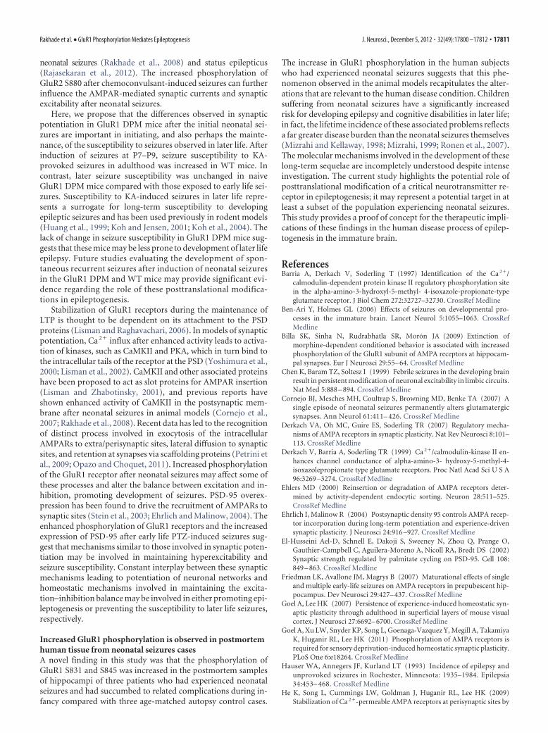
Increased phosphorylation of GluR1 observed in postmortemhuman brain tissue from cases of neonatal seizuresBecause we observed increased phosphorylation of the GluR1receptor in multiple models of neonatal seizures, we examinedwhether there was evidence for phosphorylation at the GluR S831or S845 site in human postmortem tissue from cases of neonatalseizures. Western blot analysis of GluR1 receptor subunit expres-sion and its phosphorylation was performed using postmortemhippocampal tissue obtained from infants that had been diag-nosed with neonatal seizures secondary to hypoxic encephalop-athy (n � 3) (Table 1) and compared with age-matched controls(control tissue obtained from infants �6 months age at death).Hippocampal samples from neonates with confirmed seizuresshowed increased phosphorylation of the GluR1 S831 (296 �83%, n � 3, p � 0.05) compared with brain tissue from autopsycontrols (100 � 19%, p � 0.05) and S845 receptor subunit(232 � 72%, n � 3, p � 0.05) compared with brain tissue fromautopsy controls (100 � 16%, p � 0.05) (Fig. 8). These resultsshow that phosphorylation of GluR1 S831 and S845 can be mea-sured in human postmortem brain tissue and support the
experimental animal data that this post-translational modification may be atherapeutic target in human neonatalseizures.
DiscussionThe present study is the first direct evi-dence to suggest a critical role for post-translational modification of GluR1 S831and S845 in the genesis of seizure-inducednetwork excitability and that this mayrepresent a potential therapeutic target inhuman brain tissue. Early life seizures inWT C57BL/6N mice pups lead to in-creased phosphorylation of the GluR1subunit at S831 and S845 and commensu-rate increases in AMPAR-mediated mEP-SCs in the hippocampal CA1 neurons.Furthermore, seizure-induced phosphor-ylation of GluR1 S831 and S845 was asso-ciated with increases in the synapticscaffolding protein PSD-95. GluR1 DPMmice lacking the ability to phosphorylateS831 and S845 are less susceptible to PTZ
and hypoxia-induced seizures and lacked the seizure-inducedAMPAR potentiation, PSD-95 overexpression, as well as an at-tenuated long-term hyperexcitability that was observed in WTmice. These data implicate GluR1 phosphorylation as an impor-tant step upstream of mechanisms involved in initiation andmaintenance of seizure-induced network hyperexcitability. Im-portantly, there was also evidence of increased phosphorylationof these subunits in human postmortem brain tissue from casesof neonatal seizures compared with control cases.
Neonatal seizures lead to alterations in seizure susceptibilityin C57BL/6N miceInfants experiencing neonatal seizures have a significantly higherincidence of development of epilepsy, autism, and other cogni-tive and neurobehavioral disabilities. The pathological processesunderlying these changes are likely to be multifactorial, but theimbalance between excitation and inhibition in the neuronal cir-cuits plays a critical role in epileptogenesis (McNamara et al.,2006; Rakhade and Jensen, 2009). Given the relative preponder-ance of GluR2-lacking, Ca 2�-permeable AMPARs during thiswindow, the effects of any change in synaptic plasticity signalingcascades are likely to be accentuated. Here we report a new modeland methods for inducing hypoxia-induced seizures in C57BL/6mice, because previous studies were performed in rats that dem-onstrated functional enhancement of AMPARs associated withincreased phosphorylation of GluR1 S831 and S845 within 24 hafter HS (Rakhade et al., 2008; Zhou et al., 2011). Unambiguousproof of a role for these novel posttranslational modifications inepilepsy required reestablishing this model in a transgenic mouselacking the ability to phosphorylate these sites (Lee et al., 2003).In P7–P9 WT mice, seizures induced by either PTZ or globalhypoxia increased phosphorylation of GluR1 S831 and S845. Theincrease in GluR1 phosphorylation was observed as early as 1 hafter the PTZ seizures, providing a mechanism for early altera-tions in the transition of the normal hippocampal circuits intohyperexcitable circuits.
Enhanced excitability and synaptic potentiation mediated byincreased synaptic AMPARs and occluded LTP have been ob-served previously in the hippocampal CA1 neurons immediately
Figure 7. GluR1 DPM mice exposed to neonatal seizures fail to show an increase in susceptibility to later life seizures. A, GluR1DPM mice and WT littermate controls were exposed to hypoxia-induced neonatal seizures at P9; the susceptibility to later lifeKA-induced seizures was studied at P40. Onset of behavioral seizures and the percentage of animals experiencing seizures after KAinjection (35 mg/kg, i.p.) in WT and GluR1 DPM mice that have been exposed to HS at P9 compared with control littermates.Progressive analysis of percentage of animals reaching stage 3 seizures compared with latency to onset of seizures shows asignificant difference in the WT animals exposed to HS compared with normoxic controls. B, Latency time for onset of tonic– clonicseizures was significantly faster in WT mice exposed to HS at P9 (68 � 12%, n � 14, p � 0.05) compared with normoxic WT mice(100 � 11%, n � 16). Latency time was not significantly different in the GluR1 DPM (�/�) mice experiencing neonatal seizures(154 � 21%, n � 12) compared with those not experiencing neonatal seizures (152 � 19%, n � 12, p � 0.4). *p � 0.05.
Rakhade et al. • GluR1 Phosphorylation Mediates Epileptogenesis J. Neurosci., December 5, 2012 • 32(49):17800 –17812 • 17809

after early life seizures in the rat model of hypoxia-induced sei-zures (Jensen et al., 1998; Rakhade et al., 2008; Zhou et al., 2011).These alterations in synaptic potentiation and hippocampal ex-citability are not observed after induction of hypoxia alone; sei-zures are required for initiating these changes (Zhou et al., 2011).In rats, neonatal seizures can lead to later life alterations in plas-ticity at glutamatergic synapses in the hippocampus (Cornejo etal., 2007), accompanied by alterations in synaptic neurotransmit-ters, silent synapses and deficits in spatial memory, and impairedLTP and learning (Mikati et al., 2005; Zhou et al., 2011). In addi-tion, this study establishes a model of hypoxia-induced neonatalseizures in WT mice that exhibit similar consequences in post-translational GluR1 modifications and seizure susceptibility asthe rat model (Rakhade et al., 2008). Chemically induced seizuresin WT mice also lead to these alterations in hippocampal synapticexcitability and susceptibility to later life seizures.
GluR1 phosphorylation contributes to seizure-inducedincreases in AMPAR-mediated synaptic transmission andlater life in vivo seizure susceptibilityGluR1 phosphorylation has been shown to result in changes inAMPAR kinetics and amplitude, synaptic trafficking, and inser-tion of AMPARs in the synaptic membrane (Shepherd andHuganir, 2007) and plays a critical role in mediating LTP andlong-term depression after appropriate stimuli (Mammen et al.,1997; Derkach et al., 1999, 2007). GluR1 phosphorylation at S845leads to an increase in the reinsertion of GluR1 subunits at thePSD and phosphorylation at S831 leads to an increase in theconductance of AMPARs during the induction of LTP. The first 2postnatal weeks in rodents is a critical period in development,with multiple changes that affect the balance of excitation andinhibition in the brain (Rakhade and Jensen, 2009). Here, wehave observed that acute seizures increase AMPAR-mediatedmEPSC amplitude in WT mice, similar to the results describedpreviously in the rat model of neonatal seizures (Rakhade et al.,
2008). However, neonatal seizures in the GluR1 DPM mice resultin a decrease in AMPAR-mediated mEPSC amplitude, this para-doxical effect suggesting that the phosphorylation of GluR1 re-ceptors plays an important role for mediating the excitabilityobserved 24 h after the initial seizures. The decrease in mEPSCamplitude observed after PTZ-induced seizures in the GluR1DPM mice may involve interactions between phosphorylationevents that promote homeostatic events, such as trafficking andstabilization of the AMPARs via internalization and lysosomaldegradation of these receptors (Shepherd and Huganir, 2007;Heine et al., 2008; He et al., 2009). We hypothesize that thesynaptic changes observed after neonatal seizures may be attrib-utable to complex interactions between the AMPAR phosphor-ylation and trafficking, in combination with homeostaticmechanisms involved in maintaining the excitation–inhibitionimbalance. Indeed, previous studies have suggested that GluR1S845 phosphorylation plays an important role in stabilizingCa 2�-permeable AMPARs and preventing their lysosomal deg-radation (Ehlers, 2000; Goel and Lee, 2007; Man et al., 2007; He etal., 2009; Goel et al., 2011). The lack of GluR1 S845 phosphory-lation in the GluR1 DPM mice may significantly enhance thelysosomal degradation of the internalized GluR1-containing re-ceptors and may affect the perisynaptic AMPARs that are avail-able for “ready insertion ” in response to neuronal activity (He etal., 2009). Furthermore, enhanced GluR2 phosphorylation atS880 observed after neonatal seizure may contribute to receptorinternalization and expression of Ca 2�-permeable AMPARs atthe synaptic surface (Rakhade et al., 2008).
Recent studies using single molecule tracking to detect themovement of AMPARs have shown that GluR1-containingAMPARs freely diffuse in and out of the synapse within the PSD(Heine et al., 2008; Petrini et al., 2009), and anchoring them at thesynapse may require PDZ domain interactions. Future studiesmay reveal the mechanisms involved in mediating this effect onAMPAR-mediated mEPSCs after PTZ-induced seizures, andcompensatory changes in the excitation–inhibition imbalancemay provide information regarding the specific mechanism in-volved in the paradoxical change observed. Animal models ofseizures induced by use of chemoconvulsants in early life haveshown multifactorial changes, including alterations in the GABAreceptors and AMPARs (Zhang et al., 2004; Silva et al., 2005;Friedman et al., 2007).
GluR1 DPM mice retain the ability to exhibit seizures in thepresence of PTZ but display an increased latency to the onset ofthese behavioral seizures. There does not appear to be significantchanges in baseline expression of AMPAR GluR1 or GluR2 sub-units or in baseline mEPSCs in hippocampal neurons studied inthe GluR1 DPM and WT mice. Furthermore, we did not observea difference in the rise time, decay time, and frequency ofAMPAR-mediated mEPSCs between the naive WT and GluR1DPM mice at P8. Consistently, previous reports comparingGluR1 DPM mice with WT did not show any significant abnor-malities in anatomical structure, receptor subunit distribution,baseline synaptic transmission, and transport of receptors to thesynaptic surface (Lee et al., 2003).
Phosphorylation of the GluR1 subunit has been reported tobe critical for multiple synaptic potentiation events, includingtrafficking of GluR1-containing receptors, stabilization at thesynaptic surface, maintenance of LTP, as well as lysosomalinternalization and degradation of Ca 2�-permeable AMPARs(Shepherd and Huganir, 2007; Heine et al., 2008). Previousreportshavesuggestedthat increasedGluR2S880phosphorylationmayresult in the enhanced persistence of Ca2�-permeable AMPARs after
Figure 8. Increase in phosphorylation of GluR1 receptor in hippocampi from early childhoodcases with neonatal seizures. A, Representative Western blots depicting increased phosphory-lation of GluR1 S831 and GluR1 S845 in hippocampal tissue obtained from subjects who hadexperienced hypoxic encephalopathy before death at term; comparison were made with hip-pocampal tissue obtained from age-matched autopsy controls. Phosphorylation of GluR1 S831was increased in hippocampal tissue (average of 296 � 83%, n � 3, p � 0.05) compared withage-matched controls. B, Similarly, increase in phosphorylation of GluR1 S845 was also de-tected (average of 232 � 72%, n � 3, p � 0.05) compared with brain tissue from autopsycontrols. *p � 0.05.
17810 • J. Neurosci., December 5, 2012 • 32(49):17800 –17812 Rakhade et al. • GluR1 Phosphorylation Mediates Epileptogenesis
neonatal seizures (Rakhade et al., 2008) and status epilepticus(Rajasekaran et al., 2012). The increased phosphorylation ofGluR2 S880 after chemoconvulsant-induced seizures can furtherinfluence the AMPAR-mediated synaptic currents and synapticexcitability after neonatal seizures.
Here, we propose that the differences observed in synapticpotentiation in GluR1 DPM mice after the initial neonatal sei-zures are important in initiating, and also perhaps the mainte-nance, of the susceptibility to seizures observed in later life. Afterinduction of seizures at P7–P9, seizure susceptibility to KA-provoked seizures in adulthood was increased in WT mice. Incontrast, later seizure susceptibility was unchanged in naiveGluR1 DPM mice compared with those exposed to early life sei-zures. Susceptibility to KA-induced seizures in later life repre-sents a surrogate for long-term susceptibility to developingepileptic seizures and has been used previously in rodent models(Huang et al., 1999; Koh and Jensen, 2001; Koh et al., 2004). Thelack of change in seizure susceptibility in GluR1 DPM mice sug-gests that these mice may be less prone to development of later lifeepilepsy. Future studies evaluating the development of spon-taneous recurrent seizures after induction of neonatal seizuresin the GluR1 DPM and WT mice may provide significant evi-dence regarding the role of these posttranslational modifica-tions in epileptogenesis.
Stabilization of GluR1 receptors during the maintenance ofLTP is thought to be dependent on its attachment to the PSDproteins (Lisman and Raghavachari, 2006). In models of synapticpotentiation, Ca 2� influx after enhanced activity leads to activa-tion of kinases, such as CaMKII and PKA, which in turn bind tothe intracellular tails of the receptor at the PSD (Yoshimura et al.,2000; Lisman et al., 2002). CaMKII and other associated proteinshave been proposed to act as slot proteins for AMPAR insertion(Lisman and Zhabotinsky, 2001), and previous reports haveshown enhanced activity of CaMKII in the postsynaptic mem-brane after neonatal seizures in animal models (Cornejo et al.,2007; Rakhade et al., 2008). Recent data has led to the recognitionof distinct process involved in exocytosis of the intracellularAMPARs to extra/perisynaptic sites, lateral diffusion to synapticsites, and retention at synapses via scaffolding proteins (Petrini etal., 2009; Opazo and Choquet, 2011). Increased phosphorylationof the GluR1 receptor after neonatal seizures may affect some ofthese processes and alter the balance between excitation and in-hibition, promoting development of seizures. PSD-95 overex-pression has been found to drive the recruitment of AMPARs tosynaptic sites (Stein et al., 2003; Ehrlich and Malinow, 2004). Theenhanced phosphorylation of GluR1 receptors and the increasedexpression of PSD-95 after early life PTZ-induced seizures sug-gest that mechanisms similar to those involved in synaptic poten-tiation may be involved in maintaining hyperexcitability andseizure susceptibility. Constant interplay between these synapticmechanisms leading to potentiation of neuronal networks andhomeostatic mechanisms involved in maintaining the excita-tion–inhibition balance may be involved in either promoting epi-leptogenesis or preventing the susceptibility to later life seizures,respectively.
Increased GluR1 phosphorylation is observed in postmortemhuman tissue from neonatal seizures casesA novel finding in this study was that the phosphorylation ofGluR1 S831 and S845 was increased in the postmortem samplesof hippocampi of three patients who had experienced neonatalseizures and had succumbed to related complications during in-fancy compared with three age-matched autopsy control cases.
The increase in GluR1 phosphorylation in the human subjectswho had experienced neonatal seizures suggests that this phe-nomenon observed in the animal models recapitulates the alter-ations that are relevant to the human disease condition. Childrensuffering from neonatal seizures have a significantly increasedrisk for developing epilepsy and cognitive disabilities in later life;in fact, the lifetime incidence of these associated problems reflectsa far greater disease burden than the neonatal seizures themselves(Mizrahi and Kellaway, 1998; Mizrahi, 1999; Ronen et al., 2007).The molecular mechanisms involved in the development of theselong-term sequelae are incompletely understood despite intenseinvestigation. The current study highlights the potential role ofposttranslational modification of a critical neurotransmitter re-ceptor in epileptogenesis; it may represent a potential target in atleast a subset of the population experiencing neonatal seizures.This study provides a proof of concept for the therapeutic impli-cations of these findings in the human disease process of epilep-togenesis in the immature brain.
ReferencesBarria A, Derkach V, Soderling T (1997) Identification of the Ca 2�/
calmodulin-dependent protein kinase II regulatory phosphorylation sitein the alpha-amino-3-hydroxyl-5-methyl- 4-isoxazole-propionate-typeglutamate receptor. J Biol Chem 272:32727–32730. CrossRef Medline
Ben-Ari Y, Holmes GL (2006) Effects of seizures on developmental pro-cesses in the immature brain. Lancet Neurol 5:1055–1063. CrossRefMedline
Billa SK, Sinha N, Rudrabhatla SR, Moron JA (2009) Extinction ofmorphine-dependent conditioned behavior is associated with increasedphosphorylation of the GluR1 subunit of AMPA receptors at hippocam-pal synapses. Eur J Neurosci 29:55– 64. CrossRef Medline
Chen K, Baram TZ, Soltesz I (1999) Febrile seizures in the developing brainresult in persistent modification of neuronal excitability in limbic circuits.Nat Med 5:888 – 894. CrossRef Medline
Cornejo BJ, Mesches MH, Coultrap S, Browning MD, Benke TA (2007) Asingle episode of neonatal seizures permanently alters glutamatergicsynapses. Ann Neurol 61:411– 426. CrossRef Medline
Derkach VA, Oh MC, Guire ES, Soderling TR (2007) Regulatory mecha-nisms of AMPA receptors in synaptic plasticity. Nat Rev Neurosci 8:101–113. CrossRef Medline
Derkach V, Barria A, Soderling TR (1999) Ca 2�/calmodulin-kinase II en-hances channel conductance of alpha-amino-3- hydroxy-5-methyl-4-isoxazolepropionate type glutamate receptors. Proc Natl Acad Sci U S A96:3269 –3274. CrossRef Medline
Ehlers MD (2000) Reinsertion or degradation of AMPA receptors deter-mined by activity-dependent endocytic sorting. Neuron 28:511–525.CrossRef Medline
Ehrlich I, Malinow R (2004) Postsynaptic density 95 controls AMPA recep-tor incorporation during long-term potentiation and experience-drivensynaptic plasticity. J Neurosci 24:916 –927. CrossRef Medline
El-Husseini Ael-D, Schnell E, Dakoji S, Sweeney N, Zhou Q, Prange O,Gauthier-Campbell C, Aguilera-Moreno A, Nicoll RA, Bredt DS (2002)Synaptic strength regulated by palmitate cycling on PSD-95. Cell 108:849 – 863. CrossRef Medline
Friedman LK, Avallone JM, Magrys B (2007) Maturational effects of singleand multiple early-life seizures on AMPA receptors in prepubescent hip-pocampus. Dev Neurosci 29:427– 437. CrossRef Medline
Goel A, Lee HK (2007) Persistence of experience-induced homeostatic syn-aptic plasticity through adulthood in superficial layers of mouse visualcortex. J Neurosci 27:6692– 6700. CrossRef Medline
Goel A, Xu LW, Snyder KP, Song L, Goenaga-Vazquez Y, Megill A, TakamiyaK, Huganir RL, Lee HK (2011) Phosphorylation of AMPA receptors isrequired for sensory deprivation-induced homeostatic synaptic plasticity.PLoS One 6:e18264. CrossRef Medline
Hauser WA, Annegers JF, Kurland LT (1993) Incidence of epilepsy andunprovoked seizures in Rochester, Minnesota: 1935–1984. Epilepsia34:453– 468. CrossRef Medline
He K, Song L, Cummings LW, Goldman J, Huganir RL, Lee HK (2009)Stabilization of Ca 2�-permeable AMPA receptors at perisynaptic sites by
Rakhade et al. • GluR1 Phosphorylation Mediates Epileptogenesis J. Neurosci., December 5, 2012 • 32(49):17800 –17812 • 17811

GluR1–S845 phosphorylation. Proc Natl Acad Sci U S A 106:20033–20038. CrossRef Medline
Heine M, Groc L, Frischknecht R, Beique JC, Lounis B, Rumbaugh G,Huganir RL, Cognet L, Choquet D (2008) Surface mobility of postsyn-aptic AMPARs tunes synaptic transmission. Science 320:201–205.CrossRef Medline
Hu H, Real E, Takamiya K, Kang MG, Ledoux J, Huganir RL, Malinow R(2007) Emotion enhances learning via norepinephrine regulation ofAMPA-receptor trafficking. Cell 131:160 –173. CrossRef Medline
Huang L, Cilio MR, Silveira DC, McCabe BK, Sogawa Y, Stafstrom CE, Hol-mes GL (1999) Long-term effects of neonatal seizures: a behavioral,electrophysiological, and histological study. Brain Res Dev Brain Res 118:99 –107. CrossRef Medline
Jensen FE (2011) Epilepsy as a spectrum disorder: Implications from novelclinical and basic neuroscience. Epilepsia 52 [Suppl 1]:1– 6.
Jensen FE, Wang C, Stafstrom CE, Liu Z, Geary C, Stevens MC (1998) Acuteand chronic increases in excitability in rat hippocampal slices after peri-natal hypoxia in vivo. J Neurophysiol 79:73– 81. Medline
Kapus G, Szekely JI, Durand J, Ruiz A, Tarnawa I (2000) AMPA receptorantagonists, GYKI 52466 and NBQX, do not block the induction of long-term potentiation at therapeutically relevant concentrations. Brain ResBull 52:511–517. CrossRef Medline
Koh S, Jensen FE (2001) Topiramate blocks perinatal hypoxia-induced sei-zures in rat pups. Ann Neurol 50:366 –372. CrossRef Medline
Koh S, Tibayan FD, Simpson JN, Jensen FE (2004) NBQX or topiramatetreatment following perinatal hypoxia-induced seizures prevents later in-creases in seizure-induced neuronal injury. Epilepsia 45:569 –575.CrossRef Medline
Kumar SS, Bacci A, Kharazia V, Huguenard JR (2002) A developmentalswitch of AMPA receptor subunits in neocortical pyramidal neurons.J Neurosci 22:3005–3015. Medline
Lee HK, Barbarosie M, Kameyama K, Bear MF, Huganir RL (2000) Regula-tion of distinct AMPA receptor phosphorylation sites during bidirec-tional synaptic plasticity. Nature 405:955–959. CrossRef Medline
Lee HK, Takamiya K, Han JS, Man H, Kim CH, Rumbaugh G, Yu S, Ding L,He C, Petralia RS, Wenthold RJ, Gallagher M, Huganir RL (2003) Phos-phorylation of the AMPA receptor GluR1 subunit is required for synapticplasticity and retention of spatial memory. Cell 112:631– 643. CrossRefMedline
Li P, Kerchner GA, Sala C, Wei F, Huettner JE, Sheng M, Zhuo M (1999)AMPA receptor-PDZ interactions in facilitation of spinal sensory syn-apses. Nat Neurosci 2:972–977. CrossRef Medline
Lisman J, Raghavachari S (2006) A unified model of the presynaptic andpostsynaptic changes during LTP at CA1 synapses. Sci STKE 2006:re11.CrossRef Medline
Lisman JE, Zhabotinsky AM (2001) A model of synaptic memory: a CaMKII/PP1 switch that potentiates transmission by organizing an AMPA recep-tor anchoring assembly. Neuron 31:191–201. CrossRef Medline
Lisman J, Schulman H, Cline H (2002) The molecular basis of CaMKIIfunction in synaptic and behavioural memory. Nat Rev Neurosci 3:175–190. CrossRef Medline
Mammen AL, Kameyama K, Roche KW, Huganir RL (1997) Phosphoryla-tion of the alpha-amino-3-hydroxy-5-methylisoxazole4-propionic acidreceptor GluR1 subunit by calcium/calmodulin-dependent kinase II.J Biol Chem 272:32528 –32533. CrossRef Medline
Man HY, Sekine-Aizawa Y, Huganir R (2007) Regulation of a-amino-3-hydroxy-5-methyl-4-isoxazolepropionic acid receptor trafficking throughPKA phosphorylation of the Glu receptor 1 subunit. Proc Natl Acad Sci U S A104:3579–3584. CrossRef Medline
McNamara JO, Huang YZ, Leonard AS (2006) Molecular signaling mecha-nisms underlying epileptogenesis. Sci STKE 2006:re12. CrossRef Medline
Mikati MA, Zeinieh MP, Kurdi RM, Harb SA, El Hokayem JA, Daderian RH,Shamseddine A, Obeid M, Bitar FF, El Sabban M (2005) Long-termeffects of acute and of chronic hypoxia on behavior and on hippocampalhistology in the developing brain. Brain Res Dev Brain Res 157:98 –102.CrossRef Medline
Mizrahi EM (1999) Acute and chronic effects of seizures in the developingbrain: lessons from clinical experience. Epilepsia 40 [Suppl 1]:S42–S50.
Mizrahi EM, Kellaway P (1998) Diagnosis and management of neonatalseizures. Philadelphia: Lippincott-Raven.
Opazo P, Choquet D (2011) A three-step model for the synaptic recruit-ment of AMPA receptors. Mol Cell Neurosci 46:1– 8. CrossRef Medline
Petrini EM, Lu J, Cognet L, Lounis B, Ehlers MD, Choquet D (2009) Endo-cytic trafficking and recycling maintain a pool of mobile surface AMPAreceptors required for synaptic potentiation. Neuron 63:92–105.CrossRef Medline
Rajasekaran K, Todorovic M, Kapur J (2012) Calcium-permeable AMPAreceptors are expressed in a rodent model of status epilepticus. Ann Neu-rol 72:91–102. CrossRef Medline
Rakhade SN, Jensen FE (2009) Epileptogenesis in the immature brain:emerging mechanisms. Nat Rev Neurol 5:380 –391. CrossRef Medline
Rakhade SN, Zhou C, Aujla PK, Fishman R, Sucher NJ, Jensen FE (2008)Early alterations of AMPA receptors mediate synaptic potentiation in-duced by neonatal seizures. J Neurosci 28:7979 –7990. CrossRef Medline
Rakhade SN, Klein PM, Huynh T, Hilario-Gomez C, Kosaras B, Rotenberg A,Jensen FE (2011) Development of later life spontaneous seizures in a rodentmodel of hypoxia-induced neonatal seizures. Epilepsia 52:753–765. CrossRefMedline
Ramzan IM, Levy G (1985) Kinetics of drug action in disease states. XV.Effect of pregnancy on the convulsive activity of pentylenetetrazol in rats.J Pharm Sci 74:1233–1235. CrossRef Medline
Ronen GM, Buckley D, Penney S, Streiner DL (2007) Long-term prognosisin children with neonatal seizures: a population-based study. Neurology69:1816 –1822. CrossRef Medline
Sanchez RM, Koh S, Rio C, Wang C, Lamperti ED, Sharma D, Corfas G,Jensen FE (2001) Decreased glutamate receptor 2 expression and en-hanced epileptogenesis in immature rat hippocampus after perinatalhypoxia-induced seizures. J Neurosci 21:8154 – 8163. Medline
Sanchez RM, Dai W, Levada RE, Lippman JJ, Jensen FE (2005a) AMPA/kainate receptor-mediated downregulation of GABAergic synaptic trans-mission by calcineurin after seizures in the developing rat brain.J Neurosci 25:3442–3451. CrossRef Medline
Sanchez RM, Jensen FE, Pitanken A, Schwartzkroin PA, Moshe SL (2005b)Modeling hypoxia-induced seizures and hypoxic encephalopathy in theneonatal period. In: Models of seizures and epilepsy. San Diego: Elsevier.
Shepherd JD, Huganir RL (2007) The cell biology of synaptic plasticity:AMPA receptor trafficking. Annu Rev Cell Dev Biol 23:613– 643.CrossRef Medline
Silva AV, Regondi MC, Cipelletti B, Frassoni C, Cavalheiro EA, Spreafico R(2005) Neocortical and hippocampal changes after multiple pilocarpine-induced status epilepticus in rats. Epilepsia 46:636 – 642. CrossRefMedline
Silverstein FS, Jensen FE (2007) Neonatal seizures. Ann Neurol 62:112–120.CrossRef Medline
Sogawa Y, Monokoshi M, Silveira DC, Cha BH, Cilio MR, McCabe BK, Liu X,Hu Y, Holmes GL (2001) Timing of cognitive deficits following neona-tal seizures: relationship to histological changes in the hippocampus.Brain Res Dev Brain Res 131:73– 83. CrossRef Medline
Song I, Huganir RL (2002) Regulation of AMPA receptors during synapticplasticity. Trends Neurosci 25:578 –588. CrossRef Medline
Stein V, House DR, Bredt DS, Nicoll RA (2003) Postsynaptic density-95mimics and occludes hippocampal long-term potentiation and enhanceslong-term depression. J Neurosci 23:5503–5506. Medline
Talos DM, Fishman RE, Park H, Folkerth RD, Follett PL, Volpe JJ, Jensen FE(2006) Developmental regulation of alpha-amino-3-hydroxy-5-methyl-4-isoxazole-propionic acid receptor subunit expression in forebrain andrelationship to regional susceptibility to hypoxic/ischemic injury. I. Ro-dent cerebral white matter and cortex. J Comp Neurol 497:42– 60.CrossRef Medline
Talos DM, Sun H, Zhou X, Fitzgerald EC, Jackson MC, Klein PM, Lan VJ,Joseph A, Jensen FE (2012) The interaction between early life epilepsyand autistic-like behavioral consequences: a role for the mammalian tar-get of rapamycin (mTOR) pathway. PLoS One 7:e35885. CrossRefMedline
Velisek L, Kubova H, Pohl M, Stankova L, Mares P, Schickerova R (1992)Pentylenetetrazol-induced seizures in rats: an ontogenetic study. NaunynSchmiedebergs Arch Pharmacol 346:588 –591. CrossRef Medline
Wenthold RJ, Yokotani N, Doi K, Wada K (1992) Immunochemical char-acterization of the non-NMDA glutamate receptor using subunit-specificantibodies. J Biol Chem 267:501–507. Medline
Wyllie DJ, Nicoll RA (1994) A role for protein kinases and phosphatases inthe Ca 2�-induced enhancement of hippocampal AMPA receptor-mediated synaptic responses. Neuron 13:635– 643. CrossRef Medline
Yoshimura Y, Aoi C, Yamauchi T (2000) Investigation of protein substrates
17812 • J. Neurosci., December 5, 2012 • 32(49):17800 –17812 Rakhade et al. • GluR1 Phosphorylation Mediates Epileptogenesis

of Ca 2�/calmodulin-dependent protein kinase II translocated to thepostsynaptic density. Brain Res Mol Brain Res 81:118 –128. CrossRefMedline
Zhang G, Raol YH, Hsu FC, Coulter DA, Brooks-Kayal AR (2004) Effects ofstatus epilepticus on hippocampal GABAA receptors are age-dependent.Neuroscience 125:299 –303. CrossRef Medline
Zhou C, Lippman JJ, Sun H, Jensen FE (2011) Hypoxia-induced neonatalseizures diminish silent synapses and long-term potentiation in hip-pocampal CA1 neurons. J Neurosci 31:18211–18222. CrossRef Medline
Rakhade et al. • GluR1 Phosphorylation Mediates Epileptogenesis J. Neurosci., December 5, 2012 • 32(49):17800 –17812 • 17812a