Glutamate Transporter GLAST Is Expressed in the Radial ... · Glutamate Transporter GLAST Is...
8
Glutamate Transporter GLAST Is Expressed in the Radial Glia–Astrocyte Lineage of Developing Mouse Spinal Cord Takashi Shibata, 1,2 Keiko Yamada, 1 Masahiko Watanabe, 1 Kazuhiro Ikenaka, 3 Keiji Wada, 4 Kohichi Tanaka, 4 and Yoshiro Inoue 1 Departments of 1 Anatomy and 2 Urology, Hokkaido University School of Medicine, Sapporo 060, Japan, 3 National Institute for Physiological Sciences, Okazaki National Research Institutes, Okazaki 444, Japan, and 4 Department of Degenerative Neurological Diseases, National Institute of Neuroscience, National Center of Neurology and Psychiatry, Kodaira 187, Japan The glutamate transporter GLAST is localized on the cell mem- brane of mature astrocytes and is also expressed in the ven- tricular zone of developing brains. To characterize and follow the GLAST-expressing cells during development, we examined the mouse spinal cord by in situ hybridization and immunohis- tochemistry. At embryonic day (E) 11 and E13, cells expressing GLAST mRNA were present only in the ventricular zone, where GLAST immunoreactivity was associated with most of the cell bodies of neuroepithelial cells. In addition, GLAST immunore- activity was detected in radial processes running through the mantle and marginal zones. From this characteristic cytology, GLAST-expressing cells at early stages were judged to be radial glia cells. At E15, cells expressing GLAST mRNA first appeared in the mantle zone, and GLAST-immunopositive punctate or reticular protrusions were formed along the radial processes. From E18 to postnatal day (P) 7, GLAST mRNA or its immuno- reactivity gradually decreased from the ventricular zone and dis- appeared from radial processes, whereas cells with GLAST mRNA spread all over the mantle zone and GLAST-immunopositive punctate/reticular protrusions predominated in the neuropils. At P7, GLAST-expressing cells were immunopositive for glial fibrillary acidic protein, an intermediate filament specific to astrocytes. Therefore, the glutamate transporter GLAST is expressed from radial glia through astrocytes during spinal cord development. Furthermore, the distinct changes in the cell position and mor- phology suggest that both the migration and transformation of radial glia cells begin in the spinal cord between E13 and E15, when the active stage of neuronal migration is over. Key words: glutamate transporter; GLAST; radial glia; astro- cyte; spinal cord; development; in situ hybridization; immuno- histochemistry; mouse Glutamate is a major neurotransmitter mediating the fast excita- tory transmission at central synapses, and it also plays critical roles in synaptic plasticity and development (Mayer and Westbrook, 1987). The extracellular glutamate concentration has to be kept low enough to terminate glutamate receptor activation and to protect neurons from glutamate excitotoxicity (Hertz, 1979; Choi, 1992; Tanaka K et al., 1997). The low extracellular concentration is attributable to the action of high-affinity, sodium-dependent gluta- mate transporters. Molecular biological studies have identified four subtypes of the glutamate transporter with distinct structures, functions, and distributions (Kanai et al., 1997). In the brain, GLAST (GluT-1 or EAAT1) and GLT1 (EAAT2) are the astro- cytic transporters, being highly concentrated in the cerebellum and the telencephalon, respectively (Kanai and Hediger, 1992; Storck et al., 1992; Rothstein et al., 1994; Torp et al., 1994; Lehre et al., 1995; Shibata et al., 1996). On the other hand, EAAC1 (EAAT3) and EAAT4 are the neuronal transporters; the former is expressed widely in the brain (Kanai et al., 1995; Kiryu et al., 1995; Shibata et al., 1996), whereas the latter expression is confined to cerebellar Purkinje cells (Yamada et al., 1996). Immunoelectron microscopy has clarified that GLAST and GLT1 are more abundant on astro- cytic cell membrane that surrounds synapses than that facing dendritic shafts, vessels, pial surface, and neighboring astrocytes (Chaudhry et al., 1995). EAAT4 is selectively localized at the extrajunctional site of Purkinje cell spines contacting parallel fibers or climbing fibers (Yamada et al., 1996; Tanaka J et al., 1997). The preferential and highly regulated localizations at synaptic regions provide a molecular– anatomical basis for glutamate transporter function in rapid removal of glutamate or its related amino acids from the synaptic cleft. Four glutamate transporter subtypes also exhibit distinct ex- pression and distribution in the fetal brain, even before synapto- genesis (Shibata et al., 1996; Sutherland et al., 1996; Yamada et al., 1997). Of these, GLAST is characterized by prominent ex- pression of the mRNA in the ventricular zone of various brain regions, a transient zone that produces most neurons and neuro- glia. The expression of GL AST mRNA from early stages suggests that the glutamate transporter is involved not only in the regula- tion of synaptic transmission, but it also plays some role in the development and differentiation of the CNS. As the first step to test this possibility, we attempted to characterize and follow cells expressing GLAST. In the present study, we examined the devel- oping mouse spinal cord by taking advantage of its simple tube- like structure throughout development and of well documented histogenesis and cytogenesis (Choi, 1981; Altman and Bayer, Received June 19, 1997; revised Sept. 11, 1997; accepted Sept. 19, 1997. This investigation was supported in part by grants-in-aid for Scientific Research on Priority Areas from the Ministry of Education, Science, Sports, and Culture of Japan and also by research grants from the Ministry of Health and Welfare of Japan, the Science and Technology Agency of Japan, the Suhara Foundation, and the Ichiro Kanehara Foundation. We thank Dr. Yasuhiro Tomooka at the Science University of Tokyo for his kind gift of rabbit anti-nestin antibody, and also Mr. Hideo Umeda and Mr. Yoshihiko Ogawa at the Hokkaido University School of Medicine for their technical assistance. T.S. and K.Y. contributed equally to this work. Correspondence should be addressed to Masahiko Watanabe, Department of Anatomy, Hokkaido University School of Medicine, Sapporo 060, Japan. Copyright © 1997 Society for Neuroscience 0270-6474/97/179212-08$05.00/0 The Journal of Neuroscience, December 1, 1997, 17(23):9212–9219
Transcript of Glutamate Transporter GLAST Is Expressed in the Radial ... · Glutamate Transporter GLAST Is...

Glutamate Transporter GLAST Is Expressed in the RadialGlia–Astrocyte Lineage of Developing Mouse Spinal Cord
Takashi Shibata,1,2 Keiko Yamada,1 Masahiko Watanabe,1 Kazuhiro Ikenaka,3 Keiji Wada,4 Kohichi Tanaka,4and Yoshiro Inoue1
Departments of 1Anatomy and 2Urology, Hokkaido University School of Medicine, Sapporo 060, Japan, 3National Institute forPhysiological Sciences, Okazaki National Research Institutes, Okazaki 444, Japan, and 4Department of DegenerativeNeurological Diseases, National Institute of Neuroscience, National Center of Neurology and Psychiatry, Kodaira 187, Japan
The glutamate transporter GLAST is localized on the cell mem-brane of mature astrocytes and is also expressed in the ven-tricular zone of developing brains. To characterize and followthe GLAST-expressing cells during development, we examinedthe mouse spinal cord by in situ hybridization and immunohis-tochemistry. At embryonic day (E) 11 and E13, cells expressingGLAST mRNA were present only in the ventricular zone, whereGLAST immunoreactivity was associated with most of the cellbodies of neuroepithelial cells. In addition, GLAST immunore-activity was detected in radial processes running through themantle and marginal zones. From this characteristic cytology,GLAST-expressing cells at early stages were judged to be radialglia cells. At E15, cells expressing GLAST mRNA first appearedin the mantle zone, and GLAST-immunopositive punctate orreticular protrusions were formed along the radial processes.From E18 to postnatal day (P) 7, GLAST mRNA or its immuno-
reactivity gradually decreased from the ventricular zone and dis-appeared from radial processes, whereas cells with GLAST mRNAspread all over the mantle zone and GLAST-immunopositivepunctate/reticular protrusions predominated in the neuropils. AtP7, GLAST-expressing cells were immunopositive for glial fibrillaryacidic protein, an intermediate filament specific to astrocytes.Therefore, the glutamate transporter GLAST is expressed fromradial glia through astrocytes during spinal cord development.Furthermore, the distinct changes in the cell position and mor-phology suggest that both the migration and transformation ofradial glia cells begin in the spinal cord between E13 and E15,when the active stage of neuronal migration is over.
Key words: glutamate transporter; GLAST; radial glia; astro-cyte; spinal cord; development; in situ hybridization; immuno-histochemistry; mouse
Glutamate is a major neurotransmitter mediating the fast excita-tory transmission at central synapses, and it also plays critical rolesin synaptic plasticity and development (Mayer and Westbrook,1987). The extracellular glutamate concentration has to be keptlow enough to terminate glutamate receptor activation and toprotect neurons from glutamate excitotoxicity (Hertz, 1979; Choi,1992; Tanaka K et al., 1997). The low extracellular concentration isattributable to the action of high-affinity, sodium-dependent gluta-mate transporters. Molecular biological studies have identified foursubtypes of the glutamate transporter with distinct structures,functions, and distributions (Kanai et al., 1997). In the brain,GLAST (GluT-1 or EAAT1) and GLT1 (EAAT2) are the astro-cytic transporters, being highly concentrated in the cerebellum andthe telencephalon, respectively (Kanai and Hediger, 1992; Storcket al., 1992; Rothstein et al., 1994; Torp et al., 1994; Lehre et al.,1995; Shibata et al., 1996). On the other hand, EAAC1 (EAAT3)and EAAT4 are the neuronal transporters; the former is expressed
widely in the brain (Kanai et al., 1995; Kiryu et al., 1995; Shibataet al., 1996), whereas the latter expression is confined to cerebellarPurkinje cells (Yamada et al., 1996). Immunoelectron microscopyhas clarified that GLAST and GLT1 are more abundant on astro-cytic cell membrane that surrounds synapses than that facingdendritic shafts, vessels, pial surface, and neighboring astrocytes(Chaudhry et al., 1995). EAAT4 is selectively localized at theextrajunctional site of Purkinje cell spines contacting parallel fibersor climbing fibers (Yamada et al., 1996; Tanaka J et al., 1997). Thepreferential and highly regulated localizations at synaptic regionsprovide a molecular–anatomical basis for glutamate transporterfunction in rapid removal of glutamate or its related amino acidsfrom the synaptic cleft.
Four glutamate transporter subtypes also exhibit distinct ex-pression and distribution in the fetal brain, even before synapto-genesis (Shibata et al., 1996; Sutherland et al., 1996; Yamada etal., 1997). Of these, GLAST is characterized by prominent ex-pression of the mRNA in the ventricular zone of various brainregions, a transient zone that produces most neurons and neuro-glia. The expression of GLAST mRNA from early stages suggeststhat the glutamate transporter is involved not only in the regula-tion of synaptic transmission, but it also plays some role in thedevelopment and differentiation of the CNS. As the first step totest this possibility, we attempted to characterize and follow cellsexpressing GLAST. In the present study, we examined the devel-oping mouse spinal cord by taking advantage of its simple tube-like structure throughout development and of well documentedhistogenesis and cytogenesis (Choi, 1981; Altman and Bayer,
Received June 19, 1997; revised Sept. 11, 1997; accepted Sept. 19, 1997.This investigation was supported in part by grants-in-aid for Scientific Research on
Priority Areas from the Ministry of Education, Science, Sports, and Culture ofJapan and also by research grants from the Ministry of Health and Welfare of Japan,the Science and Technology Agency of Japan, the Suhara Foundation, and the IchiroKanehara Foundation. We thank Dr. Yasuhiro Tomooka at the Science Universityof Tokyo for his kind gift of rabbit anti-nestin antibody, and also Mr. Hideo Umedaand Mr. Yoshihiko Ogawa at the Hokkaido University School of Medicine for theirtechnical assistance.
T.S. and K.Y. contributed equally to this work.Correspondence should be addressed to Masahiko Watanabe, Department of
Anatomy, Hokkaido University School of Medicine, Sapporo 060, Japan.Copyright © 1997 Society for Neuroscience 0270-6474/97/179212-08$05.00/0
The Journal of Neuroscience, December 1, 1997, 17(23):9212–9219

1984). Here we show that GLAST is expressed in the radialglia–astrocyte lineage in the developing spinal cord. Furthermore,characteristic temporal changes in the cell position and morphol-ogy, as visualized by in situ hybridization and immunohistochem-istry for GLAST, suggest that both the migration and transforma-tion of radial glia cells begin at a specific stage of development.
MATERIALS AND METHODSAnimals and tissues. In the present study, we used the lumbar spinal cordof C57BL/6J mice at embryonic day (E) 9, E11, E13, E15, and E18,postnatal day (P) 1, P7, P14, and P21, and 4 months (adult) as well as theadult brain. The day after overnight mating was counted as E0. Forisotopic in situ hybridization, fresh frozen tissues were prepared frommice at E11 through the adult stage under deep pentobarbital anesthesia.For histological examination, immunohistochemistry, and nonisotopic insitu hybridization, fetuses from E9 to E18 were immersed overnight inthe Bouin fixative at 4°C, and postnatal mice were perfused transcardiallywith 4% paraformaldehyde in 0.1 M sodium phosphate buffer, pH 7.2.
Histology. Cross sections (5 mm) were prepared from the spinal cord,embedded in paraffin wax, and stained with hematoxylin. From fivesections, the mean number of nuclear profiles per single sections wascalculated for the ventricular or mantle zone.
In situ hybridization. For isotopic detection of the mouse GLASTmRNA, two nonoverlapping antisense 45-mer oligonucleotide probeswere labeled with 35S-dATP using terminal deoxyribonucleotidyl trans-ferase (BRL, Bethesda, MD) to a specific activity of 0.5 3 10 9 dpm/mgDNA. The probe sequence was complementary to nucleotide residues11–56 or 669–714 of the mouse GLAST cDNA (Tanaka, 1993). Freshfrozen sections (20 mm in thickness) were treated at room temperaturewith the following incubations: fixation with 4% paraformaldehyde for 10min, acetylation with 0.25% acetic anhydride in 0.1 M triethanolamine-HCl, pH 8.0, for 10 min, and prehybridization for 1 hr in a buffercontaining 50% formamide, 50 mM Tris-HCl, pH 7.5, 0.02% Ficoll,0.02% polyvinylpyrrolidone, 0.02% bovine serum albumin, 0.6 M NaCl,0.25% SDS, 200 mg/ml tRNA, 1 mM EDTA, and 10% dextran sulfate.Hybridization was performed at 42°C for 10 hr in the prehybridizationbuffer supplemented with 10,000 cpm/ml of 35S-labeled oligonucleotideprobes and 0.1 M dithiothreitol. The slides were washed twice at 55°C for40 min in 0.1 3 SSC containing 0.1% sarcosyl and exposed to eitherHyperfilm-b max (Amersham, Arlington Heights, IL) or nuclear trackemulsion (Kodak NTB-2, Rochester, NY).
For nonisotopic detection, digoxigenin (DIG)-labeled sense and anti-sense cRNA probes were prepared, using linearized Bluescript SK (2)plasmid containing a 733 base pair cDNA fragment corresponding tonucleotide residues 1746–2478 (Tanaka, 1993). The transcribed cRNAswere alkali-digested to an average length of 150 bases. Procedures fornonisotopic in situ hybridization were the same as for isotopic hybrid-ization, except that paraffin sections were partially digested, beforefixation, with 100 mg/ml of pepsin (Dako, Carpinteria, California) in 0.2N HCl for 10 min at 37°C and hybridization was performed at 50°C. Thehybridization reaction was visualized using DIG Nucleic Acid DetectionKit (Boehringer Mannheim, Mannheim, Germany).
Antibody preparation. To produce GLAST-specific antibody, an aminoacid sequence (KKPYQLIAQDNEPEKPVADSETKM) correspondingto the C terminus of the mouse GLAST (Tanaka, 1993) was synthesized. Acysteine residue was introduced at the C terminus of the synthetic peptide,to facilitate the conjugation to keyhole limpet hemocyanin (KLH) with the3-maleidobenzoic acid N-hydroxy succinimide ester (MBS) methods. Thepeptide/KLH conjugates emulsified with Freund’s complete adjuvant(Difco, Detroit, MI) were injected subcutaneously into a New ZealandWhite rabbit or a guinea pig at intervals of 2–4 weeks. Two weeks after thelast injection, the IgG fraction was purified from the antiserum, usingProtein G Sepharose (Pharmacia Biotec AB, Uppsala, Sweden). Thepeptide-specific IgG was affinity-purified, using CNBr-activated Sepharose4B (Pharmacia Biotech AB).
Immunoblot. Membrane extracts from the mouse forebrain and cerebel-lum were prepared by homogenization in 8–10 vol of ice-cold buffercontaining 250 mM sucrose, 25 mM KCl, 50 mM Tris-HCl, pH 7.5, and 4 mMMgCl2 , using a Potter homogenizer with 15 strokes at 800 rpm. Proteinconcentration of the supernatants after 1000 3 g centrifugation for 10 minwas determined by Lowry’s method. Ten micrograms of the protein sam-ples were analyzed on an 11% SDS-polyacrylamide gel under reducingconditions. Proteins in the gel were electroblotted onto a nitrocellulose
membrane (Schleicher & Schuell, Dassel, Germany). The membrane wasincubated with affinity-purified GLAST antibody at a concentration of 1mg/ml in PBS containing 0.1% Tween 20 and 5% skim milk and visualizedwith an ECL chemiluminescence detection system (Amersham).
Immunohistochemistry. For immunoperoxidase staining, paraffin or mi-croslicer sections (50 mm) were incubated overnight with the rabbit anti-GLAST antibody at a concentration of 0.1–0.25 mg/ml at room tempera-ture and further incubated with biotinylated second antibodies for 1 hr andavidin–biotin–peroxidase complex for 30 min (Histofine SAB-PO(R) kit,Nichirei, Tokyo, Japan). Immunoreaction was visualized with 3,39-diaminobenzidine. After nonisotopic in situ hybridization, some sectionswere processed for immunoperoxidase staining with rabbit anti-glial fibril-lary acid protein (GFAP) antibody (1:10; Dako).
For double-labeling immunofluorescence, paraffin sections were incu-bated overnight at room temperature with guinea pig anti-GLAST anti-body (2.5 mg/ml) and with rabbit anti-nestin antibody (1:500) (Tomooka etal., 1993), rabbit anti-GFAP antibody (1:1), or rabbit anti-synaptophysinantibody (1:200) (Biotechnik, Heidelberg, Germany), respectively. Immu-noreaction by the guinea pig antibody was visualized with biotinylated goatanti-guinea pig IgG for 2 hr (1:500) (Jackson ImmunoResearch, WestGrove, PA) and streptavidin–Texas Red for 30 min (10 mg/ml) (Vector,Burlingame, CA), whereas that by the rabbit antibodies was visualized withFITC-labeled donkey anti-rabbit IgG for 2 hr (1:200). Some sections werecounterstained with 10 mM propidium iodide (Molecular Probes, Eugene,OR). Sections were photographed by a confocal laser scanning microscope(MRC 1024, Bio-Rad, Hercules, CA).
RESULTSGeneral histologyCross sections of developing mouse spinal cords were histologicallyexamined by hematoxylin (Fig. 1). At E9, a large lumen of thecentral canal was encompassed, with the neural wall consisting oflateral plates, floor plate, and roof plate (Fig. 1A). At this stage, thelateral plate was occupied exclusively by the ventricular zone,whereas the mantle and marginal zones were not clearly recog-nized (Fig. 1B). At E11, thin mantle and marginal zones appearedbetween the ventricular zone and pial surface (Fig. 1C). Themantle zone in the ventral cord was observed as the ventral horn,a small oval bulging in the ventrolateral portion (Fig. 1D). At E13,the thickness of the ventricular zone was reduced to a two- tothree-cell-thick layer, whereas the mantle zone in the ventral anddorsal cords was expanded (Fig. 1E,F). In the dorsal cord, acell-dense layer transiently appeared between the ventricular zoneand dorsal horn (arrowheads in Fig. 1E). At E15, the cell-denselayer as well as the ventricular zone disappeared from the dorsalcord, and instead, cells in the dorsal horn showed a remarkableincrease (Fig. 1G). In the ventral cord, the ventricular zone wasfound as a short neuroepithelial layer surrounding a narrow slit-likecentral canal (Fig. 1G,H). At this stage, cells with small dark nucleiappeared in the mantle and marginal zones, being scattered amongneurons with large pale nuclei or nerve bundles, respectively (insetin Fig. 1H). At E18, the ventricular zone became a monolayerepithelium (ependymal layer) surrounding a small round centralcanal, except for the dorsalmost portion (Fig. 1I,J). Parallel withthe histological reduction of the ventricular zone, the number ofneuroepithelial cells was greatly reduced from E11 to E18; themean nuclear profile number per single cross section was countedas 2887 6 63 at E11, 640 6 25 at E13, 112 6 4 at E15, and 31 6 1at E18 (mean 6 SEM; n 5 5). On the contrary, the cell number inthe mantle zone was prominently increased from E11 to E13; themean nuclear profile number per single paraffin section was 776 68 at E11, 2908 6 42 at E13, 3102 6 145 at E15, and 2831 6 67 atE18 (n 5 5). At P7, the floor plate disappeared, and the centralcanal was completely encircled by the ependymal layer, resulting inan adult-like appearance (Fig. 1K). After this, the size of the spinalcord increased slightly (Fig. 1L).
Shibata et al. • Glutamate Transporter GLAST in Radial Glia J. Neurosci., December 1, 1997, 17(23):9212–9219 9213

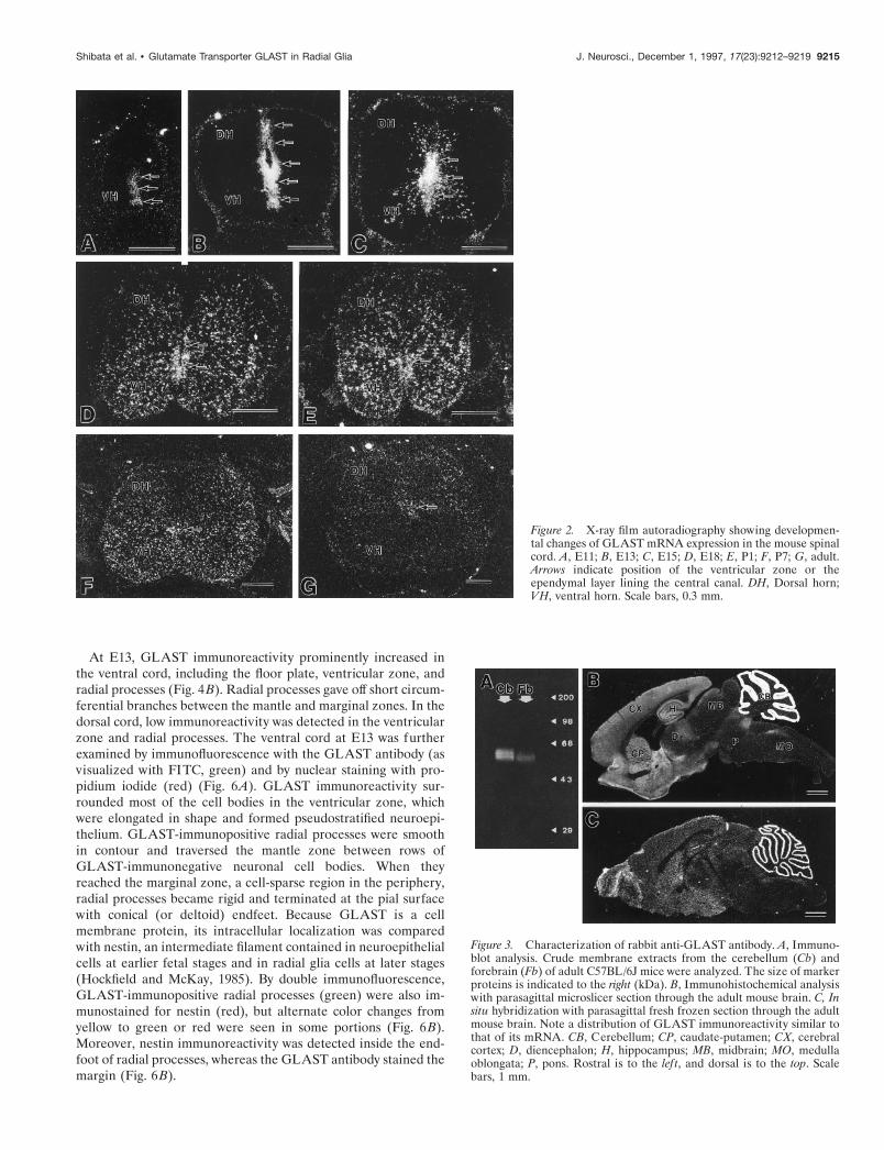
In situ hybridizationTo clarify the cell position of GLAST-expressing cells, in situhybridization was used in developing spinal cords, using 35S-labeled antisense oligonucleotide probe (Fig. 2). At E11, low butsignificant signals were detected in the ventral half of the ventric-ular zone (Fig. 2A). At E13, GLAST mRNA was also confined tothe ventricular zone, in which the ventral half including the floorplate was associated with higher signals than the dorsal half (Fig.2B). At E15, prominent signals were observed in the ventricularzone and also in cells dispersed in the mantle zone (Fig. 2C). TheGLAST mRNA-expressing cells in the mantle zone were distrib-uted in proximity to the ventricular zone. From E18 to P7, signallevels in the ventricular zone were gradually reduced and disap-peared, whereas a number of cells expressing GLAST mRNAspread all over the mantle zone (gray matter) and a few weredistributed in the marginal zone (white matter) (Fig. 2D–F).Thereafter, the transcription levels were gradually decreased invarious regions of the gray and white matters, and at the adultstage very faint signals were detectable only in the dorsal hornand around the central canal (Fig. 2G).
Characterization of anti-GLAST antibodyTo characterize morphological and immunochemical propertiesof GLAST-expressing cells, affinity-purified anti-mouse GLASTantibody was raised in the rabbit and guinea pig. The specificity ofantibodies was checked using the adult mouse brain, where thedistribution of GLAST has been well characterized (Rothstein etal., 1994; Torp et al., 1994; Lehre et al., 1995). An immunoblot
analysis showed that the rabbit antibody recognized heteroge-neous protein bands at 60–65 kDa in membrane extracts from theadult mouse brain, showing a higher concentration in the cere-bellum than in the forebrain (Fig. 3A). Immunohistochemistrywith microslicer sections revealed a predominant distribution ofGLAST in the molecular layer of the cerebellum, with lowerlevels in the telencephalon (Fig. 3B). In the preabsorption test,GLAST immunoreactivity decreased with the increase of antigenpeptides added to the GLAST antibody; the immunoreactivitywas completely abolished at the peptide concentration of 1 mg/mlin the telencephalon and at 10 mg/ml in the cerebellum (data notshown). The general distribution of GLAST immunoreactivitywas consistent with that of GLAST mRNA (Fig. 3C). Similarresults were obtained with the guinea pig antibody (data notshown). On the basis of these findings, these antibodies werejudged to be specific to the glutamate transporter GLAST.
ImmunohistochemistryThe spinal cord was examined by single-labeling (Figs. 4 and 5) anddouble-labeling (Fig. 6) immunohistochemistry. No immunoreac-tivity was detected at E9 (data not shown). At E11, GLASTimmunoreactivity was detected in the ventral cord (Fig. 4A).Within the ventricular zone, higher levels were associated with thefloor plate and ventricular side of the lateral wall. However, anarrow ventrobasal region adjacent to the floor plate lacked theimmunoreactivity (arrowheads in Fig. 4A). Faint immunoreactivitywas found in radially oriented fibrous processes and ran throughthe ventricular zone and ventral horn and reached the pial surface.
Figure 1. Histology of the developing spinal cord at E9 (A,B), E11 (C, D), E13 (E, F ), E15 (G, H ), E18 (I, J ), P7 (K ),and P21 (L). Hematoxylin-stained paraffin sections. Whitecircles in D indicate the border between the ventricular zone(Vn) and ventral horn (VH ). Arrowheads in E indicate atransient cell-dense layer formed between the ventricularzone and dorsal horn (DH ). Arrows in H indicate putativeglia cells with small dark nuclei. F, Floor plate; R, roof plate;*, central canal. Scale bars, 100 mm.
9214 J. Neurosci., December 1, 1997, 17(23):9212–9219 Shibata et al. • Glutamate Transporter GLAST in Radial Glia
At E13, GLAST immunoreactivity prominently increased inthe ventral cord, including the floor plate, ventricular zone, andradial processes (Fig. 4B). Radial processes gave off short circum-ferential branches between the mantle and marginal zones. In thedorsal cord, low immunoreactivity was detected in the ventricularzone and radial processes. The ventral cord at E13 was furtherexamined by immunofluorescence with the GLAST antibody (asvisualized with FITC, green) and by nuclear staining with pro-pidium iodide (red) (Fig. 6A). GLAST immunoreactivity sur-rounded most of the cell bodies in the ventricular zone, whichwere elongated in shape and formed pseudostratified neuroepi-thelium. GLAST-immunopositive radial processes were smoothin contour and traversed the mantle zone between rows ofGLAST-immunonegative neuronal cell bodies. When theyreached the marginal zone, a cell-sparse region in the periphery,radial processes became rigid and terminated at the pial surfacewith conical (or deltoid) endfeet. Because GLAST is a cellmembrane protein, its intracellular localization was comparedwith nestin, an intermediate filament contained in neuroepithelialcells at earlier fetal stages and in radial glia cells at later stages(Hockfield and McKay, 1985). By double immunofluorescence,GLAST-immunopositive radial processes (green) were also im-munostained for nestin (red), but alternate color changes fromyellow to green or red were seen in some portions (Fig. 6B).Moreover, nestin immunoreactivity was detected inside the end-foot of radial processes, whereas the GLAST antibody stained themargin (Fig. 6B).
Figure 2. X-ray film autoradiography showing developmen-tal changes of GLAST mRNA expression in the mouse spinalcord. A, E11; B, E13; C, E15; D, E18; E, P1; F, P7; G, adult.Arrows indicate position of the ventricular zone or theependymal layer lining the central canal. DH, Dorsal horn;VH, ventral horn. Scale bars, 0.3 mm.
Figure 3. Characterization of rabbit anti-GLAST antibody. A, Immuno-blot analysis. Crude membrane extracts from the cerebellum (Cb) andforebrain (Fb) of adult C57BL/6J mice were analyzed. The size of markerproteins is indicated to the right (kDa). B, Immunohistochemical analysiswith parasagittal microslicer section through the adult mouse brain. C, Insitu hybridization with parasagittal fresh frozen section through the adultmouse brain. Note a distribution of GLAST immunoreactivity similar tothat of its mRNA. CB, Cerebellum; CP, caudate-putamen; CX, cerebralcortex; D, diencephalon; H, hippocampus; MB, midbrain; MO, medullaoblongata; P, pons. Rostral is to the lef t, and dorsal is to the top. Scalebars, 1 mm.
Shibata et al. • Glutamate Transporter GLAST in Radial Glia J. Neurosci., December 1, 1997, 17(23):9212–9219 9215

At E15, GLAST immunoreactivity was lowered in the ventric-ular zone, whereas intense immunoreactivity was detected in thefloor plate and in radial processes of both the ventral and dorsalcords (Fig. 4C). Between the radial processes, GLAST-immunopositive punctate or reticular structures were observed inthe mantle and marginal zones. By double immunofluorescence,shafts of radial processes were labeled for both GLAST (green)and nestin (red) (Fig. 6C,D). When compared with E13, GLAST-immunopositive cell membrane at E15 was slightly dissociatedfrom the nestin-immunopositive filaments (Fig. 6, B vs D). Thepunctate/reticular structures between radial processes were im-munopositive for GLAST only and were often continuous withradial processes (arrowheads in Fig. 6C,D).
From E18 to P7, GLAST-immunoreactive punctate/reticularstructures increased in number and predominated throughout thegray matter (mantle zone), whereas GLAST immunoreactivity wasdiminished or had disappeared from the floor plate, ventricularzone (ependymal layer), radial processes, and marginal zone (whitematter) (Figs. 4D, 5A,B). To characterize the GLAST-expressingcells at P7, sections were double-labeled for GLAST mRNA andGFAP, an intermediate filament specific to astrocytes (Fig. 6E). Adigoxigenin-labeled GLAST cRNA probe stained small cell bodiesin the gray matter, but not large neuronal ones. From the labeledcell bodies, GFAP-immunopositive short fibers extended in variousdirections (Fig. 6E). Double immunofluorescence for GLAST andGFAP showed that GLAST-immunopositive punctate/reticularstructures were distributed around GFAP-immunopositive fibers,showing little overlap (Fig. 6F). Using antibody against synapto-
physin, a synaptic vesicle protein, these GLAST-immunopositivepunctate/reticular structures were shown to be apposed closely tonerve terminals (Fig. 6G).
At P14 and P21, the GLAST immunoreactivity displayed aremarkable increase in neuropil regions throughout the graymatter, in which GLAST-immunonegative neuronal cell bodiesand thick dendrites were observed as pale silhouettes (Fig. 5C,D).Thereafter, the immunoreactivity decreased substantially, andonly low to moderate levels were detected in superficial layers ofthe dorsal horn and around the central canal in the adult stage(Fig. 5E), consistent with a previous report (Lehre et al., 1995).
DISCUSSIONGLAST is expressed in radial glia–astrocyte lineageRadial glia cells are a distinct cell class of the neuroglia. At earlystages, the cell bodies are located in the ventricular or subven-tricular zone, and their long radial processes span the neural tube,ending at the pial surface or blood vessels with conical endfeet(Ramon y Cajal, 1911; Rakic, 1971a, 1972; Choi, 1981; Gressenset al., 1992). Radial glia cells exist transiently at neurogenesis andneuronal migration, and they migrate and transform into astro-cytes. Expressions of intermediate filaments also exhibit devel-opmental changes; in various nonprimate mammals, vimentin,nestin (Rat-401 antigen), and RC1 and RC2 antigens constituteintermediate filaments or filament-related antigens in radial gliacells and immature astrocytes, whereas GFAP is predominant inmature astrocytes (Schnitzer et al., 1981; Bignami et al., 1982;Bovolenta et al., 1984; Pixley and de Vellis, 1984; Hockfield and
Figure 4. Immunohistochemical localization of GLAST inthe fetal spinal cord. A, E11; B, E13; C, E15; D, E18. Asterisksand arrows indicate the central canal. Arrowheads in A and Bindicate a ventrobasal region of the ventricular zone, which isassociated with little or low GLAST immunoreactivity. DH,Dorsal horn; F, floor plate; Mg, marginal zone; R, roof plate;VH, ventral horn; Vn, ventricular zone. Scale bars, 100 mm.
9216 J. Neurosci., December 1, 1997, 17(23):9212–9219 Shibata et al. • Glutamate Transporter GLAST in Radial Glia

McKay, 1985; McDermott and Lantos, 1989; Voigt, 1989; Edwardset al., 1990; Ghooray and Martin, 1993). In this study at E11 andE13, cells expressing GLAST were localized in the ventricularzone and displayed such cytological characteristics of the radial gliacells. At P7, GLAST mRNA was expressed in GFAP-immunopositive astrocytes, and GLAST immunoreactivity wasobserved as synapse-associated punctate/reticular structures in thegray matter. Between these two stages, the immunoreactivity wasdetected in both the radial processes and punctate/reticular struc-tures. Therefore, it is reasonable to judge that GLAST is expressedfrom radial glia cells through astrocytes in the mouse spinal cord.The present study has also shown that the localization restricted tothe dorsal horn and around the central canal at the adult stage isestablished through downregulation in mature astrocytes.
GLAST appears on radial glia cells duringneuronal migrationSpinal cord neurons are generated at early stages (Fujita, 1964;Nornes and Das, 1974; Sims and Vaughn, 1979; Altman andBayer, 1984). According to Sims and Vaughn (1979), lateralmotor neurons in the mouse cervical cord are mostly generatedbetween E9 and E10.5, and dorsal horn neurons in the laminaeI–III are born at about E12. Radial glia cells are present fromthe stage of neurogenesis, as shown by Golgi technique and byimmunohistochemistry (Choi, 1981; Hockfield and McKay,1985; Edwards et al., 1990). For example, RC1 antibody stainsradial glia cells in the mouse spinal cord as early as E9, andRC1-immunopositive radial processes span the ventral anddorsal cords at E12 (Edwards et al., 1990). When comparedwith the previous radial glial cell markers, the appearance of
GLAST immunoreactivity is relatively late and shows a dis-tinct ventral-to-dorsal gradient. In the ventral cord, GLASTimmunoreactivity was first detected at E11, when a smallventral horn begins to be formed. It became intense by E13,when the ventral horn had become enlarged and contained anumber of neuronal cell bodies. In the dorsal cord, GLASTimmunoreactivity appeared at E13 and became intense at E15,during which period cells in the dorsal horn increased innumber. Therefore, the GLAST appears on preexisting radialglia cells at stages when spinal cord neurons migrate from theventricular zone to the mantle zone.
Radial glial cells migrate after neuronal migrationBy 3H-thymidine autoradiography and Feulgen cytophotometry,Fujita (1965, 1973) has shown that DNA synthesizing cells firstappear in the mantle and marginal zones at the 8 week stage inthe human spinal cord and at the 8 d stage in the chick spinal cord.In the human spinal cord at the 7.5 week stage, cell bodies ofradial glia cells begin to be displaced from the wall of theventricular zone (Choi, 1981). In the present study, we observedthat cells expressing GLAST mRNA first emerged outside theventricular zone at E15. At this stage, glia-like cells, which con-tain small nuclei stained heavily with hematoxylin, were distin-guished in the mantle and marginal zones. All of these findingssuggest that radial glia cells retaining mitotic activities begin tomigrate at a specific stage of development. In the mouse spinalcord, the migration begins between E13 and E15. On the otherhand, a substantial number of neuroepithelial cells were lost fromthe ventricular zone between E11 and E13, when cells in theventral and dorsal horns had increased, representing a massive
Figure 5. Immunohistochemical localization of GLAST inthe postnatal spinal cord. A, P1; B, P7; C, P14; D, P21; E,adult. Note strong GLAST immunoreactivity throughout thegray matter at P14 and P21, whereas it disappears from mostregions of the gray matter at the adult stage. Arrows indicatethe ependymal layer lining the central canal. DH, Dorsalhorn; F, floor plate; VH, ventral horn. Scale bars, 100 mm.
Shibata et al. • Glutamate Transporter GLAST in Radial Glia J. Neurosci., December 1, 1997, 17(23):9212–9219 9217

neuronal migration. Therefore, the migration of radial glia cellsoccurs after active neuronal migration.
Concomitant transformation and migration of radialglia cellsBy taking advantage of the cell membrane localization, immuno-histochemistry for GLAST enabled visualization of cytologicalchanges of radial glia cells. Radial glial processes were smooth incontour at E13, whereas a number of punctate/reticular protru-sions were formed at E15 along or between radial processes. Thelatter structures gradually increased and predominated in thegray matter at early postnatal stages. Therefore, the cytologicalchange between E13 and E15 represents an initial sign of theradial glial transformation. Thus, it seems likely that the transfor-mation and migration of radial glia cells occur concomitantly.This is consistent with a previous finding from Golgi studies inwhich numerous lamellate expansions appear along radial pro-cesses at the stage when the cell bodies begin to be displaced fromthe ventricular zone (Schmechel and Rakic, 1979; Choi, 1981).
Speculated roles of GLAST on radial glia cellsMature astrocytes surround various structures in the CNS, includ-ing synapses, dendritic shafts, neuronal cell bodies, capillaries, andpia mater. Dense localization on astrocytic membrane apposingsynapses is demonstrated for GLAST and also for GLT1, anotherglutamate transporter expressed in astrocytes (Chaudhry et al.,1995). The extracellular glutamate concentration is elevated in rats
infused intraventricularly with GLAST or GLT1 antisense oligo-nucleotide (Rothstein et al., 1996). Mice defective in GLT1 displayprolonged high glutamate levels in the synaptic cleft, epilepsy, andneuronal cell death (Tanaka K et al., 1997). It is thus clear thatglutamate transporters on mature astrocytes play an essential rolein the rapid removal of extracellular glutamate, to keep the levellow enough to prevent neurons from excitotoxicity. What can bespeculated as a role of GLAST on radial glia cells?
Radial glia cells are thought to serve as a guide for neuronalmigration (Rakic, 1971a, 1972; Gregory et al., 1988). In thedeveloping cerebellum, migrating granule cells are directly ap-posed to Bergmann glial fibers (Rakic, 1971b). Komuro andRakic (1993) have shown using slice preparations that the rate ofgranule cell migration is increased by the activation of NMDAreceptor channels. Because synapses are not yet differentiated inmigrating granule cells, the cell migration is facilitated by activa-tion of nonsynaptic NMDA receptor channels. Interestingly, thecell migration is also accelerated by the administration of theglutamate uptake inhibitor p-chloromercuriphenylsulfonic acid(Komuro and Rakic, 1993). In this regard, the dense localizationof GLAST on radial glial processes seems to be of great interest,in terms of regulation of the glutamate level along the course ofmigrating neurons. If GLAST on radial glial processes lowers theglutamate level, it would slow the neuronal migration and protectmigrating neurons from excessive receptor activation leading toexcitotoxicity. Alternatively, if GLAST supplies glutamate to
Figure 6. Double-labeling showing morphological and immu-nochemical properties of GLAST-expressing cells at E13 (A,B), E15 (C, D), and P7 (E–G). A, GLAST immunoreactivity( green) and nuclear staining with propidium iodide (red). B–D,GLAST immunoreactivity ( green) and nestin immunoreactiv-ity (red ) in the marginal zone (B, D) and mantle zone (C ).Arrowheads indicate GLAST-immunopositive punctate or ir-regular protrusions from radial processes. Arrows indicate con-ical endfeet of radial processes. E, GLAST mRNA ( purple)and GFAP immunoreactivity (brown). Arrows indicate cellbodies labeled for the GLAST mRNA. F, GLAST immuno-reactivity ( green) and GFAP immunoreactivity (red). G,GLAST immunoreactivity ( green) and synaptophysin immu-noreactivity (red). Mg, Marginal zone; Mn, mantle zone; N,neuronal cell body; P, pial surface; Vn, ventricular zone; *,central canal. Scale bars, A–D, F, 10 mm; E, 50 mm; G, 5 mm.
9218 J. Neurosci., December 1, 1997, 17(23):9212–9219 Shibata et al. • Glutamate Transporter GLAST in Radial Glia
migrating neurons, it would facilitate the migration. It has beenpostulated that early in development the reversed transporteroperation from intracellular to extracellular space is the source ofneurotransmitters (Taylor and Gordon-Weeks, 1991). It shouldalso be taken into consideration that GLAST is involved indifferentiation of the radial glia–astrocyte lineage. In the future,developmental analyses of mice lacking GLAST will provide animportant insight into its role in neural development.
REFERENCESAltman J, Bayer SA (1984) The development of the rat spinal cord. Adv
Anat Embryol Cell Biol 85:1–168.Bignami A, Raju T, Dahl D (1982) Localization of vimentin, the non-
specific intermediate filament protein, in embryonal glia and in earlydifferentiating neurons. In vivo and in vitro immunofluorescence studyof the rat embryo with vimentin and neurofilament antisera. Dev Biol91:286–295.
Bovolenta P, Liem RKH, Mason CA (1984) Development of cerebellarastroglia: transitions in form and cytoskeletal content. Dev Biol102:248–259.
Chaudhry FA, Lehre KP, Campagne ML, Ottersen OP, Danbolt NC,Storm-Mathisen J (1995) Glutamate transporters in glial plasma mem-branes: highly differentiated localizations revealed by quantitative ul-trastructural immunocytochemistry. Neuron 15:711–720.
Choi BH (1981) Radial glia of developing human fetal spinal cord:Golgi, immunohistochemical and electron microscopic study. Dev BrainRes 1:249–267.
Choi DW (1992) Excitotoxic cell death. J Neurobiol 23:1261–1276.Edwards MA, Yamamoto M, Caviness Jr VS (1990) Organization of
radial glia and related cells in the developing murine CNS. An analysisbased upon a new monoclonal antibody marker. Neuroscience36:121–144.
Fujita S (1964) Analysis of neuron differentiation in the central nervoussystem by tritiated thymidine autoradiography. J Comp Neurol122:311–328.
Fujita S (1965) An autoradiographic study on the origin and fate of thesub-pial glioblast in the embryonic chick spinal cord. J Comp Neurol124:41–60.
Fujita S (1973) Genesis of glioblasts in the human spinal cord as revealedby Feulgen cytophotometry. J Comp Neurol 151:25–34.
Ghooray GT, Martin G (1993) Development of radial glia and astrocytesin the spinal cord of the North American opossum (Didelphis virg ini-ana): an immunohistochemical study using anti-vimentin and anti-glialfibrillary acidic protein. Glia 9:1–9.
Gregory WA, Edmondson JC, Hatten ME, Mason CA (1988) Cytologyand neuron-glial apposition of migrating cerebellar granule cells invitro. J Neurosci 8:1728–1738.
Gressens P, Richelme C, Kadhim HJ, Gadisseux J-F, Evrard P (1992) Thegerminative zone produces the most cortical astrocytes after neuronalmigration in the developing mammalian brain. Biol Neonate 61:4–24.
Hertz L (1979) Functional interactions between neurons and astrocytes.I. Turnover and metabolism of putative amino acid transmitters. ProgNeurobiol 13:277–323.
Hockfield S, McKay RD (1985) Identification of major cell classes in thedeveloping mammalian nervous system. J Neurosci 5:3310–3328.
Kanai Y, Hediger MA (1992) Primary structure and functional charac-terization of a high-affinity glutamate transporter. Nature 360:467–471.
Kanai Y, Bhide PG, DiFiglia M, Hediger MA (1995) Neuronal high-affinity glutamate transport in the rat central nervous system. Neuro-Report 6:2357–2362.
Kanai Y, Trotti D, Nussberger S, Hediger MA (1997) The high affinityglutamate transporter family: structure, function, and physiologicalrelevance. In: Neurotransmitter transporters. Structure, function, andregulation (Reith MEA, ed), pp 171–213. Totowa, NJ: Humana.
Kiryu S, Yao GL, Morita N, Kato H, Kiyama H (1995) Nerve injuryenhances rat neuronal glutamate transporter expression: identificationby differential display PCR. J Neurosci 15:7872–7878.
Komuro H, Rakic P (1993) Modulation of neuronal migration byNMDA receptors. Science 260:95–98.
Lehre KP, Levy LM, Ottersen OP, Storm-Mathisen J, Danbolt NC(1995) Differential expression of two glial glutamate transporters in therat brain: quantitative and immunocytochemical observations. J Neu-rosci 15:1835–1853.
Mayer BL, Westbrook GL (1987) The physiology of excitatory amino acidsin the vertebrate central nervous system. Prog Neurobiol 28:197–276.
McDermott KWG, Lantos PL (1989) The distribution of glial fibrillaryacidic protein and vimentin in postnatal marmoset (Callithrix jacchus)brain. Dev Brain Res 45:169–177.
Nornes HO, Das GD (1974) Temporal patter of neurogenesis in spinalcord of rat. I. An autoradiographic study-time and sites of origin andmigration and settling patterns of neuroblasts. Brain Res 73:121–138.
Pixley SR, de Vellis J (1984) Transition between immature radial gliaand mature astrocytes studied with a monoclonal antibody to vimentin.Dev Brain Res 15:201–209.
Rakic P (1971a) Guidance of neurons migrating to the fetal monkeyneocortex. Brain Res 33:471–476.
Rakic P (1971b) Neuron-glia relationship during granule cell migrationin developing cerebellar cortex. A Golgi and electronmicroscopic studyin Macacus rhesus. J Comp Neurol 141:293–312.
Rakic P (1972) Mode of cell migration to the superficial layers of fetalmonkey neocortex. J Comp Neurol 145:61–84.
Ramon y Cajal S (1911) Histologie du Systeme Nerveux de l’Homme etdes Vertebres. Tome 2. Paris Maloine. Reprinted by Consejo Superiorde Investigaciones Cientificas: Madrid, 1955.
Rothstein JD, Martin L, Levey AI, Dykes-Hoberg M, Jin L, Wu D, NashN, Kuncl RW (1994) Localization of neuronal and glial glutamatetransporters. Neuron 13:713–725.
Rothstein JD, Dykes-Hoberg M, Pardo CA, Bristol LA, Jin L, Kuncl RW,Kanai Y, Hediger MA, Wang Y, Schielke JP, Welty DF (1996) Knock-out of glutamate transporters reveals a major role for astroglial transportin excitotoxicity and clearance of glutamate. Neuron 16:675–686.
Schmechel DE, Rakic P (1979) A Golgi study of radial glial cells indeveloping monkey telencephalon: morphogenesis and transformationinto astrocytes. Anat Embryol 156:115–152.
Schnitzer J, Franke WW, Schachner M (1981) Immunocytochemicaldemonstration of vimentin in astrocytes and ependymal cells of devel-oping and adult mouse nervous system. J Cell Biol 90:435–447.
Shibata T, Watanabe M, Tanaka K, Wada K, Inoue Y (1996) Dynamicchanges in expression of glutamate transporter mRNAs in developingbrain. NeuroReport 7:705–709.
Sims TJ, Vaughn JE (1979) The generation of neurons involved in anearly reflex pathway of embryonic mouse spinal cord. J Comp Neurol183:707–720.
Storck T, Schulte S, Hofmann K, Stoffel W (1992) Structure, expression,and functional analysis of a Na 1-dependent glutamate/aspartate trans-porter from rat brain. Proc Natl Acad Sci USA 89:10955–10959.
Sutherland ML, Delaney TA, Noebels JL (1996) Glutamate transportermRNA expression in proliferative zones of the developing and adultmurine CNS. J Neurosci 16:2191–2207.
Tanaka K (1993) Cloning and expression of a glutamate transporterfrom mouse brain. Neurosci Lett 159:183–186.
Tanaka J, Ichikawa R, Watanabe M, Tanaka K, Inoue Y (1997) Extra-junctional localization of glutamate transporter EAAT4 at excitatoryPurkinje cell synapses. NeuroReport 8:2461–2464.
Tanaka K, Watase K, Manabe T, Yamada K, Watanabe M, Takahasi K,Iwama H, Nishikawa T, Ichihara N, Kikuchi T, Okuyama S, KawashimaN, Hori S, Takimoto M, Wada K (1997) Epilepsy and exacerbation ofbrain injury in mice lacking the glutamate transporter GLT-1. Science276:1699–1702.
Taylor J, Gordon-Weeks P (1991) Calcium-independent gamma-aminobutyric acid release from growth cones: role of gamma-aminobutyric acid transporter. J Neurochem 56:273–280.
Tomooka Y, Kitani H, Jing N, Matsubara M, Sakakura T (1993) Recon-struction of neural tube-like structures in vitro from primary neuralprecursor cells. Proc Natl Acad Sci USA 90:9683–9687.
Torp R, Danbolt NC, Babaie E, Bjørås M, Seeberg, E, Storm-Mathisen J,Ottersen OP (1994) Differential expression of two glial glutamatetransporters in the rat brain: an in situ hybridization study. Eur J Neu-rosci 6:936–942.
Voigt T (1989) Development of glial cells in the cerebral wall of ferrets:direct tracing of their transformation from radial glia into astrocytes.J Comp Neurol 289:74–88.
Yamada K, Watanabe M, Shibata T, Tanaka K, Wada K, Inoue Y (1996)EAAT4 is a post-synaptic glutamate transporter at Purkinje cell syn-apses. NeuroReport 7:2013–2017.
Yamada K, Wada S, Watanabe M, Tanaka K, Wada K, Inoue Y (1997)Changes in expression and distribution of the glutamate transporterEAAT4 in developing mouse Purkinje cells. Neurosci Res 27:191–198.
Shibata et al. • Glutamate Transporter GLAST in Radial Glia J. Neurosci., December 1, 1997, 17(23):9212–9219 9219


![Exploratory Clinical Trial of (4S F]fl C Transporter Using Positron … · Imaging, Diagnosis, Prognosis Exploratory Clinical Trial of (4S)-4-(3-[18F]fluoropropyl)-L-glutamate for](https://static.fdocuments.net/doc/165x107/60f90642e71e4a0af4493803/exploratory-clinical-trial-of-4s-fi-c-transporter-using-positron-imaging-diagnosis.jpg)