MUSCLE GROUP ISOLATION AND NERVE ANASTOMOSIS IN THE ...
31
The PDF of the article you requested follows this cover page. This is an enhanced PDF from The Journal of Bone and Joint Surgery 1910;s2-8:95-124. J Bone Joint Surg Am. NATHANIEL ALLISON and SIDNEY I. SCHWAB THE TREATMENT OF THE PARALYSES OF THE EXTREMITIES MUSCLE GROUP ISOLATION AND NERVE ANASTOMOSIS IN This information is current as of January 24, 2010 Reprints and Permissions Permissions] link. and click on the [Reprints and jbjs.org article, or locate the article citation on to use material from this order reprints or request permission Click here to Publisher Information www.jbjs.org 20 Pickering Street, Needham, MA 02492-3157 The Journal of Bone and Joint Surgery
Transcript of MUSCLE GROUP ISOLATION AND NERVE ANASTOMOSIS IN THE ...

The PDF of the article you requested follows this cover page.
This is an enhanced PDF from The Journal of Bone and Joint Surgery
1910;s2-8:95-124. J Bone Joint Surg Am.NATHANIEL ALLISON and SIDNEY I. SCHWAB
THE TREATMENT OF THE PARALYSES OF THE EXTREMITIESMUSCLE GROUP ISOLATION AND NERVE ANASTOMOSIS IN
This information is current as of January 24, 2010
Reprints and Permissions
Permissions] link. and click on the [Reprints andjbjs.orgarticle, or locate the article citation on
to use material from thisorder reprints or request permissionClick here to
Publisher Information
www.jbjs.org20 Pickering Street, Needham, MA 02492-3157The Journal of Bone and Joint Surgery

V.
MUSCLE GROUP ISOLATION AND NERVE ANASTO-
MOSIS IN THE TREATMENT OF
THE PARALYSES OF THE
EXTREMITIES.*
BY NATHANIEL ALLISON, M.D., AND SIDNEY I. ScHWAB,
M. D., ST. LOUIS.
In a paper read before the American Neurological Association
in 1909, with the title “The Surgical Treatment of Athetosis and
Spasticities by Muscle Group Isolation”, we made a preliminary
report of our experience with what we conceived to be a novel
method of approaching the problems presented by cases of spastic
palsies and athetoses in children. The material upon which
this paper was based consisted of four cases, one was a cerebral
hemiplegia with athetoid movements in the forearm and fingers,
the other three were cerebral diplegias, usually called Little’s
disease. This method was designated “Muscle Group Isola-
tion,” a term descriptive of a procedure which had for its aim an
isolation of a single group, or several groups of spastic muscles,
by a surgical attack upon the nerve or nerves directly involved.
As our material has grown and our experience increased, the
utility of this procedure has extended so that it has included in its
application the flaccid paralyses.
At this point it seems of advantage for the purpose of making
what is to be said clearer, to set down briefly the fundamental
principles upon which muscle group isolation is based. Atheto-
ses and spastic conditions of organic origin are similar processes.
Athetoid movements may be regarded as variants of permanent
tonic spasticities. They may be said to be due to several factors,
the most salient of which is a constantly active set of highly irrita-
* Read before the American Orthopedic Association at its twenty-fourth annual
meeting, held at Washington, May 3-5, 1910.
7

96 PARALYSES OF THE EXTREMITIES.
tive impulses arising from the cortical motor cells. These im-
pinge upon the muscles which are usually in a condition of slight
hypertonus. The overaction of the congenitally stronger mus-
cle group as compared to the weaker antagonistic group causes this
temporary state to become permanent, and there results then a
characteristic attitude and deformity. The essential mechanism
must he thought of as central in origin, meaning that the nerve or
nerves supplying the muscle group or groups, which is the con-
ducting mechanism and by which this state is produced, are the
anatomical structures which demand primary attention as the
focus of operative measures.
As we have advanced in the study of these and allied problems,
we have come to believe that definite advance follows a mutual
appreciation of surgical and neurological points of view. The
neurologist is apt to be concerned with matters pertaining to dis-
turbance of function; the surgeon, on the other hand, with postures
typifying end-results and expressed in terms of deformity. There
is needed, therefore, a clinical nomenclature which not only covers
the origin and end-results, but also the intermediate ground. This
includes the more fertile field for therapeutic effort. To accom-
plish this it is only necessary to express in terms easily understood
the nervous and muscular mechanism through which the clinical
picture is produced. For example, in describing the adduction
of the thighs seen in cerebral diplegias, we have made use of the
muscle group involved as well as the nervous agency, thinking of
it simply in terms of adductor overaction through the obturator
nerve. In like manner in paralytic flexion of the knee, we have
called it hamstring overaction through the branches of the sciatic.
We regard the various paralytic attitudes of the foot in terms of
overacting muscles through their nerve supplies.
We have thought it best to divide our clinical experience tenta-
tively into three groups; each of the groups presents a different
phase of the therapeutic problem and has for its solution a different
application of the method.
Group I. Athetoses and spasticities.
Treated by muscle group isolation
only.

NATHANIEL ALLISON. 97
Group II. Flaccid paralyses.
Treated by muscle group isolation
and nerve anastomosis.
Group III. Complicated tic movements.
Treated by induced paralysis.
In the first group we have a total of nine cases operated upon.
i. Cerebral hemiplegia with athetosis.
2. Cerebral diplegia. Little’s disease.
3. Cerebral diplegia. Little’s disease.
4. Cerebral quadriplegia.
5. Cerebral diplegia. Little’s disease.
6. Cerebral quadriplegia.
7. Spastic hemiplegia, with slight con-
genital hydrocephalus.
8. Cerebral diplegia. Little’s disease.
9. Cerebral diplegia. Little’s disease.
GROUP I.
In our preliminary report a description of the methods that
have been used in treating this class of cases was given, together
with a summary of the recent literature. From these sources and
our own experience we gave the causes for the lack of permanent
results following tenotomy, myotomy, tendon and muscle trans-
plantation as follows:
These methods, besides being merely an attack upon the end-
result, have the additional disadvantage of being but transitory in
their benefits, the condition being all too frequently reestablished.
This is due to the fact that the nerve supply, which is the con-
ducting structure by which the abnormal impulses are brought
into action, remains untouched. Furthermore, by the necessary
supplementary treatment, i.e., plaster-of-Paris bandages or
fixation apparatus, not only are the local antagonists made much
weaker, but also the whole muscular mechanism of the extremity
is seriously impaired by the confinement in bed and the tight
bandaging necessary. This criticism holds true for muscle trans-
plantation in spastic cases; in addition, the scope of this method

98 PARALYSES OF THE EXTREMITIES.
is necessarily limited to a narrowly selected group of cases in
which only a single muscle may be utilized. Since working on
these problems, two other methods designed for the surgical
relief of these cases have been reported: i. Intraperineural
neurotomy, by Dr. John Joseph Nutt.* The principles upon
which this operation is based appear to us to be unconvincing
from the point of view of the physiology of the nervous mechanism
which is involved. From a practical point of view there is an
evident objection: First, in the actual carrying out of the opera-
tion, and second, in the effect of the operation. It would be im-
possible to perform this operation upon the smaller peripheral
nerves; and, furthermore, the operation ought to be more directly
applied to the mechanics of muscle groups which are directly con-
cerned in the production of the paralyses. 2. The other procedure,
based upon an entirely different conception of the whole problem,
has been reported by Foerster and Tietze.t This method con-
sists of resection of the posterior spinal nerve roots. Our objec-
tion to this method is based upon the following facts: The
magnitude and seriousness of the operation, especially in cases of
spastic children, is a positive objection to it. Furthermore, the
operation is based on incorrect physiologic principles for these
reasons: Motor nerves contain sensory fibres which have noth-
ing particularly to do with the posterior roots, so that cortical
rest, whatever that might be, could not possibly result from mere
posterior root sections. The overlaping is so considerable that in
order to affect a whole spastic extremity, a large number of roots
would necessarily have to be cut. This would make the operation
one of inadvisable magnitude in the majority of cases. Further-
more, no matter how many roots were cut, no complete sensory
paralysis could possibly result. Admitting even that complete
sensory paralysis could result, then the very object of an operation
of this kind would be done away with, namely, the possibilities
of coordinated voluntary movement. It is a well-known fact
that in absolutely anesthetic areas no properly coordinated move-
ment can ever be possible.
* The AMERICAN JOURNAL OF ORTHOPEDIC SURGERY, VII, 2, Nov., 1909.
t Zeit. /. Or/ho. Chir., October, 22, 1908.

NATHANIEL ALLISON. 99
and retracted. The obturator nerve is discovered issuing from
The chief object of treatment in all cases of spasticity is to
enable the patient to make properly coordinated movements,
with the mechanical disability caused by spasticity rendered as
ineffective as possible. The spasticity as such is scarcely the
main object of treatment, for the reason that its origin, nature,
and localization are still matters of great obscurity. To assign
it to the cortex of the brain is neither justified by facts nor upheld
by any well-known theories.
OPERATIVE PROCEDURES.
Four operations have been performed on spasticities.
The first of these consists of isolating and injecting with alcohol
the obturator nerve at its exit from the pelvis. This nerve supplies
the gracilis, the adductor brevis, the adductor longus and some-
times also (partly) the pectineus, the hip-joint capsule, the
adductor minimus and the main portion of the adductor magnus.
It arises from the second, third, and fourth lumbar nerves, and is
the only nerve of the ph!xus that emerges at the medial border of
the psoas, behind the common iliac vessels. It runs forward and
downward to the obturator canal. It traverses this canal,
giving off muscular branches to the obturator muscles, and im-
mediately after making its exit it divides into two branches: an
anterior, or stronger, which supplies the adductor longus and
brevis, and a posterior, or weaker, which supplies the hip-joint
capsule and adductor magnus. The nerve is principally a motor
nerve though it contains some sensory fibers.
OPERATIVE TECHNIC.
An incision two inches long on anterior aspect of thigh, running
vertically, beginning at Poupart’s ligament,, one-half inch inside
its middle point, carried downward through subcutaneous fat to
fascia covering the adductor group of muscles. Fascia divided by
dissection and the internal border of the adductor brevis and
longus made out. Opening carried down to pectineus fascia at
horizontal ramus of the pubis; this is divided in line of incision

100 PARALYSES OF THE EXTREMITIES.
below the horizontal ramus of the pubis at the upper extremity
of the wound; it divides at this point into two terminal branches.
Above this division the trunk of the nerve is seized with blunt
dissecting forceps and drawn upward into the wound. At this
point it is injected with an alcoholic solution.
The second operation is planned for the relief of overaction and
spasticity in the hamstring groups. Here, the nerves to be
attacked are the branches of the great sciatic which supply the
hamstring muscles; i.e., the biceps, semimembranosus and the
semitendinosus. These branches are given off of the trunk of the
great sciatic in the upper half of the thigh.
OPERATIVE TECHNIC.
�Tertical incision four inches long over the course of the great
sciatic nerve; nerve exposed and branches isolated, their identity
being determined by the use of a sterilizable electrode and faradic
current. This feature is of especial importance and requires
considerable care and observation. When the branches are thus
isolated they are easily injected with alcohol with a fine needle
from the ordinary hypodermic syringe.
The third operation is planned for the relief of overaction of
the gastrocnemius group. The muscles here involved are mainly
the gastrocnemius and soleus which are supplied by branches
from the internal popliteal nerve.
OPERATIVE TECHNIC.
Incision two inches long, made longitudinally over the center of
the popliteal space, and carried downward through the fascia
between the two heads of the gastrocneminus to the internal pop-
liteal nerve. The branches which supply the soleus and gas-
trocnemius are isolated by careful electrical stimulation and
injected with an alcoholic solution.
The fourth operation is designed for the relief of spasticity
affecting the anterior tibial group through its nerve supply from
the anterior tibia! nerve.

H
:�
�-
NATHANIEL ALLISON. I0I
OPERATIVE TEchNIc.
Through a longitudinal incision one and one-half in�ches long
over the head of the fibula, the external popliteal nerve is easily
exposed at its bifurcation into the peroneal and anterior tihial
CASE I\, (.ROUP 1--Cerebral quaclriplegia showing marked adductur and ham-string spasticity.
nerves; it is best to identify the nerves by a careful electrical stimu-
lation. The anterior tibia! is then injected with an alcoholic
solution.
CASE IV.-C. D., twelve years of age, December 19, 1908,
cerebral quadriplegia. General condition of paralysis noticed

102 PARALYSES OF THE EXTREMITIES.
shortly after birth. Has made little progress mentally, thoughhe understands simple questions and is able to form single words
intelligently. Has never walked. General condition has been
good. Family history good, except that mother had a form offacial paralysis. Examination, December 19, 1908, showed awell developed and fairly well nourished boy. Intelligence that ofa child of three years of age No deformities of skeleton. Nothingabnormal about abdomen or thorax. Because of automaticmovements of lower muscles of face he has peculiar grimaces and
foolish expressions. Spine normal. Upper extremities spastic
from tips of fingers to shoulder-joints; lower extremities held inmarked adduction, knees crossed, legs flexed on thighs 45 degrees,
foot in position of slight dorsal flexion, and toes plantar flexed.
Spasticity of adductor groups is very marked, resisting attemptsat passive correction. The knees cannot be extended beyond20 degrees flexion. Hamstring groups are particularly spastic
and contracted. Attempts at passive movements at ankle-jointsbring on spasticity, especially in extensor groups, and the foot is
moved into slight dorsal flexion with toes plantar flexed, the greattoe overlapping the two next it. In the feet and hands areautomatic movements similar to those described in the face.Standing position: Can almost maintain balance, weight is borneon internal borders of feet, not including heel, with legs crossed,the left knee anterior to the right knee, the right knee resting on
the calf of the left leg, flexed 45 degrees. Thighs flexed on trunk35 degrees. Trunk fairly erect, but the arms are engaged in manyexcited movements in the attempt to maintain balance. Inattempting to walk both legs are moved rapidly by automaticirregular movements of two sorts, described as movements of
attempted progression which are cork-screw in character and areproduced voluntarily. During this attempt the boy’s face is con-torted and his arms are very spastic, the whole being accompanied
by foolish laughter, grimaces, and inarticulate sounds.December 21, 1908.-Operation under ether; through the in-
cisions described above, the obturator nerves were exposed on
both sides without difficulty, and injected with an 8o per cent.solution of alcohol. It was found immediately after this operationthat both thighs fell into a position of abduction. Recovery good.
December 26, 1908.-Boy had no temperature. Thighs are
abducted i� degrees and there is no spasm in the adductor groups.General condition good.
December 29, 1908.-Operation under ether; wounds of first op-eration found to be healed, except for a small space at the lowerend of the incision on the right side which has granulated. Stitches
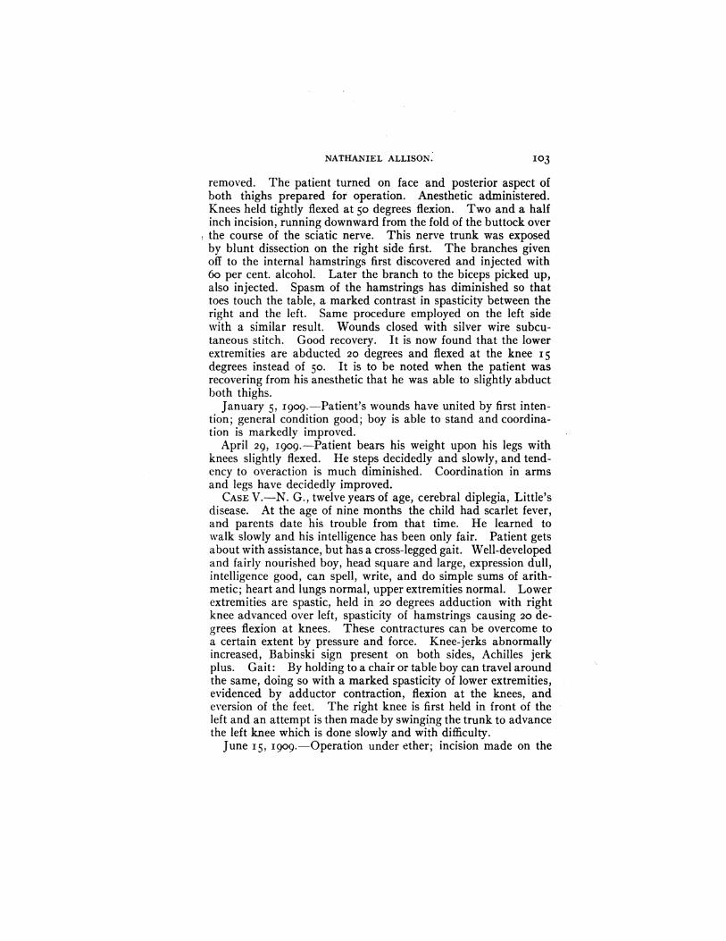
NATHANIEL ALLISON. 103
removed. The patient turned on face and posterior aspect ofboth thighs prepared for operation. Anesthetic administered.
Knees held tightly flexed at �o degrees flexion. Two and a half
inch incision, running downward from the fold of the buttock overthe course of the sciatic nerve. This nerve trunk was exposed
by blunt dissection on the right side first. The branches given
off to the internal hamstrings first discovered and injected with
60 per cent. alcohol. Later the branch to the biceps picked up,also injected. Spasm of the hamstrings has diminished so thattoes touch the table, a marked contrast in spasticity between the
right and the left. Same procedure employed on the left side
with a similar result. Wounds closed with silver wire subcu-
taneous stitch. Good recovery. It is now found that the lowerextremities are abducted 20 degrees and flexed at the knee i�
degrees instead of 50. It is to be noted when the patient wasrecovering from his anesthetic that he was able to slightly abduct
both thighs.January 5, 1909.-Patient’s wounds have united by first inten-
tion; general condition good; boy is able to stand and coordina-tion is markedly improved.
April 29, 1909.-Patient bears his weight upon his legs withknees slightly flexed. He steps decidedly and slowly, and tend-ency to overaction is much diminished. Coordination in armsand legs have decidedly improved.
CASE V.-N. G., twelve years of age, cerebral diplegia, Little’sdisease. At the age of nine months the child had scarlet fever,and parents date his trouble from that time. He learned to
walk slowly and his intelligence has been only fair. Patient gets
about with assistance, but has a cross-legged gait. Well-developed
and fairly nourished boy, head square and large, expression dull,
intelligence good, can spell, write, and do simple sums of arith-metic; heart and lungs normal, upper extremities normal. Lower
extremities are spastic, held in 20 degrees adduction with rightknee advanced over left, spasticity of hamstrings causing 20 de-
grees flexion at knees. These contractures can be overcome toa certain extent by pressure and force. Knee-jerks abnormallyincreased, Babinski sign present on both sides, Achilles jerk
plus. Gait: By holding to a chair or table boy can travel around
the same, doing so with a marked spasticity of lower extremities,
evidenced by adductor contraction, flexion at the knees, andeversion of the feet. The right knee is first held in front of the
left and an attempt is then made by swinging the trunk to advancethe left knee which is done slowly and with difficulty.
June 15, 1909.-Operation under ether; incision made on the

104 PARALYSES OF THE EXTREMITIES.
right side over the anterior aspect of the thigh at the internal
borders of the adductor muscles, and carried down through the su-
perficial fascia; muscles made out, and an attempt made to discoverthe obturator nerve and its exit into the ramus of the pubis.After one and one-half hours’ time, this search was unsuccessful
and the wound closed. The nerve was considered to have eitheran abnormal course or to be undiscoverable by the operator. At
C.�sr: V, (,Ruue 1-Cerebral (liplegia (Little’s) showing adductor and ham-string spasticity.
all events it must have received some surgical injury, for the thighat the end of the operation was abducted 30 degrees and therewas very little spasticity in the adductor group. Recovery good.
June 19, 1909.-Operation on left side, ether; incision wasmade in the usual manner and the obturator nerve was exposed
in less than 10 minutes’ time, having a normal course. Wasinjected with 8o per cent. alcohol and electrical stimulation was
employed to make out its conductivity. After the injection

NATHANIEL ALLISON. 105
this was found to be lost, a strong faradic current being usedthrew the other muscles of the thigh into a state of tetany whichlasted twenty minutes. Wounds closed with silkworm gut. Re-covery good. Operative wound at first operation healed by first
intention. After recovering from the anesthetic it was found
that both thighs were in a position of 30 degrees of abduction, nospasm of the adductor muscles on either side.
July 15, 19o9.-Operation, ether; secondary operation done on
the right side. The obturator nerve discovered after some diffi-
culty. Injection after the usual manner and the usual result.
Wound closed with silkworm suture. Good recovery.
July 22, 1909.-Operation, ether; patient placed prone uponthe table, back of thighs prepared for operation. Longitudinalincision made over each thigh, four inches long. Sciatic and itsbranches readily exposed. Each branch was picked up, stimu-
lated with mild faradism to find out what muscle or muscles itsupplied. Injected with alcohol and conductivity retested.This procedure was done on the branches to the biceps, thesemimembranosus and semitendinosus, and a branch to the
abductor magnus. Wounds closed with silkworm gut. Good
recovery. Two hours after the operation no spasms of ham-
strings. Complete extension of the leg is voluntarily possible.
Wound of second operation on right side shows primary healing.
August i, 1909.-Leaves hospital, gait improving daily.
September 29, 1909.-Boy lS going to school; legs still weak but
uses them well; feet placed squarely on floor, with no tend-
ency to adduction or flexion deformity; is having daily training.
Walks alone without support and gait is improving.
CASE VI.-L. F., thirteen years of age, cerebral quadriplegia,
October i, i#{231}o�. Mother states that child walked normally until
the fifth year, when she had a fright and a fall and was injured by
a horse. Following this there was progressive loss of power inher legs, so that when she was seven years of age she was no longer
able to stand or walk. Her hands then became involved so that
she could not use them. Her mental condition has remainedabout the same. She is able to understand her mother and tomake attempts at speech. When asked to sit up she endeavors
to do so. During the last year she has had several epileptiformattacks with loss of consciousness. She has grown normally and
her general health has been good. She has control of bladderand rectum, and has good appetite, sleeps well, and is of a pleasantdisposition. Well developed and fairly nourished girl, imbecilic,drolling expression, with intelligence that of a child five or six years.
Is unable to stand, to walk, or to use her arms on account of

106 PARALYSES OF THE EXTREMITIES.
spastic contractures. As the patient lies in bed the legs assume
position of flexion at the knees, flexion at the hip, with the spas-ticity more marked in the left than in the right. The adductor
spasm can be seen best when the legs are forcibly abducted.
Owing to the contracture at both knees the overadduction is notso clearly in evidence. The right foot is in extreme pronation
with extension in the three outer toes; athetoid movements pres-
ent. The left foot is in marked supination. The calf muscles
feel soft and flabby in spite of their evident spastic contractures.
This is due probably to the fact that the muscles have never
been used for walking. There is a shortening of the Achillestendon in both legs, likewise of the flexors at the knees. Owingto the contracture and the spastic condition surrounding thejoints the knee-jerks could be obtained only with difficulty andwere pathologically plus. On account of the mental conditionof the child no attempt at sensory examination was made.
October 2, 1909.-Operation with ether; excision made over the
external popliteal nerve on left leg. Nerve discovered withoutdifficulty and its bifurcation into the anterior tibia! and musculo-cutaneous made out. By an electrode these nerves were posi-tively determined. The anterior tibial on this side was injectedwith 60 per cent. alcohol and its conductivity tested; found to belost. On the right side the same procedure was put into effect,
with the difference that the musculo-cutaneous was injected in-stead of the anterior tibia!. Through two long vertical incisions
on the back of the thighs the sciatic nerves were laid bare and thebranches to the hamstrings were injected. Wounds were closed;good recovery. After the operation it was found that the right
foot had no longer peroneal spasticity. The left foot is held inclubbed position but not due to muscular overaction. Thespasm of the hamstrings has completely disappeared.
October 5, i9o9.-At 2.00 A. M. patient died of double pneu-monia, with a temperature of io#{231}�#{176}.
CASE VII.-Spastic hemiplegia, with slight congenital hydro-
cephalus. A. A., three and one-half years of age, April 9, 1908.
Child is one of five children. Was born when mother was forty-one years of age. Instrumental delivery was necessary which
left a scar upon the forehead. Child was nursed at breast, buthas always been weak. Dentition normal. Walked at the ageof one and one-half years. It was then noticed that child limpedwith right foot and held right arm in a peculiar way. This con-
dition has increased since. Mentality is above the average andher general health has always been good. First consultation
showed a large flabby child with good mentality. On right side

NATHANIEL ALLISON. 107
of forehead is a scar from instrumental delivery. Heart andlungs are normal. Expression slightly stupid. Head large and
of hydrocephalic appearance. The right foot is held in supi-nation and plantar flexion, the tendo Achillis contracted. Thereis a slight contraction at the knee. The gait is that of spastic
club-foot. +
Trealmenl.-April 20, 1908. The tendo Achillis was divided
and the foot was placed in a right angle position in plaster-of-Paris. Child made a good recovery and after three weekswalked with very much improved gait. Observation has ex-
tended over the period of time between this operation and the
present.
October 4, 1909.-The spasticity in the whole right extremityhas returned. There is overaction of anterior tibial and gastroc-
nemius groups in the right foot and 20 degrees flexion at theknee, due to spastic hamstrings. There is a slight tendency to
spasticity in the left lower extremity. Child’s general condition
has improved, health is good, and she walks about in a lively
manner but with this disability. Reflexes increased, more on
the right. The contractures can be overcome by steady pressure;
that is, the knee can be straightened and the foot can be broughtto a right angle, straight position with the legs, but no further.
October II, 1909.-Operation, chloroform, with patient on
face. A longitudinal incision was made on the posterior aspectof the thigh, laying bare the upper half of the great sciatic nerve.
The branches to the semimembranosus, semitendinosus, and
abductor magnus were injected with 6o per cent, alcohol, alsothe branch to the biceps. Incision closed with subcutaneoussilver wire stitch. Over the popliteal space a longitudinal inci-sion was made which laid bare the branches of the internal poplit-
eal nerve which supply the gastrocnemius and soleus; three innumber. Electrical stimulation was employed to identify these
nerves. Sixty per cent. alcohol was injected into their sheaths.Incision closed with subcutaneous silver wire stitch. Through
a longitudinal incision over the head of the fibula the externalpopliteal nerve was exposed and its bifurcation into the muscu-
locutaneous and anterior tibia! nerves made out. The anteriortibia! was injected with alcohol and the wound closed with sub-cutaneous silver wire stitch. Time of anesthetic one hour. Re-covery good.
October i�, 1909.-Discharged from hospital, all woundshealed by first intention, no temperature.
March 14, I9I0.-Spasticity of hamstrings has entirely dis-
appeared. There is still overaction of the anterior tibial group,

a
io8 PARALYSES OF THE EXTREMITIES.
also of the gastrocnemius. This is attributed to faulty technic.A secondary operation is planned.
CASE VIII.-Cerehral diplegia, Little’s disease, D. L., six
years, eleven months, October 20, 1909. Seen first on October13, 1905. Family history is negative. Child was born pre-
maturely; weight 4 pounds. At the time of first observation childwas twenty-seven months old. Has been under the care of a
doctor for malnutrition and has gained weight and strength.Child is able to speak a few words. Spasticity was noticed inthe right arm and leg. The mental development has been below
normal. Says “mamma,” “sister,” and “papa.” Weight
23 pounds. Examination showed spasticity in legs, more ma�rkedin the right, with plus knee-jerks, no clonus, no Babinski. Thelegs are freely movable as are likewise the arms in passive motion,
but a spastic condition develops and is easily observed. Thechild is only able to hold its head up for a moment or so; cannot
stand unless supported. When held up the legs become ad-
ducted in a cross-legged position.Seen again at three years of age, November 9, 1906. Patient
is not able to stand, attempts to rise up in a sitting position with
more vigor than at the last examination.November 9, 1906.-Well developed and well nourished child.
Back weak, both legs spastic, and arms spastic. Overaction of
both tendo Achilles and hamstring groups.
November io, 19o6.-Operation under ether; both tendo
Achilles divided subcutaneously, feet confined at right-angledposition with legs in plaster-of-Paris bandages. Recovery good.To be supplied with ankle braces and jacket.
November 25, 1906.-Sent home. Mother instructed to
encourage child to take the initiative in walking and using hismuscles.
May 19, 1908.-Seen in consultation with Dr. E. H. Bradford
of Boston. Child walks with feet on floor, slight pronation, slight
flexion of toes, stands with support requiring aid in balancing.Back is still weak, sits erect for a few seconds with head correctly
posed, but relaxes and faIls forward. There is marked spasm ofthe adductor groups of muscles and slight spasm of the hamstrings.
Advised to discontinue braces and have child associate withplaymates and be educated in attempting to walk.
September 27, i#{231}o#{231}.-When placed on his back child makes agood attempt to rise by flexing the whole body at an angle of 20
degrees. The spasticity in the lower limbs is very evident. There
is marked adduction spasm, producing cross-legged position.
Child is able to stand with support. The feet are flat on the floor

NATHANIEL ALLISON. 109
and there is no spasticity in the gastrocnemius group. He cansit alone with spine flexed in a long posterior curve, the head held
fairly erect. Mental condition markedly improved, generalnutrition good. Parents advised to bring child to the city for a
period of six months.
October 20, 1909.-Operation under ether; through two incisionsover the anterior aspect of both thighs at the usual point; theobturator nerves were easily found and isolated. Injected with
8o per cent. alcohol. Time of operation, forty minutes. Child’s
condition after the operation, good. Legs abducted to 50 degrees,spasticity of adductors has entirely disappeared.
October 29, 1909.-Patient discharged from hospital. Woundshealed.
January i, i9io.-Child has been doing systematic dailyphysiologically planned exercises, with massage. There has beenmarked improvement in initiative. He now attempts to makesteps. Supports himself in standing and all tendency to adductor
deformity has disappeared.April I, 1910.-Educational exercises have been continued
daily. Child is now able to make certain voluntary movementsand to hold himself upright in sitting. As yet he is unable tobalance or to stand alone.
CASE IX.-Cerebral diplegia, Little’s disease. M. M., two andone-half years of age, March 17, 1910. Birth was normal and
child presented a normal appearance. As it became older mothernoticed that lower limbs were held stiff. Mentality has been slowin its development. Can say only a few words and expression is
vacant. Has never made any attempt to walk but can standwith slight support. Well developed and well nourished child,marked spasticity of both lower extremities, legs held adducted.
When child is placed upon its feet the spasm of the adductormuscles and of the gastrocnemius groups is very marked, pro-
ducing cross-legged flexed knee position.March 17, 1910.-Operation under ether; through an incision
over the anterior aspect of thigh, just below Poupart’s ligament, the
obturator nerve was easily discovered and isolated on both sides.Into them was injected 6o per cent. alcohol. This was followedimmediately by loss of conductivity demonstrated with the
electrode, and by the thighs assuming an abducted position.Wounds closed with silver wire subcutaneous sutures. Recoverygood.
March 30, 1910.-Wounds healed by first intention, childleaves hospital in good condition. All tendency to adductor
deformity has disappeared. There is no spasm of adductor

110 PARALYSES OF THE EXTREMITIES.
groups; gastrocnemius group slightly overactive, also hamstring.
Mother instructed in physiological exercises.
In summarizing the results obtained in this group of cases it is
necessary to point to the fact that we are dealing with a class of
congenitally defective individuals, so far as the nervous system
is concerned, and that the results of operative procedures must
be given in terms of improvement only. Furthermore, the mor-
tality following operation is due to the lessened resistance charac-
teristic of the congenitally defective; for example, the two cases
that resulted fatally (Cases II and VI) died of double lobar pneu-
monia, though the operation was in neither case prolonged nor
productive of shock. With this explanation in view we believe
the following results are a just estimation of the effectiveness of
muscle group isolation in the treatment of spasticities:
One case of cerebral hemiplegia with athetosis: athetois
stopped, paralysis unaffected.
Four cases of cerebral diplegia (Little’s disease) markedly
improved: one of these to the extent that walking and coordi-
nation are practically normal; two cases relieved of cross-legged
progression and put into a position where the proper educational
exercises will develop a coordinate gait; the fourth relieved of
adductor overaction and still under treatment.
One case of quadriplegia, very extensive in character and with
extreme overaction of stronger muscle groups; standing impos-
sible. This case after operation was able to stand and has since
made considerable improvement under properly designed exer-
cises. \Valking has not been accomplished without support.
One case of spastic hemiplegia with slight congenital hydro-
cephalus in which there was overaction of the hamstring group, the
gastrocnemius group, and the anterior tibia! group. Here, the
operation was effective in overcoming the overaction of the ham-
strings. The overaction of the gastrocnemius group, due to
faulty technic, has not been checked, but the gait is much
improved.
Two cases died after operation, from pneumonia.
Cases I, II, and III in the above list were reported in detail in
our preliminary paper (The Surgical Treatment of Athetosis

NATHANIEL ALLISON. III
and Spasticities by Muscle Group Isolation, Schwab and Allison.
Journal of Nervous and Menial Diseases, Vol. XXXVI, No. 8,
August, 1909).
GROUP II.
In the second group we have a total of four cases operated
upon.
i. Traumatic paralysis of musculospiral.
2. Poliomyelitis anterior. Paralysis of quadriceps extensor.
3. Poliomyelitis anterior. Peroneal paralysis with overacting
anterior tibial group.
4. Poliomyelitis anterior. Peroneal paralysis with overacting
anterior tibia! group.
The first two cases in this group are reported to show the effi-
ciency of nerve anastomosis properly performed, and have
their bearing upon the problem under consideration in that nerve
anastomosis serves an important purpose in carrying out an
essential step in an operation of which muscle-group isolation is
the complement. This operation was used in Cases III and IV
of this group. In them the antagonist group overacting at the
expense of the flaccidly paralyzed group produced deformity.
Nerve anastomosis alone would have been insufficient unless the
unequal pull had been rendered powerless by temporarily throwing
the overacting muscle out of activity. This was accomplished
by alcoholic injection of the nerve supplying this muscle group.
CASE 1.-Traumatic paralysis of musculospiral. T. T.,twenty-one years of age, June i6, 1909. On February 17, he wasriding in the doorway of a caboose attached to a lumber train.The door was open and of a sliding character; leaning out of the
doorway, his right arm and right leg over the sill, the trainstruck a boulder and came to a sudden stop, throwing thesliding door against his right arm and right thigh, fracturing thefemur and injuring his arm. Well developed and well nourishedyoung man, heart and lungs normal, general physique good.Right femur: Small discharging sinus on inside of thigh, union
good with deformity. Right arm and forearm: There is a
characteristic wrist-drop, lack of power to supinate, lack of powerto extend the wrist, typical musculospiral palsy. The bicepsmuscle apparently crushed in two, one-third of it forming a promi-
8

CASE I, GROUP Il-Diagram of operation showing anastomosis between themusculospiral and median nerves.
112 PARALYSES OF THE EXTREMITIES.
nence just above the elbow-joint, the upper two-thirds retracted to
one inch above the middle of the arm Triceps power and brachi-alis anticus power good. The divided ends of the biceps arealso enervated. The humerus in uninjured. There is consider-
able wasting of the muscles of the forearm and shoulder.June 17, 1909.-Operation under ether; a longitudinal incision
was made over the posterior external aspect of the arm, carrieddown between muscular septa to musculospiral nerve. Just belowthe groove there is a thickening in the nerve. Electrical stimula-tion applied along the course of the nerve causes contraction of
the brachialis and triceps. No response in muscles of the forearmor wrist. Sheath of the nerve freed and the wound closed withinterrupted silkworm-gut sutures. Ruptured biceps suturedwith animal tendon.
July 3, 19o9.-All the wounds have healed by first intention.Electrical stimulation (faradism) produces no reaction either innerve or muscles on the extensor side of the arm.
July 7, 1909.-Patient is up and about and examined daily withelectricity to discover any tendency toward reestablishment ofnerve function. Up to the present time there has been no im-provement. Biceps united.

NATHANIEL ALLISON. 113
July 13, 1909.-Operation under ether, incision was made overthe course of the musculospiral nerve, following line of former in-cision, carried anteriorly over the elbow-joint down to the
muscle. The musculospiral nerve exposed does not respond to
direct electrical stimulation. It was divided in its middle fora distance of 3 inches longitudinally and the inner half cut free
at the upper extremity of the longitudinal cut. This segmentcarried under the biceps tendon and inserted into the mediannerve and sutured in place with 00 catgut. Wounds closed.Through a longitudinal incision over the dorsum of the forearmthe extensor tendons of the wrist were shortened, placing thewrist in extreme extension. Both wounds closed with silver wiresubcutaneous stitches. Plaster-of-Paris applied, holding forearmflexed, hand extended. Good recovery.
July 23, 19o9.-Paster removed. Wounds have healed by
first intention. There is absolutely no power of extension of the
wrist or fingers. Patient supplied with a tin split which can beapplied to the arm, holding the forearm flexed and the wrist andfingers extended. There is no reaction to faradism in nerve or
muscle.November ii, 1909.-Patient can hold wrist extended and use
his crutch on that side with very little difficulty. Examination of
the muscles supplied by the musculospiral nerve: There isonly a minimum amount of paresis at the wrist. The hand is
held almost horizontal with a slight indication of wrist-drop, pro-
duced largely by fatigue. There is a definite extension movementat the wrist and fingers. This movement can be observed with
the arm flexed at the elbow. The extension at the elbow is as good
as it has always been. Stimulation of the musculospiral at theusual place over the triceps muscle gives a slight reaction of thetriceps. Below the injury, however, galvanism of this nerve pro-
duces no response. Galvanic stimulation of the extensor group of
muscles gives a very definite response in the middle finger and toa lesser extent in the other fingers. This extensor response canbe shown by the movement of fingers and by the actual movementsof the tendon and by palpation as well. It seems from thisexamination that the extensor response at the wrist and at thefingers has returned and the conclusion is justified that the anas-
tomosis of the median and the distal part of the musculospiralhas been to this measure successful.
April 27, �9�o.-The forearm is held pronated and there issome disability in supination. There is considerable power inextending the wrist and extending the fingers. Along the ana-tomical course of the musculospiral nerve strong faradism gives

CASE II, GROUP 11.-Diagram showing nerve anastomosis between obturatornerve and quadriceps extensor branch of anterior crural nerve.
114 PARALYSES OF THE EXTREMITIES.
only transmitted effect on the flexor side of the hand, forearm, andwrist. There is no extensor response whatever. Over the siteof the anastomosis, at the median surface of the arm, along the
inner border of the biceps, stimulation with a light faradic currentgives prompt extension of the fingers, thumb, and wrist. Stronger
current here produces in addition normal flexion. This demon-strates that impulses along the extensor tract in the arm are trans-mitted through the median nerve and that the remnant of the
musculospiral react no longer to electrical stimulation. Thereis normal ability to use the hand and forearm.
CASE II.-Poliomyelitis anterior. Paralysis of quadriceps ex-tensor. W. W. K., six years of age, November 13, 1909. Eighteenmonths ago boy had a typical attack of poliomyelitis from whichhe recovered so that he could walk in about a week. Generalphysical condition good, mentality is above the average. Heart
and lungs are normal, skeleton normal except for 1/2-inch shorten-ing of right lower extremity. Musculature is normal except
right lower extremity. Here there is absolute loss of power in
the quadriceps extensor. The sartorius has power, the adductorsare normally strong, and there is some power in the abductors.

NATHANIEL ALLISON. 115
Hamstring group weak but active. All muscles in right leg arefunctioning but weak. There is power to extend and flex the foot
on the ankle, also to supinate and pronate the foot. He walks byswinging the right leg on the right thigh with a slight limp, charac-teristic of quadriceps extensor paralysis.
November �6, 1909.-Operation under ether; longitudinalincision made over the anterior aspect of thigh in line of femoralvessels, 4 inches long, anterior crural nerve exposed. The branch
running to quadriceps group picked up by electrical stimulation.This branch was cut off from the main trunk high up in thewound, passed under the nerve, artery, and the vein. The
obturator nerve was then exposed and the branch of the anteriorcrural was introduced into a longitudinal cut in the trunk of theobturator nerve and there sutured with two strands of oo catgut.There was no tension upon the anastomosis. The wound was
closed with a deep catgut suture and a silver wire subcutaneousstitch. Good recovery.
April 24, 1910.-The quadriceps extensor is faradically excit-
able, both at the motor, point and in the course of the muscleitself. There is a visible and palpable contraction of the patella.Power is not yet sufficient in the quadriceps to hold the leg ex-
tended on the thigh against the force of gravity, but in walkingthere is marked improvement in gait, the swing of the leg on
the thigh being very much lessened.
CASE III.-Poliomye!itis anterior, peroneal paralysis withoveracting anterior tibia! group. E. S., nine years of age. Hasalways been a healthy child, except for trouble of her left lowerextremity. Two years ago without apparent illness, so far asher parents’ observation extended, she lost the use of her entire
left extremity. It was slightly painful and tender. After a
time muscular power began to reappear.April 30, 1909.-Muscles are all good except in left lower leg,
where the peronei, with the exception of the peroneus tertius, arepowerless. In walking the foot is held in marked supination.The plantar fascia is contracted as is the gastrocnemius. Theanterior muscles of the leg; i.e., tibialis anticus, extensor com-munis digitorum and the peroneus tertius have fair power.The peroneus longus and brevis do not act. All the other muscles
of the leg are normal. There is 1/2 inch atrophy of calf.May 3, 1909.-Operation under ether; incision made 3 inches
in length, vertical in direction, over the outer aspect of the leg, hav-ing its middle point opposite the head and neck of the fibula.This was carried to the muscular layers and the external poplitealnerve was exposed in the wound. Faradic stimulation of this�

Musculo -
C.�sEs III and IV, GROUP 11.-Diagram of operation on musculocutaneous andanterior til)ial nerves. Nerve anastomosis plus muscle group isolation.
116 PARALYSES OF THE EXTREMITIES.
trunk caused a quick contraction of the extensor communis digi-
torum, tibialis anticus, and the peroneus tertius. The nerve wasfurther exposed and its bifurcation into the anterior tibia! andmusculocutaneous made out. Faradic stimulation of the mus-
culocutaneous, 1/2 inch below the bifurcation, produced noresponse whatever in the peroneus longus and peroneus brevis.
Stimulation of the anterior tibia! nerve, 1/2 inch below the bifur-cation, produced a similar strong contraction to that describedabove. The musculocutaneous nerve was divided 1/4 inchbelow the bifurcation in a longitudinal direction. This cut end
of the nerve was then inserted in a vertical slit made in the anteriortibia! nerve, 1/4 inch below the bifurcation, and was suturedwith two strands of oo catgut. One-half inch below this nerveanastomosis in the course of the anterior tibial nerve, 10 minims of
8o per. cent alcohol were injected into the sheath of the nerve.Faradic stimulation of the anterior tibia! nerve gave no response in
the group of muscles which it supplies. The wound was closedwith silver wire subcutaneous stitch. Recovery good.
May �o, 1909.-Wound healed by first intention.
September 17, �9o9.-Child has had daily electrical stimula-tion since last note. Foot examined shows power to abduct,
using the peronei to a noticeable extent.

NATHANIEL ALLISON. 117
November 13, �9o9.-The foot is held in a much improvedposition with the outer edge fairly well raised. Voluntary motionis restricted to slight movement and extension of the toes. Thereis slight movement of the outer edge of the foot Tibia! group isactive. Stimulation of the musculocutaneous nerve by galvanism
gives a response which can be seen and felt by the movement ofthe peroneal tendons.
CASE IV.-Po!iomyelitis anterior, peronea! paralysis with over-
acting anterior group, B. McL., eight years of age, May 20, 1909.
At two years of age had indefinite history of an attack of paralysis.
Since then both legs have been involved. Has had various forms
of treatment, but not of an operative character. Well developedand well nourished girl. General health good, trunk and upper
extremities normal. Right thigh abducted io degrees, flexed �odegrees, � degrees of flexion of leg on thigh, foot held in markedplantar flexion and supination. Left thigh flexed on trunk 40
degrees, abduction 30 degrees, externally rotated �o degrees,marked shortening of psoas and iliac-tibial band, leg flexed onthigh 25 degrees, foot in marked plantar flexion and slight inver-sion. Muscular power: Hamstrings on right side strong; onleft side weak, but have power. Quadriceps extensor on right
side weak; on left absent. Gastrocnemius group, right sidestrong; left side power absent. On the right side there is some
power in tibialis anticus and extensor longus hallucis. The pero-nei on both sides show no muscular power. There is a slight
power of dorsal flexion and of extending the great toe. The leftfoot shows no power in the anterior group of muscles, in the pero-nei or gastrocnemius group.
May 21, 1909.-Operation. Considering the completely para-lyzed condition of the left lower extremity, it was decided to place
this limb in a corrected position by division of tendons, delayingoperative interference until deformity is corrected. Upon theright side, considering the pull of the anterior group of muscles
and the lack of power in the opposing group, it was decided toattempt a nerve anastomosis and a blocking-off of the relativelystronger group by an alcoholic injection similar to that which wasdone in Case III with variation. An incision was made similarto that in Case III, carried down to the muscles and the external
popliteal with its two divisions (anterior tibial and the musculocu-taneous) were exposed. Stimulation of the musculocutaneouscaused no peroneal contraction; stimulation of the anterior tibia!caused contraction in the anterior group of muscles. Peronealgroup of muscles were pale and atrophied, but did not show anyevidence of degeneration. The musculocutaneous nerve was

118 PARALYSES OF THE EXTREMITIES.
split for a distance of 1/2 inch longitudinally in its course and 1/2
inch below the bifurcation of the external popliteal. A cut wasmade horizontally at the upper end of this split, and half of thenerve fibers were �turned to the inside. A slit was made in the
anterior tibia! nerve opposite this nerve division and severed endinserted and sutured with a fine catgut suture and the wound
closed over the anastomosis with a second fine catgut suture.Below this anastomosis, on the anterior tibial nerve, an alcoholic
injection was made into the nerve sheath. This immediatelyproduced a relaxation of muscles supplied by the anterior tibia!
nerve, allowing correction of the deformity. Wound was closed
with silver wire suture. Tendo Achillis was divided subcuta-neously. The limb incased in plaster-of-Paris, with the foot in a
corrected position. Good recovery.November 13, 1909.-Strong faradism over the site of the
external popliteal nerve produced in the peroneal tendons a weakcontraction. Also there is some ability to evert the foot. This,however, is very weak and is over-pulled by the anterior tibia!muscles as well as by the tibialis posticus.
In summarizing the methods used and the results obtained in
the second group of cases, attention is called to the absolutely
favorable results which followed simple nerve anastomosis, illus-
trated in Cases I and II. These results reenforce the idea upon
which the combination of operation is based; that is, in order to
have an effective transference of nerve impulses from a sound
nerve to an injured nerve the opposing factor of antagonist muscle
pull must be eliminated (this has long been recognized). Further-
more it is essential to point out emphatically that the good results
reported in the literature obtained through the agency of tendon
transference and elongations serve to strengthen the plan of oper-
ation which has been done in Cases III and IV. Given the
amount of power which would insure a good result by any mechan-
ical method whatever, a much more perfect result may be obtained
by a proper conservation of the muscles and tendons intact and
an utilization of nerve anastomosis plus properly applied muscle
group isolation. Cases III andG IV illustrate this point.
GROUP III.
In the third group we have one case to report. It is illustrative
of the wide extent to which the method of induced paralyses can

NATHANIEL ALLISON. 119
be applied. It further illustrates the fact that the total nerve
supply of a segment of the body, including both sensation and
motion, can be cut off by means of nerve alcoholization without
subsequent trophic disturbance.
CASE I.-J. H., thirty-two years of age, family history negative
in respect to his present trouble. About 8 years before first
observation the patient, working in a brush factory, noticed
jerking movements in his left arm and hand. These jerkingmovements at first were comparatively infrequent in the courseof the day, .but later increased in extent and in variety untilwork was impossible for him. For the past three years he has
been unable to do any work.
CASE I, GROUP 111.-Diagram show- CAsE I GROUP 111.-Diagram show’ing area of anesthesia immediately after ing area of anesthesia, April 7, 1910.
operation, October 30, 1909.
November 2, 19o9.-There is a peculiar and complicated group
of movements, limited largely to his left side; that is, to the wholeof the left arm, to the shoulder, and to the neck muscles on the leftside. The origin of this movement is vague, and perhaps hashad something to do with his former trade-that of a brush-maker. The movements so duplicated the physiological arrange-
ment of cortical muscles in the arm that a temporary diagnosis of
Jacksonian epilepsy was made, the diagnosis being based largelyon the successive groupings of the movements of the arm. This
movement, which we have come now to believe is a tic, is some-what as follows:

120 PARALYSES OF THE EXTREMITIES.
In the first place the movement, to a certain extent, is under the
control of the will; that is, it can be started apparently by thepatient himself, but once begun he seems unable to control it.The movement begins by an extension of the fingers and thumb,
and is followed by flexion of the wrist, by a contraction of the ex-
tensors of the forearm, by which the hand and arm are thrownclear of the body. At this point the flexed hand is seized by theright hand and forcibly extended. With this forcible extension
of the wrist the whole arm becomes a bit stiffened from the elbowdown, and is thrown by the right arm away from the body. Themuscles of the upper arm and shoulder now engage in the move-ment and the arm is finally brought at rest, adducted, and flexed
at the elbow and at the wrist. The movement is performedquickly, sharply, and accurately, never varying in the slightest,occupying thirty seconds to a minute or a minute and a half.There is never loss of consciousness, there is no pain or discom-fort in the movement. When the movement was first observed,
the muscles of the neck, the deep muscles, and the sternocleido-mastoid were not concerned. These have lately been involved
and there has been quite marked rotation of the neck with the chinto the left and intense muscular spasm. When the patient wasfirst seen the diagnosis lay between Jacksonian epilepsy and a
complicated tic. An effort was made at training the patient.Then an operation was done over the right cortical area on the
chance that there might be some cortical source of irritation. At
this operation through a large flap the middle of the Rolandic areawas laid bare. With a mild faradic current the area of the fingers
and thumb and the forearm were picked out to determine accu-rately the location of the motor center. After the dura was opened,nothing abnormal was discovered, except a group of markedly
tortuous veins, practically varicosities, lying at the upper limit ofthe Rolandic area. These were ligated. The patient’s arm was
enclosed in a plaster-of-Paris bandage after the operation, and
as long as the plaster bandage was intact the tic movements werenot observed. Shortly after the plaster dressing was removed,however, the movement returned with much of its former severity.The diagnosis now seems undoubtedly that of tic. As an addi-tional argument in favor of this diagnosis, can be cited the factthat the patient seemed to be more comfortable after the move-ment takes place and seems rather expectantly awaiting the im-
pulse, whatever it may be, for the movement to take place.October 21, 1909.-Operation, ether. Through a median
incision over the belly of the biceps muscle, above the flexure of
the elbow, the median nerve and the musculospiral nerve were

NATHANIEL ALLISON. 121
exposed and injected with 8o per cent. alcohol. Their lack ofconductivity was demonstrated with faradism. Through a longi-tudinal incision over the internal condyle the ulna nerve wasdrawn up to the wound, also injected with 8o per cent. alcohol.
Wounds closed with subcutaneous catgut suture. Good recovery.October 29, 1909.-Patient discharged from hospital. Wounds
healed.
No definite conclusion can be given concerning this case, as it
is at the present time under observation. It may be said, how-
ever, that the tic movements have totally disappeared as would be
naturally expected, on account of paralysis of the distally involved
muscles. The other muscles which took part in the tic move-
ment and which were unaffected by the operation do not show any
evidence of abnormal or uncontrolled movements. The case is
further of interest in regard to the whole question of paralyses
produced by alcoholic injections of the nerve from the point of
view of trophic disturbance and the length of time of regeneration.
Concerning the trophic functions of the nerves involved there has
been an interesting and unexpected feature in this case, although,
as can be seen from diagrams, there is a total lack of sensation in
the region supplied by the three nerves injected. The patient
showed no positive signs of trophic disturbance. About three
months after the operation the patient received a severe burn on
the back of his fingers. This healed as quickly as would a similar
burn on a normal hand. The return of sensation is extremely
slow in this case. It is now six months after the operation and
the demarcation between normal sensory areas of the skin and
those affected by the operation show only slight variation from the
first examination. Although this might seem slow, yet its con-
stant though small increase shows that there is a marked and
positive tendency toward sensory regeneration. Up to the pres-
ent time there is no evidence of motor regeneration whatever.
On the other hand, there is very little tendency toward atrophy
of the muscles supplied by the injected nerves.
DISCUSSION.
DR. SCHWAB, opening the discussion, expressed his appreciation of having
been asked to attend the meeting and, in a brief way, commented on the

122 PARALYSES OF THE EXTREMITIES.
work that he and Dr. Allison had been doing for the last three years. Hewished simply to emphasize two or three points that were probably not very
clearly understood.
The alcohol injections in spastic cases, he said, are not curative. All thatthe authors had attempted was temporarily to throw out of action the antag-onistic muscle. Meanwhile, the opposing group is exercised, so that by
the time the original nerve regenerates the muscular force has been some-what transferred to the opponent group. The obturator muscles can be
so strengthened that when regeneration takes place the leg will be heldin better position and walking be facilitated. He wished to be consideredas speaking from the neurological point of view, rather than the orthopedic.
Even when the regeneration is not complete, he said, the advantage is that
in these operations the patient can be gotten out of bed within a few days.There is no necessity for the use of a plaster cast or for putting the patient on
his back for a long time. He and Dr. Allison had had their patients walk-
ing about the ward a few days after the operation. Considering the classof patients on whom they had operated, he thought this a great ad-vantage. Their assumption was that athetosis, spasticities, and wereof the same nature. When they injected the nerves with alcohol the
athetosis completely disappeared and did not return. Whether athetosisis of so much importance from the orthopedic point of view, however, asfrom that of the neurological he did not know. He stated that two or three
operations had been done in Chicago, with the same results.
In the second group of cases, the operation of throwing out of action theantagonistic pull was very effective; by the time the nerve regenerated
the antagonizing group had not regained its original strength, so that thecondition was more hopeful of future cure than by any other method.
While the third class of cases, tics, might not have the same amount of
orthopedic interest, many cases can be attacked in a much better way than
ever before by surgical treatment. They paralyzed the whole distal group ofaffected muscles in one case of the most remarkable tic movements, and
the tic disappeared. It was assumed that there had been produced a sortof motor amnesia of this movement.
Of greater interest, perhaps, from the standpoint of the alcoholization ofnerves was the fact that while three nerves were alcoholized and there was
complete anesthesia to touch and pain, no trophic disturbance appeared inany part of the arm. The man accidentally burned himself but this burnhealed just as quickly, without suppuration, as it would have done in an
ordinary person.DR. B. E. MCKENZIE, of Toi�onto, asked Dr. Allison to tell the Society some
particulars regarding the technic of the operation and the strength of
alcohol used in the injections. He thought that the authors had omittedto state in their paper just what strength of alcohol they used.
DR. G. G. DAvIs, of Philadelphia, said that it would be interesting to find

NATHANIEL ALLISON. 123
something curative in the treatment of these cases. Treatment by tendondivision had been criticized as being devoted to the tendons, and not to the
nervous system; but Dr. Davis thought that the treatment to the nervous
system was the same, practically, as that to the tendons. Neither methodhad been curative, so far as he knew; and no curative means had been pro-posed. To his mind, the alcohol injection of nerves was similar to the divi-sion of nerves: after the nerves unite, they resume their functions; and after
the effects of the alcohol disappear, the nerves also resume their functions.Dr. Schwab had said that the nerves do not resume their functions abso�
lutely. To that extent, Dr. Davis believed the alcohol injections to be anadvance. Even in division of the tendons, however, some permanent goodis done. He thought it a question whether the new method constituted
any real improvement. He did not think that a sufficient number of goodresults had yet been shown to demonstrate that the regeneration of the nerve
does not take place entirely, or to what extent it fails to return. Dr. Nutt’s
case last year was one in which he divided the sciatic nerve. This had alocal effect, and also to a limited extent a secondary effect on the general
condition. Therefore, it might be considered superior to operation on thetendon.
Coming to other means of treatment, Dr. Davis said that in treatment bynerve anastomosis a longer time must also elapse before its value can bedetermined. Spiller and Frazier, three or more years ago, anastomosed the
brachial plexus with that object in view. As soon as the spasmodic groupis thrown out of action, improvement takes place. At first there is absolute
paralysis of this group, then an interval of more or less paralysis; after this
the muscle begins to resume its ascendency. This is what he understood hadoccurred in Frazier and Spiller’s case;’in other words, there is a resumptionof the original condition. He trusted that this resumption of the original
condition was not complete, but said that he would like to have furtherevidence. He thought that more than two years should elapse before onecould judge of the value of these procedures.
DR. VIRGIL P. GIBNEY, of New York City, thought that the Associationought to feel under lasting obligation to Drs. Allison and Schwab for bring-ing forward anything helpful in the treatment of cases of spastic type. Amonth before Dr. Gibney had seen Dr. Murphy, of Chicago, inject osmic
acid into the facial nerve for the cure of tic. Dr. Murphy had remarked thatthis procedure would give relief for twelve months and that the tic would
then return, and said that alcohol would not serve so good a purpose in thefacial nerve; in nerves farther removed from the center of circulation, how-
ever, he thought that alcohol would serve the purpose better, putting thesenerves out of commission for a long time. Dr. Gibney thought that anymember of the Association who has had these cases to treat would welcomenine or ten months’ surcease from spasticity. At the end of this time, theinjection could be again made, and the patient given a rest for nine or ten

124 PARALYSES OF THE EXTREMITIES.
months more. He did not see any objection to using the alcohol everynine or ten months. He thought it better than plaster, no matter howmuch one loves the latter. Putting a large child in plaster in spread-eagle
style so that it requires two beds is very troublesome, even if there is a hope of
putting the muscles out of commission by this method. If these gentlemen,
by attacking the nerves, could destroy them for any number of months, as
they had claimed, Dr. Gibney thought that the opposing muscles could bedeveloped in the interval, which could not be done when a plaster cast is
employed. If the children can go around, he said, their education can becarried on; so he considered it a wonderful advance in the therapeutics of
spastic paralysis.DR. WALTER G. STERN, of Cleveland, referring to the cases of athetosis
and especially to the case of Drs. Frazier and Spiller, mentioned by Dr.Davis, said that Whatever operation may be done on such a case, the athetosiswill return after a while; even if one should amputate the arm below the
shoulder, the shoulder would move. When Dr. Stern spoke to Dr. Frazier
after the reading of that paper, he mentioned that he had himself done suchan operation as theirs, but the nerve had regenerated; nerve to nerve anasto-mosis was successful, and after a month or two sensation returned; a year
or so passed, and athetoid motion again returned. He was not so blessedwith his operation as Drs. Frazier and Spiller were with theirs: their patient
is worse, but his own is a great deal worse.DR. ALLISON, closing the discussion, said, in reply to Dr. McKenzie’s
question regarding the strength of the acohol used, that they varied the
strength to a certain extent. He did not know just what per cent. shouldbe used, but stated that Dr. Schwab and himself were then engaged in someexperiments for the purpose of determining this point. They had foundthat some cases have a more lasting paralysis than have others. He thought
that the percentage of alcohol might have something to do with this. Itmust be at least 50 per cent., in order to have any effect at all; and he
had used straight alcohol in some cases