Mouse IgE ELISA Set — 555248 - BD Biosciences · Aspirate wells and wash 3 times with ≥ 300...
2
Calculation of Results Calculate the mean absorbance for each set of duplicate standards, controls and samples. Subtract the mean zero standard absorbance from each. Plot the standard curve on log-log graph paper, with IgE concentration on the x-axis and absorbance on the y-axis. Draw the best fit curve through the standard points. To determine the IgE concentration of the unknowns, find the unknown’s mean absorbance value on the y-axis and draw a horizontal line to the standard curve. At the point of intersection, draw a vertical line to the x-axis and read the IgE concentration. If samples were diluted, multiply the IgE concentration by the dilution factor. Computer data reduction may also be employed, utilizing log-log regression analysis. Typical Standard Curve This standard curve is for demonstration only. A standard curve must be run with each assay. Specificity Cross Reactivity: The following isotypes were tested in the BD OptEIA™ assay at 2 mg/mL and no cross-reactivity (value ≥ 2 ng/mL) was identified. Mouse: IgG 1 , k; IgG 1 , l; IgG 2a , k; IgG 2b , k; IgG 2b , l; IgG 3 , k; IgG 3 , l; IgM, k; IgM, l; IgA, k BD OptEIA ™ Technical Data Sheet Mouse IgE ELISA Set Cat. No. 555248 Materials Provided The BD OptEIA™ Set for mouse immunoglobulin E (IgE) contains the components necessary to develop enzyme-linked immunosorbent assays (ELISA) for mouse IgE in serum, plasma, and cell culture supernatants. Sufficient materials are provided to yield approximately 20 plates of 96-wells if the recommened storage, materials, buffer preparation, and assay procedure are followed as specified in this package. Capture Antibody Anti-Mouse IgE monoclonal antibody Detection Antibody Biotinylated Anti-Mouse IgE monoclonal antibody Enzyme Reagent Streptavidin-horseradish peroxidase conjugate (SAv-HRP) Standards Purified mouse IgE, lyophilized Instruction/ Analysis Certificate (lot-specific) 2655KI_555248 Rev.10 BD Biosciences Pharmingen 10975 Torreyana Road San Diego, CA 92121 Customer/Technical Service Tel 877.232.8995 (US) Fax 858.812.8888 www.bdbiosciences.com United States 877.232.8995 Canada 866.979.9408 Europe 32.2.400.98.95 Japan 0120.8555.90 Asia/Pacific 65.6861.0633 Latin America/Caribbean 55.11.5185.9995 Conditions: The information disclosed herein is not to be construed as a recommendation to use the above product in violation of any patents. BD Biosciences will not be held responsible for patent infringement or other violations that may occur with the use of our products. Purchase does not include or carry any right to resell or transfer this product either as a stand-alone product or as a component of another product. Any use of this product other than the permitted use without the express written authorization of Becton Dickinson and Company is strictly prohibited. Unless otherwise noted, BD, BD Logo and all other trademarks are the property of Becton, Dickinson and Company. ©2015 BD Standardization This immunoassay is calibrated against purified mouse IgE, k Assay Optimization 1. BD OptEIA™ Sets allow flexible assay design to fit individual laboratory needs. To design an immunoassay with different sensitivity and dynamic range,the following parameters can be varied: Capture, Detection Antibody titers, Incubation time, Incubation temperature, Assay Diluent formulation, Buffer pH, ionic strength, protein concentration, Type of substrate, Washing technique (i.e., number of wash repetitions and soak times) 2. “Typical Standard Curve” and 20-plate yield were obtained in the BD Biosciences Pharmingen laboratory, using the recommended procedure and manual plate-washing. Troubleshooting Poor Precision Possible Source Corrective Action • Inadequate washing/ aspiration of wells • Check function of washing system • Inadequate mixing of reagents • Ensure adequate mixing • Imprecise/ inaccurate pipetting • Check/ calibrate pipettes • Incomplete sealing of plate • Ensure complete seal on plate Poor Standard Curve Possible Source Corrective Action • Improper standard handling/ dilution • Ensure correct preparation, storage of standards • Incomplete washing/ aspiration of wells • Check function of washing system • Imprecise/ inaccurate pipetting • Check/ calibrate pipettes • Improper buffer/ diluent used • Check buffer/ diluent preparation, pH Low Absorbances Possible Source Corrective Action • Inadequate reagent volumes added to wells • Check/ calibrate pipettes • Incorrect incubation times/ temperature • Ensure sufficient incubation times/ reagents warmed to RT • Incorrect antibody titration • Check Capture Ab and Working Detector preparation • Improper buffer/diluent used • Check buffer/ diluent preparation, pH • Overly high wash/aspiration pressure from • Utilize manual washing automated plate-washer Limitations of the Procedure • Samples that generate absorbance values higher than the standard curve should be diluted with Standard Diluent and re-assayed. • Interference by drug metabolites, soluble receptors, or other binding proteins in specimens has not been thoroughly investigated. The possibility of interference cannot be excluded. • BD OptEIA™ Sets are intended for use as an integral unit. Do not mix reagents from different Set batches. Reagents from other manufacturers are not recommended for use in this Set. References 1. Olszewski M.A., et al. The Role of Macrophage Inflammatory Protein- 1a/CCL3 in Regulation of T Cell-Mediated Immunity to Cryptococcus neoformans Infection. J Immunol. 2000; 165:6429-6436.
Transcript of Mouse IgE ELISA Set — 555248 - BD Biosciences · Aspirate wells and wash 3 times with ≥ 300...
Calculation of ResultsCalculate the mean absorbance for each set of duplicate standards,controls and samples. Subtract the mean zero standard absorbancefrom each.Plot the standard curve on log-log graph paper, with IgEconcentration on the x-axis and absorbance on the y-axis. Draw thebest fit curve through the standard points.To determine the IgE concentration of the unknowns, find theunknown’s mean absorbance value on the y-axis and draw ahorizontal line to the standard curve. At the point of intersection,draw a vertical line to the x-axis and read the IgE concentration. Ifsamples were diluted, multiply the IgE concentration by the dilutionfactor.Computer data reduction may also be employed, utilizing log-logregression analysis.
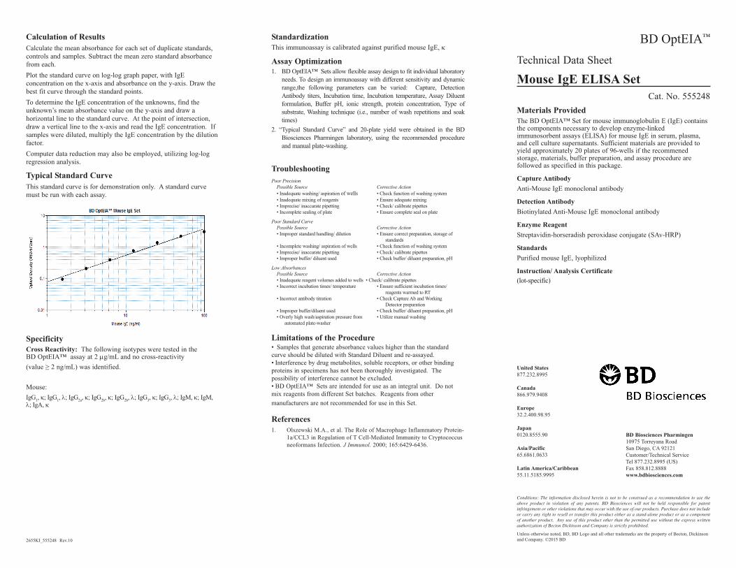
Typical Standard CurveThis standard curve is for demonstration only. A standard curvemust be run with each assay.
SpecificityCross Reactivity: The following isotypes were tested in the BD OptEIA™ assay at 2 mg/mL and no cross-reactivity (value ≥ 2 ng/mL) was identified.
Mouse:IgG1, k; IgG1, l; IgG2a, k; IgG2b, k; IgG2b, l; IgG3, k; IgG3, l; IgM, k; IgM,l; IgA, k
BD OptEIA™
Technical Data Sheet
Mouse IgE ELISA SetCat. No. 555248
Materials ProvidedThe BD OptEIA™ Set for mouse immunoglobulin E (IgE) containsthe components necessary to develop enzyme-linkedimmunosorbent assays (ELISA) for mouse IgE in serum, plasma,and cell culture supernatants. Sufficient materials are provided toyield approximately 20 plates of 96-wells if the recommenedstorage, materials, buffer preparation, and assay procedure arefollowed as specified in this package.
Capture AntibodyAnti-Mouse IgE monoclonal antibody
Detection AntibodyBiotinylated Anti-Mouse IgE monoclonal antibody
Enzyme ReagentStreptavidin-horseradish peroxidase conjugate (SAv-HRP)
StandardsPurified mouse IgE, lyophilized
Instruction/ Analysis Certificate(lot-specific)
2655KI_555248 Rev.10
BD Biosciences Pharmingen10975 Torreyana RoadSan Diego, CA 92121Customer/Technical ServiceTel 877.232.8995 (US)Fax 858.812.8888www.bdbiosciences.com
United States877.232.8995
Canada866.979.9408
Europe32.2.400.98.95
Japan0120.8555.90
Asia/Pacific65.6861.0633
Latin America/Caribbean55.11.5185.9995
Conditions: The information disclosed herein is not to be construed as a recommendation to use theabove product in violation of any patents. BD Biosciences will not be held responsible for patentinfringement or other violations that may occur with the use of our products. Purchase does not includeor carry any right to resell or transfer this product either as a stand-alone product or as a componentof another product. Any use of this product other than the permitted use without the express writtenauthorization of Becton Dickinson and Company is strictly prohibited.
Unless otherwise noted, BD, BD Logo and all other trademarks are the property of Becton, Dickinsonand Company. ©2015 BD
StandardizationThis immunoassay is calibrated against purified mouse IgE, k
Assay Optimization1. BD OptEIA™ Sets allow flexible assay design to fit individual laboratory
needs. To design an immunoassay with different sensitivity and dynamicrange,the following parameters can be varied: Capture, DetectionAntibody titers, Incubation time, Incubation temperature, Assay Diluentformulation, Buffer pH, ionic strength, protein concentration, Type ofsubstrate, Washing technique (i.e., number of wash repetitions and soaktimes)
2. “Typical Standard Curve” and 20-plate yield were obtained in the BDBiosciences Pharmingen laboratory, using the recommended procedureand manual plate-washing.
TroubleshootingPoor Precision
Possible Source Corrective Action• Inadequate washing/ aspiration of wells • Check function of washing system • Inadequate mixing of reagents • Ensure adequate mixing • Imprecise/ inaccurate pipetting • Check/ calibrate pipettes • Incomplete sealing of plate • Ensure complete seal on plate
Poor Standard CurvePossible Source Corrective Action• Improper standard handling/ dilution • Ensure correct preparation, storage of
standards• Incomplete washing/ aspiration of wells • Check function of washing system • Imprecise/ inaccurate pipetting • Check/ calibrate pipettes • Improper buffer/ diluent used • Check buffer/ diluent preparation, pH
Low AbsorbancesPossible Source Corrective Action• Inadequate reagent volumes added to wells • Check/ calibrate pipettes • Incorrect incubation times/ temperature • Ensure sufficient incubation times/
reagents warmed to RT • Incorrect antibody titration • Check Capture Ab and Working
Detector preparation• Improper buffer/diluent used • Check buffer/ diluent preparation, pH• Overly high wash/aspiration pressure from • Utilize manual washing
automated plate-washer
Limitations of the Procedure• Samples that generate absorbance values higher than the standardcurve should be diluted with Standard Diluent and re-assayed.• Interference by drug metabolites, soluble receptors, or other bindingproteins in specimens has not been thoroughly investigated. Thepossibility of interference cannot be excluded.• BD OptEIA™ Sets are intended for use as an integral unit. Do notmix reagents from different Set batches. Reagents from othermanufacturers are not recommended for use in this Set.
References1. Olszewski M.A., et al. The Role of Macrophage Inflammatory Protein-
1a/CCL3 in Regulation of T Cell-Mediated Immunity to Cryptococcusneoformans Infection. J Immunol. 2000; 165:6429-6436.

Hazard statementsMay cause an allergic skin reaction.Precautionary statementsWear protective gloves / eye protection.Wear protective clothing.Avoid breathing mist/vapours/spray.If skin irritation or rash occurs: Get medical advice/attention.IF ON SKIN: Wash with plenty of water.Dispose of contents/container in accordance with local/regional/national/international regulations.
Recommended Assay Procedure1. Coat microwells with 100 µL per well of Capture Antibody diluted in
Coating Buffer. For recommended antibody coating dilution, see lot-specific Instruction/ Analysis Certificate. Seal plate and incubateovernight at 4°C .
2. Aspirate wells and wash 3 times with ≥ 300 µL/well Wash Buffer. Afterlast wash, invert plate and blot on absorbent paper to remove any residualbuffer.
3. Block plates with ≥ 200 µL/well Assay Diluent. Incubate at roomtemperature (RT) for 1 hour.
4. Aspirate/wash as in step 2.5. Prepare standard and sample dilutions in Assay Diluent. See “Standards
Preparation and Handling”. 6. Pipette 100 µL of each standard, sample, and control into appropriate
wells. Seal plate and incubate for 2 hours at RT.7. Aspirate/ wash as in step 2, but with 5 total washes.8. Add 100 µL of prepared Working Detector (Detection Antibody + SAv-
HRP reagent) to each well. Seal plate and incubate for 1 hour at RT.9. Aspirate/ wash as in step 2, but with 7 total washes. NOTE: In this final
wash step, soak wells in wash buffer for 30 seconds to 1 minute for eachwash.
10. Add 100 µL of Substrate Solution to each well. Incubate plate (withoutplate sealer) for 30 minutes RT in the dark.
11. Add 50 µL of Stop Solution to each well.12. Read absorbance at 450 nm within 30 minutes of stopping reaction. If
wavelength correction is available, subtract absorbance 570 nm fromabsorbance 450 nm.
Assay Procedure Summary1. Add 100 µL diluted Capture Ab to each well. Incubate overnight at 4°C.2. Aspirate and wash 3 times.3. Block plates: 200 µL Assay Diluent to each well. Incubate 1 hr RT.4. Aspirate and wash 3 times.5. Add 100 µL standard or sample to each well. Incubate 2 hr RT6. Aspirate and wash 5 times.7. Add 100 µL Working Detector (Detection Ab + SAv-HRP) to each well.
Incubate 1 hr RT.8. Aspirate and wash 7 times (with 30 sec to 1 min soaks).9. Add 100 µL Substrate Solution to each well. Incubate 30 min RT in dark.10. Add 50 µL Stop Solution to each well. Read at 450 nm within
30 min with l correction at 570 nm.
Standards Preparation and Handling1. Reconstitution: After warming to room temperature, carefully open vial
to avoid loss of material. Reconstitute lyophilized Standard with 1.0 mLof deionized water to yield a stock standard. Allow the standard toequilibrate for at least 15 minutes before making dilutions. Vortex gentlyto mix.
2. Storage/ handling of reconstituted standard: After reconstitution,immediately aliquot standard stock at 50 µl per vial and freeze at -80°Cfor up to 6 months. If necessary, store at 2-8ºC for up to 8 hr prior to aliquoting/freezing. Do not leave reconstituted standard at roomtemperature.
3. Standards Preparation for Assay:a. Prepare a 100 ng/mL standard from the stock standard. Vortex to
mix.b. Add 300 µL Assay Diluent to 6 tubes. Label as 50 ng/mL, 25
ng/mL, 12.5 ng/mL, 6.3 ng/mL, 3.1 ng/mL, and 1.6 ng/mL.c. Perform serial dilutions by adding 300 µL of each standard to the
next tube and vortexing between each transfer. Assay Diluentserves as the zero standard (0 ng/mL).
Serial dilutions within the plate may also be performed, by pipetting 100 µL of AssayDiluent into each standard well except the highest (100 ng/mL), then adding 100 µLof the 100 ng/mL standard to both that well and the 50 ng/mL well, mixing the wellcontents by rinsing the pipette tip, and adding 100 µL of the 50 ng/mL standard to the25 ng/mL well. Continue these dilutions to the 1.6 ng/mL standard well, out of whichthe extra 100 µL should be discarded.
Working Detector Preparation(Note: One-step incubation of Biotin/Streptavidin reagents.) Addrequired volume of Detection Antibody to Assay Diluent. Within 15minutes prior to use, add required quantity of Enzyme Reagent, vortex ormix well. For a full 96-well plate, prepare 12 mL of Working Detector.
Warnings and Precautions1. Reagents which contain preservatives may be toxic if ingested,
inhaled, or in contact with skin.2. Handle all serum and plasma specimens in accordance with NCCLS
guidelines for preventing transmission of blood-borne infections.3. Capture Antibody contains < 0.1% sodium azide. Sodium azide yields
highly toxic hydrazoic acid under acidic conditions. Dilute azidecompounds in running water before discarding to avoid accumulationof potentially explosive deposits in plumbing.
4. Detection Antibody contains BSA and ProClin®-150 as a preservative.5. Enzyme Reagent contains BSA and ProClin®-150 as a preservative.6. Source of all serum proteins is from USDA inspected abattoirs located
in the United States.7. Warning: Purified Mouse IgE lyophilized standard (component 51-
26556E) contains 0.02% (w/w) and Detection Antibody Biotin Anti-Mouse IgE (component 51-26552E) contains 0.002% (w/w) of aCMIT/MIT mixture (3:1), which is a mixture of: 5-chloro-2-methyl-4-isothiazolin-3-one [EC No 247-500-7] and 2-methyl-4-isothiazolin-3-one [EC No 220-239-6] (3:1).
Recommended buffers, solutionsNote: Do not use sodium azide in these preparations.
Sodium azide inactivates the horseradish peroxidaseenzyme.
The BD OptEIA™ Reagent Set B (Cat. No. 550534) containingCoating Buffer, Assay Diluent, Substrate Reagents A and B, StopSolution and 20X Wash Buffer Concentrate is recommended.1. Coating Buffer- 0.1 M Sodium Carbonate, pH 9.5
7.13 g NaHCO3, 1.59 g Na2CO3; q.s. to 1.0 L; pH to 9.5 with 10 N NaOH.Freshly prepare or use within 7 days of preparation, stored at 2-8°C.
2. Assay Diluent- PBS* with 10% FBS#, pH 7.0. The BD Pharmingen™ AssayDiluent (Cat. No. 555213) is recommended.*Phosphate-Buffered Saline: 80.0 g NaCl, 11.6 g Na2HPO4, 2.0 g KH2PO4, 2.0g KCl, q.s. to 10 L; pH to 7.0.#Fetal Bovine Serum: Hyclone Cat. No. SH30088 (heat-inactivated)recommended.Freshly prepare or use within 3 days of preparation, with 2-8°C storage.
3. Wash Buffer- PBS* with 0.05% Tween-20. Freshly prepare or use within 3days of preparation, with 2-8°C storage.
4. Substrate Solution- Tetramethylbenzidine (TMB) and Hydrogen Peroxide.The BD Pharmingen™ TMB Substrate Reagent Set (Cat. No. 555214) isrecommended.
5. Stop Solution - 1 M H3PO4 or 2 N H2SO4
Additional Materials Required1. 96-well Nunc-Immuno™ polystyrene Maxisorp ELISA flat bottom
plates (ThermoFisher Scientific Cat. No. 442404) are recommended2. Microplate reader capable of measuring absorbance at 450 nm3. Precision pipettes 4. Graduated cylinder, one liter5. Deionized or distilled water6. Wash bottle or automated washer7. Log-log graph paper or automated data reduction8. Tubes to prepare standard dilutions9. Laboratory timer10. Plate sealers or parafilm
Storage Information1. Store unopened reagents at 2-8°C. Do not use reagents after expiration date,
or if precipitation or turbidity is evident.2. Before use, bring all reagents to room temperature (18-25°C). Immediately
after use, return to proper storage conditions.
Specimen Collection and HandlingSpecimens should be clear, non-hemolyzed and non-lipemic.Cell culture supernatants: Remove any particulate material bycentrifugation and assay immediately or store samples at ≤ -20°C. Avoidrepeated freeze-thaw cycles.Serum: Use a serum separator tube and allow samples to clot for 30minutes, then centrifuge for 10 minutes at 1000 x g. Remove serum andassay immediately or store samples at ≤ -20° C. Avoid repeated freeze-thaw cycles.Plasma: Collect plasma using citrate, EDTA, or heparin as anticoagulant.Centrifuge for 10 minutes at 1000 x g within 30 minutes of collection.Assay immediately or store samples at ≤-20°C. Avoid repeated freeze-thaw cycles.
Unless otherwise specified, all products are for Research Use Only.Not for use in diagnostic or therapeutic procedures. Not for resale.
ProClin® is a registered trademark of Rohm and Haas Company. For information about other BD OptEIA products, visit www.bdbiosciences.com/support/resources/