Mitral Stenosis in Young Adults - Clinics in · PDF fileEchocardiogram revealed severe Mitral...
1
Remedy Publications LLC., | http://clinicsinsurgery.com/ Clinics in Surgery 2017 | Volume 2 | Article 1548 1 Mitral Stenosis in Young Adults OPEN ACCESS *Correspondence: Syed Aitizaz Uddin, Department of Cardiac Surgery, Madinah Cardiac Center, Madinah, Po Box 6167, Saudi Arabia, E-mail: [email protected] Received Date: 20 Apr 2017 Accepted Date: 26 Jun 2017 Published Date: 07 Jul 2017 Citation: Uddin SA. Mitral Stenosis in Young Adults. Clin Surg. 2017; 2: 1548. Copyright © 2017 Uddin SA. This is an open access article distributed under the Creative Commons Attribution License, which permits unrestricted use, distribution, and reproduction in any medium, provided the original work is properly cited. Clinical Image Published: 07 Jul, 2017 Syed Aitizaz Uddin* Department of Cardiac Surgery, Madinah Cardiac Center, Saudi Arabia Clinical Image In the given Figure 1 severe calcification with Fibrosis in a young adult male patient with history of rheumatic heart disease. He had severe Mitral stenosis with moderate Mitral Regurgitation on Preoperative Echocardiogram. e valve during surgery appeared as a solid structure which had to be excised by cutting around its annulus with sharp dissection. Both leaflets and sub valve apparatus had severe fibrosis with calcification. is degree of degeneration of Rheumatic valves is not seen in the countries with access to modern health care Figure 1. is 23 years old lady had an episode of pulmonary odema during her last pregnancy which resulted in abortion. Echocardiogram revealed severe Mitral stenosis with abnormal looking Mitral valve with two papillary muscles. Imaging was reported as most probably a "supra Mitral Membrane" adherent to the Mitral valve Figure 2. During surgery the Mitral valve was found to have one Dome like structure with a 0.5 cm slit like opening in the middle. e Chordae were attached to the ventricular aspect of this opening and were crowded. ere were two distinct papillary muscles in normal position. A rare congenital anomaly described as "Unicuspid Mitral Valve" in literature. e picture is an operative photograph before excising the valve. Figure 1: Mitral Stenosis. Figure 2: Unicuspid Mitral Valve.
Transcript of Mitral Stenosis in Young Adults - Clinics in · PDF fileEchocardiogram revealed severe Mitral...
Remedy Publications LLC., | http://clinicsinsurgery.com/
Clinics in Surgery
2017 | Volume 2 | Article 15481
Mitral Stenosis in Young Adults
OPEN ACCESS
*Correspondence:Syed Aitizaz Uddin, Department of Cardiac Surgery, Madinah Cardiac
Center, Madinah, Po Box 6167, Saudi Arabia,
E-mail: [email protected] Date: 20 Apr 2017Accepted Date: 26 Jun 2017Published Date: 07 Jul 2017
Citation: Uddin SA. Mitral Stenosis in Young
Adults. Clin Surg. 2017; 2: 1548.
Copyright © 2017 Uddin SA. This is an open access article distributed under
the Creative Commons Attribution License, which permits unrestricted
use, distribution, and reproduction in any medium, provided the original work
is properly cited.
Clinical ImagePublished: 07 Jul, 2017
Syed Aitizaz Uddin*
Department of Cardiac Surgery, Madinah Cardiac Center, Saudi Arabia
Clinical ImageIn the given Figure 1 severe calcification with Fibrosis in a young adult male patient with history
of rheumatic heart disease. He had severe Mitral stenosis with moderate Mitral Regurgitation on Preoperative Echocardiogram. The valve during surgery appeared as a solid structure which had to be excised by cutting around its annulus with sharp dissection. Both leaflets and sub valve apparatus had severe fibrosis with calcification. This degree of degeneration of Rheumatic valves is not seen in the countries with access to modern health care Figure 1.
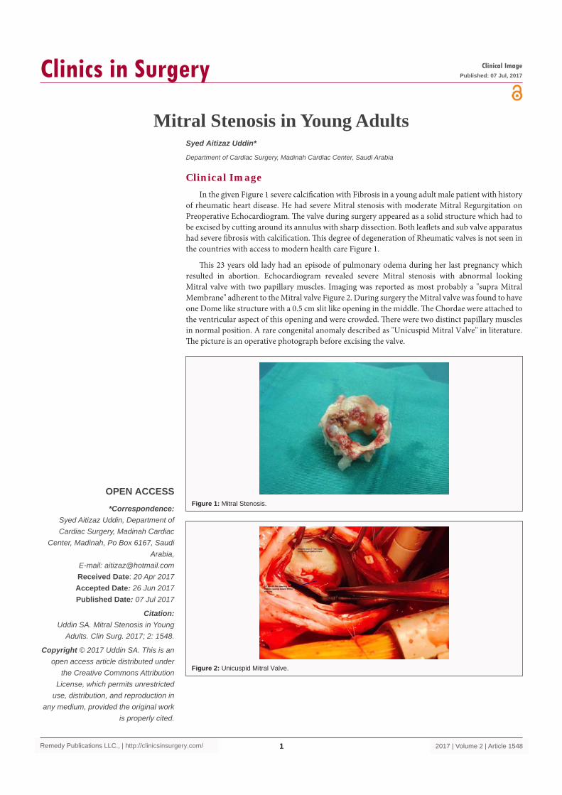
This 23 years old lady had an episode of pulmonary odema during her last pregnancy which resulted in abortion. Echocardiogram revealed severe Mitral stenosis with abnormal looking Mitral valve with two papillary muscles. Imaging was reported as most probably a "supra Mitral Membrane" adherent to the Mitral valve Figure 2. During surgery the Mitral valve was found to have one Dome like structure with a 0.5 cm slit like opening in the middle. The Chordae were attached to the ventricular aspect of this opening and were crowded. There were two distinct papillary muscles in normal position. A rare congenital anomaly described as "Unicuspid Mitral Valve" in literature. The picture is an operative photograph before excising the valve.
Figure 1: Mitral Stenosis.
Figure 2: Unicuspid Mitral Valve.