METHIONINE FORMATION BY TRANSMETHYLATION IN VITRO
12
METHIONINE FORMATION BY TRANSMETHYLATION IN VITRO BY HENRY BORSOOK AND JACOB W. DUBNOFF (From the William G. Kercklwff Laboratories of the Biological Sciences, California Institute of Technology, Pasadena) (Received for publication, February 8, 1947) We reported in a previous communication (1) that :guanidoacetic acid is methylated to creatine by rat liver slices in the presence of choline and either homocysteine or homocystine. Choline, homocystine, or homo- cysteine alone had little or no effect. These observations closed the gap which had existed between the evidence from tissue slice experiments on the one hand (2) and that from experiments in viva on the other (3-5) re- garding the transfer of choline methyl in the formation of creatine. Two transmethylation reactions, one in which methionine, the other in which creatine is formed, were thus exposed for study. We had observed that the rate of methylation of guanidoacetic acid by choline is as fast or faster with homocystine than with homocysteine as the methyl carrier (1). This was surprising, as it might have been expected, a priori, that homocysteine would be the more effective methyl carrier, and it suggested that the two transmet,hylation reactions may have different characteristics. We had found an indication earlier (2) that the methylation of guanido- acetic acid by methionine is not a simple methyl transfer between the two substances in that the reaction is inhibited when respiration is inhibited, whether by anaerobiosis or by oxidation inhibitors. We undertook a study of methionine formation by transmethylation in rat liver and kidney slices and homogenates to obtain more information on this question, and also because of the physiological significance of the reaction in itself, to which no in vitro studies had as yet been devoted. Methods The animals used were adult white rats which had been bred from the Wistar Institute strain. They were killed by stunning; the liver and kidney tissue were sliced free-hand with a straight edge razor to an average thickness of 0.4 mm. The slices were rinsed in Krebs and Henseleit (6) Ringer’s solution without the bicarbonate and finally suspended in t.hc same Ringer’s solution with such a concentration of bicarbonate that the pH, when equilibrated at 38” wit.1~5 per cent carbon dioxide, was 7.4. The reactants were dissolved in the latter version of the Krebs-Henseleit bicarbonate Ringer’s solution. 247
Transcript of METHIONINE FORMATION BY TRANSMETHYLATION IN VITRO

METHIONINE FORMATION BY TRANSMETHYLATION IN VITRO
BY HENRY BORSOOK AND JACOB W. DUBNOFF
(From the William G. Kercklwff Laboratories of the Biological Sciences, California Institute of Technology, Pasadena)
(Received for publication, February 8, 1947)
We reported in a previous communication (1) that :guanidoacetic acid is methylated to creatine by rat liver slices in the presence of choline and either homocysteine or homocystine. Choline, homocystine, or homo- cysteine alone had little or no effect. These observations closed the gap which had existed between the evidence from tissue slice experiments on the one hand (2) and that from experiments in viva on the other (3-5) re- garding the transfer of choline methyl in the formation of creatine.
Two transmethylation reactions, one in which methionine, the other in which creatine is formed, were thus exposed for study. We had observed that the rate of methylation of guanidoacetic acid by choline is as fast or faster with homocystine than with homocysteine as the methyl carrier (1). This was surprising, as it might have been expected, a priori, that homocysteine would be the more effective methyl carrier, and it suggested that the two transmet,hylation reactions may have different characteristics. We had found an indication earlier (2) that the methylation of guanido- acetic acid by methionine is not a simple methyl transfer between the two substances in that the reaction is inhibited when respiration is inhibited, whether by anaerobiosis or by oxidation inhibitors. We undertook a study of methionine formation by transmethylation in rat liver and kidney slices and homogenates to obtain more information on this question, and also because of the physiological significance of the reaction in itself, to which no in vitro studies had as yet been devoted.
Methods
The animals used were adult white rats which had been bred from the Wistar Institute strain. They were killed by stunning; the liver and kidney tissue were sliced free-hand with a straight edge razor to an average thickness of 0.4 mm. The slices were rinsed in Krebs and Henseleit (6) Ringer’s solution without the bicarbonate and finally suspended in t.hc same Ringer’s solution with such a concentration of bicarbonate that the pH, when equilibrated at 38” wit.1~ 5 per cent carbon dioxide, was 7.4. The reactants were dissolved in the latter version of the Krebs-Henseleit bicarbonate Ringer’s solution.
247

248 METHIONINE FORMED BY TBANSMETHYLATION
The reaction vessels were 20 ml. beakers. They were placed in a closed chamber which was partially immersed in a wat,er bath. The chamber was provided with a gas inlet tube through which the gas mixture was passed under slight pressure; the excess gas escaped under water through a slit in the side. The chamber could hold thirty beakers. When the latter were in place, the gas mixture was blown through vigorously for 10 minutes, after which it was slowed down. Through the period of gassing and afterwards the chamber containing the beakers was rocked at a rate of about 80 cycles per minute. By prior tests it was ascertained that the evaporation from the reaction vessels in a 4 hour run was negligible. The temperature of the water bath was 38”.
When homogenates were used, the tissue was removed and, without chilling, cut into small pieces and homogenized in the apparatus of Potter and Elvehje’m (7) in isotonic phosphate-Ringer’s solution (6) at pH 7.4. The homogenate was strained through two layers of cheese-cloth.
The lyophilized enzyme was prepared as follows. The tissue was sliced, the slices rinsed as described above, and then frozen on a block of solid carbon dioxide, transferred to flasks previously cooled to the temperature of solid carbon dioxide, and then dried under a high vacuum with the water trap kept in a bath of solid carbon dioxide in methyl cellosolve (Mefford). The thoroughly dried tissue was pulverized.
The first experiments were carried out with preparations of m-homo- cysteine and of DL-homocystine, generously given us by Professor V. du Vigneaud.
Later it seemed desirable to use the natural isomers. The starting material for their preparation was L-methionine obtained from casein by the method of Hill and Robson (8) with the following modifications. After the usual acid hydrolysis, removal of excess HCl, adjustment of the pH to 2.4, decoloration with charcoal and separation of the tyrosine, the pH was adjusted to 6.0 and the iron (presumably in the HCl and NaOH used) was removed by treatment with HzS. The removal of the iron clarified the solution further and facilitated the crystallization of the leucine and methio- nine. Methionine was separated from the leucine by the mercury pre- cipitation procedure described by Hill and Robson; after removal of the mercury the solution of impure methionine hydrochloride was concentrated nearly to dryness, dissolved in alcohol, and the solution brought to pH 6.0 with concentrated NaOH instead of with pyridine, as recommended by Hill and Robson, and set away overnight in the ice box. Methionine which had crystallized out was removed, the mother liquor concentrated, alcohol added to 80 per cent concentration, the pH adjusted to 6.0 with concentrated NaOH, and set away in the ice box overnight. The second crop of methionine thus obtained was added to the first. The methionine

H. BORSOOK AND J. W. DUBNOFF 249
was purified further by a second mercury precipitation as described by Hill and Robson, and recovered from the mercury precipitate by the same pro- cedure as after the first mercury precipitation. The methionine was puri- fied finally by two precipitations with alcohol from water.
L-Homocystine was prepared from L-methionine by the method of Butz and du Vigneaud (9), as modified by du Vigneaud and Patterson (10).
L-Homocysteine was prepared from L-homocystine by reduction with sodium in liquid ammonia, following the procedure of Riegel and du Vig- neaud (11).
Choline hydrochloride and betaine hydrochloride were used from several commercial sources. Dr. N. Horowitz kindly supplied specimens of the following compounds used: di- and monomethylethanolamine, mono- ethyldimethyl- and diethylmonomethylhydroxyethylammonium chloride, arsenocholine, phosphorylcholine, and homocysteinethiolactone.
Methionine was determined by an adaptation of the method of McC.arthy and Sullivan (12) which made the method more sensitive but somewhat less specific. The reduction in specificity was not significant under our experimental conditions, i.e. Ringer’s solution in which tissue slices or liver homogenates had been incubated and in which the metl$onine did not exceed 10 mg. per cent and the total of other amino acids 10 mg. per cent. The procedure was as follows: to 4 ml. of Ringer’s solution containing the slices were added 0.4 ml. of 30 per cent trichloroacetic acid; 2 ml. of filtrate were transferred to a 20 ml. beaker; 0.2 ml. of 5 N NaOH followed by 0.1 ml. of 1 per cent freshly made sodium nitroprusside were then added. The resulting solution was warmed with gentle rocking in a water bath at 38” for 8 minutes, after which it was cooled in ice water until the temperature was below 4“, and then 1 ml. of an acid mixture consisting of 9 volumes of concentrated HCl and 1 volume of 85 per cent H3P04 was added with shaking. The beaker was again placed in ice water for 1 to 2 minutes, after which it was warmed to room temperature and then transferred to a vacuum desiccator in which the CO2 was removed by 3 minutes suction. The color was then measured at 510 rnp in a Beckman spectrophotometer. Methionine standards were run simultaneously with the unknowns; the st,andard solution cont,aining no methionine was used as the blank in the spectrophotometer. A straight line relation is obtained between the logarithm of the transmission and the concentration of methionine over the range, 0 to 10 mg. per cent.
The method was tested by incubating known amounts of added methio- nine in Ringer’s solution with liver slices for 4 hours at 38”. The assay gave 98 per cent of the added methionine over a range of 0 to 10 mg. per cent after deducting the methionine liberated by the slices. The deficiency of 2 per cent was probably the result of oxidation of the added methionine.

250 METHIONINE FORMED BY TRANSMETHYLATION
The above modification of the McCart,hy-Sullivan method for the de- t,ermination of methioninc may be used only when the total concentration of non-methionine amino acids is of the order of 0.001 M or less. When the latter are present in higher concentrat’ion, the original recipe of McCa,rthy a.nd Sullivan is advisable.
Results1
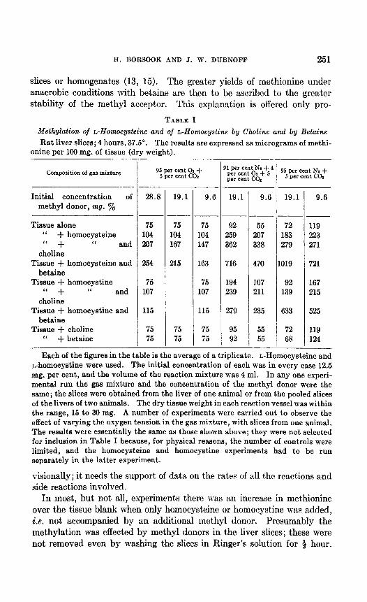
In Table I are summarized the result,s of experiments which show the formation of methionine by rat liver slices from homocysteine or homo- cystine and a suitable methyl donor. Larger yields of methionine were obtained under low oxygen t,ension (4 per cent O,), or anaerobically, than under 95 per cent 02, from homocysteine t,han from homocystine, whether under aerobic or anaerobic conditions, and with betaine than with choline as the methyl donor. The superiority of bet’aine over choline was greatest under anaerobic conditions and least under 95 per cent oxygen. The effect of oxygen tension on the yield of methionine was far less when choline was the methyl donor than when betaine was used.
The following fact.s offer a qualitative and at least partial explanation of the higher yields of methionine under nitrogen than under oxygen and of the greater relative effect. of anaerobiosis with betaine than wit,h choline as the met,hyl donor. Homocysteine is more stable under nit,rogen than under oxygen, presumably because it is protected from oxidation.2 Choline is rapidly oxidized by rat liver slices and homogenates to betaine on the alkaline (pH 7.8) and to betaine aldehyde on the acid (pH 6.7) sides of neutrality (13, 14). In its oxidation to betaine choline becomes a more effective methyl donor for methionine formation. Aerobiosis and anaero- biosis thus have opposite effects on the methyl donor and methyl acceptor when choline is the methyl donor; hence the small difference in the yield of methionine under the two conditions. Betaine is not oxidized by rat liver
1 Nearly all the results reported here were first obtained with the DL forms of homo- cysteine and of homocystine. The experiments were repeated with the natural isomers, and, as there were no essential differences between the two sets of exper- iments, only those of the latter are reported.
2 That homocysteine is protected by anaerobiosis was shown by the difference in the intensity of the transitory purple color after the addition of alkali and nitro- prusside in the first stage of the methionine determination. In the experiments under 95 per cent oxygen with homocysteine the purple color was faint, and deep when they were conducted under nitrogen, even though more homocysteine was converted to methionine in the latter solutions. Similar results were found with homocystine; no purple color was seen after experiments under 95 per cent oxygen, a faint purple after the anaerobic experiments. In the latter case the color was less intense than when homocysteine was used under the same conditions. The purple color given by homocystine in the anaerobic experiments was qualitative evidence that it was re- duced to homocysteine.
H. BORSOOK AND J. W. DUBNOFF 251
slices or homogenates (13, 15). The greater yields of methionine under anaerobic conditions with betaine are then to be ascribed to the greater stabilit,y of the methyl acceptor. This explanation is offered only pro-
TABLE I Methylation of Alomocysteine and of L-Homocystine by Choline and by Beta&e
Rat liver slices; 4 hours, 37.5”. The results are expressed as micrograms of methi- onine per 100 mg. of tissue (dry weight).
Composition of gas mixture
Initial concentration of methyl donor, mg. y.
Tissue alone “ + homocysteine “ + “ and
choline Tissue + homocysteine and
bet&e Tissue + homocystine
‘( + “ and choline
Tissue + homocystine and betaine
Tissue + choline “ + betaine
-
95 per cent 02 + 5 per cent CO8
28.8 19.1
75 104 207
254
75 107
115
75 75
75 104 167
215
75 75
-
. _
-
9.6
75 92 104 259 147 362
163 716
75 107
115
75 75
194 239
279
95 92
‘lp,Serc,c,e;t4 95 per cent Nt + per cent co* 5 per cent co2
19.1 9.6 19.1
~__ 55 72
207 183 338 279
470 1019
107 92 211 139
235 633
55 72 55 68
9.6
119 223 271
721
167 215
525
119 124
Each of the figures in the table is the average of a triplicate. L-Homocysteine and n-homocystine were used. The initial concentration of each was in every case 12.5 mg. per cent, and the volume of the reaction mixture was 4 ml. In any one experi- mental run the gas mixture and the concentration of the methyl donor were the same; the slices were obtained from the liver of one animal or from the pooled slices of the livers of two animals. The dry tissue weight in each reaction vessel was within the range, 15 to 30 mg. A number of experiments were carried out to observe the effect of varying the oxygen tension in the gas mixture, with slices from one animal. The results were essentially the same as those shown above; they were not selected for inclusion in Table I because, for physical reasons, the number of controls were limited, and the homocysteine and homocystine experiments had to be run separately in the latter experiment.
visionally; it needs the support of dat,a on the rates of all the reactions and side reactions involved.
In most, but not all, experiments there was an increase in met,hionine over the tissue blank when only homocysteine or homocystine was added, i.e. not accompanied by an additional methyl donor. Presumably the methylation was effected by methyl donors in the liver slices; these were not removed even by washing the slices in Ringer’s solution for + hour.

252 METHIONINE FORMED BY TRANSMETHYIATION
In our studies on the methylation of guanidoacetic acid (1, 2) we found a considerable methylation by liver slices, even when no methionine was added. Until a sufficiently sensitive method for the determination of methionine was available, we were uncertain whether this “tissue” methy- lation might be ascribed to methionine liberated by the liver slices during the period of incubation or whether it was necessary to invoke some other methyl donor. The method for the determination of methionine used in this study was sufficiently sensitive. With it we have found that rat liver slices liberated methionine, under the conditions of the experiments on the methylation of guanidoacetic acid, in concentrations which ranged from 0.2 to 1.0 mg. per cent; in 4 hours the total free methionine increased usually to about 3 times the zero time value. As the “tissue” methylation of guanidoacetic acid could be accounted for by a free methionine concen- tration of 3 X lo-’ M, i.e. approximately 0.5 mg. per cent, the methionine liberated by rat liver slices is sufficient, therefore, to account for the rates of methylation of guanidoacetic acid observed when no methionine is added to the Ringer’s solution.
Cyanide, azide, arsenite, and arsenate do not inhibit the methylation of homocysteine or of homocystine (Table II). In fact arsenite increased the yield of methionine even under 4 per cent oxygen. The non-suscept,i- bility of this transmethylation to oxidation inhibitors is in accord with its independence of oxygen.
The possibility suggested itself, in view of the superiority of betaine to choline as a methyl donor in methionine formation, that the methylating action of choline may be via betaine; i.e., that betaine is the immediate methyl donor to homocysteine or homocystine and that the methyl group lost by such a donation is replenished by choline. The possibility was tested in a number of experiments in which varying small amounts of be- taine were added to the reaction mixture containing the methyl acceptor and choline. The choline concentration in these experiments was 3 times the molecular equivalent of the homocysteine and 6 times that of homo- cystine; the betaine concentration was varied from one-fifth to one-hun- dredth that of the choline. The results obtained argue against a carrier function of betaine. The effect of choline plus betaine was always less than the sum of their separate effects.
In addition to bet,aine and choline a number of other possible methyl donors were tested. As Table III shows at the end of the 4 hour incu- bation with liver slices at 38”, no more and in some cases less methionine was found than when the methyl acceptor alone was added to the Ringer’s solution. The ot,her possible methyl donors tested were dimethylethanol- amine, monomethylethanolamine, diethylmethylhydroxyethylammonium chloride, monoethyldimethylhydroxyethylammonium chloride, arsenocho- line, and phosphorylcholine.

H. BORSOOK AND J. W. DUBNOFF 253
These results illustrate one of the limitations of the tissue slice technique. The tissue slices contained significant amounts of methyl donors, even after being washed 4 hour in large amounts of Ringer’s solution. The failure of the five possible methyl donors other than betaine or choline to add, under our experimental conditions, to the rate of methylation effected by t,he methyl donors in the slices does not, in itself, indicate that they could not
TABLE II Effect of Oxidation Inhibitors on Methylation of L-Homocysteine and of L-Homocystine
by Choline and by Betaine
Rat liver slices; 4 hours, 37.5”. The results are expressed as per cent of the methi- onine found after addition of homocysteine plus choline in the absence of an oxida- tion inhibitor.
Composition of gas mixture 95 per cent 01 + 5 per cent co2
- I
Inhibitor NOW
Tissue alone “ + homocys-
teine and choline Tissue + homocys-
teine and betaine Tissue + homocystine
and choline Tissue + homocystine
and betaine
46 100
115
61
64
-~ Cyanide Azide
_--
50 44 116 99
121 103
67 63
74 66
&mite Azt- None Azide -- --
28 35 21 15 163 100 100 75
324 119 112 87
78 63 59 57
113 75 65 60
91 per cent NI + 4 per cent Ox + 5 per cent co2
4rsenite Azt --
4.5 19 145 114
155 121
70 52
81 61
L-Homocysteine and L-homocystine were used at a concentration of 12.5 mg. per cent. The volume of the reaction mixture was 4 ml. In the experiments with 95 per cent oxygen the concentration of methyl donor was 28.8 mg. per cent andwith 4 per cent oxygen, 9.6 mg. per cent. The oxidation inhibitors were used in the form of their sodium salts and at a concentration of 0.001 M. In every experiment the controls consisted of tissue alone, tissue plus homocysteine, and tissue plus homo- cystine; the controls and the different reaction mixtures, all in triplicate, were run simultaneously in the absence and in the presence of one of the above oxidation inhibitors.
do so in vivo. One possibility is that under our experimental conditions the above substances are not transformed to betaine or to choline fast enough to make their contribution to that of the methyl donors in the slices noticeable.
The results summarized in Table III are in accord with those on growth (4,16) in that none of the compounds which were found to be incapable of supporting growth with homocystine exerted any additive methylating effect under our experimental conditions. Monoethyldimethylhydroxy- ethylammonium chloride and phosphorylcholine will support growth with homocystine presumably because they are converted to choline (or

254 METHIONINE FORAMED BY TRANSMETHYLATIOX
betaine) iu vivo; they exert,ed no significant ndditive methylating effect,s in our experiment’s because, presumably, they were not converted to betaine or to choline sufficiently rapidly to augment noticeably in short period experiment.s the effect, of bhe met,hyl donors in the slices.
TABLE III Relative R&es of Methylation of L-Homocysteine, L-Homocystine, and of Homocysteine-
thiolactone by Various Possible Methyl Donors
Rat liver slices; 4 hours, 37.5“. Excess methionine over tissue blank with homocysteine alone taken as 100.
-
Composition of gas mixture
____
Homocysteine ‘I + choline I‘ + betaine “ + dimethylethsnolamine ‘I + monomethylethanolamine “ + diethylmonomethylhy-
droxyethylammonium chloride Homocysteine + monoethyldimethylhy-
droxyethylammonium chloride Homocysteine + arsenocholine
‘I + phosphorylcholine Homocystine
“ + choline “ + betaine I‘ + dimethylethanolamine ‘I + monomethylethanolamine ‘I + diethylmonomethylhy-
droxyethylammonium chloride Homocystine + monoethyldimethylhy-
droxyethylammonium chloride Homocystine + arsenocholine
“ + phosphorylcholine Homocysteinethiolactone
‘I + choline “ -k betaine “ + arsenocholine “ + phosphorylcholin
35 per cent OP
+ 5 5 cent *
100 217 347 101 65
119
150
56 123 165
-42 - 138 - 138
54
-I
11 per cent N, 4 per cent c
+ 5 f%zcent -
100 220 633
95 99 91
100
76 94 51
102 180
21 11 72 92
254 20 49
loo 270
1905 90 93
-20
-120
60 104 208 27
-240 0
-20
Table III also contains a summary of results obtained with homocyskine- thiolactone. It was transformed to methionine at a rate less than that of homocysteine (50 to 75 per cent) and slightly superior t,o that of homo- cystine. Hetaine was in this instance also a more effective methyl donor t.han choline.

H. BOHSOOK .CXD J. \I-. DCBNOFF 255
Homocysteinethiolactone can replace homocyst,ine for rat, growth (Ii). Methionineless mutants of Neurospwa which can utilize homocystine as a substitut.e for methionine use homocysteinethiolactone better, and homo- cysteine still better.3 Our findings with homocysteinet,hiolactone and these are in accord.
Homogenized or lyophiliaed and dialyzed rat. liver retain the ability t,o catalyze the methylation of homocysteine and of homorystine. Some results with lyophilized liver are given in Table I\‘. Some a,ntivity is lost,
TABLE IV Methylation of L-Homocysteine and of L-Homocystine with and Gthout Choline an.d
Betaine by Lyophilized Rat Liver
Nitrogen, 4 hours, 38”. The figures are mg. per cent of met&mine in the tri- chloroacetic acid filtrates.
Reaction mixture
Blank....................... Homocysteine.
“ I‘
+ choline.. + betaine..
Homocystine. “ “
+ choline.. + betaine.....
Choline. Betsine.....................
Undialyzed powder
25 mg. 100 mg.
0.6 2.5 1.25 6.0 1.40 6.6 3.00 9.8 0.6 4.2 0.7 4.8 0.8 7.7 0.55 2.6 0.60 3.0
-
Dialyzed enzyme used in each test equivalent to 100 mg. powder
2.7 3.1 2.7 3.2 3.5 4.1 3.2 4.1 3.6 4.3 3.0 4.2 6.4 5.5 5.7 6.1
2.7 3.0 2.8 3.2 2.7 3.2 3.1 3.2
Plus iialysate Plus ash
The powder and reactants were dissolved inO. M phosphate buffer at pIE3.0. The final volume was 4 ml. Concentrations of homocysteine and of homocystine were 12.5 mg. per cent, of choline and betaine 37.5 mg. per cent.
in lyophilization and a further loss occurs in dialysis. The loss in activity indicates that there may be two mechanisms, one which uses choline, the other betaine; t:he former appears to suffer more loss in dialysis t,han the latter.
We have carried out, a number of experiments with slices of rat kidney cortex. Methyl donors within (i.e. not washed out of) the kidney slices effected about the same amount of methylation of homocysteine and homocyst,ine as did liver slices. Choline and betaine exerted only slight additive methylating effect,s. Unlike liver, kidney slices were more ef- fective under aerobic than anaerobic condSons.
3 Horowitz, N., unpublished experiments, private communication.

256 METHIONINE FORMED BY TIt4NSMETHYLATION
DISCUSSION
The results of experiments reported here have established that there are at least two categories of transmethylation reactions: One, of which the methylation of guanidoacetic acid to creatine is an example, depends on oxygen and is inhibited by oxidation inhibitors; the other, represented by the formation of methionine from homocysteine or homocystine as the methyl acceptor and choline or betaine as the methyl donor, does not re- quire oxygen and is not inhibited by oxidation inhibitors. In the first category methionine can act as a methyl donor but choline and betaine cannot unless homocysteine or homocystine is present to serve as a methyl carrier. Still another difference is that liver homogenates are unable, with or without added methionine, to methylate guanidoacetic acid; they retain their ability to catalyze methionine formation by transmethylation.4
In the first category the direction of the methyl transfer is from an S- methyl to an N-methyl and the reverse in the second category. This distinction may not be fundamental; it seems advisable to suspend judg- ment on this point at present.
Perlzweig et al. (18) reported that rat liver slices can methylate nico- tinamide. The reaction is strictly aerobic, requires unbroken cells, is usually but not always accelerated by the addition of methionine, and the direction of the methyl transfer is from S-methyl to N-methyl, character- istics which place this methylation in the same category as the methylation of guanidoacetic acid. The two methylations are similar also in that they are restricted to the liver.
Stetten (19) proposed a scheme of choline formation from betaine in which betaine is demethylated to glycine, the latter is reduced to ethanol- amine, which is then methylated to choline by methionine. The direct participation of methionine was not established and the pathway of betaine methyl was left open. The observations reported here show that a methyl group of betaine is available for choline formation by way of its incorpora- tion into methionine and thus account for the physiological properties of betaine associated with its possessing a labile methyl group. Other possi- bilities are not excluded.
Chandler and du Vigneaud (20) found that there was a lag of several days before growth was resumed after betaine was added to a diet containing homocystine and devoid of or very low in methionine and choline, whereas growth was resumed immediately after the addition of choline. Even after the lag, betaine was somewhat less effective than equivalent amounts of choline. Griffith and Mulford (21) found that 3 to 4 mg. of methionine
4 After this manuscript was submitted for publication we found that the methyla- tion of guanidoacetic acid by methionine occurs in guinea pig liver homogenates if adenosine triphosphate is provided.

H. BORSOOK AND J. 1%‘. DUBNOFF 257
or of betaine were required to give the same effect as 1 mg. of choline in preventing fatty liver or hemorrhagic kidney. As an explanation of the lesser efficiency of betaine than choline, the evidence Stetten obtained with N16 excluded direct reduction of betaine to choline. Our observations indicate that one route by which betaine methyl is made available for growth and for lipotropic purposes is via its incorporation into methionine, and hence its lower efficiency than choline for the latter function. None of the above observations excludes the possibility that ethanolamine may be methylated by betaine directly.
When choline is oxidized to betaine, at least one of its methyl groups remains labile via incorporation into methionine. There is considerable evidence that choline can be oxidized to betaine in the body. Rat liver slices and homogenates rapidly oxidize choline to betaine aldehyde and betaine (13, 14) ; betaine has been found in considerable amounts in animal tissues (22-25) and has been isolated from the urine of animals on a betaine- free diet after being fed large amounts of choline (26).
Although methyl transfer proceeds more rapidly from betaine than from choline, it seems unlikely from the evidence at hand that oxidation of choline to betaine is obligatory for this purpose. If it were, there should be little or no methionine formation with choline as the methyl donor under anaerobic conditions. Choline is effective under these conditions.
SUMMARY
1. Rat liver slices methylate homocysteine, homocystine, and homo- cysteinethiolactone to methionine. Choline or betaine can serve as the methyl donor.
2. The speed of the reaction with each of the three methyl acceptors is fastest with homocysteine and slowest with homocystine. It is faster with betaine than with choline as the methyl donor.
3. A number of possible methyl donors were tested; other than betaine and choline, none exerted a methylating effect which was additive to that of the methyl donors retained in the slices.
4. These transmethylations are independent of oxygen and are not inhibited by oxidation inhibitors.
5. Catalytic activity is retained in homogenized and in lyophilized rat liver.
6. There are two (at least) categories of methyl transfer reactions. One is dependent on oxygen, is inhibited by oxidation inhibitors, and catalytic activity is lost by homogenization and is not restored by adding methionine, which accelerates the reaction with slices. In this category is the methyla- tion by methionine of guanidoacetic acid to creatine and of nicotinamide to N’-methylnicotinamide. Characteristics of the second category are

258 METHIONINE FORMED BY TRANSMETHYLATION
independence of oxygen, nonsusceptibility to oxidation inhibitors, and per- sistence of catalytic activity after cell structure is destroyed. In this category is methionine formation by the methylation of homocysteine, homocysteinethiolactone, or homocystine by betaine or choline.
The authors wish to acknowledge the assistance of Miss I. Silberbach in the experiments reported here.
BIBLIOGRAPHY
1. Boreook, H., and Dubnoff, J. W., J. Biol. Chem., 160,635 (1945). 2. Borsook, H., and Dubnoff, J. W., J. Biol. Chem., 132,559 (1940). 3. du Vigneaud, V., Chandler, J. P., Moyer, A. W., and Keppel, D. M., J. Biol.
Chem., 131, 57 (1939). 4. du Vigneaud, V., Chandler, J. P., Cohn, M., and Brown, G. B., J. Biol. Chem.,
134, 787 (1940). 5. du Vigneaud, V., Harvey Lectures, 38.39 (1942-43). 6. Krebs, H. A., and Heneeleit, K., 2. physiol. Chem., 210, 33 (1932). 7. Potter, V. R., and Elvehjem, C. A., J. Biol. Chem., 114,495 (1936). 8. Hill, E. M., and Robson, W., Biochem. J., 26, 1008 (1934). 9. Bute, L. W., and du Vigneaud, V., J. Biol. Chem., 99,135 (1932-33).
10. du Vigneaud, V., and Patterson, W. I., J. Bid. Chem., 109, 97 (1935). 11. Riegel, B., and du Vignesud, V., J. Biol. Chem., 112, 149 (1935-36). 12. McCarthy, T. E., and Sullivan, M. X., J. Biol. Chem., 141, 871 (1941). 13. Bernheim, F., and Bernheim, M. L. C., Am. J. Physiol., 104, 438 (1933); 121,
55 (1938). 14. Mann, P. J. G., and Qunstel, J. H., Biochem. J., 31, 869 (1937). 15. Handler, P., Bernheim, hf. L. C., and Klein, J. R., J. Biol. Chem., 136,211 (1941). 16. Moyer, A. W., and du Vigneaud, V., J. Biol. Chem., 143, 373 (1942). 17. Rose, W. C., and Rice, E. E., J. BioE. Chem., 130,305 (1939). 18. Perlzweig, W. A., Bernheim, M. L. C., and Bernheim, F., J. Biol. Chem., 160,
401 (1943). 19. Stetten, D., Jr., J. Biol. Chcm., 138, 437 (1941); 140, 143 (1941). 20. Chandler, J. P., and du Vigneaud, V., J. Biol. Chem., 136, 223 (1940). 21. Griffith, W. H., and Mulford, D. J., J. Am. Chem. Sot., 63, 929 (1941). 22. Bebeschin, K., Z. phyriol. Chem., 72, 380 (1911). 23. FlBesner, O., and Kirstein, F., Z. Biol., 64, 510 (1926). 24. Ackermann, D., and Fuchs, H. G., 2. physiol. Chem., 267, 151, 153 (1939). 25. Ackermann, D., and Wasmuth, W., Z. physiol. Chem., 281,199 (1944). 26. Miiller, H., 2. physiol. C&m., 263, 243 (193940).


![Dietary supplementation with free methionine or methionine … · 2019. 6. 27. · with MHA or DL-methionine in heat stress-exposed broilers [23, 24]. In this study, we hypothesize](https://static.fdocuments.net/doc/165x107/60e337666b3f9a31a45a96d1/dietary-supplementation-with-free-methionine-or-methionine-2019-6-27-with-mha.jpg)