Management & Therapy of Dry Eye Disease - Tear Film & Ocular
16
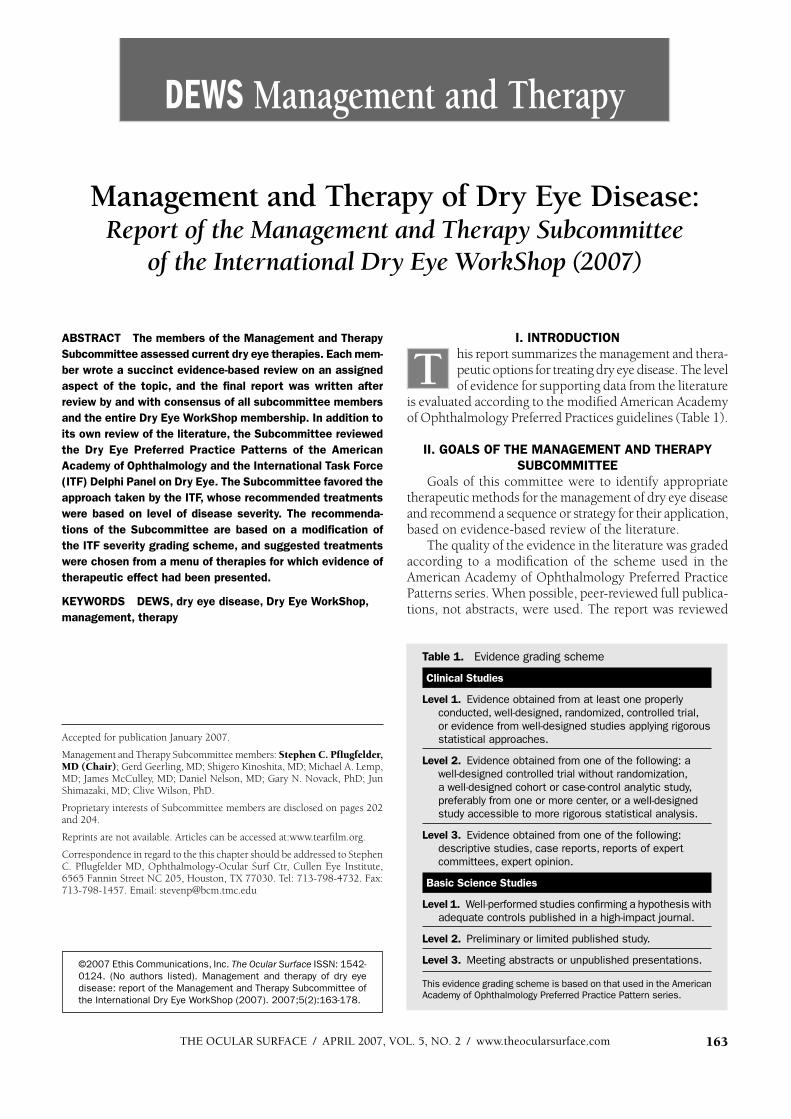
THE OCULAR SURF ACE / APRIL 2007, VOL. 5, NO. 2 / www.theocularsurface.com 163 DEWS M anagem e n t a n d Th e ra p y ©2007 Ethis Communications, Inc. The Ocular Surface ISSN : 154 2- 0124 . (N o authors listed). Management and therapy of dry eye disease: report of the Management and Therapy Subcommittee of the International Dry Eye W orkShop (2007). 2007;5(2):163-178. M anagem e n t a n d Th e ra p y o f D ry E ye D isease: Report of the Management and Therapy Subcommittee of the International D ry E ye W ork Shop (2 0 0 7 ) ABSTRACT The members of the Management and Therapy Subcommittee assessed current dry eye therapies. Each mem- ber wrote a succinct evidence-based review on an assigned aspect of the topic, and the final report was written after review by and with consensus of all subcommittee members and the entire Dry Eye WorkShop membership. In addition to its own review of the literature, the Subcommittee reviewed the Dry Eye Preferred Practice Patterns of the American Academy of Ophthalmology and the International Task Force (ITF) Delphi Panel on Dry Eye. The Subcommittee favored the approach taken by the ITF, whose recommended treatments were based on level of disease severity. The recommenda- tions of the Subcommittee are based on a modification of the ITF severity grading scheme, and suggested treatments were chosen from a menu of therapies for which evidence of therapeutic effect had been presented. KEYWORDS DEWS, dry eye disease, Dry Eye WorkShop, management, therapy I. INTRODUCTION his report summarizes the management and thera- peutic options for treating dry eye disease. The level of evidence for supporting data from the literature is evaluated according to the modified American Academy of Ophthalmology Preferred Practices guidelines (Table 1). II. GOALS OF THE MANAGEMENT AND THERAPY SUBCOMMITTEE Goals of this committee were to identify appropriate therapeutic methods for the management of dry eye disease and recommend a sequence or strategy for their application, based on evidence-based review of the literature. The quality of the evidence in the literature was graded according to a modification of the scheme used in the American Academy of Ophthalmology Preferred Practice Patterns series. W hen possible, peer-reviewed full publica- tions, not abstracts, were used. The report was reviewed Accepted for publication January 2007. Management and Therapy Subcommittee members: S tep he n C. P fl u g fe ld e r, MD (Chair); Gerd Geerling, MD; Shigero Kinoshita, MD; Michael A. Lemp, MD; James McCulley, MD; Daniel Nelson, MD; Gary N. Novack, PhD; Jun Shimazaki, MD; Clive W ilson, PhD. Proprietary interests of Subcommittee members are disclosed on pages 202 and 204. Reprints are not available. Articles can be accessed at:www.tearfilm.org. Correspondence in regard to the this chapter should be addressed to Stephen C. Pflugfelder MD, Ophthalmology-Ocular Surf Ctr, Cullen Eye Institute, 6565 Fannin Street NC 205, Houston, TX 77030. Tel: 713-798-4732. Fax: 713-798-1457. Email: [email protected] Table 1 . Ev idence grading scheme Clinical Studies Level 1 . Ev idence obtained from at least one properly conducted, w ell-designed, randomized, controlled trial, or ev idence from w ell-designed studies applying rigorous statistical approaches. Level 2 . Ev idence obtained from one of the follow ing: a w ell-designed controlled trial w ithout randomiz ation, a w ell-designed cohort or case-control analytic study, preferably from one or more center, or a w ell-designed study accessible to more rigorous statistical analysis. Level 3 . Ev idence obtained from one of the follow ing: descriptiv e studies, case reports, reports of ex pert committees, ex pert opinion. Basic Science Studies Level 1 . W ell-performed studies confi rming a hypothesis w ith adeq uate controls published in a high-impact journal. Level 2 . P reliminary or limited published study. Level 3 . Meeting abstracts or unpublished presentations. This ev idence grading scheme is based on that used in the A merican A cademy of O phthalmology P referred P ractice P attern series. T
Transcript of Management & Therapy of Dry Eye Disease - Tear Film & Ocular
THE OCULAR SURF ACE / APRIL 2007, VOL. 5, NO. 2 / www.theocularsurface.com 163
DEWS M a n a g e m e n t a n d Th e ra p y
©2007 Ethis Communications, Inc. The Ocular Surface ISSN : 154 2-
0124 . (N o authors listed). Management and therapy of dry eye
disease: report of the Management and Therapy Subcommittee of
the International Dry Eye W orkShop (2007). 2007;5(2):163-178.
M a n a g e m e n t a n d Th e ra p y o f D ry E ye D is e a s e : Report of the Management and Therapy Subcommittee
of the International D ry E ye W ork Shop (2 0 0 7 )
ABSTRACT The members of the Management and Therapy
Subcommittee assessed current dry eye therapies. Each mem-
ber wrote a succinct evidence-based review on an assigned
aspect of the topic, and the final report was written after
review by and with consensus of all subcommittee members
and the entire Dry Eye WorkShop membership. In addition to
its own review of the literature, the Subcommittee reviewed
the Dry Eye Preferred Practice Patterns of the American
Academy of Ophthalmology and the International Task Force
(ITF) Delphi Panel on Dry Eye. The Subcommittee favored the
approach taken by the ITF, whose recommended treatments
were based on level of disease severity. The recommenda-
tions of the Subcommittee are based on a modification of
the ITF severity grading scheme, and suggested treatments
were chosen from a menu of therapies for which evidence of
therapeutic effect had been presented.
KEYWORDS DEWS, dry eye disease, Dry Eye WorkShop,
management, therapy
I. INTRODUCTION
his report summarizes the management and thera-peutic options for treating dry eye disease. The level of evidence for supporting data from the literature
is evaluated according to the modified American Academy of Ophthalmology Preferred Practices guidelines (Table 1).
II. GOALS OF THE MANAGEMENT AND THERAPY
SUBCOMMITTEE
Goals of this committee were to identify appropriate therapeutic methods for the management of dry eye disease and recommend a sequence or strategy for their application, based on evidence-based review of the literature.
The quality of the evidence in the literature was graded according to a modification of the scheme used in the American Academy of Ophthalmology Preferred Practice Patterns series. W hen possible, peer-reviewed full publica-tions, not abstracts, were used. The report was reviewed
Accepted for publication January 2007.
Management and Therapy Subcommittee members: S te p he n C. P fl u g fe ld e r, MD (Chair); Gerd Geerling, MD; Shigero Kinoshita, MD; Michael A. Lemp, MD; James McCulley, MD; Daniel Nelson, MD; Gary N. Novack, PhD; Jun Shimazaki, MD; Clive W ilson, PhD.
Proprietary interests of Subcommittee members are disclosed on pages 202 and 204.
Reprints are not available. Articles can be accessed at:www.tearfilm.org.
Correspondence in regard to the this chapter should be addressed to Stephen C. Pflugfelder MD, Ophthalmology-Ocular Surf Ctr, Cullen Eye Institute, 6565 Fannin Street NC 205, Houston, TX 77030. Tel: 713-798-4732. Fax: 713-798-1457. Email: [email protected]
Table 1 . Ev idence grading scheme
Clinical Studies
Level 1 . Ev idence obtained from at least one properly
conducted, w ell-designed, randomized, controlled trial,
or ev idence from w ell-designed studies applying rigorous
statistical approaches.
Level 2 . Ev idence obtained from one of the follow ing: a
w ell-designed controlled trial w ithout randomization,
a w ell-designed cohort or case-control analytic study,
preferably from one or more center, or a w ell-designed
study accessible to more rigorous statistical analysis.
Level 3 . Ev idence obtained from one of the follow ing:
descriptiv e studies, case reports, reports of expert
committees, expert opinion.
Basic Science Studies
Level 1 . W ell-performed studies confi rming a hypothesis w ith
adeq uate controls published in a high-impact journal.
Level 2 . P reliminary or limited published study.
Level 3 . Meeting abstracts or unpublished presentations.
This ev idence grading scheme is based on that used in the A merican
A cademy of O phthalmology P referred P ractice P attern series.
T
THE OCULAR SURFACE / APRIL 2007, VOL. 5, NO. 2 / www.theocularsurface.com164
by all subcommittee members and by the entire Dry Eye WorkShop membership. Comments and suggested revi-sions were discussed by the subcommittee members and incorporated into the report where deemed appropriate by consensus.
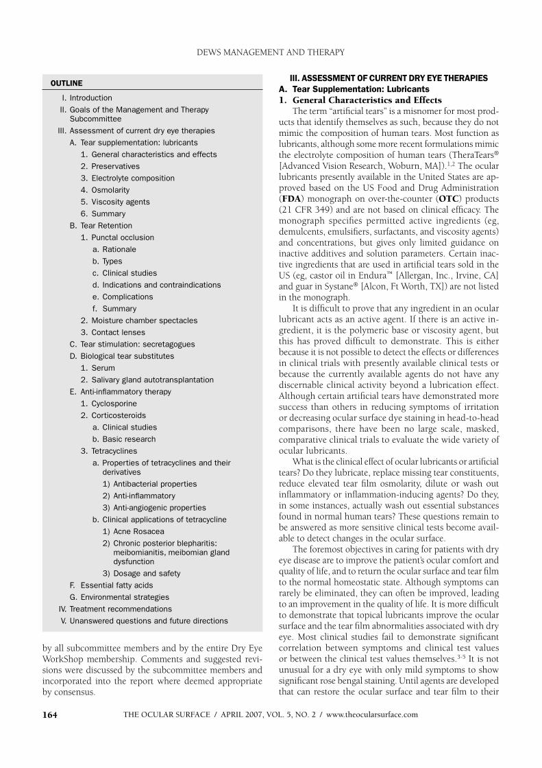
III. ASSESSMENT OF CURRENT DRY EYE THERAPIES
A. Tear Supplementation: Lubricants
1. General Characteristics and EffectsThe term “artificial tears” is a misnomer for most prod-
ucts that identify themselves as such, because they do not mimic the composition of human tears. Most function as lubricants, although some more recent formulations mimic the electrolyte composition of human tears (TheraTears®
[Advanced V ision Research, Woburn, MA]).1,2 The ocular lubricants presently available in the United States are ap-proved based on the US Food and Drug Administration (FDA) monograph on over-the-counter (OTC) products (21 CFR 349) and are not based on clinical efficacy. The monograph specifies permitted active ingredients (eg, demulcents, emulsifiers, surfactants, and viscosity agents) and concentrations, but gives only limited guidance on inactive additives and solution parameters. Certain inac-tive ingredients that are used in artificial tears sold in the US (eg, castor oil in Endura™ [Allergan, Inc., Irvine, CA] and guar in Systane® [Alcon, Ft Worth, TX]) are not listed in the monograph.
It is difficult to prove that any ingredient in an ocular lubricant acts as an active agent. If there is an active in-gredient, it is the polymeric base or viscosity agent, but this has proved difficult to demonstrate. This is either because it is not possible to detect the effects or differences in clinical trials with presently available clinical tests or because the currently available agents do not have any discernable clinical activity beyond a lubrication effect. Although certain artificial tears have demonstrated more success than others in reducing symptoms of irritation or decreasing ocular surface dye staining in head-to-head comparisons, there have been no large scale, masked, comparative clinical trials to evaluate the wide variety of ocular lubricants.
What is the clinical effect of ocular lubricants or artificial tears? Do they lubricate, replace missing tear constituents, reduce elevated tear film osmolarity, dilute or wash out inflammatory or inflammation-inducing agents? Do they, in some instances, actually wash out essential substances found in normal human tears? These questions remain to be answered as more sensitive clinical tests become avail-able to detect changes in the ocular surface.
The foremost objectives in caring for patients with dry eye disease are to improve the patient’s ocular comfort and quality of life, and to return the ocular surface and tear film to the normal homeostatic state. Although symptoms can rarely be eliminated, they can often be improved, leading to an improvement in the quality of life. It is more difficult to demonstrate that topical lubricants improve the ocular surface and the tear film abnormalities associated with dry eye. Most clinical studies fail to demonstrate significant correlation between symptoms and clinical test values or between the clinical test values themselves.3-5 It is not unusual for a dry eye with only mild symptoms to show significant rose bengal staining. Until agents are developed that can restore the ocular surface and tear film to their
OUTLINE
I. Introduction
II. Goals of the Management and Therapy Subcommittee
III. Assessment of current dry eye therapies
A. Tear supplementation: lubricants
1. General characteristics and effects
2. Preservatives
3. Electrolyte composition
4. Osmolarity
5. V iscosity agents
6. Summary
B. Tear Retention
1. Punctal occlusion
a. Rationale
b. Types
c. Clinical studies
d. Indications and contraindications
e. Complications
f. Summary
2. Moisture chamber spectacles
3. Contact lenses
C. Tear stimulation: secretagogues
D. Biological tear substitutes
1. Serum
2. Salivary gland autotransplantation
E. Anti-inflammatory therapy
1. Cyclosporine
2. Corticosteroids
a. Clinical studies
b. Basic research
3. Tetracyclines
a. Properties of tetracyclines and their derivatives
1) Antibacterial properties
2) Anti-inflammatory
3) Anti-angiogenic properties
b. Clinical applications of tetracycline
1) Acne Rosacea
2) Chronic posterior blepharitis: meibomianitis, meibomian gland dysfunction
3) Dosage and safety
F. Essential fatty acids
G. Environmental strategies
IV . Treatment recommendations
V . Unanswered questions and future directions
DEWS MANAGEMENT AND THERAPY
THE OCULAR SURFACE / APRIL 2007, VOL. 5, NO. 2 / www.theocularsurface.com 165
normal homeostatic state, the symptoms and signs of dry eye disease will continue.
Ocular lubricants are characterized by hypotonic or isotonic buffered solutions containing electrolytes, surfac-tants, and various types of viscosity agents. In theory, the ideal artificial lubricant should be preservative-free, contain potassium, bicarbonate, and other electrolytes and have a polymeric system to increase its retention time.1,6-8 Physical properties should include a neutral to slightly alkaline pH. Osmolarities of artificial tears have been measured to range from about 181 to 354 mOsm/L.9 The main variables in the formulation of ocular lubricants regard the concentration of and choice of electrolytes, the osmolarity and the type of viscosity/polymeric system, the presence or absence of preservative, and, if present, the type of preservative.
2. Preservatives The single most critical advance in the treatment of dry
eye came with the elimination of preservatives, such as benzal-konium chloride (B AK), from OTC lubricants. Because of the risk of contamination of multidose products, most either contain a preservative or employ some mechanism for minimizing contamination. The FDA has required that multidose artificial tears contain preservatives to prevent microbial growth.10 Preservatives are not required in unit dose vials that are discarded after a single use. The wide-spread availability of nonpreserved preparations allows patients to administer lubricants more frequently without concern about the toxic effects of preservatives. For patients with moderate-to-severe dry eye disease, the absence of preservatives is of more critical importance than the particu-lar polymeric agent used in ocular lubricants. The ocular surface inflammation associated with dry eye is exacerbated by preserved lubricants; however, nonpreserved solutions are inadequate in themselves to improve the surface inflam-mation and epithelial pathology seen in dry eye disease.11
Benzalkonium chloride is the most frequently used preservative in topical ophthalmic preparations, as well as in topical lubricants. Its epithelial toxic effects have been well established.12-17 The toxicity of BAK is related to its concentration, the frequency of dosing, the level or amount of tear secretion, and the severity of the ocular surface disease. In the patient with mild dry eye, BAK-preserved drops are usually well tolerated when used 4-6 times a day or less. In patients with moderate-to-severe dry eye, the potential for BAK toxicity is high, due to decreased tear secretion and decreased turnover.17 Some patients may be using other topical preparations (eg, glaucoma medications) that contain BAK, increasing their exposure to the toxic effects of BAK. Also, the potential for toxicity exists with patient abuse of other OTC products that contain BAK, such as vasoconstrictors.
BAK can damage the corneal and conjunctival epithe-lium, affecting cell-to-cell junctions and cell shape and microvilli, eventually leading to cell necrosis with sloughing of 1-2 layers of epithelial cells.17 Preservative-free formula-tions are absolutely necessary for patients with severe dry
eye with ocular surface disease and impairment of lacrimal gland secretion, or for patients on multiple, preserved topical medications for chronic eye disease. Patients with severe dry eye, greatly reduced tear secretion, and punctal occlusion are at particular risk for preservative toxicity. In such patients, the instilled agent cannot be washed out; if this risk has not been appreciated by the clinician, preserved drops might be used at high frequency.
Another additive used in OTC formulations is disodium (EDTA). It augments the preservative efficacy of BAK and other preservatives, but, by itself, it is not a sufficient pre-servative. Used in some nonpreserved solutions, it may help limit microbial growth in opened unit-dose vials. Although use of EDTA may allow a lower concentration of preservative, EDTA may itself be toxic to the ocular surface epithelium. A study comparing two preservative-free solu-tions, Hypotears PF® (Novartis Ophthalmics, East Hanover, NJ) containing EDTA and Refresh® (Allergan, Inc., Irvine, CA) without EDTA, showed that both formulations had identical safety profiles and were completely nontoxic to the rabbit corneal epithelium.18 Other studies found that EDTA-containing preparations increased corneal epithelial permeability.19,20 The potential exists that patients with severe dry eye will find that EDTA-containing preparations increase irritation.
Nonpreserved, single unit-dose tear substitutes are more costly for the manufacturer to produce, more costly for the patients to purchase, and less convenient to use than bottled ocular lubricants. For these reasons, reclosable unit dose vials (eg, Refresh Free [Allergan Inc., Irvine, CA]; Tears Natural Free® [Alcon, Fort Worth, TX]) were introduced. Less toxic preservatives, such as polyquad (polyquaternium-1), sodium chlorite (Purite®), and sodium perborate were developed to allow the use of multidose bottled lubricants and to avoid the known toxicity of BAK-containing solutions.21,22 The “vanishing” preservatives were sodium perborate and sodium chlorite (TheraTears® [Advanced Vision Research, Woburn, MA], Genteal® [Novartis, East Hanover, NJ], and Refresh Tears®
[Allergan Inc., Irvine, CA]).Sodium chlorite degrades to chloride ions and water
upon exposure to UV light after instillation. Sodium perbo-rate is converted to water and oxygen on contact with the tear film. For patients with severe dry eye, even vanishing preservatives may not totally degrade, due to a decrease in tear volume, and may be irritating. Patients prefer bottled preparations for reasons of both cost and ease of use. The ideal lubricant would come in a multidose, easy-to-use bottle that contains a preservative that completely dissipates before reaching the tear film, or is completely nontoxic and nonirritating and maintains absolute sterility with frequent use. One such multi-use, preservative-free product has been introduced to the market (Visine Pure-Tears® [Pfizer, Inc, NJ]).
Ocular ointments and gels are also used in treatment of dry eye disease. Ointments are formulated with a specific mixture of mineral oil and petrolatum. Some contain lanolin,
DEWS MANAGEMENT AND THERAPY
THE OCULAR SURFACE / APRIL 2007, VOL. 5, NO. 2 / www.theocularsurface.com166
which can be irritating to the eye and delay corneal wound healing.23 Individuals with sensitivity to wool may also be sensitive to lanolin.23 Some ointments contain parabens as preservatives, and these ointments are not well tolerated by patients with severe dry eye. In general, ointments do not support bacterial growth and, therefore, do not require preservatives. Gels containing high molecular weight cross-linked polymers of acrylic acid (carbomers) have longer retention times than artificial tear solutions, but have less visual blurring effect than petrolatum ointments.
3. Electrolyte Composition Solutions containing electrolytes and or ions have been
shown to be beneficial in treating ocular surface damage due to dry eye.1,6,20,24,25 To date, potassium and bicarbon-ate seem to be the most critical. Potassium is important to maintain corneal thickness.7 In a dry-eye rabbit model, a hypotonic tear-matched electrolyte solution (TheraTears®
[Advanced Vision Research, Woburn, MA]) increased con-junctival goblet cell density and corneal glycogen content, and reduced tear osmolarity and rose bengal staining after 2 weeks of treatment.25 The restoration of conjunctival goblet cells seen in the dry-eye rabbit model has been corroborated in patients with dry eye after LASIK.26
Bicarbonate-containing solutions promote the recovery of epithelial barrier function in damaged corneal epithelium and aid in maintaining normal epithelial ultrastructure. They may also be important for maintaining the mucin layer of the tear film.6 Ocular lubricants are available that mimic the electrolyte composition of human tears, eg, TheraTears®
(Advanced Vision Research, Woburn, MA) and BION Tears®
(Alcon, Fort Worth, TX).1,2 These also contain bicarbonate, which is critical for forming and maintaining the protec-tive mucin gel in the stomach.27 Bicarbonate may play a similar role for gel-forming mucins on the ocular surface. Because bicarbonate is converted to carbon dioxide when in contact with air and can diffuse through the plastic unit dose vials, foil packaging of the plastic vials is required to maintain stability.
4. OsmolarityTears of patients with dry eye have a higher tear film
osmolarity (crystalloid osmolarity) than do those of normal patients.28,29 Elevated tear film osmolarity causes mor-phological and biochemical changes to the corneal and conjunctival epithelium18,30 and is pro-inflammatory.31 This knowledge influenced the development of hypo-osmotic artificial tears such as Hypotears® (230 mOsm/L [Novartis Ophthalmics, East Hanover, NJ]) and subsequently Thera-Tears® (181 mOsm/L [Advance Vision Research, Woburn, MA]).32
Colloidal osmolality is another factor that varies in artificial tear formulations. While crystalloid osmolarity is related to the presence of ions, colloidal osmolality is dependent largely on macromolecule content. Colloidal osmolarity, also known as oncotic p res s u re, is involved in the control of water transport in tissues. Differences in colloidal
osmolality affect the net water flow across membranes, and water flow is eliminated by applying hydrostatic pressure to the downside of the water flow. The magnitude of this osmotic pressure is determined by osmolality differences on the two sides of the membrane. Epithelial cells swell due to damage to their cellular membranes or due to a dysfunction in the pumping mechanism. Following the addition of a fluid with a high colloidal osmolality to the damaged cell surface, deturgescence occurs, leading to a return of normal cell physiology. Theoretically, an artificial tear formulation with a high colloidal osmolality may be of value. Holly and Esquivel evaluated many different artificial tear formulations and showed that Hypotears® (Novartis Ophthalmics, East Hanover, NJ) had the highest colloidal osmolality of all of the formulations tested.33 Formulations with higher colloidal osmolality have since been marketed (Dwelle® [Dry Eye Company, Silverdale, WA]).
Protection against the adverse effects of increased os-molarity (osmoprotection) has led to development of OTC drops incorporating compatible solutes (such as glycerin, erythritol, and levocarnitine (Optive® [Allergan Inc., Irvine, CA]). It is thought that the compatible solutes distribute be-tween the tears and the intracellular fluids to protect against potential cellular damage from hyperosmolar tears.34
5. Viscosity Agents The stability of the tear film depends on the chemical-
physical characteristics of that film interacting with the conjunctival and corneal epithelium via the membrane-spanning mucins (ie, MUC-16 and MUC-4). In the classical three-layered tear film model, the mucin layer is usually thought of as a surfactant or wetting agent, acting to lower the surface tension of the relatively hydrophobic ocular surface, rendering the corneal and conjunctival cells “wet-table.”33 Currently, the tear film is probably best described as a hydrated, mucin gel whose mucin concentration decreases with distance from the epithelial cell surface. It may have a protective role similar to that of mucin in the stomach.35 It may also serve as a “sink” or storage vehicle for substances secreted by the main and accessory lacrimal glands and the ocular surface cells. This may explain why most of the available water-containing lubricants are only minimally effective in restoring the normal homeostasis of the ocular surface. In addition to washing away and diluting out irritating or toxic substances in the tear film, artificial lubricants hydrate gel-forming mucin. While some patients with dry eye have decreased aqueous lacrimal gland secretion, alterations or deficiencies involving mucin also cause dry eye.
Macromolecular complexes added to artificial lubricants act as viscosity agents. The addition of a viscosity agent in-creases residence time, providing a longer interval of patient comfort. For example, when a viscous, anionic charged carboxymethyl-cellulose (CMC, 100,000 mw) solution was compared with a neutral hydroxymethylcellulose (HPMC) solution, CMC was shown to have a significantly slower rate of clearance from the eye.36 Viscous agents in active drug
DEWS MANAGEMENT AND THERAPY

THE OCULAR SURFACE / APRIL 2007, VOL. 5, NO. 2 / www.theocularsurface.com 167
formulations may also prolong ocular surface contact, in-creasing the duration of action and penetration of the drug.
Viscous agents may also protect the ocular surface epithelium. It is known that rose bengal stains abnormal corneal and conjunctival epithelial cells expressing an al-tered mucin glycocalyx.37 Agents such as hydroxymethycel-lulose (HMC), which decrease rose bengal staining in dry eye subjects,38 may either “coat and protect” the surface epithelium or help restore the protective effect of mucins.
In the US, carboxymethyl cellulose is the most com-monly used polymeric viscosity agent (IRI Market Share Data, Chicago, IL), typically in concentrations from 0.25% to 1%, with differences in molecular weight also contrib-uting to final product viscosity. Carboxymethyl cellulose has been found to bind to and be retained by human epi-thelial cells.39 Other viscosity agents included in the FDA monograph (in various concentrations) include polyvinyl alcohol, polyethylene glycol, glycol 400, propylene glycol hydroxymethyl cellulose and hydroxypropyl cellulose.
The blurring of vision and esthetic disadvantages of cak-ing and drying on eyelashes are drawbacks of highly viscous agents that patients with mild to moderate dry eye will not tolerate. Lower molecular-weight viscous agents help to minimize these problems. Because patient compliance, comfort, and convenience are important considerations, a range of tear substitute formulations with varying viscosi-ties are needed.
Hydroxypropyl-guar (HP-guar) has been used as a gel-ling agent in a solution containing glycol 400 and propyl-ene glycol (Systane®, Alcon, Fort Worth, TX). It has been suggested that HP-guar preferentially binds to the more hydrophobic, desiccated or damaged areas of the surface epithelial cells, providing temporary protection for these cells.40,41 Several commercial preparations containing oil in the form of castor oil (Endura™ [Allergan Inc., Irvine, CA]) or mineral oil (Soothe® [Bausch & Lomb, Rochester, NY]) are purported to aid in restoring or increasing the lipid layer of the tear film.42,43 Hyaluronic acid is a viscosity agent that has been investigated for years as an “active” compound added to tear substitute formulations for the treatment of dry eye. Hyaluronic acid (0.2%) has significantly longer ocular surface residence times than 0.3 percent HPMC or 1.4 percent polyvinyl alcohol.44 Some clinical studies reported improvement in 44-48 dry eye in patients treated with sodium hyaluronate-containing solutions compared to other lubricant solutions, whereas others did not.48
Although lubricant preparations containing sodium hyal-uronate have not been approved for use in the US, they are frequently used in some countries.
6. SummaryAlthough many topical lubricants, with various viscos-
ity agents, may improve symptoms and objective findings, there is no evidence that any agent is superior to another. Most clinical trials involving topical lubricant preparations will document some improvement (but not resolution) of subjective symptoms and improvement in some objective
parameters.4 However, the improvements noted are not necessarily any better than those seen with the vehicle or other nonpreserved artificial lubricants. The elimination of preservatives and the development of newer, less toxic preservatives have made ocular lubricants better tolerated by dry eye patients. However, ocular lubricants, which have been shown to provide some protection of the ocular surface epithelium and some improvement in patient symp-toms and objective findings, have not been demonstrated in controlled clinical trials to be sufficient to resolve the ocular surface disorder and inflammation seen in most dry eye sufferers.
B. Tear Retention
1. Punctal Occlusiona. Rationale
While the concept of permanently occluding the lacri-mal puncta with cautery to treat dry eye extends back 70 years,49 and, although the first dissolvable implants were used 45 years ago,50 the modern era of punctal plug use began in 1975 with the report by Freeman.51 Freeman de-scribed the use of a dumbbell-shaped silicone plug, which rests on the opening of the punctum and extends into the canaliculus. His report established a concept of punctal oc-clusion, which opened the field for development of a variety of removable, long-lasting plugs to retard tear clearance in an attempt to treat the ocular surface of patients with deficient aqueous tear production. The Freeman style plug remains the prototype for most styles of punctal plugs.
b. T ypesPunctal plugs are divided into two main types: absorb-
able and nonabsorbable. The former are made of collagen or polymers and last for variable periods of time (3 days to 6 months). The latter nonabsorbable “permanent” plugs include the Freeman style, which consists of a surface collar resting on the punctal opening, a neck, and a wider base. In contrast, the Herrick plug (Lacrimedics [Eastsound,WA]) is shaped like a golf tee and is designed to reside within the canaliculus. It is blue for visualization; other variations are radiopaque. A newly designed cylindrical Smartplug™
(Medennium Inc [Irvine, CA]) expands and increases in diameter in situ following insertion into the canaliculus due to thermodynamic properties of its hydrophilic acrylic composition.
c. Clinical StudiesA variety of clinical studies evaluating the efficacy of
punctal plugs have been reported.52-56 These series generally fall into Level II evidence. Their use has been associated with objective and subjective improvement in patients with both Sjogren and non-Sjogren aqueous tear deficient dry eye, filamentary keratitis, contact lens intolerance, Stevens-Johnson disease, severe trachoma, neurotrophic keratopathy, post-penetrating keratoplasty, diabetic kera-topathy, and post-photorefractive keratectomy or laser in situ keratomileusis. Several studies have been performed
DEWS MANAGEMENT AND THERAPY
THE OCULAR SURFACE / APRIL 2007, VOL. 5, NO. 2 / www.theocularsurface.com168
to evaluate the effects of punctal plugs on the efficacy of glaucoma medications in reducing intraocular pressure, and these studies have reported conflicting results.57,58
Beneficial outcome in dry eye symptoms has been reported in 74-86% of patients treated with punctal plugs. Objective indices of improvement reported with the use of punctal plugs include improved corneal staining, prolonged tear film breakup time (TFBUT), decrease in tear osmolarity, and increase in goblet cell density. Overall, the clinical util-ity of punctal plugs in the management of dry eye disease has been well documented.
d. Indications and ContraindicationsIn a recent review on punctal plugs, it was reported
that in a major eye clinic, punctal plugs are considered indicated in patients who are symptomatic of dry eyes, have a Schirmer test (with anesthesia) result less than 5 mm at 5 minutes, and show evidence of ocular surface dye staining.56
Contraindications to the use of punctal plugs include allergy to the materials used in the plugs to be implanted, punctal ectropion, and pre-existing nasolacrimal duct ob-struction, which would, presumably, negate the need for punctal occlusion. It has been suggested that plugs may be contraindicated in dry eye patients with clinical ocular surface inflammation, because occlusion of tear outflow would prolong contact of the abnormal tears contain-ing proinflammatory cytokines with the ocular surface. Treatment of the ocular surface inflammation prior to plug insertion has been recommended. Acute or chronic infection of the lacrimal canaliculus or lacrimal sac is also a contraindication to use of a plug.
e. ComplicationsThe most common complication of punctal plugs is
spontaneous plug extrusion, which is particularly common with the Freeman-style plugs. Over time, an extrusion rate of 50% has been reported, but many of these extrusions took place after extensive periods of plug residence. Most extrusions are of small consequence, except for incon-venience and expense. More troublesome complications include internal migration of a plug, biofilm formation and infection,59 and pyogenic granuloma formation. Removal of migrated canalicular plugs can be difficult and may require surgery to the nasolacrimal duct system.60,61
f. SummaryThe extensive literature on the use of punctal plugs in
the management of dry eye disease has documented their utility. Several recent reports, however, have suggested that absorption of tears by the nasolacrimal ducts into sur-rounding tissues and blood vessels may provide a feedback mechanism to the lacrimal gland regulating tear produc-tion.62 In one study, placement of punctal plugs in patients with normal tear production caused a significant decrease in tear production for up to 2 weeks after plug insertion.63
This cautionary note should be considered when deciding
whether to incorporate punctal occlusion into a dry eye disease management plan.
2. Moisture Chamber SpectaclesThe wearing of moisture-conserving spectacles has for
many years been advocated to alleviate ocular discomfort associated with dry eye. However, the level of evidence sup-porting its efficacy for dry eye treatment has been relatively limited. Tsubota et al, using a sensitive moisture sensor, reported an increase in periocular humidity in subjects wearing such spectacles.64 Addition of side panels to the spectacles was shown to further increase the humidity.65
The clinical efficacy of moisture chamber spectacles has been reported in case reports.66,67 Kurihashi proposed a related treatment for dry eye patients, in the form of a wet gauze eye mask.68 Conversely, Nichols et al recently report-ed in their epidemiologic study that spectacle wearers were twice as likely as emmetropes to report dry eye disease.69
The reason for this observation was not explained.There have been several reports with relatively high
level of evidence describing the relationship between environmental humidity and dry eye. Korb et al reported that increases in periocular humidity caused a significant increase in thickness of the tear film lipid layer.70 Dry eye subjects wearing spectacles showed significantly longer interblink intervals than those who did not wear spectacles, and duration of blink (blinking time) was significantly longer in the latter subjects.70 Instillation of artificial tears caused a significant increase in the interblink interval and a decrease in the blink rate.71 Maruyama et al reported that dry eye symptoms worsened in soft contact lens wearers when environmental humidity decreased.72
3. Contact Lenses Contact lenses may help to protect and hydrate the
corneal surface in severe dry eye conditions. Several differ-ent contact lens materials and designs have been evaluated, including silicone rubber lenses and gas permeable scleral-bearing hard contact lenses with or without fenestration.73-77
Improved visual acuity and comfort, decreased corneal epitheliopathy, and healing of persistent corneal epithelial defects have been reported.73-77 Highly oxygen-permeable materials enable overnight wear in appropriate circum-stances.75 There is a small risk of corneal vascularization and possible corneal infection associated with the use of contact lenses by dry eye patients.
C. Tear Stimulation: Secretogogues
Several potential topical pharmacologic agents may stimulate aqueous secretion, mucous secretion, or both. The agents currently under investigation by pharmaceuti-cal companies are diquafosol (one of the P2Y2 receptor agonists), rebamipide, gefarnate, ecabet sodium (mucous secretion stimulants), and 15(S)-HETE (MUC1 stimulant). Among them, a diquafosol eye drop has been favorably evaluated in clinical trials. 2% diquafosol (INS365, DE-089 [Santen, Osaka, Japan]; Inspire [Durham, NC]) proved to
DEWS MANAGEMENT AND THERAPY
THE OCULAR SURFACE / APRIL 2007, VOL. 5, NO. 2 / www.theocularsurface.com 169
be effective in the treatment of dry eye in a randomized, double-masked trial in humans to reduce ocular surface staining.78 A similar study demonstrated the ocular safety and tolerability of diquafosol in a double-masked, placebo-controlled, randomized study.79 This agent is capable of stimulating both aqueous and mucous secretion in animals and humans.80-83 Beneficial effects on corneal epithelial barrier function, as well as increased tear secretion, has been demonstrated in the rat dry eye model.84 Diquafosol also has been shown to stimulate mucin release from goblet cells in a rabbit dry eye model.85,86
The effects of rebamipide (OPC-12759 [Otsuka, Rock-ville, MD]; Novartis [Basel, Switzerland]) have been evalu-ated in human clinical trials. In animal studies, rebamipide increased the mucin-like substances on the ocular surface of N-acetylcysteine-treated rabbit eyes.87 It also had hy-droxyl radical scavenging effects on UVB-induced corneal damage in mice.88
Ecabet sodium (Senju [Osaka, Japan]; ISTA [Irvine, CA]) is being evaluated in clinical trials internationally, but only limited results have yet been published. A single instillation of ecabet sodium ophthalmic solution elicited a statistically significant increase in tear mucin in dry eye patients.89 Gefarnate (Santen [Osaka, Japan]) has been evaluated in animal studies. Gefarnate promoted mucin production after conjunctival injury in monkeys.90 Gefar-nate increased PAS-positive cell density in rabbit conjunc-tiva and stimulated mucin-like glycoprotein stimulation from rat cultured corneal epithelium.91,92 An in vivo rabbit experiment showed a similar result.93,94
The agent 15(S)-HETE, a unique molecule, can stimulate MUC1 mucin expression on ocular surface epithelium.9515(S)-HETE protected the cornea in a rabbit model of desiccation-induced injury, probably because of mucin secretion.96 It has been shown to have beneficial effects on secretion of mucin-like glycoprotein by the rab-bit corneal epithelium.97 Other laboratory studies confirm the stimulatory effect of 15(S)-HETE.98-101 Some of these agents may become useful clinical therapeutic modalities in the near future.
Two orally administered cholinergic agonists, pilocar-pine and cevilemine, have been evaluated in clinical trials for treatment of Sjogren syndrome associated keratocon-junctivitis sicca (KCS). Patients who were treated with pi-locarpine at a dose of 5 mg Q ID experienced a significantly greater overall improvement than placebo-treated patients in “ocular problems” in their ability to focus their eyes dur-ing reading, and in symptoms of blurred vision compared with placebo-treated patients.102 The most commonly reported side effect from this medication was excessive sweating, which occurred in over 40% of patients. Two percent of the patients taking pilocarpine withdrew from the study because of drug-related side effects. Other stud-ies have reported efficacy of pilocarpine for ocular signs and symptoms of Sjogren syndrome KCS,103-105 including an increase in conjunctival goblet cell density after 1 and 2 months of therapy.106
Cevilemine is another oral cholinergic agonist that was found to significantly improve symptoms of dryness and aqueous tear production and ocular surface disease compared to placebo when taken in doses of 15 or 30 mg TID.107,108 This agent may have fewer adverse systemic side effects than oral pilocarpine.
D. Biological Tear Substitutes
Naturally occurring biological, ie, nonpharmaceutical fluids, can be used to substitute for natural tears. The use of serum or saliva for this purpose has been reported in humans. They are usually unpreserved. When of autologous origin, they lack antigenicity and contain various epithe-liotrophic factors, such as growth factors, neurotrophins, vitamins, immunoglobulins, and extracellular matrix proteins involved in ocular surface maintenance. Biologi-cal tear substitutes maintain the morphology and support the proliferation of primary human corneal epithelial cells better than pharmaceutical tear substitutes.109 However, despite biomechanical and biochemical similarities, rel-evant compositional differences compared with normal tears exist and are of clinical relevance.110 Additional practical problems concern sterility and stability, and a labor-intensive production process or a surgical procedure (saliva) is required to provide the natural tear substitute to the ocular surface.
1. Serum Serum is the fluid component of full blood that remains
after clotting. Its topical use for ocular surface disease was much stimulated by Tsubota’s prolific work in the late 1990s.111 The practicalities and published evidence of autologous serum application were recently reviewed.112
The use of blood and its components as a pharmaceuti-cal preparation in many countries is restricted by specific national laws. To produce serum eye drops and to use them for outpatients, a license by an appropriate national body may be required in certain countries. The protocol used for the production of serum eye drops determines their composition and efficacy. An optimized protocol for the production was recently published.113 Concentrations between 20% and 100% of serum have been used. The efficacy seems to be dose-dependent.
Because of significant variations in patient populations, production and storage regimens, and treatment protocols, the efficacy of serum eye drops in dry eyes has varied sub-stantially between studies.113 Three published prospective randomized studies with similar patient populations (pre-dominantly immune disease associated dry eye, ie, Sjogren syndrome) are available. When comparing 20% serum with 0.9% saline applied 6 times per day, Tananuvat et al foundonly a trend toward improvement of symptoms and signs of dry eyes,114 whereas Kojima et al reported significant improvement of symptom scores, fluorescein-breakup time (FBUT), and fluorescein and rose bengal staining.115
A prospective clinical cross-over trial compared 50% serum eyedrops against the commercial lubricant previously
DEWS MANAGEMENT AND THERAPY
THE OCULAR SURFACE / APRIL 2007, VOL. 5, NO. 2 / www.theocularsurface.com170
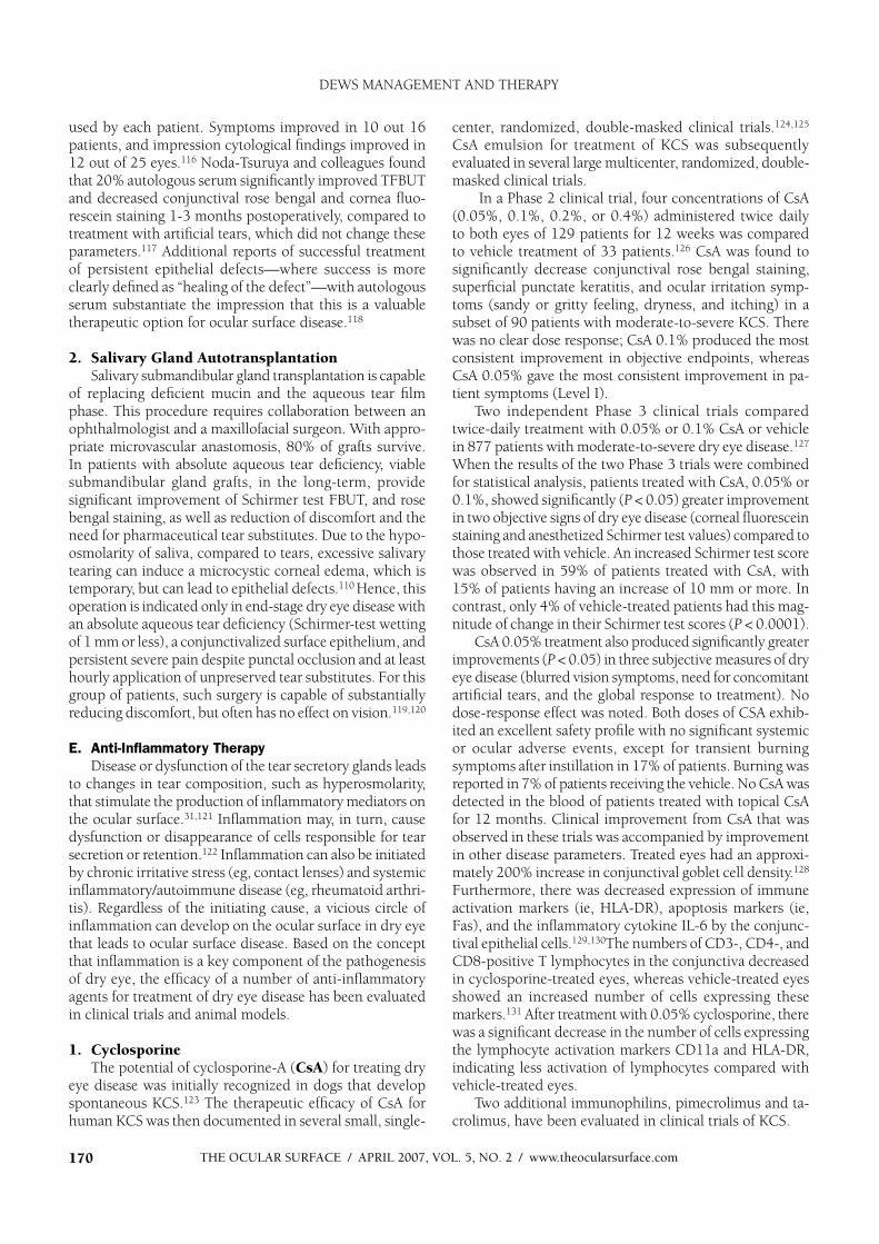
used by each patient. Symptoms improved in 10 out 16 patients, and impression cytological findings improved in 12 out of 25 eyes.116 Noda-Tsuruya and colleagues found that 20% autologous serum significantly improved TFBUT and decreased conjunctival rose bengal and cornea fluo-rescein staining 1-3 months postoperatively, compared to treatment with artificial tears, which did not change these parameters.117 Additional reports of successful treatment of persistent epithelial defects—where success is more clearly defined as “healing of the defect”—with autologous serum substantiate the impression that this is a valuable therapeutic option for ocular surface disease.118
2. Salivary Gland AutotransplantationSalivary submandibular gland transplantation is capable
of replacing deficient mucin and the aqueous tear film phase. This procedure requires collaboration between an ophthalmologist and a maxillofacial surgeon. With appro-priate microvascular anastomosis, 80% of grafts survive. In patients with absolute aqueous tear deficiency, viable submandibular gland grafts, in the long-term, provide significant improvement of Schirmer test FBUT, and rose bengal staining, as well as reduction of discomfort and the need for pharmaceutical tear substitutes. Due to the hypo-osmolarity of saliva, compared to tears, excessive salivary tearing can induce a microcystic corneal edema, which is temporary, but can lead to epithelial defects.110 Hence, this operation is indicated only in end-stage dry eye disease with an absolute aqueous tear deficiency (Schirmer-test wetting of 1 mm or less), a conjunctivalized surface epithelium, and persistent severe pain despite punctal occlusion and at least hourly application of unpreserved tear substitutes. For this group of patients, such surgery is capable of substantially reducing discomfort, but often has no effect on vision.119,120
E. Anti-Inflammatory Therapy
Disease or dysfunction of the tear secretory glands leads to changes in tear composition, such as hyperosmolarity, that stimulate the production of inflammatory mediators on the ocular surface.31,121 Inflammation may, in turn, cause dysfunction or disappearance of cells responsible for tear secretion or retention.122 Inflammation can also be initiated by chronic irritative stress (eg, contact lenses) and systemic inflammatory/autoimmune disease (eg, rheumatoid arthri-tis). Regardless of the initiating cause, a vicious circle of inflammation can develop on the ocular surface in dry eye that leads to ocular surface disease. Based on the concept that inflammation is a key component of the pathogenesis of dry eye, the efficacy of a number of anti-inflammatory agents for treatment of dry eye disease has been evaluated in clinical trials and animal models.
1. CyclosporineThe potential of cyclosporine-A (CsA) for treating dry
eye disease was initially recognized in dogs that develop spontaneous KCS.123 The therapeutic efficacy of CsA for human KCS was then documented in several small, single-
center, randomized, double-masked clinical trials.124,125
CsA emulsion for treatment of KCS was subsequently evaluated in several large multicenter, randomized, double-masked clinical trials.
In a Phase 2 clinical trial, four concentrations of CsA (0.05%, 0.1%, 0.2%, or 0.4%) administered twice daily to both eyes of 129 patients for 12 weeks was compared to vehicle treatment of 33 patients.126 CsA was found to significantly decrease conjunctival rose bengal staining, superficial punctate keratitis, and ocular irritation symp-toms (sandy or gritty feeling, dryness, and itching) in a subset of 90 patients with moderate-to-severe KCS. There was no clear dose response; CsA 0.1% produced the most consistent improvement in objective endpoints, whereas CsA 0.05% gave the most consistent improvement in pa-tient symptoms (Level I).
Two independent Phase 3 clinical trials compared twice-daily treatment with 0.05% or 0.1% CsA or vehicle in 877 patients with moderate-to-severe dry eye disease.127
When the results of the two Phase 3 trials were combined for statistical analysis, patients treated with CsA, 0.05% or 0.1%, showed significantly (P < 0.05) greater improvement in two objective signs of dry eye disease (corneal fluorescein staining and anesthetized Schirmer test values) compared to those treated with vehicle. An increased Schirmer test score was observed in 59% of patients treated with CsA, with 15% of patients having an increase of 10 mm or more. In contrast, only 4% of vehicle-treated patients had this mag-nitude of change in their Schirmer test scores (P < 0.0001).
CsA 0.05% treatment also produced significantly greater improvements (P < 0.05) in three subjective measures of dry eye disease (blurred vision symptoms, need for concomitant artificial tears, and the global response to treatment). No dose-response effect was noted. Both doses of CSA exhib-ited an excellent safety profile with no significant systemic or ocular adverse events, except for transient burning symptoms after instillation in 17% of patients. Burning was reported in 7% of patients receiving the vehicle. No CsA was detected in the blood of patients treated with topical CsA for 12 months. Clinical improvement from CsA that was observed in these trials was accompanied by improvement in other disease parameters. Treated eyes had an approxi-mately 200% increase in conjunctival goblet cell density.128
Furthermore, there was decreased expression of immune activation markers (ie, HLA-DR), apoptosis markers (ie, Fas), and the inflammatory cytokine IL-6 by the conjunc-tival epithelial cells.129,130The numbers of CD3-, CD4-, and CD8-positive T lymphocytes in the conjunctiva decreased in cyclosporine-treated eyes, whereas vehicle-treated eyes showed an increased number of cells expressing these markers.131 After treatment with 0.05% cyclosporine, there was a significant decrease in the number of cells expressing the lymphocyte activation markers CD11a and HLA-DR, indicating less activation of lymphocytes compared with vehicle-treated eyes.
Two additional immunophilins, pimecrolimus and ta-crolimus, have been evaluated in clinical trials of KCS.
DEWS MANAGEMENT AND THERAPY

THE OCULAR SURFACE / APRIL 2007, VOL. 5, NO. 2 / www.theocularsurface.com 171
2. Corticosteroids a. Clinical Studies
Corticosteroids are an effective anti-inflammatory therapy in dry eye disease. Level I evidence is published for a number of corticosteroid formulations. In a 4-week, double-masked, randomized study in 64 patients with KCS and delayed tear clearance, loteprednol etabonate 0.5% ophthalmic suspension (Lotemax [Bausch and Lomb, Rochester, NY]), q.i.d., was found to be more effective than its vehicle in improving some signs and symptoms.132
In a 4-week, open-label, randomized study in 32 pa-tients with KCS, patients receiving fluorometholone plus artificial tear substitutes (ATS) experienced lower symptom severity scores and lower fluorescein and rose bengal stain-ing than patients receiving either ATS alone or ATS plus flurbiprofen.133
A prospective, randomized clinical trial compared the severity of ocular irritation symptoms and corneal fluores-cein staining in two groups of patients, one treated with topical nonpreserved methylprednisolone for 2 weeks, followed by punctal occlusion (Group 1), with a group that received punctal occlusion alone (Group 2).134 After 2 months, 80% of patients in Group 1 and 33% of patients in Group 2 had complete relief of ocular irritation symptoms. Corneal fluorescein staining was negative in 80% of eyes in Group 1 and 60% of eyes in Group 2 after 2 months. No steroid-related complications were observed in this study.
Level III evidence is also available to support the efficacy of corticosteroids. In an open-label, non-comparative trial, extemporaneously formulated nonpreserved methylpred-nisolone 1% ophthalmic suspension was found to be clini-cally effective in 21 patients with Sjogren syndrome KCS.135
In a review, it was stated that “…clinical improvement of KCS has been observed after therapy with anti-inflamma-tory agents, including corticosteroids.”136
In the US Federal Regulations, ocular corticosteroids receiving “class labeling” are indicated for the treatment “…of steroid responsive inflammatory conditions of the palpebral and bulbar conjunctiva, cornea and anterior segment of the globe such as allergic conjunctivitis, acne rosacea, superficial punctate keratitis, herpes zoster kerati-tis, iritis, cyclitis, selected infective conjunctivitides, when the inherent hazard of steroid use is accepted to obtain an advisable diminution in edema and inflammation.” We in-terpret that KCS is included in this list of steroid-responsive inflammatory conditions.137-140
b. Basic ResearchCorticosteroids are the standard anti-inflammatory
agent for numerous basic research studies of inflamma-tion, including the types that are involved in KCS. The corticosteroid methylprednisolone was noted to preserve corneal epithelial smoothness and barrier function in an experimental murine model of dry eye.141 This was at-tributed to its ability to maintain the integrity of corneal epithelial tight junctions and decrease desquamation of apical corneal epithelial cells.142 A concurrent study showed
that methylprednislone prevented an increase in MMP-9 protein in the corneal epithelium, as well as gelatinase activity in the corneal epithelium and tears in response to experimental dry eye.141
Preparations of topically applied androgen and es-trogen steroid hormones are currently being evaluated in randomized clinical trials. A trial of topically applied 0.03% testosterone was reported to increase the percent-age of patients that had meibomian gland secretions with normal viscosity and to relieve discomfort symptoms after 6 months of treatment compared to vehicle.143 TFBUT and lipid layer thickness were observed to increase in a patient with KCS who was treated with topical androgen for 3 months.144 Tear production and ocular irritation symptoms were reported to increase following treatment with topical 17 beta-oestradiol solution for 4 months.145
3. Tetracyclinesa. Properties of Tetracyclines and Their Derivatives1) Antibacterial Properties
The antimicrobial effect of oral tetracycline treatment analogues (eg, minocycline, doxycline) has previously been discussed by Shine et al,146 Dougherty et al,147 and Ta et al.148 It is hypothesized that a decrease in bacterial flora pro-ducing lipolytic exoenzymes146,148 and inhibition of lipase production147 with resultant decrease in meibomian lipid breakdown products146 may contribute to improvement in clinical parameters in dry eye-associated diseases.
2) Anti-Inflammatory PropertiesThe tetracyclines have anti-inflammatory as well as
antibacterial properties that may make them useful for the management of chronic inflammatory diseases. These agents decrease the activity of collagenase, phospholipase A2, and several matrix metalloproteinases, and they de-crease the production of interleukin (IL)-1 and tumor necrosis factor (TN F)-alpha in a wide range of tissues, including the corneal epithelium.149-151 At high concentra-tions, tetracyclines inhibit staphylococcal exotoxin-induced cytokines and chemokines.152,153
3) Anti-angiogenic PropertiesAngiogenesis, the formation of new blood vessels, oc-
curs in many diseases. These include benign conditions (eg, rosacea) and malignant processes (eg, cancer). Minocycline and doxycycline inhibit angiogenesis induced by implanted tumors in rabbit cornea.154 The anti-angiogenic effect of tetracycline may have therapeutic implications in inflamma-tory processes accompanied by new blood vessel formation. Well-controlled studies must be performed, at both the laboratory and clinical levels, to investigate this potential.155
b. Clinical Applications of Tetracycline1) Acne Rosacea
Rosacea, including its ocular manifestations, is an in-flammatory disorder, occurring mainly in adults, with peak severity in the third and fourth decades. Current recom-
DEWS MANAGEMENT AND THERAPY
THE OCULAR SURFACE / APRIL 2007, VOL. 5, NO. 2 / www.theocularsurface.com172
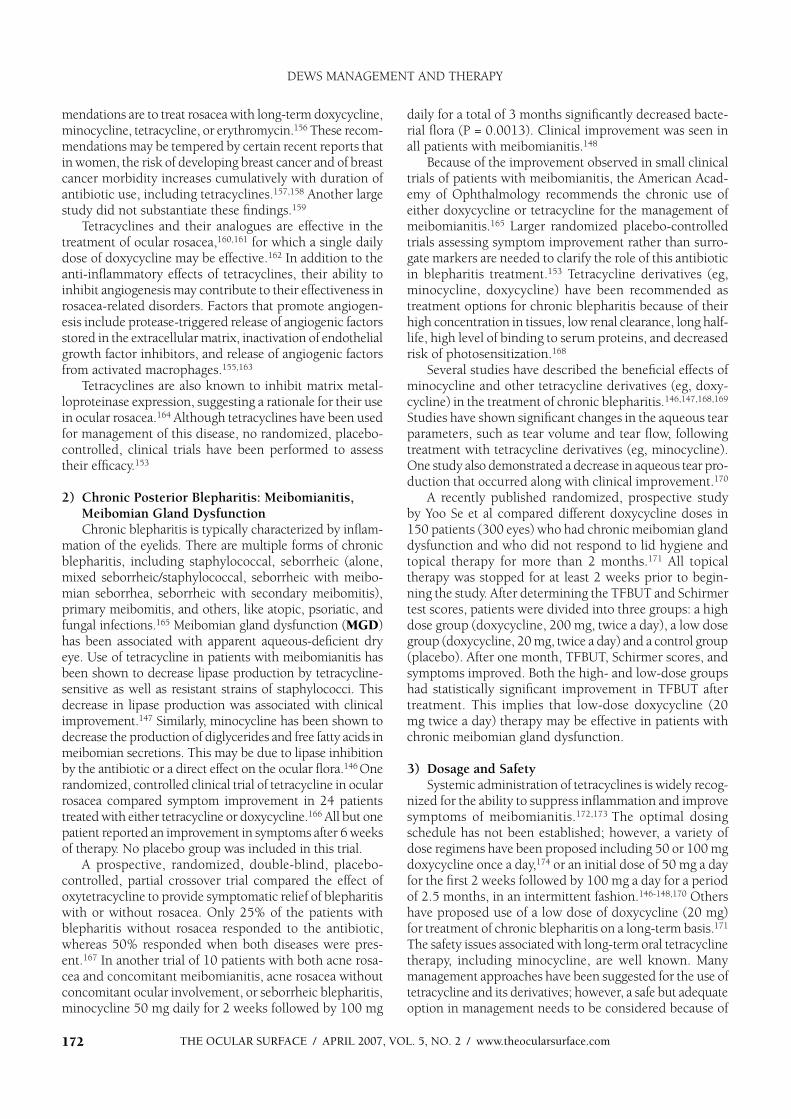
mendations are to treat rosacea with long-term doxycycline, minocycline, tetracycline, or erythromycin.156 These recom-mendations may be tempered by certain recent reports that in women, the risk of developing breast cancer and of breast cancer morbidity increases cumulatively with duration of antibiotic use, including tetracyclines.157,158 Another large study did not substantiate these findings.159
Tetracyclines and their analogues are effective in the treatment of ocular rosacea,160,161 for which a single daily dose of doxycycline may be effective.162 In addition to the anti-inflammatory effects of tetracyclines, their ability to inhibit angiogenesis may contribute to their effectiveness in rosacea-related disorders. Factors that promote angiogen-esis include protease-triggered release of angiogenic factors stored in the extracellular matrix, inactivation of endothelial growth factor inhibitors, and release of angiogenic factors from activated macrophages.155,163
Tetracyclines are also known to inhibit matrix metal-loproteinase expression, suggesting a rationale for their use in ocular rosacea.164 Although tetracyclines have been used for management of this disease, no randomized, placebo-controlled, clinical trials have been performed to assess their efficacy.153
2) Chronic Posterior Blepharitis: Meibomianitis, Meibomian Gland Dysfunction Chronic blepharitis is typically characterized by inflam-
mation of the eyelids. There are multiple forms of chronic blepharitis, including staphylococcal, seborrheic (alone, mixed seborrheic/staphylococcal, seborrheic with meibo-mian seborrhea, seborrheic with secondary meibomitis), primary meibomitis, and others, like atopic, psoriatic, and fungal infections.165 Meibomian gland dysfunction (MGD)has been associated with apparent aqueous-deficient dry eye. Use of tetracycline in patients with meibomianitis has been shown to decrease lipase production by tetracycline-sensitive as well as resistant strains of staphylococci. This decrease in lipase production was associated with clinical improvement.147 Similarly, minocycline has been shown to decrease the production of diglycerides and free fatty acids in meibomian secretions. This may be due to lipase inhibition by the antibiotic or a direct effect on the ocular flora.146 One randomized, controlled clinical trial of tetracycline in ocular rosacea compared symptom improvement in 24 patients treated with either tetracycline or doxycycline.166 All but one patient reported an improvement in symptoms after 6 weeks of therapy. No placebo group was included in this trial.
A prospective, randomized, double-blind, placebo-controlled, partial crossover trial compared the effect of oxytetracycline to provide symptomatic relief of blepharitis with or without rosacea. Only 25% of the patients with blepharitis without rosacea responded to the antibiotic, whereas 50% responded when both diseases were pres-ent.167 In another trial of 10 patients with both acne rosa-cea and concomitant meibomianitis, acne rosacea without concomitant ocular involvement, or seborrheic blepharitis, minocycline 50 mg daily for 2 weeks followed by 100 mg
daily for a total of 3 months significantly decreased bacte-rial flora (P = 0.0013). Clinical improvement was seen in all patients with meibomianitis.148
Because of the improvement observed in small clinical trials of patients with meibomianitis, the American Acad-emy of Ophthalmology recommends the chronic use of either doxycycline or tetracycline for the management of meibomianitis.165 Larger randomized placebo-controlled trials assessing symptom improvement rather than surro-gate markers are needed to clarify the role of this antibiotic in blepharitis treatment.153 Tetracycline derivatives (eg, minocycline, doxycycline) have been recommended as treatment options for chronic blepharitis because of their high concentration in tissues, low renal clearance, long half-life, high level of binding to serum proteins, and decreased risk of photosensitization.168
Several studies have described the beneficial effects of minocycline and other tetracycline derivatives (eg, doxy-cycline) in the treatment of chronic blepharitis.146,147,168,169
Studies have shown significant changes in the aqueous tear parameters, such as tear volume and tear flow, following treatment with tetracycline derivatives (eg, minocycline). One study also demonstrated a decrease in aqueous tear pro-duction that occurred along with clinical improvement.170
A recently published randomized, prospective study by Yoo Se et al compared different doxycycline doses in 150 patients (300 eyes) who had chronic meibomian gland dysfunction and who did not respond to lid hygiene and topical therapy for more than 2 months.171 All topical therapy was stopped for at least 2 weeks prior to begin-ning the study. After determining the TFBUT and Schirmer test scores, patients were divided into three groups: a high dose group (doxycycline, 200 mg, twice a day), a low dose group (doxycycline, 20 mg, twice a day) and a control group (placebo). After one month, TFBUT, Schirmer scores, and symptoms improved. Both the high- and low-dose groups had statistically significant improvement in TFBUT after treatment. This implies that low-dose doxycycline (20 mg twice a day) therapy may be effective in patients with chronic meibomian gland dysfunction.
3) Dosage and SafetySystemic administration of tetracyclines is widely recog-
nized for the ability to suppress inflammation and improve symptoms of meibomianitis.172,173 The optimal dosing schedule has not been established; however, a variety of dose regimens have been proposed including 50 or 100 mg doxycycline once a day,174 or an initial dose of 50 mg a day for the first 2 weeks followed by 100 mg a day for a period of 2.5 months, in an intermittent fashion.146-148,170 Others have proposed use of a low dose of doxycycline (20 mg) for treatment of chronic blepharitis on a long-term basis.171
The safety issues associated with long-term oral tetracycline therapy, including minocycline, are well known. Many management approaches have been suggested for the use of tetracycline and its derivatives; however, a safe but adequate option in management needs to be considered because of
DEWS MANAGEMENT AND THERAPY

THE OCULAR SURFACE / APRIL 2007, VOL. 5, NO. 2 / www.theocularsurface.com 173
the new information regarding the potentially hazardous effects of prolonged use of oral antibiotics. A recent study suggested that a 3-month course of 100 mg of minocycline might be sufficient to bring significant meibomianitis under control, as continued control was maintained for at least 3 months after cessation of therapy.170
In an experimental murine model of dry eye, topically applied doxycycline was found to preserve corneal epithe-lial smoothness and barrier function.141 It also preserved the integrity of corneal epithelial tight junctions in dry eyes, leading to a marked decrease in apical corneal epithelial cell desquamation.142 This corresponded to a decrease in MMP-9 protein in the corneal epithelium and reduced gelatinase activity in the corneal epithelium and tears.141
F. Essential Fatty Acids
Essential fatty acids are necessary for complete health. They cannot be synthesized by vertebrates and must be obtained from dietary sources. Among the essential fatty acids are 18 carbon omega-6 and omega-3 fatty acids. In the typical western diet, 20-25 times more omega-6 than omega-3 fatty acids are consumed. Omega-6 fatty acids are precursors for arachidonic acid and certain proinflamma-tory lipid mediators (PGE2 and LTB4). In contrast, certain omega-3 fatty acids (eg, EPA found in fish oil) inhibit the synthesis of these lipid mediators and block production of IL-1 and TNF-alpha.175,176
A beneficial clinical effect of fish oil omega-3 fatty ac-ids on rheumatoid arthritis has been observed in several
double-masked, placebo-controlled clinical trials.177,178 In a prospective, placebo-controlled clinical trial of the essential fatty acids, linoleic acid and gamma-linolenic acid adminis-tered orally twice daily produced significant improvement in ocular irritation symptoms and ocular surface lissamine green staining.179 Decreased conjunctival HLA-DR staining also was observed.
G. Environmental Strategies
Factors that may decrease tear production or increase tear evaporation, such as the use of systemic anticholiner-gic medications (eg, antihistamines and antidepressants) and desiccating environmental stresses (eg, low humid-ity and air conditioning drafts) should be minimized or eliminated.180-182 Video display terminals should be lowered below eye level to decrease the interpalpebral aperture, and patients should be encouraged to take pe-riodic breaks with eye closure when reading or working on a computer.183 A humidified environment is recom-mended to reduce tear evaporation. This is particularly beneficial in dry climates and high altitudes. Nocturnal lagophthalmos can be treated by wearing swim goggles, taping the eyelid closed, or tarsorrhapy.
IV. TREATMENT RECOMMENDATIONS
In addition to material presented above, the subcom-mittee members reviewed the Dry Eye Preferred Practice Patterns of the American Academy of Ophthalmology and the International Task Force (ITF) Delphi Panel on dry
Table 2. Dry eye severity grading scheme
Dry Eye Severity
Level 1 2 3 4 *
Discomfort, severity
& frequency
Mild and/or episodic
occurs under environ
stress
Moderate episodic or
chronic, stress or no
stress
Severe frequent or
constant without
stress
Severe and/or
disabling and constant
Visual symptomsNone or episodic mild
fatigue
Annoying and/or activity
limiting episodic
Annoying, chronic and/
or constant limiting
activity
Constant and/or
possibly disabling
Conjunctival injection None to mild None to mild +/– +/++
Conjunctival staining None to mild Variable Moderate to marked Marked
Corneal staining
(severity/location)None to mild Variable Marked central
Severe punctate
erosions
Corneal/tear signs None to mild Mild debris, meniscus
Filamentary keratitis,
mucus clumping,
tear debris
Filamentary keratitis,
mucus clumping,
tear debris, ulceration
L id/meibomian glands MGD variably present MGD variably present FrequentTrichiasis, keratinization,
symblepharon
TFBUT (sec) Variable 10 5 Immediate
Schirmer score
(mm/5 min)Variable 10 5 2
* Must have signs AND symptoms. TBUT: fluorescein tear break-up time. MGD: meibomian gland disease
Reprinted with permission from Behrens A, Doyle JJ, Stern L , et al. Dysfunctional tear syndrome. A Delphi approach to treatment recommendations.
Cornea 2006;25:9 0-7
DEWS MANAGEMENT AND THERAPY
THE OCULAR SURFACE / APRIL 2007, VOL. 5, NO. 2 / www.theocularsurface.com174
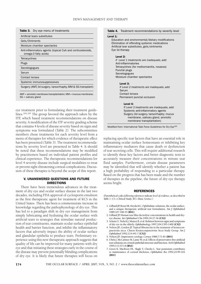
eye treatment prior to formulating their treatment guide-lines.184,185 The group favored the approach taken by the ITF, which based treatment recommendations on disease severity. A modification of the ITF severity grading scheme that contains 4 levels of disease severity based on signs and symptoms was formulated (Table 2). The subcommittee members chose treatments for each severity level from a menu of therapies for which evidence of therapeutic effect has been presented (Table 3). The treatment recommenda-tions by severity level are presented in Table 4. It should be noted that these recommendations may be modified by practitioners based on individual patient profiles and clinical experience. The therapeutic recommendations for level 4 severity disease include surgical modalities to treat or prevent sight-threatening corneal complications. Discus-sion of these therapies is beyond the scope of this report.
V. UNANSWERED QUESTIONS AND FUTURE
DIRECTIONS
There have been tremendous advances in the treat-ment of dry eye and ocular surface disease in the last two decades, including FDA approval of cyclosporin emulsion as the first therapeutic agent for treatment of KCS in the United States. There has been a commensurate increase in knowledge regarding the pathophysiology of dry eye. This has led to a paradigm shift in dry eye management from simply lubricating and hydrating the ocular surface with artificial tears to strategies that stimulate natural produc-tion of tear constituents, maintain ocular surface epithelial health and barrier function, and inhibit the inflammatory factors that adversely impact the ability of ocular surface and glandular epithelia to produce tears. Preliminary ex-perience using this new therapeutic approach suggests that quality of life can be improved for many patients with dry eye and that initiating these strategies early in the course of the disease may prevent potentially blinding complications of dry eye. It is likely that future therapies will focus on
replacing specific tear factors that have an essential role in maintaining ocular surface homeostasis or inhibiting key inflammatory mediators that cause death or dysfunction of tear secreting cells. This will require additional research to identify these key factors and better diagnostic tests to accurately measure their concentrations in minute tear fluid samples. Furthermore, certain disease parameters may be identified that will identify whether a patient has a high probability of responding to a particular therapy. Based on the progress that has been made and the number of therapies in the pipeline, the future of dry eye therapy seems bright.
REFERENCES
(Parenthetical codes following references indicate level of evidence, as described in T able 1 . C S = C linical S tu dy; B S = B asic S cience.)
1 . G ilb ard JP, Rossi SR, Hey d a K G . Op hthalmic solutions, the ocular surface, and a uniq ue therap eutic artifi cial tear formulation. A m J O p hthalm ol1 9 8 9 ;1 07:3 4 8 -55 (BS1)
2. G ilb ard JP. Human tear fi lm electroly te concentrations in health and d ry -ey e d isease. Int O p hthalm ol C lin 1 9 9 4 ;3 4 :27-3 6 (CS2)
3 . Schein O, Tielsch J, M unoz B , et al. Relation b etween sig ns and sy mp toms of d ry ey e in the eld erly. O p hthalm ology 1 9 9 7;1 04 :1 3 9 5-1 4 00 (CS2)
4 . Nelson JD, G ord on JF. Top ical fi b ronectin in the treatment of k eratocon-junctiv itis sicca. Chiron K eratoconjunctiv itis Sicca Stud y G roup . A m J O p hthalm ol 1 9 9 2;1 1 4 :4 4 1 -7 (CS2)
5. Nelson JD. Imp ression cy tolog y. C ornea 1 9 8 8 ;7:71 -8 1 (BS1)6 . Ub els J, M cCartney M , Lantz W , et al. Effects of p reserv ativ e-free artifi cial
tear solutions on corneal ep ithelial structure and function. A rch O p hthalm ol1 9 9 5;1 1 3 :3 71 -8 (BS1)
7. G reen K , M acK een DL, Slag le T, Cheek s L. Tear p otassium contrib utes to maintenance of corneal thick ness. O p hthalm ic R es 1 9 9 2;24 :9 9 -1 02 (BS1)
Table 4. Treatment recommendations by severity level
Level 1:
Education and environmental/dietary modifications
Elimination of offending systemic medications
Artificial tear substitutes, gels/ointments
Eye lid therapy
Level 2:
If Level 1 treatments are inadequate, add:
Anti-inflammatories
Tetracyclines (for meibomianitis, rosacea)
Punctal plugs
Secretogogues
Moisture chamber spectacles
Level 3:
If Level 2 treatments are inadequate, add:
Serum
Contact lenses
Permanent punctal occlusion
Level 4:
If Level 3 treatments are inadequate, add:
Systemic anti-inflammatory agents
Surgery (lid surgery, tarsorrhaphy; mucus
membrane, salivary gland, amniotic
membrane transplantation)
Modified from: International Task Force Guidelines for Dry Eye185
Table 3. Dry eye menu of treatments
Artificial tears substitutes
Gels/Ointments
Moisture chamber spectacles
Anti-inflammatory agents (topical CsA and corticosteroids,
omega-3 fatty acids)
Tetracyclines
Plugs
Secretogogues
Serum
Contact lenses
Systemic immunosuppressives
Surgery (AMT, lid surgery. tarsorrhaphy, MM & SG transplant)
AMT = amniotic membrane transplantation; MM = mucous membrane;
SG = salivary gland
DEWS MANAGEMENT AND THERAPY
THE OCULAR SURFACE / APRIL 2007, VOL. 5, NO. 2 / www.theocularsurface.com 175
8. Holly F, Lemp M. Surface chemistry of the tear film: Implications for dry eye syndromes, contact lenses, and ophthalmic polymers. Contact Lens Soc Am J 1971;5:12-9 (BS2)
9. Perrigan DM, Morgan A, Q uintero S, et al. Comparison of osmolarity values of selected ocular lubricants. ARVO 2004 poster session 449
10. Kaufman B, Novack GD. Compliance issues in manufacturing of drugs. Ocul Surf 2003;1:80-5
11. Albietz J, Bruce A. The conjunctival epithelium in dry eye subtypes: Effect of preserved and nonpreserved topical treatments. Curr E ye Res2001;22:8-18 (CS2)
12. Gasset AR, Ishii Y , Kaufman H, Miller T. Cytotox icity of ophthalmic preservatives. Am J Ophthalmol 1974;78:98-105 (BS1)
13. Wilson F. Adverse ex ternal effects of topical ophthalmic medications. Surv Ophthalmol 1979;24:57-88 (CS3)
14. Burstein N. Corneal cytotox icity of topically applied drugs, vehicles and preservatives. Surv Ophthalmol 1980;25:15-30 (CS3)
15. Burstein N. The effects of topical drugs and preservatives on the tears and corneal epithelium in dry eye. Trans Ophthalmol Soc U K. 1985;104:402-9 (CS3)
16. Brubaker R, McLaren J. Uses of the fl uorophotometer in glaucoma research. Ophthalmology 1985;92:884-90 (BS1)
17. Smith L, George M, Berdy G, Abelson M. Comparative effects of pre-servative free tear substitutes on the rabbit cornea: a scanning electron microscopic evaluation (ARVO abstract) . Invest Ophthalmol V is Sci 1991;32 (Suppl) :733 (BS1)
18. Gilbard JP, Farris RL, Santamaria J 2nd. Osmolarity of tear microvolumes in keratoconjunctivitis sicca. Arch Ophthalmol 1978;96:677-81 (BS2)
19. Lopez Bernal D, Ubels JL. Q uantitative evaluation of the corneal epithelial barrier: effect of artificial tears and preservatives. Curr E ye Res 1991;10:645-56 (BS1)
20. Bernal DL, Ubels JL. Artificial tear composition and promotion of recovery of the damaged corneal epithelium. Cornea 1993;12:115-20 (BS1)
21. Noecker R: Effects of common ophthalmic preservatives on ocular health. Adv Ther 2001;18:205-15 (CS1)
22. Tripathi BJ, Tripathi RC, Kolli SP: Cytotox icity of ophthalmic preserva-tives on human corneal epithelium. Lens E ye Tox icity Res 1992;9:361-75 (BS1)
23. Herrema J, Friedenwald J. Retardation of wound healing in the corneal epithelium by lanolin. Am J Ophthalmol 1950;33:1421 (CS3)
24. Nelson J, Drake M, Brewer J, Tuley M. Evaluation of physiologic tear substitute in patients with keratoconjunctivitis sicca. Adv E x p M ed Biol 1994;350:453-7 (CS2)
25. Gilbard JP, Rossi SR. An electrolyte-based solution that increases corneal glycogen and conjunctival goblet-cell density in a rabbit model for kera-toconjunctivitis sicca. Ophthalmology 1992;99:600-4 (BS1)
26. Lenton LM, Albietz JM: Effect of carmellose-based artificial tears on the ocular surface in eyes after laser in situ keratomileusis. J Refract Surg 1999;15(2 Suppl) :S227-S231 (CS2)
27. Slomiany BL, Slomiany A. Role of mucus in gastric mucosal protection. J Physiol Pharmacol 1991; 42:147-61 (BS1)
28. Gilbard JP. Tear film osmolarity and keratoconjunctivitis sicca. CLAO J 1985;11:243-50 (CS1)
29. Gilbard J. Tear film osmolarity and keratoconjuncitivitis sicca. Lubbock TX,Dry Eye Institute, 1986 (CS3)
30. Gilbard J, Carter J, Sang D, et al. Morphologic effect of hyperosmolarity on rabbit corneal epithelium. Ophthalmology 1984;91:1205-12 (BS1)
31. Luo L, Li D, Corrales R, Pfl ugfelder S. Hyperosmolar saline is a proinfl am-matory stress on the mouse ocular surface. E ye Contact Lens 2005;31:186-93 (BS1)
32. Gilbard JP, Kenyon KR. Tear diluents in the treatment of keratoconjunc-tivitis sicca. Ophthalmology 1985;92:646-50 (CS2)
33. Holly F, Esquivel E. Colloid osmotic pressure of artificial tears. J Ocul Pharmacol 1985;1:327-36 (BS1)
34. Y ancey PH: Organic osmolytes as compatible, metabolic and counter-acting cryoprotectants in high osmolarity and other stresses .J E x p Biol 2005;208:2819-30 (BS2)
35. Holly F, Lemp M. Wettability and wetting of corneal epithelium. E x p E ye Res 1971;11:239-50 (BS1)
36. Hawi A, Smith T, Digenis G. A quantitiative comparison of artificial tear clearance rates in humans using gamma scintigraphy (ARVO abstract) . Invest Ophthalmol V is Sci 1990;31 (Suppl) :517 (BS1)
37. Argueso P, Tisdale A, Spurr-Michaud S, et al. Mucin characteristics of hu-man corneal-limbal epithelial cells that ex clude the rose bengal anionic dye. Invest Ophthalmol V is Sci 2006;47:113-9 (BS1)
38. Versura P, Maltarello M, Stecher F, et al. Dry eye before and after therapy
with hydrox ypropylmethylcellulose. Ophthalmologica 1989;198:152-62 (CS3)
39. Simmons PA, Garrett Q , Xu S, et al. Interaction of carbox ymethylcellulose with human corneal cells. ARVO 2006, E-Abstract 2759 (BS1)
40. Christiansen M, Cohen S, Rinehart J,et al. Clinical evaluation of an HP-guar gellable lubricant eye drop for the relief of dryness of the eye. Curr E ye Res 2004;28:55-62 (CS2)
41. Di Pascuale MA, Goto E, Tseng SC. Sequential changes of lipid tear film after the instillation of a single drop of a new emulsion eye drop in dry eye patients. Ophthalmology 2004;111:783-91 (CS2)
42. Korb DR, Scaffidi RC, Greiner JV, et al. The effect of two novel lubricant eye drops on tear film lipid layer thickness in subjects with dry eye symptoms. Optom V is Sci 2005;82:594-601 (CS2)
43. Snibson GR, Greaves JL, Soper ND, et al. Precorneal residence times of sodium hyaluronate solutions studied by quantitative gamma scintigraphy. E ye 1990;4:594-602 (CS3)
44. Polack F, McNiece M. The treatment of dry eyes with NA hyaluronate (Healon) . Cornea 1982;1:1333 (CS3)
45. Stuart JC, Linn JG. Dilute sodium hyaluronate (Healon) in the treatment of ocular surface disorders. Ann Ophthalmol 1985;17:190-2 (CS3)
46. DeLuise V, Peterson W. The use of topical Healon tears in the management of refractory dry-eye syndrome. Ann Ophthalmol 1984;16:823-4 (CS3)
47. Sand B, Marner K, Norn M. Sodium hyaluronate in the treatment of keratoconjuctivitis sicca. Acta Ophthalmol 1989; 67:181-3 (CS3)
48. Nelson JD, Farris RL. Sodium hyaluronate and polyvinyl alcohol artificial tear preparations a comparison in patients with keratoconjunctivitis sicca. Arch Ophthalmol 1988;106:484-7 (CS2)
49. Beetham WP. Filamentary keratitis. Trans Am Ophthalmol Soc 1936:33:413-35 (CS1)
50. Foulds WS. Intracanalicular gelatin implants in the treatment of kerato-conjunctivitis sicca. Br J Ophthalmol 1961:45:625-7 (CS2)
51. Freeman JM. The punctum plug: evaluation of a new treatment for the dry eye. Trans Am Acad Ophthalmol Otolaryngol 1975:79:OP874-9 (CS2)
52. Tuberville AW, Frederick WR, Wood TO. Punctal occlusion in tear defi-ciency syndromes. Ophthalmology 1982;89:1170-2 (CS2)
53. Willis RM, Folberg R, Krachmer JH, et al. The treatment of aqueous-de-ficient dry eye with removable punctal plugs. A clinical and impression-cytological study. Ophthalmology 1987;94:514-8 (CS2)
54. Gilbard JP, Rossi SR, Azar DT, Gray KL. Effect of punctal occlusion by Freeman silicone plug insertion on tear osmolarity in dry eye disorders. CLAO J 1989;15:216-8 (CS2)
55. Balaram M, Schaumberg DA, Dana MR. Efficacy and tolerability outcomes after punctal occlusion with silicone plugs in dry eye syndrome. Am J Ophthalmol 2001;131:30-6 (CS1)
56. Bax ter SA, Laibson PR. Punctal plugs in the management of dry eyes Ocul Surf 2004;2:255-65 (CS3)
57. Bartlett JD, Boan K, Corliss D, Gaddie IB. Efficacy of silicone punctal plugs as adjuncts to topical pharmacotherapy of glaucoma--a pilot study. Punctal Plugs in Glaucoma Study Group. J Am Optom Assoc 1996;67:664-8 (CS2)
58. Huang TC, Lee DA. Punctal occlusion and topical medications for glau-coma. Am J Ophthalmol 1989;107:151-5 (CS2)
59. Sugita J, Y okoi N, Fullwood NJ, et al. The detection of bacteria and bacte-rial biofilms in punctal plug holes. Cornea 2001;20: 362-5 (CS3)
60. Gerding H, Kuppers J, Busse H. Symptomatic cicatrizial occlusion of canaliculi after insertion of Herrick lacrimal plugs. Am J Ophthalmol2003;136:926-8 (CS3)
61. Lee J, Flanagan JC. Complications associated with silicone intracanalicular plugs. Ophthal Plast Reconstr Surg 2001;17:465-9 (CS3)
62. Paulsen F. The human lacrimal glands. Adv Anat E mbryol Cell Biol2003;170:III-XI,1-106 (BS1)
63. Y en MT, Pfl ugfelder SC, Feuer WJ. The effect of punctal occlusion on tear production, tear clearance, and ocular surface sensation in normal subjects. Am J Ophthalmol 2001;131:314-23 (CS2)
64. Tsubota K. The effect of wearing spectacles on the humidity of the eye. Am J Ophthalmol 1989;15;108:92-3 (BS2)
65. Tsubota K, Y amada M, Urayama K. Spectacle side panels and moist inserts for the treatment of dry-eye patients. Cornea 1994;13:197-201 (BS1)
66. Gresset J, Simonet P, Gordon D. Combination of a side shield with an ocular moisture chamber. Am J Optom Physiol Opt 1984;61:610-2 (CS3)
67. Savar DE. A new approach to ocular moisture chambers. J Pediatr Oph-thalmol Strabismus 1978;15:51-3 (CS3)
68. Kurihashi K. Moisture aid during sleep for the treatment of dry eye: wet gauze eye mask. Ophthalmologica 1994;208:216-9 (CS3)
69. Nichols JJ, Z iegler C, Mitchell GL, Nichols KK. Self-reported dry eye dis-
DEWS MANAGEMENT AND THERAPY
THE OCULAR SURFACE / APRIL 2007, VOL. 5, NO. 2 / www.theocularsurface.com176
ease across refractive modalities. Invest Ophthalmol Vis Sci 2005;46:1911-4 (CS2)
70. Korb DR, Greiner JV, Glonek T, et al. Effect of periocular humidity on the tear film lipid layer. Cornea 1996;15:129-34 (BS2)
71. Tsubota K, Hata S, Okusawa Y, et al. Quantitative videographic analysis of blinking in normal subjects and patients with dry eye. Arch Ophthalmol1996;114:715-20 (BS1)
72. Maruyama K, Yokoi N, Takamata A, Kinoshita S. Effect of environmental conditions on tear dynamics in soft contact lens wearers. Invest Ophthalmol Vis Sci 2004;45:2563-8 (BS1)
73. Bacon AS, Astin C, Dart JK. Silicone rubber contact lenses for the com-promised cornea. Cornea 1994;13:422-8 (CS3)
74. Pullum KW, Whiting MA, Buckley RJ. Scleral contact lenses: the expanding role. Cornea 2005;24:269-77 (CS3)
75. Tappin MJ, Pullum KW, Buckley RJ. Scleral contact lenses for overnight wear in the management of ocular surface disorders. Eye 2001;15(Pt2):168-72 (CS3)
76. Romero-Rangel T, Stavrou P, Cotter J, et al. Gas-permeable scleral contact lens therapy in ocular surface disease. Am J Ophthalmol 2000;130:25-32 (CS3)
77. Rosenthal P, Cotter JM, Baum J. Treatment of persistent corneal epithelial defect with extended wear of a fluid-ventilated gas-permeable scleral contact lens. Am J Ophthalmol 2000;130:33-41 (CS3)
78. Tauber J, Davitt WF, Bokosky JE, et al. Double-masked, placebo-controlled safety and efficacy trial of diquafosol tetrasodium (INS365) ophthalmic solution for the treatment of dry eye. Cornea 2004;23:784-92 (CS1)
79. Mundasad MV, Novack GD, Allgood VE, et al. Ocular safety of INS365 ophthalmic solution: a P2Y(2) agonist in healthy subjects. J Ocul PharmacolT her 2001;17:173-9 (CS1)
80. Murakami T, Fujihara T, Horibe Y, Nakamura M. Diquafosol elicits in-creases in net Cl- transport through P2Y2 receptor stimulation in rabbit conjunctiva. Ophthalmic Res 2004;36:89-93 (BS1)
81. Li DQ, Lokeshwar BL, Solomon A, et al. Regulation of MMP-9 in human corneal epithelial cells. Exp Eye Res 2001;73:449-59 (BS1)
82. Murakami T, Fujita H, Fujihara T, et al. Novel noninvasive sensitive determination of tear volume changes in normal cats. Ophthalmic Res2002;34:371-4 (BS1)
83. Yerxa BR, Mundasad M, Sylvester RN, et al. Ocular safety of INS365 ophthalmic solution, a P2Y2 agonist, in patients with mild to moderate dry eye disease. Adv Exp Med Biol 2002;506(Pt B):1251-7 (BS2)
84. Fujihara T, Murakami T, Fujita H, et al. Improvement of corneal barrier function by the P2Y(2) agonist INS365 in a rat dry eye model. Invest Ophthalmol Vis Sci 2001;42:96-100 (BS1)
85. Fujihara T, Murakami T, Nagano T, et al. INS365 suppresses loss of corneal epithelial integrity by secretion of mucin-like glycoprotein in a rabbit short-term dry eye model. J Ocul Pharmacol Ther 2002;18:363-70 (BS1)
86. Yerxa BR, Douglass JG, Elena PP, et al. Potency and duration of action of synthetic P2Y2 receptor agonists on Schirmer scores in rabbits Adv Exp Med Biol 2002;506(Pt A):261-5 (BS2)
87. Urashima H, Okamoto T, Takeji Y, et al. Rebamipide increases the amount of mucin-like substances on the conjunctiva and cornea in the N-acetyl-cysteine-treated in vivo model. Cornea 2004;23:613-9 (BS1)
88. Tanito M, Takanashi T, Kaidzu S, et al. Cytoprotective effects of rebamipide and carteolol hydrochloride against ultraviolet B-induced corneal damage in mice. Invest Ophthalmol Vis Sci 2003; 44:2980-5 (BS3)
89. Masuda K, Tokushige H, Ogawa T, et al. Effect of topical ecabet sodium on mucin levels in the tear fluid of patients with dry eye. SERI-ARVO2003.
90. Toshida H, Nakata K, Hamano T, et al. Effect of gefarnate on the ocular surface in squirrel monkeys Cornea 2002;21:292-9 (BS3)
91. Nakamura M, Endo K, Nakata K, Hamano T. Gefarnate increases PASpositive cell density in rabbit conjunctiva. Br J Ophthalmol 1998;82:1320-3 (BS3)
92. Nakamura M, Endo K, Nakata K, Hamano T. Gefarnate stimulates secre-tion of mucin-like glycoproteins by corneal epithelium in vitro and protects corneal epithelium from desiccation in vivo. Exp Eye Res 1997;65:569-74 (BS3)
93. Toshida H, Nakata K, Hamano T, et al. Gefarnate stimulates goblet cell repopulation following an experimental wound to the tarsal conjunctiva in the dry eye rabbit. Adv Exp Med Biol 2002;506(Pt A):353-7 (BS 3)
94. Hamano T. Dry eye treatment with eye drops that stimulate mucin produc-tion. A dv E x p M ed B iol 1998;438:965-8 (CS3)
95. Jumblatt JE, Cunningham L, Jumblatt MM. Effects of 15(S)-HETE on human conjunctival mucin secretion. Adv Exp Med Biol 2002;506(PtA):323-7 (BS1)
96. Gamache DA, Wei ZY, Weimer LK, et al. Corneal protection by the ocular mucin secretagogue 15(S)-HETE in a rabbit model of desiccation-induced corneal defect. J Ocul Pharmacol Ther 2002;18:349-61 (BS2)
97. Jackson RS 2nd, Van Dyken SJ, McCartney MD, Ubels JL. The eicosanoid, 15-(S)-HETE, stimulates secretion of mucin-like glycoprotein by the corneal epithelium Cornea 2001;20:516-21 (BS2)
98. Azar RG, Edelhauser HF. Evaluation of the effects of 15(S)-HETE on corneal epithelial cells: an electrophysiological and cytochemical study. Adv Exp Med Biol 2002; 506(Pt A):329-33 (BS3)
99. Ubels JL, Aupperlee MD, Jackson RS 2nd, et al. Topically applied 15-(S)-HETE stimulates mucin production by corneal epithelium. Adv Exp Med Biol 2002;506(Pt A):317-21 (BS2)
100. Gamache DA, Wei ZY, Weimer LK, et al. Preservation of corneal integrity by the mucin secretagogue 15(S)-HETE in a rabbit model of desiccation-induced dry eye. Adv Exp Med Biol 2002;506(Pt A):335-40 (BS2)
101. Jumblatt JE, Cunningham LT, Li Y, Jumblatt MM. Characterization of human ocular mucin secretion mediated by 15(S)-HETE. Cornea2002;21:818-24 (BS3)
102. Vivino FB, Al-Hashimi I, Khan K, et al. Pilocarpine tablets for the treatment of dry mouth and dry eye symptoms in patients with Sjogren’s syndrome. Arch Intern Med 1999;159:174-81 (CS1)
103. Takaya M, Ichikawa Y, Yamada C, et al. Treatment with pilocarpine hydrochloride for sicca symptoms in Sjogren’s syndrome. Ryumachi1997;37:453-7 (CS2)
104. Tsifetaki N, Kitsos G, Paschides CA, et al. Oral pilocarpine for the treatment of ocular symptoms in patients with Sjogren’s syndrome: a randomised 12-week controlled study. Ann Rheum D is 2003;62:1204-7 (CS2)
105. Papas AS, Sherrer YS, Charney M, et al. Successful treatment of dry mouth and dry eye symptoms in Sjogren’s syndrome patients with oral pilocarpine: A randomized, placebo-controlled, dose-adjustment study. J Clin Rheumatol 2004;4:169-77 (CS1)
106. Aragona P, Di Pietro R, Spinella R, Mobrici M. Conjunctival epithelium improvement after systemic pilocarpine in patients with Sjogren’s syn-drome. Br J Ophthalmol 2006;90:166-70 (CS2)
107. Petrone D, Condemi JJ, Fife R, et al. Double-blind randomized placebo-controlled study of cevimeline in Sjogren’s syndrome patients with xerosto-mia and keratoconjunctivitis sicca. Arthritis Rheum 2002;46:748-54 (CS1)
108. Ono M, Takamura E, Shinozaki K, et al. Therapeutic effect of cevimeline on dry eye in patients with Sjogren’s syndrome: a randomized, double-blind clinical study. Am J Ophthalmol 2004;138:6-17 (CS1)
109. Geerling G, Daniels JT, Dart JK, et al. Toxicity of natural tear substitutes in a fully defined culture model of human corneal epithelial cells. Invest Ophthalmol Vis Sci 2001;42948-56 (BS1)
110. Geerling G, Honnicke K, Schroder C, et al. Quality of salivary tears fol-lowing autologous submandibular gland transplantation for severe dry eye. G raefes Arch Clin Exp Ophthalmol 2000;238:45-52 (BS1)
111. Tsubota K, Goto E, Fujita H, et al. Treatment of dry eye by autologous serum application in Sjogren’s syndrome. Br J Ophthalmol 1999;83:390-5 (CS2)
112. Geerling G, Hartwig D. Autologous serum eyedrops for ocular surface disorders, in Reinhard T, Larkin F (eds). Cornea and external eye disease. Berlin, Heidelberg, Springer, 2005, pp 2-19
113. Liu L, Hartwig D, Harloff S, et al. An optimised protocol for the produc-tion of autologous serum eyedrops. G raefes Arch Clin Exp Ophthalmol2005;243:706-14 (BS1)
114. Tananuvat, N, Daniell M, Sullivan LJ, et al. Controlled study of the use of autologous serum in dry eye patients. Cornea 2001;20:802-6 (CS1)
115. Kojima T, Ishida R, Dogru M, et al. The effect of autologous serum eye-drops in the treatment of severe dry eye disease: a prospective randomized case-control study. Am J Ophthalmol 2005;139:242-6 (CS1)
116. Noble BA, Loh RS, MacLennan S, et al. Comparison of autologous serum eye drops with conventional therapy in a randomised controlled crossover trial for ocular surface disease. Br J Ophthalmol 2004;88:647-52 (CS1)
117. Noda-Tsuruya T, Asano-Kato N, Toda I, Tsubota K. Autologous serum eye drops for dry eye after LASIK. J Refract Surg 2006;22:61-6 (CS1)
118. Schulze SD, Sekundo W,Kroll P.Autologous serum for the treatment of corneal epithelial abrasions in diabetic patients undergoing vitrectomy. Am J Ophthalmol 2006;142:207-11 (BS1)
119. Geerling G, Sieg P, Bastian GO, Laqua H. Transplantation of the autologous submandibular gland for most severe cases of keratoconjunctivitis sicca. Ophthalmology 1998;105:327-35 (CS2)
120. Schroder, Sieg P, Framme C, et al. [Transplantation of the submandibular gland in absolute dry eyes. Effect on the ocular surface] . Klin Monatsbl Augenheilk d 2002;219:494-501(CS2)
121. Luo L, Li DQ, Doshi A, et al. Experimental dry eye stimulates production
DEWS MANAGEMENT AND THERAPY

THE OCULAR SURFACE / APRIL 2007, VOL. 5, NO. 2 / www.theocularsurface.com 177
of inflammatory cytokines and MMP-9 and activates MAPK signaling pathways on the ocular surface. Invest Ophthalmol Vis Sci 2004;45:4293-301 (BS1)
122. Niederkorn JY, Stern ME, Pflugfelder SC, et al. Desiccating stress induces T cell-mediated Sjogren‘s syndrome-like lacrimal keratoconjunctivitis. J Immunol 2006;176:3950-7 (BS1)
123. Kaswan RL, Salisbury MA, Ward DA. Spontaneous canine keratoconjuncti-vitis sicca. A useful model for human keratoconjunctivitis sicca: treatment with cyclosporine eye drops. Arch Ophthalmol 1989;107:1210-16 (BS2)
124. Gunduz K, Ozdemir O. Topical cyclosporin treatment of keratoconjuncti-vitis sicca in secondary Sjogren’s syndrome. Acta Ophthalmol 1994;72:38-42 (CS2)
125. Laibovitz RA, Solch S, Andrianao J. Pilot trial of cyclosporin 1% oph-thalmic ointment in the treatment of keratoconjunctivitis sicca. Cornea1993;12:315-23 (CS1)
126. Stevenson D, Tauber J, Reis BL. Efficacy and safety of cyclosporin A oph-thalmic emulsion in the treatment of moderate-to-severe dry eye disease. Adose-ranging, randomized trial. Ophthalmology 2000;107:967-74 (CS1)
127. Sall K, Stevenson OD, Mundorf TK, Reis BL. Two multicenter, randomized studies of the efficacy and safety of cyclosporine ophthalmic emulsion in moderate to severe dry eye disease. Ophthalmology 2000;107:631-9 (CS1)
128. Kunert KS, Tisdale AS, Gipson IK. Goblet cell numbers and epithelial proliferation in the conjunctiva of patients with dry eye syndrome treated with cyclosporine. Arch Ophthalmol 2002;120:330-7 (BS1)
129. Brignole F, Pisella PJ, De Saint Jean M, et al. Flow cytometric analysis of inflammatory markers in KCS: 6-month treatment with topical cyclosporin A. Invest Ophthalmol Vis Sci 2001;42:90-5 (BS1)
130. Turner K, Pflugfelder SC, Ji Z, et al. Interleukin-6 levels in the conjuncti-val epithelium of patients with dry eye disease treated with cyclosporine ophthalmic emulsion. Cornea 2000;19:492-6 (BS1)
131. Kunert KS, Tisdale AS, Stern ME, Smith JA. Analysis of topical cyclospo-rine treatment of patients with dry eye syndrome: effect on conjunctival lymphocytes. Arch Ophthalmol 2000;118:1489-96 (BS1)
132. Pflugfelder SC, Maskin SL, Anderson B, et al. A randomized, double-masked, placebo-controlled, multicenter comparison of loteprednol etabonate ophthalmic suspension, 0.5% , and placebo for treatment of keratoconjunctivitis sicca in patients with delayed tear clearance. Am J Ophthalmol 2004;138:444-57 (CS1)
133. Avunduk AM, Avunduk MC, Varnell ED, Kaufman HE. The comparison of efficacies of topical corticosteroids and nonsteroidal antiinflammatory drops on dry eye patients: A clinical and immunocytochemical study. Am J Ophthalmol 2003;136:593-602 (CS1)
134. Sainz de la Maza Serra SM, Simon Castellvi C, Kabbani O. Nonpreserved topical steroids and punctal occlusion for severe keratoconjunctivitis sicca. Arch Soc Esp Oftalmol 2000;75:751-56 (CS1)
135. Marsh P, Pflugfelder SC. Topical nonpreserved methylprednisolone therapy for keratoconjunctivitis sicca in Sjogren syndrome. Ophthalmology1999;106:811-6 (CS3)
136. Pflugfelder SC. Antiinflammatory therapy for dry eye. Am J Ophthalmol2004;137:337-42 (CS3)
137. Anonymous. Certain ophthalmic antibiotic combination drugs for human use; Drug efficacy study implementation. F ed Reg 1982;47:21296
138. Anonymous. Certain steroid preparations for ophthalmic and/or otic use. F ed Reg 1980a;45:57776-80
139. Anonymous. Certain ophthalmic antibiotic combination drugs for human use; Drug efficacy study implementation. F ed Reg 1980b;45:57780-3
140. Anonymous. Certain steroid preparations for ophthalmic or otic use. F ed Reg 1976;41:34340-2
141. De Paiva CS, Corrales RM, Villarreal AL, et al. Apical corneal barrier disrup-tion in experimental murine dry eye is abrogated by methylprednisolone and doxycycline. Invest Ophthalmol Vis Sci 2006;47:2847-56 (RS1)
142. De Paiva CS, Corrales RM, Villarreal AL, et al. Corticosteroid and doxy-cycline suppress MMP-9 and inflammatory cytokine expression, MAPKactivation in the corneal epithelium in experimental dry eye. Exp Eye Res2006;83:526-35 (RS1)
143. Schiffman RM, Bradford R, Bunnell B, et al. A multicenter, double-masked, randomized, vehicle-controlled, parallel-group study to evaluate the safety and efficacy of testosterone ophthalmic solution in patients with meibomian gland dysfunction. ARVO 2006, E-Abstract 5608 (CS3)
144. Worda C, Nepp J, Huber JC, Sator MO. Treatment of keratoconjunctivitis sicca with topical androgen. Maturitas 2001;37:209-12 (CS3)
145. Sator MO, Joura EA, Golaszewski T, et al. Treatment of menopausal keratoconjunctivitis sicca with topical oestradiol. Br J Obstet Gynaecol1998;105:100-2 (CS2)
146. Shine WE, McCulley JP, Pandya AG. Minocycline effect on meibomian gland lipids in meibomianitis patients. Exp Eye Res 2003;76:417– 20 (CS2)
147. Dougherty JM, McCulley JP, Silvany RE, et al. The role of tetracycline in chronic blepharitis. Invest Ophthalmol Vis Sci 1991;32:2970– 5 (CS2)
148. Ta CN, Shine WE, McCulley JP, et al. Effects of minocycline on the ocu-lar flora of patients with acne rosacea or seborrheic blepharitis. Cornea2003;22:545– 8 (CS2)
149. Solomon A, Rosenblatt M, Li DQ, et al. Doxycycline inhibition of interleu-kin-1 in the corneal epithelium. Invest Ophthalmol Vis Sci 2000;41:2544-57 (CS2)
150. Li Y, Kuang K, Yerxa B, et al. Rabbit conjunctival epithelium transports fluid, and P2Y2(2) receptor agonists stimulate Cl(-) and fluid secretion. Am J Physiol Cell Physiol 2001;281:C595-602 (BS1)
151. Li DQ, Luo L, Chen Z, et al. JNK and ERK MAP kinases mediate induc-tion of IL-1beta, TNF-alpha and IL-8 following hyperosmolar stress in human limbal epithelial cells. Exp Eye Res 2006;82:588-96. Epub 2005 Oct 3 (BS1)
152. Krakauer T, Buckley M. Doxycycline is anti-inflammatory and inhibits staphylococcal exotoxin-induced cytokines and chemokines. Antimicrob Agents Chemother 2003;47:3630-3 (BS1)
153. Voils SA, Evans ME, Lane MT, et al. Use of macrolides and tetracyclines for chronic inflammatory diseases. Ann Pharmacother 2005;39:86-94 (CS3)
154. Tamargo RJ, Bok RA, Brem H. Angiogenesis inhibition by minocycline. Cancer Res 1991;51:672-5 (BS1)
155. Sapadin AN, Fleischmajer R. Tetracyclines: Nonantibiotic properties and their clinical implications J Am Acad D ermatol 2006;54:258-65 (CS3)
156. Habif TP. Clinical dermatology, 4th ed. St Louis:Mosby-Year Book, 2004, pp 162-89 (CS3)
157. Velicer CM, Heckbert SR, Lampe JW, et al. Antibiotic use in relation to the risk of breast cancer. JAMA 2004;291:827– 35
158. Velicer CM, Heckbert SR, Rutter C, et al. Association between antibiotic use prior to breast cancer diagnosis and breast tumour characteristics (United States). Cancer Causes Control (N etherlands) 2006;17:307-13
159. Garcıa Rodrıguez LA , Gonzalez-Perez A. Use of antibiotics and risk of breast cancer. Am J Epidemiology 2005;161:616-9
160. Macdonald A, Feiwel M. Perioral dermatitis: aetiology and treatment with tetracycline. Br J D ermatol 1972;87:315-9 (CS3)
161. Jansen T, Plewig G. Rosacea: classification and treatment. J R Soc Med1997;90:144-50 (CS3)
162. Frucht-Pery J, Chayet AS, Feldman ST, et al. The effect of doxycycline on ocular rosacea. Am J Ophthalmol 1989;107:434-5 (CS2)
163. Wilkin JK. Rosacea. pathophysiology and treatment. Arch Dermatol1994;130:359-62 (BS1)
164. Stone DU, Chodosh J. Oral tetracyclines for ocular rosacea: an evidence-based review of the literature. Cornea 2004;23:106-9 (CS1)
165. McCulley JP, Dougherty JM, Deneau DG. Classification of chronic blepha-ritis. Ophthalmology 1982;89:1173-80 (CS2)
166. Frucht-Pery J, Sagi E, Hemo I, Ever-Hadani P. Efficacy of doxycycline and tetracycline in ocular rosacea. Am J Ophthalmol 1993;116:88-92 (CS1)
167. Seal DV, Wright P, Picker L, et al. Placebo controlled trial of fusidic acid gel and oxytetracycline for recurrent blepharitis and rosacea. Br J Ophthalmol1995;79:42-5 (CS1)
168. Hoeprich PD, Warshauer DM. Entry of four tetracyclines into saliva and tears. Antimicob Agents Chemother 1974;3:330– 6 (BS1)
169. Gulbenkian A, Myers J, Freis D. Hamster flank organ hydrolase and lipase activity. J Invest Dermatol 1980;75:289– 92 (BS1)
170. Aronowicz JD, Shine WE, Oral D, et al. Short term oral minocycline treatment of meibomianitis. Br J Ophthalmol 2006;90:856-60 (CS2)
171. Yoo SE, Lee DC, Chang MH. The effect of low-dose doxycycline therapy in chronic meibomian gland dysfunction. Korean J Ophthalmol 2005;19:258-63 (CS2)
172. Browning DJ, Proia AD. Ocular rosacea. Surv Ophthalmol 1986;31:145– 58 (CS3)
173. Esterly NB, Koransky JS, Furey NL, et al. Neutrophil chemotaxis in patients with acne receiving oral tetracycline therapy. Arch Dermatol1984;120:1308-13 (BS1)
174. Gilbard JP. The scientific context and basis of the pharmacologic manage-ment of dry eyes. Ophthalmol Clin N orth Am 2005;18:475-84,v (CS3)
175. James MJ, Gibson RA, Cleland LG. Dietary polyunsaturated fatty acids and inflammatory mediator production. Am J Clin N utr 2000;71(1 Suppl):343S-8S (BS2)
176. Endres S, Ghorbani R, Kelley VE, et al. The effect of dietary supplemen-tation with n-3 polyunsaturated fatty acids on the synthesis of inter-leukin-1 and tumor necrosis factor by mononuclear cells. N Engl J Med
DEWS MANAGEMENT AND THERAPY
THE OCULAR SURFACE / APRIL 2007, VOL. 5, NO. 2 / www.theocularsurface.com178
1989;320:265-71 (BS1)177. James MJ, Cleland LG. Dietary n-3 fatty acids and therapy for rheumatoid
arthritis. Semin Arthritis Rheum 1997;27:85-97 (CS3)178. Kremer JM. n-3 fatty acid supplements in rheumatoid arthritis. Am J Clin
Nutr 2000;71(1 Suppl):349S-51S (CS1)179. Barabino S, Rolando M, Camicione P, et al. Systemic linoleic and gamma-
linolenic acid therapy in dry eye syndrome with an inflammatory com-ponent. Cornea 2003;22:97-101 (CS2)
180. Seedor JA, Lamberts D, Bergmann RB, Perry HD. Filamentary keratitis associated with diphenhydramine hydrochloride (Benadryl). Am J Oph-thalmol 1986;101:376-7 (CS3)
181. Moss SE, Klein R, Klein BE. Prevalence of and risk factors for dry eye syndrome. Arch Ophthalmol 2000;118:1264-68
182. Mader TH, Stulting RD. Keratoconjunctivitis sicca caused by diphenox-ylate hydrochloride with atropine sulfate (Lomotil). Am J Ophthalmol 1991;111:377-8 (CS2)
183. Tsubota K, Nakamori K. Dry eyes and video display terminals. N Engl J Med 1993;25;328:584 (CS2)
184. Matoba AY, Harris DJ, Mark DB, et al. Dry eye syndrome, American Academy of Ophthalmology, 2003
185. Behrens A, Doyle JJ, Stern L, et al. Dysfunctional tear syndrome: A Delphi approach to treatment recommendations. Cornea 2006;25:900-7
DEWS MANAGEMENT AND THERAPY


![Contact Lens and Anterior Eye · [32], the Symptom Assessment in Dry Eye (SANDE) [33] and the Dry Eye Questionnaire (DEQ-5; short version) [34]. 2.2.2. Tear film osmolarity Tear](https://static.fdocuments.net/doc/165x107/5feb70c08fd7cf124839e0cf/contact-lens-and-anterior-eye-32-the-symptom-assessment-in-dry-eye-sande-33.jpg)