The Dry Eye Story… Disclosures “A Real Tear Jerker”texas.aoa.org/Documents/TX/2018...
19
2/1/2018 1 The Dry Eye Story… “A Real Tear Jerker” Seema Nanda, OD Clinical Professor: UHCO & Texas Eye Institute Texas Optometry Assoc. Austin, TX 25 th February 2018, Sun. 8 - 10AM Disclosures • University of Houston College of Optometry • nJoy Vision Center • BioTissue , Inc. • OCuSOFT , Inc. • Shire Pharmaceuticals Overview • Dry Eye – Definition & Prevalence – Diagnostic techniques • Rx Treatment – Cyclosporine-A: Restasis – Lifetigrast: Xiidra • Amniotic Membrane – Cryo-preserved 2. Restasis DTC Market Receptivity Final Report, G&S Research Inc., Feb 2004. Restasis Brand Review – February 14, 2005. 3. Simmons PA, et al. ARVO Meeting Program; May 2003. #2448. NEI Definition of Dry Eye • Dry eye is: – Disorder of tear film – Due to tear deficiency or – Excessive evaporation, • Causing damage to the ocular surface and • Associated with symptoms of discomfort. Mild Moderate Severe Severity as Percent of Patients 2 55% 32% 12% Prevalence of Dry Eye • Results from Gallup Poll – Projects an increase in the number of adults who frequently experience Dry eye. – Over 26.4 million Americans Suffer from signs and symptoms of Dry Eyes Visits to Eye Care Professionals *P < 0.05 0% 5% 10% 15% 20% 25% 30% Primary Reason for Visit Secondary Reason for Visit Percentage of Responses 0% 5% 10% 15% 20% 25% 30% Primary Reason for Visit Secondary Reason for Visit Percentage of Responses MD’s OD’s Average N = 635 Average N = 376 PhysPulse ® Study. 2005.
Transcript of The Dry Eye Story… Disclosures “A Real Tear Jerker”texas.aoa.org/Documents/TX/2018...
2/1/2018
1
The Dry Eye Story…
“A Real Tear Jerker”
Seema Nanda, ODClinical Professor: UHCO & Texas Eye Institute
Texas Optometry Assoc. Austin, TX
25th February 2018, Sun. 8-10AM
Disclosures
• University of Houston
College of Optometry
• nJoy Vision Center
• BioTissue, Inc.
• OCuSOFT, Inc.
• Shire Pharmaceuticals
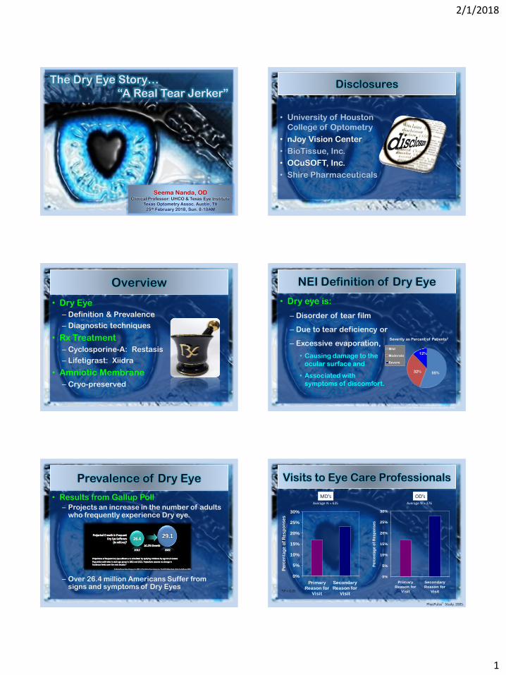
Overview
• Dry Eye
– Definition & Prevalence
– Diagnostic techniques
• Rx Treatment
– Cyclosporine-A: Restasis
– Lifetigrast: Xiidra
• Amniotic Membrane
– Cryo-preserved
1. Lemp MA. CLAO J. 1995;21(4):221-232.2. Restasis DTC Market Receptivity Final Report, G&S Research Inc., Feb 2004. Restasis Brand Review – February 14, 2005.
3. Simmons PA, et al. ARVO Meeting Program; May 2003. #2448.
NEI Definition of Dry Eye
• Dry eye is:
– Disorder of tear film
– Due to tear deficiency or
– Excessive evaporation,
• Causing damage to the
ocular surface and
• Associated with
symptoms of discomfort.
Mild
Moderate
Severe
Severity as Percent of Patients2
55%32%
12%
Prevalence of Dry Eye
• Results from Gallup Poll
– Projects an increase in the number of adults who frequently experience Dry eye.
– Over 26.4 million Americans Suffer from signs and symptoms of Dry Eyes
Visits to Eye Care Professionals
*P < 0.05
0%
5%
10%
15%
20%
25%
30%
Primary
Reason for
Visit
Secondary
Reason for
Visit
Pe
rce
nta
ge
of
Re
sp
on
se
s
0%
5%
10%
15%
20%
25%
30%
Primary
Reason for
Visit
Secondary
Reason for
Visit
Perc
en
tag
e o
f R
esp
on
ses
MD’s OD’sAverage N = 635 Average N = 376
PhysPulse® Study. 2005.
2/1/2018
2
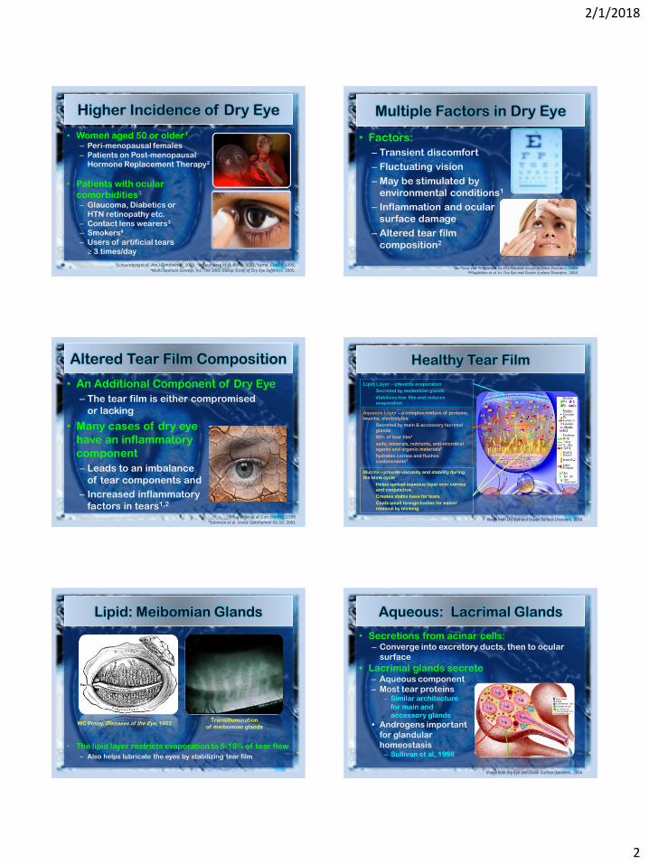
Higher Incidence of Dry Eye
• Women aged 50 or older1
– Peri-menopausal females
– Patients on Post-menopausal
Hormone Replacement Therapy2
• Patients with ocular
comorbidities3
– Glaucoma, Diabetics or
HTN retinopathy etc.
– Contact lens wearers3
– Smokers4
– Users of artificial tears
≥ 3 times/day
1Schaumberg et al. Am J Ophthalmol. 2003; 2Schaumberg et al. JAMA. 2001;3Lemp. CLAO J. 1995;4Multi-Sponsor Surveys, Inc. The 2005 Gallup Study of Dry Eye Sufferers. 2005.
Multiple Factors in Dry Eye
• Factors:
– Transient discomfort
– Fluctuating vision
– May be stimulated by
environmental conditions1
– Inflammation and ocular
surface damage
– Altered tear film
composition2
1de Paiva and Pflugfelder. In: Dry Eye and Ocular Surface Disorders. 2004;2Pflugfelder et al. In: Dry Eye and Ocular Surface Disorders. 2004.
Altered Tear Film Composition
• An Additional Component of Dry Eye
– The tear film is either compromised
or lacking
• Many cases of dry eye
have an inflammatory
component
– Leads to an imbalance
of tear components and
– Increased inflammatory
factors in tears1,2
1Pflugfelder et al. Curr Eye Res. 1999;2Solomon et al. Invest Ophthalmol Vis Sci. 2001.
Healthy Tear Film
Image from Dry Eye and Ocular Surface Disorders, 2004
Lipid Layer – prevents evaporation
Secreted by meibomian glands
stabilizes tear film and reduces
evaporation
Aqueous Layer – a complex mixture of proteins,
mucins, electrolytes
Secreted by main & accessory lacrimal
glands
90% of tear film1
salts, minerals, nutrients, anti-microbial
agents and organic materials2
hydrates cornea and flushes
contaminants3
Mucins – provide viscosity and stability during
the blink cycle
Helps spread aqueous layer over cornea
and conjunctiva
Creates stable base for tears
Coats small foreign bodies for easier
removal by blinking
Lipid: Meibomian Glands
• The lipid layer restricts evaporation to 5-10% of tear flow
– Also helps lubricate the eyes by stabilizing tear film
WC Posey, Diseases of the Eye, 1902Transillumination
of meibomian glands
Aqueous: Lacrimal Glands
• Secretions from acinar cells:– Converge into excretory ducts, then to ocular
surface
• Lacrimal glands secrete– Aqueous component
– Most tear proteins– Similar architecture
for main and
accessory glands
• Androgens important
for glandular
homeostasis– Sullivan et al, 1998
Image from Dry Eye and Ocular Surface Disorders, 2004

2/1/2018
3
Mucin: Goblet Cells
• 5-20% of conjunctival epithelial cells are mucin-producing goblet cells
• Soluble mucins - essential for viscosity of the normal tear film
– Helps resist thin spots and tear break-up
Superficial layer of bulbar
conjunctiva. Goblet cells
violet, epithelial cells blue
Images from Dry Eye and Ocular Surface Disorders, 2004
Goblet cells secreting mucins
(arrows) surrounded by
epithelial cells
Healthy Tears
• A complex mixture of proteins, mucin, and
electrolytes:
– Antimicrobial proteins:
Lysozyme, lactoferrin
– Growth factors &
suppressors of
inflammation: EGF, IL-1RA
– Soluble mucin 5-AC
secreted by goblet cells
for viscosity
– Electrolytes for proper
osmolarity
Stern et al. In: Dry Eye and Ocular Surface Disorders. 2004,Image adapted from: Dry Eye and Ocular Surface Disorders. 2004.
Functions of Healthy Tear Film
• Optical clarity, refractive power
• Ocular surface comfort, lubrication
• Protection from environmental & infectious insults
– Antibacterial proteins, antibodies, complement
– Reflex tears flush away particles
• Trophic environment for corneal epithelium
– Necessary electrolytes maintain pH
– Protein factors for growth and wound healing
– Antioxidants
Rolando et al. Dry Eye and Ocular Surface Disorders. 2004.Stern et al. In: Dry Eye and Ocular Surface Disorders. 2004.
Tears in Chronic Dry Eye
• CDE Tears:– Decrease in many proteins
– Decreased growth factor
concentrations
– Altered cytokine balance
promotes inflammation
– Soluble mucin 5-AC greatly
decreased • Due to goblet cell loss
• Impacts viscosity of
tear film
– Proteases activated
– Increased electrolytes
Solomon et al. Invest Ophthalmol Vis Sci. 2001.Zhao et al. Cornea. 2001. Ogasawara et al. Graefes Arch Clin Exp Ophthalmol. 1996.Image adapted from: Dry Eye and Ocular Surface Disorders. 2004.
Healthy vs. Dry Eye Tears Consequence of Altered Tears
• Altered tears of ocular surface tissues has:– Increased Osmolarity
– Imbalanced growth factors
and cytokines fail to
promote normal epithelial
growth
– Poor viscosity can cause
thin spots in tear film and
tear break-up – Lubrication compromised
• Ocular surface damage:– Loss of corneal epithelial integrity
– Squamous metaplasia of conjunctival epithelium
Pflugfelder. Am J Ophthalmol. 2004.

2/1/2018
4
Conclusion: CDE vs. Healthy Tears
• Chronic Dry Eye Tears: – Altered composition
poor viscosity
– Provide unfavorable environment, leading to ocular surface damage
• Artificial tears:– Provide temporary palliative relief of symptoms
• Natural, healthy tears: – Complex mixture of proteins, mucins,
other factors
– Essential for optical clarity, ocular comfort
– Provide environment supporting health of ocular surface tissues
Identifying Dry Eye Patients
Yes No
1. Do your eyes feel dry, painful, or sore?
2. Do you experience episodes or periodsof blurred vision?
3. How often are your eyes sensitive to light?
4. Do you have problems with your eyes when you are working on a computer, watching TV, or reading?
5. Do you use artificial tears three or more times a day?
Diagnosing Dry Eye Disease
• Questionnaire:
– Patients who answer “yes” to any one of
the questions should be evaluated
for dry eye disease.
• Many clinicians
use clinical tests
• Plus symptoms
and patient
history to diagnose1
1Nichols et al. Cornea. 2000.
Dry Eye Severity Level 1 2 3 4
Symptoms Mild Moderate Mod-ly Severe Severe
Conjunctival Staining
Mild Moderate Marked Scarring
Corneal Staining
--Mild
punctate
Marked punctate central
Severe punctate erosions
Tear Film -- Visual signs
OtherFilamentary
keratitis
LissamineStaining
Tear Film Breakup Time
< 12 3-7 < 3 < 3
Schrimer’s Score > 10 6-10 < 5 < 2
Current Testing for Dry Eye
McDonnell et al. ARVO. 2004.
Diagnostic Testing: Osmolarity
• Definition of Osmolarity
• Impact on Ocular Surface Health
• Impact on
Visual Stability
• Device use and
Patient Prep
What Is Osmolarity?
Osmolarity is the concentrationof solutes in the tear film.
Cytokines
LipidsProteins

2/1/2018
5
In any form of dry eye, abnormal osmolarityis an early indicator of DED.
Osmolarity Impact on Ocular Surface Osmolarity Impact on Visual Stability
• Abnormal osmolarity is defined by:• An elevated reading, >308 mOsm/L,
indicating loss of homeostasis -OR-
• When the inter-eye difference is >8 mOsm/L,
indicating instability of the tear film.
Osmolarity Definition
DIAGNOSETest both eyes to uncover abnormal
osmolarity and determine severity.
MANAGE
Use Tear Lab Osmolarity data:
• To inform your treatment plan based on
disease severity and
• Manage patient progress by evaluating
therapeutic effectiveness.
Osmolarity: Diagnosis & Management
• TearLab allows clinicians to track therapeutic response
• Abnormal osmolarity will decrease witheffective treatment
Monitoring Therapeutic Response Effect of Osmolarity on Untreated Eye
Abnormal osmolarity leads to:epithelial cell death & visual fluctuations

2/1/2018
6
As Inflammation and Tear Film
Instability Resolve...
• Corneal staining improves1
– Improved ocular surface integrity
• Blurred vision improves1
– Moisture balance of corneal epithelial cells
• Goblet cells increase2
– Normalization of ocular
surface
1Sall et al. Ophthalmology. 2000;2Kunert et al. Arch Ophthalmol. 2002.
TearLab Osmolarity: How it works
How Test Cards Work: Lab-on-a-Chip
• Lab ‘On-a-Chip’:
– Gold chip located
on the underside
of the test card
– 50nL of tear fluid is
collected & analyzed.
• TearLab analyzer:
– Test card with the pen
– Contains all the technology for nano-fluidic
collection and analysis.
• Each system actually has two analyzers (pens)
that work independent of each other.
TearLab Osmolarity Patient Preparation
• Patient Preparation:• Always perform a TearLab
test FIRST before any other
diagnostic examinations.
• No procedure that alters
the tear fluid should be performed
within 2 hours prior to TearLab testing,
including: – Tonometry: Goldmann, Air Puff
– Ocular Surface Staining
– Schirmer’s testing
– Tear Break Up Time (TBUT)
– Slit lamp exam
Patient Preparation
• Patient Preparation:
– YOU CAN TEST with
contact lenses on.
• Patients that are currently
being treated with:
• Punctal plugs
• Oral meds – ex: Omega-3s
• Eye drops may have reduced osmolarity,
• Patient may still present with symptoms of
dry eyes
Collecting Tears
• Collecting Tears:– Seat the patient with
their head tilted back,
looking up and away • Collect sample from lower
eyelid margin at the
lateral tear lake– Avoid corneal contact
– Tip should be lowered
onto the tear meniscus
• Do NOT pull the lower lid
away from the globe– This will reduce the
tear meniscus height and
may prevent tear collection

2/1/2018
7
Summary: Tear Osmolarity
• Dry eye is a prevalent yet underdiagnoseddisease ranging from mild to severe, episodic or chronic
– Episodic dry eye can be due to external factors
– Chronic dry eye can be a progressive diseasewith underlying pathophysiology of inflammation and altered tear composition
• For Dry Testing with Osmolarity:
– Remember: • Over 308 mOSm/l - OR -
• A difference of 8 between the both eyes is NOT great!
Pathophysiology of Dry Eye Disease
LacrimalGlands
SecretomotorNerve Impulses
Tears Support and MaintainOcular Surface
Ocular SurfaceNeural Stimulation
Stern et al. Cornea. 1998:17:584
Normal tearing
depends on a
neuronal feedback loop
Healthy Tear Film
Lacrimal Glands:
• Neurogenic inflammation
• T-cell activation
• Cytokine secretion into tears
Interrupted Secretomotor Nerve Impulses
Tears Inflamed Ocular Surface
Cytokines Disrupt Neural Arc
Inflammation disrupts normal neuronalcontrol of tearing
Stern et al. Cornea. 1998:17:584
Dry Eye Disease: Immune-Mediated
Inflammation
Lacrimal GlandConjunctiva
T-Cell Infiltrations
(Dark-stained cells; Canine biopsy)
Stern et al. Cornea. 1998:17:584
Inflammation in Dry Eye Disease
• Environment
• Medications
• Contact Lens
• Surgery
• Allergens
• Rheumatoid
Arthritis
• Lupus
• Sjögren’s
• Graft vs Host
• Postmenopausal women
• Meibomian Gland Disease
Symptoms of Ocular Surface Disease
Inflammation
Tear
Deficiency/
Instability
Irritation
Triggers of Dry Eye Disease
2/1/2018
8
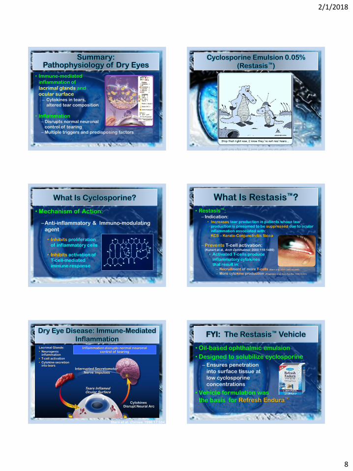
Summary:Pathophysiology of Dry Eyes
• Immune-mediated
inflammation of
lacrimal glands and
ocular surface– Cytokines in tears,
altered tear composition
• Inflammation – Disrupts normal neuronal
control of tearing
– Multiple triggers and predisposing factors
Cyclosporine Emulsion 0.05%
(Restasis™)
What Is Cyclosporine?
• Mechanism of Action:
–Anti-inflammatory & Immuno-modulating
agent
• Inhibits proliferation
of inflammatory cells
• Inhibits activation of
T-cell-mediated
immune response
What Is Restasis™?
• Restasis™:– Indication:
• Increases tear production in patients whose tear
production is presumed to be suppressed due to ocular
inflammation associated with:
KCS – Kerato-Conjunctivitis Sicca
–Prevents T-cell activation:(Kunert et al, Arch Ophthalmol. 2000;118:1489)
• Activated T-cells produce
inflammatory cytokines
that result in:– Recruitment of more T-cells (Stern et al, IOVS. 2002;43:2609)
– More cytokine production (Pflugfelder et al, Curr Eye Res. 1999;19:201)
Lacrimal Glands:
• Neurogenic inflammation
• T-cell activation
• Cytokine secretion into tears
Interrupted Secretomotor Nerve Impulses
Tears Inflamed Ocular Surface
Cytokines Disrupt Neural Arc
Inflammation disrupts normal neuronalcontrol of tearing
Stern et al. Cornea. 1998:17:584
Dry Eye Disease: Immune-Mediated
InflammationFYI: The Restasis™ Vehicle
• Oil-based ophthalmic emulsion
• Designed to solubilize cyclosporine
– Ensures penetration
into surface tissue at
low cyclosporine
concentrations
• Vehicle formulation was
the basis for Refresh Endura™

2/1/2018
9
Clinical Variables for FDA Approval
Primary
• Objective
– Corneal and Conjunctival staining
– Schirmerwith anesthesia
• Subjective
– Blurred vision
– Artificial tear reliance
Secondary
• Subjective
– Photophobia
– Sandy /gritty feeling
– Burning/stinging
– Itching
– Dryness
– Pain
Tertiary
• Objective
– Conjunctival biopsies
– Presence of inflammatory mediators
– Number of T-cells
– Goblet Cell Density
Lemp, 1995; Marsh et al, 1999
Slit lamp
Fluorescein
Dye Stain
Mild Severe
Clinical Presentation
Varies in Severity
Schirmer’s Testing
• Reflex Tearing– Without anesthesia
– Measures reflex
tear secretion
• Basal Tearing– With anesthesia
– Eliminates
stimulated tearing
Sall et al. Ophthalmol. 2000;107:631
Restasis Improves Schirmer Test Scores vs Vehicle
• Improvement from Baseline– 59% of Restasis™ users achieve a 1 - 10 mm or more improvement
from baseline in Schirmer scores at 6 months (n = 238)
• Statistically significant increases in Schirmer
with anesthesia wetting of 10 mm or more at 6 months– 15% increase with Restasis™ vs 5% with vehicle
1-9 mm
>10 mm 15%
44%
Percentage of Patients
Improvement
From
Baseline
in Schirmer
scores
• CD-3 Stained T Lymphocytes in Conjunctival Biopsies
Kunert et al. Arch Ophthalmol. 2000;118:1489
Baseline
CsA 0.05%
6 Months
2291
cells/mm2
Non-Sjögren’s
819
cells/mm2
Sjögren’s
762
cells/mm2
3965
cells/mm2
Decreased Inflammation in Sjögren’s and NonSjögren’s Pts.
Restasis™ Safety:
Ocular Adverse Events (%)
Data on file, Allergan, Inc.
17
3
3
3
2
2
1
1
7
1
2
2
1
4
1
0
0.05%
Cyclosporine Vehicle
Burning upon
instillation
Stinging
Discharge
Foreign-body sensation
Conjunctival hyperemia
Visual disturbance
Pain
Epiphora
2/1/2018
10
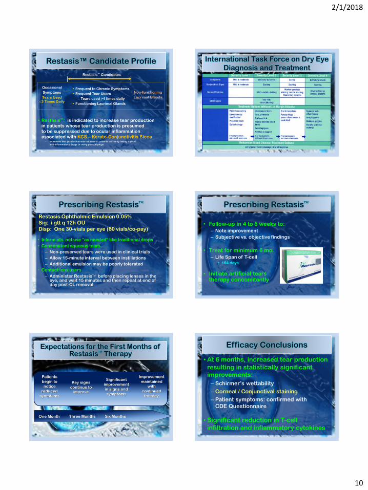
Restasis™ Candidate Profile
• Restasis™: is indicated to increase tear production
in patients whose tear production is presumed
to be suppressed due to ocular inflammation
associated with KCS – Kerato-Conjunctivitis Sicca– Increased tear production was not seen in patients currently taking topical
anti-inflammatory drugs or using punctal plugs
Occasional
Symptoms
Tears Used 3 Times Daily
Non-functioning
Lacrimal Glands
• Frequent to Chronic Symptoms
• Frequent Tear Users
– Tears used >4 times daily
• Functioning Lacrimal Glands
Restasis™ Candidates
International Task Force on Dry Eye
Diagnosis and Treatment
Prescribing Restasis
Restasis Ophthalmic Emulsion 0.05%
Sig: i gtt q 12h OU
Disp: One 30-vials per eye (60 vials/co-pay)
• Inform pts. not use “as needed” like traditional drops
• Concomitant aqueous tears
– Non-preserved tears were used in clinical trials
– Allow 15-minute interval between instillations
– Additional emulsion may be poorly tolerated
• Contact lens users
– Administer Restasis before placing lenses in the eye, and wait 15 minutes and then repeat at end of day post-CL removal.
Prescribing Restasis
• Follow-up in 4 to 6 weeks to: – Note improvement
– Subjective vs. objective findings
• Treat for minimum 6 mo.
– Life Span of T-cell• 164 days
• Initiate artificial tears therapy concomitantly
Patients
begin to
notice
reduced
symptoms
Key signs
continue to
improve
Significant
improvement
in signs and
symptoms
Improvement
maintained
with
continued
therapy
One Month Three Months Six Months
Expectations for the First Months of Restasis Therapy
Efficacy Conclusions
• At 6 months, increased tear production
resulting in statistically significant
improvements:
– Schirmer’s wettability
– Corneal / Conjunctival staining
– Patient symptoms: confirmed with
CDE Questionnaire
• Significant reduction in T-cell
infiltration and inflammatory cytokines

2/1/2018
11
Lifitegrast™ Latest in Dry Eyes: Lifitegrast 5%
• Progression of Dry Eye:– A receptor on the surface of T-cells is called
ICAM-1 (Inter-Cellular Adhesion Molecule-1) binds to LFA-1.
– ICAM-1 may be overexpressed in the corneal and conjunctival tissues in Dry Eye Diseasepatients1
– This interaction can result in T-cell activation and migration to target tissues1
• What Lifitegrast Does:– IIDRA blocks the
interaction of ICAM-1 to LFA-1
LFA-1 ICAM-1
Mechanism of Action
• By binding to LFA-1, Lifitegrast blocks the
ICAM-1 & LFA-1 interaction.1
– In vitro studies demonstrated that Lifitegrast may
inhibit T-cell adhesion to ICAM-1
and the secretion of pro-inflammatory cytokines.1
• The exact
mechanism of
action of
Lifitegrast in
Dry Eye Disease
is not known.1
1)Xiidra [Prescribing Information]. Lexington, MA: Shire US.
Mechanism of Action Video
• https://www.xiidra-
ecp.com/mechanism-of-
action?gclid=CKvUudWDmM8CFUiIfgo
dE6UPXg
Study Overview
• Evaluated for safety and efficacy
– Four randomized, double-masked, 12-week trials with total of 2133 patients.
– Assessed by improvement in:• Signs: measured by
Inferior Corneal Staining Score
• Symptoms: measured by Eye Dryness Score (N=2,133)1
– Vehicle consisted of a sterile buffered solution with: • pH range of 7.0-8.0
• Osmolality range of 200-330 mOsmol/kg
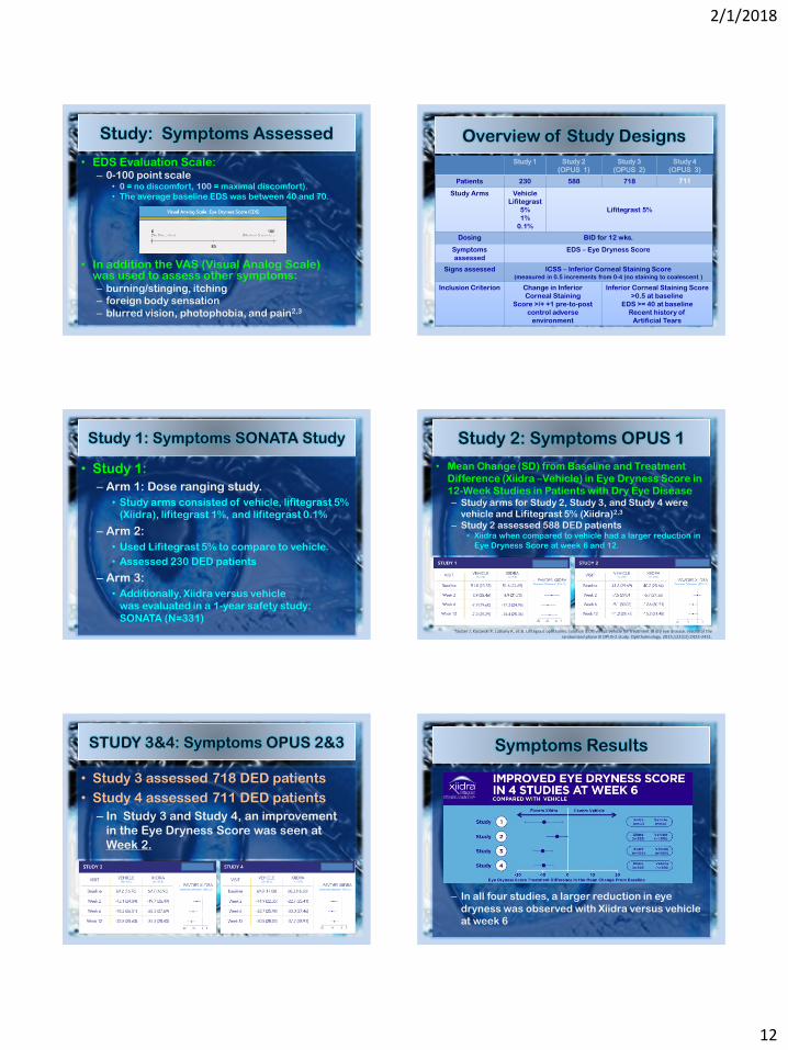
Study: Symptoms Assessed
• Each of the 4 studies assessed the
effect of Xiidra vs. Vehicle (saline) on:
– Both the signs and symptoms of Dry Eye
at: baseline and weeks 2, 6, and 12.
– Assessment of symptoms was based on
change from baseline in patient-reported
Eye Dryness Score (EDS):
2/1/2018
12
Study: Symptoms Assessed
• EDS Evaluation Scale:– 0-100 point scale
• 0 = no discomfort, 100 = maximal discomfort).
• The average baseline EDS was between 40 and 70.1
• In addition the VAS (Visual Analog Scale)was used to assess other symptoms: – burning/stinging, itching
– foreign body sensation
– blurred vision, photophobia, and pain2,3
2.Data on file. SHP606 (SAR 1118) (2.5 Clinical Overview). Shire US Inc; 2015.
Overview of Study Designs
Study 1 Study 2
(OPUS 1)
Study 3
(OPUS 2)
Study 4
(OPUS 3)
Patients 230 588 718 711
Study Arms Vehicle
Lifitegrast
5%
1%
0.1%
Lifitegrast 5%
Dosing BID for 12 wks.
Symptoms
assessed
EDS – Eye Dryness Score
Signs assessed ICSS – Inferior Corneal Staining Score(measured in 0.5 increments from 0-4 (no staining to coalescent )
Inclusion Criterion Change in Inferior
Corneal Staining
Score >/+ +1 pre-to-post
control adverse
environment
Inferior Corneal Staining Score
>0.5 at baseline
EDS >= 40 at baseline
Recent history of
Artificial Tears
2.Data on file. SHP606 (SAR 1118) (2.5 Clinical Overview). Shire US Inc; 2015.
Study 1: Symptoms SONATA Study
• Study 1:
– Arm 1: Dose ranging study.
• Study arms consisted of vehicle, lifitegrast 5%
(Xiidra), lifitegrast 1%, and lifitegrast 0.1%
– Arm 2:
• Used Lifitegrast 5% to compare to vehicle.
• Assessed 230 DED patients
– Arm 3:
• Additionally, Xiidra versus vehicle
was evaluated in a 1-year safety study:
SONATA (N=331)2
Study 2: Symptoms OPUS 1
• Mean Change (SD) from Baseline and Treatment
Difference (Xiidra –Vehicle) in Eye Dryness Score in
12-Week Studies in Patients with Dry Eye Disease– Study arms for Study 2, Study 3, and Study 4 were
vehicle and Lifitegrast 5% (Xiidra)2,3
– Study 2 assessed 588 DED patients• Xiidra when compared to vehicle had a larger reduction in
Eye Dryness Score at week 6 and 12.
Tauber J, Karpecki P, Latkany R, et al. Lifitegrast ophthalmic solution 5.0% versus vehicle for treatment of dry eye disease: results of the randomized phase III OPUS-2 study. Ophthalmology. 2015;122(12):2423-2431.
STUDY 3&4: Symptoms OPUS 2&3
• Study 3 assessed 718 DED patients
• Study 4 assessed 711 DED patients
– In Study 3 and Study 4, an improvement
in the Eye Dryness Score was seen at
Week 2.
Symptoms Results
– In all four studies, a larger reduction in eye
dryness was observed with Xiidra versus vehicle
at week 6

2/1/2018
13
Study: Signs - Corneal Staining
• Assessment of signs was measured by:
– Inferior Corneal Staining Score (ICSS)
– On a scale of 0 to 4 in increments of 0.5.
• The average baseline ICSS was approximately
1.8 in Studies 1 and 2 and 2.4 in Studies 3 and 4.
ICSS: Study 1 & 2
• Mean change (SD)
from baseline and
Treatment
Difference
(Xiidra – Vehicle)
in Inferior
Corneal
Staining
Score1
ICSS: Study 3 & 4
• In 3 of the 4 studies, at day 84 (after 12 weeks)
a larger reduction of Inferior Corneal Staining
was noted in Xiidra patients compared to
vehicle.
– In three out of the four studies (Study 1, Study 2, and Study 4), a larger reduction in inferior corneal staining was observed with Xiidra versus vehicle at week 12
Study: Signs Corneal Staining
Let There Be Light…NOT?
• Instructions:– Take a foil pouch out of the Xiidra box.
– Pull off 1 single use vial.
– Put the remaining strip of single use
containers back in the pouch
and fold the edge to close the pouch.
• Storing Xiidra– Store at room temperature
between 68°F to 77°F(20°C to 25°C).
– Store in the original foil pouch
to protect it from light.
– Do not open the foil pouch until
you are ready to use.
– Return unused single use containers
to their original foil pouch to protect from
excessive light exposure.
What’s So Hot?
• Fast acting:
– relief of symptoms in 2 to 4 weeks
• Great for patients with:
– Sjogren’s Syndrome
– Severe KCS
– Inflammatory tears
• Allows for patients to:
– Decrease use of concomitant tears
– Decrease use of adjunctive steroids

2/1/2018
14
And What’s Not So Hot?
• Dysgeusia – “FUNKY” taste
– tastes like heavy metal –“iron man”
• Burning upon instillation
– Phlegm in throat days to weeks of use
• Blurred vision
– Lasts 30min to 2 hrs.
• Increased Lacrimation:
– “Floods” eyes with reflex tearing
– Unable to “blink away” excess tearing
Important Safety Information
• In clinical trials, the most common adverse reactions reported in 5 to 25%of patients:– instillation site irritation,
– dysgeusia and
– reduced visual acuity.
• Other adverse reactions reported in 1% to 5% of the patients:– Blurred vision, increased lacrimation
– conjunctival hyperemia, eye irritation,
– headache, eye discharge, eye discomfort,
– eye pruritus and sinusitis.
My Patient Instructions
• Use drop in the morning– Pinch nose and tilt head
downward
– This will prevent burning
and drainage to back of throat
• Do not drink or eat anything for 30 min post-instillation– This will lessen “metallic” taste
– Brush your teeth post-gtts. Instillation
– Shower afterwards, if blurring of vision occurs.
• Wait 15 min. before
inserting SCLs
Texas Eye: Clinical Results
0%
5%
10%
15%
20%
25%
30%
35%
dysguesia
excess lacrimation
staining
burning
phlegm
stay on Xii
Latest in Dry Eyes: Lifitegrast 5%
• FDA Indication:
– For the treatment of the “signs & symptoms”of Dry Eye Disease
• Dosage:
– Used twice daily
• LFA-1 Antagonist Medication:
– Lymphocyte Functioning-associated Agonist-1 (LFA-1) Antagonist
– New drug class
Cryo-Preserved Amniotic Membrane

2/1/2018
15
• Dry eye disease (DED) is:
– One of the most commonly
encountered conditions
in our practice.
– Common denominator is:
• Tear film instability and
ocular surface
inflammation
Dana et al. (2002) Role of immunity and inflammation in corneal and ocular surface disease associated with dry eye. Adv Exp Med Biol.Benítez et al. (2004) An in vivo confocal masked study on corneal epithelium and subbasal nerves in patients with dry eye. IOVS
Regenerative Healing:
Dry Eye Disease• Corneal nerves play a significant role
in the maintenance of corneal
sensation and ocular surface health
Dana et al. (2002) Role of immunity and inflammation in corneal and ocular surface disease associated with dry eye. Adv Exp Med Biol.Benítez et al. (2004) An in vivo confocal masked study on corneal epithelium and subbasal nerves in patients with dry eye. IOVS
LacrimalGlands
SecretomotorNerve Impulses
Tears Support and MaintainOcular Surface
Ocular SurfaceNeural Stimulation
Normal tearing
depends on a
neuronal feedback loop
Dry Eye Disease: Background
• DED is accompanied with reduced
corneal nerve density.
– This results in compromised ocular
surface and reduced tear function
Dana et al. (2002) Role of immunity and inflammation in corneal and ocular surface disease associated with dry eye. Adv Exp Med Biol.Benítez et al. (2004) An in vivo confocal masked study on corneal epithelium and subbasal nerves in patients with dry eye. IOVS
Dry Eye Disease: Background Hypothesis
• Although there is an inflammatory component :
– Not all patients respond to topical anti-inflammatory
specifically when the nerves are compromised.
– To the best of our knowledge, there is no current
treatment for nerve degeneration.
Touhami A, et al. Invest Ophthalmol Vis Sci. 2002 Apr;43(4):987-94.Cheng A, et al. Ocul Surf. 2016 Jan;14(1):56-63.Sheha H, et al. Ocul Surf Disorders. JP Medical London; 2013 (39) 325-329.
Hypothesis
• Cryopreserved Amniotic Membrane (CAM)
– Rich in nerve growth factor
– Possesses a potent anti-inflammatory effect
– Successfully used to treat DED with ocular
surface involvement
• Therefore CAM may help corneal
nerve regeneration.
– To prove this hypothesis a
Randomized Clinical Trial
was designed.
Touhami A, et al. Invest Ophthalmol Vis Sci. 2002 Apr;43(4):987-94., Cheng A, et al. Ocul Surf. 2016 Jan;14(1):56-63.Sheha H, et al. Ocul Surf Disorders. JP Medical London; 2013 (39) 325-329.
Thomas John, MDCorneal Specialist Loyola University of Chicago
Massachusetts Eye and Ear Infirmary, Fellowship in Clinical CorneaScheffer Tseng, MD, PhD, Anny Cheng, MD, Hosam El Sheha, MD, PhD, Sean Tighe, MSc
Study: Corneal Nerve Regeneration after Self-
Retained Amniotic Membrane Use
for Dry-Eye Disease (ASCRS 2016)

2/1/2018
16
Objectives
• To evaluate:
– Efficacy of cryopreserved self-retained
amniotic membrane in restoring
corneal nerve density and
– Improving
corneal sensitivity
in patients with
dry eye disease
(DED).
Study Design
• A prospective, controlled
study to compare:
– Self-retained
amniotic membrane
– Conventional treatment
in patients with moderate
to severe DED (DEWS 2-4).
• 20 subjects were enrolled
and randomized to receive:
– CAM – Cryo-preserved AM
(Study Group) or
– Conventional
maximum medical treatment
(control group).
ITF-DEWS: International Task Force
Dry Eye Work Shop Study Design
• Observations: evaluated at baseline,
1 month, and 3 months
– Changes in clinical signs &
symptoms,
– Corneal topography,
– Corneal sensitivity, and
– Corneal nerve density
(using in vivo confocal
microscopy)
Testing and Results
• 20 Patients enrolled – 17 Patients completed the 1 and
3 months follow-up visits
• Dry eye signs and symptoms testing:– SPEED
“Standard Patient Evaluation of Eye Dryness Score”
– Pain score
– Fluorescein staining
– TBUT
– DEWS grading
• Significant improvement
in the study group compared
to no change in the control group.
SPEED Questionnaire

2/1/2018
17
Results: SPEED Score
• Statistically significant decrease:
– SPEED score compared to control group at:
• 1 month (p<0.0001, n=17)
• 3 months (p<0.0001, n=12)
Results: Corneal Staining
• Less Corneal Staining:
Before Prokera
After Prokera
Results: Pain Scoring
• Pain was graded 0 to 10, 10 being the most severe pain score.
– Study showed statistically significant decrease in these parameters from baseline to:• 1 month (p<0.005, n=9) and pain score showed statistical
significant decrease from 1 month to 3 month (p<0.05, n=7)
ITF-DEWS: International Task Force
Dry Eye Work Shop
Results: DEWS Scoring
P<0.0001
* *
P<0.005
Results: Corneal Sensitivity
• Significant increase in corneal sensitivity
– from 3.25 ± 0.6 to 5.2 ± 0.5 at 1 month
– 5.6 ± 0.4 cm at 3 months, p<0.001).
P<0.0005
**
2/1/2018
18
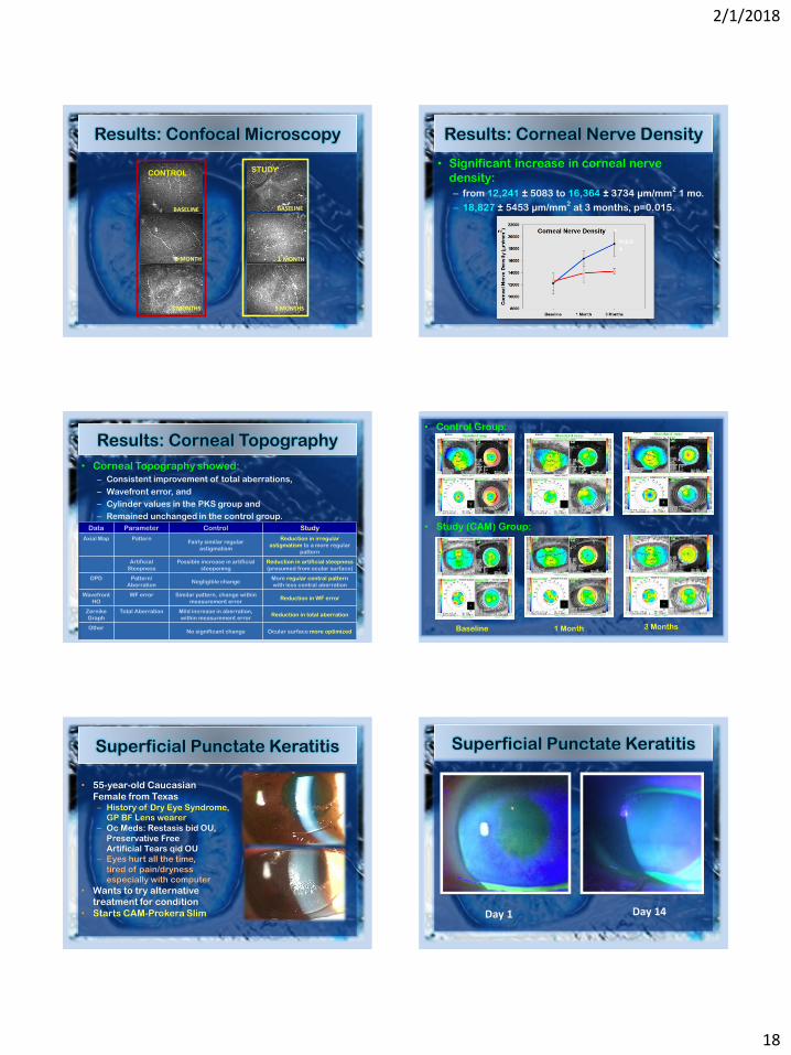
Results: Confocal Microscopy
BASELINE
1 MONTH
3 MONTHS
STUDY
BASELINE
1 MONTH
3 MONTHS
CONTROL
Results: Corneal Nerve Density
• Significant increase in corneal nerve
density:
– from 12,241 ± 5083 to 16,364 ± 3734 µm/mm2
1 mo.
– 18,827 ± 5453 µm/mm2
at 3 months, p=0.015.
*P=0.04
Results: Corneal Topography
• Corneal Topography showed:
– Consistent improvement of total aberrations,
– Wavefront error, and
– Cylinder values in the PKS group and
– Remained unchanged in the control group.
Data Parameter Control Study
Axial Map PatternFairly similar regular
astigmatism
Reduction in irregular
astigmatism to a more regular
pattern
Artificial
Steepness
Possible increase in artificial
steepening
Reduction in artificial steepness
(presumed from ocular surface)
OPD Pattern/
AberrationNegligible change
More regular central pattern
with less central aberration
Wavefront
HO
WF error Similar pattern, change within
measurement error Reduction in WF error
Zernike
Graph
Total Aberration Mild increase in aberration,
within measurement errorReduction in total aberration
OtherNo significant change Ocular surface more optimized
• Control Group:
• Study (CAM) Group:
Baseline 1 Month 3 Months
• 55-year-old Caucasian
Female from Texas– History of Dry Eye Syndrome,
GP BF Lens wearer
– Oc Meds: Restasis bid OU,
Preservative Free
Artificial Tears qid OU
– Eyes hurt all the time,
tired of pain/dryness
especially with computer
• Wants to try alternative
treatment for condition
• Starts CAM-Prokera Slim
Superficial Punctate Keratitis Superficial Punctate Keratitis
Day 1 Day 14

2/1/2018
19
Study Conclusion
• Placement of Cryopreserved amniotic
membrane is a: – Promising therapy for corneal nerve
regeneration AND– Accelerated recovery of the
ocular surface health in
patients with Dry Eye Disease.
– It effectively suppresses
inflammation, promotes
regenerative healing with
a lasting effect and helps
avoid further deterioration.
Summary
• Rx Treatment
– Cyclosporine-A: Restasis
– Lifetigrast: Xiidra
• Amniotic Membrane
– Cryo-preserved tissue
• What’s Next….
– Neural Stimulation !!!
The Dry Eye Story…
“A Real Tear Jerker”
Thank
You!