Lymph-Node-Sampler-Based Nucleic Acid Detection Helps to ...
23
Lymph-Node-Sampler-Based Nucleic Acid Detection Helps to Diagnose and Control African Swine Fever in Pig Production Xiaowen Li College of Veterinary Science and Animal Husbandry: Sam Higginbottom University of Agriculture Technology and Sciences Yang Li Shandong New Hope Liuhe Agriculture and Animal Husbandry Technology Co.,Ltd. Mingyu Fan Shandong new hope liuhe agruculture and animal husbandry technology co.,ltd. Shiran Fan Shandong newhope liuhe group co.,ltd. Wenchao Gao Shandong newhope liuhe group co.,ltd. Peng Yuan Shandong newhope liuhe group co.,ltd. Jing Ren Dezhou University Jingtao Li Shandong new hope liuhe group co.,ltd. Jincheng Yu Shandong new hope liuhe group co.,ltd. Weisheng Wu Shandong new hope liuhe group co.,ltd. Junxian Li Shandong new hope liuhe group co.,ltd. Qiannan Yu Shandong new hope liuhe group co.,ltd. Zhenwen Shao Shandong new hope liuhe group co.,ltd. Zhichun Yan ( [email protected] ) Shandong New Hope Liuhe Co.,Ltd. Short communication
Transcript of Lymph-Node-Sampler-Based Nucleic Acid Detection Helps to ...

Lymph-Node-Sampler-Based Nucleic Acid DetectionHelps to Diagnose and Control African Swine Feverin Pig ProductionXiaowen Li
College of Veterinary Science and Animal Husbandry: Sam Higginbottom University of AgricultureTechnology and SciencesYang Li
Shandong New Hope Liuhe Agriculture and Animal Husbandry Technology Co.,Ltd.Mingyu Fan
Shandong new hope liuhe agruculture and animal husbandry technology co.,ltd.Shiran Fan
Shandong newhope liuhe group co.,ltd.Wenchao Gao
Shandong newhope liuhe group co.,ltd.Peng Yuan
Shandong newhope liuhe group co.,ltd.Jing Ren
Dezhou UniversityJingtao Li
Shandong new hope liuhe group co.,ltd.Jincheng Yu
Shandong new hope liuhe group co.,ltd.Weisheng Wu
Shandong new hope liuhe group co.,ltd.Junxian Li
Shandong new hope liuhe group co.,ltd.Qiannan Yu
Shandong new hope liuhe group co.,ltd.Zhenwen Shao
Shandong new hope liuhe group co.,ltd.Zhichun Yan ( [email protected] )
Shandong New Hope Liuhe Co.,Ltd.
Short communication

Keywords: African swine fever, lymph node, sampler, real-time PCR, diagnostics
Posted Date: September 14th, 2021
DOI: https://doi.org/10.21203/rs.3.rs-882660/v1
License: This work is licensed under a Creative Commons Attribution 4.0 International License. Read Full License
1
Lymph-Node-Sampler-Based Nucleic Acid Detection Helps to Diagnose and 1
Control African Swine Fever in Pig Production 2
Running title: Lymph Node Sampler Helps to Diagnose ASF 3
Xiaowen Li1,2, Yang Li1,2, Mingyu Fan1,2, Shiran Fan1,2, Wenchao Gao1,2, Peng Yuan1,2, 4
Jing Ren3, Jingtao Li2, Jincheng Yu2, Weisheng Wu2, Junxian Li2, Qiannan Yu2, 5
Zhenwen Shao1,2, Zhichun Yan1,2 6
1 Shandong New Hope Liuhe Agriculture and Animal Husbandry Technology Co., Ltd, 7
Dezhou, China 8
2 Shandong New Hope Liuhe Group Co., Ltd, Qingdao, China 9
3 Shandong Provincial Laboratory of Swine Health Big Data and Intelligent Monitoring, 10
Dezhou, China 11
E-mail for correspondence: Dr Zhichun Yan; [email protected]. 12

2
Abstract 13
Background: African swine fever (ASF) is a highly contagious hemorrhagic and 14
transboundary animal disease. It has rapidly spread to many regions of the world and is 15
responsible for serious production and economic losses. However, clinical diagnosis is 16
impractical for the similar classic symptoms of ASF and several other normal diseases. 17
How to make a definite diagnosis is a top priority. 18
Results: In the present study, lymph node samples were collected by the patented 19
“lymph node sampler”, which makes the sampling safer, more efficient and minimally 20
invasive. The ASF virus (ASFV) contents of lymph node sample as well as serum, oral 21
fluid, nasal and rectal swab samples from pig production line were detected by real-22
time PCR. The big data results demonstrated that the lymph node samples contained 23
more stable and strongly positive ASFV nucleic acid than the porcine exudate and 24
serum samples. Taking the lymph node sample is one of the assistant ways in 25
diagnosing pigs suspected of having ASF. We supervised twenty farms that realized the 26
accurate diagnosis and eliminating of the risk in the shortest time period by using lymph 27
node sampler. 28
Conclusions: Our findings suggest that the lymph node sample is an ideal tissue for 29
diagnosing ASFV infection. The lymph node sampler is a convenient tool for lymph 30
node sampling for practitioners. 31
32
Keywords: African swine fever; lymph node; sampler; real-time PCR; diagnostics 33
3
1. Background 34
African swine fever (ASF) is a devastating swine viral disease that is reportable to 35
the World Organization for Animal Health (OIE). It causes high fever, severe depression, 36
ataxia and hemorrhages in domestic swine and results in a mortality rate approaching 37
100% (Ge et al., 2018). ASF is a highly contagious infectious disease and can be 38
transmitted by exposure to infectious pigs or tissues, contaminated swills, transport 39
vehicles, the clothing of animal workers and infected soft ticks (Penrith and Vosloo, 40
2009; Guinat et al., 2016; Olesen et al., 2018). Airborne transmission has been 41
demonstrated at close range (Wilkinson and Donaldson, 1977; Olesen et al., 2017). 42
Since its emergence in Kenya in the 1920s, ASF remains endemic in sub-Saharan Africa, 43
the Indian Ocean region and eastern Europe (Costard et al., 2009; Oganesyan et al., 44
2013; Gallardo et al., 2014). In 2007, ASF was confirmed in Georgia and Russia, posing 45
the risk of further dissemination into neighboring countries (Chapman et al., 2011). 46
Since August 2018, ASF has spread to China, the world's largest pork producer and a 47
leading pork importer (Ma et al., 2020). Ongoing outbreaks of ASF afflicted the 48
livestock industry and wiped out 40% of the nation’s pigs, leading to severe 49
socioeconomic losses (Li and Tian, 2018; Zhou et al., 2018; Lu et al., 2020). 50
The causative agent of the disease, ASF virus (ASFV), belongs to the genus 51
Asfivirus within the Asfarviridae family (Gogin et al., 2013). ASFV is a complex 52
enveloped virus containing a large double-stranded DNA genome that ranges in length 53
from 170 to 193 kbp (Tulman et al., 2009; Dixon et al., 2013). Although scientists 54
4
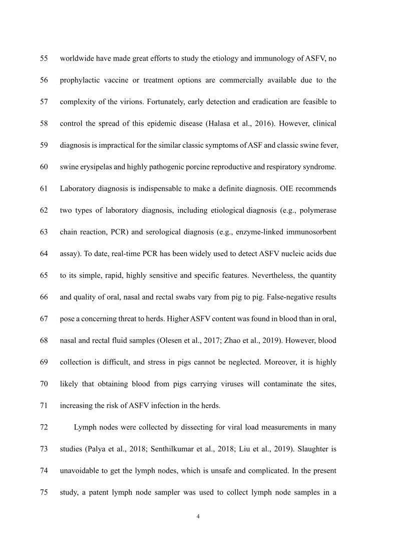
worldwide have made great efforts to study the etiology and immunology of ASFV, no 55
prophylactic vaccine or treatment options are commercially available due to the 56
complexity of the virions. Fortunately, early detection and eradication are feasible to 57
control the spread of this epidemic disease (Halasa et al., 2016). However, clinical 58
diagnosis is impractical for the similar classic symptoms of ASF and classic swine fever, 59
swine erysipelas and highly pathogenic porcine reproductive and respiratory syndrome. 60
Laboratory diagnosis is indispensable to make a definite diagnosis. OIE recommends 61
two types of laboratory diagnosis, including etiological diagnosis (e.g., polymerase 62
chain reaction, PCR) and serological diagnosis (e.g., enzyme-linked immunosorbent 63
assay). To date, real-time PCR has been widely used to detect ASFV nucleic acids due 64
to its simple, rapid, highly sensitive and specific features. Nevertheless, the quantity 65
and quality of oral, nasal and rectal swabs vary from pig to pig. False-negative results 66
pose a concerning threat to herds. Higher ASFV content was found in blood than in oral, 67
nasal and rectal fluid samples (Olesen et al., 2017; Zhao et al., 2019). However, blood 68
collection is difficult, and stress in pigs cannot be neglected. Moreover, it is highly 69
likely that obtaining blood from pigs carrying viruses will contaminate the sites, 70
increasing the risk of ASFV infection in the herds. 71
Lymph nodes were collected by dissecting for viral load measurements in many 72
studies (Palya et al., 2018; Senthilkumar et al., 2018; Liu et al., 2019). Slaughter is 73
unavoidable to get the lymph nodes, which is unsafe and complicated. In the present 74
study, a patent lymph node sampler was used to collect lymph node samples in a 75

5
minimally invasive way (Li et al., 2020). The viral loads of lymph nodes as well as oral 76
fluid, nasal and rectal swab samples from the pig production line were investigated. The 77
application of lymph node samplers in twenty farms shows great potential for the 78
accurate diagnosis and eradication of ASFV. 79
80
2. Materials and methods 81
2.1 The structure and usage of the lymph node sampler. 82
The lymph node sampler is a simple and easy-to-operate tool. It consists of a needle, 83
a syringe, a handle and a connection rod inside the needle (Figure 1A, 1B). The needle 84
contains a barb, which can remove the lymphoid tissue from the muscle (Figure 1C, 85
1D). The connection rod can be inserted into the needle to extrude the tissue. 86
2.2 How to use the lymph node sampler. 87
First, the pig was restrained to access its inguinal lymph nodes (Figure 2A). Then, 88
the skin was punctured vertically with the sampler to ensure that the barb entirely 89
entered the tissue (Figure 2B). The sampler was pulled out, and the handle was pressed 90
to push the tissue out of the needle (Figure 2C). The tissue was placed into an Eppendorf 91
(EP) tube and submerged in nucleic acid protective fluid. 92
Plenty of needles should be prepared before the sampling. The sampler can be 93
disassembled for recycling by removing the residual blood and tissue, soaking the 94
sampler for 30 minutes in a sodium hypochlorite solution (3%), and washing the 95
sampler with clean water. The sampler can then be reassembled and dried for the next 96
6
usage. 97
2.3 Sampling methods of other tissues. 98
For serum samples, collect the blood from the porcine anterior vena cava into an 99
anticoagulation tube. Let stand for one hour and get the upper serum. For oral fluid 100
samples, tie a cotton rope in front of the pig. Squeeze out the liquid after the sufficient 101
chew by the pig. For throat swabs, insert a long spermaduct with a sponge on the top 102
into the throat quickly. Elute the mucus with 1mL nucleic acid protective fluid (refer to 103
2.4). The cotton nasal swabs and rectal swabs were also eluted with the nucleic acid 104
protective fluid. The nasal-rectal swab and nasal-throat-rectal swab samples were the 105
mixture of the individuals. 106
2.4 Preventing sample degradation 107
The sample should be protected in case of degradation. In this study, a patented 108
customized nucleic acid protective fluid was used, which contains 109
ethylenediaminetetraacetic acid (EDTA) to stop DNase activity by chelating 110
magnesium, manganese and ferric ions (Yu et al., 2017; Ren et al., 2020). The 111
concentrated solution was diluted with purified water. Working solutions of nucleic acid 112
protective fluid normally last for up to 7 days, but fresh solution is recommended. 113
For the lymph node samples, 1 mL of nucleic acid protective fluid was added to 114
soak the tissue. For the swab samples, the sticks were broken and 1 mL of nucleic acid 115
protective fluid was added. The samples were sent for detection at room temperature 116
within one day. 117
7
2.5 Quantitative real-time PCR (qPCR). 118
ASFV DNA was quantified with qPCR analysis in a “one step” way without the 119
process of nucleic acid extraction. The kit from Beijing MingRiDa Technology 120
Development Co., Ltd. (010688870) was used according to the user guide. Briefly, 121
lymph node samples were homogenized. Swab samples were oscillated and centrifuged 122
at 8000 g for 2 min. Five microliters of supernatant, negative control or positive control 123
fluid was mixed with 20 µL of operating fluid. Twenty microliters of mixture was taken 124
for qPCR detection. 125
126
3. Results 127
3.1 The ASFV content of lymph nodes was higher than that of other collected 128
sample tissues. 129
Suspected ASF samples from the pig production line in Northwestern Shandong 130
Province from January 2020 to March 2021 were collected for nucleic acid detection 131
by qPCR. The results showed that 37 serum samples, 243 lymph node samples, 237 132
oral fluid, 17 throat swab, 102 nasal swab, 25 rectal swab, 1341 nasal-rectal swab and 133
26 nasal-throat-rectal swab samples were true positive for ASFV. The CT values were 134
collected and are shown in Figure 3. Serum samples are widely used for many 135
pathogenic diagnoses. The nasal, nasal-rectal and nasal-throat-rectal swab samples had 136
almost the same CT value as the serum samples, suggesting that the virus contents of 137
nasal and serum samples were comparable. Saliva is other common sampling point. 138
8
However, our results showed that the mean CT value of saliva samples was much higher 139
than those of other sites, suggesting that the viral loads in saliva were lower than those 140
in other samples. The lymph node samples were the only samples with lower CT values 141
than the serum samples. The standard deviation (SD) value of the lymph node sample 142
CT value was much smaller than that of serum samples. These data suggest that the 143
ASFV content of lymph node samples is more stable than that of serum samples and is 144
higher than that of other samples. 145
3.2 A larger proportion of strongly positive results was found in lymph node 146
samples. 147
The distribution of CT values between the samples was also analyzed. Generally, 148
CT values over 35 are considered hard to judge, while values less than 30 can be 149
identified as strongly positive results. The percentage of strongly positive lymph node 150
samples was 70.9%, while the relative percentages of strongly positive serum, nasal 151
swab and oral-nasal-rectal swab samples were 45.9%, 37.3%, 37.4%, respectively 152
(Figure 4). These data suggest that the ASFV content of lymph nodes is much higher 153
than that of other sites, which has great potential for the accurate diagnosis of ASFV. 154
3.3 ASFV-positive pigs were removed within 3 weeks with the help of lymph node 155
samplers. 156
To check whether lymph node samples can help to eliminate ASFV-positive pigs 157
in production, lymph node samplers were immediately applied on farms that tested 158
positive for ASFV nucleic acid. Once abnormal symptoms appeared, such as fever, 159
9
inappetence, blood spots, emesis or death, the surrounding samples and porcine lymph 160
node samples were collected and detected for the first time. The positive pigs were 161
removed from the herd according to the biosafety requirements. The surroundings were 162
disinfected repeatedly using 3% sodium hypochlorite until free from nucleic acid. 163
Among all twenty farms we monitored, the time from ASFV positivity to negativity 164
ranged from 1 day to 18 days (Figure 5). These results demonstrated that lymph node 165
sampling is workful for the removal of ASFV-positive pigs. 166
167
4. Discussion 168
ASF is an acute viral infection that has a major impact on global pig production. 169
Stockbreeders displayed passivity in the diffusion of practices without effective 170
vaccines or drugs. However, the highly contagious characteristic of ASF transmission 171
indicates the potential for possible interventions to manage the risk of ASF introduction 172
and transmission (Lichoti et al., 2016; Govoeyi et al., 2020). Biosecurity is a high 173
priority for pig farmers due to its high efficiency in controlling this disease (De Lorenzi 174
et al., 2020; Jackman et al., 2020). It contains at least two parts: how to prevent ASF 175
infection entirely and how to remove infected pigs rapidly and accurately (Álvarez et 176
al., 2019). 177
Immunology and nucleic acid detection are two main methods to diagnose ASF. 178
qPCR assays have many advantages compared with immunology methods, such as 179
simple operation processes, high sensitivity, high specificity and excellent repeatability. 180
10
Thus, qPCR is the most commonly used laboratory test method to diagnose ASFV at 181
present, along with the ASFV antigen detection method recommended by the OIE (King 182
et al., 2003). However, it is difficult to determine CT values larger than 35. The main 183
reason for the high CT values is the low viral content of the samples. The degradation 184
of nucleic acids due to improper preservation and transportation methods also results 185
in confusing data. The present study took advantage of a patented lymph node sampler 186
to puncture the skin in a minimally invasive manner to efficiently obtain lymph node 187
tissue. Moreover, an innovative nucleic acid protective fluid was used to prevent the 188
degradation of ASFV DNA. The qPCR data revealed that the ASFV content of the 189
lymph node samples was higher than that of other common kinds of samples, including 190
serum, oral fluid and nasal-rectal swabs, suggesting that the lymph node is an ideal 191
tissue for the diagnosis of ASF. 192
Practice has indicated that our synthetic solutions work well in the prevention and 193
control of ASF according to the contingency plan (Ministry-of-Agriculture-and-Rural-194
Affairs, 2020). In general, we require all the pigs, materials and workers that intend to 195
enter the farm to be ASFV free by taking corresponding measures. Breeders should 196
inspect the herds daily and determine abnormal symptoms, including depression, fever, 197
inappetence, purple skin, blood spots, emesis, nosebleed, abortion, hematochezia and 198
death. The nasal swab, rectal swab and lymph node samples were collected immediately 199
and sent to a testing laboratory nearby. The laboratory was continuously running for 200
24-hour full-time service to give feedback to the decision makers within 8 hours. The 201
11
positive pigs were removed with minimum contamination. The oral fluid and nasal-202
rectal swab samples of the whole herd, as well as the whole divisional ground, were 203
collected at once. The positive pigs were removed again. The disinfection of the whole 204
farm was immediately executed using potent disinfectors. Seven days later, the next 205
cycle of census, elimination and disinfection were carried out until no positive results 206
occurred. Supported by these powerful biosecurity measures, lymph-node-sample-207
based rapid and accurate definite diagnosis plays a dominant role in the systematic 208
projects. 209
In conclusion, this study took full advantage of the lymph node sampler to collect 210
lymph node samples from pigs in a minimally invasive way. The viral content of lymph 211
nodes, rather than serum or other swab samples of porcine secretions, was proven to be 212
the highest. The lymph-node-sampler-based sampling method helps to realize the 213
accurate diagnosis and elimination of ASF. 214
215
Declarations 216
Ethics approval and consent to participate 217
The authors confirm that all the procedures were conducted in adherence with the 218
the guidelines of New Hope Group Subcommittee of Experimental Animal Ethics 219
with no. of 151920. 220
Consent for publication 221
Not applicable. 222
12
Availability of data and materials 223
The dataset generated in this study is available from the first author and 224
corresponding author on reasonable request. 225
Competing interests 226
The authors declare that there are no conflicts of interest. 227
Funding 228
This work was supported by the Integration and Demonstration of 229
Comprehensive Prevention and Control of ASFV by the Ministry of Science and 230
Technology (No. 2018YFC0840405), the New Hope Group Science and Technology 231
Leading 100-person Program Fund (NH201806), the Shandong Provincial Key 232
Laboratory Open Fund (SD2019BP102) and the China National Science Foundation 233
(No. 31701424). 234
Authors’ contributions 235
Study design and planning: Zhichun Yan, Xiaowen Li; animal experiments: 236
Wenchao Gao, Peng Yuan, Jingtao Li, Jincheng Yu, Weisheng Wu, Junxian Li; 237
virological work: Zhenwen Shao, Qiannan Yu, Yang Li; data processing and analysis: 238
Jing Ren, Mingyu Fan, Shiran Fan; drafting manuscript: Xiaowen Li, Yang Li; 239
critically revising manuscript prior to the submission: Zhichun Yan. All authors read, 240
edited, and approved the manuscript. 241
Acknowledgements 242
The authors thank the conveniences from Shandong Provincial Laboratory of 243
13
Swine Health Big Data and Intelligent Monitoring. 244
245
References 246
Álvarez, J., Bicout, D., Boklund, A., Bøtner, A., Depner, K., More, S.J., Roberts, H., 247 Stahl, K., Thulke, H.H., Viltrop, A., Antoniou, S.E., Cortiñas Abrahantes, J., 248 Dhollander, S., Gogin, A., Papanikolaou, A., Van der Stede, Y., González Villeta, 249 L.C., Gortázar Schmidt, C., 2019. Research gap analysis on African swine fever. 250 Efsa j 17, e05811. 251
Chapman, D.A., Darby, A.C., Da Silva, M., Upton, C., Radford, A.D., Dixon, L.K., 252 2011. Genomic analysis of highly virulent Georgia 2007/1 isolate of African 253 swine fever virus. Emerg Infect Dis 17, 599-605. 254
Costard, S., Wieland, B., de Glanville, W., Jori, F., Rowlands, R., Vosloo, W., Roger, F., 255 Pfeiffer, D.U., Dixon, L.K., 2009. African swine fever: how can global spread 256 be prevented? Philos Trans R Soc Lond B Biol Sci 364, 2683-2696. 257
De Lorenzi, G., Borella, L., Alborali, G.L., Prodanov-Radulović, J., Štukelj, M., Bellini, 258 S., 2020. African swine fever: A review of cleaning and disinfection procedures 259 in commercial pig holdings. Research in veterinary science 132, 262-267. 260
Dixon, L.K., Chapman, D.A., Netherton, C.L., Upton, C., 2013. African swine fever 261 virus replication and genomics. Virus Res 173, 3-14. 262
Gallardo, C., Fernandez-Pinero, J., Pelayo, V., Gazaev, I., Markowska-Daniel, I., 263 Pridotkas, G., Nieto, R., Fernandez-Pacheco, P., Bokhan, S., Nevolko, O., 264 Drozhzhe, Z., Perez, C., Soler, A., Kolvasov, D., Arias, M., 2014. Genetic 265 variation among African swine fever genotype II viruses, eastern and central 266 Europe. Emerg Infect Dis 20, 1544-1547. 267
Ge, S., Li, J., Fan, X., Liu, F., Li, L., Wang, Q., Ren, W., Bao, J., Liu, C., Wang, H., Liu, 268 Y., Zhang, Y., Xu, T., Wu, X., Wang, Z., 2018. Molecular Characterization of 269 African Swine Fever Virus, China, 2018. Emerg Infect Dis 24, 2131-2133. 270
Gogin, A., Gerasimov, V., Malogolovkin, A., Kolbasov, D., 2013. African swine fever 271 in the North Caucasus region and the Russian Federation in years 2007-2012. 272 Virus Res 173, 198-203. 273
Govoeyi, B., Agbokounou, A.M., Camara, Y., Ahounou, S.G., Dotche, I.O., Kiki, P.S., 274 Abdou Karim, I.Y., Delabouglise, A., Antoine-Moussiaux, N., 2020. Social 275 network analysis of practice adoption facing outbreaks of African Swine Fever. 276 Prev Vet Med 179, 105008. 277
Guinat, C., Gubbins, S., Vergne, T., Gonzales, J.L., Dixon, L., Pfeiffer, D.U., 2016. 278 Experimental pig-to-pig transmission dynamics for African swine fever virus, 279 Georgia 2007/1 strain. Epidemiol Infect 144, 25-34. 280
Halasa, T., Botner, A., Mortensen, S., Christensen, H., Toft, N., Boklund, A., 2016. 281 Control of African swine fever epidemics in industrialized swine populations. 282
14
Vet Microbiol 197, 142-150. 283 Jackman, J.A., Hakobyan, A., Zakaryan, H., Elrod, C.C., 2020. Inhibition of African 284
swine fever virus in liquid and feed by medium-chain fatty acids and glycerol 285 monolaurate. J Anim Sci Biotechnol 11, 114. 286
King, D.P., Reid, S.M., Hutchings, G.H., Grierson, S.S., Wilkinson, P.J., Dixon, L.K., 287 Bastos, A.D., Drew, T.W., 2003. Development of a TaqMan PCR assay with 288 internal amplification control for the detection of African swine fever virus. 289 Journal of virological methods 107, 53-61. 290
Li, X., Fan, S., Liu, C., Zhang, Z., Fan, M., 2020. A medical tissue sampler for livestock. 291 CN211749759U. 292
Li, X., Tian, K., 2018. African swine fever in China. The Veterinary record 183, 300-293 301. 294
Lichoti, J.K., Davies, J., Kitala, P.M., Githigia, S.M., Okoth, E., Maru, Y., Bukachi, 295 S.A., Bishop, R.P., 2016. Social network analysis provides insights into African 296 swine fever epidemiology. Prev Vet Med 126, 1-10. 297
Liu, J., Li, Z., Ren, X., Li, H., Lu, R., Zhang, Y., Ning, Z., 2019. Viral load and 298 histological distribution of atypical porcine pestivirus in different tissues of 299 naturally infected piglets. Arch Virol 164, 2519-2523. 300
Lu, G., Pan, J., Zhang, G., 2020. African swine fever virus in Asia: Its rapid spread and 301 potential threat to unaffected countries. J Infect 80, 350-371. 302
Ma, J., Chen, H., Gao, X., Xiao, J., Wang, H., 2020. African swine fever emerging in 303 China: Distribution characteristics and high-risk areas. Prev Vet Med 175, 304 104861. 305
Ministry-of-Agriculture-and-Rural-Affairs, 2020. African Swine Fever Response 306 Implementation Plan. 307 http://www.moa.gov.cn/nybgb/2020/202006/202007/t20200714_6348531.htm 308 (accessed 20 April 2021). 309
Murphy, C., Bashiruddin, J.B., Quan, M., Zhang, Z., Alexandersen, S., 2010. Foot-and-310 mouth disease viral loads in pigs in the early, acute stage of disease. The 311 Veterinary record 166, 10-14. 312
Oganesyan, A.S., Petrova, O.N., Korennoy, F.I., Bardina, N.S., Gogin, A.E., Dudnikov, 313 S.A., 2013. African swine fever in the Russian Federation: spatio-temporal 314 analysis and epidemiological overview. Virus Res 173, 204-211. 315
Olesen, A.S., Lohse, L., Boklund, A., Halasa, T., Belsham, G.J., Rasmussen, T.B., 316 Bøtner, A., 2018. Short time window for transmissibility of African swine fever 317 virus from a contaminated environment. Transbound Emerg Dis 65, 1024-1032. 318
Olesen, A.S., Lohse, L., Boklund, A., Halasa, T., Gallardo, C., Pejsak, Z., Belsham, G.J., 319 Rasmussen, T.B., Botner, A., 2017. Transmission of African swine fever virus 320 from infected pigs by direct contact and aerosol routes. Vet Microbiol 211, 92-321 102. 322
Palya, V., Homonnay, Z.G., Mato, T., Kiss, I., 2018. Characterization of a PCV2d-2 323 isolate by experimental infection of pigs. Virol J 15, 185. 324
15
Penrith, M.L., Vosloo, W., 2009. Review of African swine fever: transmission, spread 325 and control. J S Afr Vet Assoc 80, 58-62. 326
Ren, J., Li, X., Fan, M., Wang, J., Yu, J., Gao, W., 2020. An oral fluid virus DNA 327 protector and its preparation method and application. CN202010716933.6. 328
Senthilkumar, D., Rajukumar, K., Sen, A., Kumar, M., Shrivastava, D., Kalaiyarasu, S., 329 Gautam, S., Singh, F., Kulkarni, D.D., Singh, V.P., 2018. Pathogenic 330 characterization of porcine reproductive and respiratory syndrome virus of 331 Indian origin in experimentally infected piglets. Transbound Emerg Dis 65, 332 1522-1536. 333
Tulman, E.R., Delhon, G.A., Ku, B.K., Rock, D.L., 2009. African swine fever virus. 334 Curr Top Microbiol Immunol 328, 43-87. 335
Wilkinson, P.J., Donaldson, A.I., 1977. Transmission studies with African swine fever 336 virus. The early distribution of virus in pigs infected by airborne virus. J Comp 337 Pathol 87, 497-501. 338
Yu, G., Hatta, A., Periyannan, S., Lagudah, E., Wulff, B.B.H., 2017. Isolation of Wheat 339 Genomic DNA for Gene Mapping and Cloning. Methods in molecular biology 340 1659, 207-213. 341
Zhao, D., Liu, R., Zhang, X., Li, F., Wang, J., Zhang, J., Liu, X., Wang, L., Zhang, J., 342 Wu, X., Guan, Y., Chen, W., Wang, X., He, X., Bu, Z., 2019. Replication and 343 virulence in pigs of the first African swine fever virus isolated in China. Emerg 344 Microbes Infect 8, 438-447. 345
Zhou, X., Li, N., Luo, Y., Liu, Y., Miao, F., Chen, T., Zhang, S., Cao, P., Li, X., Tian, 346 K., Qiu, H.J., Hu, R., 2018. Emergence of African Swine Fever in China, 2018. 347 Transbound Emerg Dis 65, 1482-1484. 348
349
Figure legends 350
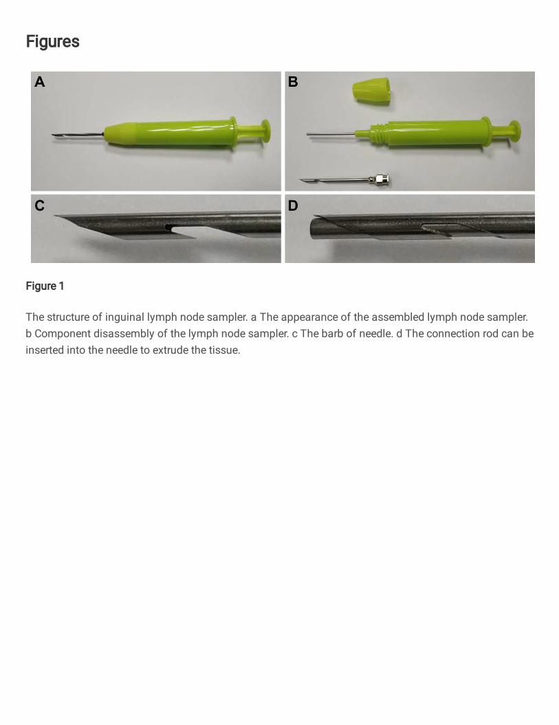
Figure 1. The structure of inguinal lymph node sampler. a The appearance of the 351
assembled lymph node sampler. b Component disassembly of the lymph node 352
sampler. c The barb of needle. d The connection rod can be inserted into the needle to 353
extrude the tissue. 354
355
Figure 2. Three steps to obtain inguinal lymph node samples using a sampler. a 356
Find and pinch the inguinal lymph node. b Puncture the skin and tissue. c Pull out the 357
16
sampler and press the handle several times to squeeze the tissue out. d Enlarged view 358
of squeezed lymph node tissue. 359
360
Figure 3. The ASFV content of lymph nodes and other kinds of collected 361
samples. Eight kinds of porcine samples were collected, and their ASFV content was 362
detected by qPCR. Individual CT values are presented. 363
364
Figure 4. A larger proportion of strongly positive results was found in lymph 365
node samples. The CT values were sorted into different ranges. The distribution of 366
each 5 CT ranges is shown in the figure. Values smaller than 30 were identified as 367
strongly positive results. 368
369
Figure 5. ASFV-positive pigs were removed within 3 weeks according to the 370
viral content of lymph nodes. The ASFV-positive farms judged the suspected pigs by 371
the viral content of the lymph nodes. The positive pigs were removed from the herd, 372
and the surroundings were disinfected thoroughly by irrigation with 3% sodium 373
hypochlorite until ASFV nucleic acid was undetectable. The results of nucleic acid 374
detection are shown. Red: positivity; green: negativity. 375
Figures
Figure 1
The structure of inguinal lymph node sampler. a The appearance of the assembled lymph node sampler.b Component disassembly of the lymph node sampler. c The barb of needle. d The connection rod can beinserted into the needle to extrude the tissue.
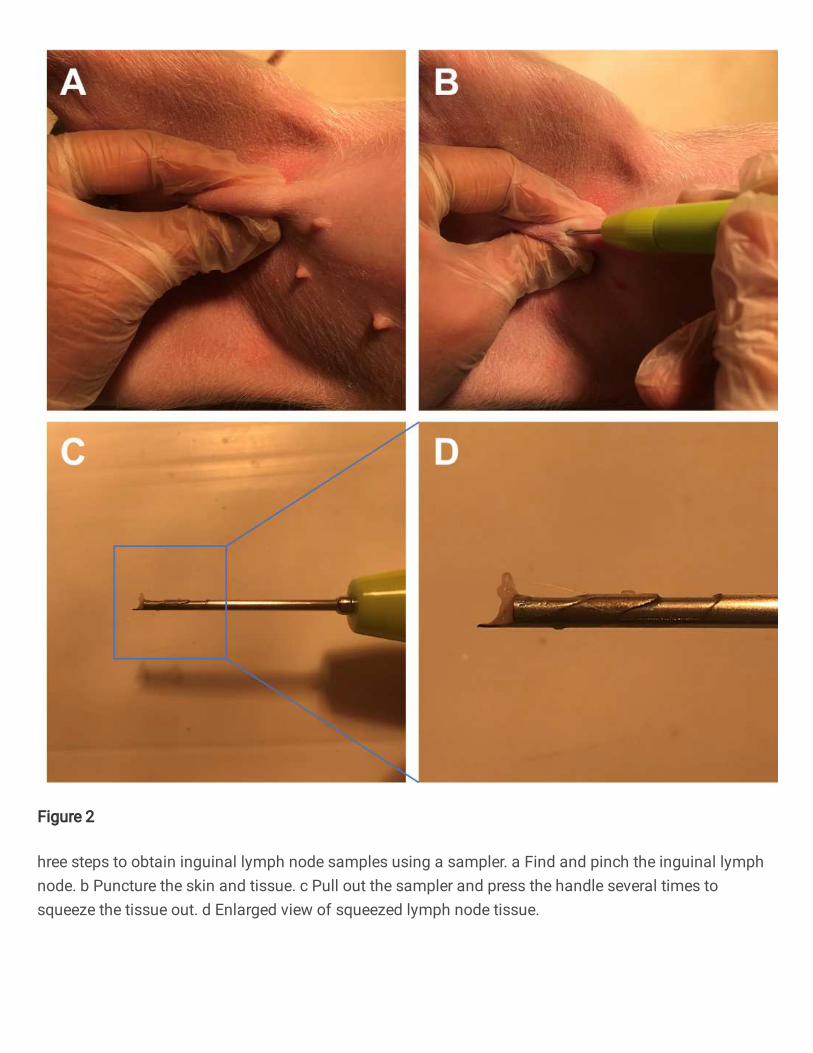
Figure 2
hree steps to obtain inguinal lymph node samples using a sampler. a Find and pinch the inguinal lymphnode. b Puncture the skin and tissue. c Pull out the sampler and press the handle several times tosqueeze the tissue out. d Enlarged view of squeezed lymph node tissue.
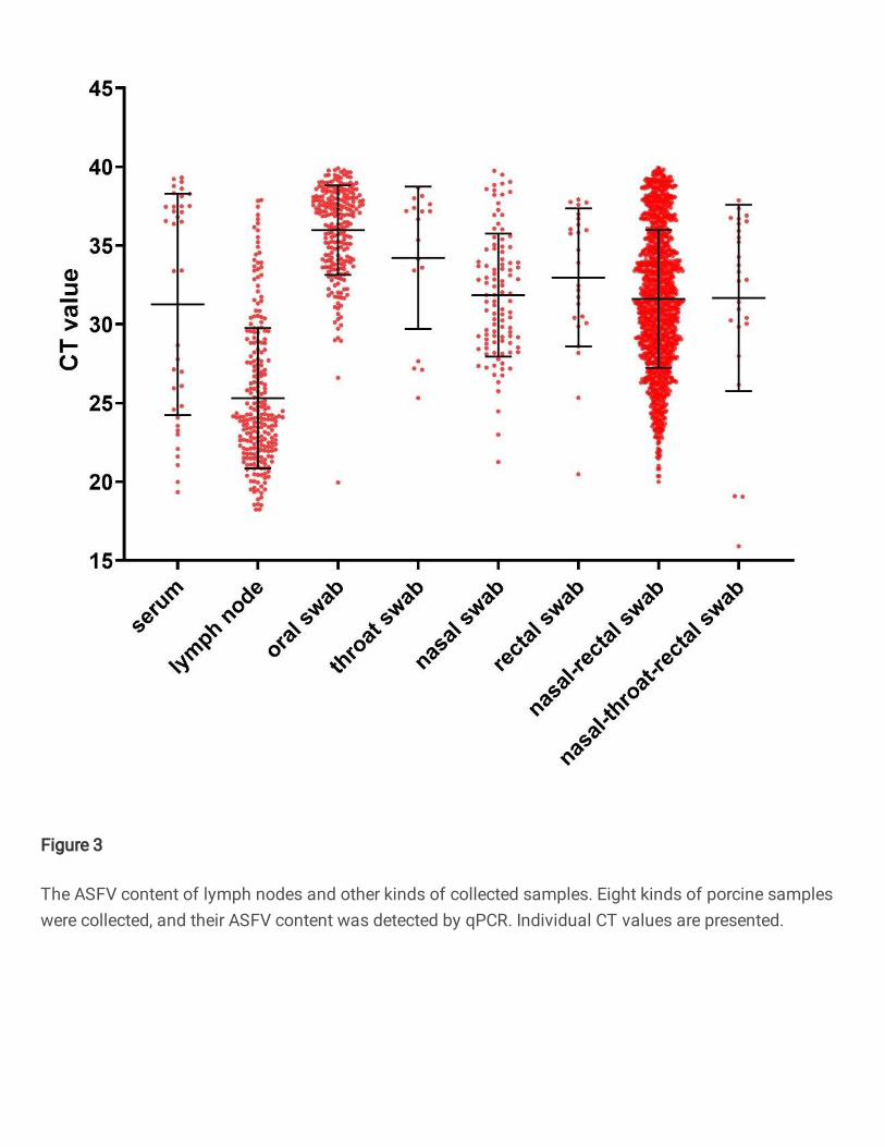
Figure 3
The ASFV content of lymph nodes and other kinds of collected samples. Eight kinds of porcine sampleswere collected, and their ASFV content was detected by qPCR. Individual CT values are presented.
Figure 4
A larger proportion of strongly positive results was found in lymph node samples. The CT values weresorted into different ranges. The distribution of each 5 CT ranges is shown in the �gure. Values smallerthan 30 were identi�ed as strongly positive results.

Figure 5
ASFV-positive pigs were removed within 3 weeks according to the viral content of lymph nodes. TheASFV-positive farms judged the suspected pigs by the viral content of the lymph nodes. The positive pigswere removed from the herd, and the surroundings were disinfected thoroughly by irrigation with 3%sodium hypochlorite until ASFV nucleic acid was undetectable. The results of nucleic acid detection areshown. Red: positivity; green: negativity.


