Quantification of Liver Fat in Mice: Comparing Dual-Echo Dixon ...

LIVER BIOLOGY AND PATHOBIOLOGYBIOLOGY
Liver Tissue Engineering at Extrahepatic Sites in Miceas a Potential New Therapy for Genetic Liver Diseases
Kazuo Ohashi,1,3 Jacob M. Waugh,2 Michael D. Dake,2 Takashi Yokoyama,3 Hiroyuki Kuge,3 Yoshiyuki Nakajima,3
Masaki Yamanouchi,4 Hiroyuki Naka,4 Akira Yoshioka,4 and Mark A. Kay1
Liver tissue engineering using hepatocyte transplantation has been proposed as an alterna-tive to whole-organ transplantation or liver-directed gene therapy to correct various types ofhepatic insufficiency. Hepatocytes are not sustained when transplanted under the kidneycapsule of syngeneic mice. However, when we transplanted hepatocytes with the extracellu-lar matrix components extracted from Engelbreth-Holm-Swarm cells, hepatocytes survivedfor at least 140 days and formed small liver tissues. Liver engineering in hemophilia A micereconstituted 5% to 10% of normal clotting activity, enough to reduce the bleeding time andhave a therapeutic benefit. Conversely, the subcutaneous space did not support the persistentsurvival of hepatocytes with Engelbreth-Holm-Swarm gel matrix. We hypothesized thatestablishing a local vascular network at the transplantation site would reduce graft loss. Totest this idea, we provided a potent angiogenic agent before hepatocyte transplantation intothe subcutaneous space. With this procedure, persistent survival was achieved for the lengthof the experiment (120 days). To establish that these engineered liver tissues also retainedtheir native regeneration potential in vivo, we induced two different modes of proliferativestimulus to the naı̈ve liver and confirmed that hepatocytes within the extrahepatic tissuesregenerated with activity similar to that of naı̈ve liver. In conclusion, our studies indicatethat liver tissues can be engineered and maintained at extrahepatic sites, retain their capacityfor regeneration in vivo, and used to successfully treat genetic disorders. (HEPATOLOGY 2005;41:132–140.)
Development of cellular-based therapies, includ-ing hepatocyte transplantation and liver tissueengineering, has been attempted to treat differ-
ent forms of liver diseases as an alternative to liver organtransplantation.1–4 By transplanting isolated hepatocytesthrough the portal circulation into the liver, encouragingresults were reported in patients with Criglar-Najjar syn-
drome and a glycogen storage disease.1,2 However, this ap-proach of cell transplantation through the portal vein islimited in the number of cells that can be transplanted at onetime because of potential life-threatening complications.3,5,6
To increase the utility of hepatocyte-based therapy, it is im-portant to develop a method that allows for the engraftmentof a greater number of hepatocytes. In this context, trans-planting hepatocytes at an extrahepatic site is attractive be-cause it would provide additional space to maintain a greaternumber of cells, and with fewer complications.3,7–9 This typeof approach is extremely viable from the standpoint of livertissue engineering. Researchers have transplanted hepato-cytes at several different extrahepatic sites, including the in-traperitoneal cavity, pancreas, mesenteric leaves, lungparenchyma, under the kidney capsule, and in the subcuta-neous space.3,5,8,10,11 Irrespective of the sites used, the studiesreported inefficient engraftment of hepatocytes and veryshort-term survival. To overcome this issue, we have recentlyused an agonistic antibody that stimulates the HGF/cMetpathway to achieve persistent survival of human hepatocytes(�140 days) grafted into mice under the kidney capsulespace.8,12 These successes have encouraged us to establishmore clinically feasible methods for liver tissue engineering at
Abbreviations: hAAT, human alpha-1 antitrypsin; EHS, Engelbreth-Holm-Swarm; aFGF, acidic fibroblast growth factor; MS, microspheres; DH, direct hy-perplasia; TCPOBOP, 1,4-bis[2-(3,5-dicholoropyridyloxy)] benzene; BrdU,5-bromo-2-deoxyuridine.
From the Departments of 1Pediatrics and Genetics and 2Cardiovascular andInterventional Radiology, Department of Radiology, Stanford University MedicalCenter, Stanford, CA; and the 3First Department of Surgery and 4Department ofPediatrics, Nara Medical University, Nara, Japan.
Received April 26, 2004; accepted September 20, 2004.Supported by NIH U19-AI40034 (M.A.K.), Grant-in-Aid from the Scientific
Research from the Ministry of Education, Science, Sports and Culture of Japan(Y.N. and K.O.), and Japan Society for the Promotion of Science Fellowship (K.O.).
Address reprint requests to: Mark A. Kay, M.D., Ph.D., Departments of Pediat-rics and Genetics, Stanford University Medical Center, Rm G305, 300 Pasteur Dr.,Stanford, CA 94305-5208. E-mail: [email protected]; fax: 650-498-6540.
Copyright © 2004 by the American Association for the Study of Liver Diseases.Published online in Wiley InterScience (www.interscience.wiley.com).DOI 10.1002/hep.20484Conflict of interest. Nothing to report.
132
extrahepatic sites that do not require the administration ofadditional compounds.
Materials and Methods
Hepatocyte Isolation. Hepatocytes were isolatedfrom 10- to 15-week-old human alpha-1 antitrypsin(hAAT) transgenic mice (hA1AT-FVB/N, kindly pro-vided by Dr. Bumgardner, Ohio State University, Co-lumbus, OH) by in situ collagenase perfusion(Collagenase D, Boehringer Mannheim, Indianapolis,IN) of the liver through the inferior vena cava.3,8 Cellswere filtered and hepatocytes were separated from non-parenchymal cells by 3 rounds of low-speed centrifuga-tion at 50g. Hepatocytes with viabilities more than 80%,as quantified by trypan blue exclusion, were used in thepresent studies. Cells were stored at 4°C before transplan-tation. Hepatocytes also were isolated from C57Bl/6 mice(CLEA Japan Inc., Tokyo, Japan) for the hemophiliamouse study.
Transplantation Procedures. All animal studies usedthe institutional guidelines set forth by Stanford Univer-sity and the Nara Medical University Animal Care Com-mittee. In all studies except for the hemophilia A mouseexperiments, FVB/N mice (Jackson Laboratories, BarHarbor, ME) were used as a recipient. Right before thecell transplantation, hA1AT-FVB/N hepatocytes were re-suspended (to a ratio of 1 � 107 cells per milliliter) withcold Williams E Medium (Invitrogen Corp., Carlsbad,CA) without serum or with an equal volume of WilliamsE Medium and cold Engelbreth-Holm-Swarm (EHS) gel(Matrigel, BD Biosciences, Bedford, MA). For under-the-kidney capsule transplantation, a total of 3 � 106
hepatocytes were transplanted by dividing the dose be-tween the two kidney capsule spaces. For subcutaneoustransplantation, 6 � 106 hepatocytes were transplantedinto the subcutaneous space between the scapulae. Be-cause EHS-gel quickly polymerizes into a 3-dimensionalgel at room temperature, all of the procedures were doneat 4°C. In some experiments, we placed acidic fibroblastgrowth factor microspheres (aFGF-MS) (described be-low) into the subcutaneous space of the back and trans-planted hepatocytes at the same location 10 daysafterwards.
In the hemophilia mouse study, we used a mousemodel of hemophilia A in the C57Bl/6 background, thatlack functional factor VIII activity (kindly provided byDr. Kazazian Jr., University of Pennsylvania, Philadel-phia, PA). Hepatocytes were isolated from C57Bl/6 miceas a syngeneic combination. Hepatocytes resuspendedwith EHS-gel then were transplanted into either the uni-lateral or the bilateral kidney capsule spaces of factor VII-
I–deficient hemophilia mice at a ratio of 1.5 � 106 perkidney.13 To avoid uncontrolled bleeding during surgery,we infused 90 U/kg factor VIII concentrates into the peri-toneum (Confact F, Chemo-Sero-Therapeutic Inc., Ku-mamoto, Japan), 30 minutes before the hepatocytetransplantation or sham operation. The half-life of theinfused factor VIII was less than 12 hours.
Measurement of the Mice Factor VIII Activity. Theserum factor VIII biological activity was quantitated bythe 1-step clotting assay based on the activated partialthrombin time using human FVIII-deficient plasma(bioMerieux Inc., Durham, NC). The advantage of thismethod over the chromogenic assay in terms of accuracyhas been previously described.14 Pooled human plasma(bioMerieux Inc.) was used as the FVIII activity standard.Before performing the assay, we confirmed that the FVIIIactivity in the FVIII-KO mouse pooled plasma repre-sented less than 2%, whereas the activity in the normalpooled mice plasma showed higher than 100% of normalhuman FVIII activity. All standards and samples weremeasured in duplicate. Mouse tail-clip bleeding time as-say was performed by cutting 1 cm from the tip of themouse tail.15 The mouse was returned to a separate cage,and the bleeding time was measured. If the mouse did notstop bleeding at 30 minutes, we cauterized the wounds tosave the mouse.
Production of Acidic FGF-Microspheres. To pro-vide local release of growth factor into the subcutaneousspace, different doses of potent angiogenic agent, aFGF(R&D Systems, Minneapolis, MN, lot # CQ089111)with stabilization by heparin, were incorporated into themicrospheres (MS). Bioerodible polyethylene glycol-poly-lactide-co-glycolide (Polysciences, Warrington, PA) MSwere prepared as a modification of previously describedtechniques.16,17 An 8:1 ratio of polylactide-co-glycolide(PLGA; 75:25; Polysciences, Warrington, PA) to PEG-8000 (polyethylene glycol, MW 8000, Sigma, St. Louis,MO) was employed with the double emulsion techniqueto generate MS of final mean diameter 8 to 10 �m. Ad-ditionally, a pH buffer of 7.4 was incorporated in all MSpreparations as a modification of other techniques to limitlocal pH changes. MS were prepared to give a continuousmean daily release of 0 ng (saline), 0.0167 ng, 0.167 ng,and 1.67 ng for 14 days in aFGF-MS-G1, aFGF-MS-G2,aFGF-MS-G3, and aFGF-MS-G4, respectively. All of thegroups of MS also incorporated 32 units heparin per 5 mgMS. Five micrograms of MS were resuspended with 250�L Williams E Medium for 8 hours and injected into thesubcutaneous space of the back with admixing 250 �LEHS-gel. Ten days later, we removed the subcutaneoustissues for histological analysis of the vascular network ortransplanted hepatocytes at the same location.
HEPATOLOGY, Vol. 41, No. 1, 2005 OHASHI ET AL. 133
Enzyme-Linked Immunosorbent Assay. Recipientserum hAAT concentrations were assayed by using anti-body against hAAT (Dia Sorin, Stillwater, MN) as de-scribed elsewhere.8,18
Liver Proliferation Stimulus. Two different modesof liver proliferation were induced at 70 days after hepa-tocyte transplantation: (1) Hepatectomy group: compen-satory regeneration was induced by performing a two-third partial hepatectomy by surgically removing themedian and lateral lobes19,20 under general anesthesia us-ing methoxyflurane (Metafane, Mallinkrodt, IL); and (2)Direct hyperplasia (DH) group: direct hyperplasia modeof hepatocyte proliferation was induced by a single intra-gastric injection of the primary mitogen 1,4-bis [2-(3,5-dichloropyridyloxy)] benzene (TCPOBOP, kindlyprovided by Dr. B. Diwan, National Cancer Institute,Bethesda, MD) at a dose of 3 mg/kg.19,20 For the analysisof cell cycle proliferation in naı̈ve and engineered livers,we administered 5-bromo-2-deoxyuridine (BrdU) subcu-taneously (Sigma, St. Louis, MO) at a dose of 1 mg/dthrough the use of an osmotic mini-pump (model 2001;Alzet, Palo Alto, CA) for a period of 14 days starting at thesame day as giving the regenerative stimulus. Fourteendays later, livers, engineered liver tissues including kid-neys and surrounding subcutaneous tissues, and duode-num were removed and processed for histologicalanalysis.
Histological Analysis and Immunohistochemistry.aFGF-MS–treated subcutaneous tissues, livers, engi-neered liver tissues including kidneys and surroundingsubcutaneous tissues, and duodenum (for positive con-trol) were fixed in 10% formalin before embedding intoparaffin as previously described.21 Sections (5 �m) wereprocessed for the following immunohistochemical analy-sis and hematoxylin & eosin staining. In the aFGF-MSstudy, vascular numbers were counted at the area sur-rounding the EHS-gel-plug at 10 randomly selected areas(total of 0.1 mm2) on the sections stained by hematoxylin& eosin. For immunohistochemistry, after deparaffiniza-tion, sections were treated with 3% hydrogen peroxidesolution for 20 minutes and then were incubated with 2NHCl for 90 minutes. Nonspecific binding sites wereblocked with unconjugated human anti-mouse IgG (Vec-tor Laboratories, Burlingame, CA) and 10% normal goatserum (Vector Laboratories). Sections were incubatedovernight at 4°C with mouse anti-BrdU antibody (1:30,Becton Dickinson, San Jose, CA) and rabbit anti-hAATantibody (1:200, Boehringer Mannheim). After rinses,sections were then incubated for 30 minutes at room tem-perature with Texas-Red–conjugated goat anti-mouseIgG (1:200, Molecular Probes Inc., Eugene, OR) andFITC-conjugated goat anti-rabbit IgG (1:500, Molecular
Probes Inc.). Preparations were counterstained with4�,6�-diamidino-2-phenylindole (DAPI, 1 �g/mL,Sigma) for 30 minutes, extensively rinsed, mounted, andcoverslipped. In each mouse, the BrdU labeling indices ofhepatocytes in naı̈ve liver and engineered liver were deter-mined separately by counting a total of 1,000 hepatocytesin 20 to 30 randomly selected liver fields and expressed asa percentage of all positive nuclei. The duodenum wasused as a positive control tissue for this experiment.
Statistical Analysis. The significance of differences inthe value of hAAT serum between groups were tested byStudent t test, and differences in the vessel number andBrdU index among groups were tested by a 1-wayANOVA with the use of StatView 5.0 software (SAS In-stitute Inc., Cary, NC). If a probability P value of less than.05 was obtained, the Tukey test was used for comparisonfor each individual group with the appropriate control.
ResultsTo engineer liver tissues under the kidney capsule of
mice, we isolated hepatocytes from transgenic mice(hA1AT-FVB/N) that express the serum marker protein,human alpha-1 antitrypsin (hAAT) driven by the liver-specific alpha-1 antitrypsin promoter, and transplantedthem under the kidney capsule. The maintenance of thetransplants in vivo was determined by periodic serummeasurement of the hAAT transgene product.4,8,22 Thetransplanted hepatocytes sharply declined to approxi-mately 30% of the day-3 value by 3 weeks and continu-ously deteriorated afterward (Fig. 1A). Becauseextrahepatic sites may lack the proper microenvironmentnecessary for hepatocyte attachment and differentiation,we studied whether transplanting extracellular matrixcomponents and bound growth factors with the hepato-cytes would increase their survival. When we transplantedhepatocytes resuspended in extracellular matrix compo-nents extracted from EHS cells, hepatocytes persisted forat least 20 weeks (the length of this experiment; Fig. 1A).Histological examination at week 20 confirmed that thehepatocyte-specific hAAT originated from the trans-planted hepatocytes. In addition, these cells showed spe-cific characteristics of differentiated hepatocytes,including periodic acid-Schiff (PAS) staining for glycogensynthesis (Fig. 1B-D). We transplanted the same numberof hepatocytes (1.5 � 106 hepatocytes) into two differentsites and found that the established kidney capsule hepa-tocyte transplantation technique resulted in higher sur-vival compared with the hepatocyte transplantation intothe liver through the portal vein (Fig. 1E).
To set out to evaluate the liver tissue engineering ap-proach in a clinically relevant model of human disease, wetransplanted isogenic wild-type hepatocytes into the kid-
134 OHASHI ET AL. HEPATOLOGY, January 2005

ney capsule space of factor VIII–deficient hemophilia Amice. Therapeutic levels of factor VIII activity wereachieved, reaching approximately 5% and 10% of thenormal activity in unilateral and bilateral kidney trans-planted groups, respectively (Fig. 2). Hemophilia A micethat underwent a sham operation did not show any de-tectable serum factor VIII activities after the procedure. In
humans, achievement of these levels of factor VIII wouldconvert a patient from a severe form of the disease to amild one, basically eliminating the bleeding diathesis ex-cept in times of substantial trauma. Nevertheless, to es-tablish phenotypic correction, we measured the bleedingtime 2 weeks after the transplantation. The liver-treatedmice showed similar bleeding times as the control wild-type mouse group and showed significantly shortenedbleeding times compared with the sham-treated mice(13.3 � 2.1, 18.3 � 2.9, 16.0 � 2.9, and 30.0 � 0.1minutes, in the wild-type control, unilateral transplant,bilateral transplant, and sham-treated groups of Hemo-philia A mice, respectively; n � 3 in each group; P � .01sham group versus unilateral or bilateral transplantgroups).
Because the liver possesses the ability to undergo activeproliferation during pathological states, we studied thepotential of the engineered liver tissues for proliferationcapacity. We induced two distinct pathways of liverproliferation in the mice 70 days after hepatocellular en-graftment into the kidney capsule. Compensatory regen-eration was induced by performing a two-third partial
Fig. 1. Engineering extrahepatic liver tissues under the kidney capsule in mice. (A–D) hA1AT-FVB/N mouse hepatocytes were transplanted at day0. (closed triangle, mice received hepatocytes resuspended with WE medium; closed circle, mice received hepatocytes resuspended with WEmedium plus EHS-gel). (A) Mouse serum hAAT levels after hepatocyte transplantation. *P � .05 between WE medium plus EHS-gel group vs. WEmedium group at time points of day 9 and thereafter. (B–D) Histological analysis of the transplants at week 20 of mice engrafted hepatocytesresuspended with WE medium plus EHS-gel; hematoxylin-eosin staining (B), hAAT immunostaining (C), or PAS staining (D). Original magnification�200. T, transplanted hepatocytes. F, Mouse serum hAAT levels after hepatocyte transplantation under the kidney capsule or into the liver throughportal vein (open triangle, mice received hepatocytes into the liver; open circle, mice received hepatocytes resuspended with WE medium plusEHS-gel under the kidney capsule). Each mouse received 1.5 � 106 hepatocytes. *P � .05 between groups at time points of day 9 and thereafter.
Fig. 2. Plasma factor VIII activities in hemophilia A mice. IsolatedC57Bl/6 mouse hepatocytes resuspended with EHS-gel were trans-planted under the kidney capsule. Factor VIII– deficient mice receivedtransplants into unilateral (open circle), bilateral (closed circle) kidneyat day 0. Control mice that received a sham operation were used ascontrols (closed triangle).
HEPATOLOGY, Vol. 41, No. 1, 2005 OHASHI ET AL. 135

hepatectomy, and the DH mode of regeneration was in-duced by intragastric injection of the primary mitogen1,4-bis [2-(3,5-dichloropyridyloxy)] benzene (TCPOBOP).To assess cell proliferation status of the naive hepatocytesand grafted hepatocytes, BrdU, a cell cycle marker, wasadministered for 14 days, and BrdU-positive cells weredetected by immunohistochemistry. In the naı̈ve liver, theBrdU labeling indexes were 12.1% � 2.5%, 95.5% �3.3%, and 94.9% � 2.6% in the non–liver manipulatedcontrol (control), hepatectomy, and DH groups, respec-tively (Fig. 3E-G; Table 1). The weight of the naive liversin the hepatectomy group returned to near normal levels,and that of the DH groups showed a significant increase
in liver mass compared with the normal liver (data notshown), both of which were consistent with our previousstudy.19 In the engineered liver tissues, hepatocytesshowed 12.7% � 2.1%, 91.9% � 3.3%, and 88.5% �3.7% BrdU labeling in the control, hepatectomy, andDH groups, respectively (Fig. 3H-J; Table 1). At day 84(2 weeks post–proliferative stimulus), we confirmed thatthe serum hAAT levels in mice increased to 237% �18%, and 205% � 17% in the hepatectomy and DHgroups compared with the pretreatment value at day 70(Fig. 3A).
We next attempted to engineer liver tissues into thesubcutaneous space by transplanting hA1AT-FVB/N
Fig. 3. Proliferation of the naı̈ve liver and liver tissues under the kidney capsule. At day 0, hA1AT-FVB/N mouse hepatocytes resuspended withWE medium plus EHS-gel were transplanted, and at day 70 mice received either two-thirds partial hepatectomy (hepatectomy group) (C, F, I),intragastric injection of TCPOBOP (DH group) (D, G, J), or sham operation as a control (B, E, H). We delivered BrdU continuously for 14 days (days70–84). (A) Mouse serum hAAT levels after the proliferation stimulus. At days 70, 78, and 84, the serum hAAT levels were measured byenzyme-linked immunosorbent assay, and the relative levels of hAAT were expressed by comparing values with that of at day 70 in each mouse. Opencircle, mice in the hepatectomy group; closed circle, mice in the DH group; closed triangle, mice in the control group. *P � .05 control group vs.other 2 groups. (B–D) Representative photomicrographs of BrdU immunostaining from duodenum epithelium sections of control group (B),hepatectomy group (C), and DH group (D). BrdU-positive nuclei were colored with Texas red. That all the epithelial cells showed positive staining intheir nuclei confirmed that BrdU had continuously been infused. (E–G) Representative photomicrographs of BrdU immunostaining from the naı̈ve liversections of control group (E), hepatectomy group (F), and DH group (G). (H–J) Representative photomicrographs of BrdU and hAAT co-immunostainingfrom engrafted hepatocytes in the kidney sections of control group (H), hepatectomy group (I), and DH group (J). hAAT-positive cytoplasm werecolored with fluorescein isothiocyanate, and the BrdU-positive nuclei were colored with Texas red. Original magnification, �100 in B–D and �200in E–J.
Table 1. Regeneration of the Naı̈ve Livers and Extrahepatic Liver Tissues Engineered at the Kidney and Subcutaneous Space(BrdU Was Delivered for 14 Days After Giving Each Mode of Regeneration Stimulus)
Tissue Engineered SiteMode of Regeneration
StimulusBrdU LI of Hepatocytes
in the Naı̈ve LiverBrdU LI of Hepatocytes in the
Engineered Liver Tissue
Kidney capsule Control (3) 12.1 � 2.5 12.7 � 2.1CR (4) 95.5 � 3.3* 91.9 � 3.3*DH (4) 94.9 � 2.6* 88.5 � 3.7*
Subcutaneous space Control (3) 12.5 � 3.4 10.5 � 3.3CR (3) 94.2 � 4.2* 83.3 � 4.7*DH (3) 93.3 � 4.9* 75.6 � 6.7*
NOTE. Seventy days after hepatocyte transplantation, we induced liver regeneration. To detect proliferated cells, we delivered BrdU continuously for 14 days throughthe Osmotic mini-pump. Numbers of mice examined in parentheses.
Abbreviations: CR, compensatory regeneration induced by performing two-third hepatectomy; DH, direct hyperplasia induced by the injection of TCPOBOP.*P � .005 vs. control group.
136 OHASHI ET AL. HEPATOLOGY, January 2005

mouse hepatocytes with or without co-transplantingEHS-gel. Although hepatocytes with the EHS-gel groupshowed significantly higher survival as compared with thenon–EHS-gel group, the transplanted hepatocytes inboth of these groups exhibited a sharp drop in survivalover time (Fig. 4A). Based on the lack of sufficient vascu-lar support for the transplanted hepatocytes (not shown),we hypothesized that establishing a local vascular networkat the transplantation site would allow nutrient and gastransport to the grafts, and this would reduce graft loss.
To do this, we created polylactide-co-glycolide-co-polyethylene glycol MS incorporating different doses of apotent angiogenic agent, aFGF,23 to provide continuouslocal delivery (aFGF-MS-G1 to G4). Five microgramsMS (resuspended in EHS-gel) were placed into the sub-cutaneous space of FVB/N mice, and histological analyseswere performed by using subcutaneous tissue samples
taken 10 days later. Quantitative increases in local tissuevascularity around the EHS-gel plug were demonstratedin the aFGF-MS-G3–treated animals, with the optimaldosage being a mean daily release of 0.167 ng (Fig. 4B-D).We then placed aFGF-MSs-G3 into the subcutaneousspace and transplanted hepatocytes in EHS-gel in thesame location 10 days later. When active vascular net-works were induced with aFGF-MS-G3, persistent hepa-tocyte survival was achieved for the duration of thisextended experiment (140 days) (Fig. 4E).
To further establish whether the engineered liver tis-sues in the subcutaneous space also possessed proliferativecapacity, we applied the 2 different modes of proliferation(hepatectomy and DH) to mice 70 days after the hepato-cyte engraftment by using another set of mice that alsohad been treated with aFGF-MS-G3. We delivered BrdUfor 14 days from the day of the stimulus. Hepatocytes in
Fig. 4. Engineering extrahepatic liver tissues into the subcutaneous space. (A) Mouse serum hAAT levels after the transplantation. hA1AT-FVB/Nmouse hepatocytes were transplanted into the subcutaneous space of the back at day 0 (closed triangle, mouse received hepatocytes resuspendedwith WE medium; closed circle, mouse received hepatocytes resuspended with WE medium plus EHS-gel). (B–D) Establishment of vascular networkinto the subcutaneous space. Microspheres incorporating different doses of aFGF were resuspended with an equal volume of WE medium andEHS-gel. Five micrograms MS were placed into the subcutaneous space of FVB/N mice and analyzed 10 days later. Histological analysis(hematoxylin-eosin staining) of subcutaneous space around the EHS-gel containing either aFGF-MS-G1 (B) or aFGF-MS-G3 (C). Arrows denote vesselsincluding blood cells within the channel. (D) Number of vessels into the subcutaneous space around the aFGF microspheres. #P � .001 betweenaFGF-MS-G3 vs. other groups. (E) Mouse serum hAAT level after transplantation. Different groups of aFGF-MS were placed into the subcutaneousspace, and hepatocytes were transplanted at the same location 10 days later. The relative level of hAAT was compared with the value obtained 3days after the transplantation (open triangle, closed triangle, closed circle, open square; aFGF-MS-G1, aFGF-MS-G2, aFGF-MS-G3, aFGF-MS-G4,respectively). Note that aFGF-MS-G3, which induced high level of vascular network (see panel D), showed persistent survival of hepatocytes (closedcircle).
HEPATOLOGY, Vol. 41, No. 1, 2005 OHASHI ET AL. 137
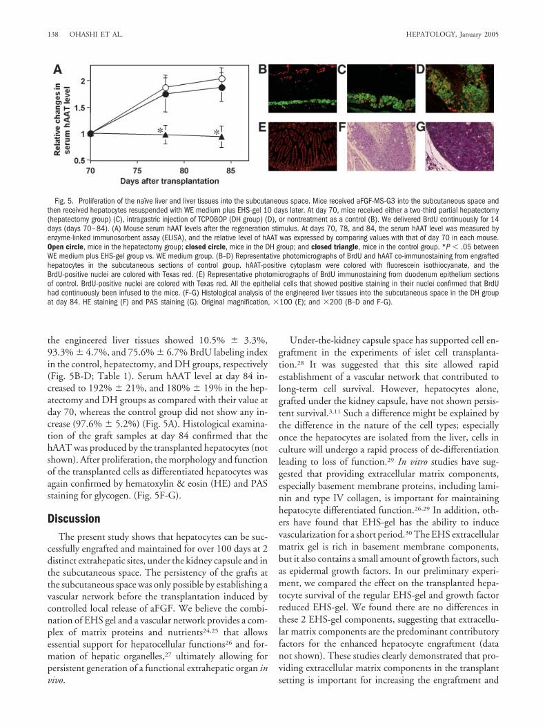
the engineered liver tissues showed 10.5% � 3.3%,93.3% � 4.7%, and 75.6% � 6.7% BrdU labeling indexin the control, hepatectomy, and DH groups, respectively(Fig. 5B-D; Table 1). Serum hAAT level at day 84 in-creased to 192% � 21%, and 180% � 19% in the hep-atectomy and DH groups as compared with their value atday 70, whereas the control group did not show any in-crease (97.6% � 5.2%) (Fig. 5A). Histological examina-tion of the graft samples at day 84 confirmed that thehAAT was produced by the transplanted hepatocytes (notshown). After proliferation, the morphology and functionof the transplanted cells as differentiated hepatocytes wasagain confirmed by hematoxylin & eosin (HE) and PASstaining for glycogen. (Fig. 5F-G).
DiscussionThe present study shows that hepatocytes can be suc-
cessfully engrafted and maintained for over 100 days at 2distinct extrahepatic sites, under the kidney capsule and inthe subcutaneous space. The persistency of the grafts atthe subcutaneous space was only possible by establishing avascular network before the transplantation induced bycontrolled local release of aFGF. We believe the combi-nation of EHS gel and a vascular network provides a com-plex of matrix proteins and nutrients24,25 that allowsessential support for hepatocellular functions26 and for-mation of hepatic organelles,27 ultimately allowing forpersistent generation of a functional extrahepatic organ invivo.
Under-the-kidney capsule space has supported cell en-graftment in the experiments of islet cell transplanta-tion.28 It was suggested that this site allowed rapidestablishment of a vascular network that contributed tolong-term cell survival. However, hepatocytes alone,grafted under the kidney capsule, have not shown persis-tent survival.3,11 Such a difference might be explained bythe difference in the nature of the cell types; especiallyonce the hepatocytes are isolated from the liver, cells inculture will undergo a rapid process of de-differentiationleading to loss of function.29 In vitro studies have sug-gested that providing extracellular matrix components,especially basement membrane proteins, including lami-nin and type IV collagen, is important for maintaininghepatocyte differentiated function.26,29 In addition, oth-ers have found that EHS-gel has the ability to inducevascularization for a short period.30 The EHS extracellularmatrix gel is rich in basement membrane components,but it also contains a small amount of growth factors, suchas epidermal growth factors. In our preliminary experi-ment, we compared the effect on the transplanted hepa-tocyte survival of the regular EHS-gel and growth factorreduced EHS-gel. We found there are no differences inthese 2 EHS-gel components, suggesting that extracellu-lar matrix components are the predominant contributoryfactors for the enhanced hepatocyte engraftment (datanot shown). These studies clearly demonstrated that pro-viding extracellular matrix components in the transplantsetting is important for increasing the engraftment and
Fig. 5. Proliferation of the naı̈ve liver and liver tissues into the subcutaneous space. Mice received aFGF-MS-G3 into the subcutaneous space andthen received hepatocytes resuspended with WE medium plus EHS-gel 10 days later. At day 70, mice received either a two-third partial hepatectomy(hepatectomy group) (C), intragastric injection of TCPOBOP (DH group) (D), or nontreatment as a control (B). We delivered BrdU continuously for 14days (days 70–84). (A) Mouse serum hAAT levels after the regeneration stimulus. At days 70, 78, and 84, the serum hAAT level was measured byenzyme-linked immunosorbent assay (ELISA), and the relative level of hAAT was expressed by comparing values with that of day 70 in each mouse.Open circle, mice in the hepatectomy group; closed circle, mice in the DH group; and closed triangle, mice in the control group. *P � .05 betweenWE medium plus EHS-gel group vs. WE medium group. (B–D) Representative photomicrographs of BrdU and hAAT co-immunostaining from engraftedhepatocytes in the subcutaneous sections of control group. hAAT-positive cytoplasm were colored with fluorescein isothiocyanate, and theBrdU-positive nuclei are colored with Texas red. (E) Representative photomicrographs of BrdU immunostaining from duodenum epithelium sectionsof control. BrdU-positive nuclei are colored with Texas red. All the epithelial cells that showed positive staining in their nuclei confirmed that BrdUhad continuously been infused to the mice. (F–G) Histological analysis of the engineered liver tissues into the subcutaneous space in the DH groupat day 84. HE staining (F) and PAS staining (G). Original magnification, �100 (E); and �200 (B–D and F–G).
138 OHASHI ET AL. HEPATOLOGY, January 2005

stable survival of the hepatocytes, leading to liver tissueengineering. However, further experiments are needed toanalyze which of the extracellular matrix element(s) arenecessary for facilitating hepatocyte engraftment.
Toward a novel treatment for liver diseases, we thenassessed the therapeutic efficacy of our approach for livertissue engineering in a mouse model of hemophilia A.Hemophilia A is a bleeding disorder, occurring in 1 ofapproximately 5,000 men who lack the production offunctional factor VIII. Protein replacement therapy withinfusion of plasma-derived or recombinant factor VIII hasbeen a standard for this disease. Because the liver is amajor contributory organ for factor VIII production,31
recent experience in transplanting whole or part of theliver into hemophilia patients have resulted in curativelevels (�50%) of factor VIII production and clotting ac-tivity.32,33 However, raising these levels into the range of2% to 3% will result in a significant phenotypic improve-ment in the disease, and this prompted us to establishwhether engineering liver tissues would have a therapeuticbenefit on hemophilia A.34 As shown in this study, by engi-neering liver tissue with 3 � 106 hepatocytes that are equiv-alent to approx 3% of the naı̈ve liver, we could demonstratetherapeutic efficacy in reconstituting 5% to 10% of normalclotting activity, enough to reduce the bleeding time to nearnormal values. These findings establish the proof-of-conceptthat extrahepatic liver tissue engineering is a viable therapyfor patients with hepatodeficiency conditions.
The subcutaneous space is a particularly attractive sitefor cell transplantation and tissue engineering, because itis an easy site to access and manipulate, and it holds aremarkably large capacity for engrafting.3,7 However, sev-eral studies have not been able to achieve persistent sur-vival of hepatocytes transplanted into the subcutaneousspace.8,35 In the previous and present studies, we experi-enced a significant decrease in cell survival within the first2 weeks after the transplantation even in the presence ofan extracellular matrix component and HGF/cMet stim-ulation. We hypothesized that a major cause of cell deathwas insufficient vascularity at the transplantation site.Our strategy, establishing a vascular network using con-tinuous delivery of aFGF before transplantation, clearlydemonstrated its importance for persistent survival lead-ing to liver tissue engineering. Other studies have re-ported that transplanting hepatocytes transfected withvascular endothelial growth factor genes or transplantinghepatocytes with a bFGF-releasing device showed someeffects on survival when cells were transplanted into theabdominal cavity.24,25 However, persistent survival of thehepatocytes was not achieved. In prior strategies, oncecells were implanted in the recipient, metabolic needsbegan immediately, but it took several days for the growth
of new blood vessels that would deliver oxygen and nutri-ents to the grafts.7 Ready access to the subcutaneous spacerenders a staged procedure such as that presented heremore clinically feasible.
The liver has a number of fascinating properties be-cause of its proliferative capacity18,36–38 through 2 distinctpathways, compensatory regeneration or direct hyperpla-sia.19,20,39 A large number of molecules are involved in theliver proliferation process in both endocrine and paracrinefashions.36,39 We have previously reported that the acti-vation of HGF/cMet is one of the important pathways forhepatocytes to grow at the extrahepatic sites.12 Althoughit has been suggested that shunting the portal circulationto the engrafted hepatocytes (to diverse growth factorsfrom portal to a general circulation) is necessary for theircell proliferation,40 the present study clearly demonstratesthat engineered liver tissues at two different extrahepaticsites could regenerate with similar activity as their naı̈velivers without the need of portal blood supply. It is alsoimportant to note that the onset and completion of theproliferation process of the engineered liver tissues oc-curred within the same period as naı̈ve liver. These find-ings, along with the morphological, and enzymatical(PAS staining) analyses, proved that the liver tissues com-prising donor hepatocytes were recognized as a part oftheir own liver in vivo.
In summary, the present studies show that liver tissue,which was recognized as a part of the host naı̈ve liver in termsof the proliferation profile, could be engineered at a heterol-ogous site that does not have access to the portal circulation.This approach is further supported by the recent interest inthe ability to generate mature hepatocytes from embryonic,hematopoietic, or somatic stem cells as an alternative cellsource.41 These cells could be transplanted in a similar man-ner, offering new medical therapy for many patients with amyriad of different diseases.3,34 Taken together, the currentwork thus can serve as the basis to enable further develop-ment of tissue engineering, stem cell work, and molecularmedicine approaches to liver disease.
Acknowledgment: The authors thank Dr. G.L. Bum-gardner for providing the hA1AT-FVB/N mouse line,Dr. H. Kazazian Jr. for providing factor VIII–deficientmice, and Dr. B. Diwan for providing TCPOCOP. Theauthors also thank Thu Thao T. Pham, Kimbary Wiges,and Rika Hongo for technical assistance in histology andenzyme-linked immunosorbent assay.
References1. Fox IJ, Chowdhury JR, Kaufman SS, Goertzen TC, Chowdhury NR,
Warkentin PI, et al. Treatment of the Crigler-Najjar syndrome type I withhepatocyte transplantation. N Engl J Med 1998;338:1422–1426.
HEPATOLOGY, Vol. 41, No. 1, 2005 OHASHI ET AL. 139

2. Muraca M, Gerunda G, Neri D, Vilei MT, Granato A, Feltracco P, et al.Hepatocyte transplantation as a treatment for glycogen storage disease type1a. Lancet 2002;359:317–318.
3. Ohashi K, Park F, Kay MA. Hepatocyte transplantation: clinical and ex-perimental application. J Mol Med 2001;79:617–630.
4. Ponder KP, Gupta S, Leland F, Darlington G, Finegold M, DeMayo J, etal. Mouse hepatocytes migrate to liver parenchyma and function indefi-nitely after intrasplenic transplantation. Proc Natl Acad Sci U S A 1991;88:1217–1221.
5. Gupta S, Chowdhury JR. Hepatocyte transplantation. In: Arias IM, BoyerJL, Fausto N, Jakoby WB, Schachter D, Shafritz DA, eds. The Liver:Biology and Pathobiology. 3rd ed. New York, NY: Raven Press, 1994:1519–1536.
6. Schneider A, Attaran M, Gratz KF, Bleck JS, Winkler M, Manns MP, et al.Intraportal infusion of 99mtechnetium-macro-aggregrated albumin parti-cles and hepatocytes in rabbits: assessment of shunting and portal hemo-dynamic changes. Transplantation 2003;75:296–302.
7. Griffith LG, Naughton G. Tissue engineering-current challenges and ex-panding opportunities. Science 2002;295:1009–1014.
8. Ohashi K, Marion PL, Nakai H, Meusu L, Cullen JM, Bordier BB, et al.Sustained survival of human hepatocytes in mice: a model for in vivoinfection with human hepatitis B and hepatitis delta viruses. Nat Med2000;6:327–331.
9. Allen JW, Bhatia SN. Engineering liver therapies for the future. Tissue Eng2002;8:725–737.
10. Jirtle RL, Michalopoulos G. Effects of partial hepatectomy on transplantedhepatocytes. Cancer Res 1982;42:3000–3004.
11. Ricordi C, Lacy PE, Callery MP, Park PW, Flye MW. Trophic factorsfrom pancreatic islets in combined hepatocyte-islet allografts enhance hep-atocellular survival. Surgery 1989;105(2 Pt 1):218–223.
12. Ohashi K, Meuse L, Schwall R, Kay MA. cMet activation allows persistentengraftment of ectopically transplanted xenogenic human hepatocytes inmice. Transplant Proc 2001;33:587–588.
13. Bi L, Lawler AM, Antonarakis SE, High KA, Gearhart JD, Kazazian HHJr. Targeted disruption of the mouse factor VIII gene produces a model ofhaemophilia A. Nat Genet 1995;10:119–121.
14. Shima M, Matsumoto T, Fukuda K, Kubota Y, Tanaka I, Nishiya K, et al.The utility of activated partial thromboplastin time (apt) clot waveform inthe investigation of hemophilia A patients with very low levels of factorVIII activity (FVIII:C). Thromb Haemost 2002;87:436–441.
15. Wang L, Zoppe M, Hackeng TM, Griffin JH, Lee KF, Verma IM. A factorIX-deficient mouse model for hemophilia B gene therapy. Proc Natl AcadSci U S A 1997;94:11563–11566.
16. Yuksel E, Weinfeld AB, Cleek R, Waugh JM, Jensen J, Boutros S, et al. Denovo adipose tissue generation through long-term, local delivery of insulinand insulin-like growth factor-1 by PLGA/PEG microspheres in an in vivorat model: a novel concept and capability. Plast Reconstr Surg 2000;105:1721–1729.
17. Elkins C, Waugh JM, Amabile PG, Minamiguchi H, Uy M, Sugimoto K,et al. Development of a platform to evaluate and limit in-stent restenosis.Tissue Engineering 2002;8:395–407.
18. Ponder KP, Gupta S, Leland F, Darlington G, Finegold M, Demayo J, etal. Mouse hepatocytes migrate to liver parenchyma and function indefi-nitely after intrasplenic transplantation. Proc Natl Acad Sci U S A 1991;88:1217–1221.
19. Ohashi K, Park F, Kay MA. Role of hepatocyte direct hyperplasia inlentivirus-mediated liver transduction in vivo. Hum Gene Ther 2002;13:653–663.
20. Columbano A, Ledda-Columbano GM, Pibiri M, Piga R, Shinozuka H,DeLuca V, et al. Increased expression of c-fos, c-jun and LRF-1 is notrequired for in vivo priming of hepatocytes by the mitogen TCPOBOP.Oncogene 1997;14:857–863.
21. Ohashi K, Tsutsumi M, Tsujiuchi T, Kobitsu K, Okajima E, Nakajima Y,et al. Enhancement of N-nitrosodiethylamine-initiated hepatocarcinogen-
esis caused by a colchicines-induced cell cycle disturbance in partially hep-atectomized rats. Cancer Res 1996;56:3474–3479.
22. Bumgardner GL, Heininger M, Li J, Xia D, Parker-Thornburg J, FergusonRM, et al. A functional model of hepatocyte transplantation for in vivoimmunologic studies. Transplantation 1998;65:53–61.
23. Passaniti A, Taylor RM, Pili R, Guo Y, Long PV, Haney JA, et al. A simple,quantitative method for assessing angiogenesis and antiangiogenic agentsusing reconstituted basement membrane, heparin, and fibroblast growthfactor. Lab Invest 1992;67:519–528.
24. Ajioka T, Akaike T, Watanabe Y. Expression of vascular endothelialgrowth factor promotes colonization, vascularization, and growth of trans-planted hepatic tissues in the mouse. HEPATOLOGY 1999;29:396–402.
25. Lee H, Cusick RA, Browne F, Ho Kim T, Ma PX, Utsunomiya H, et al.Local delivery of basic fibroblast growth factor increases both angiogenesisand engraftment of hepatocytes in tissue-engineered polymer devices.Transplantation 2002;73:1589–1593.
26. Bissell DM, Arenson DM, Maher JJ, Roll FJ . Support of cultured hepa-tocytes by a laminin-rich gel: evidence for a functionally significant suben-dothelial matrix in normal rat liver. J Clin Invest 1987;79:801–812.
27. Mitaka T, Sato F, Mizuguchi T, Yokono T, Mochizuki Y. Reconstructionof hepatic organoid by rat small hepatocytes and hepatic nonparenchymalcells. HEPATOLOGY 1999;29:111–125.
28. Kumagai N, O’Neil JJ, Barth RN, LaMattina JC, Utsugi R, Moran SG, etal. Vascularized islet-cell transplantation in miniature swine. I. Preparationof vascularized islet kidneys. Transplantation 2002;74:1223–1230.
29. Runge D, Runge DM, Drenning SD, Bowen WC Jr, Grandis JR, Micha-lopoulos GK. Growth and differentiation of rat hepatocytes: changes intranscription factors HNF-3, HNF-4, STAT-3, and STAT-5. BiochemBiophys Res Commun 1998;250:762–768.
30. Fridman R, Sweeney TM, Zain M, Martin GR, Kleinman HK. Malignanttransformation of NIH-3T3 cells after subcutaneous co-injection with areconstituted basement membrane (Matrigel). Int J Cancer 1992;51:740–744.
31. Wion KL, Kelly D, Summerfield JA, Tuddenham EG, Lawn RM. Distri-bution of factor VIII mRNA and antigen in human liver and other tissues.Nature 1985;317:726–729.
32. Wilde J, Teixeira P, Bramhall SR, Gunson B, Mutimer D, Mirza DF. Livertransplantation in haemophilia. Br J Haematol 2002;117:952–956.
33. Horita K, Matsunami H, Shimizu Y, Shimizu A, Kurimoto M, Suzuki K,et al. Treatment of a patient with hemophilia A and hepatitis C virus-related cirrhosis by living-related liver transplantation from an obligatecarrier donor. Transplantation 2002;73:1909–1912.
34. High KA. Gene therapy: a 2001 perspective. Hemophilia 2001;7(Suppl1):23–27.
35. Demetriou AA, Whiting JF, Feldman D, Levenson SM, Chowdhury NR,Moscioni AD, et al. Replacement of liver function in rats by transplanta-tion of microcarrier-attached hepatocytes. Science 986;233:1190–1192.
36. Michalopoulos GK, DeFrancis ME. Liver regeneration. Science 1997;276:60–66.
37. Ohashi K, Tsutsumi M, Nakajima Y, Kobitsu K, Nakano H, Konishi Y.Telomere changes in human hepatocellular carcinomas and hepatitis virusinfected noncancerous livers. Cancer 1996;77: S1747–S1751.
38. Higgins GM, Anderson RM. Experimental pathology of the liver: restora-tion of the liver of the white rat following partial surgical removal. ArchPathol 1931;12:186–202.
39. Columbano A, Shinozuka H. Liver regeneration versus direct hyperplasia.FASEB J 1996;10:1118–1128.
40. Kaufmann PM, Sano K, Uyama S, Takeda T, Vacanti JP. Heterotopichepatocyte transplantation: assessing the impact of hepatotrophic stimula-tion. Transplant Proc 1994;26:2240–2241.
41. Jones EA, Tosh D, Wilson DI, Lindsay S, Forrester LM. Hepatic differ-entiation of murine embryonic stem cells. Exp Cell Res 2002;272:15–22.
140 OHASHI ET AL. HEPATOLOGY, January 2005