


Quantification of Liver Fat in Mice: Comparing Dual-Echo Dixon ...
31
1 Quantification of Liver Fat in Mice: Comparing Dual-Echo Dixon Imaging, Chemical Shift Imaging and 1 H-MR Spectroscopy Xin-Gui Peng(彭新桂) 1* , Shenghong Ju(居胜红) 1* , Yujiao Qi(秦雨姣) 1 , Fang Fang (方芳) 1 , Xin Cui(崔鑫) 2 , George Liu 2 , Yicheng Ni(倪以成) 3 , Gao-Jun Teng(滕皋军) 1 1 Laboratory of Molecular Imaging, Department of Radiology, Zhongda Hospital, Southeast University, Nanjing 210009, China 2 Institute of Cardiovascular Sciences and Key Laboratory of Molecular Cardiovascular Sciences, Ministry of Education, Peking University Health Science Center, Beijing, 10091, China 3 Department of Radiology, University Hospitals, Catholic University of Leuven, Leuven B-3000, Belgium * These authors contributed equally Original research Correspondence to: Dr. Shenghong Ju Laboratory of Molecular Imaging, Department of Radiology Zhongda Hospital Medical School of Southeast University 87 Ding Jia Qiao Road Nanjing 210009, China Tel.: 086 25 83272121 Fax: 086 25 83311083 Email: [email protected] Abbreviated title: In Vivo MR Quantification of Liver Fat Content in Mice by guest, on April 4, 2018 www.jlr.org Downloaded from
Transcript of Quantification of Liver Fat in Mice: Comparing Dual-Echo Dixon ...

1
Quantification of Liver Fat in Mice: Comparing Dual-Echo Dixon Imaging,
Chemical Shift Imaging and 1H-MR Spectroscopy
Xin-Gui Peng(彭新桂)1*, Shenghong Ju(居胜红)1*, Yujiao Qi(秦雨姣)1, Fang Fang (方芳)1,
Xin Cui(崔鑫)2, George Liu2, Yicheng Ni(倪以成)3, Gao-Jun Teng(滕皋军)1 1Laboratory of Molecular Imaging, Department of Radiology, Zhongda Hospital, Southeast
University, Nanjing 210009, China
2Institute of Cardiovascular Sciences and Key Laboratory of Molecular Cardiovascular
Sciences, Ministry of Education, Peking University Health Science Center, Beijing, 10091,
China
3Department of Radiology, University Hospitals, Catholic University of Leuven, Leuven
B-3000, Belgium
* These authors contributed equally
Original research
Correspondence to:
Dr. Shenghong Ju
Laboratory of Molecular Imaging, Department of Radiology
Zhongda Hospital
Medical School of Southeast University
87 Ding Jia Qiao Road
Nanjing 210009, China
Tel.: 086 25 83272121
Fax: 086 25 83311083
Email: [email protected]
Abbreviated title: In Vivo MR Quantification of Liver Fat Content in Mice
by guest, on April 4, 2018
ww
w.jlr.org
Dow
nloaded from

2
Abbreviations used: 1H = hydrogen
BSCL2 = Berardinelli-Seip congenital lipodystrophy type 2
CGL = Congenital generalized lipodystrophy
CSI = chemical shift imaging
CT = computed tomography
FC = fat content
HIS-S = semi-automatic vacuole segmentation procedure
IP-OP = in-phase and out-of-phase
LI = liver index
LL = liver lipid
MR = magnetic resonance
MRS = magnetic resonance spectroscopy
NAFLD = non-alcoholic fatty liver disease
ob/ob = obese/obese
ROI = region of interest
SD = standard deviation
SI = signal intensity
SKO = human BSCL2/seipin gene knock-out
TR = repetition time
TE = echo time
WT = wild type
by guest, on April 4, 2018
ww
w.jlr.org
Dow
nloaded from

3
ABSTRACT
We evaluate dual-echo Dixon (in-phase and out-of-phase, IP-OP), chemical shift imaging
(CSI), and 1H MRS in estimating fat content (FC) in phantoms and livers of mice. Phantoms
were made according to the volume percentage of fat ranging from 0% to 100%. Three MR
methods were performed to measure FCs in phantoms, livers of ob/ob, human BSCL2/seipin
gene knock-out (SKO) and wild type (WT) mice. The results were compared to known FCs in
phantoms and to reference standard from mice by histological semi-automatic vacuole
segmentation procedure (HIS-S) and liver lipid (LL) chemical analysis. In phantoms, CSI
underestimated FC in range of 50-100%, to a smaller extent than IP-OP. In vivo, liver FCs in
ob/ob and SKO mice measured by three MR methods were all significantly higher than that of
WT mice. Liver FC measured by IP-OP are significantly lower than that measured by CSI
and MRS with no significant difference between CSI and MRS. CSI and MRS showed a
linear correlation with LL analysis and with each other. IP-OP underestimated FC, while CSI
and MRS are more accurate for quantifying fat in both phantoms and liver. CSI and MRS
have the potential to replace HIS-S and LL analysis in longitudinal studies.
Supplementary key words: non-alcoholic fatty liver disease; obesity; triglyceride; spin-spin
relaxation time T2; spectroscopy; in vivo
by guest, on April 4, 2018
ww
w.jlr.org
Dow
nloaded from

4
INTRODUCTION
During the past decade, there has been evidence of an epidemic increase in the
non-alcoholic fatty liver disease (NAFLD), which affects 10%–30% of adults (1-3) and 13%
of children (4) in the general population. NAFLD has a strong association with type II
diabetes mellitus, obesity, hyperlipidemia, and other diseases of the metabolic syndrome (5).
The hallmark of NAFLD is fatty infiltration of hepatocytes. Hepatic steatosis is also reflective
to the toxicity of drugs such as amiodarone, tamoxifen, and antiretrovirals (6).
Thus, liver fat quantification has generated considerable interest; it may be of clinical
importance to be able to reliably measure liver fat content (FC). Liver biopsy and histological
analysis are considered as the diagnostic reference standard. But in human, the biopsy
procedure is both invasive and painful, and presents risks for the patients (7, 8). In addition,
the very small liver sample may not be representative of the heterogeneous fat distribution (9).
The analogous procedure in animals involves killing the animals and directly analyzing liver
FC, which is not ideal for longitudinal follow-up studies.
Hydrogen (1H) magnetic resonance (MR) imaging offers several non-invasive methods to
obtain separate fat and water images for the liver FC quantification. In 1984, the Dixon
technique for water and fat imaging was described (10). The authors presented two imaging
sequences, one of them was a conventional spin echo imaging with water and fat signals
in-phase, and the second one had the readout gradient slightly shifted to create 180°
out-of-phase of the water and fat signals. Over the last decade, the Dixon technique has been
improved extensively in the aspects of phase errors, noise and artifacts. Triple-echo and
multiecho in- and out-of-phase MR imaging were proposed, which allow correction for T2*
decay and lipid quantification (11, 12). Iterative decomposition of water and fat with echo
asymmetry and least-squares estimation (IDEAL) has also improved fat quantification and fat
suppression (13, 14). In addition to the Dixon technique, in 1985, Haase et al. proposed
chemical shift selective imaging technique, which relies on a single frequency-selective
excitation pulse with a flip angle of 90° followed by a dephasing gradient to distinguish H2O
and CH2 difference (15). Sbarbati et al. and Lunati et al. then applied chemical shift imaging
to in vivo quantify lipid of brown adipose tissue (16, 17).
Animal models of hepatic steatosis and steatohepatitis have improved our understanding
by guest, on April 4, 2018
ww
w.jlr.org
Dow
nloaded from

5
of the pathogenesis of NAFLD. Continued studies in animals will further clarify the
pathogenesis of these disorders and, therefore, will probably improve the diagnosis and
treatment of human NAFLD. The ob/ob mouse is an excellent model of hepatic steatosis
because of its expression of many NAFLD traits (18). These mice are genetically leptin
deficient, which causes excessive overeating and development of obesity, steatosis,
steatohepatitis, and diabetes, among other symptoms of NAFLD (19).
Berardinelli-Seip congenital lipodystrophy type 2 (BSCL2) is a recessive disorder
characterized by an almost complete loss of adipose tissue, insulin resistance and fatty liver
(20, 21). BSCL2 encodes a protein, seipin, the function of which is largely unknown (22, 23).
Recently, Cui et al. created the first murine model of BSCL2 by targeted disruption of seipin
and suggested a possible tissue-autonomous role of seipin in liver lipid storage (24). To better
understand the effect of seipin levels on fatty liver, we have measured liver fat content in a
human BSCL2/seipin gene knock-out (SKO) mouse model in vivo and in vitro.
The purpose of this study was to evaluate the dual-echo Dixon MR imaging (in-phase
and out-of-phase, IP-OP), chemical shift imaging (CSI) selective for either fat or water
protons imaging, and 1H MR spectroscopy (MRS) for fat measurement in phantoms and in the
liver of ob/ob and human BSCL2/seipin gene knock-out (SKO) mice, and to compare these
MR-derived data with those from histological and chemical analysis.
by guest, on April 4, 2018
ww
w.jlr.org
Dow
nloaded from

6
MATERIALS AND METHODS
Animals
All animal experiments were approved by the institutional Committee on Animal
Research. Obese leptin-deficient mice (C57BL/∧OlaHsd-Lep; ob/ob) and wild type (WT)
C57BL/6J mice were obtained from Laboratory Animal Center of Academy of Military
Medical Science and Shanghai Model Animal Research Center, respectively. They were fed
normally before the experiment and kept at temperature of 20-24°C in 12 hour day/night
periods. Six male ob/ob and WT mice each, of ten-week-old with an average weight of 47.5 ±
1.54 g and 26 ± 0.71 g respectively, were used in this study. Four human BSCL2/seipin gene
knock-out (SKO) and four wild type (WT) C57BL/6J mice were presented by Peking
University Health Science Center, ten-week-old with an average weight of 21.8 ± 1.61 g and
22.6 ± 1.25 g respectively. After MR scan, they were euthanized with an overdose of
pentobarbital.
Fat/ Water Phantoms
According to Poon et al. (25), phantoms were made by mixing known amounts of water
(doped with 0.2 mM MnCl2) and vegetable oil (extra virgin olive oil, Olivoila, Italy).
Percentages of oil by volume ranged from 0 to 100 % in steps of 10 %. To improve the
stability of the mixture, 2 % of Tween 80 (polyoxyethylene sorbitan monooleate) by volume
of oil was added. The mixture was blended homogeneously using an ultrasonic homogenizer
(KQ-400KQE, Kunshan Ultrasonic Equipment CO, LTD, Kunshan, China). Plastic tubes, of
15 mm in diameter, containing the suspensions were placed longitudinally into the magnet.
Phantoms were scanned and analyzed with the same experimental protocol as that in vivo.
MR Imaging and 1H MRS Protocol
For in vivo MR acquisition, anesthesia was induced by inhalation of a mixture of oxygen
and 5% isoflurane and maintained by a mixture of oxygen containing 0.5 % to 1 % isoflurane.
All MR experiments were carried out using a 7T small animal magnetic resonance system
(Bruker PharmaScan, Ettlingen, Germany) that is interfaced to a Bruker console. The
horizontal bore system is equipped with a 15 cm diameter gradient set capable of generating
by guest, on April 4, 2018
ww
w.jlr.org
Dow
nloaded from

7
375 mT/m gradient strengths in all three directions. A 31 mm inner diameter transmit-receive
quatrature coil was used for MR data collection.
T1-weighted images were acquired with a respiratory-gated spin echo sequence, 500/15
ms; section thickness, 2 mm; matrix, 256×256; field of view, 3.5×3.5 cm; and number of
excitations, 4. The T1-weighted images were used to study the distribution of fat stores and
measure the volume of the liver using ImageJ software.
A point-resolved spectroscopy sequence for localized 1H MRS sequence was used with
the following parameters: repetition time (TR)/echo time (TE), 2500/20 ms; voxel, 3 × 3 × 3
mm; and number of excitations, 128. To correct for T2 decay, seven consecutive spectra were
acquired with echo times of 10, 20, 30, 40, 50, 70 and 90 msec. A 9-mm3 region of interest
(ROI) was placed over the left lobe of the liver, avoiding intrahepatic blood vessels. Before
measurement, the automatic shimming procedure FASTMAP was used to achieve optimal
uniformity of the magnetic field across the voxel volume. Water suppression was never used
in all spectroscopy sequences during the measurement. The free induction decay signals were
Fourier-transformed. The phase and the baseline of the spectra were also corrected with great
care using TOPSPIN (Bruker BioSpin MRI GmbH). Spectra were used only if homogeneity
after shimming was better than 0.45 ppm, measured as the full width at 50 % peak height.
Spin-spin relaxation times (T2) was determined for nine different peaks (in the range from 0.9
to 5.32 ppm) by fitting the monoexponential model function MTE = M0 × exp(-TE/T2) to the
measured peak integrals at the different TEs, and the correction factors (M0/MTE) for the nine
different proton resonances of triacylglycerols and the proton resonance of the water peak
were caculated (26-28). The degree of FC was calculated using the following formula:
FCMRS = 100 × (integral value of fat peak) / (integral value of fat peak + integral value of
water peak) [1]
Fast low angle shot imaging of the entire liver were obtained for both in-phase and
out-of-phase transverse dual-echoes. Imaging parameters were as follows: 500/1.47
(out-of-phase) or 1.97 (in-phase); flip angle, 40°; section thickness, 2 mm; matrix, 256×256;
field of view, 3.5×3.5cm. A radiologist used these images to position one voxel in the liver
parenchyma outside the area of the great vessels for spectroscopy. And the size and location
of the ROI matched those of the original 1H MRS voxel on three consecutive in-phase and
by guest, on April 4, 2018
ww
w.jlr.org
Dow
nloaded from

8
out-of-phase images. Signal intensity (SI) was measured at ParaVision4.0 (Bruker BioSpin
MRI). The SI for each ROI (3×3mm) was recorded separately, and an average SI was
calculated from the three images to gather information for the entire 1H MR spectroscopy
voxel volume. We used the following equation:
FCIP-OP = 100 × [(SIIP - SIOP)/ (2 × SIIP)] [2]
where SIIP is SI measured on the in-phase; SIOP, SI measured on the out-of-phase image. The
in-phase and out-of-phase images were used to study the liver FC.
CSI were also used to study liver FC. Two series of chemical shift selective images, one
for fat protons and one for water protons, were derived using rapid acquisition with relaxation
enhancement (RARE) sequence. Both series were performed by the 180° refocusing pulse (16,
17). Both the 90° and the 180° pulses were band-selective Gaussian pulses; however, the 90°
pulse was applied when the slice-selection gradient was on, and when the 180° pulse is
applied, all gradients were off to refocus only in the selected chemical-shift range. The 180°
pulse was a 2.3 ms Gaussian pulse, which excited a 700 Hz bandwidth. This was appropriate
to discriminate fat and water peaks which, at 7T, have a separation of about 1000 Hz. All
images were acquired using the following parameters: 1000/9.9 ms, flip angle, 180°; section
thickness, 1mm; matrix, 256×256; field of view, 3.5×3.5cm; and number of excitations, 4. An
ROI was drawn at the same site as the voxel used for 1H MRS in the left lobe. The SI in the
ROI was recorded for both fat selective images (SIfat) and water selective images (SIwater).
Liver FC was computed as follows:
FCCSI=100× SIfat/ (SIfat+SIwater×R) [3]
where R is the ratio of the fat to water proton densities in their pure form: a value R = 0.9 has
been used in literature (25). The water fraction by volume of the sample is thus 1- FC.
Histopathology
Each mouse were perfused transcardially with phosphate-buffered saline, followed by
freshly prepared 4 % paraformaldehyde in 0.1 M phosphate buffer (pH 7.4); Parts of liver
were fixed, dehydrated, embedded and transversely sectioned into 5 μm pieces for
hematoxylin and eosin staining. Other parts of liver were frozen in dry ice and then cryostat
sectioned at a thickness of 7 um onto poly-l-lysine slides for lipid deposition analysis by Oil
by guest, on April 4, 2018
ww
w.jlr.org
Dow
nloaded from

9
Red O and hematoxylin staining.
All histopathology slides were examined and FC analysis was performed with a
semi-automatic vacuole segmentation procedure (HIS-S) developed with the MATLAB
software (The MathWorks, Natick, Mass.) as describe by Gaspard et al. (29). The artificial
areas such as blood vessels were manually excluded by a pathologist. The percent fat fraction
(FCHIS-S) was calculated by following formula:
FCHIS-S = the area of fat / the total tissue area [4]
Liver Lipid (LL) Analysis
Approximately 100 mg of liver (wet weight) was weighed and homogenized in 1 ml of
PBS. Lipid was extracted by homogenizing with 2: 1 chloroform-methanol (v/v) and
separating into three phase by centrifugation, the upper, lower liquid phases and the middle of
solid phase. The upper phase was removed as much as possible by siphoning and extracted
once more. The lower phase was reserved and dried. Lipid was dissolved in 100ul 3% Triton
X-100. The determination of triglyceride was carried out using enzymatic methods as
described by Folch et al. (30).
Statistical Analysis
All statistical analyses were performed using SPSS software (SPSS for Windows, version
11.0, 2001; SPSS, Chicago, IL). Numerical data were reported as mean values ± standard
deviation (SD). For statistical comparisons, independent-sample t test, paired-sample t test
and correlation test were applied. A P value of less than 0.05 was considered to indicate a
statistically significant difference.
MR methods determined the volume fractions of lipids in the liver tissue and
histological method calculated the percentage of hepatocytes showing visible fat droplets in
the microscopic view, but the chemical method measured the TG content of fresh liver tissue.
For statistical comparisons, correlation test was applied to assess the relationship between LL
and FC measured by three MR and histological methods, both paired-sample t test and
correlation test were applied to assess difference and relationship between MR and
histological methods.
by guest, on April 4, 2018
ww
w.jlr.org
Dow
nloaded from
10
RESULTS
Quantitation in Fat/ Water Phantoms
Three MR-based methods of quantitative FC evaluation have been tested in fat/water
phantoms (Fig. 1, 2). Results for fat and water quantitation are shown in Table 1. The
dual-echo method significantly underestimated the fat concentration compared to the
gravimetric reference standard when FC was in range of 10% to 100%. There is a weak
correlation between FC calculated with dual-echo method and the known gravimetric
reference standard (r=0.03, P>0.05). When the actual FC is smaller than 50% (water content
higher than FC), fat concentration can be calculated using the formula: FCIP-OP = 100 × [(SIIP -
SIOP)/ (2 × SIIP)] but when the actual FC is higher than 50% (water content lower than FC), fat
concentration can be calculated using:WIP-OP = 100 × [(SIIP - SIOP)/ (2 × SIIP)], FCIP-OP =1-
WIP-OP. With the visual analysis of 1H MRS fat–water peak size to guide for fat and water
dominance on IP-OP images, the corrected IP-OP underestimated fat concentration when FC
was in range of 10% to 60%: (4.5±0)%, (9.2±2.0)%, (16.0±1.0)%, (26.2±0.1)%, (31.9±1.0)%
and (56.1±0.2)% respectively. There is a strong correlation (r = 0.972, P < 0.01) between FC
calculated with corrected IP-OP (FCIP-OP correction) and the known gravimetric reference
standard.
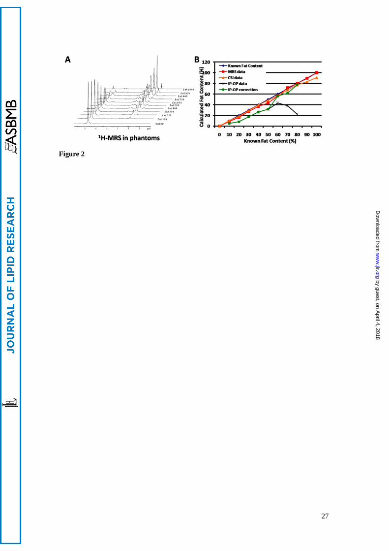
CSI underestimated fat concentration when FC was in range of 50% to 100%:
(48.0±1.0)%, (57.0±0.5)%, (67.3±0.6)%, (77.3±0.6)%, (83.3±0.6)% and (91.0±1.0)%
respectively (P<0.05). MRS overestimated slightly fat concentration when FC was in range of
80%: (81.6±0.6)% (P<0.05). CSI and MRS have a high linear correlation with the known
gravimetric reference standard (CSI: r=0.998, MRS: r=0.999, P<0.01), but correlation
between corrected IP-OP and the known FC is slightly weakest (r=0.972, P<0.01) (Fig. 3).
There are strong correlations between FC calculated with corrected IP-OP and MRS
(r=0.972, P<0.01), between CSI and MRS (r=0.998, P<0.01), and between corrected IP-OP
and CSI (r=0.963, P<0.01) (Fig. 3).
In vivo MR imaging and 1H MRS of mice
The liver FCs measured by all three MR methods (Fig. 4, 6), HIS-S (Fig. 5) and LL
analysis (Fig. 6) are listed in Table 2. There are significant differences in liver FC between
by guest, on April 4, 2018
ww
w.jlr.org
Dow
nloaded from

11
ob/ob mice and WT mice and between SKO mice and WT mice by using all methods
(P<0.001), with liver FC of the ob/ob mice and SKO mice all being higher than that of WT
mice. Both in vivo T1-weighted imaging and in vitro liver volume calculation prove the liver
volume of ob/ob and SKO mice being significantly larger than that of WT mice(P<0.001),
and there is no significant difference between these two methods (P=0.798). Liver index (LI)
was calculated as the volume of liver from MR and gravimetric measure divided by the total
body weight (LIMR, LIGM). Both LIMR and LIGM of ob/ob and SKO mice were significantly
higher than that of WT mice (P<0.001).
Liver FC measured by histological method and the corrected IP-OP is smaller than that
by CSI and MRS (P<0.001), but no significant difference is observed between FCIP-OP correction
and FCHIS-S (P=0.556). Liver FC measured by CSI and MRS are also no significant difference
(P=0.230) (Table 3). There is a strong correlation between FC calculated with histological
method and three MR methods (corrected IP-OP: r=0.882, CSI: r=0.984, MRS: r=0.978
respectively, P=0.000); and also a significant linear correlation is observed between LL
measured by chemical method and the FC calculated by all three MR methods and HIS-S
(corrected IP-OP: r=0.867, CSI: r=0.986, MR spectroscopy: r=0.977, HIS-S: r=0.960,
P=0.000)(Table 4, Fig.6) .
A significant linear correlation is observed between liver FC calculated by three MR
methods. The FCCSI and FCMRS correlations is slightly stronger (FCCSI vs. FCMRS: r=0.992,
P<0.001) than the FCIP-OP correction and FCCSI, FCMRS (FCIP-OP correction vs. FCCSI: r=0.891, FCIP-OP
correction vs. FCMRS: r=0.917, P=0.000) (Table 4, Fig.6).
by guest, on April 4, 2018
ww
w.jlr.org
Dow
nloaded from

12
DISCUSSION
There are currently three different types of imaging techniques that allow noninvasive
detection of liver FC: 1) ultrasound; 2) computed tomography (CT); and 3) MR methods.
However, ultrasound is not considered as a sufficient quantitative tool for fat determination in
liver. One major weakness of ultrasound is its operator dependency. CT requires undesired
radiation exposure in the examinations of subjects (31). MR methods therefore become the
most desirable and useful techniques. A number of MR methods have been proposed for the
detection of fatty liver infiltration: 1) Fat-sensitive MR imaging techniques based on signal
phase, two-point Dixon and three-point IDEAL, also known as iterative decomposition of
water and fat with echo asymmetry and least-squares estimation; 2) Fat-selective MR imaging
based on frequency selective excitation, chemical shift selective images, also called spectral
fat or water selective imaging; 3) 1H-MRS (31). 1H MRS has been by far the most promising
and most sensitive noninvasive method to assess liver FC (32).
In our study, liver FCs measured by two-point Dixon IP-OP, CSI and 1H-MR
spectroscopy were compared. Two-point Dixon is a routine clinical method for the
semi-quantitative assessment of liver FC. However, this method neglects T2* signal decay and
assumes that the signal difference between IP and OP echoes is due to fat-water signal
interference only. With OP-then-IP sequential acquisition, the confounding T2* effect is
known to cause fat fraction underestimation (33-35), which is also shown in this study.
At 7T MR, the resonance frequencies of methylene and water have a separation of about
1000Hz, the first echo time of in-phase time is 0.5 ms, and of out-of-phase time is 1.0 ms. Our
MR machine can only reach the echo time of in-phase time at 1.5ms and out-of-phase time at
2.0 ms. The corresponding in-phase and out-of-phase image signals were thus lower and the
difference between them became smaller (12). Our results agreed with our expectation by
proving that two-point Dixon method systematically resulted in an underestimation of FC in
both phantom and in vivo evaluations, and the degree of underestimation increased along with
increasing FC.
In an in-phase image, the fat and water signals within a voxel are additive; in an
opposed-phase image, they are subtracted from each other. The decrease in signal intensity
from the in-phase to an opposed-phase image indicates the presence of both water and fat
by guest, on April 4, 2018
ww
w.jlr.org
Dow
nloaded from

13
within a voxel. When the actual FC is higher than 50% (water content lower than FC), fat
concentration were corrected though above formulate. But IP-OP method also presented
greater underestimation compared to other methods due to the higher FC in the liver of ob/ob
and SKO mice.
CSI includes two series, selective fat-protons imaging and selective water-protons
imaging. Our CSI method was derived partly from the method described by Sbandrea et al.
(16), who evaluated the accuracy of CSI on brown adipose tissue in rats using a 4.7T MR
scanner. In phantoms, we proved using a 7T MR system that CSI was accurate in lipid
quantification when FC was lower than 50 %, and approximately 9 % underestimation of fat
occurred from 50 % to 100 % of FC. Since centers of excitation frequency are methylene and
water proton, the degree of FC underestimation increased in higher FC. Liver FC usually is
lower than 50 %, CSI is thus able to not only accurately measure the FC, but also evaluate its
distribution.
Besides liver histological and chemical analysis, as current standard for diagnosis and
grading of hepatic FC, 1H MRS also allows non-invasive studies in the molecular
composition of tissues in vivo. 1H MRS recorded from liver tissue usually shows two
dominant signal portions, namely the water signal (positioned at 4.7 ppm) and the signal from
methylene protons of fatty acids (positioned at 1.3 ppm). In 1H MRS, peak area is
proportional to concentration of the metabolite containing the relevant nuclei which is also
influenced by T1 and T2 relaxation times (36). To minimize T1 effects, one sequence with a
long repetition time (10 s) and one sequence with 1.8 s of repetition time were used by Strobel
et al (26). A correction factor was also used on the difference of the two spectra. In order to
correct T2 relaxation, a sequence with a series of different echo times was used to calculate
the spin-spin relaxation time (T2). We also used a series of different echo time to correct the
spectral data. In phantom experiments, we proved that there was more accurate that
quantifying FC with MRS method than with CSI when FC was in range of 50% to 100%.
MRS allows quantifying FC in different tissues, and CSI allows identifying the fat
distribution. Therefore, CSI and MRS should be used together in order to evaluate liver FC
comprehensively.
Interestingly, the percentage values of liver FC from MRS and CSI were slight different
by guest, on April 4, 2018
ww
w.jlr.org
Dow
nloaded from

14
compared to HIS-S examination. Gaspard et al. suggested that this might be due to the fact
that MR techniques determined the volume fractions of lipids in the liver tissue rather than the
percentage of hepatocytes showing visible fat droplets in the microscopic view (29). In
addition, this discrepancy might also be explained by the difficulty of quantifying
microvesicular fat. In fact, automated software estimated the area occupied by fat droplets
with a minimum diameter of 5 μm. Fat existing in droplets of diameters less than 5 μm was
therefore not included. Since the histological image was obtained by light transmitting
through the slice, droplets failed to pass through the slice completely would not be quantified.
Similarly, the results obtained by automated calculation software from histogical slides
and by isolation and purification of LL were not comparable to each other because the
automated software measured the specific surface area of fat droplets in the microscopic field,
whereas LL analysis measured the quantity of lipid. However, the data of LL chemical
analysis and CSI and 1H MRS correlated very well. This may be because volume fractions of
lipid in liver tissue were measured in MR methods and the TG content of fresh liver tissue
was detected in chemical method, rather than surface area of fat droplets in the HIS-S method.
In addition to the above mentioned MR methods, traditional T1-wighted imaging method
was also used to observe the anatomic details of liver, calculate liver volume, and identify the
distribution of fat in subcutaneous and internal organs (data not shown). Ob/ob mice had
significantly larger liver than other groups, indicating that severity of the fatty liver positively
correlate with the liver volume.
This study provided some information about fatty liver that may be useful when ob/ob
mice are used as an animal model of disease. The liver is the organ most often involved in fat
accumulation. In its parenchyma, the fat depot appears to be homogeneous, and it is
quantified by in vivo localized spectroscopy, MR imaging and by anatomical histological and
chemical surveys. Considering that ob/ob mice are a model for hepatic steatosis and SKO
mice are frequently with fat accumulation in liver, 1H MR spectroscopy and imaging seem to
be promising tools to follow the time course of fat accumulation.
The limitations of our study must be acknowledged. Firstly, the number of mice used in
the experiments was fairly small. Secondly, the amount of fat measured by HIS-S and liver
lipid analysis might not be representative of the amount of fat throughout the entire liver (9,
by guest, on April 4, 2018
ww
w.jlr.org
Dow
nloaded from

15
37). Thirdly, the FC measured using our MR based methods represents the signal derived
from protons contained in fatty acid molecules, as opposed to the weight of lipid per unit of
liver volume.
CONCLUSION
T1WI can be used to measure liver volume and observe fat distribution; IP-OP method
gives a FC that is significantly lower than the actual value, especially in higher fat
concentrations; CSI and 1H MRS are accurate in quantifying fat in both phantoms and liver in
vivo. Given their excellent correlation and concordance with LL analysis, CSI and MRS may
replace liver fat histological and chemical analysis in longitudinal studies.
by guest, on April 4, 2018
ww
w.jlr.org
Dow
nloaded from

16
This work was Supported by National Nature Science Foundation of China (NSFC,
No.30830039, No. 81071125) and Major State Basic Research Development Program of
China (973 Program) (No. 2010CB933903).
by guest, on April 4, 2018
ww
w.jlr.org
Dow
nloaded from
17
REFERENCES
1. Angulo, P. 2002. Nonalcoholic fatty liver disease. N. Engl. J. Med. 346: 1221-1231.
2. Wieckowska, A., A.J. McCullough, A.E. Feldstein. 2007. Noninvasive diagnosis and
monitoring of nonalcoholic steatohepatitis: present and future. Hepatology. 46: 582-589.
3. Gastaldelli, A., K. Cusi, M. Pettiti, J. Hardies, Y. Miyazaki, R. Berria, E. Buzzigoli, A.M.
Sironi, E. Cersosimo, E. Ferrannini. 2007. Relationship between hepatic/visceral fat and
hepatic insulin resistance in nondiabetic and type 2 diabetic subjects. Gastroenterology. 133:
496-506.
4. Schwimmer, J.B., R. Deutsch, T. Kahen, J.E. Lavine, C. Stanley, C. Behling. 2006.
Prevalence of fatty liver in children and adolescents. Pediatrics. 118: 1388-1393.
5. Harrison, S.A., B.A. Neuschwander-Tetri. 2004. Nonalcoholic fatty liver disease and
nonalcoholic steatohepatitis. Clin. Liver Dis. 8: 861-879.
6. Begriche, K., A. Igoudjil, D. Pessayre, B. Fromenty. 2006. Mitochondrial dysfunction in
NASH: causes, consequences and possible means to prevent it. Mitochondrion. 6: 1-28.
7. Castera, L., I. Negre, K. Samii, C. Buffet. 1999. Pain experienced during percutaneous
liver biopsy. Hepatology. 30: 1529-1530.
8. Joy, D., V.R. Thava, B.B. Scott. 2003. Diagnosis of fatty liver disease: is biopsy
necessary? Eur. J gastroenterol Hepatol. 15: 539-543.
9. Ratziu, V., F. Charlotte, A. Heurtier, S. Gombert, P. Giral, E. Bruckert, A. Grimaldi, F.
Capron, T. Poynard. 2005. Sampling variability of liver biopsy in nonalcoholic fatty liver
disease. Gastroenterology. 128: 1898-1906.
10. Dixon, W.T. 1984. Simple proton spectroscopic imaging. Radiology. 153: 189-194.
11. Guiu, B., J.M. Petit, R. Loffroy, D. Ben Salem, S. Aho, D. Masson, P. Hillon, D. Krause,
J.P. Cercueil. 2009. Quantification of liver fat content: comparison of triple-echo chemical
shift gradient-echo imaging and in vivo proton MR spectroscopy. Radiology. 250: 95-102.
12. O'Regan, D.P., M.F. Callaghan, M. Wylezinska-Arridge, J. Fitzpatrick, R.P. Naoumova,
J.V. Hajnal, S.A. Schmitz. 2008. Liver Fat Content and T2*: Simultaneous Measurement by
Using Breath-hold Multiecho MR Imaging at 3.0 T-feasibility. Radiology. 247: 550-557.
13. Hines, C.D., H. Yu, A. Shimakawa, C.A. McKenzie, T.F. Warner, J.H. Brittain, S.B.
Reeder. 2010. Quantification of hepatic steatosis with 3-T MR imaging: validation in ob/ob
by guest, on April 4, 2018
ww
w.jlr.org
Dow
nloaded from

18
mice. Radiology. 254: 119-128.
14. Reeder, S.B., A.R. Pineda, Z. Wen, A. Shimakawa, H. Yu, J.H. Brittain, G.E. Gold, C.H.
Beaulieu, N.J. Pelc. 2005. Iterative decomposition of water and fat with echo asymmetry and
least-squares estimation (IDEAL): application with fast spin-echo imaging. Magn. Reson.
Med. 54: 636-644.
15. Haase, A., J. Frahm, W. Hanicke, D. Matthaei. 1985. 1H NMR chemical shift selective
(CHESS) imaging. Phys. Med. Biol. 30: 341-344.
16. Sbarbati, A., U. Guerrini, P. Marzola, R. Asperio, F. Osculati. 1997. Chemical shift
imaging at 4.7 tesla of brown adipose tissue. J. Lipid Res. 38: 343-347.
17. Lunati, E., P. Marzola, E. Nicolato, M. Fedrigo, M. Villa, A. Sbarbati. 1999. In vivo
quantitative lipidic map of brown adipose tissue by chemical shift imaging at 4.7 Tesla. J.
Lipid Res. 40: 1395-1400.
18. Nanji, A.A. 2004. Animal models of nonalcoholic fatty liver disease and steatohepatitis.
Clin. Liver Dis. 8: 559-574.
19. Koteish, A., A.M. Diehl. 2001. Animal models of steatosis. Semin. Liver Dis. 21: 89-104.
20. Agarwal, A.K., A. Garg. 2006. Genetic basis of lipodystrophies and management of
metabolic complications. Annu. Rev. Med. 57: 297-311.
21. Hegele, R.A., T.R. Joy, S.A. Al-Attar, B.K. Rutt. 2007. Thematic review series:
Adipocyte Biology. Lipodystrophies: windows on adipose biology and metabolism. J. Lipid
Res. 48: 1433-1444.
22. Agarwal, A.K., A. Garg. 2004. Seipin: a mysterious protein. Trends Mol. Med. 10:
440-444.
23. Magre, J., M. Delepine, E. Khallouf, T. Gedde-Dahl, Jr., L. Van Maldergem, E. Sobel, J.
Papp, M. Meier, A. Megarbane, A. Bachy, A. Verloes, F.H. d'Abronzo, E. Seemanova, R.
Assan, N. Baudic, C. Bourut, P. Czernichow, F. Huet, F. Grigorescu, M. de Kerdanet, D.
Lacombe, P. Labrune, M. Lanza, H. Loret, F. Matsuda, J. Navarro, A. Nivelon-Chevalier, M.
Polak, J.J. Robert, P. Tric, N. Tubiana-Rufi, C. Vigouroux, J. Weissenbach, S. Savasta, J.A.
Maassen, O. Trygstad, P. Bogalho, P. Freitas, J.L. Medina, F. Bonnicci, B.I. Joffe, G. Loyson,
V.R. Panz, F.J. Raal, S. O'Rahilly, T. Stephenson, C.R. Kahn, M. Lathrop, J. Capeau. 2001.
Identification of the gene altered in Berardinelli-Seip congenital lipodystrophy on
by guest, on April 4, 2018
ww
w.jlr.org
Dow
nloaded from

19
chromosome 11q13. Nat. Genet. 28: 365-370.
24. Cui, X., Y. Wang, Y. Tang, Y. Liu, L. Zhao, J. Deng, G. Xu, X. Peng, S. Ju, G. Liu, H.
Yang. 2011. Seipin ablation in mice results in severe generalized lipodystrophy. Hum. Mol.
Genet. In press.
25. Poon, C.S., J. Szumowski, D.B. Plewes, P. Ashby, R.M. Henkelman. 1989. Fat/water
quantitation and differential relaxation time measurement using chemical shift imaging
technique. Magn. Reson. Imaging. 7: 369-382.
26. Strobel, K., J. van den Hoff, J. Pietzsch. 2008. Localized proton magnetic resonance
spectroscopy of lipids in adipose tissue at high spatial resolution in mice in vivo. J. Lipid Res.
49: 473-480.
27. Zancanaro, C., R. Nano, C. Marchioro, A. Sbarbati, A. Boicelli, F. Osculati. 1994.
Magnetic resonance spectroscopy investigations of brown adipose tissue and isolated brown
adipocytes. J. Lipid Res. 35: 2191-2199.
28. Lunati, E., P. Farace, E. Nicolato, C. Righetti, P. Marzola, A. Sbarbati, F. Osculati. 2001.
Polyunsaturated fatty acids mapping by (1)H MR-chemical shift imaging. Magn. Reson. Med.
46: 879-883.
29. d'Assignies, G., M. Ruel, A. Khiat, L. Lepanto, M. Chagnon, C. Kauffmann, A. Tang, L.
Gaboury, Y. Boulanger. 2009. Noninvasive quantitation of human liver steatosis using
magnetic resonance and bioassay methods. Eur. Radiol. 19: 2033-2040.
30. Folch, J., M. Lees, G.H. Sloane Stanley. 1957. A simple method for the isolation and
purification of total lipides from animal tissues. J. Biol. Chem. 226: 497-509.
31. Schwenzer, N.F., F. Springer, C. Schraml, N. Stefan, J. Machann, F. Schick. 2009.
Non-invasive assessment and quantification of liver steatosis by ultrasound, computed
tomography and magnetic resonance. J. Hepatol. 51: 433-445.
32. Szczepaniak, L.S., P. Nurenberg, D. Leonard, J.D. Browning, J.S. Reingold, S. Grundy,
H.H. Hobbs, R.L. Dobbins. 2005. Magnetic resonance spectroscopy to measure hepatic
triglyceride content: prevalence of hepatic steatosis in the general population. Am. J. Physio.l
Endocrinol. Metab. 288: E462-468.
33. Yu, H., C.A. McKenzie, A. Shimakawa, A.T. Vu, A.C. Brau, P.J. Beatty, A.R. Pineda, J.H.
Brittain, S.B. Reeder. 2007. Multiecho reconstruction for simultaneous water-fat
by guest, on April 4, 2018
ww
w.jlr.org
Dow
nloaded from

20
decomposition and T2* estimation. J. Magn. Reson. Imaging. 26: 1153-1161.
34. Bydder, M., T. Yokoo, G. Hamilton, M.S. Middleton, A.D. Chavez, J.B. Schwimmer, J.E.
Lavine, C.B. Sirlin. 2008. Relaxation effects in the quantification of fat using gradient echo
imaging. Magn. Reson. Imaging. 26: 347-359.
35. Siegelman, E.S. 1997. MR imaging of diffuse liver disease. Hepatic fat and iron. Magn.
Reson. Imaging Clin. N. Am. 5: 347-365.
36. Jansen, J.F., W.H. Backes, K. Nicolay, M.E. Kooi. 2006. 1H MR spectroscopy of the
brain: absolute quantification of metabolites. Radiology. 240: 318-332.
37. Hussain, H.K., T.L. Chenevert, F.J. Londy, V. Gulani, S.D. Swanson, B.J. McKenna, H.D.
Appelman, S. Adusumilli, J.K. Greenson, H.S. Conjeevaram. 2005. Hepatic fat fraction: MR
imaging for quantitative measurement and display--early experience. Radiology. 237:
1048-1055.
by guest, on April 4, 2018
ww
w.jlr.org
Dow
nloaded from

21
CAPTIONS FOR ILLUSTRATION
Figure 1:Quantitative MR imaging of diluted fat/water phantoms. A: chemical shift imaging
(selective fat protons imaging and selective water protons imaging, CSI-fat and CSI-water); B:
Dixon dual-echo MR imaging (in-phase and out-of-phase).
Figure 2: 1H-MRS of fat/water phantoms (A) and results of fat content calculated by using
IP-OP, corrected IP-OP, CSI, and MRS (B).
Figure 3: Graph shows correlation between known FC and FC measured by CSI (r=0.998,
P<0.01, A), and MRS (r=0.999, P<0.01, B) in phantom; and relationship between FCCSI and
FCMRS (r=0.998, P<0.01, C).
Figure 4: In vivo Dixon dual-echo IP-OP MR imaging, CSI and 1H MRS in mice liver.
Figure 5: Histology of liver (scale bar, 20 μm). A: H&E staining (×400); B: Oil Red O
staining (×400).
Figure 6: In vivo and in vitro measurements of liver FC and LL in mice. A, B: liver FC
measured by IP-OP, CSI and 1H MRS in vivo and by HIS-S in vitro from ob/ob and SKO mice,
respectively; C: LL calculated by chemical method. Graph shows correlation between Liver
FC by chemical method and FC measured by CSI (r=0.986, P<0.001, D), and MRS (r=0.977,
P<0.001, E); and relationship between CSI and MRS (r=0.992, P<0.001, F).
by guest, on April 4, 2018
ww
w.jlr.org
Dow
nloaded from

22
Table 1: Known gravimetric FC and measured FC by MR imaging and MR spectroscopy in
phantoms.
Known
FC (%)
FCIP-OP
(%)
P Corrected
FCIP-OP (%)
P FCCSI
(%)
P FCMRS
(%)
P
0 0. 1±0 0.004* 0. 1±0 0.004* 0 1.000 0 1.000
10 4.5±0 0.003* 4.5±0 0.003* 10.0±1.0 1.000 9.8±0.9 0.825
20 9.6±2.0 0.085 9.6±2.0 0.085 20.0±1.0 1.000 19.7±0.7 0.585
30 16.0±1.0 0.033* 16.0±1.0 0.033* 29.0±1.0 0.184 29.4±0.3 0.363
40 26.2±0.1 0.004* 26.2±0.1 0.004* 39.0±1.0 0.225 40.7±1.2 0.452
50 31.9±1.0 0.029* 31.9±1.0 0.029* 48.0±1.0 0.038* 50.6±1.7 0.638
60 43.9±0.2 0.007* 56.1±0.2 0.016* 57.0±0.5 0.015* 60.8±1.2 0.363
70 37.4±1.8 0.025* 62.6±1.8 0.073 67.3±0.6 0.015* 71.2±0.9 0.160
80 22.0±0.3 0.002* 77.0±0.3 0.063 77.3±0.6 0.015* 81.6±0.6 0.045*
90 2.6±1.0 0.006* 97.4±1.0 0.072 83.3±0.6 0.002* 91.6±1.6 0.215
100 0.4±0 0.000* 99.6±0 0.258 91.0±1.0 0.008* 1 1.000
All values are means ± SD. Statistical analysis was done with independent-samples t test. n
represents number of tested phantoms, n = 3. *P<0.05.
by guest, on April 4, 2018
ww
w.jlr.org
Dow
nloaded from

23
Table 2: Liver FC measured by IP-OP, CSI, MR spectroscopy, HIS-S and LL, and volume of
liver and LI obtained by MR T1-weigted imaging and volumetric methods in ob/ob and WT
mice
Ob/ob WT P SKO WT P
FCIP-OP (%) 23.62±5.18 1.50±1.90 0.000* 2.90±0.30 1.70±1.40 0.000*
FCCSI (%) 54.44±5.02 4.90±1.10 0.000* 17.66±4.45 3.04±0.80 0.000*
FCMRS (%) 55.83±7.01 3.30±0.99 0.000* 14.78±4.10 1.31±0.88 0.000*
FCHIS-S (%) 23.03±2.59 0.53±0.19 0.000* 7.81±0.89 0.49±0.18 0.000*
LL (mg/g) 55.30±4.3 5.40±0.78 0.000* 18.50±6.42 4.81±0.93 0.000*
Body weight (g) 47.50±1.54 26.00±0.71 0.000* 21.80±1.61 22.60±1.25 0.000*
VolumeMR (mm3) 4260±547 1503±199 0.000* 2100±221 922±183 0.000*
VolumeGM (mm3) 4397±416 1410±205 0.000* 1720±328 1109±123 0.000*
LIMR 89.54±9.22 56.77±6.85 0.000* 96.77±13.40 40.79±7.79 0.000*
LIGM 92.45±6.10 54.25±7.68 0.000* 78.95±6.19 49.04±2.94 0.000*
All values are means ± SD. Statistical analysis was done by independent-samples t test. *P<
0.05. VolumeMR (Volume from MR measure), VolumeGM (Volume from gravimetric measure),
LIMR (Volume from MR measure/Body weight), LIGM (Volume from gravimetric measure
/Body weight).
by guest, on April 4, 2018
ww
w.jlr.org
Dow
nloaded from

24
Table 3: Differences between MR and histological HIS-S were measured for mice.
Parameter 1 Parameter 2 t, P
FCHIS-S FCIP-OP -0.600, 0.556
FCCSI 4.394, 0.000*
FCMRS 3. 548, 0.002*
FCMRS FCIP-OP -3.695, 0.002*
FCCSI 2.494, 0.230
FCCSI FCIP-OP 4.432, 0.000*
Statistical analysis was done with paired-samples t test. *P < 0.05.
by guest, on April 4, 2018
ww
w.jlr.org
Dow
nloaded from

25
Table 4: Correlations between MR, histological HIS-S and chemical LL analysis were
measured for mice.
Parameter 1 Parameter 2 r, P
FCHIS-S FCIP-OP 0.882, 0.000*
FCCSI 0.984, 0.000*
FCMRS 0.978, 0.000*
LL FCIP-OP 0.867, 0.000*
FCCSI 0.986, 0.000*
FCMRS 0.977, 0.000*
FCHIS-S 0.960, 0.000*
FCMRS FCIP-OP 0.917, 0.000*
FCCSI 0.992, 0.000*
FCCSI FCIP-OP 0.891, 0.000*
Statistical analysis was done with correlation test. *P < 0.05.
by guest, on April 4, 2018
ww
w.jlr.org
Dow
nloaded from


![U.S. v. Dixon, 509 U.S. 688 (1993) - Columbus School of Lawclinics.law.edu/res/docs/US-v-Dixon.pdfU.S. v. Dixon, 509 U.S. 688 (1993) Dixon, Dixon. and [1] Dixon. *698. order. Dixon.](https://static.fdocuments.net/doc/165x107/5ac1e6007f8b9ad73f8d6ea8/us-v-dixon-509-us-688-1993-columbus-school-of-v-dixon-509-us-688.jpg)