Should clopidogrel be discontinued before laparoscopic cholecystectomy?
2
Laparoscopic Management of Difficult Cholecystectomy
Mushtaq Chalkoo, Shahnawaz Ahangar, Ab Hamid Wani, Asim Laharwal, Umar Younus, Faud Sadiq Baqal and Sikender Iqbal
Department of Surgery, Government Medical College Srinagar, Kashmir, India
1. Introduction
Medicine is an ever changing art and needs to be shared with the progeny. Since the advent
of laparoscopy, a new beginning started in the art of surgical craft. Many innovations and
technical modifications are on the way for the satisfaction of the patient and the surgeon
dealing with minimal access procedures. Laparoscopic cholecystectomy has revolutionized
the whole globe and does not need any special mention. At the beginning surgeons would
feel comfortable dealing with simple gallbladders but with the increase in expertise and
introduction of newer armamentarium, difficult gallbladders are being subsequently dealt
with. As of now, laparoscopic cholecystectomy can safely be declared as the gold standard
for dealing with any kind of benign gallbladder disorder. However, before going to deal
with the inflamed gallbladders; the skill of the surgeon, experience in laparoscopic
techniques and thorough knowledge of risk factors are collectively important for a safe
outcome. Even in the present era, the laparoscopic surgeon, amidst of such a substantial
advance in laparoscopy, should have low threshold for conversion to open technique in case
of difficulty. We strongly believe, from the experience we carry in dealing with these
inflamed gallbladders, that every gallbladder is a book in itself which needs to be read time
and again for a better and a safe outcome. Looking at the literature, the difficult thing to
understand is to define the word ‘difficult gallbladder.’ However, we believe difficulty is a
relative term and there are certain general principles that need to be followed before
embarking on laparoscopic cholecystectomy. The aim of the operating surgeon should not
only be giving the benefits of minimal access surgery but also avoiding the operative
complications and lessen the postoperative morbidity.
The laparoscopic cholecystectomy is one of the common procedures performed globally so the errors are commonly reported. The beginners should start with the simple cases and the ideal patient would be one who is not obese, with no history of previous upper abdomen surgery, with a solitary stone in gallbladder and without features of cholecystitis. As the learning curve of the surgeon graphically increases, he can then deal with the difficult cases. We recommend the learning should be taken in a step ladder pattern. The patients having thick walled gallbladders, chronic cholecystitis, mucocele, inflamed Calot’s triangle, previous upper abdomen surgeries, Mirrizi’s, syndrome and obesity can be managed
www.intechopen.com
Advances in Laparoscopic Surgery 14
subsequently. Suture is future in laparoscopy. One needs to understand that difficult laparoscopy is a step ahead in this craft.
2. Risk factors
‘Safety saves’ is a golden principle in handling any surgical or operative procedure. A good
navigator knows the trick of saving himself from the tides of misfortune. The risk is a part of
surgical play and cannot be avoided but dealt with meticulously. The risk factors can be
called the predictors of difficulty while performing the surgery. The clinical risk factors on
history would be a stocky male patient, the reason of which is not clear till date, previous
upper abdominal surgery, cirrhosis of liver, previous/present acute cholecystitis and/or
acute pancreatitis, previous interventions like percutaneous drainage or cholecystostomy. A
robust male patient is difficult to handle with respect to port creation, so is a very thin and
lean patient. In the former the first port creation is difficult as lot of force is required for
lifting the abdominal wall and then to to thrust in the first trocar while as in the latter, the
possibility of intraabdominal injuries is common should the force required to put in the
trocar not be guarded. The closely placed ports also pose a problem in the face of a simple
gallbladder as it might result in sword fighting of the instruments.
Ultrasonography is a very important tool not only for diagnosing the gallbladder pathology
but also predicting the difficulty during surgery. It is mandatory on the part of surgeon to
know about the wall thickness, status of gallbladder (distended/contracted),
solitary/multiple stones, cystic duct length and diameter, intrahepatic/extrahepatic
gallbladder and above all the status of the common bile duct. The ultrasonic criteria for a
difficult cholecystectomy can be categorized as under:
1. Thick walled gallbladder. 2. Contracted gallbladder 3. Gallbladder packed with stones. 4. A large calcified gallbladder 5. An acutely inflamed gallbladder, pericholecystic fluid collection and air in the
gallbladder (emphysematous cholecystitis), xanthogranulomatous cholecystitis, acute gangrenous cholecystitis.
6. Left sided gallbladder. 7. Sessile gallbladder.
The risk factors that can arise while performing laparoscopic cholecystectomy are usually
technical in nature. They can be enumerated as below
1. Difficult entry to the right hypochondrium owing to the adhesions. 2. Difficulty in exposure can also arise due to diseased gallbladder and Liver. 3. Acutely inflamed and tense gallbladder 4. Gallbladder packed with stones 5. Thick walled gallbladder 6. Fibrotic gallbladder 7. Gallbladder mass. 8. Abnormality can also arise due to anomalous anatomy of hepatobiliary system like
situs inversus, malposition of the gallbladder, arterial anomalies and short cystic duct, a
www.intechopen.com
Laparoscopic Management of Difficult Cholecystectomy 15
huge stone impacted in the cystic duct, Hartmann’s pouch adherent to the common hepatic duct and anomalous insertion of the cystic duct.
3. Safety measures
The difficulty encompasses a gamut of factors that arise from the patient, the surgical scene and the surgeon himself. The various safety measures in performing a safe laparoscopic cholecystectomy should not be undermined and left to the oblivion. The surgeon needs to give a due importance and weightage to all those techniques that will safeguard him for a smooth travel. One should resort to open laparoscopy in all those difficulties which make closed laparoscopy dangerous for the patient. The surgeon needs to be familiar with the angled scopes. Intraoperative cholangiography or laparoscopic ultrasound, if available, needs to be performed to identify the biliary anatomy and common duct stones. The adequate instrumentation is the key to a successful procedure. Toothed graspers are required to retract or grasp a thick walled gallbladder. The specialized needle drivers and holders are required. Hydrodissection is a boon to a safe surgery. One should not hesitate to create accessory ports for a speedy, safe and efficient outcome. One should be trained in suturing and knotting to encounter any difficulty that may arise while performing the procedure. Last, but not the least, one should always put in a drain in difficult circumstances. The proper positioning of patient adds ease to the surgeon. Partial or subtotal cholecystectomy sometimes proves to be the only alternative to the surgeon. One should not hesitate to leave the posterior wall intact in situations like fibrotic, intrahepatic and gangrenous gallbladders to avoid sinus opening and consequent bleeding.
4. Complications and technical difficulties
The various complications and the technical difficulties that we have come across from our experience of working with simple or difficult gallbladders for the last 10 years can be analyzed and discussed under the following categories
4.1 The problems related to the access to the operative site
4.1.1 Adhesions
It is a challenge to operate in the face of adhesions that could arise due to severe inflammatory conditions of gallbladder and as a result of any previous surgery. Owing to the operative scars in the lower abdomen designed for one or the other surgery, some amount of omental and bowel adhesions are very much common. In these situations it is better to avoid umbilicus as the initial site of veress needle insertion. It is better that one either resorts to open laparoscopy or else choose a safe site for the creation of pneumoperitoneum. One can even choose the site of the proposed epigastric port, slightly above the transpyloric plane in the midline. Some surgeons feel comfortable doing it in the left hypochondrium 2 cm below the subcostal margin in the midclavicular line with the due care to rule out spleenomegaly. One should not hesitate to use accessory ports to release the lower abdomen adhesions. The benefit of entering the abdomen this way avoids any inadvertent injury at the umbilicus as the port is put under visual guidance. The optical port can then be shifted from the epigastric to the umbilical site. One can also encounter small incisional hernias at the previous scar site which can then be repaired using polypropylene suture with or without mesh depending upon the size of the
www.intechopen.com
Advances in Laparoscopic Surgery 16
defect. Most of the previous surgeries done for appendix, ulcer disease or pancreas create a significant problem for the first port access. It is wise to resort to Hasson’s technique or else go to the site that lies diagonal to the previous scar. The conversion rates to the tune of 25% have been reported in patients with extensive upper abdomen adhesions2 .We do not recommend complete lysis of all the adhesions but would suggest only the obstructing adhesions to be lysed to clear the path for the camera port. All the bleeding points have to be controlled during adhesiolysis. These ports can be interchanged as camera and working trocars in order to get better exposure. We have an experience of going to laparoscopic cholecystectomy in patients with previous right upper paramedian incisions. We invariably would succeed to have a peep into the organ by a safe manipulation and rotation of the laparoscope to create a window through the adhesion. However, it may not be out of place to mention that the subsequent travel to the operative site for the camera assistant is a difficult job. The camera assistant needs to understand the negotiation of camera for a smooth and speedy outcome. Some surgeons use special trocars like visi-port or opti-view trocars to avoid any injury. But, they add cost to the procedure. Many studies have shown that the incidence of hollow viscus injury following open access for pneumoperitoneum and closed veress needle technique are the same.3
Inflammatory adhesions are very common due to acute cholecystitis or acute pancreatitis; but luckily they are usually flimsy and can easily be dealt with the suction nozzle. However, if the adhesions are dense, one should resort to the careful sharp dissection and control the diffuse self limiting oozes either with mild electrocautery or with a gauze piece. The predictors of dense adhesions in the subhepatic space from our experience can be grouped as under:-
1. Peptic ulcer surgery. 2. Right hemicolectomy. 3. Previous gastric surgery. 4. Hydatid cyst of liver. 5. Pancreaticodudenectomy. 6. Liver abscess surgery.
It is recommended, however, in face of dense adhesions, one can resort to additional ports, retrograde fundus-first technique or even modified cholecystectomy.
4.1.2 Incisional hernias
During laparoscopic cholecystectomy a surgeon can also deal with any concomitant hernias due to previous scars. However, one needs to understand and follow the principles of hernia repair. The problem of hernia is dealt with according to the site and size of the defect. The placement of mesh has to be avoided for small defects and in acute inflammatory or infective cases. The small hernias in and around umbilicus can usually be managed laparoscopically. We have a good experience of dealing with cholecystectomy and concomitant epigastric hernias laparoscopically in a single stage.
4.1.3 Morbid obesity
Obesity is associated with increased incidence of gallstone disease and it may also pose a problem of access. The patients who are morbidly obese are at risk of anesthetic and postoperative complications. The surgery in these patients is associated with a high incidence of pulmonary and thrombotic complications.4 Pneumoperitoneum and steep
www.intechopen.com
Laparoscopic Management of Difficult Cholecystectomy 17
Trendelenberg position augment the risk of deep vein thrombosis. Obesity poses a difficulty for the beginners so far as the insertion of veress needle and first trocar is concerned. Calot’s triangle is usually loaded with thick fat hence the identification of cystic duct and artery should be done carefully. Precautions that avoid any untoward event while operating on a patient with abdominal obesity are as follows:-
1. Adequate padding of the pressure points should be done. Lanz transumblical veress needle insertion technique may be applied. Two Lanz tissue holding forceps can be used to lift the umbilicus and with adequate muscle relaxation the surgeon can safely go into the abdomen perpendicular to the umbilicus after making a small incision. One should take proper care not to injure the skin at the site while lifting or holding the umbilicus. The surgeon needs to have a control on the thrust on insertion of Veress needle and the direction of the needle should be towards the pubic symphysis.
2. Sometimes the trocar length is a problem of concern for access. However, we have never used other than conventional trocars. The problem lies in the insertion of trocar. The amount of tension required to introduce trocar in abdominally obese patient is also a matter of concern. Most of the times the trocar might go into the parities and not into the abdominal cavity as such. Additionally, the trocar may go in an oblique fashion which creates difficulty in dissection for the surgeon.
3. Sometimes a thick fat laden falciform ligament creates a problem for the epigastric port. In difficult situations one can use a percutaneous silk stitch to lift it up.
4. The fat laden Calot’s triangle sometimes obscures the anatomy. The dissection might sometimes cause torrential bleed from cystic artery if left unidentified. One can put a gauze piece and pack the area, relax for a minute and then proceed. The other way round is to get the fundus of gallbladder to the Calot’s triangle and compress the area.
5. Sometimes the left lobe of the liver is enlarged and obscures the operating field. Left lateral tilting with placement of a sandbag behind the right costal margin moves it away from the field of surgery. However, from our working experience in such troubled matters we have learnt that the surgeon needs to be tricky to use his epigastric working port as a retractor as well as a dissector.
4.2 Technical difficulties that arise during cholecystectomy
A good navigator before embarking on his job should know his domain for a successful and
smooth outcome. The technical difficulties that can arise while performing Laparoscopic
cholecystectomy are varied in number. It is not possible to describe them in detail.
Nevertheless, few of them do need a mention as under:-
4.2.1 Hidden gallbladder
Sometimes when the endoscope moves in, the surgeon doing diagnostic laparoscopy cannot find the gallbladder due to extensive adhesions. These adhesions between the inferior surface of the liver and the posterior parietal peritoneum together with hepatic flexure of colon and the omentum collectively seem to bury the gallbladder behind them. The duodenum may be adherent to the infundibulum. There may be even a fistulous communication between gallbladder and stomach or duodenum. The crux of the technique lies in moving in with suction nozzle and hydrodissection. The electrocautery should be carefully used. The surgeon needs to resort to careful sharp and blunt dissection. The fistulous communication needs to be
www.intechopen.com

Advances in Laparoscopic Surgery 18
looked for and if present should be repaired by intracorporeal suturing. If the gallbladder is hugely distended and tense, an initial decompression may ease the surgeon. However, we strongly recommend that mildly distended gallbladders should not be decompressed as the surgical planes become difficult to negotiate for the surgeon with the instruments. If the gallbladder is too thick and rigid, it is difficult for the surgeon to hold the gallbladder with his left hand and may further increase the difficulty. One can use toothed grasper to hold thickened gallbladder. Small and fibrotic gallbladders also add frustration to the surgeon and one needs to be patient to handle them safely.
4.2.2 Difficult retraction
The thick walled gallbladder is a problem for the assistant to hold and retract. The wall thickness beyond 4mm is a predictor of difficult retraction. Specialized toothed graspers with long and wide mouth can facilitate laparoscopic cholecystectomy. Similar maneuvers are also helpful in contracted gallbladder and gallbladders packed with stones. Long gallbladders, usually comma shaped, also pose a problem in retraction.
4.2.3 Bleeding
Bleeding is inherent to any surgical procedure. However, managing it intraoperatively is
sometimes challenging for the surgeon. From our experience of 1000 laparoscopic
cholecystectomies we have learnt that bleeding in no way should be considered an
immediate reason for conversion. However, in rare circumstances, one should not hesitate
to convert on account of profuse bleeding should the life of the patient be in jeopardy.
During surgery bleeding may occur from injury to cystic artery or right hepatic artery. The
bleeding from Calot’s vascular arcade is usually mild and self limited which can be
controlled by initial compression, clearing the field by suction nozzle followed by
application of a clip, ligature or rarely electrocautery. The golden principle in laparoscopy
look, hook and cook should always be kept in mind. Bleeding from cystic artery is
sometimes profuse. Herein lies the test of patience of the surgeon. One should not panic and
apply clips without having adequate vision. The cranial traction from the fundus of the
gallbladder is released and the infundibulum is used to compress the bleeder. A gauze piece
can also help in this situation. Most often bleeding stops due to spasm of the vessel. If
bleeding is persistent one should be ready with the suction cannula to suck out the blood
clots and with the left hand grasper, grasp the bleeding vessel. Meanwhile, after the area is
clean, the clips are applied to the bleeder. Sometimes there is injury to the right hepatic
artery which can be clipped if the liver is normal and there is no portal vein thrombosis.
Bleeding can also arise from gallbladder bed which is usually diffuse ooze and can be
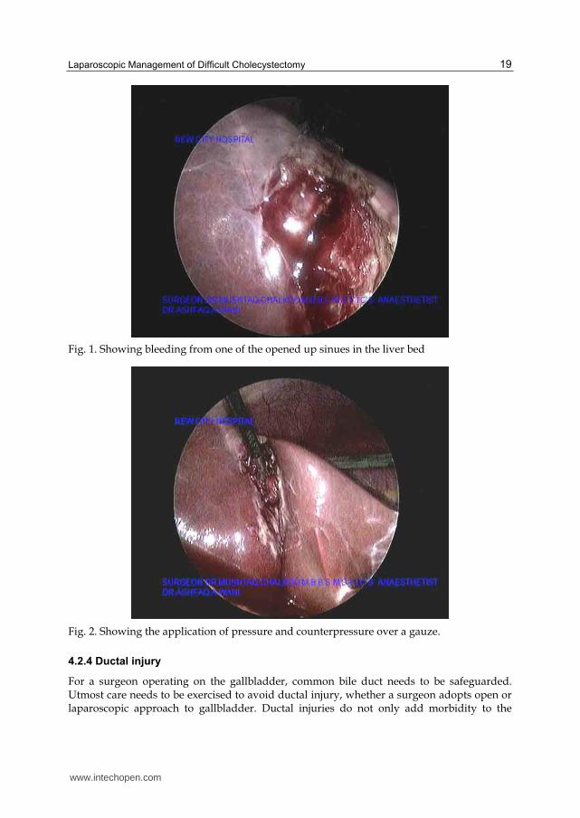
controlled with an electrocautery (figure1). We have found that gel foams do not help much
where as surgicel (oxydized cellulose polymer) is most effective in controlling bleeding from
the liver bed. We also advocate packing of liver bed in case of opening up of a sinus with
surgicel on top of which a wet-gauze should be placed and compressed by right hand
forceps. Then the counter pressure should be maintained by the left hand forceps on the
liver. This bimanual compression should be maintained continuously by watch for a period
of 5 minutes (figure 2). We strongly believe that any kind of sinus bleed dealt in this way
can be handled and conversion to open approach avoided to a great extent.
www.intechopen.com
Laparoscopic Management of Difficult Cholecystectomy 19
Fig. 1. Showing bleeding from one of the opened up sinues in the liver bed
Fig. 2. Showing the application of pressure and counterpressure over a gauze.
4.2.4 Ductal injury
For a surgeon operating on the gallbladder, common bile duct needs to be safeguarded. Utmost care needs to be exercised to avoid ductal injury, whether a surgeon adopts open or laparoscopic approach to gallbladder. Ductal injuries do not only add morbidity to the
www.intechopen.com
Advances in Laparoscopic Surgery 20
patient but can even at times prove to be fatal. If the injury is recognized intraoperatively and treated immediately, the patient may do well. In our series of 1000 cholecystectomies done by laparoscopic approach fortunately we never encountered one. Nevertheless, two of our patients presented either with a bilioma (8th – 10th postoperative day) or biliary peritonitis (5th-7th postoperative day). Both of these were managed by re-laparoscopy wherein clearing the bile from the peritoneal cavity and putting in a wide bore 28F tube drain was done. One of these had a persistent leak through the drain till 35th postoperative day. The patient was later on subjected to ERCP and a biliary stent was put in which was then removed in the third month. Small biliomas in the Morrison’s pouch or suprahepatic space can also be drained by percutaneous ultrasound guided technique. The ductal injury is a catastrophe that can result and hepaticojejunostomy can prove to be the only alternative for the operating surgeon. Sometimes accessory duct of Luscka is a cause for bilioma that a surgeon might not have recognized and dealt with in the initial go. In such a circumstance, re-laparoscopy with identification of accessory duct and clipping is recommended. If there is any bile leak which lasts beyond 5th postoperative day, distal block either with a stone or stenosis is likely. Our policy is to do ERCP and sphincterotomy with the extraction of the stone and stenting. In all such cases it takes just 24 hours for the leak to stop.
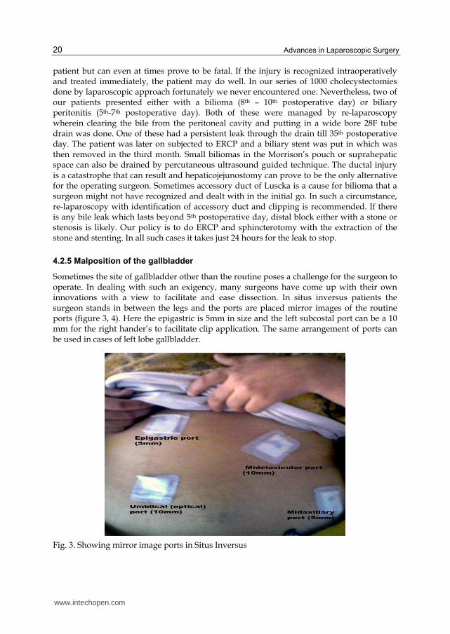
4.2.5 Malposition of the gallbladder
Sometimes the site of gallbladder other than the routine poses a challenge for the surgeon to operate. In dealing with such an exigency, many surgeons have come up with their own innovations with a view to facilitate and ease dissection. In situs inversus patients the surgeon stands in between the legs and the ports are placed mirror images of the routine ports (figure 3, 4). Here the epigastric is 5mm in size and the left subcostal port can be a 10 mm for the right hander’s to facilitate clip application. The same arrangement of ports can be used in cases of left lobe gallbladder.
Fig. 3. Showing mirror image ports in Situs Inversus
www.intechopen.com

Laparoscopic Management of Difficult Cholecystectomy 21
Fig. 4. Showing Critical view of safety in a situs inversus case
4.3 The problems related to the concomitant disease of gallbladder and nearby viscera
The gallbladder surgery has taken repute of many surgeons even at the distal end of their careers. A wise surgeon is one who thinks that the gallbladder, he operates on is his first one, amidst of huge experience he may carry in dealing with this organ. The difficulty while operating on an inflamed gallbladder cannot be defined. However, one needs to dissect safely to ease down the procedure. There are many problems and diseases of gallbladder that pose technical difficulties for the surgeon to remove this organ. One cannot generalize the principles for handling difficult gallbladders as each one of them poses a peculiar problem during dissection. The ones that need a mention herein are as follows:-
4.3.1 Impacted stone, hydrops, empyema, early Mirrizi’s of gallbladder
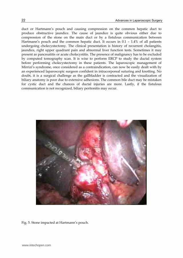
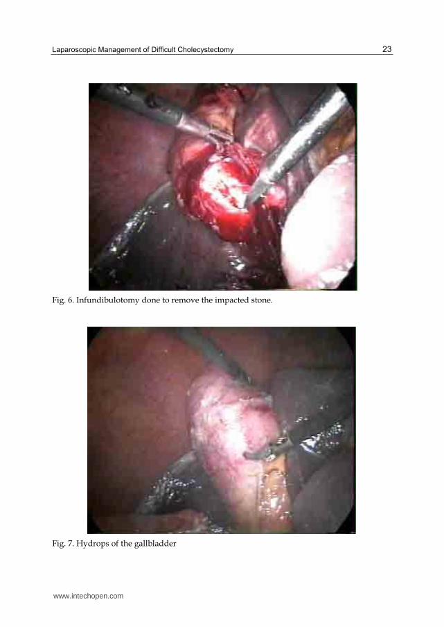
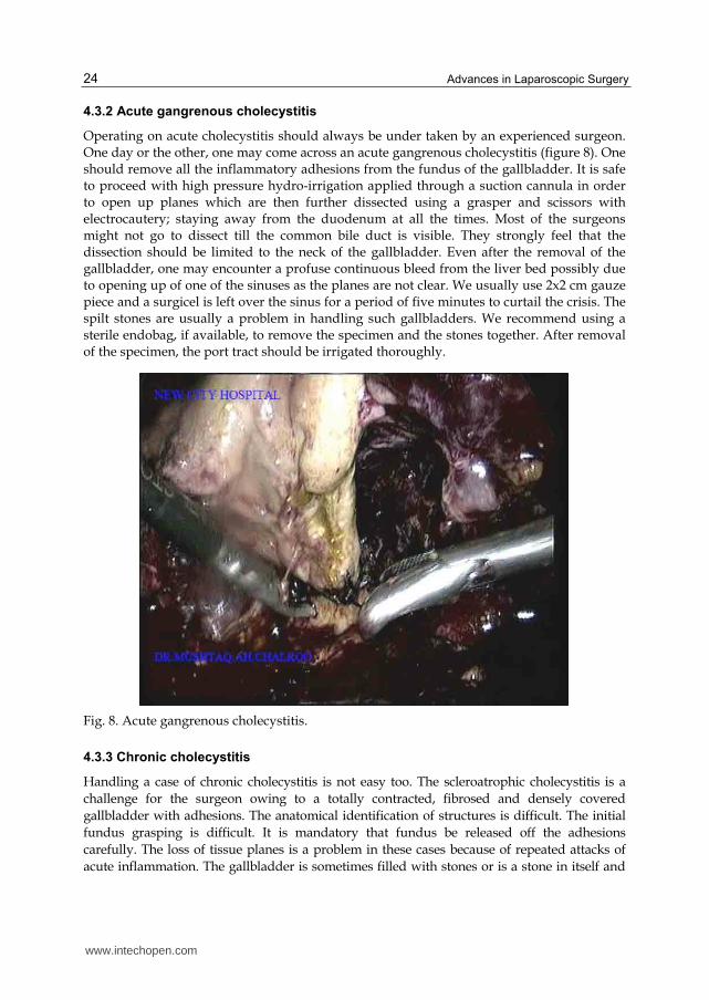
In a situation where a huge stone is impacted in the neck of the gallbladder with resultant hydrops or empyema, the easy way out to handle such a gallbladder is to aspirate it after opening the fundus with a hot hook to perform suction irrigation (figure 5). One can make an incision on the neck of the gallbladder approximately 2-3 cm above the junction of cystic duct and the neck. This incision should be generous enough to allow for the exteriorization of the stone like an enucleation of the mass (figure 6). We have usually found that in such cases the cystic duct is either small or absent. In these big stones impacted at the neck or pouch, technical problem lies in holding the pouch by the left hand and consequent addition to the fatigue of the surgeon (figure 7).
Mirrizi’s syndrome needs a mention. This syndrome was first described in 1948 by P.L. Mirrizi. He talked about an unusual complication of gallstones impacted either in the cystic
www.intechopen.com
Advances in Laparoscopic Surgery 22
duct or Hartmann’s pouch and causing compression on the common hepatic duct to produce obstructive jaundice. The cause of jaundice is quite obvious either due to compression of the stone on the main duct or by a fistulous communication between Hartmann’s pouch and the common hepatic duct. It occurs in 0.1 – 1.4% of all patients undergoing cholecystectomy. The clinical presentation is history of recurrent cholangitis, jaundice, right upper quadrant pain and abnormal liver function tests. Sometimes it may present as pancreatitis or acute cholecystitis. The presence of malignancy has to be excluded by computed tomography scan. It is wise to perform ERCP to study the ductal system before performing cholecystectomy in these patients. The laparoscopic management of Mirrizi’s syndrome, once considered as a contraindication, can now be easily dealt with by an experienced laparoscopic surgeon confident in intracorporeal suturing and knotting. No doubt, it is a surgical challenge as the gallbladder is contracted and the visualization of biliary anatomy is poor due to extensive adhesions. The common bile duct may be mistaken for cystic duct and the chances of ductal injuries are more. Lastly, if the fistulous communication is not recognized, biliary peritonitis may occur.
Fig. 5. Stone impacted at Hartmann’s pouch.
www.intechopen.com
Laparoscopic Management of Difficult Cholecystectomy 23
Fig. 6. Infundibulotomy done to remove the impacted stone.
Fig. 7. Hydrops of the gallbladder
www.intechopen.com
Advances in Laparoscopic Surgery 24
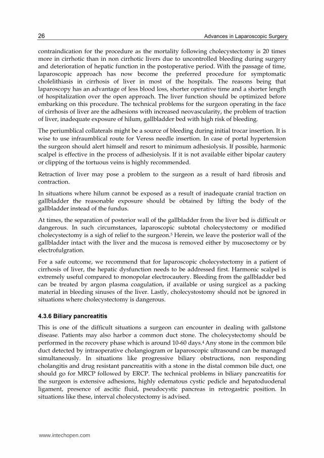
4.3.2 Acute gangrenous cholecystitis
Operating on acute cholecystitis should always be under taken by an experienced surgeon. One day or the other, one may come across an acute gangrenous cholecystitis (figure 8). One should remove all the inflammatory adhesions from the fundus of the gallbladder. It is safe to proceed with high pressure hydro-irrigation applied through a suction cannula in order to open up planes which are then further dissected using a grasper and scissors with electrocautery; staying away from the duodenum at all the times. Most of the surgeons might not go to dissect till the common bile duct is visible. They strongly feel that the dissection should be limited to the neck of the gallbladder. Even after the removal of the gallbladder, one may encounter a profuse continuous bleed from the liver bed possibly due to opening up of one of the sinuses as the planes are not clear. We usually use 2x2 cm gauze piece and a surgicel is left over the sinus for a period of five minutes to curtail the crisis. The spilt stones are usually a problem in handling such gallbladders. We recommend using a sterile endobag, if available, to remove the specimen and the stones together. After removal of the specimen, the port tract should be irrigated thoroughly.
Fig. 8. Acute gangrenous cholecystitis.
4.3.3 Chronic cholecystitis
Handling a case of chronic cholecystitis is not easy too. The scleroatrophic cholecystitis is a
challenge for the surgeon owing to a totally contracted, fibrosed and densely covered
gallbladder with adhesions. The anatomical identification of structures is difficult. The initial
fundus grasping is difficult. It is mandatory that fundus be released off the adhesions
carefully. The loss of tissue planes is a problem in these cases because of repeated attacks of
acute inflammation. The gallbladder is sometimes filled with stones or is a stone in itself and
www.intechopen.com

Laparoscopic Management of Difficult Cholecystectomy 25
the retraction becomes difficult. There are more chances of injury to the common bile duct in
handling this eventuality. One needs to be tricky to handle such a situation. The surgeon needs
to have first glimpse of the gallbladder by releasing the adhesions. Additional port placement
may help in this case. The retrograde technique may be then performed but should be done
carefully. Intraoperative cholangiography may be done, if possible. The cystic duct is usually
thick walled and difficult to occlude by clips. One should either use an endoloop or transfix
the cystic duct after milking the duct towards the gallbladder to displace any stone, if present.
4.3.4 Cholecystoenteric fistula
This is an incidental finding during laparoscopic cholecystectomy and can account for 0.5-
7% cases done laparoscopically for biliary disease (figure 9).6 The diagnosis is suspected by
noting the presence of air in the biliary tree associated with contracted gallbladder. The
conditions where air is present within the biliary tree are infections by gas forming
organisms, incompetent sphincter of Oddi, congenital anomalies, ERCP with
sphincterotomy. Cholecystoduodenal, cholecystogastric and cholecystocolic are the common
internal biliary fistulae. The common symptoms are pain, fever, diarrhea and jaundice. The
most common cause for internal biliary fistulae is gallstones (90%) whereas; peptic ulcer,
malignancy and trauma account for the rest 10% of the cases. Ultrasonography is useful and
CT scan may show contracted, thick walled gallbladder with stones, pneumobilia and
duodenal thickening. ERCP may localize the fistulous tract. Barium meal, enema and
colonoscopy may be useful in the diagnosis.
Fig. 9. Cholecystoenteric fistula.
4.3.5 Disorders of liver
It is a challenge for the surgeon to perform laparoscopic cholecystectomy in a patient with hepatic disorders especially cirrhosis. In near past this used to be considered a relative
www.intechopen.com
Advances in Laparoscopic Surgery 26
contraindication for the procedure as the mortality following cholecystectomy is 20 times more in cirrhotic than in non cirrhotic livers due to uncontrolled bleeding during surgery and deterioration of hepatic function in the postoperative period. With the passage of time, laparoscopic approach has now become the preferred procedure for symptomatic cholelithiasis in cirrhosis of liver in most of the hospitals. The reasons being that laparoscopy has an advantage of less blood loss, shorter operative time and a shorter length of hospitalization over the open approach. The liver function should be optimized before embarking on this procedure. The technical problems for the surgeon operating in the face of cirrhosis of liver are the adhesions with increased neovascularity, the problem of traction of liver, inadequate exposure of hilum, gallbladder bed with high risk of bleeding.
The periumblical collaterals might be a source of bleeding during initial trocar insertion. It is
wise to use infraumblical route for Veress needle insertion. In case of portal hypertension
the surgeon should alert himself and resort to minimum adhesiolysis. If possible, harmonic
scalpel is effective in the process of adhesiolysis. If it is not available either bipolar cautery
or clipping of the tortuous veins is highly recommended.
Retraction of liver may pose a problem to the surgeon as a result of hard fibrosis and contraction.
In situations where hilum cannot be exposed as a result of inadequate cranial traction on gallbladder the reasonable exposure should be obtained by lifting the body of the gallbladder instead of the fundus.
At times, the separation of posterior wall of the gallbladder from the liver bed is difficult or dangerous. In such circumstances, laparoscopic subtotal cholecystectomy or modified cholecystectomy is a sigh of relief to the surgeon.5 Herein, we leave the posterior wall of the gallbladder intact with the liver and the mucosa is removed either by mucosectomy or by electrofulgration.
For a safe outcome, we recommend that for laparoscopic cholecystectomy in a patient of cirrhosis of liver, the hepatic dysfunction needs to be addressed first. Harmonic scalpel is extremely useful compared to monopolar electrocautery. Bleeding from the gallbladder bed can be treated by argon plasma coagulation, if available or using surgicel as a packing material in bleeding sinuses of the liver. Lastly, cholecystostomy should not be ignored in situations where cholecystectomy is dangerous.
4.3.6 Biliary pancreatitis
This is one of the difficult situations a surgeon can encounter in dealing with gallstone
disease. Patients may also harbor a common duct stone. The cholecystectomy should be
performed in the recovery phase which is around 10-60 days.4 Any stone in the common bile
duct detected by intraoperative cholangiogram or laparoscopic ultrasound can be managed
simultaneously. In situations like progressive biliary obstructions, non responding
cholangitis and drug resistant pancreatitis with a stone in the distal common bile duct, one
should go for MRCP followed by ERCP. The technical problems in biliary pancreatitis for
the surgeon is extensive adhesions, highly edematous cystic pedicle and hepatoduodenal
ligament, presence of ascitic fluid, pseudocystic pancreas in retrogastric position. In
situations like these, interval cholecystectomy is advised.
www.intechopen.com
Laparoscopic Management of Difficult Cholecystectomy 27
We recommend an early cholecystectomy in a patient with multiple small stones to avoid recurrent bouts of pancreatitis. It may also be useful to categorize the patients into mild and severe degrees for directing appropriate management. Currently the best method to assess the severity of acute pancreatitis is contrast enhanced computed tomography scan. It is understood that biliary pancreatitis is due to a transient block of the ampulla of Vater by a migrating stone from gallbladder. It is a proven fact that the stone passes to the duodenum in majority of cases within hours of the onset of pancreatitis. If the acute process is increasing and reveals persistent obstruction, ERCP should be performed. Endoscopic sphincterotomy and extraction of the stone allows the acute pancreatitis to settle down. In mild pancreatitis laparoscopic cholecystectomy can be performed safely at the initial admission within first week depending on the condition of the patient. It is wise also to have an intraoperative cholangiogram as the risk of concurrent common bile duct stones is 14-20% associated with biliary pancreatitis. It may not be out of place to mention the value of MRCP and ERCP in severe cases. Currently an endoscopist and a laparoscopist work together to manage the problem of common bile duct stones as laparoscopic exploration of common bile duct is considered a high risk procedure in the phase of acute biliary pancreatitis.
4.3.7 Porcelain gallbladder
Here the gallbladder wall is deposited with calcium. The prevalence in cholecystectomy
specimens ranges from 0.06 – 0.08%. 7,8 It usually occurs in elderly persons with gallstones
who are predominantly females. X-ray abdomen may show a calcified lesion in the right
upper quadrant. Ultrasonography and CT scan rule out the presence of an associated
carcinoma. It increases the risk of carcinoma gallbladder by 12.5 – 60%.4 However, it is still
debatable. The removal of gallbladder is the treatment of choice. It is a technically
demanding surgery. However, adequate care should be taken to prevent dissemination of
malignant cells in the abdominal cavity and port site, if present. The specimen bags are
essential for gallbladder retrieval.
4.3.8 Carcinoma gallbladder
Carcinoma of the gallbladder is estimated to be present in 1% of cholecystectomy specimens.
It is the most common malignancy of the biliary tract. It is usually common in females.4 85%
of the cases are associated with the gallstones. It is recommended by the current medicine
that a gallbladder polyp of more than 10mm size should undergo cholecystectomy.4 The
smaller polyps should be followed up at 6 monthly periods and any increase in size is an
indication for cholecystectomy. The incidental gallbladder carcinoma is diagnosed
postoperatively by the histopathological examination of the specimen removed for the
gallstone disease. During surgery it is advocated that the surgeon should avoid inadvertent
disruption of gallbladder to avoid spillage of malignant cells into the peritoneal cavity and
the gallbladder should be removed in an endobag to avoid portal metastasis.
5. Conclusion
We strongly believe that every difficulty has a loop-hole that needs to be exploited to ease
down the procedure. Before embarking on difficult cholecystectomies, a surgeon needs to be
trained in all the technical aspects of laparoscopy. A due care should be given to suturing
www.intechopen.com
Advances in Laparoscopic Surgery 28
skills. Every gallbladder should be dealt with as if the surgeon, regardless of the experience,
is operating on the first gallbladder. The general principles in laparoscopy and the critical
view of safety should always be born in mind. The theatre staff especially the cameraman
should be properly trained. The instruments should be quite friendly and a surplus of them
should be available. The surgeon should have had a good amount of experience with simple
gallbladders before handling the difficult ones. With the advent of gratifying improvements
in the imaging technology, instrumentation and innovative techniques, the difficult
gallbladders now fall in the domain of simple surgeries. However, the intrinsic error in the
surgical technique cannot be avoided and whenever it comes onto that, open approach
should always be given a weightage.
6. References
[1] Mushtaq Chalkoo.,2009. Laparoscopic Cholecystectomy in a Mucocele of Gallbladderwith a Phrygian Cap- A Case Report,[online]. Available at: web address: http://www.google.co.in/search?sourceid=chrome&ie=UTF8&q=phyicians+academy+online+mushtaq+chalkoo
[2] Jorgensen JO, Hunt DR. Laparoscopic cholecystectomy. A prospective analysis of the potential causes of failure. Surg Laparosc Endosc. 1993 Feb;3(1):49-53.
[3] Patel M, Smart D. Aust N Z J Surg. Laparoscopic cholecystectomy and previous abdominal surgery: a safe technique. 1996 May;66(5):309-11
[4] Palanivelu C., 2007. Art of laparoscopic surgery. Ist ed. New Delhi: Jaypee. [5] K. Michalowski, P. C. Bornman, Professor J. E. J. Krige, P. J. Gallagher, J. Terblanche.
Laparoscopic subtotal cholecystectomy in patients with complicated acute cholecystitis or fibrosis. Volume 85, Issue 7, pages 904–906, July 1998
[6] Nadu A, Gallili Y, Soffer D, Kluger Y. J Trauma. Disruption of cholecystoenteric fistula induced by minor blunt trauma. 1996 Nov;41(5):914-5.
[7] Puia IC, Vlad L, Iancu C, Al-Hajjar N, Pop F, Bălă O, Munteanu D. Chirurgia (Bucur). Laparoscopic cholecystectomy for porcelain gallbladder. 2005 Mar-Apr;100(2): 187-9.
[8] Polk HC Jr. carcinoma and the calcified gallbladder. Gastroenterology 1966;50: 582-5.
www.intechopen.com
Advances in Laparoscopic SurgeryEdited by Dr Arshad Malik
ISBN 978-953-307-933-2Hard cover, 148 pagesPublisher InTechPublished online 01, February, 2012Published in print edition February, 2012
InTech EuropeUniversity Campus STeP Ri Slavka Krautzeka 83/A 51000 Rijeka, Croatia Phone: +385 (51) 770 447 Fax: +385 (51) 686 166www.intechopen.com
InTech ChinaUnit 405, Office Block, Hotel Equatorial Shanghai No.65, Yan An Road (West), Shanghai, 200040, China
Phone: +86-21-62489820 Fax: +86-21-62489821
Laparoscopic surgery, also called minimal access surgery, has revolutionized the field of surgery over the pastfew years. It has gained worldwide popularity and acceptance by surgeons and patients alike. Minimalscarring, less pain, and shorter hospital stay are the main reasons behind the global appeal of this noveltechnique. There has been a tremendous improvement in the technique, as well as in the instruments. Thetechnique has passed through the stages of simple laparoscopic surgery to advanced levels, where morecomplicated procedures are being successfully attempted. The recent introduction of robotic surgery is alsogaining popularity, in additional to single port laparoscopic surgery (SILS), which can be scarless surgery. Mostof the surgical procedures, which were considered contraindication for the laparoscopic approach, haveeventually become the most common and acceptable indications today. This book is intended to provide anoverview of the most common procedures performed laparoscopically, as well as some recent advancementsin the field.
How to referenceIn order to correctly reference this scholarly work, feel free to copy and paste the following:
Mushtaq Chalkoo, Shahnawaz Ahangar, Ab Hamid Wani, Asim Laharwal, Umar Younus, Faud Sadiq Baqaland Sikender Iqbal (2012). Laparoscopic Management of Difficult Cholecystectomy, Advances in LaparoscopicSurgery, Dr Arshad Malik (Ed.), ISBN: 978-953-307-933-2, InTech, Available from:http://www.intechopen.com/books/advances-in-laparoscopic-surgery/laparoscopic-management-of-difficult-cholecystectomy


![Left Sided Laparoscopic Cholecystectomy: Case Report and ...open cholecystectomy - before laparoscopic era [2] and 1 case in 2008 [3] and about 50 cases of laparoscopic cholecystectomy](https://static.fdocuments.net/doc/165x107/5f6509906579645fd7227a11/left-sided-laparoscopic-cholecystectomy-case-report-and-open-cholecystectomy.jpg)
