Four-channel single incision laparoscopic cholecystectomy ...€¦ · Four-channel single incision...
6
Annals of Surgical Treatment and Research 81 pISSN 2288-6575 • eISSN 2288-6796 http://dx.doi.org/10.4174/astr.2014.87.2.81 Annals of Surgical Treatment and Research ORIGINAL ARTICLE Four-channel single incision laparoscopic cholecystectomy using a snake retractor: comparison between 3- and 4-channel SILC 4-channel single incision cholecystectomy Nak Song Sung, In Seok Choi, Ju Ik Moon, Yu Mi Ra, Sang Eok Lee, Won Jun Choi Department of Surgery, Konyang University Hospital, Konyang University College of Medicine, Daejeon, Korea INTRODUCTION Since Phillipe Mouret of France performed the first laparo- scopic cholecystectomy (LC) in 1987, multiport conventional LC (CLC) has become the gold standard for the treatment of gallbladder (GB) disease [1], with advantages of better cosmesis, less scar, less pain, and shorter hospitalization [2]. Surgeons have attempted to reduce number of ports and incisions with accumulation of experience and development of equipments for the past few decades. Single incision laparoscopic chole- cystectomy (SILC) was first performed by Navarra et al. [3] in 1997, but did not regain much popularity since the development. However, the recent increase in use of SILC can be attributed to new surgical techniques and equipment, such as the articulating instruments and access ports capable of allowing several instrument to be inserted in a single ports [4- 6]. Nowadays, the 3-channel method with multiport access is commonly used in SILC [7]. Despite the new techniques and Received April 4, 2014, Reviewed April 18, 2014, Accepted May 9, 2014 Corresponding Author: In Seok Choi Department of Surgery, Konyang University Hospital, Konyang University College of Medicine, 158 Gwanjeodong-ro, Seo-gu, Daejeon 302-718, Korea Tel: +82-42-600-8833, Fax: +82-42-543-8956 E-mail: [email protected] Copyright ⓒ 2014, the Korean Surgical Society cc Annals of Surgical Treatment and Research is an Open Access Journal. All articles are distributed under the terms of the Creative Commons Attribution Non- Commercial License (http://creativecommons.org/licenses/by-nc/3.0/) which permits unrestricted non-commercial use, distribution, and reproduction in any medium, provided the original work is properly cited. Purpose: Single incision laparoscopic cholecystectomy (SILC) is a widely used method of performing cholecystectomy. A common technique used in SILC is a 3-channel method. However, exposure of Calot’s triangle is limited in conventional 3-channel SILC. Therefore, we herein report the adequacy and feasibility of 4-channel SILC using a snake retractor. Methods: Four hundred and fifteen SILC cases were performed between April 2010 and February 2013. We performed 326 SILC cases between April 2010 and September 2012 using the 3-channel method. We introduced a snake retractor for liver traction in October 2012, and 89 cases of 4-channel SILC using snake retractor have been performed since. Results: Thirty patients (9.2%) in the 3-channel SILC group, and 23 patients (25.8%) in the 4-channel SILC group, were treated with percutaneous transhepatic gallbladder drainage insertion because of acute inflammation of the gallbladder (GB) before operation (P < 0.001). The mean operating time was 53.0 ± 25.8 minutes in the 3-channel SILC group and 51.9 ± 18.6 minutes in the 4-channel SILC group (P = 0.709). In the 3-channel SILC group, mean hospital stay was 3.0 ± 3.3 days whereas it was 2.6 ± 0.9 days in the 4-channel SILC group (P = 0.043). There were a total 9 cases (2.1%) of additional port usages, 6 cases (1.8%) in the 3-channel SILC group and 3 cases (3.4%) in the 4-channel SILC group (P = 0.411), due to cystic artery bleeding and bile leakage from gallbladder bed, but there were no open conversions. Conclusion: Benign diseases of the GB can be operated on using SILC with the 4-channel method using a snake retractor. [Ann Surg Treat Res 2014;87(2):81-86] Key Words: Cholecystectomy, Laparoscopy, Single incision, 4-channel
Transcript of Four-channel single incision laparoscopic cholecystectomy ...€¦ · Four-channel single incision...
Annals of Surgical Treatment and Research 81
pISSN 2288-6575 • eISSN 2288-6796http://dx.doi.org/10.4174/astr.2014.87.2.81Annals of Surgical Treatment and Research
ORIGINAL ARTICLE
Four-channel single incision laparoscopic cholecystectomy using a snake retractor: comparison between 3- and 4-channel SILC 4-channel single incision cholecystectomyNak Song Sung, In Seok Choi, Ju Ik Moon, Yu Mi Ra, Sang Eok Lee, Won Jun ChoiDepartment of Surgery, Konyang University Hospital, Konyang University College of Medicine, Daejeon, Korea
INTRODUCTIONSince Phillipe Mouret of France performed the first laparo
scopic cholecystectomy (LC) in 1987, multiport conventional LC (CLC) has become the gold standard for the treatment of gallbladder (GB) disease [1], with advantages of better cosmesis, less scar, less pain, and shorter hospitalization [2]. Surgeons have attempted to reduce number of ports and incisions with accumulation of experience and development of equipments
for the past few decades. Single incision laparoscopic cholecystectomy (SILC) was first performed by Navarra et al. [3] in 1997, but did not regain much popularity since the development. However, the recent increase in use of SILC can be attributed to new surgical techniques and equipment, such as the articulating instruments and access ports capable of allowing several instrument to be inserted in a single ports [46]. Nowadays, the 3channel method with multiport access is commonly used in SILC [7]. Despite the new techniques and
Received April 4, 2014, Reviewed April 18, 2014, Accepted May 9, 2014
Corresponding Author: In Seok ChoiDepartment of Surgery, Konyang University Hospital, Konyang University College of Medicine, 158 Gwanjeodong-ro, Seo-gu, Daejeon 302-718, KoreaTel: +82-42-600-8833, Fax: +82-42-543-8956E-mail: [email protected]
Copyright ⓒ 2014, the Korean Surgical Society
cc Annals of Surgical Treatment and Research is an Open Access Journal. All articles are distributed under the terms of the Creative Commons Attribution Non-Commercial License (http://creativecommons.org/licenses/by-nc/3.0/) which permits unrestricted non-commercial use, distribution, and reproduction in any medium, provided the original work is properly cited.
Purpose: Single incision laparoscopic cholecystectomy (SILC) is a widely used method of performing cholecystectomy. A common technique used in SILC is a 3-channel method. However, exposure of Calot’s triangle is limited in conventional 3-channel SILC. Therefore, we herein report the adequacy and feasibility of 4-channel SILC using a snake retractor. Methods: Four hundred and fifteen SILC cases were performed between April 2010 and February 2013. We performed 326 SILC cases between April 2010 and September 2012 using the 3-channel method. We introduced a snake retractor for liver traction in October 2012, and 89 cases of 4-channel SILC using snake retractor have been performed since.Results: Thirty patients (9.2%) in the 3-channel SILC group, and 23 patients (25.8%) in the 4-channel SILC group, were treated with percutaneous transhepatic gallbladder drainage insertion because of acute inflammation of the gallbladder (GB) before operation (P < 0.001). The mean operating time was 53.0 ± 25.8 minutes in the 3-channel SILC group and 51.9 ± 18.6 minutes in the 4-channel SILC group (P = 0.709). In the 3-channel SILC group, mean hospital stay was 3.0 ± 3.3 days whereas it was 2.6 ± 0.9 days in the 4-channel SILC group (P = 0.043). There were a total 9 cases (2.1%) of additional port usages, 6 cases (1.8%) in the 3-channel SILC group and 3 cases (3.4%) in the 4-channel SILC group (P = 0.411), due to cystic artery bleeding and bile leakage from gallbladder bed, but there were no open conversions. Conclusion: Benign diseases of the GB can be operated on using SILC with the 4-channel method using a snake retractor.[Ann Surg Treat Res 2014;87(2):81-86]
Key Words: Cholecystectomy, Laparoscopy, Single incision, 4-channel
82
Annals of Surgical Treatment and Research 2014;87(2):8186
exponential improvement of equipment, there still remains some limitations, such as difficulty in exposure of the Calot’s triangle, narrow indications (exclusion criteria: high body mass index [BMI], previous abdominal surgery, acute cholecystitis with severe gallbladder [GB] inflammation), and higher bile duct injury rate with the 3channel SILC [8,9]. For these reasons, adequacy and feasibility of SILC is still controversial. Previously, we had also performed 3channel SILC with 2 instruments and a flexible telescope, but encountered similar limitations. Therefore, we have replaced our standard setting into a 4channel SILC with an addition of a snake retractor for resolution of the aforementioned limitations. In this study, we hereby report an adequacy and feasibility of 4channel single incision laparoscopic cholecystectomy using a snake retractor.
METHODSWe have conducted retrospective review of 415 patients
that underwent SILC between April 2010 and February 2013. From April 2010 through September 2012, 326 patients had been treated with 3channel method using a handmade single port, a flexible telescope and two articulating instruments. In the early period, we excluded those who were older than 70 years of age with radiologic or pulmonologic comorbidities, or acute GB inflammation, but did not exclude patients with high BMI or history of abdominal surgery. After 50 cases, with accumulation of experience, the inclusion criteria of SILC was expanded to all the patients who would typically be considered for CLC, but excluded the patients with suspected earlystaged GB malignancy [10]. After October 2012, we have modified our 3channel method to a 4channel SILC by adding a snake retractor for liver retraction which we later named as “modified Konyang standard method”. Eightynine patients have been treated with the 4channel SILC using a snake retractor, and the inclusion criteria for the 4channel SILC had been applied equally.
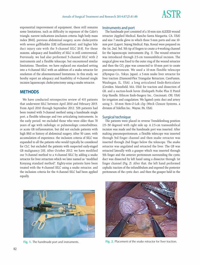
Instruments and portThe handmade port consisted of a 10mm size ALEXIS wound
retractor (Applied Medical, Rancho Santa Margarita, CA, USA) and size 7 sterile glove in which three 5mm ports and one 10mm port (Laport, Sejong Medical, Paju, Korea) were prepared on the 1st, 2nd, 3rd, 5th tip of fingers to create a 4working channel for the laparoscopic instruments (Fig. 1). The wound retractor was introduced through 2.5cm transumbilical incision. The surgical glove was fixed to the outer ring of the wound retractor and then the CO2 pipe was connected to 10mm port to create pneumoperitoneum. We used a 10mm flexible telescope (Olympus Co., Tokyo, Japan), a 5mm snake liver retractor for liver traction (DiamondFlex Triangular Retractors, CareFusion, Waukegan, IL, USA), a long articulated EndoRoticulator (Coviden, Mansfield, MA, USA) for traction and dissection of GB, and a suctionhook bovie (Endopath Probe Plus II Pistol Grip Handle, Ethicon EndoSurgery Inc., Cincinnati, OH, USA) for irrigation and coagulation. We ligated cystic duct and artery using 5, 10mm HemOLok clip (Weck Closure Systems, a division of Teleflex Inc., Wayne, PA, USA).
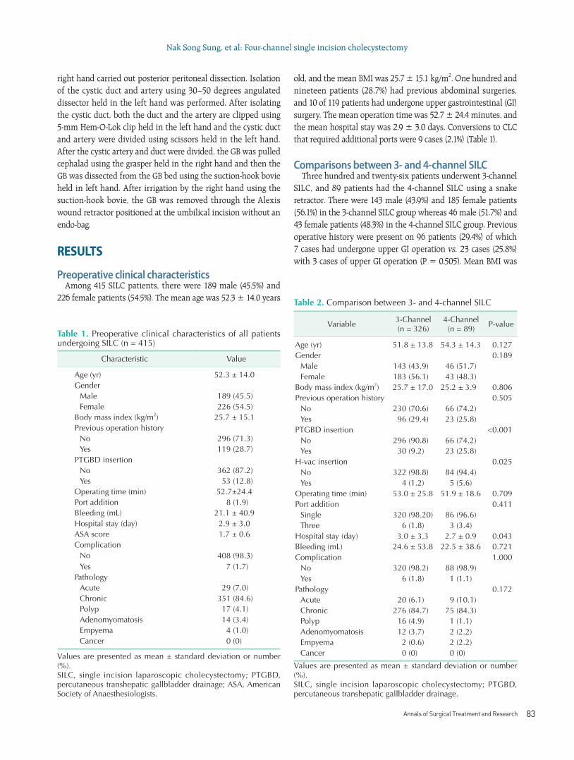
Surgical techniqueThe patients were placed in reverse Trendelenburg position
(15–30 degrees) with right side up. A 2.5cm transumbilical incision was made and the handmade port was inserted. After making pneumoperitoneum, a flexible telescope was inserted through 3rd finger channel and then snake retractor was inserted through 2nd finger below the telescope. The snake retractor was angulated and retracted the liver. The GB was retracted laterally with a grasper which was inserted through 5th finger and the anterior peritoneum surrounding the cystic duct was dissected by left hand using a dissector through 1st finger channel (Fig. 2). After that, the left hand performed cephalic traction of the infundibulum and exposed the posterior peritoneum of the cystic duct, and then the grasper held in the
Fig. 1. The handmade port and instruments. Fig. 2. Placement of the snake retractor for liver traction.
Annals of Surgical Treatment and Research 83
right hand carried out posterior peritoneal dissection. Isolation of the cystic duct and artery using 30–50 degrees angulated dissector held in the left hand was performed. After isolating the cystic duct, both the duct and the artery are clipped using 5mm HemOLok clip held in the left hand and the cystic duct and artery were divided using scissors held in the left hand. After the cystic artery and duct were divided, the GB was pulled cephalad using the grasper held in the right hand and then the GB was dissected from the GB bed using the suctionhook bovie held in left hand. After irrigation by the right hand using the suctionhook bovie, the GB was removed through the Alexis wound retractor positioned at the umbilical incision without an endobag.
RESULTS
Preoperative clinical characteristicsAmong 415 SILC patients, there were 189 male (45.5%) and
226 female patients (54.5%). The mean age was 52.3 ± 14.0 years
old, and the mean BMI was 25.7 ± 15.1 kg/m2. One hundred and nineteen patients (28.7%) had previous abdominal surgeries, and 10 of 119 patients had undergone upper gastrointestinal (GI)sur gery. The mean operation time was 52.7 ± 24.4 minutes, and the mean hospital stay was 2.9 ± 3.0 days. Conversions to CLC that required additional ports were 9 cases (2.1%) (Table 1).
Comparisons between 3- and 4-channel SILC Three hundred and twentysix patients underwent 3channel
SILC, and 89 patients had the 4channel SILC using a snake retractor. There were 143 male (43.9%) and 185 female patients (56.1%) in the 3channel SILC group whereas 46 male (51.7%) and 43 female patients (48.3%) in the 4channel SILC group. Previous operative history were present on 96 patients (29.4%) of which 7 cases had undergone upper GI operation vs. 23 cases (25.8%) with 3 cases of upper GI operation (P = 0.505). Mean BMI was
Table 1. Preoperative clinical characteristics of all patients undergoing SILC (n = 415)
Characteristic Value
Age (yr) 52.3 ± 14.0Gender
Male 189 (45.5)Female 226 (54.5)
Body mass index (kg/m2) 25.7 ± 15.1Previous operation history
No 296 (71.3)Yes 119 (28.7)
PTGBD insertionNo 362 (87.2)Yes 53 (12.8)
Operating time (min) 52.7±24.4Port addition 8 (1.9)Bleeding (mL) 21.1 ± 40.9Hospital stay (day) 2.9 ± 3.0ASA score 1.7 ± 0.6Complication
No 408 (98.3)Yes 7 (1.7)
PathologyAcute 29 (7.0)Chronic 351 (84.6)Polyp 17 (4.1)Adenomyomatosis 14 (3.4)Empyema 4 (1.0)Cancer 0 (0)
Values are presented as mean ± standard deviation or number (%).SILC, single incision laparoscopic cholecystectomy; PTGBD, percutaneous transhepatic gallbladder drainage; ASA, American Society of Anaesthesiologists.
Table 2. Comparison between 3- and 4-channel SILC
Variable 3-Channel (n = 326)
4-Channel (n = 89) P-value
Age (yr) 51.8 ± 13.8 54.3 ± 14.3 0.127Gender 0.189
Male 143 (43.9) 46 (51.7)Female 183 (56.1) 43 (48.3)
Body mass index (kg/m2) 25.7 ± 17.0 25.2 ± 3.9 0.806Previous operation history 0.505
No 230 (70.6) 66 (74.2)Yes 96 (29.4) 23 (25.8)
PTGBD insertion <0.001No 296 (90.8) 66 (74.2)Yes 30 (9.2) 23 (25.8)
H-vac insertion 0.025No 322 (98.8) 84 (94.4)Yes 4 (1.2) 5 (5.6)
Operating time (min) 53.0 ± 25.8 51.9 ± 18.6 0.709Port addition 0.411
Single 320 (98.20) 86 (96.6)Three 6 (1.8) 3 (3.4)
Hospital stay (day) 3.0 ± 3.3 2.7 ± 0.9 0.043Bleeding (mL) 24.6 ± 53.8 22.5 ± 38.6 0.721Complication 1.000
No 320 (98.2) 88 (98.9)Yes 6 (1.8) 1 (1.1)
Pathology 0.172Acute 20 (6.1) 9 (10.1)Chronic 276 (84.7) 75 (84.3)Polyp 16 (4.9) 1 (1.1)Adenomyomatosis 12 (3.7) 2 (2.2)Empyema 2 (0.6) 2 (2.2)Cancer 0 (0) 0 (0)
Values are presented as mean ± standard deviation or number (%).SILC, single incision laparoscopic cholecystectomy; PTGBD, percutaneous transhepatic gallbladder drainage.
Nak Song Sung, et al: Fourchannel single incision cholecystectomy
84
Annals of Surgical Treatment and Research 2014;87(2):8186
25.7 ± 17.0 kg/m2 vs. 25.2 ± 3.9 kg/m2 (P = 0.806), 30 patients (9.2%) vs. 23 patients (25.8%) were treated with percutaneous transhepatic gallbladder drainage (PTGBD) insertion before operation (P < 0.001), mean operating time was 53.0 ± 25.8 minutes vs. 51.9 ± 18.6 minutes (P = 0.709) and mean hospital stay was 3.0 ± 3.3 days vs. 2.6 ± 0.9 days (P = 0.043). There were total 9 cases (2.1%) of port addition, 6 cases (1.8%) in the 3channel group and 3 cases (3.4%) in the 4channel group (P = 0.411), due to cystic artery bleeding and bile leakage from the GB bed, but there were no open conversions. There was no statistically significant difference between the 3channel SILC and the 4channel SILC group except for PTGBD insertion (P < 0.001) and mean hospital stay (Table 2).
Postoperative complications between 3- and 4-channel SILCPostoperative complications were observed in total of 7
patients, 6 cases in the 3channel group and 1 case in the 4channel group (Table 3). Of the 7 patients, 2 were major complications requiring additional treatments (1 bile duct injury and 1 duodenal perforation), and the remaining 5 were minor complications (4 wound infections and 1 incisional hernia). In the 4channel SILC group, 1 wound infection was observed. There was no mortality. Bile duct injury occurred in a patient with severe GB inflammation, was determined to be type E by Strasberg’s classification, detected at postoperative day 2, and was treated with hepaticojejunostomy. For the patient with duodenal perforation, there was severe adhesion between the duodenum and the liver from prior subtotal gastrectomy. The duodenal perforation had most likely occurred during dissection of the duodenum that was firmly adhered to the liver. Signs and symptoms of peritonitis were observed at postoperative day 2, and it was treated with laparoscopic primary repair of perforation site. The four patients with wound infections were treated with simple dressing. Incisional hernia occurred in a patient who was treated for wound infection after postoperative month 3, and herniorrhaphy was performed. All complications occurred in the 3channel SILC group except for 1 simple wound infection.
DISCUSSIONSince Phillipe Mouret of France performed the first LC in
1987 [1], the superiority of LC as the standard procedure for GB removal compared to the open cholecystectomy has not been questioned due to better cosmetic satisfaction, less scar, less pain, and shorter hospitalization [2]. Ever since the initial development of LC, surgeons had attempted to reduce the number and the size of ports. In these efforts, more challenging and innovative methods like natural orifice transluminal endoscopic surgery (NOTES) have been invented and developed. Since the first NOTES transvaginal cholecystectomy by Bessler et al. [11] of the United States in 2007, numerous NOTES cholecystectomies have been performed. However, some challenges still exist, including longer operating time, poor triangulation of instruments, difficulty in standardization of the procedure, and increased risks of leakage and fistula, which clearly indicates that improvement of the NOTES surgical techniques and instruments is much needed [7,11]. Nowadays, surgeons have switched their focus to single incision laparoscopic surgery as an alternative minimally invasive surgery to the NOTES. Since the first SILC by Navarra et al. [3] in 1997, various new techniques and instruments have been introduced and modified. SILC has become more popular, and several studies including review of over hundred cases or metaanalysis of the operative data have been reported [1216]. The SILC certainly has superior cosmetic outcome compared to the CLC. However, a surgeon should not choose SILC solely based on superior cosmesis, patient’ safety still remains as a major concern. Joseph et al. [8] reported that the bile duct injury rate in SILC (0.72%) was higher than CLC (0.2%). Trastulli et al. [16] reported a metaanalysis data including 13 randomized controlled trial studies comparing SILC vs. CLC, the failure rate was 8.4% vs. 0.7%, conversion rate to open was 0.2% vs. 0.2%, overall complication rate was 7.3% vs. 6.6% and a bile leakage rate was 0.4% vs. 0.7%. Sajid et al. [13] reported postoperative pain and complications, the hospital length of stay, cosmesis score, conversion rate, and ports addition rate were statistically similar between SILC and CLC in their metaanalysis study. Although there were various studies on SILC, the inconsistencies of results among studies ranged too wide to be certain of the safety in SILC. As we mentioned earlier, a major problem when comparing CLC and SILC is the difficulty in exposure of the Calot’s triangle. Aprea et al. [17], Cao et al. [18], Edwards et al. [19], etc., many surgeons have used percutaneous anchorage with sutures and Lirici et al. [20] used needlescopic grasper for GB retraction and reducing complications. The most difficult problems of the 3channel SILC in our department were similar to those reported from other institutes, which were inadequate visualization of the Calot’s triangle and clash between the instruments and the telescope. In order to resolve
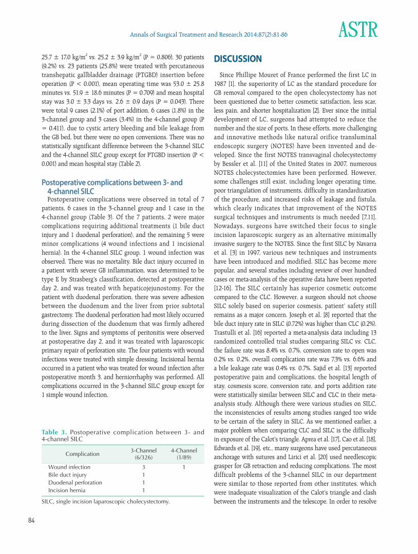
Table 3. Postoperative complication between 3- and 4-channel SILC
Complication 3-Channel (6/326)
4-Channel (1/89)
Wound infection 3 1Bile duct injury 1Duodenal perforation 1Incision hernia 1
SILC, single incision laparoscopic cholecystectomy.
Annals of Surgical Treatment and Research 85
these issues, we used a flexible telescope and articulating long instruments, and standardized our method, which was to retract the GB inferolaterally by right hand and to dissect the cystic duct and artery by left hand while positioning the flexible telescope at the left side of the working instruments [21]. Despite the standardization of our method, visualization of the Calot’s triangle was still incomplete, especially when the GB was acutely inflamed. Thus, we incorporated a snake tractor to address the remaining issue. We had initially proposed that our modified 4channel SILC wound have certain benefits, such as for better exposure of the Calot’s triangle, and decrease in ports addition, conversion rate, and complication rate. We performed SILC in a selected patient population without severe comorbidities, or acute cholecystitis in the early period in the formerly published studies [8,22,23], but we expanded indications for SILC while increasing the number of the SILC cases, and performed 3 and 4channel SILC on patients with major comorbidities, obesity, previous abdominal surgery, and acute cholecystitis pretreated with PTGBD, but excluded patients with possible early stage GB malignancy. A proportion of patients with acute cholecystitis, who treated with PTGBD before an operation, was 30 (9.2%) in 3channel SILC and 23 (25.8%) in 4channel SILC. This raises a possibility that the 4channel SILC could be applicable for more patients with acute cholecystitis. There were total 9 cases of CLC conversion including 6 cases in the 3channel SILC group and 3 cases in
the 4channel SILC group, due to the cystic artery bleeding. We recommended reducing this problem by ligating the cystic artery before the cystic duct ligation. There was only single wound infection case in the 4channel SILC group and no major complication like the bile duct injury. However, there were some limitations in our study. One is the small sample size, and the other is a lower conversion and complication rate compared to other studies [13]. The latter is most likely due to the amount of experience by the surgeon who performed the 4channel SILC, since he had previously performed over 2,000 cases of the CLC. Surely, we need larger sized samples and longterm followup studies. But we considered that the 4channel SILC can become one of the primary treatments of benign GB disease in the future.
In conclusion, 4channel single incision laparoscopic cholecystectomy has certain advantages such as better visibility and accessibility for dissection of the Calot’s triangle, and reproducible methods in experienced hands. 4channel SILC is deemed safe and feasible; therefore, most benign diseases of GB could be treated with the 4channel SILC using a snake retractor.
CONFLICTS OF INTERESTNo potential conflict of interest relevant to this article was
reported.
1. Kaiser AM, Corman ML. History of lapa
roscopy. Surg Oncol Clin N Am 2001;
10:48392.
2. Townsend CM Jr, Beauchamp RD, Evers
BM, Mattox KL. Sabiston textbook of
surgery: the biological basis of modern
surgical practice. 19th ed. Philadelphia:
Elsevier Saunders; 2012.
3. Navarra G, Pozza E, Occhionorelli S,
Carcoforo P, Donini I. Onewound laparo
scopic cholecystectomy. Br J Surg 1997;
84:695.
4. Chow A, Purkayastha S, Aziz O, Paraskeva
P. Singleincision laparoscopic surgery for
cholecystectomy: an evolving technique.
Surg Endosc 2010;24:70914.
5. Romanelli JR, Mark L, Omotosho PA.
Single port laparoscopic cholecystectomy
with the TriPort system: a case report.
Surg Innov 2008;15:2238.
6. Yoshida M, Furukawa T, Morikawa Y,
Kitagawa Y, Kitajima M. The develop
ments and achievements of endoscopic
surgery, robotic surgery and function
preserving surgery. Jpn J Clin Oncol 2010;
40:8639.
7. Rao PP, Rao PP, Bhagwat S. Singleincision
laparoscopic surgery: current status and
controversies. J Minim Access Surg 2011;
7:616.
8. Joseph M, Phillips MR, Farrell TM, Rupp
CC. Single incision laparoscopic chole
cystectomy is associated with a higher
bile duct injury rate: a review and a word
of caution. Ann Surg 2012;256:16.
9. Lai EC, Yang GP, Tang CN, Yih PC, Chan
OC, Li MK. Prospective randomized
comparative study of single incision
laparo scopic cholecystectomy versus
con ventional fourport laparoscopic
cholecystectomy. Am J Surg 2011;202:254
8.
10. Park SM, Choi IS, Moon JI, Ra YM, Lee
SE, Choi WJ, et al. Comparison of single
incision and three port laparoscopic
cholecystectomy. J Minim Invasive Surg
2013;16:15.
11. Bessler M, Stevens PD, Milone L, Parikh M,
Fowler D. Transvaginal laparoscopically
assisted endoscopic cholecystectomy: a
hybrid approach to natural orifice surgery.
Gastrointest Endosc 2007;66:12435.
12. Song T, Liao B, Liu J, Yin Y, Luo Q, Cheng
N. Singleincision versus conventional
laparoscopic cholecystectomy: a syste
matic review of available data. Surg
Laparosc Endosc Percutan Tech 2012;22:
REFERENCES
Nak Song Sung, et al: Fourchannel single incision cholecystectomy
86
Annals of Surgical Treatment and Research 2014;87(2):8186
e1906.
13. Sajid MS, Ladwa N, Kalra L, Hutson KK,
Singh KK, Sayegh M. Singleincision
laparoscopic cholecystectomy versus
conventional laparoscopic chole cystec
tomy: metaanalysis and systematic
re view of randomized controlled trials.
World J Surg 2012;36:264453.
14. Hall TC, Dennison AR, Bilku DK, Metcalfe
MS, Garcea G. Singleincision laparoscopic
cholecystectomy: a systematic review.
Arch Surg 2012;147:65766.
15. Curcillo PG 2nd, Wu AS, Podolsky ER,
Graybeal C, Katkhouda N, Saenz A, et al.
Singleportaccess (SPA) cholecystectomy:
a multiinstitutional report of the first
297 cases. Surg Endosc 2010;24:185460.
16. Trastulli S, Cirocchi R, Desiderio J,
Guarino S, Santoro A, Parisi A, et al.
Systematic review and metaanalysis of
randomized clinical trials comparing
singleincision versus conventional
laparo scopic cholecystectomy. Br J Surg
2013;100:191208.
17. Aprea G, Coppola Bottazzi E, Guida F,
Masone S, Persico G. Laparoendoscopic
single site (LESS) versus classic video
laparoscopic cholecystectomy: a rando
mized prospective study. J Surg Res 2011;
166:e10912.
18. Cao ZG, Cai W, Qin MF, Zhao HZ, Yue P,
Li Y. Randomized clinical trial of single
incision versus conventional laparoscopic
cholecystectomy: shortterm operative
outcomes. Surg Laparosc Endosc Percutan
Tech 2011;21:3113.
19. Edwards C, Bradshaw A, Ahearne P,
Dematos P, Humble T, Johnson R, et al.
Singleincision laparoscopic cholecy
stectomy is feasible: initial experience
with 80 cases. Surg Endosc 2010;24:22417.
20. Lirici MM, Califano AD, Angelini P,
Corcione F. Laparoendoscopic single
site cholecystectomy versus standard
laparoscopic cholecystectomy: results
of a pilot randomized trial. Am J Surg
2011;202:4552.
21. Son JI. Single incision laparoscopic cho
lecystectomy using Konyang standard
method [dissertation]. Daejeon: Konyang
University; 2013
22. Yilmaz H, Alptekin H, Acar F, Ciftci I,
Tekin A, Sahin M. Experiences of single
incision cholecystectomy. Int J Med Sci
2013;10:738.
23. Erbella J Jr, Bunch GM. Singleincision
laparoscopic cholecystectomy: the first
100 outpatients. Surg Endosc 2010;24:
195861.