Emergency laparoscopic cholecystectomy for intraabdominal ...
5
CASE REPORT Open Access Emergency laparoscopic cholecystectomy for intraabdominal hemorrhage in a patient with a left ventricular assist device: a case report Akihiko Takagi 1* , Erina Nagai 1 , Takeo Toda 1 , Hayato Kosaka 1 , Hisato Ishimatsu 1 , Yusuke Kyoden 1 , Takehide Akimoto 2 , Hideyuki Kanemoto 1 and Noriyuki Oba 1 Abstract Background: Continuous-flow left ventricular assist devices (LVADs), called “second generation LVADs,” have significantly improved the survival and quality of life outcomes. Accordingly, non-cardiac surgery in a patient with LVADs has required for conditions not directly related to their LVADs. And the management of bleeding in non- cardiac site remains one of long-term critical topics. Laparoscopic approach is useful in a patient with LVADs; however, there have been only few clinical reports. This report describes the first case of laparoscopic cholecystectomy (LC) for intraabdominal hemorrhage from the gallbladder serosa in a patient with LVADs. Case presentation: A 56-year-old man with an LVAD had undergone LVAD (Jarvik 2000™; Jarvik Heart, Inc., New York, NY, USA) implantation at 53 years of age. He was in shock, and contrast-enhanced computed tomography revealed abdominal hemorrhage from the gallbladder serosa. Emergency laparoscopic cholecystectomy was performed. We could avoid injury of the LVADs driveline, which was located across the upper abdominal midline, near the right hypochondriac region, by laparoscopic approach. LVADs (Jarvik 2000) did not disturb the operating field because of its smaller size. There were no intra- and postoperative complications. Conclusions: Laparoscopic approach is useful and safe in a patient with LVADs for abdominal surgery. We could perform LC for intraabdominal hemorrhage from gallbladder serosa safety. Keywords: Intraabdominal hemorrhage, Laparoscopic cholecystectomy, Ventricular assist device Background Continuous-flow left ventricular assist devices (LVADs), called “second generation LVADs,” have significantly im- proved the survival and quality of life outcomes [1]. However, they have been associated with the develop- ment of non-cardiac complications over time [2]. In abdominal surgery, it is very important to avoid injury of the LVAD driveline, which connects the main body with the controller running under the abdominal skin. Thus, laparoscopic approach is useful in these patients; how- ever, there have been only few clinical reports. We herein report a first case of emergency laparoscopic cholecystectomy (LC) for intraabdominal hemorrhage from the gallbladder serosa in a patient with LVADs (Jarvik 2000™; Jarvik Heart, Inc., New York, NY, USA). Jarvik 2000 is a second-generation LVAD that features a miniaturized intraventricular pump (Fig. 1a). Case presentation A 56-year-old man was admitted to our hospital due to right heart failure and epistaxis that could not be controlled by pressure hemostasis. He had undergone LVAD (Jarvik 2000) implantation at 53 years of age due to severe heart failure after cardiovascular surgery (mi- tral valve replacement due to mitral regurgitation after mitral valve plasty and tricuspid annuloplasty for dilated © The Author(s). 2019 Open Access This article is distributed under the terms of the Creative Commons Attribution 4.0 International License (http://creativecommons.org/licenses/by/4.0/), which permits unrestricted use, distribution, and reproduction in any medium, provided you give appropriate credit to the original author(s) and the source, provide a link to the Creative Commons license, and indicate if changes were made. * Correspondence: [email protected] 1 Department of Gastroenterological Surgery, Shizuoka General Hospital, 4-27-1 Kita-ando, Aoi-ku, Shizuoka-shi, Shizuoka 420-8527, Japan Full list of author information is available at the end of the article Takagi et al. Surgical Case Reports (2019) 5:196 https://doi.org/10.1186/s40792-019-0756-9
Transcript of Emergency laparoscopic cholecystectomy for intraabdominal ...
CASE REPORT Open Access
Emergency laparoscopic cholecystectomyfor intraabdominal hemorrhage in a patientwith a left ventricular assist device: a casereportAkihiko Takagi1* , Erina Nagai1, Takeo Toda1, Hayato Kosaka1, Hisato Ishimatsu1, Yusuke Kyoden1,Takehide Akimoto2, Hideyuki Kanemoto1 and Noriyuki Oba1
Abstract
Background: Continuous-flow left ventricular assist devices (LVADs), called “second generation LVADs,” havesignificantly improved the survival and quality of life outcomes. Accordingly, non-cardiac surgery in a patient withLVADs has required for conditions not directly related to their LVADs. And the management of bleeding in non-cardiac site remains one of long-term critical topics. Laparoscopic approach is useful in a patient with LVADs;however, there have been only few clinical reports. This report describes the first case of laparoscopiccholecystectomy (LC) for intraabdominal hemorrhage from the gallbladder serosa in a patient with LVADs.
Case presentation: A 56-year-old man with an LVAD had undergone LVAD (Jarvik 2000™; Jarvik Heart, Inc., NewYork, NY, USA) implantation at 53 years of age. He was in shock, and contrast-enhanced computed tomographyrevealed abdominal hemorrhage from the gallbladder serosa. Emergency laparoscopic cholecystectomy wasperformed. We could avoid injury of the LVADs driveline, which was located across the upper abdominal midline,near the right hypochondriac region, by laparoscopic approach. LVADs (Jarvik 2000) did not disturb the operatingfield because of its smaller size. There were no intra- and postoperative complications.
Conclusions: Laparoscopic approach is useful and safe in a patient with LVADs for abdominal surgery. We couldperform LC for intraabdominal hemorrhage from gallbladder serosa safety.
Keywords: Intraabdominal hemorrhage, Laparoscopic cholecystectomy, Ventricular assist device
BackgroundContinuous-flow left ventricular assist devices (LVADs),called “second generation LVADs,” have significantly im-proved the survival and quality of life outcomes [1].However, they have been associated with the develop-ment of non-cardiac complications over time [2]. Inabdominal surgery, it is very important to avoid injury ofthe LVAD driveline, which connects the main body withthe controller running under the abdominal skin. Thus,laparoscopic approach is useful in these patients; how-ever, there have been only few clinical reports. We
herein report a first case of emergency laparoscopiccholecystectomy (LC) for intraabdominal hemorrhagefrom the gallbladder serosa in a patient with LVADs(Jarvik 2000™; Jarvik Heart, Inc., New York, NY, USA).Jarvik 2000 is a second-generation LVAD that features aminiaturized intraventricular pump (Fig. 1a).
Case presentationA 56-year-old man was admitted to our hospital due toright heart failure and epistaxis that could not becontrolled by pressure hemostasis. He had undergoneLVAD (Jarvik 2000) implantation at 53 years of age dueto severe heart failure after cardiovascular surgery (mi-tral valve replacement due to mitral regurgitation aftermitral valve plasty and tricuspid annuloplasty for dilated
© The Author(s). 2019 Open Access This article is distributed under the terms of the Creative Commons Attribution 4.0International License (http://creativecommons.org/licenses/by/4.0/), which permits unrestricted use, distribution, andreproduction in any medium, provided you give appropriate credit to the original author(s) and the source, provide a link tothe Creative Commons license, and indicate if changes were made.
* Correspondence: [email protected] of Gastroenterological Surgery, Shizuoka General Hospital,4-27-1 Kita-ando, Aoi-ku, Shizuoka-shi, Shizuoka 420-8527, JapanFull list of author information is available at the end of the article
Takagi et al. Surgical Case Reports (2019) 5:196 https://doi.org/10.1186/s40792-019-0756-9
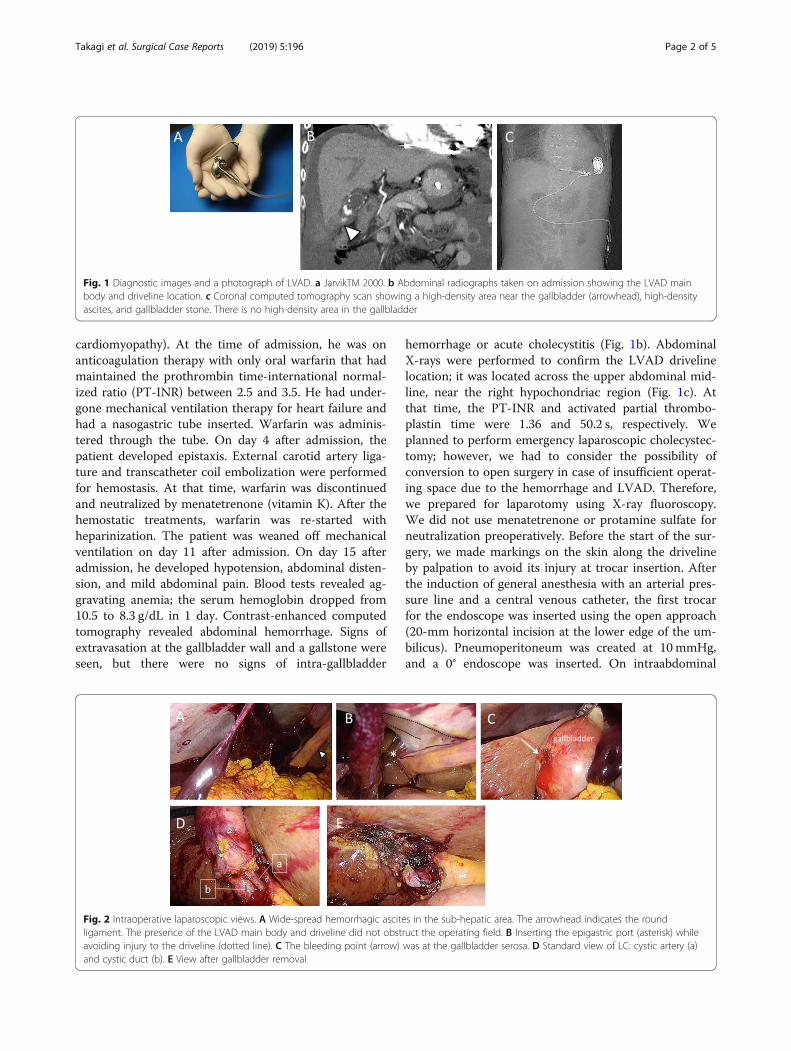
cardiomyopathy). At the time of admission, he was onanticoagulation therapy with only oral warfarin that hadmaintained the prothrombin time-international normal-ized ratio (PT-INR) between 2.5 and 3.5. He had under-gone mechanical ventilation therapy for heart failure andhad a nasogastric tube inserted. Warfarin was adminis-tered through the tube. On day 4 after admission, thepatient developed epistaxis. External carotid artery liga-ture and transcatheter coil embolization were performedfor hemostasis. At that time, warfarin was discontinuedand neutralized by menatetrenone (vitamin K). After thehemostatic treatments, warfarin was re-started withheparinization. The patient was weaned off mechanicalventilation on day 11 after admission. On day 15 afteradmission, he developed hypotension, abdominal disten-sion, and mild abdominal pain. Blood tests revealed ag-gravating anemia; the serum hemoglobin dropped from10.5 to 8.3 g/dL in 1 day. Contrast-enhanced computedtomography revealed abdominal hemorrhage. Signs ofextravasation at the gallbladder wall and a gallstone wereseen, but there were no signs of intra-gallbladder
hemorrhage or acute cholecystitis (Fig. 1b). AbdominalX-rays were performed to confirm the LVAD drivelinelocation; it was located across the upper abdominal mid-line, near the right hypochondriac region (Fig. 1c). Atthat time, the PT-INR and activated partial thrombo-plastin time were 1.36 and 50.2 s, respectively. Weplanned to perform emergency laparoscopic cholecystec-tomy; however, we had to consider the possibility ofconversion to open surgery in case of insufficient operat-ing space due to the hemorrhage and LVAD. Therefore,we prepared for laparotomy using X-ray fluoroscopy.We did not use menatetrenone or protamine sulfate forneutralization preoperatively. Before the start of the sur-gery, we made markings on the skin along the drivelineby palpation to avoid its injury at trocar insertion. Afterthe induction of general anesthesia with an arterial pres-sure line and a central venous catheter, the first trocarfor the endoscope was inserted using the open approach(20-mm horizontal incision at the lower edge of the um-bilicus). Pneumoperitoneum was created at 10 mmHg,and a 0° endoscope was inserted. On intraabdominal
Fig. 1 Diagnostic images and a photograph of LVAD. a JarvikTM 2000. b Abdominal radiographs taken on admission showing the LVAD mainbody and driveline location. c Coronal computed tomography scan showing a high-density area near the gallbladder (arrowhead), high-densityascites, and gallbladder stone. There is no high-density area in the gallbladder
Fig. 2 Intraoperative laparoscopic views. A Wide-spread hemorrhagic ascites in the sub-hepatic area. The arrowhead indicates the roundligament. The presence of the LVAD main body and driveline did not obstruct the operating field. B Inserting the epigastric port (asterisk) whileavoiding injury to the driveline (dotted line). C The bleeding point (arrow) was at the gallbladder serosa. D Standard view of LC: cystic artery (a)and cystic duct (b). E View after gallbladder removal
Takagi et al. Surgical Case Reports (2019) 5:196 Page 2 of 5
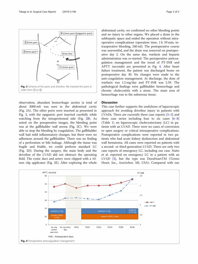
observation, abundant hemorrhagic ascites (a total ofabout 3000 ml) was seen in the abdominal cavity(Fig. 2A). The other ports were inserted as presented inFig. 3, with the epigastric port inserted carefully whilewatching from the intraperitoneal side (Fig. 2B). Asnoted on the preoperative images, the bleeding pointwas at the gallbladder wall serosa (Fig. 2C). We wereable to stop the bleeding by coagulation. The gallbladderwall had mild inflammatory changes, but there were noadhesions around the gallbladder. There was no findingof a perforation or bile leakage. Although the tissue wasfragile and friable, we could perform standard LC(Fig. 2D). During the surgery, the main body and thedriveline of the LVAD did not obstruct the operatingfield. The cystic duct and artery were clipped with a 10-mm clip applicator (Fig. 2E). After exploring the whole
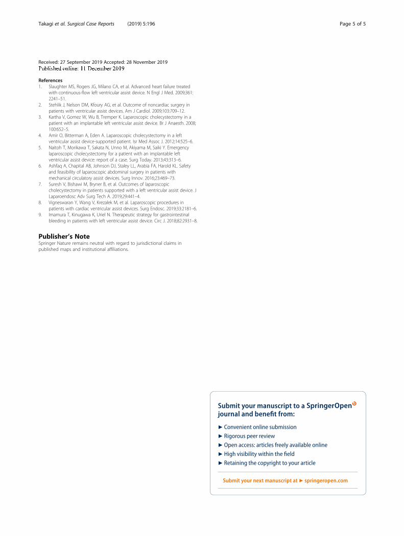
abdominal cavity, we confirmed no other bleeding pointsand no injury to other organs. We placed a drain in thesubhepatic space and ended the operation without intra-operative complications (operation time, 1 h 59min; in-traoperative bleeding, 240 ml). The postoperative coursewas uneventful, and the drain was removed on postoper-ative day 2. On the same day, warfarin and heparinadministration was re-started. The perioperative anticoa-gulation management and the trend of PT-INR andAPTT (seconds) are presented in Fig. 4. After heartfailure treatment, the patient was discharged home onpostoperative day 30. No changes were made to theanti-coagulation management. At discharge, the dose ofwarfarin was 1.5 mg/day and PT-INR was 2.59. Thepathological findings were gallbladder hemorrhage andchronic cholecystitis with a stone. The main area ofhemorrhage was in the subserous tissue.
DiscussionThis case further supports the usefulness of laparoscopicapproach for avoiding driveline injury in patients withLVADs. There are currently three case reports [3–5] andthree case series including four to six cases [6–8](Table 1) on laparoscopic cholecystectomy (LC) in pa-tients with an LVAD. There were no cases of conversionto open surgery or critical intraoperative complications.Postoperative complications were reported in two pa-tients who had acute kidney dysfunction and abdominalwall hematoma. All cases were reported on patients witha second- or third-generation LVAD. There are only twocase reports of emergency LC, including our case. Naitoet al. reported on emergency LC in a patient with anLVAD [5], but the type was DuraHeartTM (TermoHeart, Inc., AnnArbor, MI, USA). Compared with our
Fig. 3 Schema of the ports and driveline. We inserted the ports inorder from ① to ④
Fig. 4 Perioperative anticoagulation management
Takagi et al. Surgical Case Reports (2019) 5:196 Page 3 of 5

case, a protruding main body and driveline of the LVADand mild to severe adhesions to them were observed.We thus speculate that, almost certainly, laparoscopicsurgery is easier to perform in patients with Jarvik 2000than in those with other types of LVADs because of itssmaller size.Furthermore, the diagnosis in our case was
hemorrhage and the bleeding point was the gallbladderserosa without perforation, severe cholecystitis. Therewas no traumatic episode; he had been in bed for treat-ment of heart failure and uncontrollable epistaxis. Thisfinding is extremely rare, and there have been no similarreports. The factor of chronic cholecystitis by gallstonesis one of the reasons, but only that usually do not occurintraabdominal hemorrhage. It remains possible that fac-tors peculiar to a patient with LVAD contribute to thecondition. The reasons for increased risk of bleeding,particularly gastrointestinal bleeding (GIB), in thesepatients are reported to be acquired Von Willebrandsyndrome and arteriovenous malformation (AVM) [9].The continuous-flow ventricular pump of these LVADsinduces shear stress to the blood vessels, which does notexist in a physiological heart movement, and reducesVon Willebrand factor activity by loss of high-molecular-weight multimers. And approximately 33–50% of all GIB episodes in patients with LVADs arereported to be due to AVMs. In these cases,angiogenesis-related signaling cascade leading to angio-dysplasia caused by tumor necrosis factor alpha has beenreported as the underlying mechanism. Interestingly, thepatient also had refractory epistaxis, which has been re-ported to be caused by AVMs in patients with second-generation LVADs [9]. However, neither the preopera-tive computed tomography nor the pathological findingsrevealed an AVM; this may be due to the intraoperativecoagulation.
ConclusionLaparoscopic approach is useful and safe in a patientwith LVADs for abdominal surgery. We could performLC for intraabdominal hemorrhage from gallbladderserosa safety. Jarvik 2000, type of LVAD, did not disturbthe operating field because of its smaller size.
AbbreviationsAVM: Arteriovenous malformation; GIB: Gastrointestinal bleeding;LC: Laparoscopic cholecystectomy; LVAD: Left ventricular assist device; PT-INR: Prothrombin time-international normalized ratio
AcknowledgementsWe would like to thank Editage (www.editage.jp/) for the English languageediting.
Authors’ contributionsAT and EN drafted the manuscript and provided the original pictures. TT,HaK, HI, YK, HiK, TA, and NO reviewed the manuscript. All authors read andapproved the final manuscript.
FundingThis work was not supported by any funding agencies in the public,commercial, or not-for-profit sectors.
Availability of data and materialsThere is no available data and materials to be shared.
Ethics approval and consent to participateNot applicable.
Consent for publicationWritten informed consent for the publication of this case report wasobtained from the patient.
Competing interestsThe authors declare that they have no competing interests.
Author details1Department of Gastroenterological Surgery, Shizuoka General Hospital,4-27-1 Kita-ando, Aoi-ku, Shizuoka-shi, Shizuoka 420-8527, Japan.2Department of Cardiovascular Surgery, Shizuoka General Hospital, Shizuoka,Japan.
Table 1 Reports of laparoscopic cholecystectomy in patients with a left ventricular assist device
Author(s) Year Number ofcases
Diagnosis (number of cases) LVAD type Conversion Postoperativecomplications
Kartha et al. [3] 2008 1 Cholelithiasis HeartMate II† No No
Amir et al. [4] 2012 1 Cholecystitis and pancreatitis HeartMate II† No No
Naito et al. [5] 2013 1 Cholecystitis DuraHeart‡ No No
Ashfaq et al. [6] 2016 4 Cholecystitis (3), gallstonepancreatitis with symptomaticcholelithiasis (1)
HeartMate II† No 1 (acute kidney injury)
Suresh et al. [7] 2019 5 Cholelithiasis (4), cholecystitis (1) NA No 1 (abdominal wall hematoma)
Vigneswaran et al. [8] 2019 6 Cholelithiasis (4), cholecystitis (1),gallstone pancreatitis (1)
HeartMate II† andHeartWare§
No No
Our case 2019 1 Abdominal hemorrhage Jarvik 2000 No No
NA not applicable, LVAD left ventricular assist device†HeartMate II™ (Thoratec Corp., Pleasanton, CA); second-generation LVAD‡DuraHeart™ (Termo Heart, Inc., AnnArbor, MI); third-generation LVAD§HeartWare™ (International Inc., Framingham, MA); third-generation LVAD
Takagi et al. Surgical Case Reports (2019) 5:196 Page 4 of 5
Received: 27 September 2019 Accepted: 28 November 2019
References1. Slaughter MS, Rogers JG, Milano CA, et al. Advanced heart failure treated
with continuous-flow left ventricular assist device. N Engl J Med. 2009;361:2241–51.
2. Stehlik J, Nelson DM, Kfoury AG, et al. Outcome of noncardiac surgery inpatients with ventricular assist devices. Am J Cardiol. 2009;103:709–12.
3. Kartha V, Gomez W, Wu B, Tremper K. Laparoscopic cholecystectomy in apatient with an implantable left ventricular assist device. Br J Anaesth. 2008;100:652–5.
4. Amir O, Bitterman A, Eden A. Laparoscopic cholecystectomy in a leftventricular assist device-supported patient. Isr Med Assoc J. 2012;14:525–6.
5. Naitoh T, Morikawa T, Sakata N, Unno M, Akiyama M, Saiki Y. Emergencylaparoscopic cholecystectomy for a patient with an implantable leftventricular assist device: report of a case. Surg Today. 2013;43:313–6.
6. Ashfaq A, Chapital AB, Johnson DJ, Staley LL, Arabia FA, Harold KL. Safetyand feasibility of laparoscopic abdominal surgery in patients withmechanical circulatory assist devices. Surg Innov. 2016;23:469–73.
7. Suresh V, Bishawi M, Bryner B, et al. Outcomes of laparoscopiccholecystectomy in patients supported with a left ventricular assist device. JLaparoendosc Adv Surg Tech A. 2019;29:441–4.
8. Vigneswaran Y, Wang V, Krezalek M, et al. Laparoscopic procedures inpatients with cardiac ventricular assist devices. Surg Endosc. 2019;33:2181–6.
9. Imamura T, Kinugawa K, Uriel N. Therapeutic strategy for gastrointestinalbleeding in patients with left ventricular assist device. Circ J. 2018;82:2931–8.
Publisher’s NoteSpringer Nature remains neutral with regard to jurisdictional claims inpublished maps and institutional affiliations.
Takagi et al. Surgical Case Reports (2019) 5:196 Page 5 of 5