KrasG12D and p53 Mutation Cause Primary Intrahepatic...
12
Tumor and Stem Cell Biology Kras G12D and p53 Mutation Cause Primary Intrahepatic Cholangiocarcinoma Michael R. O'Dell 1 , Jing Li Huang 1 , Christa L. Whitney-Miller 2 , Vikram Deshpande 4 , Paul Rothberg 2 , Valerie Grose 1 , Randall M. Rossi 1 , Andrew X. Zhu 4 , Hartmut Land 3 , Nabeel Bardeesy 4 , and Aram F. Hezel 1 Abstract Intrahepatic cholangiocarcinoma (IHCC) is a primary cancer of the liver with an increasing incidence and poor prognosis. Preclinical studies of the etiology and treatment of this disease are hampered by the relatively small number of available IHCC cell lines or genetically faithful animal models. Here we report the development of a genetically engineered mouse model of IHCC that incorporates two of the most common mutations in human IHCC, activating mutations of Kras (Kras G12D ) and deletion of p53. Tissue-specific activation of Kras G12D alone resulted in the development of invasive IHCC with low penetrance and long latency. Latency was shortened by combining Kras G12D activation with heterozygous or homozygous deletion of p53 (mean survival of 56 weeks vs. 19 weeks, respectively), which also resulted in widespread local and distant metastasis. Serial analysis showed that the murine models closely recapitulated the multistage histopathologic progression of the human disease, including the development of stroma-rich tumors and the premalignant biliary lesions, intraductal papillary biliary neoplasms (IPBN), and Von Meyenburg complexes (VMC; also known as biliary hamartomas). These findings establish a new genetically and histopathologically faithful model of IHCC and lend experimental support to the hypothesis that IPBN and VMC are precursors to invasive cancers. Cancer Res; 72(6); 1557–67. Ó2012 AACR. Introduction Intrahepatic cholangiocarcinoma (IHCC) is thought to arise from the intrahepatic biliary system based on common histo- logic and molecular properties (1). These tumors are charac- terized by an aggressive course and early metastasis. The incidence of IHCC is on the rise and although effective palli- ative chemotherapy has recently been defined, treatment options for the majority of patients remain limited (2, 3). Many aspects of its biology and genetics remain incompletely defined including key questions relating to (i) the cell(s)-of-origin, (ii) the precursor lesions in the liver, their molecular profiles, and their relationship to fully established IHCC, and (iii) the functional impact of oncogenes and tumor suppressor genes on malignant progression. Although the genetic basis for IHCC has not been fully elucidated, mutations in a number of established oncogenes and tumor suppressors are well described. Recurrent muta- tions have been observed in the KRAS oncogene, which is activated in 20% to 50% of tumors (4–6). Mutations in BRAF were described in 2 European cohorts but have not been reported in the U.S. studies (1). p53 inactivation is the most common tumor suppressor lesion, observed in 37% of IHCC (7). In addition, subsets of IHCC show mutations or deletions of SMAD4 and p16 INK4A (4, 8). Pathologic studies of the biliary system in diseased livers and of biliary lesions adjacent to IHCC have led to a proposed multistage progression model for the development of invasive cancers from the normal hepatic epithelium (9). In particular, lesions known as biliary intraepithelial neoplasia (BilIN) and intraductal papillary biliary neoplasms (IPBN) are thought to be precursors of IHCC and have been graded according to the degrees of architectural distortion and cellular atypia (10, 11). Additional lesions of the biliary tract include malformations of the ductal systems referred to as biliary hamartomas or Von Meyenburg complexes (VMC), although the relationship of these lesions to IHCC is less clear (12, 13). Importantly, despite these detailed pathologic descriptions, the genetic features of the different biliary ductal lesions and the capacity of these lesions to give rise to IHCC remain undefined. Experimental model systems have been central to providing basic and preclinical insights into many cancer types. Although a number of advances have been made in this regard in IHCC, Authors' Affiliations: 1 James P. Wilmot Cancer Center; Departments of 2 Pathology and Laboratory Medicine and 3 Biomedical Genetics Medicine, University of Rochester School of Medicine, Rochester, New York; and 4 Massachusetts General Hospital, Harvard Medical School, Boston, Massachusetts Note: M.R. O'Dell, J-L. Huang, and C.L. Whitney-Miller contributed equally to this work. Corresponding Author: Aram F. Hezel, James P. Wilmot Cancer Center, University of Rochester School of Medicine, 300 Elmwood Avenue, Roche- ster, NY 14642. Phone: 585-273-4150; Fax: 585-276-0337; E-mail: [email protected] doi: 10.1158/0008-5472.CAN-11-3596 Ó2012 American Association for Cancer Research. Cancer Research www.aacrjournals.org 1557 on May 24, 2018. © 2012 American Association for Cancer Research. cancerres.aacrjournals.org Downloaded from Published OnlineFirst January 20, 2012; DOI: 10.1158/0008-5472.CAN-11-3596
Transcript of KrasG12D and p53 Mutation Cause Primary Intrahepatic...

Tumor and Stem Cell Biology
KrasG12D and p53 Mutation Cause Primary IntrahepaticCholangiocarcinoma
Michael R. O'Dell1, Jing Li Huang1, Christa L. Whitney-Miller2, Vikram Deshpande4,Paul Rothberg2, Valerie Grose1, Randall M. Rossi1, Andrew X. Zhu4, Hartmut Land3,Nabeel Bardeesy4, and Aram F. Hezel1
AbstractIntrahepatic cholangiocarcinoma (IHCC) is a primary cancer of the liver with an increasing incidence and poor
prognosis. Preclinical studies of the etiology and treatment of this disease are hampered by the relatively smallnumber of available IHCC cell lines or genetically faithful animal models. Here we report the development of agenetically engineered mouse model of IHCC that incorporates two of the most common mutations in humanIHCC, activating mutations of Kras (KrasG12D) and deletion of p53. Tissue-specific activation of KrasG12D aloneresulted in the development of invasive IHCC with low penetrance and long latency. Latency was shortened bycombining KrasG12D activation with heterozygous or homozygous deletion of p53 (mean survival of 56 weeks vs.19weeks, respectively), which also resulted inwidespread local anddistantmetastasis. Serial analysis showed thatthe murine models closely recapitulated the multistage histopathologic progression of the human disease,including the development of stroma-rich tumors and the premalignant biliary lesions, intraductal papillarybiliary neoplasms (IPBN), and Von Meyenburg complexes (VMC; also known as biliary hamartomas). Thesefindings establish a newgenetically andhistopathologically faithfulmodel of IHCCand lend experimental supportto the hypothesis that IPBN and VMC are precursors to invasive cancers. Cancer Res; 72(6); 1557–67. �2012AACR.
IntroductionIntrahepatic cholangiocarcinoma (IHCC) is thought to arise
from the intrahepatic biliary system based on common histo-logic and molecular properties (1). These tumors are charac-terized by an aggressive course and early metastasis. Theincidence of IHCC is on the rise and although effective palli-ative chemotherapy has recently been defined, treatmentoptions for the majority of patients remain limited (2, 3). Manyaspects of its biology and genetics remain incompletely definedincluding key questions relating to (i) the cell(s)-of-origin, (ii)the precursor lesions in the liver, their molecular profiles, andtheir relationship to fully established IHCC, and (iii) thefunctional impact of oncogenes and tumor suppressor geneson malignant progression.
Although the genetic basis for IHCC has not been fullyelucidated, mutations in a number of established oncogenesand tumor suppressors are well described. Recurrent muta-tions have been observed in the KRAS oncogene, which isactivated in 20% to 50% of tumors (4–6). Mutations in BRAFwere described in 2 European cohorts but have not beenreported in the U.S. studies (1). p53 inactivation is the mostcommon tumor suppressor lesion, observed in 37%of IHCC (7).In addition, subsets of IHCC show mutations or deletions ofSMAD4 and p16INK4A (4, 8).
Pathologic studies of the biliary system in diseased livers andof biliary lesions adjacent to IHCC have led to a proposedmultistage progression model for the development of invasivecancers from the normal hepatic epithelium (9). In particular,lesions known as biliary intraepithelial neoplasia (BilIN) andintraductal papillary biliary neoplasms (IPBN) are thought tobe precursors of IHCC and have been graded according to thedegrees of architectural distortion and cellular atypia (10, 11).Additional lesions of the biliary tract include malformations ofthe ductal systems referred to as biliary hamartomas or VonMeyenburg complexes (VMC), although the relationship ofthese lesions to IHCC is less clear (12, 13). Importantly, despitethese detailed pathologic descriptions, the genetic features ofthe different biliary ductal lesions and the capacity of theselesions to give rise to IHCC remain undefined.
Experimental model systems have been central to providingbasic and preclinical insights intomany cancer types. Althougha number of advances have been made in this regard in IHCC,
Authors' Affiliations: 1James P. Wilmot Cancer Center; Departments of2Pathology and Laboratory Medicine and 3Biomedical Genetics Medicine,University of Rochester School of Medicine, Rochester, New York; and4Massachusetts General Hospital, Harvard Medical School, Boston,Massachusetts
Note:M.R. O'Dell, J-L. Huang, and C.L. Whitney-Miller contributed equallyto this work.
Corresponding Author: Aram F. Hezel, James P. Wilmot Cancer Center,University of Rochester School ofMedicine, 300 ElmwoodAvenue, Roche-ster, NY 14642. Phone: 585-273-4150; Fax: 585-276-0337; E-mail:[email protected]
doi: 10.1158/0008-5472.CAN-11-3596
�2012 American Association for Cancer Research.
CancerResearch
www.aacrjournals.org 1557
on May 24, 2018. © 2012 American Association for Cancer Research. cancerres.aacrjournals.org Downloaded from
Published OnlineFirst January 20, 2012; DOI: 10.1158/0008-5472.CAN-11-3596
there is currently an incomplete array of systems for the studyof this malignancy. For example, only a small number of IHCCcell lines are reported in the literature, with most publishedexperimental studies employing no more than 2 or 3 lines.Alternatively, studies often employ a combination of IHCC,EHCC, and gallbladder cell lines, although these different typesof biliary cancer carry distinctmutational profiles. A number ofcarcinogen-induced models of primary liver tumors in mam-malian systems have been described (14–17) and the trans-duction of viral oncoproteins has also enabled transformationof carcinogen-treated hepatic epithelium both in vitro and invivo (18, 19). Genetically engineeredmouse (GEM)models havealso been developed tomodel tumorswith similarities to IHCC.p53mutant mice develop cholangiocarcinomas upon repeatedcarcinogen exposure, although there is long latency in thismodel (20). Liver-targeted delivery of mouse polyoma virusmiddle T antigen (PyMT) using a transgenic avian retroviralsystem induces focal regions of IHCC as well as more prom-inent hepatocellular carcinoma (HCC) lesions in Trp53 andInk4a/Arf knockout mice (21). Mixed HCC/IHCC histology isalso seen in mice with liver-specific inactivation of the NF2,Sav1, and Mst1/Mst2 tumor suppressor genes, although HCCis the predominant component in each case (reviewed inref. 22). Combined homozygous deletion of conditional Smad4and Pten alleles in the liver via crosses to the Albumin-Crestrain causes tumors histologically similar to IHCC (23).Although providing important systems to study malignanttransformation of liver cells, these models have not beenreported to exhibit progressive precursor lesions of the biliarytract nor do they accurately incorporate the most commongenetic lesions seen in the human disease.
GEMmodels designed tomimic both genetic and pathologicaspects of cancer have proven critical to drug developmentefforts, biomarker identification, and the study of early disease(24, 25). To create a model of IHCC based on oncogenicmutations commonly observed in the human disease, wegenerated compound mutant mice with Albumin-Cre–medi-ated somatic activation of KrasG12D and deletion of p53 in thehepatic parenchyma. We report that cooperation betweenthese 2 relevant genetic alterations in the hepatic epitheliumleads to a model of IHCC that recapitulates the histologic andmolecular features of multistage progression of human IHCC.We employ thismodel to study the role of preinvasive lesions asprecursors to IHCC and use a panel of IHCC-derived cell linesto show that autophagy may be an important targetablepathway in this malignancy as in some other Kras-drivencarcinomas. Thus, our work establishes a relevant and faithfulpreclinical model system with which to study this challengingdisease.
Materials and MethodsMice: mutant mouse strains
All animal studies were conducted in accordance with theAAALAC accredited University Committee on AnimalResources (UCAR). All mouse strains used in these studieshave been previously described and characterized (26–28).Specifically KrasG12D, p53L/L, and Alb-Cre mutants strained
were intercrossed to achieve the desired cohorts as outlinedabove. The genetic background was mixed. Individual micewithin experimental cohorts were followed until signs of illnessincluding poor grooming, abdominal bloating, diminishedactivity, or weight loss, at which point a full necropsy wasdone followed by histologic analysis.
HistologyTwo board-certified pathologists with a specialization in
hepatic histopathology independently reviewed and classifiedtumors. In all cases there was agreement about the histologicdiagnosis. Lymph nodes, lungs, and spleen were included in asurvey for metastasis in all individuals with tumors.
Tumor cell linesAfter sampling of tumors for histology and molecular
profiling, 3- to 5-mm samples of tumor derived from mutantmouse strains described above were cut adjacent to sampleevaluated histologically and subjected to collagenase/tryp-sin digestion. After washing, cells were placed in Dulbecco'smodified Eagle's medium (DMEM) with 10% fetal calf serumand fed until a confluent monolayer was formed. Cells werepassaged 3 to 4 times before molecular characterization thatincluded IF staining with CK-19 to establish ductal originand repeated genotyping for Kras and p53 to establishhepatic origin.
ImmunohistochemistryFormalin-fixed paraffin sections were hydrated and heat-
mediated antigen retrieval was carried out when necessary.Sections were then incubated with primary antibody overnightat 4�C. Species- and isotype-matched IgG were used in place ofthe primary antibodies as a negative control.
Western blottingWhole tissues (liver and whole tumor) were snap frozen
in liquid nitrogen and crushed immediately using an elec-tronic pestle into ice-cold lysis buffer. Cells used weregrown to 60% to 80% confluence in the presence of freshcomplete media rinsed 2� with PBS and scraped off theplate in the presence of ice-cold lysis buffer. Cell Signalinglysis buffer (catalog no. 9803) including Sigma ProteaseInhibitor Cocktail (catalog no. P8340), Sigma PhosphataseInhibitor Cocktail 2 (catalog no. P5726), and Sigma Phos-phatase Inhibitor Cocktail 3 (catalog no. P0044) were usedfor all experiments. Protein concentration was determinedusing the Bradford assay.
AntibodiesCell Signaling: glyceraldehyde-3-phosphate dehydrogenase
(GAPDH; 14C10) #2118, P-Akt Ser473 #9271, Akt #9272, p44/42mitogen-activated protein kinase (MAPK; Erk1/2; 137F5)#4695, P-p44/42 MAPK (Erk1/2; Thr202/Tyr204; D13.14.4E)#4695, p53 (1C12) #2524, LC3A (D50G8) #4599. Santa Cruz:alpha-Tubulin (TU-02) sc-8035, p21 (H-164) sc-756, p16(M-156) sc-1207, Smad4 (B-8) sc-7966, cytokeratin 19 (M-17):sc-33111. Abcam: p19ARF ab80. Progen: p62. Dako: pan-CK#Z0622 1:1000., AFP (#A0008) 1:400.
O'Dell et al.
Cancer Res; 72(6) March 15, 2012 Cancer Research1558
on May 24, 2018. © 2012 American Association for Cancer Research. cancerres.aacrjournals.org Downloaded from
Published OnlineFirst January 20, 2012; DOI: 10.1158/0008-5472.CAN-11-3596
Autophagy assaysCulture conditions: cells were grown in DMEM with 10%
FBS, L-glutamine, and no antibiotics, and media was chan-ged every 1 to 2 days or the morning before analysis. pBabe-LC3-GFP (Addgene, 11546) was used to produce virus usingNIH3T3 cells. Standard infection protocol with polybrenewas followed to establish low-passage cell explants with theGFP-LC3 autophagy reporter. To determine GFP-LC3 focicounts cells were plated onto cover slips in the presence andabsence of chloroquine (CQ) at the indicated dosage andfixed with 4% paraformaldehyde. Confocal images weretaken with FV1000 Olympus laser scanning microscope. Atleast 50 cells were counted for each cell line under differenttreatment conditions. Cells containing more than 5 punctawere considered positive for autophagy. For the cell prolif-eration assays, cells were seeded in 24-well plates at 10,000cells per well. Treatment with CQ at the indicated dose wasdone the day after plating. At the indicated time points, cellswere fixed with 10% formalin and stained with 0.1% crystalviolet. Dye was extracted with 10% acetic acid and opticaldensity at 595 nm was determined as a measure of relativecell density.
Statistical analysisAll statistical analysis was done using Prism statistical
software version 4.0a May 11, 2003. Survival was determinedusing the Kaplan–Meier method and comparisons betweentreatment groups were determined using the Log-rank test.Animals that displayed signs of illness and were found tohave advanced cancers on necropsy were included as eventsas were all animals that died before developing signs ofillness that had a liver mass identified at autopsy. Animalsthat died for reasons other than advanced cancer, as deter-mined by the absence of liver pathology on autopsy werecensored.
ResultsMutations in Kras and p53 are among the most commonly
described genetic changes in IHCC. To model these geneticfeatures in the mouse, we used a conditionally activated allelefor KrasG12D (LSL-KrasG12D) and a conditional knockout allelefor p53 (p53Lox; refs. 26, 27). The LSL-KrasG12D allele isexpressed at endogenous levels after Cre-mediated excisionof a transcriptional stopper element and the p53Lox allele isengineered to sustain Cre-mediated excision of exons 2 to 10rendering the gene functionally inactive. Although the cell oforigin of IHCC is not established, both the differentiated biliaryepithelial cells as well as resident progenitor cells within theliver, commonly referred to as "oval cells," have been implicated(29). Further complicating the understanding of the ontogenyof IHCC is the finding that human IHCC harbors regions ofabnormal intermediate hepatocytes and that HCC expressesmarkers typically seen in IHCC (30). Given the undefinedcellular origins of these tumors, we sought an approach thatwould target mutations throughout the adult liver. To accom-plish this, we used the Albumin-Cre (Alb-Cre) transgene that isinitially active in liver progenitors during late embryogenesisand has been shown to produce effective recombination offloxed alleles in both the adult hepatocytes and cholangiocytes(Fig. 1A; ref. 28). Cohorts of compound mutant mice werecreated with the experimental genotypes Alb-Cre;KrasG12D
(n ¼ 8), Alb-Cre;KrasG12D;p53L/L (n ¼ 20), and Alb-Cre;KrasG12D;p53L/þ(n ¼ 22)—from here on designated Kras,Kras-p53L/L, and Kras-p53L/þ mice, respectively.
KrasG12D cooperates with p53 inactivation to causehepatic transformation
Kras, Kras-p53L/þ, and Kras-p53L/L cohorts were producedat the expected ratios and showed no evidence of earlydevelopmental abnormalities. These animals were monitoreduntil they developed signs of illness (abdominal bloating,
A
C
Alb-CRE B
0 25 50 75
0
50
100
Kras p53
Kras p53 L/+
L/L
Kras
Weeks of age
% S
urv
ival
CRECRECCCRECRECRERERRCRECREC
i ii
p53
KrasG12D STOP
►
►
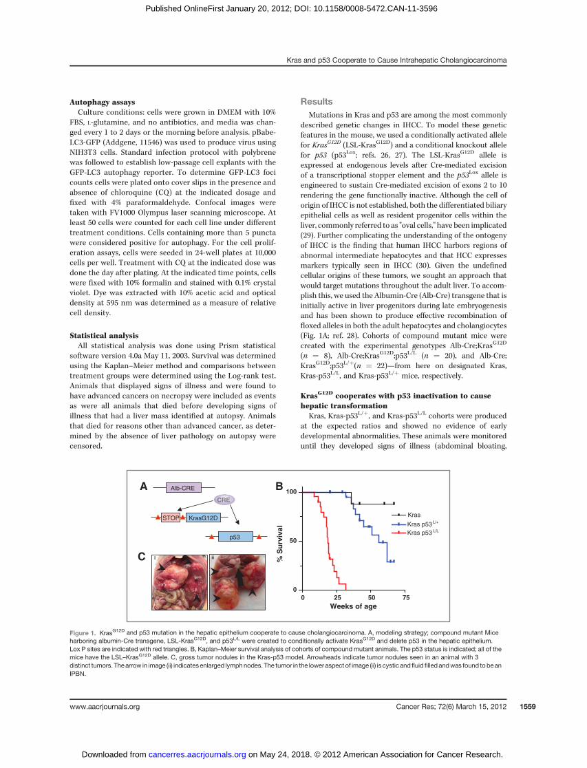
Figure 1. KrasG12D and p53 mutation in the hepatic epithelium cooperate to cause cholangiocarcinoma. A, modeling strategy; compound mutant Miceharboring albumin-Cre transgene, LSL-KrasG12D, and p53L/L were created to conditionally activate KrasG12D and delete p53 in the hepatic epithelium.Lox P sites are indicated with red triangles. B, Kaplan–Meier survival analysis of cohorts of compound mutant animals. The p53 status is indicated; all of themice have the LSL–KrasG12D allele. C, gross tumor nodules in the Kras-p53 model. Arrowheads indicate tumor nodules seen in an animal with 3distinct tumors. Thearrow in image (ii) indicatesenlarged lymphnodes. The tumor in the lower aspect of image (ii) is cystic andfluid filled andwas found tobeanIPBN.
Kras and p53 Cooperate to Cause Intrahepatic Cholangiocarcinoma
www.aacrjournals.org Cancer Res; 72(6) March 15, 2012 1559
on May 24, 2018. © 2012 American Association for Cancer Research. cancerres.aacrjournals.org Downloaded from
Published OnlineFirst January 20, 2012; DOI: 10.1158/0008-5472.CAN-11-3596
diminished activity, and cachexia). Most of the Kras mice werehealthy up to an age of 75 weeks, although 1 of 8 developed alethal liver mass at 36 weeks of age. p53 deficiency greatlyincreased the penetrance and accelerated disease onset as theKras-p53L/þ and Kras-p53L/L mice developed tumors as earlyas 32 and 9 weeks, respectively (mean survival 52 weeks and 19weeks; Fig. 1B). Upon necropsy these animals were found tohave solid liver tumors ranging in size from 2 to 20 mm andpresented as isolated nodules (n ¼ 14 mice) or as multipleindependent lesions (n ¼ 6 mice; Table 1). In many animals,cystic fluid-filled lesions were found adjacent to the solidmasses (Fig. 1C). Hepatic lesions frequently showed hemor-rhage into the peritoneal cavity and had evidence of tumornecrosis. Tumors were highly metastatic, with 75% of tumor-bearing animals displaying gross evidence of invasion intoadjacent organs (diaphragm, bowel, pancreas, and stomach) ordistant metastasis (with dissemination to the lymph nodes,spleen, lungs, and peritoneal cavity; Fig 2B and Table 1). MiceharboringAlb-Cre;p53L/L (n¼ 10)were followed to 1 year of agewithout evidence of illness, and subsequent necropsy and
histologic survey did not reveal abnormal hepatic pathology(data not shown). Thus, KrasG12D promotes metastatic livertumorigenesis that is significantly accelerated by the hetero-zygous and homozygous inactivation of p53.
Albumin-Cre; Kras GEM models recapitulate thehistopathologic features of human IHCC
Microscopic analysis of the liver tumors revealed that 83%showed IHCC histology, with 66% (n ¼ 19) containing exclu-sively IHCC and 17% (n ¼ 5) showing a mixed phenotype ofIHCC and HCC elements. A smaller number had exclusivelyHCC histology (17% n ¼ 5; Table 1, and Fig. 2A). The IHCCshowed a range of cellular differentiation, including well-differentiated tumors with clear glandular architecture andareas of mucin production, as well as poorly differentiatedtumors with minimal gland formation and large pleomorphiccells showing nuclear atypia and frequent mitosis (Fig. 2A).Often different grades of differentiation were seen in the sametumor. The histologic spectrumof IHCCwas similar among thedifferent mouse cohorts.
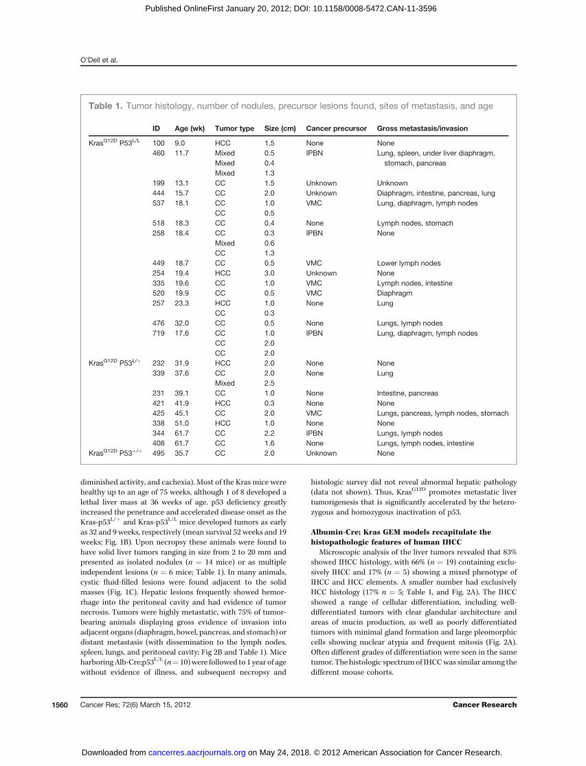
Table 1. Tumor histology, number of nodules, precursor lesions found, sites of metastasis, and age
ID Age (wk) Tumor type Size (cm) Cancer precursor Gross metastasis/invasion
KrasG12D P53L/L 100 9.0 HCC 1.5 None None460 11.7 Mixed 0.5 IPBN Lung, spleen, under liver diaphragm,
stomach, pancreasMixed 0.4Mixed 1.3
199 13.1 CC 1.5 Unknown Unknown444 15.7 CC 2.0 Unknown Diaphragm, intestine, pancreas, lung537 18.1 CC 1.0 VMC Lung, diaphragm, lymph nodes
CC 0.5518 18.3 CC 0.4 None Lymph nodes, stomach258 18.4 CC 0.3 IPBN None
Mixed 0.6CC 1.3
449 18.7 CC 0.5 VMC Lower lymph nodes254 19.4 HCC 3.0 Unknown None335 19.6 CC 1.0 VMC Lymph nodes, intestine520 19.9 CC 0.5 VMC Diaphragm257 23.3 HCC 1.0 None Lung
CC 0.3476 32.0 CC 0.5 None Lungs, lymph nodes719 17.6 CC 1.0 IPBN Lung, diaphragm, lymph nodes
CC 2.0CC 2.0
KrasG12D P53L/þ 232 31.9 HCC 2.0 None None339 37.6 CC 2.0 None Lung
Mixed 2.5231 39.1 CC 1.0 None Intestine, pancreas421 41.9 HCC 0.3 None None425 45.1 CC 2.0 VMC Lungs, pancreas, lymph nodes, stomach338 51.0 HCC 1.0 None None344 61.7 CC 2.2 IPBN Lungs, lymph nodes408 61.7 CC 1.6 None Lungs, lymph nodes, intestine
KrasG12D P53þ/þ 495 35.7 CC 2.0 Unknown None
O'Dell et al.
Cancer Res; 72(6) March 15, 2012 Cancer Research1560
on May 24, 2018. © 2012 American Association for Cancer Research. cancerres.aacrjournals.org Downloaded from
Published OnlineFirst January 20, 2012; DOI: 10.1158/0008-5472.CAN-11-3596
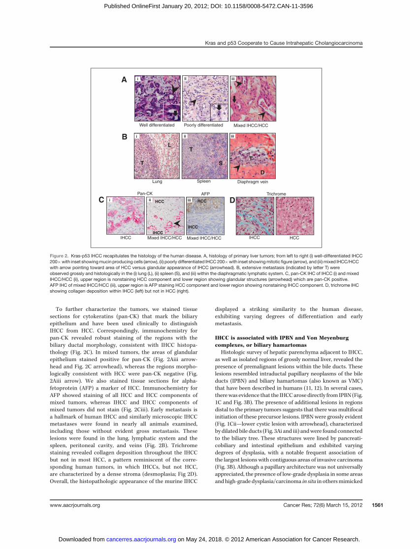
To further characterize the tumors, we stained tissuesections for cytokeratins (pan-CK) that mark the biliaryepithelium and have been used clinically to distinguishIHCC from HCC. Correspondingly, immunochemistry forpan-CK revealed robust staining of the regions with thebiliary ductal morphology, consistent with IHCC histopa-thology (Fig. 2C). In mixed tumors, the areas of glandularepithelium stained positive for pan-CK (Fig. 2Aiii arrow-head and Fig. 2C arrowhead), whereas the regions morpho-logically consistent with HCC were pan-CK negative (Fig.2Aiii arrow). We also stained tissue sections for alpha-fetoprotein (AFP) a marker of HCC. Immunochemistry forAFP showed staining of all HCC and HCC components ofmixed tumors, whereas IHCC and IHCC components ofmixed tumors did not stain (Fig. 2Ciii). Early metastasis isa hallmark of human IHCC and similarly microscopic IHCCmetastases were found in nearly all animals examined,including those without evident gross metastasis. Theselesions were found in the lung, lymphatic system and thespleen, peritoneal cavity, and veins (Fig. 2B). Trichromestaining revealed collagen deposition throughout the IHCCbut not in most HCC, a pattern reminiscent of the corre-sponding human tumors, in which IHCCs, but not HCC,are characterized by a dense stroma (desmoplasia; Fig 2D).Overall, the histopathologic appearance of the murine IHCC
displayed a striking similarity to the human disease,exhibiting varying degrees of differentiation and earlymetastasis.
IHCC is associated with IPBN and Von Meyenburgcomplexes, or biliary hamartomas
Histologic survey of hepatic parenchyma adjacent to IHCC,as well as isolated regions of grossly normal liver, revealed thepresence of premalignant lesions within the bile ducts. Theselesions resembled intraductal papillary neoplasms of the bileducts (IPBN) and biliary hamartomas (also known as VMC)that have been described in humans (11, 12). In several cases,therewas evidence that the IHCCarose directly from IPBN (Fig.1C and Fig. 3B). The presence of additional lesions in regionsdistal to the primary tumors suggests that there wasmultifocalinitiation of these precursor lesions. IPBN were grossly evident(Fig. 1Cii—lower cystic lesion with arrowhead), characterizedby dilated bile ducts (Fig. 3Ai and iii) andwere found connectedto the biliary tree. These structures were lined by pancreati-cobiliary and intestinal epithelium and exhibited varyingdegrees of dysplasia, with a notable frequent association ofthe largest lesions with contiguous areas of invasive carcinoma(Fig. 3B). Although a papillary architecture was not universallyappreciated, the presence of low-grade dysplasia in some areasandhigh-grade dysplasia/carcinoma in situ in othersmimicked
A
B
iii
Lung Spleen Diaphragm vein
L
T
T
S
D
T
Well differentiated Poorly differentiated Mixed IHCC/HCC
ii i
i iiiii
DC
IHCC Mixed IHCC/HCC IHCC HCC
Pan-CK Trichrome
Mixed IHCC/HCC
AFP
i ii iii HCCHCC
IHCC
IHCC
Figure 2. Kras-p53 IHCC recapitulates the histology of the human disease. A, histology of primary liver tumors; from left to right (i) well-differentiated IHCC200�with inset showingmucin producing cells (arrow), (ii) poorly differentiated IHCC200�with inset showingmitotic figure (arrow), and (iii) mixed IHCC/HCCwith arrow pointing toward area of HCC versus glandular appearance of IHCC (arrowhead). B, extensive metastasis (indicated by letter T) wereobserved grossly and histologically in the (i) lung (L), (ii) spleen (S), and (iii) within the diaphragmatic lymphatic system. C, pan-CK IHC of IHCC (i) and mixedIHCC/HCC (ii), upper region is nonstaining HCC component and lower region showing glandular structures (arrowhead) which are pan-CK positive.AFP IHC of mixed IHCC/HCC (iii), upper region is AFP staining HCC component and lower region showing nonstaining IHCC component. D, trichrome IHCshowing collagen deposition within IHCC (left) but not in HCC (right).
Kras and p53 Cooperate to Cause Intrahepatic Cholangiocarcinoma
www.aacrjournals.org Cancer Res; 72(6) March 15, 2012 1561
on May 24, 2018. © 2012 American Association for Cancer Research. cancerres.aacrjournals.org Downloaded from
Published OnlineFirst January 20, 2012; DOI: 10.1158/0008-5472.CAN-11-3596
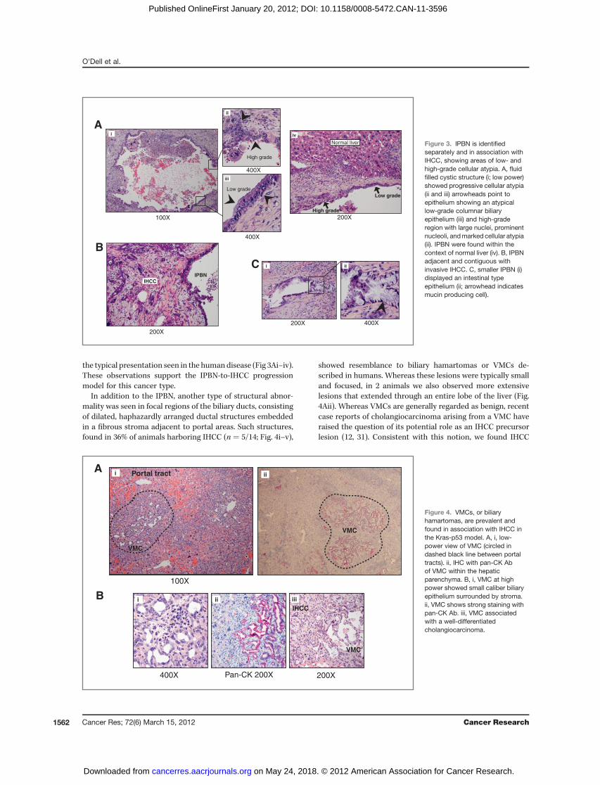
the typical presentation seen in the humandisease (Fig 3Ai–iv).These observations support the IPBN-to-IHCC progressionmodel for this cancer type.
In addition to the IPBN, another type of structural abnor-mality was seen in focal regions of the biliary ducts, consistingof dilated, haphazardly arranged ductal structures embeddedin a fibrous stroma adjacent to portal areas. Such structures,found in 36% of animals harboring IHCC (n ¼ 5/14; Fig. 4i–v),
showed resemblance to biliary hamartomas or VMCs de-scribed in humans. Whereas these lesions were typically smalland focused, in 2 animals we also observed more extensivelesions that extended through an entire lobe of the liver (Fig.4Aii). Whereas VMCs are generally regarded as benign, recentcase reports of cholangiocarcinoma arising from a VMC haveraised the question of its potential role as an IHCC precursorlesion (12, 31). Consistent with this notion, we found IHCC
C
400X200X
A
Low grade
High grade
Normal liver
100X
High grade
400X
Low grade
400X
B
200X
IPBN
IHCC
i
ii
iii
iv
ii i
200X
Figure 3. IPBN is identifiedseparately and in association withIHCC, showing areas of low- andhigh-grade cellular atypia. A, fluidfilled cystic structure (i; low power)showed progressive cellular atypia(ii and iii) arrowheads point toepithelium showing an atypicallow-grade columnar biliaryepithelium (iii) and high-graderegion with large nuclei, prominentnucleoli, andmarked cellular atypia(ii). IPBN were found within thecontext of normal liver (iv). B, IPBNadjacent and contiguous withinvasive IHCC. C, smaller IPBN (i)displayed an intestinal typeepithelium (ii; arrowhead indicatesmucin producing cell).
A
100X
Portal tract
VMC
Pan-CK 200X400X 200X
IHCC
VMC
i ii
iB ii iii
VMC
Figure 4. VMCs, or biliaryhamartomas, are prevalent andfound in association with IHCC inthe Kras-p53 model. A, i, low-power view of VMC (circled indashed black line between portaltracts). ii, IHC with pan-CK Abof VMC within the hepaticparenchyma. B, i, VMC at highpower showed small caliber biliaryepithelium surrounded by stroma.ii, VMC shows strong staining withpan-CK Ab. iii, VMC associatedwith a well-differentiatedcholangiocarcinoma.
O'Dell et al.
Cancer Res; 72(6) March 15, 2012 Cancer Research1562
on May 24, 2018. © 2012 American Association for Cancer Research. cancerres.aacrjournals.org Downloaded from
Published OnlineFirst January 20, 2012; DOI: 10.1158/0008-5472.CAN-11-3596
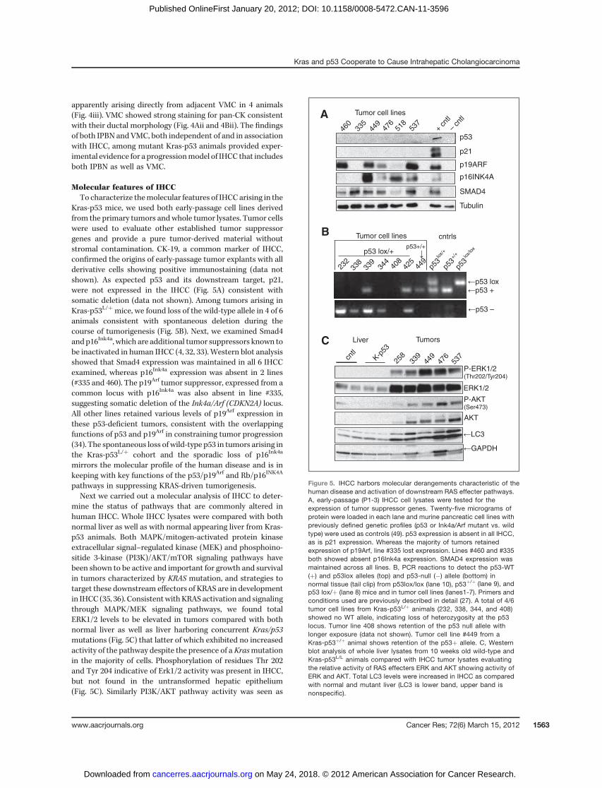
apparently arising directly from adjacent VMC in 4 animals(Fig. 4iii). VMC showed strong staining for pan-CK consistentwith their ductal morphology (Fig. 4Aii and 4Bii). The findingsof both IPBN andVMC, both independent of and in associationwith IHCC, among mutant Kras-p53 animals provided exper-imental evidence for a progressionmodel of IHCC that includesboth IPBN as well as VMC.
Molecular features of IHCCTo characterize themolecular features of IHCC arising in the
Kras-p53 mice, we used both early-passage cell lines derivedfrom the primary tumors andwhole tumor lysates. Tumor cellswere used to evaluate other established tumor suppressorgenes and provide a pure tumor-derived material withoutstromal contamination. CK-19, a common marker of IHCC,confirmed the origins of early-passage tumor explants with allderivative cells showing positive immunostaining (data notshown). As expected p53 and its downstream target, p21,were not expressed in the IHCC (Fig. 5A) consistent withsomatic deletion (data not shown). Among tumors arising inKras-p53L/þmice, we found loss of the wild-type allele in 4 of 6animals consistent with spontaneous deletion during thecourse of tumorigenesis (Fig. 5B). Next, we examined Smad4andp16Ink4a, which are additional tumor suppressors known tobe inactivated in human IHCC (4, 32, 33). Western blot analysisshowed that Smad4 expression was maintained in all 6 IHCCexamined, whereas p16Ink4a expression was absent in 2 lines(#335 and 460). The p19Arf tumor suppressor, expressed from acommon locus with p16Ink4a was also absent in line #335,suggesting somatic deletion of the Ink4a/Arf (CDKN2A) locus.All other lines retained various levels of p19Arf expression inthese p53-deficient tumors, consistent with the overlappingfunctions of p53 and p19Arf in constraining tumor progression(34). The spontaneous loss of wild-type p53 in tumors arising inthe Kras-p53L/þ cohort and the sporadic loss of p16Ink4a
mirrors the molecular profile of the human disease and is inkeeping with key functions of the p53/p19Arf and Rb/p16INK4A
pathways in suppressing KRAS-driven tumorigenesis.Next we carried out a molecular analysis of IHCC to deter-
mine the status of pathways that are commonly altered inhuman IHCC. Whole IHCC lysates were compared with bothnormal liver as well as with normal appearing liver from Kras-p53 animals. Both MAPK/mitogen-activated protein kinaseextracellular signal–regulated kinase (MEK) and phosphoino-sitide 3-kinase (PI3K)/AKT/mTOR signaling pathways havebeen shown to be active and important for growth and survivalin tumors characterized by KRAS mutation, and strategies totarget these downstream effectors of KRAS are in developmentin IHCC (35, 36). ConsistentwithKRAS activation and signalingthrough MAPK/MEK signaling pathways, we found totalERK1/2 levels to be elevated in tumors compared with bothnormal liver as well as liver harboring concurrent Kras/p53mutations (Fig. 5C) that latter of which exhibited no increasedactivity of the pathway despite the presence of a Krasmutationin the majority of cells. Phosphorylation of residues Thr 202and Tyr 204 indicative of Erk1/2 activity was present in IHCC,but not found in the untransformed hepatic epithelium(Fig. 5C). Similarly PI3K/AKT pathway activity was seen as
A
B
p19ARF
p21
Tubulin
SMAD4
p16INK4A
p5346
033
544
947
651
853
7+
cntl
– cn
tlTumor cell lines
←p53 lox←p53 +
←p53 –
Tumor cell lines
p53
p53
p53
lox/+
+/+
lox/lox
232
338
339
344
408
425
449
p53 lox/+p53+/+
↓
cntrls
P-ERK1/2 (Thr202/Tyr204)
ERK1/2
cntl
K-p53
TumorsLiver
←GAPDH
P-AKT (Ser473)
AKT
←LC3
258
339
449
476
537
C
Figure 5. IHCC harbors molecular derangements characteristic of thehuman disease and activation of downstream RAS effecter pathways.A, early-passage (P1-3) IHCC cell lysates were tested for theexpression of tumor suppressor genes. Twenty-five micrograms ofprotein were loaded in each lane and murine pancreatic cell lines withpreviously defined genetic profiles (p53 or Ink4a/Arf mutant vs. wildtype) were used as controls (49). p53 expression is absent in all IHCC,as is p21 expression. Whereas the majority of tumors retainedexpression of p19Arf, line #335 lost expression. Lines #460 and #335both showed absent p16Ink4a expression. SMAD4 expression wasmaintained across all lines. B, PCR reactions to detect the p53-WT(þ) and p53lox alleles (top) and p53-null (�) allele (bottom) innormal tissue (tail clip) from p53lox/lox (lane 10), p53þ/þ (lane 9), andp53 lox/þ (lane 8) mice and in tumor cell lines (lanes1-7). Primers andconditions used are previously described in detail (27). A total of 4/6tumor cell lines from Kras-p53L/þ animals (232, 338, 344, and 408)showed no WT allele, indicating loss of heterozygosity at the p53locus. Tumor line 408 shows retention of the p53 null allele withlonger exposure (data not shown). Tumor cell line #449 from aKras-p53þ/þ animal shows retention of the p53þ allele. C, Westernblot analysis of whole liver lysates from 10 weeks old wild-type andKras-p53L/L animals compared with IHCC tumor lysates evaluatingthe relative activity of RAS effecters ERK and AKT showing activity ofERK and AKT. Total LC3 levels were increased in IHCC as comparedwith normal and mutant liver (LC3 is lower band, upper band isnonspecific).
Kras and p53 Cooperate to Cause Intrahepatic Cholangiocarcinoma
www.aacrjournals.org Cancer Res; 72(6) March 15, 2012 1563
on May 24, 2018. © 2012 American Association for Cancer Research. cancerres.aacrjournals.org Downloaded from
Published OnlineFirst January 20, 2012; DOI: 10.1158/0008-5472.CAN-11-3596
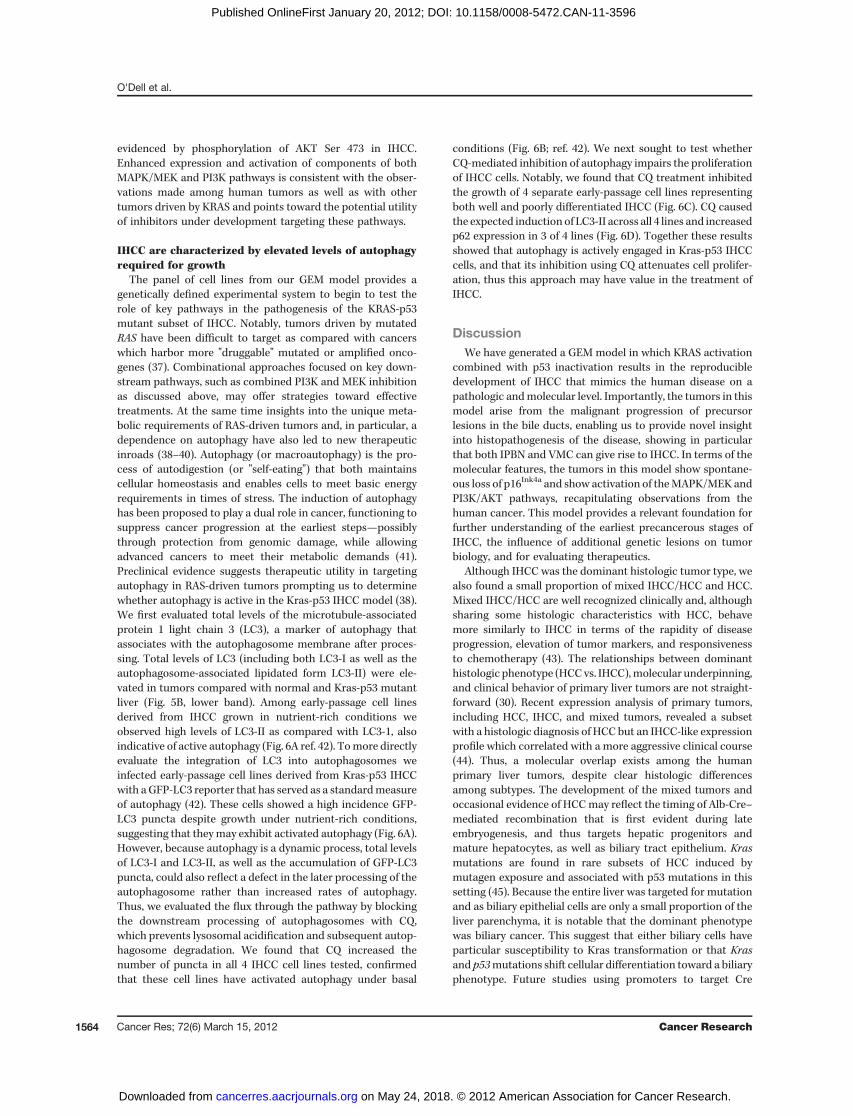
evidenced by phosphorylation of AKT Ser 473 in IHCC.Enhanced expression and activation of components of bothMAPK/MEK and PI3K pathways is consistent with the obser-vations made among human tumors as well as with othertumors driven by KRAS and points toward the potential utilityof inhibitors under development targeting these pathways.
IHCC are characterized by elevated levels of autophagyrequired for growth
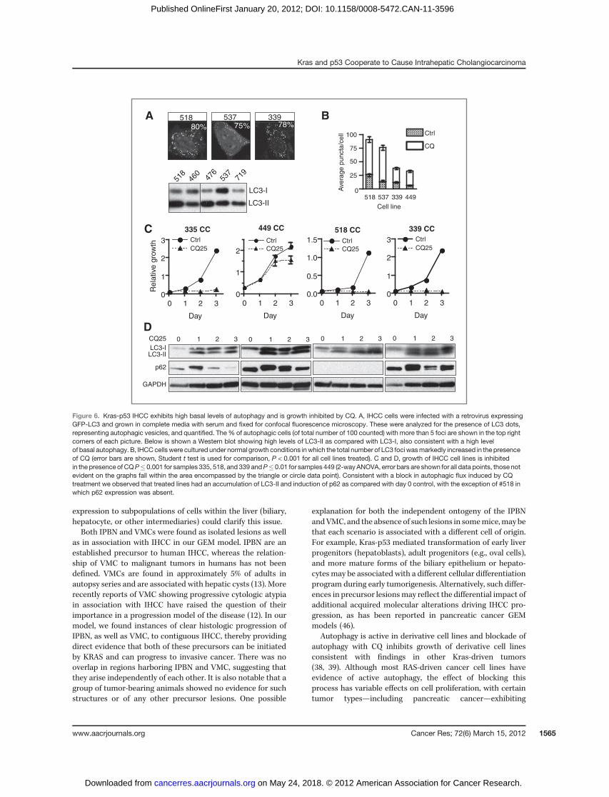
The panel of cell lines from our GEM model provides agenetically defined experimental system to begin to test therole of key pathways in the pathogenesis of the KRAS-p53mutant subset of IHCC. Notably, tumors driven by mutatedRAS have been difficult to target as compared with cancerswhich harbor more "druggable" mutated or amplified onco-genes (37). Combinational approaches focused on key down-stream pathways, such as combined PI3K and MEK inhibitionas discussed above, may offer strategies toward effectivetreatments. At the same time insights into the unique meta-bolic requirements of RAS-driven tumors and, in particular, adependence on autophagy have also led to new therapeuticinroads (38–40). Autophagy (or macroautophagy) is the pro-cess of autodigestion (or "self-eating") that both maintainscellular homeostasis and enables cells to meet basic energyrequirements in times of stress. The induction of autophagyhas been proposed to play a dual role in cancer, functioning tosuppress cancer progression at the earliest steps—possiblythrough protection from genomic damage, while allowingadvanced cancers to meet their metabolic demands (41).Preclinical evidence suggests therapeutic utility in targetingautophagy in RAS-driven tumors prompting us to determinewhether autophagy is active in the Kras-p53 IHCC model (38).We first evaluated total levels of the microtubule-associatedprotein 1 light chain 3 (LC3), a marker of autophagy thatassociates with the autophagosome membrane after proces-sing. Total levels of LC3 (including both LC3-I as well as theautophagosome-associated lipidated form LC3-II) were ele-vated in tumors compared with normal and Kras-p53 mutantliver (Fig. 5B, lower band). Among early-passage cell linesderived from IHCC grown in nutrient-rich conditions weobserved high levels of LC3-II as compared with LC3-1, alsoindicative of active autophagy (Fig. 6A ref. 42). Tomore directlyevaluate the integration of LC3 into autophagosomes weinfected early-passage cell lines derived from Kras-p53 IHCCwith aGFP-LC3 reporter that has served as a standardmeasureof autophagy (42). These cells showed a high incidence GFP-LC3 puncta despite growth under nutrient-rich conditions,suggesting that theymay exhibit activated autophagy (Fig. 6A).However, because autophagy is a dynamic process, total levelsof LC3-I and LC3-II, as well as the accumulation of GFP-LC3puncta, could also reflect a defect in the later processing of theautophagosome rather than increased rates of autophagy.Thus, we evaluated the flux through the pathway by blockingthe downstream processing of autophagosomes with CQ,which prevents lysosomal acidification and subsequent autop-hagosome degradation. We found that CQ increased thenumber of puncta in all 4 IHCC cell lines tested, confirmedthat these cell lines have activated autophagy under basal
conditions (Fig. 6B; ref. 42). We next sought to test whetherCQ-mediated inhibition of autophagy impairs the proliferationof IHCC cells. Notably, we found that CQ treatment inhibitedthe growth of 4 separate early-passage cell lines representingboth well and poorly differentiated IHCC (Fig. 6C). CQ causedthe expected induction of LC3-II across all 4 lines and increasedp62 expression in 3 of 4 lines (Fig. 6D). Together these resultsshowed that autophagy is actively engaged in Kras-p53 IHCCcells, and that its inhibition using CQ attenuates cell prolifer-ation, thus this approach may have value in the treatment ofIHCC.
DiscussionWe have generated a GEMmodel in which KRAS activation
combined with p53 inactivation results in the reproducibledevelopment of IHCC that mimics the human disease on apathologic andmolecular level. Importantly, the tumors in thismodel arise from the malignant progression of precursorlesions in the bile ducts, enabling us to provide novel insightinto histopathogenesis of the disease, showing in particularthat both IPBN and VMC can give rise to IHCC. In terms of themolecular features, the tumors in this model show spontane-ous loss of p16Ink4a and show activation of theMAPK/MEK andPI3K/AKT pathways, recapitulating observations from thehuman cancer. This model provides a relevant foundation forfurther understanding of the earliest precancerous stages ofIHCC, the influence of additional genetic lesions on tumorbiology, and for evaluating therapeutics.
Although IHCC was the dominant histologic tumor type, wealso found a small proportion of mixed IHCC/HCC and HCC.Mixed IHCC/HCC are well recognized clinically and, althoughsharing some histologic characteristics with HCC, behavemore similarly to IHCC in terms of the rapidity of diseaseprogression, elevation of tumor markers, and responsivenessto chemotherapy (43). The relationships between dominanthistologic phenotype (HCC vs. IHCC),molecular underpinning,and clinical behavior of primary liver tumors are not straight-forward (30). Recent expression analysis of primary tumors,including HCC, IHCC, and mixed tumors, revealed a subsetwith a histologic diagnosis of HCC but an IHCC-like expressionprofile which correlated with a more aggressive clinical course(44). Thus, a molecular overlap exists among the humanprimary liver tumors, despite clear histologic differencesamong subtypes. The development of the mixed tumors andoccasional evidence of HCCmay reflect the timing of Alb-Cre–mediated recombination that is first evident during lateembryogenesis, and thus targets hepatic progenitors andmature hepatocytes, as well as biliary tract epithelium. Krasmutations are found in rare subsets of HCC induced bymutagen exposure and associated with p53 mutations in thissetting (45). Because the entire liver was targeted for mutationand as biliary epithelial cells are only a small proportion of theliver parenchyma, it is notable that the dominant phenotypewas biliary cancer. This suggest that either biliary cells haveparticular susceptibility to Kras transformation or that Krasand p53mutations shift cellular differentiation toward a biliaryphenotype. Future studies using promoters to target Cre
O'Dell et al.
Cancer Res; 72(6) March 15, 2012 Cancer Research1564
on May 24, 2018. © 2012 American Association for Cancer Research. cancerres.aacrjournals.org Downloaded from
Published OnlineFirst January 20, 2012; DOI: 10.1158/0008-5472.CAN-11-3596
expression to subpopulations of cells within the liver (biliary,hepatocyte, or other intermediaries) could clarify this issue.Both IPBN and VMCs were found as isolated lesions as well
as in association with IHCC in our GEM model. IPBN are anestablished precursor to human IHCC, whereas the relation-ship of VMC to malignant tumors in humans has not beendefined. VMCs are found in approximately 5% of adults inautopsy series and are associated with hepatic cysts (13). Morerecently reports of VMC showing progressive cytologic atypiain association with IHCC have raised the question of theirimportance in a progression model of the disease (12). In ourmodel, we found instances of clear histologic progression ofIPBN, as well as VMC, to contiguous IHCC, thereby providingdirect evidence that both of these precursors can be initiatedby KRAS and can progress to invasive cancer. There was nooverlap in regions harboring IPBN and VMC, suggesting thatthey arise independently of each other. It is also notable that agroup of tumor-bearing animals showed no evidence for suchstructures or of any other precursor lesions. One possible
explanation for both the independent ontogeny of the IPBNandVMC, and the absence of such lesions in somemice,may bethat each scenario is associated with a different cell of origin.For example, Kras-p53 mediated transformation of early liverprogenitors (hepatoblasts), adult progenitors (e.g., oval cells),and more mature forms of the biliary epithelium or hepato-cytes may be associated with a different cellular differentiationprogram during early tumorigenesis. Alternatively, such differ-ences in precursor lesionsmay reflect the differential impact ofadditional acquired molecular alterations driving IHCC pro-gression, as has been reported in pancreatic cancer GEMmodels (46).
Autophagy is active in derivative cell lines and blockade ofautophagy with CQ inhibits growth of derivative cell linesconsistent with findings in other Kras-driven tumors(38, 39). Although most RAS-driven cancer cell lines haveevidence of active autophagy, the effect of blocking thisprocess has variable effects on cell proliferation, with certaintumor types—including pancreatic cancer—exhibiting
Re
lative
gro
wth
A B
C 449 CC
0 1 2 30
1
2
Ctrl
CQ25
Day
335 CC
0 1 2 30
1
2
3 Ctrl
CQ25
Day
339 CC
0 1 2 30
1
2
3 Ctrl
CQ25
Day
518
460
476
537
719
LC3-I
LC3-II
518 CC
0 1 2 30.0
0.5
1.0
1.5 Ctrl
CQ25
Day
33978%
53775%
51880%
LC3-ILC3-II
p62
GAPDH
CQ25 0 1 2 3 0 1 2 3 0 1 2 3 0 1 2 3
DA
vera
ge p
uncta
/cell
518 537 339 4490
25
50
75
100 Ctrl
CQ
Cell line
Figure 6. Kras-p53 IHCC exhibits high basal levels of autophagy and is growth inhibited by CQ. A, IHCC cells were infected with a retrovirus expressingGFP-LC3 and grown in complete media with serum and fixed for confocal fluorescence microscopy. These were analyzed for the presence of LC3 dots,representing autophagic vesicles, and quantified. The% of autophagic cells (of total number of 100 counted) with more than 5 foci are shown in the top rightcorners of each picture. Below is shown a Western blot showing high levels of LC3-II as compared with LC3-I, also consistent with a high levelof basal autophagy. B, IHCCcellswere cultured under normal growth conditions inwhich the total number of LC3 foci wasmarkedly increased in the presenceof CQ (error bars are shown, Student t test is used for comparison, P < 0.001 for all cell lines treated). C and D, growth of IHCC cell lines is inhibitedin the presence of CQP� 0.001 for samples 335, 518, and 339 andP� 0.01 for samples 449 (2-wayANOVA, error bars are shown for all data points, those notevident on the graphs fall within the area encompassed by the triangle or circle data point). Consistent with a block in autophagic flux induced by CQtreatment we observed that treated lines had an accumulation of LC3-II and induction of p62 as compared with day 0 control, with the exception of #518 inwhich p62 expression was absent.
Kras and p53 Cooperate to Cause Intrahepatic Cholangiocarcinoma
www.aacrjournals.org Cancer Res; 72(6) March 15, 2012 1565
on May 24, 2018. © 2012 American Association for Cancer Research. cancerres.aacrjournals.org Downloaded from
Published OnlineFirst January 20, 2012; DOI: 10.1158/0008-5472.CAN-11-3596
marked sensitivity to CQ, whereas growth of lung cancer celllines was effected to a lesser extent. We find that cell lines fromour model are significantly growth inhibited with a corre-sponding block in autophagy byCQ. Thisfitswell with evidencefrom human tumors and cell lines suggesting the importanceof autophagy in the human disease and provides impetus tofurther evaluate CQ and other drugs that may impact thisprocess in IHCC (47, 48).
In summary, we have created a tractable IHCC GEM modelon the basis of the genetics of the human disease. The mul-tistage evolution of the tumors provides a context to explorequestions about the role of different cells of origin and themolecular and histologic progression of precancerous stages ofIHCC. In addition, the highly reproducible development ofIHCC in this model provides a foundation for testing theinfluence of additional genetic lesions on tumor biology anda platform for the preclinical testing of promising newtherapies.
Disclosure of Potential Conflicts of InterestNo potential conflicts of interest were disclosed.
AcknowledgmentsThe authors thankCatherineHarmer for contributing to the organization and
preparation of the manuscript and Alec Kimmelman for critical reading of themanuscript.
Grant SupportThis work was supported by the Granara-Skerry Trust and was accomplished
in the Translational Research Core Facility of the Wilmot Cancer Center at theUniversity of Rochester Medical Center.
A.F. Hezel is supported by a HHMI early career award and the Edelman-Gardner foundation award. N. Bardeesy is supported by grants from Target-Cancer Foundation and from the NIH (1R01CA136567-01A1).
The costs of publication of this article were defrayed in part by thepayment of page charges. This article must therefore be hereby markedadvertisement in accordance with 18 U.S.C. Section 1734 solely to indicatethis fact.
Received October 28, 2011; revised January 12, 2012; accepted January 16, 2012;published OnlineFirst January 20, 2012.
References1. Hezel AF, Deshpande V, Zhu AX. Genetics of biliary tract cancers and
emerging targeted therapies. J Clin Oncol 2011;28:3531–40.2. Shaib Y, El-Serag HB. The epidemiology of cholangiocarcinoma.
Semin Liver Dis 2004;24:115–25.3. Valle J, Wasan H, Palmer DH, Cunningham D, Anthoney A, Maraveyas
A, et al. Cisplatin plus gemcitabine versus gemcitabine for biliary tractcancer. N Engl J Med 2011;362:1273–81.
4. Tannapfel A, Benicke M, Katalinic A, Uhlmann D, K€ockerling F, HaussJ, et al. Frequency of p16(INK4A) alterations and K-ras mutations inintrahepatic cholangiocarcinoma of the liver. Gut 2000;47:721–7.
5. Tannapfel A, Sommerer F, Benicke M, Katalinic A, Uhlmann D, Wit-zigmann H, et al. Mutations of the BRAF gene in cholangiocarcinomabut not in hepatocellular carcinoma. Gut 2003;52:706–12.
6. Deshpande V, Nduaguba A, Zimmerman SM, Kehoe SM, MacconaillLE, LauwersGY, et al.Mutational profiling revealsPIK3CAmutations ingallbladder carcinoma. BMC Cancer 2011;11:60.
7. Tannapfel A,Weinans L,Geissler F, Sch€utzA, Katalinic A, K€ockerling F,et al. Mutations of p53 tumor suppressor gene, apoptosis, and pro-liferation in intrahepatic cholangiocellular carcinoma of the liver. DigDis Sci 2000;45:317–24.
8. Argani P, Shaukat A, Kaushal M, Wilentz RE, Su GH, Sohn TA, et al.Differing rates of loss of DPC4 expression and of p53 overexpressionamong carcinomas of the proximal and distal bile ducts. Cancer2001;91:1332–41.
9. Wu TT, Levy M, Correa AM, Rosen CB, Abraham SC. Biliary intrae-pithelial neoplasia in patients without chronic biliary disease: analysisof liver explants with alcoholic cirrhosis, hepatitis C infection, andnoncirrhotic liver diseases. Cancer 2009;115:4564–75.
10. Zen Y, Fujii T, Itatsu K, Nakamura K, Minato H, Kasashima S, et al.Biliary papillary tumors share pathological features with intraductalpapillary mucinous neoplasm of the pancreas. Hepatology 2006;44:1333–43.
11. Zen Y, Adsay NV, Bardadin K, Colombari R, Ferrell L, Haga H, et al.Biliary intraepithelial neoplasia: an international interobserver agree-ment study and proposal for diagnostic criteria. Mod Pathol 2007;20:701–9.
12. XuAM,Xian ZH, ZhangSH,ChenXF. Intrahepatic cholangiocarcinomaarising in multiple bile duct hamartomas: report of two cases andreview of the literature. Eur J Gastroenterol Hepatol 2009;21:580–4.
13. Redston MS, Wanless IR. The hepatic von Meyenburg complex:prevalence and association with hepatic and renal cysts among2843 autopsies [corrected]. Mod Pathol 1996;9:233–7.
14. Kiguchi K, Carbajal S, Chan K, Beltr�an L, Ruffino L, Shen J, et al.Constitutive expression of ErbB-2 in gallbladder epithelium
results in development of adenocarcinoma. Cancer Res 2001;61:6971–6.
15. Elmore LW, Sirica AE. Phenotypic characterization of metaplasticintestinal glands and ductular hepatocytes in cholangiofibrotic lesionsrapidly induced in the caudate liver lobe of rats treated with furan.Cancer Res 1991;51:5752–9.
16. Lai GH, Zhang Z, Shen XN, Ward DJ, Dewitt JL, Holt SE, et al. erbB-2/neu transformed rat cholangiocytes recapitulate key cellular andmolecular features of human bile duct cancer. Gastroenterology2005;129:2047–57.
17. Sirica AE, Zhang Z, Lai GH, Asano T, Shen XN, Ward DJ, et al. A novel"patient-like" model of cholangiocarcinoma progression based on bileduct inoculation of tumorigenic rat cholangiocyte cell lines. Hepatol-ogy 2008;47:1178–90.
18. Braun L, Goyette M, Yaswen P, Thompson NL, Fausto N. Growth inculture and tumorigenicity after transfection with the ras oncogeneof liver epithelial cells from carcinogen-treated rats. Cancer Res1987;47:4116–24.
19. Lin YZ, Brunt EM, BowlingW,Hafenrichter DG, Kennedy SC, FlyeMW,et al. Ras-transduceddiethylnitrosamine-treated hepatocytesdevelopinto cancers of mixed phenotype in vivo. Cancer Res 1995;55:5242–50.
20. Farazi PA, Zeisberg M, Glickman J, Zhang Y, Kalluri R, DePinho RA.Chronic bile duct injury associated with fibrotic matrix microenviron-ment provokes cholangiocarcinoma in p53-deficientmice. CancerRes2006;66:6622–7.
21. Chen YW, Klimstra DS, Mongeau ME, Tatem JL, Boyartchuk V, LewisBC. Loss of p53 and Ink4a/Arf cooperate in a cell autonomous fashionto induce metastasis of hepatocellular carcinoma cells. Cancer Res2007;67:7589–96.
22. Avruch J, Zhou D, Fitamant J, Bardeesy N. Mst1/2 signalling to Yap:gatekeeper for liver size and tumour development. Br J Cancer2011;104:24–32.
23. Xu X, Kobayashi S, QiaoW, Li C, Xiao C, Radaeva S, et al. Induction ofintrahepatic cholangiocellular carcinoma by liver-specific disruption ofSmad4 and Pten in mice. J Clin Invest 2006;116:1843–52.
24. Sharpless NE, Depinho RA. Themighty mouse: genetically engineeredmouse models in cancer drug development. Nat Rev Drug Discov2006;5:741–54.
25. Tuveson D, Hanahan D. Translational medicine: Cancer lessons frommice to humans. Nature 2011;471:316–7.
26. Jackson EL, Willis N, Mercer K, Bronson RT, Crowley D, Montoya R,et al. Analysis of lung tumor initiation andprogressionusing conditionalexpression of oncogenic K-ras. Genes Dev 2001;15:3243–8.
O'Dell et al.
Cancer Res; 72(6) March 15, 2012 Cancer Research1566
on May 24, 2018. © 2012 American Association for Cancer Research. cancerres.aacrjournals.org Downloaded from
Published OnlineFirst January 20, 2012; DOI: 10.1158/0008-5472.CAN-11-3596
27. Jonkers J, Meuwissen R, van der Gulden H, Peterse H, van der ValkM,Berns A. Synergistic tumor suppressor activity of BRCA2 and p53 in aconditional mouse model for breast cancer. Nat Genet 2001;29:418–25.
28. Postic C, Magnuson MA. DNA excision in liver by an albumin-Cretransgene occurs progressively with age. Genesis 2000;26:149–50.
29. Duncan AW, Dorrell C, Grompe M. Stem cells and liver regeneration.Gastroenterology 2009;137:466–81.
30. Komuta M, Spee B, Vander Borght S, De Vos R, Verslype C, Aerts R,et al. Clinicopathological study on cholangiolocellular carcinomasuggesting hepatic progenitor cell origin. Hepatology 2008;47:1544–56.
31. Jain D, Sarode VR, Abdul-Karim FW, Homer R, Robert ME. Evidencefor the neoplastic transformation of Von-Meyenburg complexes. Am JSurg Pathol 2000;24:1131–9.
32. Tannapfel A, Sommerer F, BenickeM,Weinans L, Katalinic A, GeisslerF, et al. Genetic and epigenetic alterations of the INK4a-ARF path-way in cholangiocarcinoma. J Pathol 2002;197:624–31.
33. Taniai M, Higuchi H, Burgart LJ, Gores GJ. p16INK4a promotermutations are frequent in primary sclerosing cholangitis (PSC) andPSC-associated cholangiocarcinoma. Gastroenterology 2002;123:1090–8.
34. Sherr CJ. Principles of tumor suppression. Cell 2004;116:235–46.35. Engelman JA, Chen L, Tan X, Crosby K, Guirmaraes AR, Upadhyay R,
et al. Effective use of PI3K and MEK inhibitors to treat mutant KrasG12D and PIK3CA H1047R murine lung cancers. Nat Med2008;14:1351–6.
36. Bekaii-Saab T, Phelps MA, Li X, Saji M, Goff L, Kauh JS, et al. Multi-institutional phase II study of selumetinib in patients with metastaticbiliary cancers. J Clin Oncol 2011;29:2357–63.
37. McCormick F. Success and failure on the ras pathway. Cancer BiolTher 2007;6:1654–9.
38. Yang S, Wang X, Contino G, Liesa M, Sahin E, Bause A, et al.Pancreatic cancers require autophagy for tumor growth. Genes Dev2011;25:717–29.
39. GuoJY,ChenHY,MathewR, Fan J, StrokeckerAM,Karsii-UzunbasG,et al. Activated Ras requires autophagy to maintain oxidative meta-bolism and tumorigenesis. Genes Dev 2011;25:460–70.
40. Lock R, Roy S, Kenific CM, Su JS, Salas S, Ronen SM, et al. Auto-phagy facilitates glycolysis during Ras-mediated oncogenic trans-formation. Mol Biol Cell 2011;22:165–78.
41. Kimmelman AC. The dynamic nature of autophagy in cancer. GenesDev 2011;25:1999–2010.
42. Klionsky DJ, Abeliovich H, Agostinis P, Agrawal DK, Aliev G, AskewDS, et al. Guidelines for the use and interpretation of assays for moni-toring autophagy in higher eukaryotes. Autophagy 2008;4:151–75.
43. Jarnagin WR, Weber S, Tickoo SK, Koca JB, Oblekwe S, Fong Y, et al.Combined hepatocellular and cholangiocarcinoma: demographic,clinical, and prognostic factors. Cancer 2002;94:2040–6.
44. Woo HG, Lee JH, Yoon JH, Kinm CY, Lee HS, Jang JJ, et al. Iden-tification of a cholangiocarcinoma-like gene expression trait in hepa-tocellular carcinoma. Cancer Res 2011;70:3034–41.
45. Weihrauch M, Benicke M, Lehnert G, Wittekind C, Wrbitzky R, Tan-napfel A. Frequent k-ras-2 mutations and p16(INK4A)methylation inhepatocellular carcinomas in workers exposed to vinyl chloride. Br JCancer 2001;84:982–9.
46. Bardeesy N, Cheng KH, Berger JH, Chi GC, Pahler J, Olson P, et al.Smad4 is dispensable for normal pancreas development yet critical inprogression and tumor biology of pancreas cancer. Genes Dev2006;20:3130–46.
47. Hou YJ, Dong LW, Tan YX, Yang GZ, Pan YF, Li Z, et al. Inhibition ofactive autophagy induces apoptosis and increases chemosensitivity incholangiocarcinoma. Lab Invest 2011;91:1146–57.
48. Dong LW, Hou YJ, Tan YX, Pan YO, Wang M, Wang HY, et al.Prognostic significance of Beclin 1 in intrahepatic cholangiocellularcarcinoma. Autophagy 2011;7:1222–9.
49. Bardeesy N, Aguirre AJ, ChuGC, Cehng KH, Lopez LV, Hezel AF, et al.Both p16(Ink4a) and the p19(Arf)-p53 pathway constrain progressionof pancreatic adenocarcinoma in the mouse. Proc Natl Acad Sci U S A2006;103:5947–52.
Kras and p53 Cooperate to Cause Intrahepatic Cholangiocarcinoma
www.aacrjournals.org Cancer Res; 72(6) March 15, 2012 1567
on May 24, 2018. © 2012 American Association for Cancer Research. cancerres.aacrjournals.org Downloaded from
Published OnlineFirst January 20, 2012; DOI: 10.1158/0008-5472.CAN-11-3596
2012;72:1557-1567. Published OnlineFirst January 20, 2012.Cancer Res Michael R. O'Dell, Jing Li Huang, Christa L. Whitney-Miller, et al. Cholangiocarcinoma
Mutation Cause Primary Intrahepaticp53 and G12DKras
Updated version
10.1158/0008-5472.CAN-11-3596doi:
Access the most recent version of this article at:
Cited articles
http://cancerres.aacrjournals.org/content/72/6/1557.full#ref-list-1
This article cites 49 articles, 16 of which you can access for free at:
Citing articles
http://cancerres.aacrjournals.org/content/72/6/1557.full#related-urls
This article has been cited by 11 HighWire-hosted articles. Access the articles at:
E-mail alerts related to this article or journal.Sign up to receive free email-alerts
Subscriptions
Reprints and
To order reprints of this article or to subscribe to the journal, contact the AACR Publications Department at
Permissions
Rightslink site. Click on "Request Permissions" which will take you to the Copyright Clearance Center's (CCC)
.http://cancerres.aacrjournals.org/content/72/6/1557To request permission to re-use all or part of this article, use this link
on May 24, 2018. © 2012 American Association for Cancer Research. cancerres.aacrjournals.org Downloaded from
Published OnlineFirst January 20, 2012; DOI: 10.1158/0008-5472.CAN-11-3596