KERATOCONJUNCTIVITIS SICCA DUE TO SARCOIDOSIS*KERATOCONJUNCTIVITIS SICCA DUE TO SARCOIDOSIS 159...
8
Brit. J. Ophthal. (1957) 41, 153. KERATOCONJUNCTIVITIS SICCA DUE TO SARCOIDOSIS* BY BARRIE R. JONES AND C. J. STEVENSON The London Hospital SARCOIDOSIS is well recognized as a cause of swelling of the lacrimal glands and enters into the differential diagnosis of tumours of this region (Duke- Elder, 1952). Such swellings may be followed, in some cases, by a form of keratoconjunctivitis sicca (Ainslie and James, 1956; Gruber, 1956). The association of systemic sarcoidosis with keratoconjunctivitis sicca in the absence of swelling of the lacrimal glands has also been described (Ainslie and James, 1956; James, 1956; Ramage and Kinnear, 1956), but in these cases the histology of the ocular lesions was not obtained; it was presumed, however, that they were of the same pathology. In the case to be described, a patient, who had been treated in an ophthal- mic clinic for 3 years for symptoms of conjunctival irritation, was found on mass radiography of the chest to have the typical radiological features of sarcoidosis. In view of this finding the patient was referred, after proof of systemic sarcoidosis had been obtained by liver biopsy, for investigation of her ocular condition. The patient's eyes lacked the generally accepted features of ocular sarcoidosis, namely swelling of the lacrimal glands, yellowish conjunctival follicles (Crick, Hoyle, and Mather, 1955; Crick, 1956), or chronic uveitis, but presented the clinical features of kerato- conjunctivitis sicca. In addition she had complained of dryness of the nose and throat. Biopsy of lacrimal gland and conjunctiva, however, revealed that the ocular disorders were in fact due to sarcoidosis. Case Report A married woman aged 42, was admitted to The London Hospital for investigation in January, 1956. For 3 years she had complained of,sticky threads forming in her eyes during the night. These were associated with a pricking sensation and discomfort which lessened during the day but worsened in the evening. For 3 years she had been treated for chronic conjunctivitis with a variety of drops, including penicillin and cortisone, without relief, and had been worsened by a course of silvering. She also noticed dryness of her nose and throat and would awaken with pharyngeal irritation and dryness, relieved by coughing up a tenacious plug of mucus. For 2 years her chest had been under radiological observation since the discovery of hilar shadows and reticulation on mass radiography. With the exception of intermittent pain in the right shoulder during the past 5 years, * Received for publication November 26, 1956. 153 on February 21, 2020 by guest. Protected by copyright. http://bjo.bmj.com/ Br J Ophthalmol: first published as 10.1136/bjo.41.3.153 on 1 March 1957. Downloaded from
Transcript of KERATOCONJUNCTIVITIS SICCA DUE TO SARCOIDOSIS*KERATOCONJUNCTIVITIS SICCA DUE TO SARCOIDOSIS 159...
Brit. J. Ophthal. (1957) 41, 153.
KERATOCONJUNCTIVITIS SICCADUE TO SARCOIDOSIS*
BY
BARRIE R. JONES AND C. J. STEVENSONThe London Hospital
SARCOIDOSIS is well recognized as a cause of swelling of the lacrimal glandsand enters into the differential diagnosis of tumours of this region (Duke-Elder, 1952). Such swellings may be followed, in some cases, by a form ofkeratoconjunctivitis sicca (Ainslie and James, 1956; Gruber, 1956).The association of systemic sarcoidosis with keratoconjunctivitis sicca in
the absence of swelling of the lacrimal glands has also been described(Ainslie and James, 1956; James, 1956; Ramage and Kinnear, 1956), butin these cases the histology of the ocular lesions was not obtained; it waspresumed, however, that they were of the same pathology.
In the case to be described, a patient, who had been treated in an ophthal-mic clinic for 3 years for symptoms of conjunctival irritation, was found onmass radiography of the chest to have the typical radiological features ofsarcoidosis. In view of this finding the patient was referred, after proof ofsystemic sarcoidosis had been obtained by liver biopsy, for investigation ofher ocular condition. The patient's eyes lacked the generally acceptedfeatures of ocular sarcoidosis, namely swelling of the lacrimal glands,yellowish conjunctival follicles (Crick, Hoyle, and Mather, 1955; Crick,1956), or chronic uveitis, but presented the clinical features of kerato-conjunctivitis sicca. In addition she had complained of dryness of the noseand throat. Biopsy of lacrimal gland and conjunctiva, however, revealedthat the ocular disorders were in fact due to sarcoidosis.
Case ReportA married woman aged 42, was admitted to The London Hospital for investigation inJanuary, 1956. For 3 years she had complained of,sticky threads forming in her eyesduring the night. These were associated with a pricking sensation and discomfort whichlessened during the day but worsened in the evening. For 3 years she had been treatedfor chronic conjunctivitis with a variety of drops, including penicillin and cortisone,without relief, and had been worsened by a course of silvering. She also noticed drynessof her nose and throat and would awaken with pharyngeal irritation and dryness, relievedby coughing up a tenacious plug of mucus.For 2 years her chest had been under radiological observation since the discovery of
hilar shadows and reticulation on mass radiography.With the exception of intermittent pain in the right shoulder during the past 5 years,
* Received for publication November 26, 1956.153
on February 21, 2020 by guest. P
rotected by copyright.http://bjo.bm
j.com/
Br J O
phthalmol: first published as 10.1136/bjo.41.3.153 on 1 M
arch 1957. Dow
nloaded from
BARRIE R. JONES AND C. J. STEVENSON
she had remained in good health until the onset of the ocular symptoms. Since then shehad complained of lack of energy. She had one brother who died from tuberculosis.
Examination.-She was an athletic, well-nourished woman. Apart from the ocularfeatures, the only abnormal finding was in the upper respiratory tract, where the nasalmucosa was relatively atrophic and rather dry. The mouth, larynx, and pharynx werenormal. No abnormality of the skin, nails, salivary glands, lymphatic glands, or jointswas discovered. The respiratory and cardio-vascular systems were clinically normal;blood pressure 130/90. The liver and spleen were not palpable and no abnormality wasevident in the nervous system.
Laboratory InvestigationsChest x-Ray.-Diffuse infiltration in the lung fields especially in the upper lobes. Marked
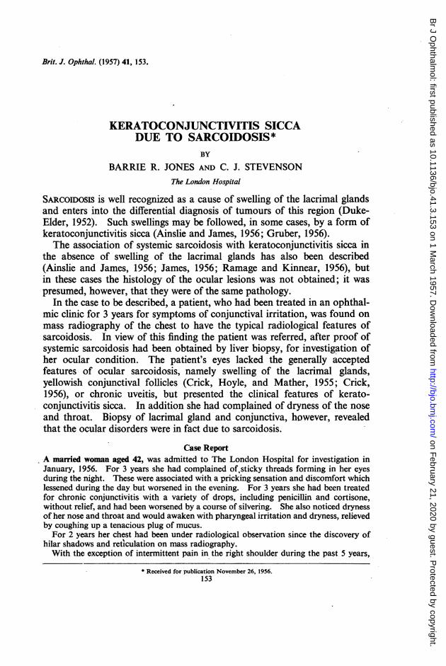
enlargement of the hilar lymph glands.Sputum and Gastric Lavage.-No tubercle bacilli on smear or culture.Mantoux Test.-Negative to 1 in 100 old tuberculin.Erythrocyte Sedimentation Rate.-15 mm. in one hour.Plasma Proteins.-Albumin/Globulin ratio 4-1/3-4.Haemoglobin.-13-9 g. 100 ml.Liver Biopsy (Fig. 1).-Needle biopsy showed non-caseous epithelioid cell tubercles with
earlyperipheralfibrosis,morphologicallycompatiblewithsarcoidosis. Noacid-fastbacilli.
. .
% I~~~~~~~~~~~~~FIG.1.Lve ios showing a non-caseating granulomaconsisti
& f
epithelioid cells surrounded by a narrow zone of lymphocytes. Haematoxylinand eosin x 150.
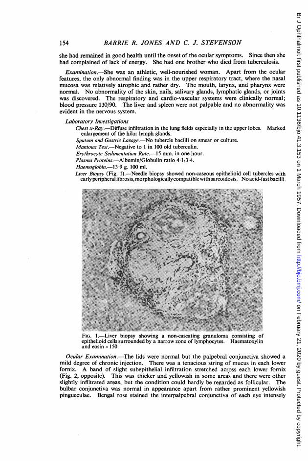
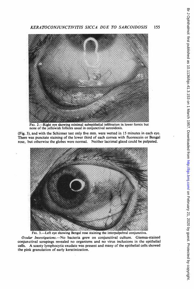
Ocular Examination.-The lids were normal but the palpebral conjunctiva showed amild degree of chronic injection. There was a tenacious string of mucus in each lowerfornix. A band of slight subepithelial infiltration stretched across each lower fornix(Fig. 2, opposite). This was thicker and yellowish in some areas and there were otherslightly infiltrated areas, but the condition could hardly be regarded as follicular. Thebulbar conjunctiva was normal in appearance apart from rather prominent yellowishpingueculae. Bengal rose stained the interpalpebral conjunctiva of each eye intensely
154
on February 21, 2020 by guest. P
rotected by copyright.http://bjo.bm
j.com/
Br J O
phthalmol: first published as 10.1136/bjo.41.3.153 on 1 M
arch 1957. Dow
nloaded from
KERA TOCONJUNCTIVITIS SICCA DUE TO SARCOIDOSIS
FIG. 2.-Right eye showing minimal subepithelial infiltration in lower fornix butnone of the yellowish follicles usual in conjunctival sarcoidosis.
(Fig. 3), and with the Schirmer test only five mm. were wetted in 15 minutes in each eye.There was punctate staining of the lower third of each cornea with fluorescein or Bengalrose, but otherwise the globes were normal. Neither lacrimal gland could be palpated.
_--s0----- ---- ------ -- --- ---- -- ---FIG. 3.-Left eye showing Bengal rose staining the interpalpebral conjunctiva.
Ocular Investigations.-No bacteria grew on conjunctival culture. Giemsa-stainedconjunctival scrapings revealed no organisms and no virus inclusions in the epithelialcells. A scanty lymphocytic exudate was present and many of the epithelial cells showedthe pink granulation of early keratinization.
155
on February 21, 2020 by guest. P
rotected by copyright.http://bjo.bm
j.com/
Br J O
phthalmol: first published as 10.1136/bjo.41.3.153 on 1 M
arch 1957. Dow
nloaded from
BARRIE R. JONES AND C. J. STEVENSON
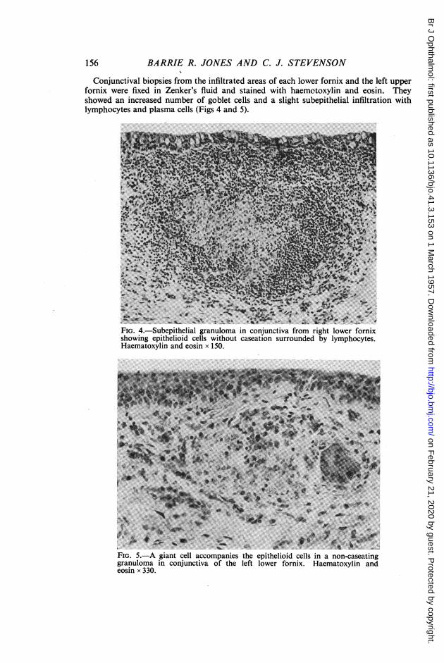
Conjunctival biopsies from the infiltrated areas of each lower fornix and the left upperfornix were fixed in Zenker's fluid and stained with haemotoxylin and eosin. Theyshowed an increased number of goblet cells and a slight subepithelial infiltration withlymphocytes and plasma cells (Figs 4 and 5).
Na.~~~~~~~~~a*>_e~~~~~~~..4*A.<e w .
FIG. 4.-Subepithelial granuloma in conjunctiva from right lower fornixshowing epithelioid cells without caseation surrounded by lymphocytes.Haematoxylin and eosin x 150.
V ~ ~s*~~~~~~~~~~~~~~~g~~~ ~ ~~~~~~~~z
FIG. 5.-A giant cell accompanies the epithelioid cells in a non-caseatinggranuloma in conjunctiva of the left lower fornix. Haematoxylin andeosin x 330.
156
on February 21, 2020 by guest. P
rotected by copyright.http://bjo.bm
j.com/
Br J O
phthalmol: first published as 10.1136/bjo.41.3.153 on 1 M
arch 1957. Dow
nloaded from
KERATOCONJUNCTIVITIS SICCA DUE TO SARCOIDOSIS 157
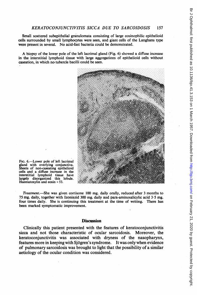
Small scattered subepithelial granulomata consisting of large eosinophilic epithelioidcells surrounded by small lymphocytes were seen, and giant cells of the Langhans typewere present in several. No acid-fast bacteria could be demonstrated.
A biopsy of the lower pole of the left lacrimal gland (Fig. 6) showed a diffuse increasein the interstitial lymphoid tissue with large aggregations of epithelioid cells withoutcaseation, in which no tubercle bacilli could be seen.
4<Jlgl,4r,
FiG. 6.-Lower pole of left lacrimnalgland with overlying conjunctiva.Sheets of non-caseating epithelioidcells and a diffuse increase in the ;; Cinterstitial lymphoid tissue havelargely disorganized this lobule. . MlHaemato-xylin and eosin x 53. t.
Treatment.-She was given cortisone 100 mg. daily oraUy, reduced after 3 months to75 mg. daily, together with Izoniazid 300 mg. daily and para-aminosalicylic acid 3 5 mg.four times daily. She is continuing this treatment at the time of writing. There hasbeen marked symptomatic improvement.
DiscussionClinically this patient presented with the features of keratoconjunctivitis
sicca and not those characteristic of ocular sarcoidosis. Moreover, thekeratoconjunctivitis was associated with dryness of the nasopharynx,features more in keeping with Sj6gren's syndrome. Itwas onlywhen evidenceof pulmonary sarcoidosis was brought to light that the possibility of a similaraetiology of the ocular condition was considered.
on February 21, 2020 by guest. P
rotected by copyright.http://bjo.bm
j.com/
Br J O
phthalmol: first published as 10.1136/bjo.41.3.153 on 1 M
arch 1957. Dow
nloaded from
BARRIE R. JONES AND C. J. STEVENSON
Sjogren's Syndrome.-Sj6gren and many others (reviewed by Duke-Elder,1952) have emphasized that keratoconjunctivitis most often arises withoutpreceding ocular disease in middle-aged women and is often associated withpolyarthritis, xerostomia, salivary gland enlargement, and rhinitis, pharyn-gitis and laryngitis sicca. Many other abnormalities have been recordedin patients with dry eyes (Duke-Elder, 1952) and added to the syndrome; inmany cases the syndrome is incomplete. There is an unfortunate tendencyfor keratoconjunctivitis sicca and almost any associated feature to bediagnosed as "Sj6gren's disease" without further systemic investigation ortreatment.
Nature of Sjogren's Syndrome.-There are four main views of this syn-drome. Firstly, it has been regarded as a disease in its own right which givesrise to some or many of the possible clinical features (Sj6gren, 1933; Hender-son, 1950; Coverdale, 1948, 1952; Cardell, and Gurling, 1954).
Secondly, the syndrome has been regarded as a manifestation or complica-tion ofrheumatoid arthritis. Thompson and Eadie (1956) recently supportedthis view by an extensive investigation of patients with rheumatoid arthritis,in which they established that keratoconjunctivitis sicca is the commonestocular complication of rheumatoid arthritis. However, rheumatoid arthritiscannot be the aetiology of those fairly common cases of Sj6gren's syndromewithout arthritis unless they represent anarthritic rheumatoid disease(Bagratuni, 1956).
Thirdly, since keratoconjunctivitis sicca has been described in associationwith several collagen diseases other than rheumatoid arthritis, namely,rheumatic fever, polyarteritis nodosa, disseminated lupus erythematosis,and scleroderma, it has been suggested that Sjogren's syndrome is merelyone variant of collagen disease (Ramage and Kinnear, 1956; British MedicalJournal Editorial, 1956).
Fourthly, the syndrome- may be regarded as a syndrome and no more, thatis to say a symptom complex which may arise from a variety of pathologicalprocesses. Having recognized the syndrome in any particular case, weshould attempt to make an aetiological diagnosis. Thorough systemicexamination may establish the presence of one of the collagen diseases. Itis at this point that sarcoidosis should also be considered, and the patientcarefully investigated for systemic and ocular sarcoidosis. The present caseclearly demonstrates the simplicity and value of conjunctival and lacrimalbiopsy in making the diagnosis and in establishing that the systemic diseaseand the ocular condition are in fact due to the same cause.
From among patients with keratoconjunctivitis sicca some will also berecognized who are presenting the early stages of a disease resembling ocularpemphigus.
158
on February 21, 2020 by guest. P
rotected by copyright.http://bjo.bm
j.com/
Br J O
phthalmol: first published as 10.1136/bjo.41.3.153 on 1 M
arch 1957. Dow
nloaded from
KERATOCONJUNCTIVITIS SICCA DUE TO SARCOIDOSIS 159
Features of Sjogren's Syndrome which can arise in Sarcoidosis.-Thepresent case shows that keratoconjunctivitis sicca can result from lacrimaland conjunctival sarcoidosis even without swelling of the lacrimal gland, asdescribed by Gruber (1956), or manifest conjunctival follicles as describedby Crick and others (1955). Swelling of parotid and other salivary glandshas been recorded in numerous cases of sarcoidosis (Longcope and Pierson,1937; Pautrier, 1938; Scott, 1938; Schultz, 1945). Xerostomia would beexpected in a patient whose salivary glands were infiltrated by sarcoid depositsor disorganized by subsequent fibrosis (James, 1956). The present patientalso had symptoms of rhinitis and pharyngitis sicca and the nasal mucosahad a dry and somewhat atrophic appearance. Infiltration of the nasal,paranasal, pharyngeal, and laryngeal mucosa in sarcoid have been demon-strated (Poe, 1942; Bordley and Proctor, 1942; Wille, 1946). Enlargedsuperficial lymph nodes, spleen, and liver are all well-recognized occurrencesin sarcoidosis. Like other possible features of Sjogren's syndrome, theirsignificance varies with the presence or absence of arthritis. If a patientwith keratoconjunctivitis sicca has rheumatoid arthritis and enlargement oflymph nodes and spleen, then she is probably presenting Felty's syndrome.If she has no arthritis then sarcoidosis is more likely. Mild pyrexia, raisederythrocyte sedimentation rate, and hyperglobulinaemia not uncommonlyoccur in sarcoidosis and are often found in cases of Sj6gren's syndromeassociated with rheumatoid arthritis.
Features of Sjogren's Syndrome not expected to arise from Sarcoidosis.Polyarthritis of the rheumatoid type does not result from sarcoidosis, sothat the presence of definite rheumatoid arthritis in any case makes sarcoidosismuch less likely. James, Thomson, and Willcox (1956) have recently drawnattention to the occurrence of an acute polyarthritis in association with theerythema nodosum which may arise in sarcoidosis and this must not beconfused with a true rheumatoid arthritis. The dry brownish-yellowlongitudinally fissured nails described by Thompson and Eadie (1956)similarly appear to be a feature in those cases of Sj6gren's syndrome asso-ciated with rheumatoid arthritis.
SummaryA case of keratoconjunctivitis sicca with symptoms of dryness at the back
of the mouth was shown by biopsy to be due to lacrimal and conjunctivalsarcoidosis. Systemic sarcoidosis was demonstrated by chest x ray and liverbiopsy.The relationship of Sj6gren's syndrome to rheumatoid arthritis and to
sarcoidosis is discussed, and it is suggested that general investigation, con-junctival biopsy and possibly lacrimal biopsy should be used to elucidatethe aetiology in those patients who do not have a rheumatoid arthritis
on February 21, 2020 by guest. P
rotected by copyright.http://bjo.bm
j.com/
Br J O
phthalmol: first published as 10.1136/bjo.41.3.153 on 1 M
arch 1957. Dow
nloaded from
BARRIE R. JONES AND C. J. STEVENSON
accompanying their dry eyes. If the keratoconjunctivitis sicca is accompaniedby yellowish conjunctival follicles, swelling of the lacrimal glands, or chronicuveitis, then sarcoidosis is more likely.Although sarcoidosis may well account for only a small proportion of
cases of keratoconjunctivitis sicca, these cases are worth recognizing sincethe disease is known to respond to cortisone and it is reasonable to supposethat if treatment can be maintained until it becomes inactive then permanentdamage by fibrosis can be prevented.
We should like to express our indebtedness to Dr. Owen Clarke, Chest Physician of Canterbury,who suspected that both the eye and lung changes might be due to sarcoidosis and referred thepatient to the London Hospital; also to Dr. D. Davies of Whitstable, the patient's GeneralPractitioner, for his assistance in carrying out the treatment. We are indebted to Dr. N. LloydRusby and Mr. J. E. M. Ayoub for their encouragement and permission to publish this case; toDr. J. R. Pridie for the liver biopsy; to Dr. R. G. F. Parker and Dr. C. Wood for the histologicalreports; and to the photographic department of The London Hospital for the illustrations.
REFERENCESAINSLIE, D., and JAMES, D. G. (1956). Brit. med. J., 1, 954.BAGRATUNI, L. (1956). Lancet, 2, 694.BORDLEY, J. E., and PROCTOR, D. F. (1942). Arch. Otolaryng. (Chicago), 36, 740.BRMSH MEDICAL JOURNAL EDrrORIAL (1956). Brit. med. J., 2, 470.CARDELL, B. S., and GURLING, K. J. (1954). J. Path. Bact., 68, 137.COVERDALE, H. (1948). British Journal of Ophthalmology, 32, 669.
(1952). Trans. ophthal. Soc. N.Z., 6, 36.CRICK, R. Prrrs (1956). Trans. ophthal. Soc. U.K, 76, 403.
HOYLE, C., and MATHER, G. (1955). Brit. med. J., 2, 1180.DUKE-ELDER (1952). "Text-book of Ophthalmology", vol. 5, p. 5229. Kimpton, London.GRUBER, E. (1956). A.M.A. Arch. Ophthal., 55, 42.HENDERSON, J. W. (1950). Amer. J. Ophthal., 33, 197.JAMES, D. G. (1956). Brit. med. J., 2, 900.--, THOMSON, A. D., and WILLCOx, A. (1956). Lancet, 2, 218.LONGCOPE, W. T., and PIERSON, J. W. (1937). Bull. Johns Hopk. Hosp., 60, 223.PAUTRIER, L. M. (1938). Ann. Derm. (Paris), 7 ser., 9, 161.POE, D. L. (1942). Ann. Otol. (St. Louis), 51, 430.RAMAGE, J. H., and KINNEAR, W. F. (1956). British Journal of Ophthalmology, 40, 416.SCHULTZ, A. (1945). Amer. J. Ophthal., 28, 1010.SCorr, R. B. (1938). Brit. med. J., 2, 777.SJOGREN, H. (1933). Acta ophthal. (Kbh.), Suppl. 2, "Zur Kenntnis der Keratoconjunctivitis
sicca", trans. J. B. Hamilton (1943). "A New Conception of Keratoconjunctivitis Sicca".Australasian Medical Publishing Co., Sydney.
THOMPSON, M., and EADIE, S. (1956). Ann. rheum. Dis., 15, 21.WILLE, C. (1946). Acta oto-laryng. (Stockh.), 34, 182.
160
on February 21, 2020 by guest. P
rotected by copyright.http://bjo.bm
j.com/
Br J O
phthalmol: first published as 10.1136/bjo.41.3.153 on 1 M
arch 1957. Dow
nloaded from