
Katsuya Yamazoe, MD, Takefumi Yamaguchi, MD, Kazuki Hotta, MD, Yoshiyuki Satake, MD, Kenji Konomi,...
20
Katsuya Yamazoe, MD, Takefumi Yamaguchi, Katsuya Yamazoe, MD, Takefumi Yamaguchi, MD, Kazuki Hotta, MD, Yoshiyuki Satake, MD, Kenji Konomi, MD, Kazuki Hotta, MD, Yoshiyuki Satake, MD, Kenji Konomi, MD, Seika Den, MD, Jun Shimazaki, MD MD, Seika Den, MD, Jun Shimazaki, MD Presented by: Abdulrahman AlDarrab Presented by: Abdulrahman AlDarrab
-
Upload
antonia-burke -
Category
Documents
-
view
226 -
download
2
Transcript of Katsuya Yamazoe, MD, Takefumi Yamaguchi, MD, Kazuki Hotta, MD, Yoshiyuki Satake, MD, Kenji Konomi,...
Katsuya Yamazoe, MD, Takefumi Yamaguchi, MD, Kazuki Hotta, Katsuya Yamazoe, MD, Takefumi Yamaguchi, MD, Kazuki Hotta, MD, Yoshiyuki Satake, MD, Kenji Konomi, MD, Seika Den, MD, Yoshiyuki Satake, MD, Kenji Konomi, MD, Seika Den,
MD, Jun Shimazaki, MDMD, Jun Shimazaki, MD
Presented by: Abdulrahman AlDarrabPresented by: Abdulrahman AlDarrab
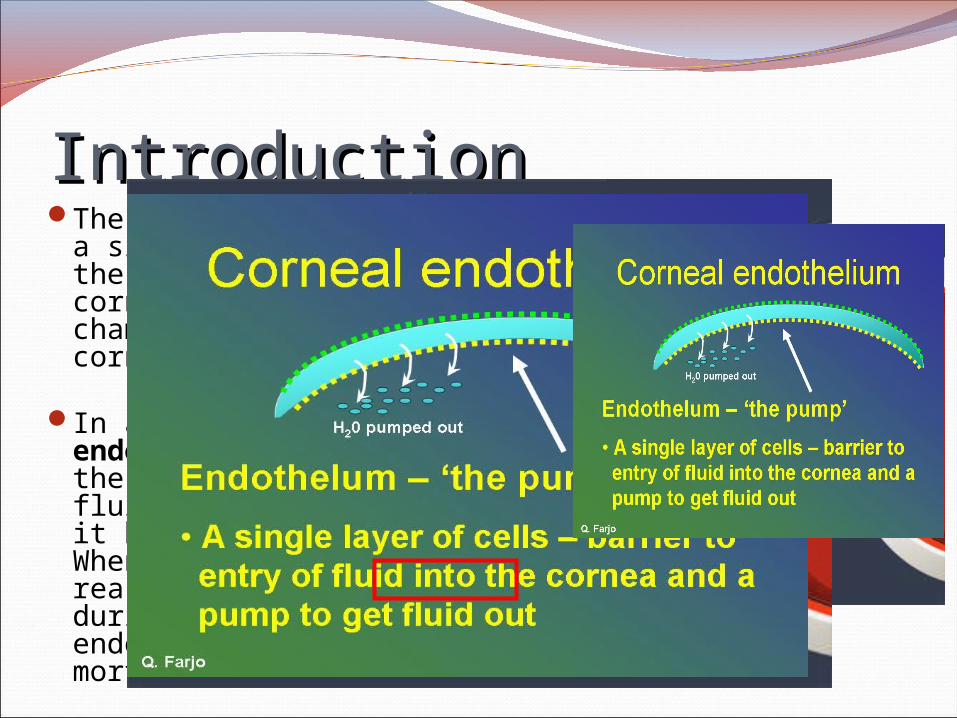
IntroductionIntroductionThe corneal endothelium is
a single layer of cells on the inner surface of the cornea. It faces the chamber formed between the cornea and the iris.
In a healthy cornea, endothelial cells keeps the tissue from excess fluid absorption, pumping it back into the aqueous. When affected by some reason, such as trauma during cataract removal, endothelial cells suffer mortality or damage.

IntroductionIntroductionWhen endothelial cell
counts drop too low, the pump starts failing to function and fluid moves anterior into the stroma and epithelium. The excess fluid precipitates swelling of the cornea. As fluid accumulates between the basal epithelium cells, blister like formations form (bullae) and they undergo painful ruptures releasing their fluid content to the surface, Bullous keratopathy.
PurposePurposeTo evaluate the surgical outcomes of cataract
surgery in eyes with a low preoperative corneal endothelial cell density (ECD) and analyze factors affecting the prognosis.
DesignDesignStudy Design:Retrospective studyNoncomparative case series
Location:Tokyo Dental College, Ichikawa General
Hospital, Chiba, Japan.
MethodsMethodsPatients with a clear cornea and an ECD less
than 1000 cells/mm2 preoperatively were identified from those who had cataract surgery at Tokyo Dental College between January 2006 and May 2010 and were included in this retrospective study.
All patients provided written informed consent.
MethodsMethodsEvaluation:Patients had slitlamp microscopy and Landolt
corrected distance visual acuity (CDVA), fundus, intraocular pressure (IOP), and ECD testing before and after cataract surgery.
The central corneal ECD was measured using the EM-3000 device before surgery and at each follow up visit.
The ECD at the final visit was taken as representing the patient’s postoperative ECD.

MethodsMethodsSurgical technique:Standard endocapsular phacoemulsification
of the nucleus was performed using the phaco- chop technique.
A foldable acrylic IOL was placed in the capsular bag.
Performed by 1 of 4 experienced surgeons.
MethodsMethodsMain outcome measures:Proportional loss of endothelial cells at the
patient’s final visit.
Incidence of bullous keratopathy.
ResultsResults61 eyes of 53 patients had a preoperative ECD
less than 1000 cells/mm2.
The most frequent preoperative diagnosis or factor regarded as causing endothelial cell loss was Fuchs dystrophy (32.8%) followed by laser iridotomy (26.2%) and keratoplasty (16.4%).
Diabetes mellitus presented in 9 eyes (14.8%) and hypertension in 23 eyes (37.7%).
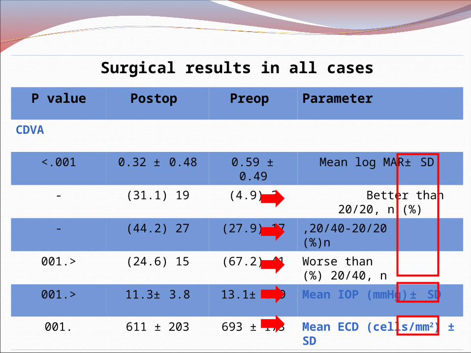
Surgical results in all cases
ParameterPreop Postop P value
CDVA
Mean log MAR± SD 0.59 ± 0.490.32 ± 0.48<.001
Better than 20/20, n (%)
3( 4.9)19( 31.1)-
20/40-20/20 ,n)%(17( 27.9)27( 44.2)-
Worse than 20/40, n )%(
41( 67.2)15( 24.6).<001
Mean IOP (mmHg) ± SD
13.1± 3.911.3± 3.8.<001
Mean ECD (cells/mm2) ± SD
693 ± 173611 ± 203.001
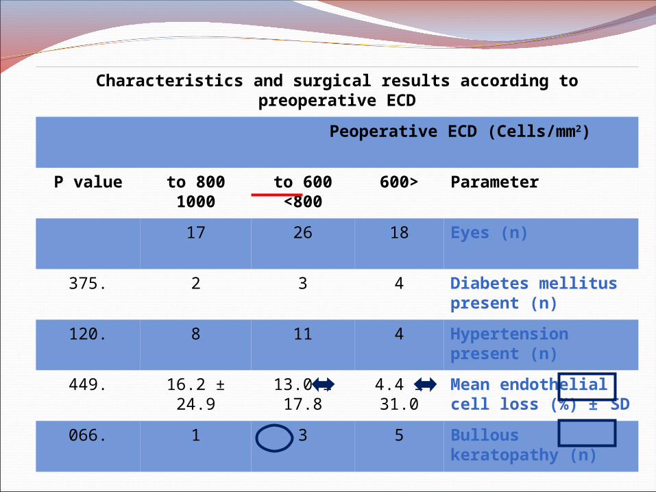
Characteristics and surgical results according to preoperative ECD
Peoperative ECD (Cells/mm2)
Parameter <600600 to <800
800 to 1000
P value
Eyes (n)182617
Diabetes mellitus present (n)
432.375
Hypertension present (n)
4118.120
Mean endothelial cell loss (%) ± SD
4.4 ± 31.0
13.0 ± 17.8
16.2 ± 24.9.449
Bullous keratopathy (n)
531.066
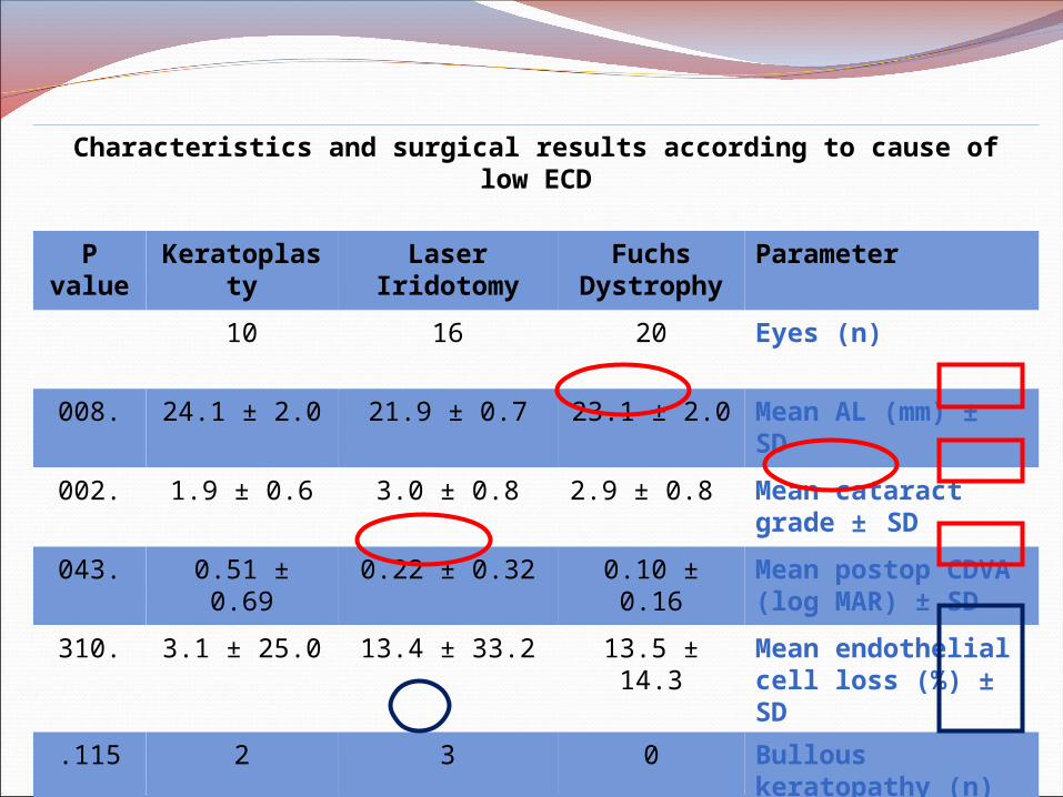
Characteristics and surgical results according to cause of low ECD
Parameter Fuchs Dystrophy
Laser Iridotomy
Keratoplasty
P value
Eyes (n)201610
Mean AL (mm) ± SD
23.1 ± 2.021.9 ± 0.724.1 ± 2.0.008
Mean cataract grade ± SD
2.9 ± 0.8 3.0 ± 0.81.9 ± 0.6.002
Mean postop CDVA (log MAR) ± SD
0.10 ± 0.160.22 ± 0.320.51 ± 0.69.043
Mean endothelial cell loss (%) ± SD
13.5 ± 14.313.4 ± 33.23.1 ± 25.0.310
Bullous keratopathy (n)
032.115
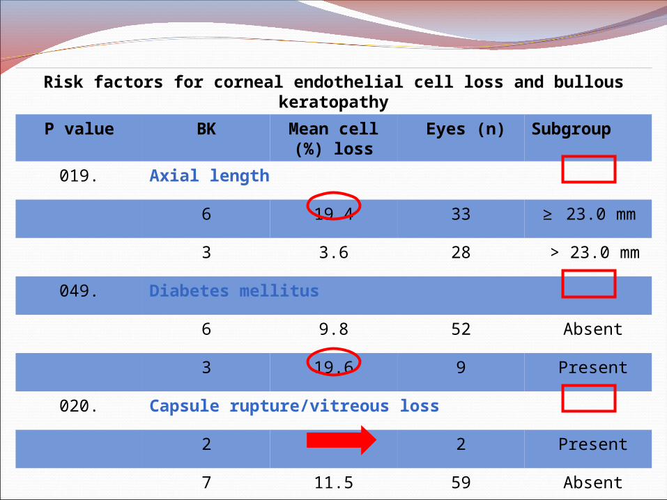
Risk factors for corneal endothelial cell loss and bullous keratopathy
SubgroupEyes (n) Mean cell loss)%(
BKP value
Axial length.019
≥ 23.0 mm
3319.46
> 23.0 mm
283.63
Diabetes mellitus.049
Absent 529.86
Present 919.63
Capsule rupture/vitreous loss.020
Present 2-2
Absent 5911.57
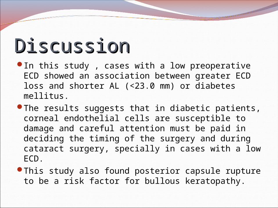
DiscussionDiscussionIn this study , cases with a low preoperative ECD
showed an association between greater ECD loss and shorter AL (<23.0 mm) or diabetes mellitus.
The results suggests that in diabetic patients, corneal endothelial cells are susceptible to damage and careful attention must be paid in deciding the timing of the surgery and during cataract surgery, specially in cases with a low ECD.
This study also found posterior capsule rupture to be a risk factor for bullous keratopathy.
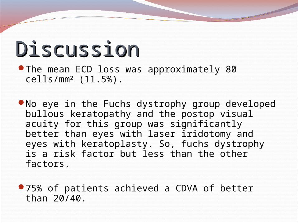
DiscussionDiscussionThe mean ECD loss was approximately 80
cells/mm2 (11.5%).
No eye in the Fuchs dystrophy group developed bullous keratopathy and the postop visual acuity for this group was significantly better than eyes with laser iridotomy and eyes with keratoplasty. So, fuchs dystrophy is a risk factor but less than the other factors.
75% of patients achieved a CDVA of better than 20/40.
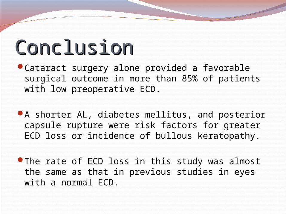
ConclusionConclusionCataract surgery alone provided a favorable
surgical outcome in more than 85% of patients with low preoperative ECD.
A shorter AL, diabetes mellitus, and posterior capsule rupture were risk factors for greater ECD loss or incidence of bullous keratopathy.
The rate of ECD loss in this study was almost the same as that in previous studies in eyes with a normal ECD.
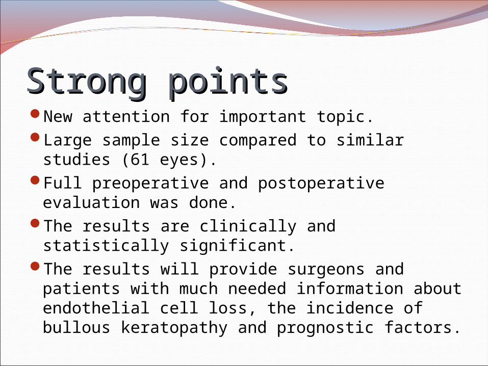
Strong pointsStrong pointsNew attention for important topic.Large sample size compared to similar studies
(61 eyes).Full preoperative and postoperative
evaluation was done.The results are clinically and statistically
significant.The results will provide surgeons and patients
with much needed information about endothelial cell loss, the incidence of bullous keratopathy and prognostic factors.
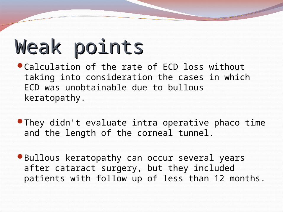
Weak pointsWeak pointsCalculation of the rate of ECD loss without
taking into consideration the cases in which ECD was unobtainable due to bullous keratopathy.
They didn't evaluate intra operative phaco time and the length of the corneal tunnel.
Bullous keratopathy can occur several years after cataract surgery, but they included patients with follow up of less than 12 months.