
jrv140004
11
Copyright 2014 American Medical Association. All rights reserved. The Pathophysiology and Treatment of Glaucoma A Review Robert N. Weinreb, MD; Tin Aung, MD, PhD; Felipe A. Medeiros, MD, PhD T he glaucomas are a group of optic neuropathies character- ized by progressive degeneration of retinal ganglion cells. These are central nervous system neurons that have their cell bodies in the inner retina and axons in the optic nerve. Degen- eration of these nerves results in cupping, a characteristic appear- ance of the optic disc and visual loss. 1 The biological basis of glau- coma is poorly understood and the factors contributing to its progression have not been fully characterized. 2 Glaucoma affects more than 70 million people worldwide with approximately 10% being bilaterally blind, 3 making it the leading cause of irreversible blindness in the world. Glaucoma can remain asymptomatic until it is severe, resulting in a high likelihood that the number of affected individuals is much higher than the number known to have it. 4,5 Population-level surveys suggest that only 10% to 50% of people with glaucoma are aware they have it. 4-8 Glaucomas can be classified into 2 broad categories: open-angle glaucoma and angle-closure glaucoma. In the United States, more than 80% of cases are open-angle glaucoma; however, angle-closure glaucoma is responsible for a disproportionate number of patients with se- vere vision loss. 9,10 Both open-angle and angle-closure glaucoma can be primary diseases. Secondary glaucoma can result from trauma, certain medications such as corticosteroids, inflammation, tumor, or conditions such as pigment dispersion or pseudo-exfoliation. A recent JAMA Rational Clinical Examination systematic re- view of primary open-angle glaucoma diagnosis found that the risk of glaucoma was highest when examination revealed an increased cup-disk ratio (CDR), CDR asymmetry, disc hemorrhage, or el- evated intraocular pressure. 11 Primary open-angle glaucoma was also more likely when there was a family history of the disease, black race, or advanced age (Box). The primary care physician also should be aware of the risk of developing glaucoma in patients being treated with systemic or topical corticosteroids. 12 Patients at risk should be referred to an eye care practitioner. This review explores patho- physiology of the disease and its treatment. IMPORTANCE Glaucoma is a worldwide leading cause of irreversible vision loss. Because it may be asymptomatic until a relatively late stage, diagnosis is frequently delayed. A general understanding of the disease pathophysiology, diagnosis, and treatment may assist primary care physicians in referring high-risk patients for comprehensive ophthalmologic examination and in more actively participating in the care of patients affected by this condition. OBJECTIVE To describe current evidence regarding the pathophysiology and treatment of open-angle glaucoma and angle-closure glaucoma. EVIDENCE REVIEW A literature search was conducted using MEDLINE, the Cochrane Library, and manuscript references for studies published in English between January 2000 and September 2013 on the topics open-angle glaucoma and angle-closure glaucoma. From the 4334 abstracts screened, 210 articles were selected that contained information on pathophysiology and treatment with relevance to primary care physicians. FINDINGS The glaucomas are a group of progressive optic neuropathies characterized by degeneration of retinal ganglion cells and resulting changes in the optic nerve head. Loss of ganglion cells is related to the level of intraocular pressure, but other factors may also play a role. Reduction of intraocular pressure is the only proven method to treat the disease. Although treatment is usually initiated with ocular hypotensive drops, laser trabeculoplasty and surgery may also be used to slow disease progression. CONCLUSIONS AND RELEVANCE Primary care physicians can play an important role in the diagnosis of glaucoma by referring patients with positive family history or with suspicious optic nerve head findings for complete ophthalmologic examination. They can improve treatment outcomes by reinforcing the importance of medication adherence and persistence and by recognizing adverse reactions from glaucoma medications and surgeries. JAMA. 2014;311(18):1901-1911. doi:10.1001/jama.2014.3192 Author Video Interview at jama.com JAMA Patient Page page 1934 Author Affiliations: Hamilton Glaucoma Center, Shiley Eye Center and Department of Ophthalmology, University of California, San Diego, La Jolla (Weinreb, Medeiros); Singapore National Eye Center, Singapore, Singapore (Aung); Yong Loo Lin School of Medicine, National University of Singapore, Singapore (Aung). Corresponding Author: Robert N. Weinreb, MD, UC San Diego, Shiley Eye Center, 9500 Gilman Dr, MC 0946, La Jolla, CA 92093-0946 ([email protected]). Section Editor: Mary McGrae McDermott, MD, Senior Editor. Clinical Review & Education Review jama.com JAMA May 14, 2014 Volume 311, Number 18 1901 Copyright 2014 American Medical Association. All rights reserved. Downloaded From: http://jama.jamanetwork.com/ by anna galvan on 03/15/2015
description
mkk
Transcript of jrv140004
Copyright 2014 American Medical Association. All rights reserved.
The Pathophysiology and Treatment of GlaucomaA ReviewRobert N. Weinreb, MD; Tin Aung, MD, PhD; Felipe A. Medeiros, MD, PhD
T he glaucomas are a group of optic neuropathies character-ized by progressive degeneration of retinal ganglion cells.These are central nervous system neurons that have their
cell bodies in the inner retina and axons in the optic nerve. Degen-eration of these nerves results in cupping, a characteristic appear-ance of the optic disc and visual loss.1 The biological basis of glau-coma is poorly understood and the factors contributing to itsprogression have not been fully characterized.2
Glaucoma affects more than 70 million people worldwide withapproximately 10% being bilaterally blind,3 making it the leadingcause of irreversible blindness in the world. Glaucoma can remainasymptomatic until it is severe, resulting in a high likelihood that thenumber of affected individuals is much higher than the numberknown to have it.4,5 Population-level surveys suggest that only 10%to 50% of people with glaucoma are aware they have it.4-8Glaucomascan be classified into 2 broad categories: open-angle glaucoma andangle-closure glaucoma. In the United States, more than 80% of
cases are open-angle glaucoma; however, angle-closure glaucomais responsible for a disproportionate number of patients with se-vere vision loss.9,10 Both open-angle and angle-closure glaucoma canbe primary diseases. Secondary glaucoma can result from trauma,certain medications such as corticosteroids, inflammation, tumor,or conditions such as pigment dispersion or pseudo-exfoliation.
A recent JAMA Rational Clinical Examination systematic re-view of primary open-angle glaucoma diagnosis found that the riskof glaucoma was highest when examination revealed an increasedcup-disk ratio (CDR), CDR asymmetry, disc hemorrhage, or el-evated intraocular pressure.11 Primary open-angle glaucoma was alsomore likely when there was a family history of the disease, black race,or advanced age (Box). The primary care physician also should beaware of the risk of developing glaucoma in patients being treatedwith systemic or topical corticosteroids.12 Patients at risk should bereferred to an eye care practitioner. This review explores patho-physiology of the disease and its treatment.
IMPORTANCE Glaucoma is a worldwide leading cause of irreversible vision loss. Because itmay be asymptomatic until a relatively late stage, diagnosis is frequently delayed. A generalunderstanding of the disease pathophysiology, diagnosis, and treatment may assist primarycare physicians in referring high-risk patients for comprehensive ophthalmologic examinationand in more actively participating in the care of patients affected by this condition.
OBJECTIVE To describe current evidence regarding the pathophysiology and treatment ofopen-angle glaucoma and angle-closure glaucoma.
EVIDENCE REVIEW A literature search was conducted using MEDLINE, the Cochrane Library,and manuscript references for studies published in English between January 2000 andSeptember 2013 on the topics open-angle glaucoma and angle-closure glaucoma. From the4334 abstracts screened, 210 articles were selected that contained information onpathophysiology and treatment with relevance to primary care physicians.
FINDINGS The glaucomas are a group of progressive optic neuropathies characterized bydegeneration of retinal ganglion cells and resulting changes in the optic nerve head. Loss ofganglion cells is related to the level of intraocular pressure, but other factors may also play arole. Reduction of intraocular pressure is the only proven method to treat the disease.Although treatment is usually initiated with ocular hypotensive drops, laser trabeculoplastyand surgery may also be used to slow disease progression.
CONCLUSIONS AND RELEVANCE Primary care physicians can play an important role in thediagnosis of glaucoma by referring patients with positive family history or with suspiciousoptic nerve head findings for complete ophthalmologic examination. They can improvetreatment outcomes by reinforcing the importance of medication adherence and persistenceand by recognizing adverse reactions from glaucoma medications and surgeries.
JAMA. 2014;311(18):1901-1911. doi:10.1001/jama.2014.3192
Author Video Interview atjama.com
JAMA Patient Page page 1934
Author Affiliations: HamiltonGlaucoma Center, Shiley Eye Centerand Department of Ophthalmology,University of California, San Diego, LaJolla (Weinreb, Medeiros); SingaporeNational Eye Center, Singapore,Singapore (Aung); Yong Loo LinSchool of Medicine, NationalUniversity of Singapore, Singapore(Aung).
Corresponding Author: Robert N.Weinreb, MD, UC San Diego, ShileyEye Center, 9500 Gilman Dr, MC0946, La Jolla, CA 92093-0946([email protected]).
Section Editor: Mary McGraeMcDermott, MD, Senior Editor.
Clinical Review & Education
Review
jama.com JAMA May 14, 2014 Volume 311, Number 18 1901
Copyright 2014 American Medical Association. All rights reserved.
Downloaded From: http://jama.jamanetwork.com/ by anna galvan on 03/15/2015
Copyright 2014 American Medical Association. All rights reserved.
Methods
A literature search was conducted using MEDLINE, the CochraneLibrary, and manuscript references for studies published in Englishbetween January 2000 and September 2013 on the topics open-angle and angle-closure glaucoma. From the 4334 abstractsscreened, 210 articles were selected that contained information onpathophysiology and treatment with relevance to primary care phy-sicians.
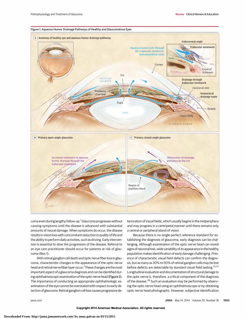
Primary Open-Angle GlaucomaPathophysiologyAlthough the pathogenesis of glaucoma is not fully understood, thelevel of intraocular pressure is related to retinal ganglion cell death.The balance between secretion of aqueous humor by the ciliary bodyand its drainage through 2 independent pathways—the trabecularmeshwork and uveoscleral outflow pathway—determines the intra-ocular pressure. In patients with open-angle glaucoma, there is in-creased resistance to aqueous outflow through the trabecular mesh-work. In contrast, the access to the drainage pathways is obstructedtypically by the iris in patients with angle-closure glaucoma (Figure 1).
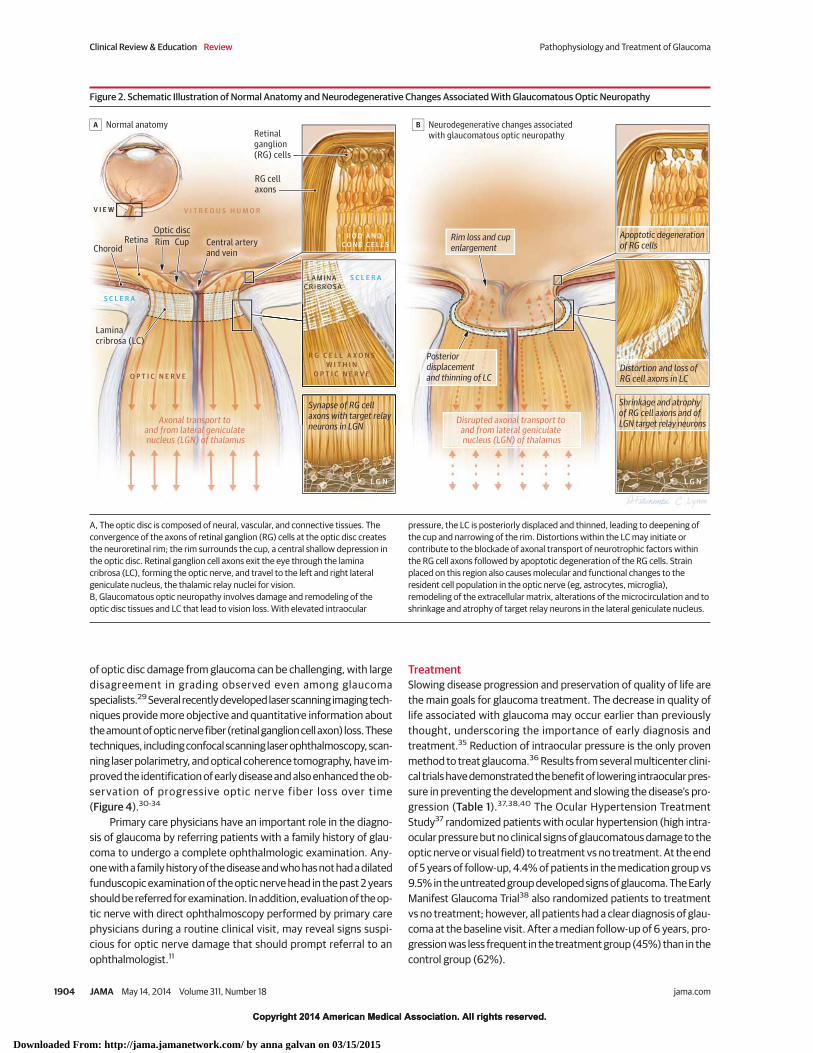
Intraocular pressure can cause mechanical stress and strain onthe posterior structures of the eye, notably the lamina cribrosa andadjacent tissues (Figure 2).13 The sclera is perforated at the laminawhere the optic nerve fibers (retinal ganglion cell axons) exit the eye.The lamina is the weakest point in the wall of the pressurized eye.Intraocular pressure–induced stress and strain may result in com-pression, deformation, and remodeling of the lamina cribrosa withconsequent mechanical axonal damage and disruption of axonaltransport14,15 that interrupts retrograde delivery of essential tro-phic factors to retinal ganglion cells from their brainstem target (re-lay neurons of the lateral geniculate nucleus). Studies involving catsand monkeys with experimentally induced ocular hypertension havedemonstrated blockade of both orthograde and retrograde axonaltransport at the level of the lamina cribrosa.16 Disrupted axonal trans-port occurs early in the pathogenesis of glaucoma in experimentalsystems resulting in collections of vesicles and disorganization of mi-crotubules and neurofilaments in the prelaminar and postlaminarregions. Similar ultrastructural changes in optic nerve fibers are seenin postmortem human eyes that have glaucoma.13 Because there alsomay be mitochondrial dysfunction in retinal ganglion cells andastrocytes,17 high levels of energy demand may be difficult to meetduring periods of intraocular pressure–induced metabolic stress.
Glaucomatous optic neuropathy can occur in individuals withintraocular pressures within the normal range. In such patients, theremay be an abnormally low cerebrospinal fluid pressure in the opticnerve subarachnoid space resulting in a large pressure gradient acrossthe lamina.18,19 Impaired microcirculation, altered immunity, exci-totoxicity, and oxidative stress may also cause glaucoma. Primaryneural pathological processes may cause secondary neurodegen-eration of other retinal neurons and cells in the central visual path-way by altering their environment and increasing susceptibility todamage.20.
GeneticsSeveral genes—including myocilin (MYOC, GLC1A) (CCDS1297.1),21
optineurin (OPTN, GLC1E) (CCDS7094.1),22 and WD repeat
domain 36 (GLC1G) (CCDS4102.1)23—are associated with a mono-genic, autosomal dominant trait; however, these genes account forless than 10% of all glaucoma cases.24 The first reported locus forprimary open-angle glaucoma was located on chromosome 1 (GLC1A).The relevant gene at the GLC1A locus is MYOC, which encodes theprotein myocilin. Disease-associated mutations of myocilin gener-ally occur in the juvenile or early adult form of primary open-angleglaucoma, usually characterized by very high levels of intraocularpressure. In populations of adults with primary open-angle glau-coma, the prevalence of myocilin mutations varies from 3% to 5%.24
Carriers of disease-associated mutations develop the glaucoma phe-notype in an estimated 90% of the cases.24 The mechanism of myo-cilin-related glaucoma has not been fully elucidated.24 It appears thatmutations alter the myocilin protein in a way that disrupts normalregulation of intraocular pressure. Disease-associated forms of myo-cilin interfere with protein trafficking and result in intracellular ac-cumulation of misfolded protein. Failure to adequately secrete theprotein is thought to somehow cause the intraocular pressure to in-crease.
In contrast to individuals with the MYOC gene, those with theOPTN gene have normal levels of intraocular pressure.22 Althoughthe mechanism relating the OPTN gene variants to glaucoma havenot been elucidated, there is evidence suggesting that optineurinmay have a neuroprotective role by reducing the susceptibility ofretinal ganglion cells to apoptotic stimuli.
A growing number of studies use genome-wide scans to lookfor glaucoma susceptibility loci. The CAV1/CAV2 (HGNC:1527/HGNC:1528) locus on 7q34 may be associated with primary open-angle glau-coma in European-derived populations. This finding has been rep-licated by independent studies.25 These genes encode proteins(caveolins) involved in the generation and function of caveola, whichare invaginations of the cell membrane involved in cell signaling andendocytosis. The CDKN2BAS (HGNC:34341) locus on 9p21 wasshown to be related to glaucoma risk in multiple cohorts.26 Themechanism by which these genes might contribute to primary open-angle glaucoma is not clear, but they may interact with transform-ing growth factor β, a molecule regulating cell growth and survivalthroughout the body. Despite promising results, susceptibility genesthat have been identified to date for primary open-angle glaucomaonly have a modest effect size in explaining glaucoma risk.
Clinical Presentation and DiagnosisAlthough elevated intraocular pressure is a very consistent risk fac-tor for the presence of glaucoma, several population-based studiesfound intraocular pressure was lower than 22 mm Hg in 25% to 50%of individuals with glaucoma.1,14 Despite the strong association be-tween elevated intraocular pressure and glaucoma, substantial num-bers of people with elevated intraocular pressure never develop glau-
Box 1. Risk Factors That Should Prompt Referral to an Eye CarePractitioner for Evaluation for Glaucoma
Older age
Family history of glaucoma
Black race
Use of systemic or topical corticosteroids
High intraocular pressure
Clinical Review & Education Review Pathophysiology and Treatment of Glaucoma
1902 JAMA May 14, 2014 Volume 311, Number 18 jama.com
Copyright 2014 American Medical Association. All rights reserved.
Downloaded From: http://jama.jamanetwork.com/ by anna galvan on 03/15/2015
Copyright 2014 American Medical Association. All rights reserved.
coma even during lengthy follow-up.1 Glaucoma progresses withoutcausing symptoms until the disease is advanced with substantialamounts of neural damage. When symptoms do occur, the diseaseresults in vision loss with concomitant reduction in quality of life andthe ability to perform daily activities, such as driving. Early interven-tion is essential to slow the progression of the disease. Referral toan eye care practitioner should occur for patients at risk of glau-coma (Box 1).
With retinal ganglion cell death and optic nerve fiber loss in glau-coma, characteristic changes in the appearance of the optic nervehead and retinal nerve fiber layer occur.1 These changes are the mostimportant aspect of a glaucoma diagnosis and can be identified dur-ing ophthalmoscopic examination of the optic nerve head (Figure 3).The importance of conducting an appropriate ophthalmologic ex-amination of the eye cannot be overstated with respect to early de-tection of glaucoma. Retinal ganglion cell loss causes progressive de-
terioration of visual fields, which usually begins in the midperipheryand may progress in a centripetal manner until there remains onlya central or peripheral island of vision.
Because there is no single perfect reference standard for es-tablishing the diagnosis of glaucoma, early diagnosis can be chal-lenging. Although examination of the optic nerve head can revealsigns of neuronal loss, wide variability of its appearance in the healthypopulation makes identification of early damage challenging. Pres-ence of characteristic visual field defects can confirm the diagno-sis, but as many as 30% to 50% of retinal ganglion cells may be lostbefore defects are detectable by standard visual field testing.13,27
Longitudinal evaluation and documentation of structural damage tothe optic nerve is, therefore, a critical component of the diagnosisof the disease.28 Such an evaluation may be performed by observ-ing the optic nerve head using an ophthalmoscope or by obtainingoptic nerve head photographs. However, subjective identification
Figure 1. Aqueous Humor Drainage Pathways of Healthy and Glaucomatous Eyes
Posteriorchamber
Ciliary body
Sclera
Iris
Cornea
Episcleral vein
Pupil
Aqueous humor exits throughthe trabecular meshwork
and uveoscleral route
Iridocorneal angle
IRIS
LENS
Canal ofSchlemm
Trabecular meshwork
Drainage throughtrabecular meshwork
Uveoscleraldrainage route
ANTERIOR
CHAMBER
Obstruction of drainagepathways by the iris
Increased resistance to aqueoushumor drainage through thetrabecular meshwork
B Primary open-angle glaucoma
A Anatomy of healthy eye and aqueous humor drainage pathways
C Primary closed-angle glaucoma
Region ofpupillary block
V I T R E O U S H U M O R
Pathophysiology and Treatment of Glaucoma Review Clinical Review & Education
jama.com JAMA May 14, 2014 Volume 311, Number 18 1903
Copyright 2014 American Medical Association. All rights reserved.
Downloaded From: http://jama.jamanetwork.com/ by anna galvan on 03/15/2015
Copyright 2014 American Medical Association. All rights reserved.
of optic disc damage from glaucoma can be challenging, with largedisagreement in grading observed even among glaucomaspecialists.29 Several recently developed laser scanning imaging tech-niques provide more objective and quantitative information aboutthe amount of optic nerve fiber (retinal ganglion cell axon) loss. Thesetechniques, including confocal scanning laser ophthalmoscopy, scan-ning laser polarimetry, and optical coherence tomography, have im-proved the identification of early disease and also enhanced the ob-servation of progressive optic nerve fiber loss over time(Figure 4).30-34
Primary care physicians have an important role in the diagno-sis of glaucoma by referring patients with a family history of glau-coma to undergo a complete ophthalmologic examination. Any-one with a family history of the disease and who has not had a dilatedfunduscopic examination of the optic nerve head in the past 2 yearsshould be referred for examination. In addition, evaluation of the op-tic nerve with direct ophthalmoscopy performed by primary carephysicians during a routine clinical visit, may reveal signs suspi-cious for optic nerve damage that should prompt referral to anophthalmologist.11
TreatmentSlowing disease progression and preservation of quality of life arethe main goals for glaucoma treatment. The decrease in quality oflife associated with glaucoma may occur earlier than previouslythought, underscoring the importance of early diagnosis andtreatment.35 Reduction of intraocular pressure is the only provenmethod to treat glaucoma.36 Results from several multicenter clini-cal trials have demonstrated the benefit of lowering intraocular pres-sure in preventing the development and slowing the disease’s pro-gression (Table 1).37,38,40 The Ocular Hypertension TreatmentStudy37 randomized patients with ocular hypertension (high intra-ocular pressure but no clinical signs of glaucomatous damage to theoptic nerve or visual field) to treatment vs no treatment. At the endof 5 years of follow-up, 4.4% of patients in the medication group vs9.5% in the untreated group developed signs of glaucoma. The EarlyManifest Glaucoma Trial38 also randomized patients to treatmentvs no treatment; however, all patients had a clear diagnosis of glau-coma at the baseline visit. After a median follow-up of 6 years, pro-gression was less frequent in the treatment group (45%) than in thecontrol group (62%).
Figure 2. Schematic Illustration of Normal Anatomy and Neurodegenerative Changes Associated With Glaucomatous Optic Neuropathy
Optic disc
Rim Central arteryand vein
Rim loss and cupenlargement
Posterior displacementand thinning of LC
S C L E R A
Choroid
Laminacribrosa (LC)
S C L E R A
Retina
RG cell axons
Synapse of RG cell axons with target relay neurons in LGN
V I E W V I T R E O U S H U M O R
O P T I C N E R V E
L G N L G N
Cup
A Normal anatomy B Neurodegenerative changes associatedwith glaucomatous optic neuropathy
ROD AND
CONE CELLS
Retinal ganglion(RG) cells
Apoptotic degeneration of RG cells
Distortion and loss of RG cell axons in LC
Shrinkage and atrophyof RG cell axons and ofLGN target relay neuronsDisrupted axonal transport to
and from lateral geniculate nucleus (LGN) of thalamus
Axonal transport to and from lateral geniculate nucleus (LGN) of thalamus
R G C E L L A X O N S
W I T H I N
O P T I C N E R V E
LAMINA
CRIBROSA
A, The optic disc is composed of neural, vascular, and connective tissues. Theconvergence of the axons of retinal ganglion (RG) cells at the optic disc createsthe neuroretinal rim; the rim surrounds the cup, a central shallow depression inthe optic disc. Retinal ganglion cell axons exit the eye through the laminacribrosa (LC), forming the optic nerve, and travel to the left and right lateralgeniculate nucleus, the thalamic relay nuclei for vision.B, Glaucomatous optic neuropathy involves damage and remodeling of theoptic disc tissues and LC that lead to vision loss. With elevated intraocular
pressure, the LC is posteriorly displaced and thinned, leading to deepening ofthe cup and narrowing of the rim. Distortions within the LC may initiate orcontribute to the blockade of axonal transport of neurotrophic factors withinthe RG cell axons followed by apoptotic degeneration of the RG cells. Strainplaced on this region also causes molecular and functional changes to theresident cell population in the optic nerve (eg, astrocytes, microglia),remodeling of the extracellular matrix, alterations of the microcirculation and toshrinkage and atrophy of target relay neurons in the lateral geniculate nucleus.
Clinical Review & Education Review Pathophysiology and Treatment of Glaucoma
1904 JAMA May 14, 2014 Volume 311, Number 18 jama.com
Copyright 2014 American Medical Association. All rights reserved.
Downloaded From: http://jama.jamanetwork.com/ by anna galvan on 03/15/2015
Copyright 2014 American Medical Association. All rights reserved.
Current management guidelines from the American Academyof Ophthalmology Preferred Practice Pattern recommend lower-ing the intraocular pressure toward a target level, which is a valueor range of values at which the clinician believes that the rate of dis-ease progression will be slowed sufficiently to avoid functional im-pairment from the disease.42 Target intraocular pressure levels fora particular eye are established from pretreatment pressure levelsthat were associated with retinal damage, the severity of damage,risk factors for progression, life expectancy, and potential for ad-verse effects from treatment. In general, the initial target aims for a20% to 50% reduction in pressure; however, the target pressureneeds to be continuously reassessed during patient follow-up, de-pending on the evolution of the disease.42 For example, if there iscontinued disease progression (optic nerve changes or visual fieldloss) despite pressure levels at the initial target value, the target willneed to be lowered.
The target intraocular pressure should be achieved with the few-est medications and minimum adverse effects. Several differentclasses of pressure-lowering medications are available (Table 2).Medication choice may be influenced by cost, adverse effects, anddosing schedules. In general, prostaglandin analogues are the first-line of medical therapy. These drugs reduce intraocular pressure byreducing outflow resistance resulting in increased aqueous humorflow through the uveoscleral pathway.43 These drugs are adminis-tered once nightly and have few, if any, systemic adverse effects.However, they can cause local adverse effects such as conjunctivalhyperemia, elongation and darkening of eyelashes, loss of orbital fat(so-called prostaglandin-associated periorbitopathy), induced irisdarkening, and periocular skin pigmentation.
Other classes of topical medications are less effective in loweringintraocular pressure than prostaglandin analogues.44 They are used assecond-line agents or when there is a contraindication or intoleranceto the use of prostaglandin analogues (Table 2). Prostaglandin ana-logues and carbonic anhydrase inhibitors lower intraocular pressureduring both the day and night. Other drugs such as the β-adrenergicblockers and α-adrenergic agonists are effective only during the dayand not at night.45 Some of these agents, such as β-adrenergic block-ers, may have significant systemic adverse effects and are contraindi-cated in patients with history of chronic pulmonary obstructive dis-ease, asthma, or bradycardia. To decrease systemic absorption oftopical medications, it is advisable for patients to use gentle punctalocclusion or eyelid closure for 2 minutes after drug instillation. Gen-eral practitioners and internists should be aware that topical medica-tions used by patients with glaucoma, including topical β-blockers, forexample, may incur significant or even life-threatening adverse ef-fects. Success of treatment can be enhanced by reinforcing the im-portance of compliance to the treatment regimen.
Considerable efforts have been made to develop neuroprotec-tive glaucoma treatments that prevent optic nerve damage. Unfor-tunately, no good evidence exists that these agents can prevent dis-ease progression in patients with glaucoma. In part, neuroprotectionhas not succeeded because of incomplete understanding of thepathophysiological mechanisms associated with optic nerve dam-age, the limited identification of drugs that can medicate the knownpathways, and lack of a viable regulatory pathway for drugapproval.46
When medical treatment does not achieve adequate intraocu-lar pressure reduction with acceptable adverse effects, laser or in-
cisional surgeries are indicated. The annual number of incisional glau-coma surgeries performed per million people in the United Stateshas been estimated at 274.47 In poorly adherent patients or in thosewith severe disease, surgery may sometimes be offered as a first-line therapy. Laser trabeculoplasty lowers intraocular pressure by in-ducing biological changes in the trabecular meshwork resulting inincreased aqueous outflow. The procedure has an excellent safetyprofile and is performed during an office visit. Although substantialintraocular pressure reductions can be achieved in the majority ofpatients, the effect decreases gradually over time with a failure rateof about 10% per year.48-50
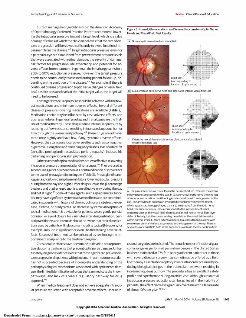
Figure 3. Normal, Glaucomatous, and Severe Glaucomatous Optic NerveHeads and Visual Field Test Results
A Normal optic nerve head and visual field
B Glaucomatous optic nerve head and associated inferior visual field loss
C Extensive neural tissue loss in severe glaucoma and associatedsevere visual field loss
Rim
CupBlind spot (corresponding tolocation of optic nerve)
Blind spot (corresponding tolocation of optic nerve)
A, The pink area of neural tissue forms the neuroretinal rim, whereas the centralempty space corresponds to the cup. B, Glaucomatous optic nerve showing lossof superior neural retinal rim (thinning) and excavation with enlargement of thecup. The arrowheads point to an associated retinal nerve fiber layer defect,which appears as a wedge-shaped dark area emanating from the optic nervehead. The superior neural losses correspond to the inferior defect (blackscotoma) seen on the visual field. There is also a small retinal nerve fiber layerdefect inferiorly, but the corresponding hemifield of the visual field remainswithin normal limits. C, More extensive neural tissue loss from glaucoma withsevere neuroretinal rim loss, excavation, and enlargement of the cup. There issevere loss of visual field both in the superior as well as in the inferior hemifield.
Pathophysiology and Treatment of Glaucoma Review Clinical Review & Education
jama.com JAMA May 14, 2014 Volume 311, Number 18 1905
Copyright 2014 American Medical Association. All rights reserved.
Downloaded From: http://jama.jamanetwork.com/ by anna galvan on 03/15/2015
Copyright 2014 American Medical Association. All rights reserved.
Trabeculectomy is the most commonly performed incisionalsurgical procedure to lower intraocular pressure. It consists ofexcision of a small portion of the trabecular meshwork and oradjacent corneoscleral tissue to provide a drainage route foraqueous humor from within the eye to underneath the conjunc-tiva where it is absorbed. Antiscarring agents are frequentlyapplied to the surgical site to decrease fibroproliferative responseand increase success rates of the surgery, but may increase therate of complications such as infection and damage from very lowintraocular pressure. Devices that drain aqueous humor to anexternal reservoir are an alternative to trabeculectomy that aresimilarly effective in lowering intraocular pressure.51 Several alter-natives to these procedures have been proposed and are beinginvestigated. These so-called minimally invasive glaucoma surger-ies potentially incur less risk of sight-threatening complications.52
To date, these procedures have not had the same intraocularpressure–lowering efficacy as trabeculectomy; however, theymay be indicated for selected cases for which risk-benefit consid-erations are more favorable than those with trabeculectomy. Arecent meta-analysis comparing trabeculectomy with nonpen-
etrating surgeries (deep sclerectomy, viscocanalostomy, andcanaloplasty) concluded that while trabeculectomy was moreeffective in reducing the pressure, it carried a higher risk ofcomplications.53
Primary Closed-Angle GlaucomaThe main feature distinguishing primary closed-angle glaucoma fromprimary open-angle glaucoma is that the angle, the site of aqueousoutflow in the eye, is obstructed by apposition of the iris, resultingin an anatomically closed angle (defined if at least 270° of the angleis occluded). Like open-angle glaucoma, closed-angle glaucoma ispredominantly an asymptomatic disease with individuals often un-aware they have the disorder until advanced visual loss has oc-curred. In less than a third of cases, patients may present with acuteprimary angle closure, a clinical condition characterized by markedconjunctival hyperemia, corneal edema, a middilated unreactive pu-pil, a shallow anterior chamber, and very high intraocular pressure,usually greater than 30 mm Hg. Such patients often complain of ocu-lar pain, nausea, vomiting, and intermittent blurring of vision withhaloes noticed around lights.
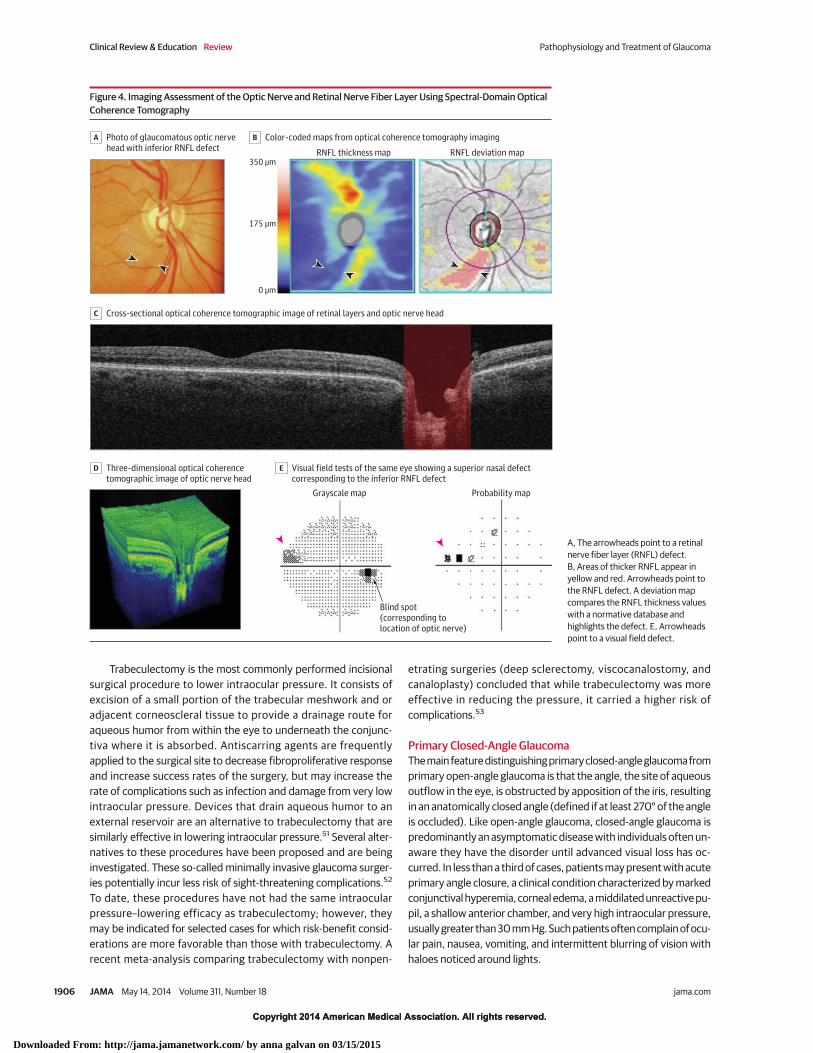
Figure 4. Imaging Assessment of the Optic Nerve and Retinal Nerve Fiber Layer Using Spectral-Domain OpticalCoherence Tomography
A Photo of glaucomatous optic nervehead with inferior RNFL defect
C Cross-sectional optical coherence tomographic image of retinal layers and optic nerve head
D Three-dimensional optical coherencetomographic image of optic nerve head
E Visual field tests of the same eye showing a superior nasal defectcorresponding to the inferior RNFL defect
RNFL thickness map
Probability mapGrayscale map
RNFL deviation map
B Color-coded maps from optical coherence tomography imaging
350 µm
175 µm
0 µm
Blind spot (corresponding to location of optic nerve)
A, The arrowheads point to a retinalnerve fiber layer (RNFL) defect.B, Areas of thicker RNFL appear inyellow and red. Arrowheads point tothe RNFL defect. A deviation mapcompares the RNFL thickness valueswith a normative database andhighlights the defect. E, Arrowheadspoint to a visual field defect.
Clinical Review & Education Review Pathophysiology and Treatment of Glaucoma
1906 JAMA May 14, 2014 Volume 311, Number 18 jama.com
Copyright 2014 American Medical Association. All rights reserved.
Downloaded From: http://jama.jamanetwork.com/ by anna galvan on 03/15/2015
Copyright 2014 American Medical Association. All rights reserved.
Primary closed-angle glaucoma is caused by disorders of the iris,the lens, and retrolenticular structures. Pupillary block is the mostcommon mechanism of angle closure and is caused by resistance toaqueous humor flow from the posterior to anterior chambers at the
pupil. Aqueous humor accumulates behind the iris increasing its con-vexity causing angle closure (Figure 1). Nonpupil block mecha-nisms such as a plateaulike iris configuration may be responsible fora significant proportion of angle closure in Asian patients.54 Closed-
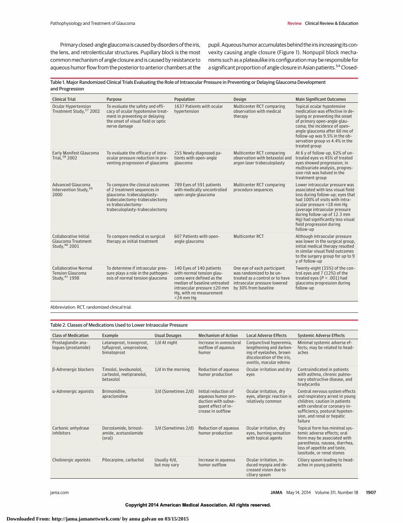
Table 1. Major Randomized Clinical Trials Evaluating the Role of Intraocular Pressure in Preventing or Delaying Glaucoma Developmentand Progression
Clinical Trial Purpose Population Design Main Significant OutcomesOcular HypertensionTreatment Study,37 2002
To evaluate the safety and effi-cacy of ocular hypotensive treat-ment in preventing or delayingthe onset of visual field or opticnerve damage
1637 Patients with ocularhypertension
Multicenter RCT comparingobservation with medicaltherapy
Topical ocular hypotensivemedication was effective in de-laying or preventing the onsetof primary open-angle glau-coma; the incidence of open-angle glaucoma after 60 mo offollow-up was 9.5% in the ob-servation group vs 4.4% in thetreated group
Early Manifest GlaucomaTrial,38 2002
To evaluate the efficacy of intra-ocular pressure reduction in pre-venting progression of glaucoma
255 Newly diagnosed pa-tients with open-angleglaucoma
Multicenter RCT comparingobservation with betaxolol andargon laser trabeculoplasty
At 6 y of follow-up, 62% of un-treated eyes vs 45% of treatedeyes showed progression; inmultivariate analysis, progres-sion risk was halved in thetreatment group
Advanced GlaucomaIntervention Study,39
2000
To compare the clinical outcomesof 2 treatment sequences inglaucoma: trabeculoplasty-trabeculectomy-trabeculectomyvs trabeculectomy-trabeculoplasty-trabeculectomy
789 Eyes of 591 patientswith medically uncontrolledopen-angle glaucoma
Multicenter RCT comparingprocedure sequences
Lower intraocular pressure wasassociated with less visual fieldloss during follow-up; eyes thathad 100% of visits with intra-ocular pressure <18 mm Hg(average intraocular pressureduring follow-up of 12.3 mmHg) had significantly less visualfield progression duringfollow-up
Collaborative InitialGlaucoma TreatmentStudy,40 2001
To compare medical vs surgicaltherapy as initial treatment
607 Patients with open-angle glaucoma
Multicenter RCT Although intraocular pressurewas lower in the surgical group,initial medical therapy resultedin similar visual field outcomesto the surgery group for up to 9y of follow-up
Collaborative NormalTension GlaucomaStudy,41 1998
To determine if intraocular pres-sure plays a role in the pathogen-esis of normal tension glaucoma
140 Eyes of 140 patientswith normal tension glau-coma were defined as themedian of baseline untreatedintraocular pressure ≤20 mmHg, with no measurement>24 mm Hg
One eye of each participantwas randomized to be un-treated as a control or to haveintraocular pressure loweredby 30% from baseline
Twenty-eight (35%) of the con-trol eyes and 7 (12%) of thetreated eyes (P < .001) hadglaucoma progression duringfollow-up
Abbreviation: RCT, randomized clinical trial.
Table 2. Classes of Medications Used to Lower Intraocular Pressure
Class of Medication Example Usual Dosages Mechanism of Action Local Adverse Effects Systemic Adverse EffectsProstaglandin ana-logues (prostamide)
Latanoprost, travoprost,tafluprost, unoprostone,bimatoprost
1/d At night Increase in uveoscleraloutflow of aqueoushumor
Conjunctival hyperemia,lengthening and darken-ing of eyelashes, browndiscoloration of the iris,uveitis, macular edema
Minimal systemic adverse ef-fects; may be related to head-aches
β-Adrenergic blockers Timolol, levobunolol,carteolol, metipranolol,betaxolol
1/d In the morning Reduction of aqueoushumor production
Ocular irritation and dryeyes
Contraindicated in patientswith asthma, chronic pulmo-nary obstructive disease, andbradycardia
α-Adrenergic agonists Brimonidine,apraclonidine
3/d (Sometimes 2/d) Initial reduction ofaqueous humor pro-duction with subse-quent effect of in-crease in outflow
Ocular irritation, dryeyes, allergic reaction isrelatively common
Central nervous system effectsand respiratory arrest in youngchildren; caution in patientswith cerebral or coronary in-sufficiency, postural hypoten-sion, and renal or hepaticfailure
Carbonic anhydraseinhibitors
Dorzolamide, brinzol-amide, acetazolamide(oral)
3/d (Sometimes 2/d) Reduction of aqueoushumor production
Ocular irritation, dryeyes, burning sensationwith topical agents
Topical form has minimal sys-temic adverse effects; oralform may be associated withparesthesia, nausea, diarrhea,loss of appetite and taste,lassitude, or renal stones
Cholinergic agonists Pilocarpine, carbachol Usually 4/d,but may vary
Increase in aqueoushumor outflow
Ocular irritation, in-duced myopia and de-creased vision due tociliary spasm
Ciliary spasm leading to head-aches in young patients
Pathophysiology and Treatment of Glaucoma Review Clinical Review & Education
jama.com JAMA May 14, 2014 Volume 311, Number 18 1907
Copyright 2014 American Medical Association. All rights reserved.
Downloaded From: http://jama.jamanetwork.com/ by anna galvan on 03/15/2015
Copyright 2014 American Medical Association. All rights reserved.
angle glaucoma may also be caused by dynamic physiological fac-tors, such as an increase in iris volume with pupil dilation and cho-roidal effusion.55
Risk FactorsRisk factors for angle closure include female sex, older age, and Asianethnicity (eg, Chinese). Eyes with angle closure tend to share cer-tain biometric characteristics. The main ocular risk factor for angleclosure involves having a crowded anterior segment in a small eye,with a shallow central anterior chamber depth, a thicker and moreanteriorly positioned lens, and short axial length of the eye.55-57 Withanterior segment optical coherence tomography, other anatomicalrisk factors for angle closure have been recently identified such assmaller anterior chamber width, area and volume, thicker irides withgreater iris curvature, and a greater lens vault.57
GeneticsA genetic etiology for angle closure is supported by epidemiologi-cal findings: first-degree relatives of patients with it are at greaterrisk than the general population, the high heritability of anatomicalrisk factors (such as anterior chamber depth), and ethnic variationsin the prevalence.58,59 Recently, a genome-wide association studyinvolving more than 20 000 individuals from 7 countries found 3new genetic loci for angle closure: rs11024102 at PLEKHA7, rs3753841
at COL11A1 (HGNC:2186), and rs1015213 located between PCMTD1(HGNC:30483) and ST18 (HGNC:18695) on chromosome 8q.59 Thisindicates that open-angle and closed-angle glaucoma are distinct ge-netic entities with different genes associated with each disease.
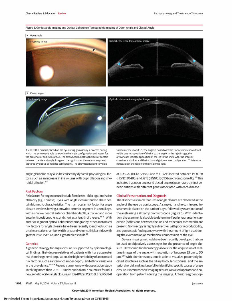
Clinical Presentation and DiagnosisThe distinctive clinical features of angle closure are observed in theangle of the eye by gonioscopy. A simple, handheld, mirrored in-strument is placed on the patient’s eye, followed by examination ofthe angle using a slit-lamp biomicroscope (Figure 5). With indenta-tion, the examiner is also able to determine if peripheral anterior syn-echiae (adhesions between the iris and trabecular meshwork) arepresent. Gonioscopy is highly subjective, with poor reproducibility,and gonioscopic findings may vary with the amount of light used dur-ing the examination or mechanical compression of the eye.
Several imaging methods have been recently developed that canbe used to objectively assess eyes for the presence of angle clo-sure. Ultrasound biomicroscopy allows for the acquisition of real-time images of the angle, with resolution of between 25 μm to 50μm.60 With biomicroscopy, one is able to visualize posteriorly lo-cated structures such as the ciliary body, lens zonules, and the an-terior choroid, making it useful for identifying specific causes of angleclosure. Biomicroscopic imaging requires a skilled operator and co-operation from patients during the imaging. Anterior segment op-
Figure 5. Gonioscopic Imaging and Optical Coherence Tomographic Imaging of Open-Angle and Closed-Angle
B
Gonioscopy image
A
Gonioscopy image Optical coherence tomographic image
Optical coherence tomographic image
Open angle
Closed angle
A lens with a prism is placed on the eye during gonioscopy, a process duringwhich the examiner is able to examine the angle configuration and assess forthe presence of angle closure. A, The arrowhead points to the lack of contactbetween the iris and angle. Image on the right shows the anterior segmentcaptured by optical coherence tomography. The arrowheads point to visible
trabecular meshwork. B, The angle is closed with the trabecular meshwork notvisible due to apposition of the iris to the angle. In the right image, thearrowheads indicate apposition of the iris to the angle wall; the anteriorchamber is shallow and the iris has a slightly convex configuration. This is morenoticeable in the region of the iris on the right.
Clinical Review & Education Review Pathophysiology and Treatment of Glaucoma
1908 JAMA May 14, 2014 Volume 311, Number 18 jama.com
Copyright 2014 American Medical Association. All rights reserved.
Downloaded From: http://jama.jamanetwork.com/ by anna galvan on 03/15/2015
Copyright 2014 American Medical Association. All rights reserved.
tical coherence tomography is a noncontact imaging device that ac-quires high-resolution cross-sectional images of the anterior chamber(Figure 5). The incorporation of automated image analysis soft-ware allows for rapid measurement of anterior segment para-meters. Comparison studies found a higher rate of diagnosis of closedangles with tomography than with gonioscopy.61
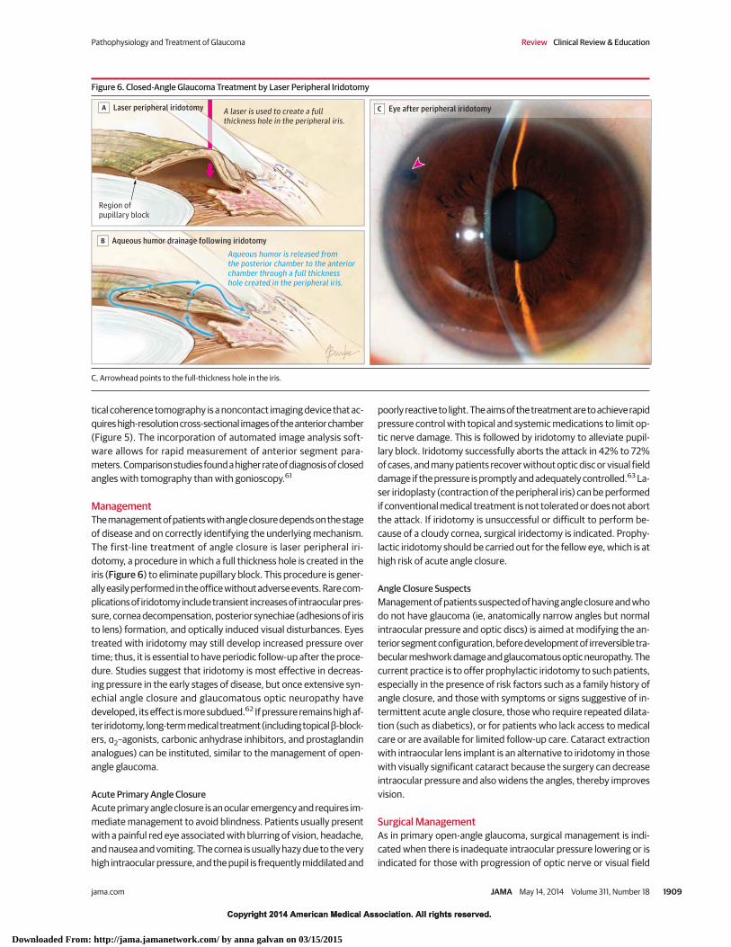
ManagementThe management of patients with angle closure depends on the stageof disease and on correctly identifying the underlying mechanism.The first-line treatment of angle closure is laser peripheral iri-dotomy, a procedure in which a full thickness hole is created in theiris (Figure 6) to eliminate pupillary block. This procedure is gener-ally easily performed in the office without adverse events. Rare com-plications of iridotomy include transient increases of intraocular pres-sure, cornea decompensation, posterior synechiae (adhesions of iristo lens) formation, and optically induced visual disturbances. Eyestreated with iridotomy may still develop increased pressure overtime; thus, it is essential to have periodic follow-up after the proce-dure. Studies suggest that iridotomy is most effective in decreas-ing pressure in the early stages of disease, but once extensive syn-echial angle closure and glaucomatous optic neuropathy havedeveloped, its effect is more subdued.62 If pressure remains high af-ter iridotomy, long-term medical treatment (including topical β-block-ers, α2–agonists, carbonic anhydrase inhibitors, and prostaglandinanalogues) can be instituted, similar to the management of open-angle glaucoma.
Acute Primary Angle ClosureAcute primary angle closure is an ocular emergency and requires im-mediate management to avoid blindness. Patients usually presentwith a painful red eye associated with blurring of vision, headache,and nausea and vomiting. The cornea is usually hazy due to the veryhigh intraocular pressure, and the pupil is frequently middilated and
poorly reactive to light. The aims of the treatment are to achieve rapidpressure control with topical and systemic medications to limit op-tic nerve damage. This is followed by iridotomy to alleviate pupil-lary block. Iridotomy successfully aborts the attack in 42% to 72%of cases, and many patients recover without optic disc or visual fielddamage if the pressure is promptly and adequately controlled.63 La-ser iridoplasty (contraction of the peripheral iris) can be performedif conventional medical treatment is not tolerated or does not abortthe attack. If iridotomy is unsuccessful or difficult to perform be-cause of a cloudy cornea, surgical iridectomy is indicated. Prophy-lactic iridotomy should be carried out for the fellow eye, which is athigh risk of acute angle closure.
Angle Closure SuspectsManagement of patients suspected of having angle closure and whodo not have glaucoma (ie, anatomically narrow angles but normalintraocular pressure and optic discs) is aimed at modifying the an-terior segment configuration, before development of irreversible tra-becular meshwork damage and glaucomatous optic neuropathy. Thecurrent practice is to offer prophylactic iridotomy to such patients,especially in the presence of risk factors such as a family history ofangle closure, and those with symptoms or signs suggestive of in-termittent acute angle closure, those who require repeated dilata-tion (such as diabetics), or for patients who lack access to medicalcare or are available for limited follow-up care. Cataract extractionwith intraocular lens implant is an alternative to iridotomy in thosewith visually significant cataract because the surgery can decreaseintraocular pressure and also widens the angles, thereby improvesvision.
Surgical ManagementAs in primary open-angle glaucoma, surgical management is indi-cated when there is inadequate intraocular pressure lowering or isindicated for those with progression of optic nerve or visual field
Figure 6. Closed-Angle Glaucoma Treatment by Laser Peripheral Iridotomy
Aqueous humor is released fromthe posterior chamber to the anteriorchamber through a full thickness hole created in the peripheral iris.
A laser is used to create a fullthickness hole in the peripheral iris.
A Laser peripheral iridotomy
B Aqueous humor drainage following iridotomy
C Eye after peripheral iridotomy
Region ofpupillary block
C, Arrowhead points to the full-thickness hole in the iris.
Pathophysiology and Treatment of Glaucoma Review Clinical Review & Education
jama.com JAMA May 14, 2014 Volume 311, Number 18 1909
Copyright 2014 American Medical Association. All rights reserved.
Downloaded From: http://jama.jamanetwork.com/ by anna galvan on 03/15/2015
Copyright 2014 American Medical Association. All rights reserved.
damage despite medical and laser treatment. Trabeculectomy, eitheralone or in combination with lens extraction should be consideredif the pressure control remains too high despite laser and medicaltreatment, especially in more advanced cases of open-angle glau-coma. Lens extraction is also performed when lens-related mecha-nisms predominate, especially in cases in which a significant cata-ract impairs vision. Finally, glaucoma drainage implants may be usedin patients with chronic angle closure similarly to open-angle glau-coma when trabeculectomy has failed to control pressure, or in eyesthat are deemed to be at high risk of failure with trabeculectomy.
Conclusions
Glaucoma is a leading cause of blindness. Early diagnosis and treat-ment can prevent vision loss from the disease. Primary care physi-cians should consider referring patients with a family history of thedisease for a complete ophthalmologic examination. In addition,evaluation of the optic nerve by direct ophthalmoscopy may iden-tify suspicious signs of optic nerve damage that should also promptreferral to an eye care specialist.
ARTICLE INFORMATION
Author Contributions: Drs Weinreb, Aung, andMedeiros had full access to all of the data in thestudy and take responsibility for the integrity of thedata and the accuracy of the data analysis.Study concept and design: All authors.Acquisition, analysis, or interpretation of data: Allauthors.Drafting of the manuscript: All authors.Critical revision of the manuscript for importantintellectual content: All authors.Statistical analysis: All authors.Obtained funding: Medeiros.Administrative, technical, or material support:Weinreb, Medeiros.Study supervision: All authors.
Conflict of Interest Disclosures: All authors havecompleted and submitted the ICMJE Form forDisclosure of Potential Conflicts of Interest. DrWeinreb reported that he has worked as aconsultant for Alcon, Allergan, Anakem, Aquesys,Bausch and Lomb, Carl Zeiss Meditec, Quark,Sensimed, Solx, Topcon and has received researchsupport from National Eye Institute, Nidek,Genentech, Quark, and Topcon. Dr Aung reportedthat he has worked as a consultant for Alcon,Allergan, Bausch and Lomb, MSD, and Quark; hasreceived research support from Alcon, Allergan,Aquesys, Carl Zeiss Meditec, Ellex, and OcularTherapeutics; and has received lecture fees fromAlcon, Allergan, Carl Zeiss Meditec, Ellex, Pfizer, andSanten. Dr Medeiros reported that he has receivedresearch support from the National Eye Institute,Alcon, Allergan, Merck, Carl-Zeiss Meditec,Heidelberg Engineering, Sensimed, and Reichert.
Funding/Support: This study was supported inpart by grants EY019692 (Weinreb), EY021818(Medeiros), from the National Institutes ofHealth/National Eye Institute; and an unrestrictedgrant from Research to Prevent Blindness. Dr Aungis supported by grants from the National MedicalResearch Council, Singapore, and the NationalResearch Foundation, Singapore.
Role of the Sponsor: The study sponsors had norole in the design and conduct of the study;collection, management, analysis, andinterpretation of the data; preparation, review, orapproval of the manuscript; and decision to submitthe manuscript for publication.
Submissions:We encourage authors to submitpapers for consideration as a Review. Pleasecontact Mary McGrae McDermott, MD, at [email protected].
REFERENCES
1. Weinreb RN, Khaw PT. Primary open-angleglaucoma. Lancet. 2004;363(9422):1711-1720.
2. Nickells RW, Howell GR, Soto I, John SW. Underpressure: cellular and molecular responses duringglaucoma, a common neurodegeneration withaxonopathy. Annu Rev Neurosci. 2012;35:153-179.
3. Quigley HA, Broman AT. The number of peoplewith glaucoma worldwide in 2010 and 2020. Br JOphthalmol. 2006;90(3):262-267.
4. Leite MT, Sakata LM, Medeiros FA. Managingglaucoma in developing countries. Arq BrasOftalmol. 2011;74(2):83-84.
5. Rotchford AP, Kirwan JF, Muller MA, Johnson GJ,Roux P. Temba glaucoma study: a population-basedcross-sectional survey in urban South Africa.Ophthalmology. 2003;110(2):376-382.
6. Hennis A, Wu SY, Nemesure B, Honkanen R,Leske MC; Barbados Eye Studies Group. Awarenessof incident open-angle glaucoma in a populationstudy: the Barbados Eye Studies. Ophthalmology.2007;114(10):1816-1821.
7. Sathyamangalam RV, Paul PG, George R, et al.Determinants of glaucoma awareness andknowledge in urban Chennai. Indian J Ophthalmol.2009;57(5):355-360.
8. Budenz DL, Barton K, Whiteside-de Vos J, et al;Tema Eye Survey Study Group. Prevalence ofglaucoma in an urban West African population: theTema Eye Survey. JAMA Ophthalmol.2013;131(5):651-658.
9. Friedman DS, Wolfs RC, O’Colmain BJ, et al; EyeDiseases Prevalence Research Group. Prevalence ofopen-angle glaucoma among adults in the UnitedStates. Arch Ophthalmol. 2004;122(4):532-538.
10. Day AC, Baio G, Gazzard G, et al. Theprevalence of primary angle closure glaucoma inEuropean derived populations: a systematic review.Br J Ophthalmol. 2012;96(9):1162-1167.
11. Hollands H, Johnson D, Hollands S, Simel DL,Jinapriya D, Sharma S. Do findings on routineexamination identify patients at risk for primaryopen-angle glaucoma? JAMA. 2013;309(19):2035-2042.
12. Kersey JP, Broadway DC. Corticosteroid-induced glaucoma: a review of the literature. Eye(Lond). 2006;20(4):407-416.
13. Quigley HA, Addicks EM, Green WR, MaumeneeAE. Optic nerve damage in human glaucoma, II: thesite of injury and susceptibility to damage. ArchOphthalmol. 1981;99(4):635-649.
14. Fechtner RD, Weinreb RN. Mechanisms of opticnerve damage in primary open angle glaucoma.Surv Ophthalmol. 1994;39(1):23-42.
15. Burgoyne CF, Downs JC, Bellezza AJ, Suh JK,Hart RT. The optic nerve head as a biomechanicalstructure: a new paradigm for understanding therole of IOP-related stress and strain in the
pathophysiology of glaucomatous optic nerve headdamage. Prog Retin Eye Res. 2005;24(1):39-73.
16. Quigley HA, McKinnon SJ, Zack DJ, et al.Retrograde axonal transport of BDNF in retinalganglion cells is blocked by acute IOP elevation inrats. Invest Ophthalmol Vis Sci. 2000;41(11):3460-3466.
17. Ju WK, Kim KY, Lindsey JD, et al. Intraocularpressure elevation induces mitochondrial fissionand triggers OPA1 release in glaucomatous opticnerve. Invest Ophthalmol Vis Sci. 2008;49(11):4903-4911.
18. Wang N, Xie X, Yang D, et al Orbitalcerebrospinal fluid space in glaucoma: the BeijingIntracranial and Intraocular Pressure (iCOP) study.Ophthalmology. 2012;119(10):2065e1-2073e1.
19. Ren R, Jonas JB, Tian G, et al. Cerebrospinalfluid pressure in glaucoma: a prospective study.Ophthalmology. 2010;117(2):259-266.
20. Almasieh M, Wilson AM, Morquette B, CuevaVargas JL, Di Polo A. The molecular basis of retinalganglion cell death in glaucoma. Prog Retin Eye Res.2012;31(2):152-181.
21. Stone EM, Fingert JH, Alward WL, et al.Identification of a gene that causes primary openangle glaucoma. Science. 1997;275(5300):668-670.
22. Rezaie T, Child A, Hitchings R, et al. Adult-onsetprimary open-angle glaucoma caused by mutationsin optineurin. Science. 2002;295(5557):1077-1079.
23. Monemi S, Spaeth G, DaSilva A, et al.Identification of a novel adult-onset primaryopen-angle glaucoma (POAG) gene on 5q22.1. HumMol Genet. 2005;14(6):725-733.
24. Kwon YH, Fingert JH, Kuehn MH, Alward WL.Primary open-angle glaucoma. N Engl J Med.2009;360(11):1113-1124.
25. Thorleifsson G, Walters GB, Hewitt AW, et al.Common variants near CAV1 and CAV2 areassociated with primary open-angle glaucoma. NatGenet. 2010;42(10):906-909.
26. Wiggs JL, Yaspan BL, Hauser MA, et al.Common variants at 9p21 and 8q22 are associatedwith increased susceptibility to optic nervedegeneration in glaucoma. PLoS Genet.2012;8(4):e1002654.
27. Harwerth RS, Wheat JL, Fredette MJ, AndersonDR. Linking structure and function in glaucoma.Prog Retin Eye Res. 2010;29(4):249-271.
28. Medeiros FA, Alencar LM, Zangwill LM, BowdC, Sample PA, Weinreb RN. Prediction of functionalloss in glaucoma from progressive optic discdamage. Arch Ophthalmol. 2009;127(10):1250-1256.
Clinical Review & Education Review Pathophysiology and Treatment of Glaucoma
1910 JAMA May 14, 2014 Volume 311, Number 18 jama.com
Copyright 2014 American Medical Association. All rights reserved.
Downloaded From: http://jama.jamanetwork.com/ by anna galvan on 03/15/2015

Copyright 2014 American Medical Association. All rights reserved.
29. Jampel HD, Friedman D, Quigley H, et alAgreement among glaucoma specialists inassessing progressive disc changes fromphotographs in open-angle glaucoma patients. AmJ Ophthalmol. 2009;147(1):39e1-44 e1. .
30. Medeiros FA, Vizzeri G, Zangwill LM, AlencarLM, Sample PA, Weinreb RN. Comparison of retinalnerve fiber layer and optic disc imaging fordiagnosing glaucoma in patients suspected ofhaving the disease. Ophthalmology.2008;115(8):1340-1346.
31. Medeiros FA, Zangwill LM, Bowd C, WeinrebRN. Comparison of the GDx VCC scanning laserpolarimeter, HRT II confocal scanning laserophthalmoscope, and stratus OCT opticalcoherence tomograph for the detection ofglaucoma. Arch Ophthalmol. 2004;122(6):827-837.
32. Chauhan BC, O’Leary N, Almobarak FA, et al.Enhanced detection of open-angle glaucoma withan anatomically accurate optical coherencetomography-derived neuroretinal rim parameter.Ophthalmology. 2013;120(3):535-543.
33. Medeiros FA, Zangwill LM, Anderson DR, et alEstimating the rate of retinal ganglion cell loss inglaucoma. Am J Ophthalmol. 2012;154(5):814e1-24e1.
34. Strouthidis NG, Gardiner SK, Sinapis C,Burgoyne CF, Garway-Heath DF. The spatial patternof neuroretinal rim loss in ocular hypertension.Invest Ophthalmol Vis Sci. 2009;50(8):3737-3742.
35. McKean-Cowdin R, Wang Y, Wu J, et al. Impactof visual field loss on health-related quality of life inglaucoma: the Los Angeles Latino Eye Study.Ophthalmology. 2008;115(6):941e1-948e1.
36. Boland MV, Ervin AM, Friedman DS, et al.Comparative effectiveness of treatments foropen-angle glaucoma: a systematic review for theUS Preventive Services Task Force. Ann Intern Med.2013;158(4):271-279.
37. Kass MA, Heuer DK, Higginbotham EJ, et al.The Ocular Hypertension Treatment Study:a randomized trial determines that topical ocularhypotensive medication delays or prevents theonset of primary open-angle glaucoma. ArchOphthalmol. 2002;120(6):701-713.
38. Heijl A, Leske MC, Bengtsson B, Hyman L,Bengtsson B, Hussein M; Early Manifest GlaucomaTrial Group. Reduction of intraocular pressure andglaucoma progression. Arch Ophthalmol.2002;120(10):1268-1279.
39. The AGIS Investigators. The AdvancedGlaucoma Intervention Study (AGIS), 7: therelationship between control of intraocularpressure and visual field deterioration. Am JOphthalmol. 2000;130(4):429-440.
40. Lichter PR, Musch DC, Gillespie BW, et al;CIGTS Study Group. Interim clinical outcomes in theCollaborative Initial Glaucoma Treatment Studycomparing initial treatment randomized tomedications or surgery. Ophthalmology.2001;108(11):1943-1953.
41. Collaborative Normal-Tension Glaucoma StudyGroup. Comparison of glaucomatous progressionbetween untreated patients with normal-tensionglaucoma and patients with therapeuticallyreduced intraocular pressures. Am J Ophthalmol.1998;126(4):487-497.
42. American Academy of OphthalmologyPreferred Practice Patterns Committee GP.Preferred practice pattern: primary open-angleglaucoma. In: Ophthalmology. Chicago, Illinois:American Academy of Ophtalmology: 2010.
43. Gaton DD, Sagara T, Lindsey JD, Gabelt BT,Kaufman PL, Weinreb RN. Increased matrixmetalloproteinases 1, 2, and 3 in the monkeyuveoscleral outflow pathway after topicalprostaglandin F(2 alpha)-isopropyl ester treatment.Arch Ophthalmol. 2001;119(8):1165-1170.
44. Stewart WC, Konstas AG, Nelson LA, Kruft B.Meta-analysis of 24-hour intraocular pressurestudies evaluating the efficacy of glaucomamedicines. Ophthalmology. 2008;115(7):1117e1-1122e1.
45. Liu JH, Kripke DF, Weinreb RN. Comparison ofthe nocturnal effects of once-daily timolol andlatanoprost on intraocular pressure. Am JOphthalmol. 2004;138(3):389-395.
46. Weinreb RN, Kaufman PL. Glaucoma researchcommunity and FDA look to the future, II: NEI/FDAGlaucoma Clinical Trial Design and EndpointsSymposium: measures of structural change andvisual function. Invest Ophthalmol Vis Sci.2011;52(11):7842-7851.
47. Mansouri K, Medeiros FA, Weinreb RN. Globalrates of glaucoma surgery. Graefes Arch Clin ExpOphthalmol. 2013;251(11):2609-2615.
48. Odberg T, Sandvik L. The medium andlong-term efficacy of primary argon lasertrabeculoplasty in avoiding topical medication inopen angle glaucoma. Acta Ophthalmol Scand.1999;77(2):176-181.
49. Shingleton BJ, Richter CU, Dharma SK, et al.Long-term efficacy of argon laser trabeculoplasty:a 10-year follow-up study. Ophthalmology.1993;100(9):1324-1329.
50. Shingleton BJ, Richter CU, Bellows AR,Hutchinson BT, Glynn RJ. Long-term efficacy ofargon laser trabeculoplasty. Ophthalmology.1987;94(12):1513-1518.
51. Gedde SJ, Schiffman JC, Feuer WJ, et al.Treatment outcomes in the Tube Versus
Trabeculectomy (TVT) study after five years offollow-up. Am J Ophthalmol. 2012;153(15):789e2-803e2.
52. Ayyala RS, Chaudhry AL, Okogbaa CB,Zurakowski D. Comparison of surgical outcomesbetween canaloplasty and trabeculectomy at 12months’ follow-up. Ophthalmology.2011;118(12):2427-2433.
53. Rulli E, Biagioli E, Riva I, et al. Efficacy andsafety of trabeculectomy vs nonpenetratingsurgical procedures: a systematic review andmeta-analysis. JAMA Ophthalmol.2013;131(12):1573-1582.
54. He M, Foster PJ, Johnson GJ, Khaw PT.Angle-closure glaucoma in East Asian and Europeanpeople: different diseases? Eye (Lond).2006;20(1):3-12.
55. Sakai H, Morine-Shinjyo S, Shinzato M,Nakamura Y, Sakai M, Sawaguchi S. Uveal effusionin primary angle-closure glaucoma. Ophthalmology.2005;112(3):413-419.
56. Lavanya R, Wong TY, Friedman DS, et al.Determinants of angle closure in olderSingaporeans. Arch Ophthalmol. 2008;126(5):686-691.
57. Nongpiur ME, Ku JY, Aung T. Angle closureglaucoma: a mechanistic review. Curr OpinOphthalmol. 2011;22(2):96-101.
58. Amerasinghe N, Zhang J, Thalamuthu A, et al.The heritability and sibling risk of angle closure inAsians. Ophthalmology. 2011;118(3):480-485.
59. Vithana EN, Khor CC, Qiao C, et al.Genome-wide association analyses identify threenew susceptibility loci for primary angle closureglaucoma. Nat Genet. 2012;44(10):1142-1146.
60. Sakata LM, Lavanya R, Friedman DS, et al.Comparison of gonioscopy and anterior segmentocular coherence tomography in detecting angleclosure in different quadrants of the anteriorchamber angle. Ophthalmology. 2008;115(5):769-774.
61. Wong HT, Lim MC, Sakata LM, et al.High-definition optical coherence tomographyimaging of the iridocorneal angle of the eye. ArchOphthalmol. 2009;127(3):256-260.
62. Alsagoff Z, Aung T, Ang LP, Chew PT. Long-termclinical course of primary angle-closure glaucoma inan Asian population. Ophthalmology.2000;107(12):2300-2304.
63. Aung T, Ang LP, Chan SP, Chew PT. Acuteprimary angle-closure: long-term intraocularpressure outcome in Asian eyes. Am J Ophthalmol.2001;131(1):7-12.
Pathophysiology and Treatment of Glaucoma Review Clinical Review & Education
jama.com JAMA May 14, 2014 Volume 311, Number 18 1911
Copyright 2014 American Medical Association. All rights reserved.
Downloaded From: http://jama.jamanetwork.com/ by anna galvan on 03/15/2015


