Phosphorylation within the Amino-Terminal Acidic Domain I ...
The eIF1A C-terminal domain promotes initiationcomplex assembly, scanning and AUG selectionin vivo
Christie A Fekete1, Drew J Applefield2,Stephen A Blakely1, Nikolay Shirokikh3,Tatyana Pestova3, Jon R Lorsch2
and Alan G Hinnebusch1,*1Laboratory of Gene Regulation and Development, National Institute ofChild Health and Human Development, NIH, Bethesda, MD, USA,2Department of Biophysics and Biophysical Chemistry, Johns HopkinsUniversity School of Medicine, Baltimore, MD, USA and 3Department ofMicrobiology and Immunology, State University of New York HealthScience Center at Brooklyn, Brooklyn, NY, USA
Translation initiation factor 1A stimulates 40S-binding of
the eukaryotic initiation factor 2 (eIF2)/GTP/Met-tRNAiMet
ternary complex (TC) and promotes scanning in vitro.
eIF1A contains an OB-fold present in bacterial IF1 plus
N- and C-terminal extensions. Truncating the C-terminus
(DC) or mutating OB-fold residues (66–70) of eIF1A re-
duced general translation in vivo but increased GCN4
translation (Gcd� phenotype) in a manner suppressed by
overexpressing TC. Consistent with this, both mutations
diminished 40S-bound TC, eIF5 and eIF3 in vivo, and DCimpaired TC recruitment in vitro. The assembly defects
of the OB-fold mutation can be attributed to reduced
40S-binding of eIF1A, whereas DC impairs eIF1A function
on the ribosome. A substitution in the C-terminal helix
(98–101) also reduced 43S assembly in vivo. Rather than
producing a Gcd� phenotype, however, 98–101 impairs
GCN4 derepression in a manner consistent with defective
scanning by reinitiating ribosomes. Indeed, 98–101 allows
formation of aberrant 48S complexes in vitro and increases
utilization of non-AUG codons in vivo. Thus, the OB-fold is
crucial for ribosome-binding and the C-terminal domain of
eIF1A has eukaryotic-specific functions in TC recruitment
and scanning.
The EMBO Journal (2005) 24, 3588–3601. doi:10.1038/
sj.emboj.7600821; Published online 29 September 2005
Subject Categories: proteins
Keywords: eIF1A; eIF2; GCN4; scanning; translation
Introduction
Translation initiation begins with the binding of Met-tRNAiMet
to the 40S ribosomal subunit in a ternary complex (TC) with
eukaryotic initiation factor 2 (eIF2) and GTP. This reaction is
stimulated in vitro by eIF1, eIF1A and eIF3 (Hinnebusch,
2000; Algire et al, 2002; Majumdar et al, 2003; Kolupaeva
et al, 2005). The 43S preinitiation complex (PIC) thus formed
binds to the 50-end of mRNA and scans the leader until Met-
tRNAiMet base-pairs with an AUG, triggering GTP hydrolysis
by eIF2 in a reaction stimulated by eIF5 (Hershey and
Merrick, 2000). Biochemical evidence indicates that mamma-
lian eIF1 and eIF1A promote scanning and formation of a 48S
complex at the start codon (Pestova et al, 1998). eIF2, eIF5,
eIF1 and eIF3 have been implicated in AUG selection, as
mutations in these factors permit increased initiation at a
UUG codon in yeast (Donahue, 2000; He et al, 2003; Valasek
et al, 2004). Consistent with this, eIF1 inhibits 48S assembly
at near-cognate triplets or AUGs in a poor context in vitro
(Pestova and Kolupaeva, 2002).
While biochemical studies have implicated eIF3, eIF1 and
eIF1A in 40S-binding of TC, their relative importance in vivo
and functions in 43S assembly are poorly understood. The TC
in yeast cells is linked to eIF3, eIF5 and eIF1 in a multifactor
complex (MFC) (Asano et al, 2000), and evidence is accu-
mulating that these interactions promote cooperative binding
of MFC components to the 40S ribosome in vivo (Asano et al,
2001; Valasek et al, 2002, 2003; Nielsen et al, 2004; Singh
et al, 2004). We presented genetic evidence that eIF1A is
required for wild-type (WT) binding of TC to 40S subunits in
yeast, as deleting the C-terminal domain and adjacent 310helix of eIF1A (the D108–153 mutation) increased translation
of GCN4 mRNA in cells lacking protein kinase GCN2 (Olsen
et al, 2003). GCN4 translation is normally induced in amino-
acid-starved cells by phosphorylation of eIF2 (Hinnebusch,
1996). The ensuing reduction in TC concentration allows 40S
subunits that translate the first upstream open-reading frame
(uORF1) and resume scanning to bypass the three remaining
uORFs and reinitiate downstream at GCN4. In nonstarved WT
or gcn2D cells, by contrast, a high level of TC ensures that all
40S subunits scanning downstream from uORF1 quickly
rebind TC, reinitiate at uORFs 2, 3 or 4 and dissociate from
the mRNA without translating GCN4 (Hinnebusch, 1996).
Importantly, the derepressed GCN4 translation in gcn2Dcells (Gcd� phenotype) produced by the D108–153 mutation
was suppressed by overexpressing the TC. This suggested
that the rate of TC binding to 40S subunits scanning down-
stream from uORF1 was reduced by D108–153, allowing a
fraction to bypass uORFs 2–4 and reinitiate at GCN4 instead
(Olsen et al, 2003). However, this interpretation was not
confirmed biochemically; nor was it determined whether
D108–153 impairs 40S-binding of eIF1A or disrupts its bio-
chemical function in 43S assembly.
eIF1A is related to bacterial IF1, and both proteins contain
the b-barrel ‘OB’-fold (Sette et al, 1997; Battiste et al, 2000),
while eIF1A also contains an a-helical domain and random-
coil N- and C-terminal extensions (NTD and CTD)
(Figure 1A). As IF1 binds to the A-site of the 30S subunitReceived: 2 June 2005; accepted: 26 August 2005; published online:29 September 2005
*Corresponding author. Laboratory of Gene Regulation andDevelopment, National Institute of Child Health and HumanDevelopment, NIH, Building 6A/Room B1A-13, Bethesda, MD 20892,USA. Tel.: þ 1 301 496 4480; Fax: þ 1 301 496 6828;E-mail: [email protected]
The EMBO Journal (2005) 24, 3588–3601 | & 2005 European Molecular Biology Organization |All Rights Reserved 0261-4189/05
www.embojournal.org
The EMBO Journal VOL 24 | NO 20 | 2005 &2005 European Molecular Biology Organization
EMBO
THE
EMBOJOURNAL
THE
EMBOJOURNAL
3588

A
B
C
FL-TIF11 alleleGrowth
30°C 37°C 3-AT
Expression
WT
1
2
3
4
5
6
7
8
KGGKK7 − 11 AAAAA
GRRGK12 − 16 AAAAA
NDSDG17 − 21 AAAAA
hc DGNK53 − 56 AANA
RKKVW66 − 70 AAAVA
DEAR98 − 101 AAAA
F131A, F133A
+++++ +++++ −
lethal lethal N/A
lethal lethal N/A
++ ++ −
++ + +
++++
+++ +++ −
+ + +
++
+++++++ +
*
*
DIDDI149 − 153 AAAAA
GKKNT2 − 6 AAAAA
PKRELIY22 − 28 AAAAAAA
KEEGQE29 − 34 AAAAQA
MLGN41 − 44 ALAA
GRVE45 − 48 AAVA
hc RDFQD82 − 86 AAAAA
KYN94 − 96 AYA
PE110 − 111 AA
KN104 − 105 AA
DVNFE128 − 132 AAAAA
FGNAD133 − 137 AAAAA
FESDE123 − 127 AAAAA
EDDEE138 − 142 AAAAA
GEDEEL143 − 148 AAAAAA
+++ +++ −
+++ ++ −+++ ++ −
++++ +++ −++++ −
+ + −+++ + −
+++++ ++++ −+++++ +++++ −
+++++ +++++ −++++ +++ ++
++++ +++ +++++ +++ −+++ +++ −+++ ++ −
*
ND
NE116 − 117 AA lethal lethal N/A
R62D lethal lethal N/A
TDNFG118 − 122 AAAAA +++++ +++++ −
NTD
OB
Helical
CTD
NTD OB-fold Helical CTD
1 34 95 112 153
Random coil α2 Random coil
eIF1A/TIF11
1 2 34 5 6
8
7
Δ108 − 153
FL 3−10
NC
1 23
4
5
6
7
8
N
123
4
5
6
7
8
NC
180°
>
8 8
hc Δ108 − 153
−
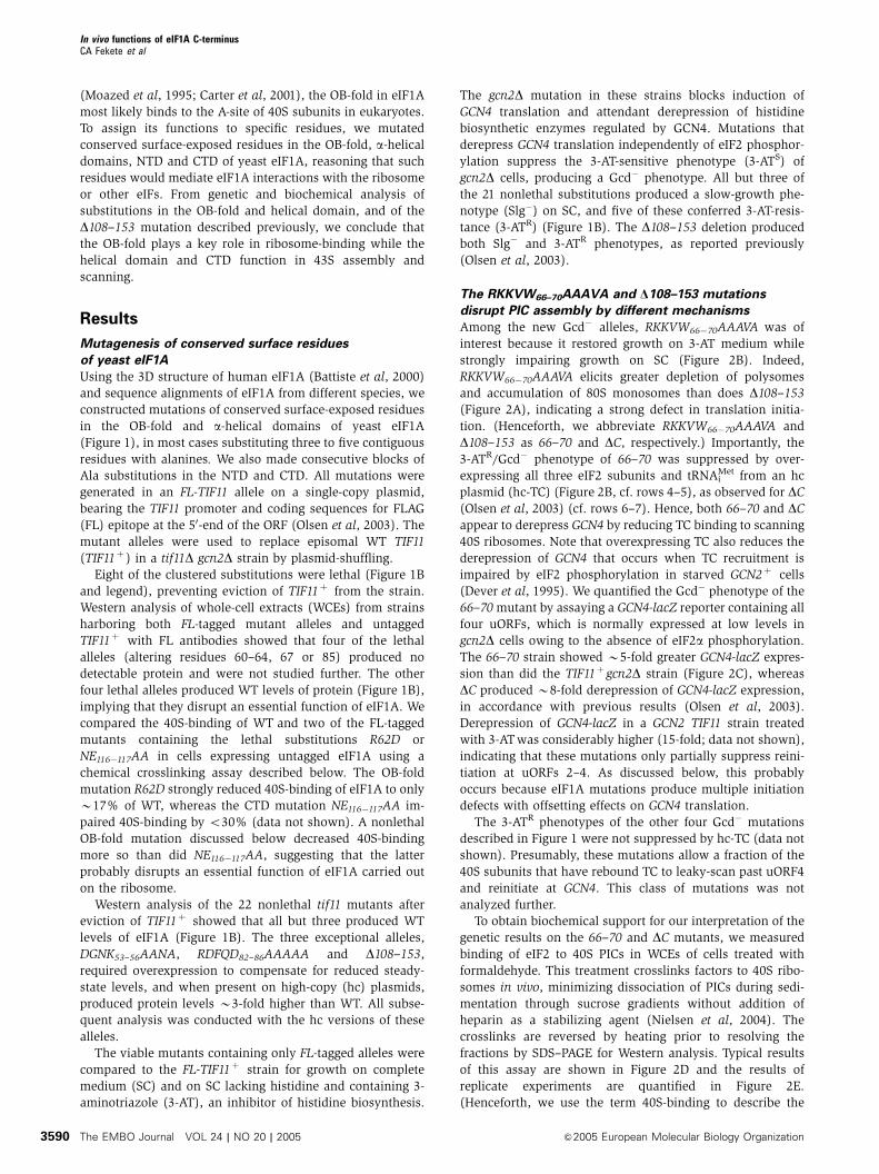
Figure 1 Phenotypes of eIF1A mutations. (A) Schematic representation of eIF1A domain organization, including the FLAG (FL) epitope,amino-acid positions (above) and locations of key mutations (1–8; underlined below). (B) Growth of gcn2D strains harboring the indicatedmutant or WT FL-TIF11 alleles on SC medium lacking Leu (SC-L) at 30 or 371C and on SC medium lacking leucine and histidine (SC–LH) andcontaining 10mM 3-AT at 301C. Relative growth is summarized on a scale from (�) to (þ þ þ þ þ ). The domain locations of the mutationsare listed on the left along with designations of selected mutations 1–8 as indicated in panel A. The far-right column (Expression) containsresults of Western analysis of WCEs from the same strains analyzed for growth phenotypes (except for the lethal alleles) using FLAG antibodies.Separate probing of the blots with antibodies against eIF2a ensured similar protein loading for each extract (not shown). Three FL-TIF11 alleles(*) were expressed on hc plasmids; ND, not detectable. Four additional lethal alleles were identified for which no protein products weredetected: K64A, K67D, Q85A and HIRGK60�64AAAA (data not shown). (C) Predicted locations of residues altered by mutations 1–8 (describedin panel A) in the 3D structure of eIF1A, designated red (viable alleles) or green (lethal alleles), as determined using MacPyMOL (DeLano,2002).
In vivo functions of eIF1A C-terminusCA Fekete et al
&2005 European Molecular Biology Organization The EMBO Journal VOL 24 | NO 20 | 2005 3589
(Moazed et al, 1995; Carter et al, 2001), the OB-fold in eIF1A
most likely binds to the A-site of 40S subunits in eukaryotes.
To assign its functions to specific residues, we mutated
conserved surface-exposed residues in the OB-fold, a-helicaldomains, NTD and CTD of yeast eIF1A, reasoning that such
residues would mediate eIF1A interactions with the ribosome
or other eIFs. From genetic and biochemical analysis of
substitutions in the OB-fold and helical domain, and of the
D108–153 mutation described previously, we conclude that
the OB-fold plays a key role in ribosome-binding while the
helical domain and CTD function in 43S assembly and
scanning.
Results
Mutagenesis of conserved surface residues
of yeast eIF1A
Using the 3D structure of human eIF1A (Battiste et al, 2000)
and sequence alignments of eIF1A from different species, we
constructed mutations of conserved surface-exposed residues
in the OB-fold and a-helical domains of yeast eIF1A
(Figure 1), in most cases substituting three to five contiguous
residues with alanines. We also made consecutive blocks of
Ala substitutions in the NTD and CTD. All mutations were
generated in an FL-TIF11 allele on a single-copy plasmid,
bearing the TIF11 promoter and coding sequences for FLAG
(FL) epitope at the 50-end of the ORF (Olsen et al, 2003). The
mutant alleles were used to replace episomal WT TIF11
(TIF11þ ) in a tif11D gcn2D strain by plasmid-shuffling.
Eight of the clustered substitutions were lethal (Figure 1B
and legend), preventing eviction of TIF11þ from the strain.
Western analysis of whole-cell extracts (WCEs) from strains
harboring both FL-tagged mutant alleles and untagged
TIF11þ with FL antibodies showed that four of the lethal
alleles (altering residues 60–64, 67 or 85) produced no
detectable protein and were not studied further. The other
four lethal alleles produced WT levels of protein (Figure 1B),
implying that they disrupt an essential function of eIF1A. We
compared the 40S-binding of WT and two of the FL-tagged
mutants containing the lethal substitutions R62D or
NE116�117AA in cells expressing untagged eIF1A using a
chemical crosslinking assay described below. The OB-fold
mutation R62D strongly reduced 40S-binding of eIF1A to only
B17% of WT, whereas the CTD mutation NE116�117AA im-
paired 40S-binding by o30% (data not shown). A nonlethal
OB-fold mutation discussed below decreased 40S-binding
more so than did NE116�117AA, suggesting that the latter
probably disrupts an essential function of eIF1A carried out
on the ribosome.
Western analysis of the 22 nonlethal tif11 mutants after
eviction of TIF11þ showed that all but three produced WT
levels of eIF1A (Figure 1B). The three exceptional alleles,
DGNK53–56AANA, RDFQD82–86AAAAA and D108–153,required overexpression to compensate for reduced steady-
state levels, and when present on high-copy (hc) plasmids,
produced protein levels B3-fold higher than WT. All subse-
quent analysis was conducted with the hc versions of these
alleles.
The viable mutants containing only FL-tagged alleles were
compared to the FL-TIF11þ strain for growth on complete
medium (SC) and on SC lacking histidine and containing 3-
aminotriazole (3-AT), an inhibitor of histidine biosynthesis.
The gcn2D mutation in these strains blocks induction of
GCN4 translation and attendant derepression of histidine
biosynthetic enzymes regulated by GCN4. Mutations that
derepress GCN4 translation independently of eIF2 phosphor-
ylation suppress the 3-AT-sensitive phenotype (3-ATS) of
gcn2D cells, producing a Gcd� phenotype. All but three of
the 21 nonlethal substitutions produced a slow-growth phe-
notype (Slg�) on SC, and five of these conferred 3-AT-resis-
tance (3-ATR) (Figure 1B). The D108–153 deletion produced
both Slg� and 3-ATR phenotypes, as reported previously
(Olsen et al, 2003).
The RKKVW66–70AAAVA and D108–153 mutations
disrupt PIC assembly by different mechanisms
Among the new Gcd� alleles, RKKVW66�70AAAVA was of
interest because it restored growth on 3-AT medium while
strongly impairing growth on SC (Figure 2B). Indeed,
RKKVW66�70AAAVA elicits greater depletion of polysomes
and accumulation of 80S monosomes than does D108–153(Figure 2A), indicating a strong defect in translation initia-
tion. (Henceforth, we abbreviate RKKVW66�70AAAVA and
D108–153 as 66–70 and DC, respectively.) Importantly, the
3-ATR/Gcd� phenotype of 66–70 was suppressed by over-
expressing all three eIF2 subunits and tRNAiMet from an hc
plasmid (hc-TC) (Figure 2B, cf. rows 4–5), as observed for DC(Olsen et al, 2003) (cf. rows 6–7). Hence, both 66–70 and DCappear to derepress GCN4 by reducing TC binding to scanning
40S ribosomes. Note that overexpressing TC also reduces the
derepression of GCN4 that occurs when TC recruitment is
impaired by eIF2 phosphorylation in starved GCN2þ cells
(Dever et al, 1995). We quantified the Gcd� phenotype of the
66–70mutant by assaying a GCN4-lacZ reporter containing all
four uORFs, which is normally expressed at low levels in
gcn2D cells owing to the absence of eIF2a phosphorylation.
The 66–70 strain showed B5-fold greater GCN4-lacZ expres-
sion than did the TIF11þgcn2D strain (Figure 2C), whereas
DC produced B8-fold derepression of GCN4-lacZ expression,
in accordance with previous results (Olsen et al, 2003).
Derepression of GCN4-lacZ in a GCN2 TIF11 strain treated
with 3-ATwas considerably higher (15-fold; data not shown),
indicating that these mutations only partially suppress reini-
tiation at uORFs 2–4. As discussed below, this probably
occurs because eIF1A mutations produce multiple initiation
defects with offsetting effects on GCN4 translation.
The 3-ATR phenotypes of the other four Gcd� mutations
described in Figure 1 were not suppressed by hc-TC (data not
shown). Presumably, these mutations allow a fraction of the
40S subunits that have rebound TC to leaky-scan past uORF4
and reinitiate at GCN4. This class of mutations was not
analyzed further.
To obtain biochemical support for our interpretation of the
genetic results on the 66–70 and DC mutants, we measured
binding of eIF2 to 40S PICs in WCEs of cells treated with
formaldehyde. This treatment crosslinks factors to 40S ribo-
somes in vivo, minimizing dissociation of PICs during sedi-
mentation through sucrose gradients without addition of
heparin as a stabilizing agent (Nielsen et al, 2004). The
crosslinks are reversed by heating prior to resolving the
fractions by SDS–PAGE for Western analysis. Typical results
of this assay are shown in Figure 2D and the results of
replicate experiments are quantified in Figure 2E.
(Henceforth, we use the term 40S-binding to describe the
In vivo functions of eIF1A C-terminusCA Fekete et al
The EMBO Journal VOL 24 | NO 20 | 2005 &2005 European Molecular Biology Organization3590
A
B SC 3AT
TIF11
66 − 70
GCN2 TIF11
gcn2
Δ
GCN4-lacZ1
Vector
hc-TC
Vector
hc-TC
ΔCVector
hc-TC
2 3 4C
TIF11 66 − 70 ΔCG
CN
4-la
cZ e
xpre
ssio
n (U
)
0
10
20
30
40
D
eIF3ieIF2γ
eIF2α
eIF1A
eIF1RPS2(40S)
Top
eIF5
IN
40S
eIF3ieIF2γ
eIF2α
eIF1A
eIF1RPS2(40S)
eIF5
eIF3ieIF2γ
eIF2α
eIF1AeIF1RPS2(40S)
eIF5
TopIN
TopIN
TIF11 66 − 70 ΔC
40S 40S
40S
60S
80S
Polysomes
40S60S
80S
66 − 70TIF11P/M = 1.8 (0.1) P/M = 0.3 (0.1) P/M = 0.7 (0.1)
40S60S
80SΔC
E
Rat
io a
bsol
ute
40S
sig
nal
(mut
ant/W
T)
0.0
0.5
1.0
1.5
2.0
2.5
eIF1A eIF2γ eIF2α eIF5eIF3i RPS2(40S)
66 − 70ΔC
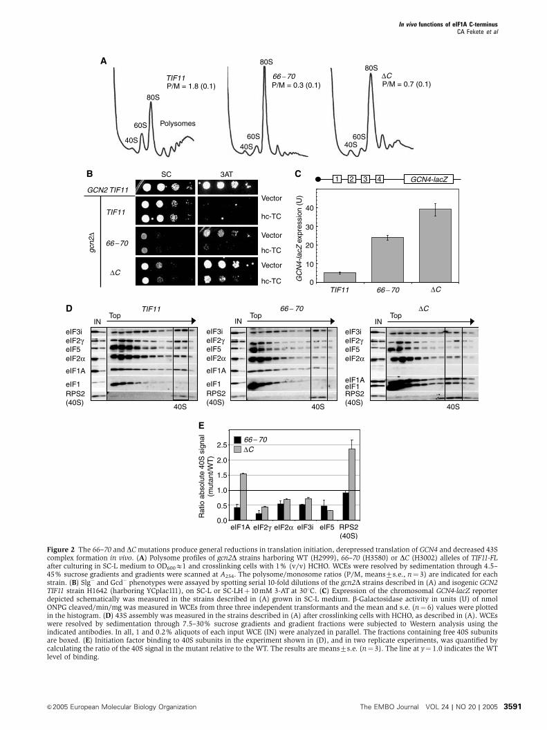
Figure 2 The 66–70 and DCmutations produce general reductions in translation initiation, derepressed translation of GCN4 and decreased 43Scomplex formation in vivo. (A) Polysome profiles of gcn2D strains harboring WT (H2999), 66–70 (H3580) or DC (H3002) alleles of TIF11-FLafter culturing in SC-L medium to OD600E1 and crosslinking cells with 1% (v/v) HCHO. WCEs were resolved by sedimentation through 4.5–45% sucrose gradients and gradients were scanned at A254. The polysome/monosome ratios (P/M, means7s.e., n¼ 3) are indicated for eachstrain. (B) Slg� and Gcd� phenotypes were assayed by spotting serial 10-fold dilutions of the gcn2D strains described in (A) and isogenic GCN2TIF11 strain H1642 (harboring YCplac111), on SC-L or SC-LHþ 10mM 3-AT at 301C. (C) Expression of the chromosomal GCN4-lacZ reporterdepicted schematically was measured in the strains described in (A) grown in SC-L medium. b-Galactosidase activity in units (U) of nmolONPG cleaved/min/mg was measured in WCEs from three three independent transformants and the mean and s.e. (n¼ 6) values were plottedin the histogram. (D) 43S assembly was measured in the strains described in (A) after crosslinking cells with HCHO, as described in (A). WCEswere resolved by sedimentation through 7.5–30% sucrose gradients and gradient fractions were subjected to Western analysis using theindicated antibodies. In all, 1 and 0.2% aliquots of each input WCE (IN) were analyzed in parallel. The fractions containing free 40S subunitsare boxed. (E) Initiation factor binding to 40S subunits in the experiment shown in (D), and in two replicate experiments, was quantified bycalculating the ratio of the 40S signal in the mutant relative to the WT. The results are means7s.e. (n¼ 3). The line at y¼ 1.0 indicates the WTlevel of binding.
In vivo functions of eIF1A C-terminusCA Fekete et al
&2005 European Molecular Biology Organization The EMBO Journal VOL 24 | NO 20 | 2005 3591
association of factors with either free 40S subunits or 43S/48S
PICs, which cannot be distinguished in this assay.)
The 40S-binding of the 66–70 mutant protein was reduced
to o50% of WT, whereas the DC protein bound at a greater
than WT level, probably reflecting its overexpression (Figures
2D and E). Interestingly, 66–70 also reduced the 40S-binding
of the TC components eIF2a and eIF2g, eIF3 and eIF5 to
p50% of WT. Similar results were obtained for the DCmutant (Figures 2D and E). (We chose not to quantify the
40S-binding of eIF1 in these experiments because it is over-
expressed in the mutants and it frequently does not show an
obvious peak in the 40S fractions of WT cells.) The total
amounts of eIF3i, eIF2g and eIF5 over the entire gradient
were somewhat lower in the 66–70 and DC mutants versus
WT (Figure 2D), even though these factors were present at
WT levels in the WCEs applied to the gradients (cf. ‘IN’
lanes). Hence, it appears that the unbound factors are more
susceptible to degradation during centrifugation in the mu-
tant extracts. This phenomenon was described previously for
mutations in the c/NIP1 subunit of eIF3 (Valasek et al, 2004).
Despite the reductions in total levels of eIF3i, eIF2g and eIF5
in the gradients, their distributions were altered such that the
proportions in the 40S fractions were reduced with corre-
sponding increases in fractions closer to the top of the
gradient (Supplementary Figures S1 and S2). Thus, it appears
that eIF1A is required for optimal 40S-binding of TC, eIF3 and
eIF5, and that both 66–70 and DC impair these assembly
reactions in vivo.
In accordance with the fact that hc-TC suppressed the
Gcd� phenotypes of 66–70 and DC, hc-TC restored 40S-
binding of eIF2a and eIF2g to WT or higher levels in both
mutants, and eIF3i binding in the 66–70 strain (cf. Figures 3A
and B; quantification in Figure 3C). By contrast, hc-TC did not
rescue 40S-binding of eIF5 in either mutant (Figure 3C). The
large amounts of eIF2 subunits in the extracts containing hc-
TC made it somewhat difficult to discern 40S-binding of eIF2
from trailing of eIF2 from the upper fractions into the 40S
fractions. To confirm our conclusions, we resedimented the
40S fractions from gradients similar to those shown in Figures
3A and B and quantified the factors that cosedimented a
second time with the 40S subunits. The results in Figure 3D
confirm that 66–70 and DC impair 40S-binding of eIF2, eIF3
and eIF5, and also that hc-TC restores 40S-binding of eIF2,
but not eIF5, by mass action. Hence, eIF1A promotes eIF5
recruitment independent of its effect on TC, and it enhances
eIF3 recruitment by mechanisms both TC-dependent
(evident in 66–70 cells) and TC-independent (evident in DCcells).
The fact that hc-TC did not restore WT 40S-binding of eIF5
in both mutants fits with our finding that the Slg� phenotypes
of these mutants were not suppressed by hc-TC (Figure 2B),
indicating a rate-limiting defect distinct from TC recruitment.
Consistent with this, overexpressing eIF5 partially suppressed
the Slg� phenotype of 66–70 (Figure 3E) and increased 40S-
binding of eIF5, but did not alter 40S-binding of other factors
or of the mutant eIF1A itself (Supplementary Figure S3).
Thus, reduced 40S-binding of eIF5 is partly responsible for
the strong initiation defect in 66–70 cells. Overexpressing
eIF5 did not reduce the Slg� phenotype of the DC mutant,
indicating an additional rate-limiting defect in this strain.
As shown below, DC impairs ribosomal scanning and AUG
recognition.
The 40S-binding of eIF1A was reduced by 66–70 to B50%
of the WT level (Figures 2D and E). From the crystal structure
of the IF1–30S complex (Carter et al, 2001), it can be
predicted that this mutation substitutes OB-fold residues
that interact with the A-site of the 40S subunit. Hence, the
defects conferred by 66–70 could result from diminished
ribosome binding of the mutant eIF1A. Supporting this
idea, introducing 66–70 on an hc plasmid partly suppressed
its Slg� and 3-ATR/Gcd� phenotypes (Figure 4A) and pro-
duced a greater than WT level of 40S-binding by the mutant
eIF1A protein (Figures 4B and C) Thus, the 40S-binding
defect of the 66–70 protein can be corrected by mass action.
The hc 66–70 allele also increased 40S-binding of eIF2, eIF3
and eIF5 compared to that seen with sc 66–70, albeit not to
WT levels (Figures 4B and C). The residual 43S assembly
defects and Slg� phenotype (Figure 4A) in cells with hc 66–70
suggest that eIF1A function in PIC assembly is impaired by
this mutation apart from its effects on 40S-binding of eIF1A
itself.
To determine whether 66–70 impairs direct contact be-
tween eIF1A and the 40S subunit, we analyzed its effect on
binding of fluorescently tagged eIF1A to purified 40S ribo-
somes. Tetramethylrhodamine (TAMRA)-labeled WT eIF1A
was prebound to 40S subunits and unlabeled mutant or WT
eIF1A was added to compete for binding of labeled eIF1A.
The decrease in fluorescence anisotropy as a function of the
concentration of mutant proteins allowed us to calculate the
Kd for each mutant (Maag and Lorsch, 2003). As shown in
Figures 4D and E, the 66–70mutant has a Kd B90-fold greater
than that of WT eIF1A, whereas the DC mutant binds to 40S
subunits with WT or higher affinity. These data indicate that
66–70, but not DC, reduces the intrinsic affinity of eIF1A for
the 40S ribosome.
We also asked whether the DC and 66–70mutations impair
the rate of PIC assembly in vitro. Preformed TC containing
[35S]-Met-tRNAiMet was incubated with 40S subunits, eIF1, a
model mRNA, and either mutant or WT eIF1A, and the
fraction of labeled [35S]-Met-tRNAiMet bound to 40S subunits
was measured with a native gel assay (Algire et al, 2002). As
shown in Figure 4F, DC greatly reduced the rate of TC binding
to 40S subunits in this assay. The DC mutant retains some
activity, as the rate of TC binding was X2-fold higher than in
the absence of eIF1A and reached the same end point given
by WT protein after 20min (data not shown). The 66–70
protein also showed a strong defect in TC binding; however,
this defect was suppressed when using a concentration of 66–
70 protein large enough to overcome its 40S-binding defect
(10mM) (Figure 4F). These findings support the idea that DCdecreases the ability of 40S-bound eIF1A to stimulate TC
recruitment, whereas 66–70 primarily impairs 40S-associa-
tion of the mutant protein.
TC overexpression suppresses the Slg� and 43S
assembly defects in the helical domain DEAR98–101AAAA
mutant
Unlike the 66–70 and DC mutations, the DEAR98�101AAAA
substitution in the helical domain (Figure 1C, #6; abbreviated
below as 98–101) produced a Slg� phenotype that is partially
suppressed by hc TC (Figure 5A). Consistent with this, hc-TC
decreased polysome runoff and accumulation of 80S mono-
somes in this mutant (Figure 5C; cf. last two profiles). In vivo
analysis of 43S complexes revealed 450% reductions in
In vivo functions of eIF1A C-terminusCA Fekete et al
The EMBO Journal VOL 24 | NO 20 | 2005 &2005 European Molecular Biology Organization3592
40S-bound eIF2 subunits and also reductions in 40S-binding
of eIF3 (byB40%), eIF5 (by 80%) and eIF1A itself (by 55%),
suggesting that 98–101 causes a broad defect in 43S assembly
(Figures 6A and B, cf. TIF11/vector and 98–101/vector, and
6C). (As described above, there were reduced levels of eIF2,
eIF3 and eIF1A in fractions at the top of the gradient, but
these reductions were not evident in the input WCEs, sug-
gesting that they arise from degradation of unbound factors in
A
C
B
Rat
io a
bsol
ute
40S
sig
nal
(mut
ant/
WT
)
0.0
0.5
2.0
2.5
1.0
1.5
+ vector
+ hc-TC
ΔC
eIF3ieIF2γ eIF2αeIF1A RPS2(40S)
eIF5
+ vector+ hc-TC
Rat
io a
bsol
ute
40S
sig
nal
(mut
ant/
WT
)
eIF3ieIF2γ eIF2αeIF1A RPS2(40S)
eIF50.0
0.5
2.0
1.0
1.5
66 −70
eIF3i
eIF2γ
eIF2α
eIF1A
eIF1
RPS2(40S)
TopTIF11/vector
Top Top66 −70 /vector ΔC /vector
eIF5
IN IN IN
40S 40S 40S
Δ
TIF11
66−70
Vector
hc TIF5
Vector
hc TIF5
SC, 2 days SC, 3 days
Vector
hc TIF5ΔC
ED
eIF3b
eIF2γ
eIF2α
eIF1A
RPS22(40S)
eIF5
Δ
66 −70 ΔCTIF11
+ vector + hc-TC + vector + hc-TC+ vector
TopTIF11/hc-TC
Top Top66 −70 /hc-TC ΔC/hc-TC
eIF3ieIF2γ
eIF2α
eIF1A
eIF1
RPS2(40S)
eIF5
IN IN IN
40S 40S 40S
Δ
**
**
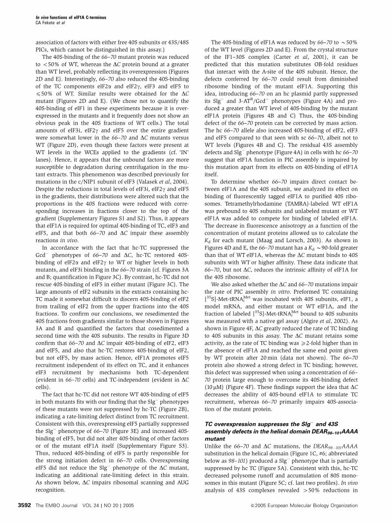
Figure 3 Overexpression of TC restores WT binding of TC, but not eIF5, in the 66–70 and DC mutants. (A, B) 43S assembly was measuredin HCHO crosslinked cells of the gcn2D strains described in Figure 2A transformed with empty vector YEp24 (A) or p1780-IMT (hc-TC) (B),exactly as described in Figure 2. ‘D’ indicates the DC product; ‘*’ indicate that darker exposures of the indicated panels were presented. (C) Theresults in (A) and (B) were quantified as in Figure 2E, showing the means7s.e. from three experiments. (D) 40S fractions from gradientssimilar to (A) and (B) were subjected to resedimentation through a second sucrose gradient. Western analysis of gradient fractions containing40S peaks in each strain is shown. (E) Overexpression of eIF5 (TIF5) partially suppresses the Slg� phenotype of the 66–70mutant. Serial 10-folddilutions of the gcn2D strains from Figure 2A transformed with empty vector YEp24 or hc TIF5 plasmid YEpTIF5-U were spotted on SC-U andgrown at 301C as indicated.
In vivo functions of eIF1A C-terminusCA Fekete et al
&2005 European Molecular Biology Organization The EMBO Journal VOL 24 | NO 20 | 2005 3593
the mutant extract during centrifugation.) Consistent with the
partial suppression of the growth defect in 98–101 cells by hc-
TC, the 40S-binding defects of eIF2, eIF3 and eIF5, and the
mutant eIF1A were diminished by hc-TC in this strain
(Figures 6A and B, cf. 98–101/vector and 98–101/hc-TC).
The fact that eIF3 and eIF5 recruitment was not fully rescued
A
TIF11
66 −70
sc
hc
SC 3AT
sc
hc
GCN2 TIF11
gcn2
Δ
E
F
C
Nor
mal
ized
ani
sotr
opy
[eIF1A] (nM)0 150
0
0.2
0.4
0.6
0.8
1.0
WT
ΔC66 −7098 −101
300 450 600 750 900
D
B
eIF3b
eIF2γ
eIF2α
eIF1A
eIF1
RPS2(40S)
eIF5
eIF3b
eIF2γ
eIF2α
eIF1A
eIF1
RPS2(40S)
eIF5
eIF3beIF2γ
eIF2α
eIF1A
eIF1
RPS2(40S)
eIF5
TopIN
40S
TIF11/sc
TopIN
40S
66 −70 /sc
TopIN
40S
66−70 /hc
Initiation factoreIF3beIF2γ eIF2αeIF1A RPS2
(40S)eIF5
Rat
io a
bsol
ute
40S
sig
nal
(mut
ant/
WT
)
0.0
0.2
0.4
0.6
0.8
1.0
1.2
1.4
1.6
1.8sc
hc
Frac
tion
TC
bou
nd
Time (s)0 10 20 30 40 50 60
0
0.05
0.1
0.15
0.2
0.25
0.3
0.35
98 −101− eIF1A
66 −70(10 μM)
WTΔC66 −70(1 μM)
TIF11 allele
98 −101
WTΔC
66 −70
27.0 ± 8.0
20.3 ± 0.15.6 ± 0.1
1820 ± 140
Kd (nM)
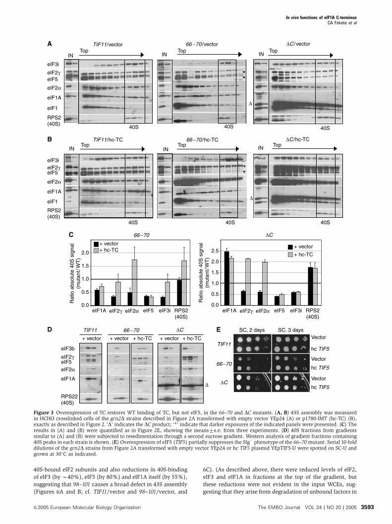
Figure 4 Overexpressing the 66–70 mutant diminishes its Slg� and Gcd� phenotypes, and the mutant is defective for 40S-binding in vitro.(A) Serial dilutions of gcn2D strains carrying the WT (H2999) or 66–70-tif11-FL alleles on sc or hc plasmids (strains H3580 and H3581,respectively), and H1642 transformed with YCplac111, were spotted on SC-L and SC-LHþ 10mM 3-AT and grown at 301C. (B, C)Overexpressing the 66–70 mutant partially suppresses its 43S assembly defect. 43S assembly was measured in HCHO crosslinked cells ofthe strains described in (A) and quantified as described in Figure 2. (D) The 66–70 mutation reduces the affinity of eIF1A for 40S subunitsin vitro. Dissociation of preformed 40S/eIF1A-TAMRA complex in the presence of increasing concentrations of unlabeled WTor mutant eIF1Aand 1mM eIF1 was measured by the decrease in fluorescence anisotropy. (E) Kd values (nM) were calculated from the curves in (D) andreported as means7s.e. (F) Rate of TC binding to 40S subunits is reduced by DC in vitro. Binding of TC to 40S subunits as a function of time (inreactions also containing eIF1, model mRNA and 1 mMWTor mutant eIF1A) was measured as the fraction of [35S]-Met-tRNAi
Met associated withthe 40S in a native gel assay. The apparent TC-binding defect in the 66–70 mutant is suppressed by increasing its concentration to 10mM.
In vivo functions of eIF1A C-terminusCA Fekete et al
The EMBO Journal VOL 24 | NO 20 | 2005 &2005 European Molecular Biology Organization3594
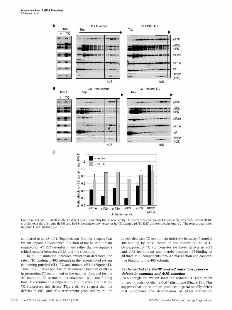
by hc-TC in the 98–101 mutant indicates that this mutation
also impairs 40S-binding of these factors by a TC-independent
mechanism.
Although 98–101 reduces 40S-binding of eIF1A in vivo by
B60% (Figure 6C), analysis of recombinant 98–101 protein
in vitro revealed no reduction in its intrinsic affinity for the
40S subunit (Figures 4D and E). This suggests that the
impaired 40S-binding of the 98–101 protein in vivo results
from defective interactions with other eIFs that promote PIC
assembly, rather than loss of a direct contact between eIF1A
and the 40S subunit. This interpretation is supported by the
fact that hc-TC increased binding of the 98–101 protein in
parallel with that of eIF3 and eIF5 (Figures 6B and C).
Overexpression of the 98–101 protein from an hc plasmid
fully suppressed its 40S-binding defect, giving 240770% of
WT binding (data not shown), but did not diminish the
growth defect (Figure 5B) or 40S-binding defects of eIF2,
eIF3 or eIF5 produced by this mutation (increases of o20%
C
SC SM
B
TIF11
98 −101
sc
hc
sc
SCA SC
TIF11
98 −101
Vector
hc-TC
Vector
hc-TC
D
40S60S
80S
Polysomes
40S60S
80S
40S60S
80S98 −101/vector 98 −101/hc-TC
40S60S
80S
TIF11/vector TIF11/hc-TC
P/M = 1.4 (0.1) P/M = 1.5 (0.1) P/M = 0.5 (0.2) P/M = 1.0 (0.1)
E
GCN2
98 −101
gcn2Δ
TIF11
GC
N2
F
eIF2α-total
eIF2α-P
U I U I
TIF11
98−10
1
Normalizedratio I/U:
1.0 1.4
GCN4-lacZ1
GCN4-lacZ1
GCN4-lacZ1 2 3(i)
TIF11
(i) (ii)
(ii)
GCN4-lacZ expression
100 (6) 16 (1.8)98 −101
U I
10 (0.9) 165 (19)11 (0.7) 56 (1.6) 112 (9) 5.2 (0.2)
4
(iii)
(iii)
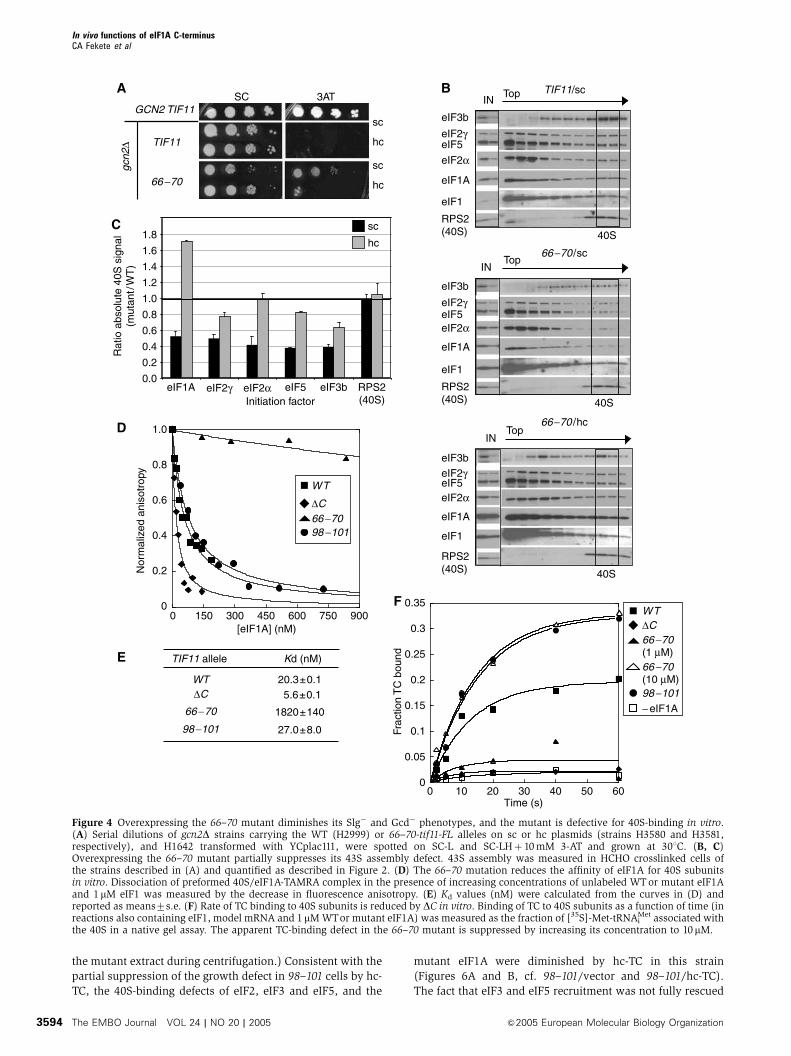
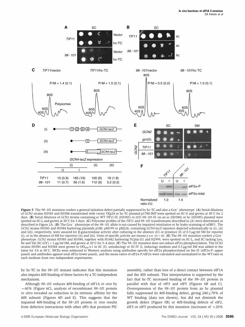
Figure 5 The 98–101mutation confers a general initiation defect partially suppressed by hc-TC and also a Gcn� phenotype. (A) Serial dilutionsof GCN2 strains H3583 and H3584 transformed with vector YEp24 or hc-TC plasmid p1780-IMTwere spotted on SC-U and grown at 301C for 2days. (B) Serial dilutions of GCN2 strains containing sc WT TIF11-FL (H3583) or tif11-98–101-FL on an sc (H3584) or hc (H3585) plasmid werespotted on SC-L and grown at 301C for 3 days. (C) Polysome profiles of the TIF11 and 98–101 transformants described in (A) were determined asdescribed in Figure 2A. (D) The Gcn� phenotype of the 98–101 allele is not caused by impaired reinitiation or by leaky-scanning of uORF1. TheGCN2 strains H3583 and H3584 harboring plasmids p180, pM199 or pM226, containing GCN4-lacZ reporters depicted schematically in (i), (ii)and (iii), respectively, were assayed for b-galactosidase activity after culturing in the absence (U) or presence (I) of 0.5mg/ml SM for reporter(i), or in the absence of SM for reporters (ii) and (iii). Units of specific activity are means7s.e. (n¼ 6). (E) The 98–101mutation confers a Gcn�
phenotype. GCN2 strains H3583 and H3584, together with H1642 harboring YCplac111 and H2999, were spotted on SC-L, and SC lacking Leu,Ile and Val (SC-LIV)þ 1mg/ml SM, and grown at 301C for 3–4 days. (F) The 98–101mutation does not reduce eIF2a phosphorylation. The GCN2strains H3583 and H3584 were grown to OD600E1 in SC (U, uninducing) or SC-IV (I, inducing) medium and 0.5mg/ml SM was added to thelatter for 3 h at 301C. WCEs were subjected to Western analysis using antibodies specific for eIF2a phosphorylated on Ser-51 (eIF2a-P; upperpanel) and antibodies against total eIF2a lower panel), and the mean ratios of eIF2a-P/eIF2a were calculated and normalized to the WTratio ineach medium from two independent experiments.
In vivo functions of eIF1A C-terminusCA Fekete et al
&2005 European Molecular Biology Organization The EMBO Journal VOL 24 | NO 20 | 2005 3595
compared to sc 98–101). Together, our findings suggest that
98–101 impairs a biochemical function of the helical domain
required for WT PIC assembly in vivo rather than disrupting a
critical contact between eIF1A and the ribosome.
The 98–101 mutation increases, rather than decreases, the
rate of TC binding to 40S subunits in the reconstituted system
containing purified eIF1, TC and mutant eIF1A (Figure 4F).
Thus, 98–101 does not disrupt an inherent function of eIF1A
in promoting TC recruitment in the manner observed for the
DC mutation. To reconcile this conclusion with our finding
that TC recruitment is impaired in 98–101 cells, and that hc-
TC suppresses this defect (Figure 6), we suggest that the
deficits in eIF5 and eIF3 recruitment produced by 98–101
in vivo decrease TC recruitment indirectly because of coupled
40S-binding by these factors in the context of the MFC.
Overexpressing TC compensates for these defects in eIF5
and eIF3 recruitment and thereby restores 40S-binding of
all three MFC components through mass action and coopera-
tive binding to the 40S subunit.
Evidence that the 98–101 and DC mutations produce
defects in scanning and AUG selection
Even though the 98–101 mutation reduces TC recruitment
in vivo, it does not elicit a Gcd� phenotype (Figure 1B). This
suggests that the mutation produces a postassembly defect
that suppresses the derepression of GCN4 translation
C
eIF3i
eIF2γ
eIF2α
eIF1A
eIF1
RPS2(40S)
98 −101/ vectorTop TopInput
v TC
B 98 −101/hc-TC
eIF5
40S40S
eIF3i
eIF2γ
eIF2α
eIF1A
eIF1
RPS2(40S)
TIF11/ vectorTop TopInput
v TC
A TIF11/hc-TC
eIF5
40S 40S
eIF3ieIF2γ eIF2αeIF1A RPS2(40S)
eIF5
Initiation factor
Rat
io a
bsol
ute
40S
sig
nal (
mut
ant/
WT
)
0.0
0.2
0.4
0.6
0.8
1.0
1.2+ vector
+ hc-TC
Figure 6 The 98–101 allele confers a defect in 43S assembly that is rescued by TC overexpression. (A–C) 43S assembly was measured in HCHOcrosslinked cells of strains H3583 and H3584 bearing empty vector or hc-TC plasmid p1780-IMT, as described in Figure 2. The results quantifiedin panel C are means7s.e., n¼ 3.
In vivo functions of eIF1A C-terminusCA Fekete et al
The EMBO Journal VOL 24 | NO 20 | 2005 &2005 European Molecular Biology Organization3596
expected from impaired TC recruitment. A similar phenom-
enon has been described for mutations in eIF5 that produce
multiple defects in the initiation pathway by disrupting the
MFC (Nielsen et al, 2004; Singh et al, 2005). Supporting this
interpretation, 98–101 produces a Gcn� phenotype in a
GCN2þ strain, decreasing GCN4-lacZ expression to B30%
of WT in response to Ile/Val starvation (Figure 5D, construct
i) and conferring sensitivity to an inhibitor of isoleucine/
valine biosynthesis (sulfometuron methyl (SM)) (Figure 5E).
The Gcn� phenotype of the 98–101 mutant does not arise
from diminished eIF2a phosphorylation in the starved cells
(Figure 5F).
One explanation for a Gcn� phenotype in cells containing
ample phosphorylated eIF2a is that ribosomes leaky-scan
through the uORF1 AUG and initiate first at uORFs 2, 3 or 4
instead. Following translation of these uORFs, the ribosomes
dissociate from the mRNA and fail to reach the GCN4 start
codon. To address this possibility, we assayed a GCN4-lacZ
reporter containing only an elongated version of uORF1 that
overlaps the GCN4 start codon (Figure 5D, construct iii).
Expression of this construct is normally very low because
nearly all 40S ribosomes translate uORF1 but fail to reinitiate
at GCN4, as elongating uORF1 destroys its ability to retain 40S
subunits that resume scanning after termination (Grant et al,
1994). Leaky-scanning of the elongated uORF1 in response to
the 98–101 mutation would be expected to increase expres-
sion of this reporter; however, we observed decreased repor-
ter expression instead (B30% of WT; Figure 5D, iii). Thus,
98–101 leads to lower, rather than higher, leaky-scanning
through uORF1.
Another possible explanation for the Gcn� phenotype of
98–101 is that it impairs the ability of 40S subunits to resume
scanning and reinitiate at GCN4 following translation of
uORF1. This mechanism can also be eliminated because
98–101 does not decrease expression of the GCN4-lacZ repor-
ter containing only WTuORF1 (Figure 5D, construct ii). This
last finding, that eliminating uORFs 2–4 suppresses the
deleterious effect of 98–101 on GCN4 translation, strongly
suggests that the Gcn� phenotype results from the inability of
rescanning 40S subunits to bypass uORFs 2–4 even when TC
recruitment is diminished by eIF2a phosphorylation. A Gcn�
phenotype with the same characteristics was observed pre-
viously when the distance between uORFs 1 and 4 was
expanded to provide increased time for rebinding of TC
before the rescanning 40S subunits reach uORF4 (Abastado
et al, 1991). Hence, 98–101 may mimic the effect of increased
uORF1–4 separation by reducing the rate of 40S scanning
between uORFs 1 and 4. A reduced efficiency of scanning
could also explain the decrease in leaky-scanning past elon-
gated uORF1 produced by 98–101 (Figure 5D, iii), as a
structural impediment to scanning was shown previously to
reduce leaky-scanning through an AUG codon (Kozak, 1990).
We sought next to obtain biochemical evidence that the
98–101 mutation decreases the efficiency of ribosomal scan-
ning. Mammalian eIF1A participates with eIF1 in allowing
the 43S complex to scan from the 50-end of mRNA and form a
stable 48S complex at the start codon in reactions containing
purified ribosomes, eIFs and b-globin mRNA. Inhibition of
primer extension by reverse transcriptase is used to map the
leading edge of the ribosome in the 48S complexes (toe-
printing) (Pestova et al, 1998). We first demonstrated that
yeast eIF1A can substitute for mammalian eIF1A in such
reactions, and then examined the effects of eIF1A mutations
on this activity.
With all human factors except eIF1A present, a significant
proportion of 48S complexes were formed with the AUG
codon in the P-site, yielding toe-prints 16 nucleotides (nt)
downstream of the start codon (complex II) (Figure 7A). A
sizable fraction of aberrant complexes also occurred with toe-
prints 16 nt from a near-cognate GUG codon. Smaller propor-
tions of aberrant complexes near the 50-terminus (complex I)
or with toe-prints 7 nt upstream of complex II (designated
complex III) also formed in the absence of eIF1A.
Supplementation with WT yeast or human eIF1A eliminated
or strongly suppressed formation of complex I and the GUG
complex, while yeast eIF1A also suppressed complex III
(Figure 7A). Thus, WT yeast eIF1A functions efficiently in
this assay to promote nearly exclusive formation of 48S PICs
at the AUG codon.
Interestingly, the 98–101 and DC proteins allowed forma-
tion of complex I and the GUG complex at levels B50% of
that observed in the absence of eIF1A, whereas a different
yeast mutant (GRRGK12�16AAAAA) showed WT inhibition of
these aberrant complexes. The latter finding is significant
because the GRRGK12�16AAAAA mutant showed a lower
affinity for 40S ribosomes in the binding assays of Figures
4D and E than did the 98–101 mutant (data not shown).
Hence, it is unlikely that the mutant phenotypes of 98–101 (or
DC) in the toe-print assays arise from reduced 40S-binding of
mutant eIF1A proteins. We interpret these results to indicate
that 98–101, and DC decrease the propensity of the 43S
complex to scan the mRNA, increasing the stability of aber-
rant 48S complexes upstream of the AUG start codon (Battiste
et al, 2000).
Finally, we asked whether the 98–101 or DCmutations lead
to increased utilization of a near-cognate start codon in vivo.
This defect can be detected by increased initiation at a UUG
triplet in the 50-end of the HIS4 ORF, allowing expression of
the his4-303 allele lacking the normal AUG start codon. The
resulting histidine auxotrophy indicates a Sui� (suppressor
of initiation codon mutation) phenotype (Donahue, 2000).
The DC mutation strongly suppressed the His� phenotype of
his4-303, conferring a robust Sui� phenotype (Figure 7C).
Furthermore, we found that DC is lethal in the presence of a
dominant Sui� allele of TIF5 encoding a hyperactive form of
eIF5 that likely produces a Sui� phenotype by increasing GTP
hydrolysis at UUG codons (Huang et al, 1997). The synthetic
lethality of the SUI5 tif11-DC combination may result from an
intolerable level of initiation at near-cognate start codons.
Although the 98–101 mutation did not suppress the His�
phenotype associated with his4-303 (data not shown), it
produced a 2.4-fold increase in translation of a his4-lacZ
reporter containing UUG versus AUG as the start codon
(Figure 7B). Hence, we believe that 98–101 also confers a
Sui� phenotype, but weaker than that of DC.
Discussion
Our finding that the DC and 66–70 mutations derepress GCN4
translation in a manner suppressed by TC overexpression
provides genetic evidence that eIF1A promotes TC recruit-
ment in vivo. Consistent with this, we showed that both
mutations impair binding of TC, eIF3 and eIF5 to 40S sub-
units in vivo, and that the defects in TC recruitment can be
In vivo functions of eIF1A C-terminusCA Fekete et al
&2005 European Molecular Biology Organization The EMBO Journal VOL 24 | NO 20 | 2005 3597

overcome through mass action by overexpressing the TC.
Both mutations also conferred reduced rates of TC binding to
40S subunits in a reconstituted in vitro assay, although the
defect produced by 66–70 was suppressed by using elevated
levels of the mutant protein to compensate for its defect in
40S-binding. In agreement with this last result, the Slg� and
Gcd� phenotypes and defects in PIC assembly produced by
66–70 in vivo were diminished by overexpressing the mutant
protein. Thus, the TC recruitment defect and attendant dere-
pression of GCN4 conferred by 66–70 arise largely from its
defect in 40S-binding. This conclusion fits with the prediction
from the 30S–IF1 complex (Carter et al, 2001) that residues
66–70 in the OB-fold of eIF1A are close to the A-site of the 40S
subunit. The DC mutation, by contrast, impairs 43S assembly
even though the mutant eIF1A is bound to 40S subunits at
WT or greater levels. Hence, the C-terminal extension or
adjacent 310 helix eliminated by DC appears to have an
important biochemical function in TC recruitment.
Whereas overexpressing TC reduced the Gcd� phenotypes
and TC recruitment defects in the 66–70 and DC mutants, it
did not suppress their Slg� phenotypes nor restore 40S-
binding of eIF5 or eIF3 (in the DC mutant) to WT levels. By
contrast, overexpressing eIF5 rescued 40S-binding of eIF5
and diminished the growth defect in 66–70 cells. These
results indicate that eIF1A promotes 40S-binding of eIF5
and eIF3 independent of its role in TC recruitment. Hence,
we conclude that eIF1A independently promotes the 40S-
binding of multiple constituents of the yeast MFC. As no
direct physical interactions between eIF1A and MFC compo-
nents have been described, the molecular basis for the
stimulatory effects of eIF1A on MFC recruitment remains to
be defined.
A
GATCHuman
eIF
1A
− eIF
1A
ΔC 98−1
01
GRRGK 12−1
6AAAAA
WTRNA o
nly
Full-length cDNA -
Complex I -
+16 nt from GUG -
Complex II -(+16 nt from AUG)
AUG
Yeast eIF1A
Complex III -
GUG
}
}
0
0.01
0.02
0.03
0.04
0.05
0.06
WT 98−101
UUG/AUGB C SC
SC - H
TIF11
ΔC
Sui−TIF11
ΔC
Figure 7 C-terminal mutations in eIF1A cause scanning/AUG recognition defects in vitro and in vivo. (A) Toe-printing analysis of 48Scomplexes on b-globin mRNA in reaction mixtures containing 40S subunits and mammalian translation initiation factors in the absence orpresence of eIF1A, as indicated. See text for explanation of different complexes labeled on the left. (B) The 98–101 mutation increasesutilization of a UUG start codon in vivo. Strains H3583 and H3584 carrying plasmids YCp50-HIS4-AUG-lacZ or YCp50-HIS4-UUG-lacZ weregrown in SC-U medium and assayed for b-galactosidase activity. The specific activities7s.e. (n¼ 6) determined for the YCp50-HIS4-UUG-lacZversus YCp50-HIS4-AUG-lacZ transformants were measured in three independent experiments and used to calculate the mean ratios7s.e.(n¼ 3) shown in the histogram. (C) The DCmutation suppresses the His� phenotype conferred by the his4-303 allele. Serial 10-fold dilutions ofstrains H3583 and H3586 were spotted on SC lacking leucine and histidine and grown at 301C for 2 days (SC, upper panel) or 14 days (SC-H,lower panel).
In vivo functions of eIF1A C-terminusCA Fekete et al
The EMBO Journal VOL 24 | NO 20 | 2005 &2005 European Molecular Biology Organization3598
The 98–101 substitution in the a-helical domain was the
only mutation that conferred a growth defect suppressible by
hc-TC, and the mutation also impaired 40S-binding binding of
TC, eIF3 and eIF5 in vivo in a manner largely corrected by
overexpressing TC. The 98–101 mutation impairs 40S-binding
of eIF1A itself in vivo, and while this defect was corrected by
overexpressing the mutant protein, the Slg� phenotype and
reductions in PIC assembly were not simultaneously sup-
pressed. Hence, we conclude that the helical domain of eIF1A
has an important function in promoting recruitment of the
MFC to the 40S subunit.
The recombinant 98–101 protein exhibits WT affinity for
40S ribosomes in vitro, indicating that reduced 40S-binding
by this mutant in vivo does not reflect loss of a contact
between eIF1A and the ribosome. Similarly, 98–101 did not
reduce the rate of TC binding to 40S subunits in the recon-
stituted assay, suggesting that the intrinsic function of eIF1A
in TC recruitment is not impaired by this mutation. To
reconcile these in vitro and in vivo findings, we suggest that
98–101 indirectly impairs TC recruitment in vivo by reducing
40S-binding of eIF3 and eIF5, thereby disrupting the coupled
association of factors in the MFC with the ribosome. We
further suggest that 40S-binding of eIF1A is coupled to that of
the MFC, so that the 98–101 mutation indirectly impairs 40S-
binding of eIF1A by diminishing recruitment of MFC compo-
nents. Indeed, we recently obtained evidence that depleting
eIF5 from cells impairs eIF1A recruitment in vivo (A
Jivotovskaya, K Nielsen and AGH, unpublished observa-
tions). We suggest that overexpressing TC most likely restores
40S-binding of the MFC in the 98–101 mutant by driving the
assembly of a TC–eIF5 binary complex on the ribosome by
mass action. This in turn would stimulate recruitment of
eIF3, as interaction with eIF2 increases the affinity of eIF5 for
eIF3 (Singh et al, 2004).
Apart from its role in PIC assembly, we found that the
C-terminal domain of eIF1A also functions in scanning and
AUG recognition. Toe-print analysis of 48S assembly in a
reconstituted mammalian system revealed that both the
98–101 and DC mutations increase formation of aberrant
complex I and the 48S complexes at an upstream GUG on
b-globin mRNA. As suggested previously (Battiste et al,
2000), formation of these aberrant complexes could result
from a decreased rate of ribosomal scanning. Consistent with
these findings, the 98–101 and DC mutations appeared to
increase utilization of non-AUG triplets as start codons on
HIS4 mRNA in vivo (Sui� phenotype).
A slower rate of scanning can also explain why the defect
in TC recruitment conferred by 98–101 in vivo does not
produce a Gcd� phenotype. In fact, this mutant displays a
Gcn� phenotype in GCN2 cells that can be attributed to the
failure of 40S ribosomes rescanning downstream from uORF1
to bypass uORFs 2–4 even though TC levels are reduced by
eIF2a phosphorylation. This type of Gcn� defect was ob-
served previously when the distance between uORFs 1 and 4
was expanded, increasing the time required for scanning 40S
subunits to reach uORF4 after terminating at uORF1. This
alteration compensates for the delay in TC binding elicited by
eIF2a phosphorylation and ensures that nearly all 40S sub-
units rebind TC before reaching uORF4 and, hence, fail to
reinitiate at GCN4 (Abastado et al, 1991). Thus, we suggest
that the 98–101 mutation mimics the effect of lengthening
the uORF1–4 interval and produces a Gcn� phenotype by
decreasing the rate of scanning.
It might be expected that the DC mutation would also
confer a Gcn� phenotype, given that it produced a scanning
defect in the toe-print assay comparable to that of 98–101.
In fact, we found that DC does confer sensitivity to SM and
a modest (20%) reduction in GCN4-lacZ expression in a
GCN2þ strain under starvation conditions, without lowering
eIF2a phosphorylation (data not shown). Perhaps the defect
in TC binding produced by DC outweighs the scanning defect
and suppresses the expected Gcn� phenotype. In 98–101
cells, by contrast, the scanning defect would outweigh
the deficit in TC binding and suppress the expected Gcd�
phenotype.
Support for this interpretation came recently from the
analysis of mutations in the eIF5-CTD that destabilize the
MFC and produce Gcd� phenotypes at 301C but Gcn� phe-
notypes at a higher temperature (361C) where the mutations
have a stronger effect on initiation. It was proposed that
Table I Plasmids used in this study
Plasmid Description Source or reference
YCplac111 sc LEU2 cloning vector Gietz and Sugino (1988)YEplac181 hc LEU2 cloning vector Gietz and Sugino (1988)p3392 sc URA3 TIF11 in YCplac33 Olsen et al (2003)p3499 sc LEU2 TIF11-FL in YCplac111 Olsen et al (2003)p3604 hc LEU2 tif11-D108–153-FL in YEplac181 Olsen et al (2003)p4399 sc LEU2 tif11-RKKVW66–70AAAVA-FL in YCplac111 This studyp4443 hc LEU2 tif11-RKKVW66–70AAAVA-FL in YEplac181 This studyp4398 sc LEU2 tif11-DEAR98–101AAAA-FL in YCplac111 This studyp4444 hc LEU2 tif11-DEAR98–101AAAA-FL in YEplac181 This studyp1780-IMT hc URA3 SUI2, GCD11, SUI3, IMT4 in YEp24 Asano et al (1999)YEp24 hc URA3 cloning vector Parent et al (1985)YEpTIF5-U hc URA3 TIF5-FL in YEplac195 Valasek et al (2004)YEplac195 hc URA3 Gietz and Sugino (1988)p4385 hc TRP1 SUI2, GCD11, SUI3, IMT4 in YEp24, ura3HTRP1:kanR This studyYEplac112 hc TRP1 Gietz and Sugino (1988)p180 sc URA3 GCN4-lacZ with WT leader Hinnebusch (1985)PM199 sc URA3 GCN4-lacZ with uORF1 only Grant et al (1994)PM226 sc URA3 GCN4-lacZ with uORF1 extending into GCN4 Grant et al (1994)p367 sc URA3 AUG-HIS4-lacZ Donahue and Cigan (1988)p391 sc URA3 AUU-HIS4-lacZ Donahue and Cigan (1988)
In vivo functions of eIF1A C-terminusCA Fekete et al
&2005 European Molecular Biology Organization The EMBO Journal VOL 24 | NO 20 | 2005 3599
impaired TC recruitment at 301C produces the Gcd� pheno-
type but more severe disruption of the MFC at 361C elicits a
scanning defect (leaky-scanning of uORF1 in this case) that
outweighs the effect of reduced TC recruitment on GCN4
translation (Singh et al, 2005). We suggest that the DC and
98–101mutations can be likened to these eIF5-CTD mutations
at 30 and 361C, respectively, in terms of their relative effects
on TC recruitment versus scanning. Singh et al (2005) also
showed that certain eIF5-CTD mutants display a Gcd� pheno-
type only when eIF1 is overexpressed, apparently because
eIF1 overexpression exacerbates a moderate defect in TC
recruitment to the point where it outweighs the scanning
defects produced by these eIF5 mutations, thus derepressing
GCN4. Consistent with this, we found that eIF1 overexpres-
sion reveals a Gcd� phenotype in the 98–101 mutant and
exacerbates the Gcd� phenotype of DC (data not shown).
Our results show that the a-helical domain and random-
coil CTD of eIF1A, regions not present in bacterial IF1,
stimulate binding of TC and other MFC components to the
40S subunit and promote efficient scanning and AUG selec-
tion. Residues 66–70 in the OB-fold, by contrast, likely
provide a critical 40S–eIF1A interaction. It makes sense that
functions of eIF1A involved in MFC recruitment and scanning
depend on domains not present in IF1, as eIF2, eIF3 and eIF5
are absent in bacteria, and there are significant differences
in Met-tRNAiMet recruitment and start codon recognition
between eukaryotes and prokaryotes.
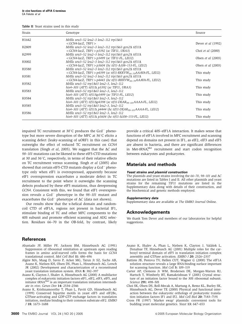
Materials and methods
Yeast strains and plasmid constructionThe plasmids and yeast strains involving the 66–70, 98–101 and DCmutations are listed in Tables I and II. All other plasmids and yeaststrains for the remaining TIF11 mutations are listed in theSupplementary data along with details of their construction, andthe biochemical and genetic methods employed.
Supplementary dataSupplementary data are available at The EMBO Journal Online.
Acknowledgements
We thank Tom Dever and members of our laboratories for helpfulsuggestions.
References
Abastado JP, Miller PF, Jackson BM, Hinnebusch AG (1991)Suppression of ribosomal reinitiation at upstream open readingframes in amino acid-starved cells forms the basis for GCN4translational control. Mol Cell Biol 11: 486–496
Algire MA, Maag D, Savio P, Acker MG, Tarun Jr SZ, Sachs AB,Asano K, Nielsen KH, Olsen DS, Phan L, Hinnebusch AG, LorschJR (2002) Development and characterization of a reconstitutedyeast translation initiation system. RNA 8: 382–397
Asano K, Clayton J, Shalev A, Hinnebusch AG (2000) A multifactorcomplex of eukaryotic initiation factors eIF1, eIF2, eIF3, eIF5, andinitiator tRNAMet is an important translation initiation intermedi-ate in vivo. Genes Dev 14: 2534–2546
Asano K, Krishnamoorthy T, Phan L, Pavitt GD, Hinnebusch AG(1999) Conserved bipartite motifs in yeast eIF5 and eIF2Be,GTPase-activating and GDP-GTP exchange factors in translationinitiation, mediate binding to their common substrate eIF2. EMBOJ 18: 1673–1688
Asano K, Shalev A, Phan L, Nielsen K, Clayton J, Valasek L,Donahue TF, Hinnebusch AG (2001) Multiple roles for the car-boxyl terminal domain of eIF5 in translation initiation complexassembly and GTPase activation. EMBO J 20: 2326–2337
Battiste JB, Pestova TV, Hellen CUT, Wagner G (2000) The eIF1Asolution structure reveals a large RNA-binding surface importantfor scanning function. Mol Cell 5: 109–119
Carter AP, Clemons Jr WM, Brodersen DE, Morgan-Warren RJ,Hartsch T, Wimberly BT, Ramakrishnan V (2001) Crystal struc-ture of an initiation factor bound to the 30S ribosomal subunit.Science 291: 498–501
Choi SK, Olsen DS, Roll-Mecak A, Martung A, Remo KL, Burley SK,Hinnebusch AG, Dever TE (2000) Physical and functional inter-action between the eukaryotic orthologs of prokaryotic transla-tion initiation factors IF1 and IF2. Mol Cell Biol 20: 7183–7191
Cross FR (1997) ‘Marker swap’ plasmids: convenient tools forbudding yeast molecular genetics. Yeast 13: 647–653
Table II Yeast strains used in this study
Strain Genotype Source
H1642 MATa ura3–52 leu2–3 leu2–112 trp1D63oGCN4-lacZ, TRP14 Dever et al (1992)
H2809 MATa ura3–52 leu2–3 leu2–112 trp1D63 gcn2D tif11DoGCN4-lacZ, TRP14p3392 (sc TIF11, URA3) Choi et al (2000)
H2999 MATa ura3–52 leu2–3 leu2–112 trp1D63 gcn2D tif11DoGCN4-lacZ, TRP14p3499 (sc TIF11-FL, LEU2) Olsen et al (2003)
H3002 MATa ura3–52 leu2–3 leu2–112 trp1D63 gcn2D tif11DoGCN4-lacZ, TRP14p3604 (hc tif11-D108–153-FL, LEU2) Olsen et al (2003)
H3580 MATa ura3–52 leu2–3 leu2–112 trp1D63 gcn2D tif11DoGCN4-lacZ, TRP14p4399 (sc tif11-RKKVW66–70AAAVA-FL, LEU2) This study
H3581 MATa ura3–52 leu2–3 leu2–112 trp1D63 gcn2D tif11DoGCN4-lacZ, TRP14p4443 (hc tif11-RKKVW66–70AAAVA-FL, LEU2) This study
H3582 MATa ura3–52 trp1D63 leu2–3, leu2–112his4–303 (ATT) tif11D p3392 (sc TIF11, URA3) This study
H3583 MATa ura3–52 trp1D63 leu2–3, leu2–112his4–303 (ATT) tif11Dp3499 (sc TIF11-FL, LEU2) This study
H3584 MATa ura3–52 trp1D63 leu2–3, leu2–112his4–303 (ATT) tif11Dp4398 (sc tif11-DEAR98–101AAAA-FL, LEU2) This study
H3585 MATa ura3–52 trp1D63 leu2–3, leu2–112his4–303 (ATT) tif11D p4444 (hc tif11-DEAR98–101AAAA-FL, LEU2) This study
H3586 MATa ura3–52 trp1D63 leu2–3, leu2–112his4–303 (ATT) tif11D p3604 (hc tif11-D108–153-FL, LEU2) This study
In vivo functions of eIF1A C-terminusCA Fekete et al
The EMBO Journal VOL 24 | NO 20 | 2005 &2005 European Molecular Biology Organization3600
DeLano WL (2002) The PyMOL Molecular Graphics System. SanCarlos, CA: DeLano Scientific. http://www.pymol.org
Dever TE, Feng L, Wek RC, Cigan AM, Donahue TD, Hinnebusch AG(1992) Phosphorylation of initiation factor 2a by protein kinaseGCN2 mediates gene-specific translational control of GCN4 inyeast. Cell 68: 585–596
Dever TE, Yang W, Astrom S, Bystrom AS, Hinnebusch AG (1995)Modulation of tRNAi
Met, eIF-2 and eIF-2B expression showsthat GCN4 translation is inversely coupled to the level ofeIF-2.GTP.Met-tRNAi
Met ternary complexes. Mol Cell Biol 15:6351–6363
Donahue T (2000) Genetic approaches to translation initiation inSaccharomyces cerevisiae. In Translational Control of GeneExpression, Sonenberg N, Hershey JWB and Mathews MB (eds),pp 487–502. Cold Spring Harbor, NY: Cold Spring HarborLaboratory Press
Donahue TF, Cigan AM (1988) Genetic selection for mutations thatreduce or abolish ribosomal recognition of the HIS4 translationalinitiator region. Mol Cell Biol 8: 2955–2963
Gietz RD, Sugino A (1988) New yeast—Escherichia coli shuttlevectors constructed with in vitromutagenized yeast genes lackingsix-base pair restriction sites. Gene 74: 527–534
Grant CM, Miller PF, Hinnebusch AG (1994) Requirements forintercistronic distance and level of eIF-2 activity in reinitiationon GCN4mRNAvaries with the downstream cistron. Mol Cell Biol14: 2616–2628
Guex N, Peitsch MC (1997) SWISS-MODEL and the Swiss-PdbViewer: an environment for comparative protein modeling.Electrophoresis 18: 2714–2723
He H, von der Haar T, Singh CR, Ii M, Li B, Hinnebusch AG,McCarthy JE, Asano K (2003) The yeast eukaryotic initiationfactor 4G (eIF4G) HEAT domain interacts with eIF1 and eIF5and is involved in stringent AUG selection. Mol Cell Biol 23:5431–5445
Hershey JWB, Merrick WC (2000) Pathway and mechanism ofinitiation of protein synthesis. In Translational Control of GeneExpression, Sonenberg N, Hershey JWB and Mathews MB (eds),pp 33–88. Cold Spring Harbor, NY: Cold Spring HarborLaboratory Press
Hinnebusch AG (1985) A hierarchy of trans-acting factors modulatetranslation of an activator of amino acid biosynthetic genes inSaccharomyces cerevisiae. Mol Cell Biol 5: 2349–2360
Hinnebusch AG (1996) Translational control of GCN4: gene-specificregulation by phosphorylation of eIF2. In Translational Control,Hershey JWB, Mathews MB and Sonenberg N (eds), pp 199–244.Cold Spring Harbor, NY: Cold Spring Harbor Laboratory Press
Hinnebusch AG (2000) Mechanism and regulation of initiatormethionyl-tRNA binding to ribosomes. In Translational Controlof Gene Expression, Sonenberg N, Hershey JWB and Mathews MB(eds), pp 185–243. Cold Spring Harbor, NY: Cold Spring HarborLaboratory Press
Huang H, Yoon H, Hannig EM, Donahue TF (1997) GTP hydrolysiscontrols stringent selection of the AUG start codon duringtranslation initiation in Saccharomyces cerevisiae. Genes Dev 11:2396–2413
Kolupaeva VG, Unbehaun A, Lomakin IB, Hellen CU, Pestova TV(2005) Binding of eukaryotic initiation factor 3 to ribosomal 40Ssubunits and its role in ribosomal dissociation and anti-associa-tion. RNA 11: 470–486
Kozak M (1990) Downstream secondary structure facilitates recog-nition of initiator codons by eukaryotic ribosomes. Proc Natl AcadSci USA 87: 8301–8305
Maag D, Fekete CA, Gryczynski Z, Lorsch JR (2005) A conforma-tional change in the eukaryotic translation preinitiation complexand release of eIF1 signal recognition of the start codon. Mol Cell17: 265–275
Maag D, Lorsch JR (2003) Communication between eukaryotictranslation initiation factors 1 and 1A on the yeast small riboso-mal subunit. J Mol Biol 330: 917–924
Majumdar R, Bandyopadhyay A, Maitra U (2003) Mammaliantranslation initiation factor eIF1 functions with eIF1A and eIF3in the formation of a stable 40S preinitiation complex. J Biol Chem278: 6580–6587
Moazed D, Samaha RR, Gualerzi C, Noller HF (1995) Specificprotection of 16S rRNA by translational initiation factors. J MolBiol 248: 207–210
Moehle CM, Hinnebusch AG (1991) Association of RAP1 bindingsites with stringent control of ribosomal protein gene transcrip-tion in Saccharomyces cerevisiae. Mol Cell Biol 11: 2723–2735
Nielsen KH, Szamecz B, Valasek L, Jivotovskaya A, Shin BS,Hinnebusch AG (2004) Functions of eIF3 downstream of 48Sassembly impact AUG recognition and GCN4 translational con-trol. EMBO J 23: 1166–1177
Olsen DS, Savner EM, Mathew A, Zhang F, Krishnamoorthy T, PhanL, Hinnebusch AG (2003) Domains of eIF1A that mediate bindingto eIF2, eIF3 and eIF5B and promote ternary complex recruitmentin vivo. EMBO J 22: 193–204
Parent SA, Fenimore CM, Bostian KA (1985) Vector systems forthe expression, analysis and cloning of DNA sequences inS.cerevisiae. Yeast 1: 83–138
Pestova TV, Borukhov SI, Hellen CUT (1998) Eukaryotic ribosomesrequire initiation factors 1 and 1A to locate initiation codons.Nature 394: 854–859
Pestova TV, Kolupaeva VG (2002) The roles of individual eukaryotictranslation initiation factors in ribosomal scanning and initiationcodon selection. Genes Dev 16: 2906–2922
Phan L, Zhang X, Asano K, Anderson J, Vornlocher HP,Greenberg JR, Qin J, Hinnebusch AG (1998) Identification ofa translation initiation factor 3 (eIF3) core complex, conservedin yeast and mammals, that interacts with eIF5. Mol Cell Biol18: 4935–4946
Sette M, van Tilborg P, Spurio R, Kaptein R, Paci M, Gualerzi CO,Boelens R (1997) The structure of the translational initiationfactor IF1 from E. coli contains an oligomer-binding motif.EMBO J 16: 1436–1443
Sherman F, Fink GR, Lawrence CW (1974) Methods of YeastGenetics, pp 61–64. Cold Spring Harbor, NY: Cold Spring HarborLaboratory Press
Singh CR, Curtis C, Yamamoto Y, Hall NS, Kruse DS, He H, HannigEM, Asano K (2005) Eukaryotic translation initiation factor 5 iscritical for integrity of the scanning preinitiation complex andaccurate control of GCN4 translation. Mol Cell Biol 25: 5480–5491
Singh CR, Yamamoto Y, Asano K (2004) Physical association ofeukaryotic initiation factor (eIF) 5 carboxyl-terminal domain withthe lysine-rich eIF2beta segment strongly enhances its binding toeIF3. J Biol Chem 279: 49644–49655
Thompson JD, Higgins DG, Gibson TJ (1994) CLUSTALW: improv-ing the sensitivity of progressive multiple sequence alignmentthrough sequence weighting, positions-specific gap penalties andweight matrix choice. Nucleic Acids Res 22: 4673–4680
Valasek L, Mathew A, Shin BS, Nielsen KH, Szamecz B, HinnebuschAG (2003) The yeast eIF3 subunits TIF32/a and NIP1/c and eIF5make critical connections with the 40S ribosome in vivo. GenesDev 17: 786–799
Valasek L, Nielsen KH, Hinnebusch AG (2002) Direct eIF2–eIF3contact in the multifactor complex is important for translationinitiation in vivo. EMBO J 21: 5886–5898
Valasek L, Nielsen KH, Zhang F, Fekete CA, Hinnebusch AG (2004)Interactions of eukaryotic translation initiation factor 3 (eIF3)subunit NIP1/c with eIF1 and eIF5 promote preinitiation complexassembly and regulate start codon selection. Mol Cell Biol 24:9437–9455
In vivo functions of eIF1A C-terminusCA Fekete et al
&2005 European Molecular Biology Organization The EMBO Journal VOL 24 | NO 20 | 2005 3601