ISSN 1948-9366 World Journal of
17
Published by Baishideng Publishing Group Inc World Journal of Gastrointestinal Surgery World J Gastrointest Surg 2015 February 27; 7(2): 15-24 ISSN 1948-9366 (online)
Transcript of ISSN 1948-9366 World Journal of
Published by Baishideng Publishing Group Inc
World Journal of Gastrointestinal SurgeryWorld J Gastrointest Surg 2015 February 27; 7(2): 15-24
ISSN 1948-9366 (online)
EDITOR-IN-CHIEFTimothy M Pawlik, Baltimore
STRATEGY ASSOCIATE EDITOR-IN-CHIEFElijah Dixon, CalgaryAntonello Forgione, MilanTobias Keck, FreiburgTsuyoshi Konishi, TokyoNatale Di Martino, Naples
GUEST EDITORIAL BOARD MEMBERSChao-Long Chen, KaohsiungChien-Hung Chen, TaipeiHsin-Yuan Fang, TaichungJong-Shiaw Jin, TaipeiChen-Guo Ker, KaohsiungKing-Teh Lee, KaohsiungWei-Jei Lee, TaoyuanShiu-Ru Lin, KaohsiungWan-Yu Lin, TaichungYan-Shen Shan, TainanYau-Lin Tseng, TainanJaw-Yuan Wang, KaohsiungLi-Wha Wu, Tainan
MEMBERS OF THE EDITORIAL BOARD
Australia
Ned Abraham, Coffs HarbourRobert Gibson, VictoriaMichael Michael, VictoriaDavid Lawson Morris, KogarahJaswinder Singh Samra, LeonardsM Wilhelm Wichmann, Mount Gambier
Austria
Harald R Rosen, ViennaFranz Sellner, Vienna
Belgium
Giovanni Dapri, BrusselsJean-François Gigot, BrusselsLerut Jan Paul Marthe, BrusselsGregory Peter Sergeant, LeuvenHans Van Vlierberghe, GentJean-Louis Vincent, Brussels
Brazil
Jose E Aguilar-Nascimento, CuiabaMario Reis Alvares-da-Silva, Porto AlegreFernando Martín Biscione, Minas GeraisJulio Coelho, CuritibaJosé Sebastião dos Santos, Ribeirão PretoMarcel Autran Machado, São PauloMarcelo AF Ribeiro, Santana de ParnaibaMarcus V Motta Valadão, Rio de JaneiroRicardo Zorron, Rio de Janeiro
Bulgaria
Krassimir Dimitrow Ivanov, VarnaBelev Vasilev Nikolai, Plovdiv Plovdiv
Canada
Runjan Chetty, OntarioLaura Ann Dawson, Ontario
Mahmoud A Khalifa, TorontoPeter C Kim, OntarioPeter Metrakos, QuebecReda S Saad, TorontoManuela Santos, Montreal
China
Yue-Zu Fan, ShanghaiWen-Tao Fang, ShanghaiYong-Song Guan, ChengduShao-Liang Han, WenzhouMichael Garnet Irwin, Hong KongLong Jiang, ShanghaiWai Lun Law, Hong KongTing-Bo Liang, HangzhouQuan-Da Liu, BeijingYu-Bin Liu, GuangdongJian-Yang Ma, ChengduKwan Man, Hong KongTang Chung Ngai, Hong KongYan-Ning Qian, NanjingAi-Wen Wu, BeijingYun-Fei Yuan, Guangzhou
Denmark
Thue Bisgaard, Koge
Finland
Helena Mariitta Isoniemi, HelsinkiIsto Henrik Nordback, Tampere
France
Mustapha Adham, Lyon Cedex
I
Editorial Board2012-2016
The World Journal of Gastrointestinal Surgery Editorial Board consists of 340 members, representing a team of worldwide experts in pediatrics. They are from 37 countries, including Australia (6), Austria (2), Belgium (6), Brazil (9), Bulgaria (2), Canada (8), China (29), Denmark (1), Finland (2), France (9), Germany (21), Greece (7), India (11), Ireland (3), Israel (3), Italy (49), Jamaica (1), Japan (47), Lithuania (1), Malaysia (1), Netherlands (11), Pakistan (1), Poland (1), Portugal (1), Russia (1), Saudi Arabia (1), Serbia (2), Singapore (5), South Korea (8), Spain (5), Sweden (2), Switzerland (3), Thailand (2), Tunisia (1), Turkey (8), United Kingdom (11), and United States (59).
February 27, 2013WJGS|www.wjgnet.com
Chapel Alain, ParisBrice Gayet, ParisJean-François Gillion, AntonyGuilhem Godlewski, Saint ChaptesD Heresbach, Rennes CedexRomaric Loffroy, Dijon CedexJacques Marescaux, Strasbourg CedexAurelie Plessier, Clichy
Germany
Hans G Beger, UlmVollmar Brigitte, RostockDieter C Broering, KielAnsgar Michael Chromik, RegensburgMarc-H Dahlke, RegensburgIrene Esposito, NeuherbergStefan Fichtner-Feigl, RegensburgBenedikt Josef Folz, Bad LippspringeHelmut Friess, MunichReinhart T Grundmann, BurghausenBertram Illert, WürzburgJakob Robert Izbicki, HamburgJörg H Kleeff, MunichAxel Kleespies, MunichUwe Klinge, AachenMartin G Mack, FrankfurtKlaus Erik Mönkemüller, BottropMatthias Peiper, DusseldorfHubert Scheidbach, MagdeburgJoerg Theisen, Munich
Greece
Teni Boulikas, AthensEelco de Bree, HerakleionStavros J Gourgiotis, AthensAndreas Manouras, AthensTheodoros E Pavlidis, ThessalonikiGeorge H Sakorafas, AthensVassilios E Smyrniotis, Athens
India
Anil Kumar Agarwal, New DelhiSamik Kumar Bandyopadhyay, KolkataShams ul Bari, KashmirSomprakas Basu, VaranasiPravin Jaiprakash Gupta, NagpurVinay Kumar Kapoor, LucknowChandra Kant Pandey, LucknowShailesh V Shrikhande, MumbaiSadiq Saleem Sikora, BangaloreRakesh K Tandon, New DelhiImtiaz Ahmed Wani, Srinagar
Ireland
Kevin CP Conlon, DublinPrem Puri, DublinEamonn Martin Quigley, Cork
Israel
Ariel Halevy, Zerifin
Jesse Lachter, HaifaHagit Tulchinsky, Tel Aviv
Italy
Angelo Andriulli, San Giovanni RotondoGiuseppe Aprile, UdineGianni Biancofiore, PisaStefania Boccia, RomeLuigi Bonavina, Piazza MalanPier Andrea Borea, FerraraGiovanni Cesana, MilanoStefano Crippa, VeronaGiovanni D De Palma, NapoliGiovanni de Simone, NapoliGiorgio Di Matteo, RomeGiorgio Ercolani, BolognaCarlo V Feo, FerraraSimone Ferrero, GenovaValenza Franco, MilanoLeandro Gennari, RozzanoFelice Giuliante, RomeCalogero Iacono, VeronaRiccardo Lencioni, PisaDottor Fabrizio Luca, MilanoGiuseppe Malleo, VeronaPaolo Massucco, CandioloGiulio Melloni, MilanPaolo Morgagni, ForliChiara Mussi, RozzanoGabriella Nesi, FlorenceAngelo Nespoli, MonzaGiuseppe R Nigri, RomeFabio Pacelli, RomeCorrado Pedrazzani, SienaRoberto Persiani, RomePasquale Petronella, NapoliPiero Portincasa, BariStefano Rausei, VareseCarla Ida Ripamonti, MilanoAntonio Russo, PalermoGiulio A Santoro, TrevisoStefano Scabini, GenoaGiuseppe S Sica, RomeGianfranco Silecchia, RomeMario Testini, BariGuido Alberto Massimo Tiberio, BresciaUmberto Veronesi, MilanoBruno Vincenzi, RomeMarco Vivarelli, BolognaAlberto Zaniboni, BresciaAlessandro Zerbi, Milano
Jamaica
Joseph Martin Plummer, Kingston
Japan
Yasunori Akutsu, ChibaRyuichiro Doi, KyotoYosuke Fukunaga, SakaiAkira Furukawa, ShigaShigeru Goto, OitaKazuhiko Hayashi, TokyoNaoki Hiki, Tokyo
Takeyama Hiromitsu, NagoyaTsujimoto Hironori, TokorozawaTsukasa Hotta, WakayamaYutaka Iida, Gifu CityKazuaki Inoue, YokohamaMasashi Ishikawa, MasaTatsuo Kanda, NiigataTatsuyuki Kawano, TokyoKeiji Koda, ChibaHajime Kubo, KyotoIruru Maetani, TokyoYoshimasa Maniwa, KobeToru Mizuguchi, HokkaidoZenichi Morise, ToyoakeYoshihiro Moriwaki, YokohamaYoshihiro Moriya, TokyoSatoru Motoyama, AkitaHiroaki Nagano, OsakaMasato Nagino, NagoyaKazuyuki Nakamura, YamaguchiShingo Noura, OsakaKazuo Ohashi, TokyoYoichi Sakurai, AichiHirozumi Sawai, NagoyaShouji Shimoyama, TokyoMasayuki Sho, NaraYasuhiko Sugawara, TokyoHiroshi Takamori, KumamotoSonshin Takao, KagoshimaKuniya Tanaka, YokohamaMasanori Tokunaga, Sunto-gunYasunobu Tsujinaka, ChibaAkira Tsunoda, ChibaToshifumi Wakai, Niigata CityJiro Watari, HyogoShinichi Yachida, KagawaYasushi Yamauchi, FukuokaHiroki Yamaue, WakayamaYutaka Yonemura, Oosaka
Lithuania
Donatas Venskutonis, Kaunas
Malaysia
Way Seah Lee, Kuala Lumpur
Netherlands
Lee H Bouwman, The HagueWim A Buuman, MaastrichtRobert Chamuleau, AmsterdamMiguel A Cuesta, AmsterdamJeroen Heemskerk, RoermondBuis Carlijn Ineke, DeventerWjhj Meijerink, AmsterdamPoortman Pieter, AmsterdamJan Stoot, SittardChj van Eijck, RotterdamAlexander Lucas Vahrmeijer, Leiden
Pakistan
Kamran Khalid, Lahore
II February 27, 2013WJGS|www.wjgnet.com
III February 27, 2013WJGS|www.wjgnet.com
Poland
Bogusław B Machalinski, Szczecin
Portugal
Jorge Correia-Pinto, Braga
Russia
Grigory G Karmazanovsky, Moscow
Saudi Arabia
Salman Y Guraya, Madina Al Munawara
Serbia
Ivan Jovanovic, BelgradeMiroslav Nikola Milicevic, Beograd
Singapore
Brian KP Goh, SingaporeJohn M Luk, SingaporeFrancis Seow-Choen, SingaporeVishalkumar G Shelat, Tan Tock SengMelissa Teo, Singapore
South Korea
Joon Koo Han, SeoulHyung-Ho Kim, SeongnamWoo Ho Kim, SeoulSang Yeoup Lee, Gyeongsangnam-doWoo Yong Lee, SeoulHyo K Lim, SeoulJae Hyung Noh, SeoulSung Hoon Noh, Seoul
Spain
Antonio M Lacy Fortuny, BarcelonaLaura Lladó Garriga, BarcelonaPrieto Jesus, PamplonaDavid Pares, Sant Boi de LlobregatFrancisco José Vizoso, Gijón
Sweden
Helgi Birgisson, UppsalaJörgen Rutegard, Umea
Switzerland
Pascal Gervaz, GenevaBucher Pascal, GenevaMarc Pusztaszeri, Carouge
Thailand
Varut Lohsiriwat, BangkokRungsun Rerknimitr, Bangkok
Tunisia
Nafaa Arfa, Sidi Daoued-Tunis
Turkey
A Ziya Anadol, BesevlerUnal Aydin, GaziantepMehmet Fatih Can, EtlikGozde Kir, Umraniye-IstanbulAdnan Narci, AfyonkarahisarIlgin Ozden, IstanbulMesut Abdulkerim Unsal, TrabzonOmer Yoldas, Ordu
United Kingdom
Graeme Alexander, CambridgeSimon R Bramhall, BirminghamBrian Ritchie Davidson, LondonAndrea Frilling, LondonGiuseppe Fusai, LondonGianpiero Gravante, LeicesterNajib Haboubi, ManchesterMohammad Abu Hilal, SouthamptonAftab Alam Khan, KentAravind Suppiah, ScarboroughCaroline S Verbeke, Leeds
United States
Eddie K Abdalla, Houston
Forse Robert Armour, OmahaMarc D Basson, LansingJames M Becker, BostonThomas David Boyer, TucsonMichael E de Vera, PittsburghAndrew J Duffy, New HavenKelli Bullard Dunn, New YorkThomas Fabian, New HavenP Marco Fisichella, MaywoodRaja M Flores, New YorkMarkus Frank, BostonNiraj J Gusani, HersheyPaul D Hansen, PortlandDouglas W Hanto, BostonJohn P Hoffman, PhiladelphiaScott A Hundahl, SacramentoMichel Kahaleh, CharlottesvilleDavid S Kauvar, San AntonioMary Margaret Kemeny, JamaicaVijay P Khatri, SacramentoJoseph Kim, DuarteAndrew Scott Klein, Los AngelesRichard A Kozarek, SeattleRobert A Kozol, FarmingtonSunil Krishnan, HoustonAtul Kumar, NorthportWei Li, SeattleKeith Douglas Lillemoe, IndianapolisHenry T Lynch, OmahaPaul Ellis Marik, PhiladelphiaRobert Clell Miller, RochesterThomas J Miner, ProvidenceRavi Murthy, HoustonAtsunori Nakao, PittsburghHirofumi Noguchi, DallasJeffrey A Norton, StanfordNicholas J Petrelli, NewarkAlessio Pigazzi, DuarteJames John Pomposelli, CarlisleMitchell C Posner, ChicagoAlexander S Rosemurgy, TampaSukamal Saha, FlintReza F Saidi, BostonAaron R Sasson, OmahaChristian Max Schmidt, IndianapolisPerry Shen, Winston-SalemAli Ahmed Siddiqui, TexasFrank A Sinicrope, RochesterJohn H Stewart, Winston-SalemPaul H Sugarbaker, WashingtonDouglas S Tyler, DurhamVic Velanovich, DetroitAlan Wilkinson, Los AngelesM Michael Wolfe, BostonChristopher L Wolfgang, BaltimoreYou-Min Wu, Little RockZhi Zhong, Charleston
Contents Monthly Volume 7 Number 2 February 27, 2015
WJGS|www.wjgnet.com I February 27, 2015|Volume 7|Issue 2|
ORIGINAL ARTICLE
Retrospectıve Study15 ResultsoftheopensurgeryafterendoscopicbasketimpactionduringERCPprocedure
Yilmaz S, Ersen O, Ozkececi T, Turel KS, Kokulu S, Kacar E, Akici M, Cilekar M, Kavak O, Arikan Y
CASE REPORT21 Hepaticportalvenousgasafterendoscopyinapatientwithanastomoticobstruction
Sadatomo A, Koinuma K, Kanamaru R, Miyakura Y, Horie H, Lefor AT, Yasuda Y
ABOUT COVER
Contents World Journal of Gastrointestinal SurgeryVolume 7 Number 2 February 27, 2015
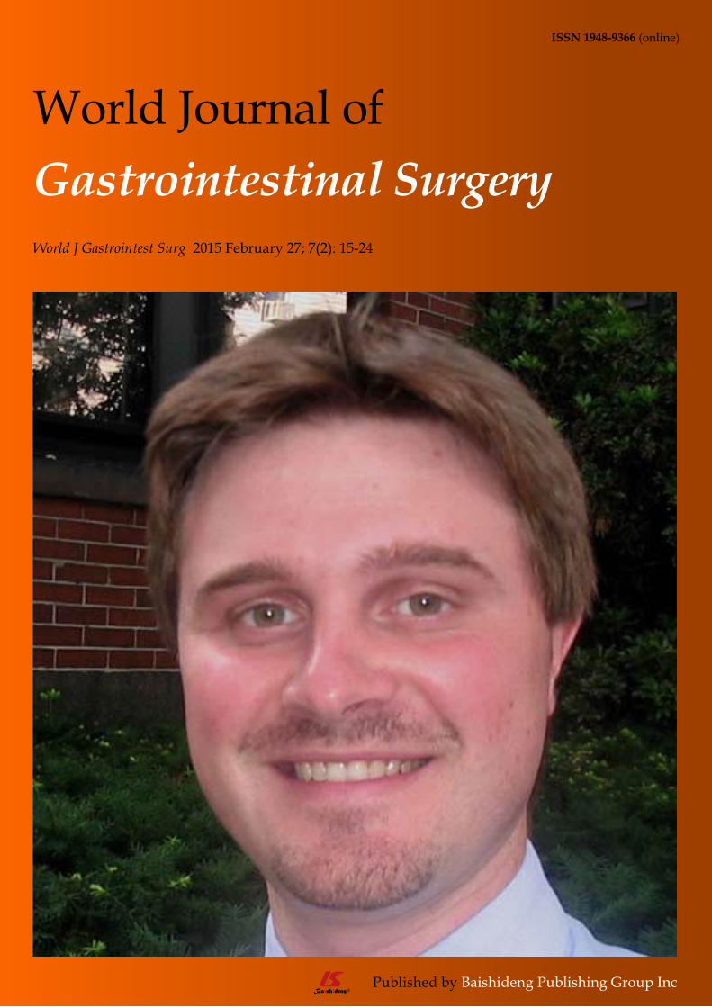
EditorialBoardMemberofWorldJournalofGastrointestinalSurgery ,StefanoCrippa,MD,DepartmentofSurgery,UniversityofVerona,Policlinico“GBRossi”,PiazzaleLAScuro,10,Verona37134,Italy
World Journal of Gastrointestinal Surgery (World J Gastrointest Surg, WJGS, online ISSN 1948-9366, DOI: 10.4240) is a peer-reviewed open access academic journal that aims to guide clinical practice and improve diagnostic and therapeutic skills of clinicians.
WJGS covers topics concerning micro-invasive surgery; laparoscopy; hepatic, biliary, pancreatic and splenic surgery; surgical nutrition; portal hypertension, as well as associated subjects. The current columns of WJGS include editorial, frontier, diagnostic advances, therapeutics advances, field of vision, mini-reviews, review, topic highlight, medical ethics, original articles, case report, clinical case conference (Clinicopathological conference), and autobiography. Priority publication will be given to articles concerning diagnosis and treatment of gastrointestinal surgery diseases. The following aspects are covered: Clinical diagnosis, laboratory diagnosis, differential diagnosis, imaging tests, pathological diagnosis, molecular biological diagnosis, immunological diagnosis, genetic diagnosis, functional diagnostics, and physical diagnosis; and comprehensive therapy, drug therapy, surgical therapy, interventional treatment, minimally invasive therapy, and robot-assisted therapy.
We encourage authors to submit their manuscripts to WJGS. We will give priority to manuscripts that are supported by major national and international foundations and those that are of great basic and clinical significance.
World Journal of Gastrointestinal Surgery is now indexed in PubMed Central, PubMed, Digital Object Identifier, and Directory of Open Access Journals.
I-III EditorialBoard
Xiu-Xia Song, Vice DirectorWorld Journal of Gastrointestinal SurgeryRoom 903, Building D, Ocean International Center, No. 62 Dongsihuan Zhonglu, Chaoyang District, Beijing 100025, ChinaTelephone: +86-10-85381891Fax: +86-10-85381893E-mail: [email protected] Desk: http://www.wjgnet.com/esps/helpdesk.aspxhttp://www.wjgnet.com
PUBLISHERBaishideng Publishing Group Inc8226 Regency Drive, Pleasanton, CA 94588, USATelephone: +1-925-223-8242Fax: +1-925-223-8243E-mail: [email protected] Desk: http://www.wjgnet.com/esps/helpdesk.aspxhttp://www.wjgnet.com
PUBLICATIONDATEFebruary 27, 2015
COPYRIGHT© 2015 Baishideng Publishing Group Inc. Articles pub-lished by this Open-Access journal are distributed under the terms of the Creative Commons Attribution Non-commercial License, which permits use, distribution, and reproduction in any medium, provided the original work is properly cited, the use is non commercial and is otherwise in compliance with the license.
SPECIALSTATEMENTAll articles published in journals owned by the Baishideng Publishing Group (BPG) represent the views and opin-ions of their authors, and not the views, opinions or policies of the BPG, except where otherwise explicitly indicated.
INSTRUCTIONSTOAUTHORSFull instructions are available online at http://www.wjgnet.com/1948-9366/g_info_20100305152206.htm
ONLINESUBMISSIONhttp://www.wjgnet.com/esps/
NAMEOFJOURNALWorld Journal of Gastrointestinal Surgery
ISSNISSN 1948-9366 (online)
LAUNCHDATENovember 30, 2009
FREQUENCYMonthly
EDITOR-IN-CHIEFTimothy M Pawlik, MD, MPH, FACS, Associate Professor of Surgery and Oncology, Hepatobiliary Surgery Program Director, Director, Johns Hopkins Medicine Liver Tumor Center Multi-Disciplinary Clinic, Co-Director of Center for Surgical Trials and Outcomes Research, Johns Hopkins Hospital, 600 N. Wolfe Street, Harvey 611, Baltimore, MD 21287, United States
EDITORIALOFFICEJin-Lei Wang, Director
EDITORS FOR THIS ISSUE
Responsible Assistant Editor: Xiang Li Responsible Science Editor: Yue-Li TianResponsible Electronic Editor: Huan-Liang Wu Proofing Editorial Office Director: Xiu-Xia SongProofing Editor-in-Chief: Lian-Sheng Ma
AIM AND SCOPE
FLYLEAF
INDEXING/ABSTRACTING
WJGS|www.wjgnet.com II February 27, 2015|Volume 7|Issue 2|
Results of the open surgery after endoscopic basket impaction during ERCP procedure
Sezgin Yilmaz, Ogun Ersen, Taner Ozkececi, Kadir S Turel, Serdar Kokulu, Emre Kacar, Murat Akici, Murat Cilekar, Ozgur Kavak, Yuksel Arikan
Sezgin Yilmaz, Ogun Ersen, Taner Ozkececi, Yuksel Arikan, Department of General Surgery, Kocatepe University, 03020 Afyon, TurkeyKadir S Turel, Murat Akici, Murat Cilekar, Ozgur Kavak, Department of General Surgery, State Hospital, 03020 Afyon, TurkeySerdar Kokulu, Department of Anaesthesiology, Kocatepe University, 03020 Afyon, TurkeyEmre Kacar, Department of Radiology, Kocatepe University, 03020 Afyon, TurkeyAuthor contributions: Yilmaz S contributed to conception and design of the study, acquisition of data, drafting the article and final approval of the version of the article, making revisions and performing the ERCP procedures, carrying out the operations; Ersen O, Turel KS, Akici M, Cilekar M and Kavak O contributed to conception and design of the study, acquisition of data, drafting the article and final approval of the version of the article; Ozkececi T contributed to conception and design of the study, acquisition of data, drafting the article and final approval of the version of the article, performing the ERCP procedures; Kokulu S contributed to conception and design of the study, acquisition of data, drafting the article and final approval of the version of the article, anesthesia protocol and postoperative care; Kacar E contributed to conception and design of the study, acquisition of data, drafting the article and final approval of the version of the article, radiological evaluations; Arikan Y contributed to conception and design of the study, acquisition of data, drafting the article and final approval of the version of the article, performing the ERCP procedures, carrying out the operations and final revision of the manuscript.Ethics approval: The study was reviewed and approved by the Afyon Kocatepe University, The Ethical Committee of Medical Faculty.Informed consent: All the patients in the study gave written informed consent prior to study enrollment.Conflict-of-interest: The authors declare that they have no conflict of interest. Data sharing: Technical appendix, statistical code and dataset are available from the corresponding author (SY, drsezgin[email protected]). Open-Access: This article is an openaccess article which was selected by an inhouse editor and fully peerreviewed by external reviewers. It is distributed in accordance with the Creative
Commons Attribution Non Commercial (CC BY-NC 4.0) license, which permits others to distribute, remix, adapt, build upon this work noncommercially, and license their derivative works on different terms, provided the original work is properly cited and the use is non-commercial. See: http://creativecommons.org/licenses/by-nc/4.0/Correspondence to: Sezgin Yilmaz, MD, Department of General Surgery, Kocatepe University, Gazligöl Yolu, Ahmet Necdet Sezer Kampusü, Gazlıgöl Yolu, 03020 Afyon, Turkey. [email protected]: +90-505-5786290Received: September 1, 2014 Peer-review started: September 2, 2014First decision: November 27, 2014Revised: December 6, 2014Accepted: January 9, 2015Article in press: January 12, 2015Published online: February 27, 2015
AbstractAIM: To report the results of open surgery for patients with basket impaction during endoscopic retrograde cholangiopancreatography (ERCP) procedure.
METHODS: Basket impaction of either classical Dormia basket or mechanical lithotripter basket with an entrapped stone occurred in six patients. These patients were immediately operated for removal of stone(s) and impacted basket. The postoperative course, length of hospital stay, diameter of the stone, complication and the surgical procedure of the patients were reported retrospectively.
RESULTS: Six patients (M/F, 0/6) were operated due to impacted basket during ERCP procedure. The mean age of the patients was 64.33 ± 14.41 years. In all cases the surgery was performed immediately after the failed ERCP procedure by making a right
ORIGINAL ARTICLE
Submit a Manuscript: http://www.wjgnet.com/esps/Help Desk: http://www.wjgnet.com/esps/helpdesk.aspxDOI: 10.4240/wjgs.v7.i2.15
World J Gastrointest Surg 2015 February 27; 7(2): 15-20ISSN 1948-9366 (online)
© 2015 Baishideng Publishing Group Inc. All rights reserved.
February 27, 2015|Volume 7|Issue 2|WJGS|www.wjgnet.com 15
Retrospective Study
subcostal incision. The baskets containing the stone were removed through longitudinal choledochotomy with the stone. The choledochotomy incisions were closed by primary closure in four patients and T tube placement in two patients. All patients were also performed cholecystectomy additionally since they had cholelithiasis. In patients with T-tube placement it was removed on the 13th day after a normal T-tube cholangiogram. The patients remained stable at postoperative period and discharged without any complication at median 7 d.
CONCLUSION: Open surgical procedures can be app-lied in patients with basket impaction during ERCP procedure in selected cases.
Key words: Biliary stone; Endoscopic retrograde cholan-giopancreatography; Basket; Impaction; Surgery
© The Author(s) 2015. Published by Baishideng Publishing Group Inc. All rights reserved.
Core tip: The impaction or wire fracture of basket is an uncommon but potentially highly dangerous complication during endoscopic retrograde cholangiopancreatography and stone extraction. Although there are several en-doscopic approaches to treat the basket impaction they require specialized equipments and experienced clinicians. So surgical approach can be an alternative to endoscopic procedures in selected patients.
Yilmaz S, Ersen O, Ozkececi T, Turel KS, Kokulu S, Kacar E, Akici M, Cilekar M, Kavak O, Arikan Y. Results of the open surgery after endoscopic basket impaction during ERCP procedure. World J Gastrointest Surg 2015; 7(2): 1520 Available from: URL: http://www.wjgnet.com/1948-9366/full/v7/i2/15.htm DOI: http://dx.doi.org/10.4240/wjgs.v7.i2.15
INTRODUCTIONEndoscopic retrograde cholangiopancreatography (ERCP) is mainly indicated for choledocholithiasis as well as pancreatic stones[1,2]. It can be performed to remove the biliary stones when combined with sphincterotomy. Bile duct stones are successfully removed with classical Dormia basket or balloon catheters in 85%-95% of the patients[3]. Mechanical lithotripsy is the second line method for non-extra-ctable stones with conventional basket aside from availability and cost[4,5]. Several types of baskets (mechanic lithotripter or wire baskets) can be used for this purpose[6]. The success rates depend on several factors as size and number of the stones, degree of the jaundice and presence of cholecystitis. Complications of ERCP have been reported to occur in 5%-10% of the cases[7]. Basket related complications can be seen as impaction around a calculus or fracture of the traction wire. Endoscopic
basket impaction is a rare and unusual complication that can be seen after an attempt for removal of biliary stones with basket. It is not encountered in classical ERCP complications in textbooks due to its extremely rare occurence. Actually it is a problem of high volume centers. It is defined as inability to withdraw the basket with stone from papillary orifice or seperate the stone from the basket in biliary channel lumen. Since there are only sporadic case reports, the precise treatment is still controversial[8]. At present endoscopic or surgical procedures can be applied for the basket impaction. Non operative maneuvers like extracorporeal shock wave, intracorporeal electrohydraulic lithotripsy, catching the basket tip with a second basket, balloon dilatation of the sphincterotomy area or laser lithotripsy are recommended at high tech-nology units[9-11]. However these procedures req-uire experienced endoscopists and sophisticated technological equipment. Therefore open surgery is still an alternative approach for such patients. In the present study, we reported the results of six patients with endoscopic basket impaction that have been treated with open surgery. To our best knowledge the present report is the largest series so far.
MATERIALS AND METHODSPatientsThis retrospective observational study was carried out between June 2008 and June 2014. A total of 1065 ERCP procedures were performed at our invasive endoscopic procedures unit for choledo-cholithiasis at this period. The total number of ERCP was 2092. Basket impaction of either classical Dormica basket or mechanical lithotripter basket with an entrapped stone were observed in six patients (0.28%). The mean age of the patients was 64.33 ± 14.41 years and they were all female. The patients were admitted with the right upper abdominal pain. Laboratory findings were consistent with cholestasis and obstructive jaundice. Ultrasonography and magnetic resonance cholangiopancreatography (MRCP) revealed one to seven pieces of biliary stones sizes ranging from 15 mm to 30 mm in diameter and dilated choledochus which had a diameter above 15 mm. A diagnosis of obstructive jaundice secondary to choledocholithiasis was made and the patients were proceeded to an ERCP with planned removal of biliary stone. The median age of our patients was 63 years (range 45-81 years). The periampullary diverticulum was present in four patients. In five patients, impacted basket was Dormia and in one impaction occured with mechanical lithotripter basket. The clinical data and the endoscopic features of the patients are represented in Tables 1 and 2. All patients in the work gave informed consent for the study prior to manuscript preparation.
Yilmaz S et al . Surgery for basket impaction during ERCP
February 27, 2015|Volume 7|Issue 2|WJGS|www.wjgnet.com 16
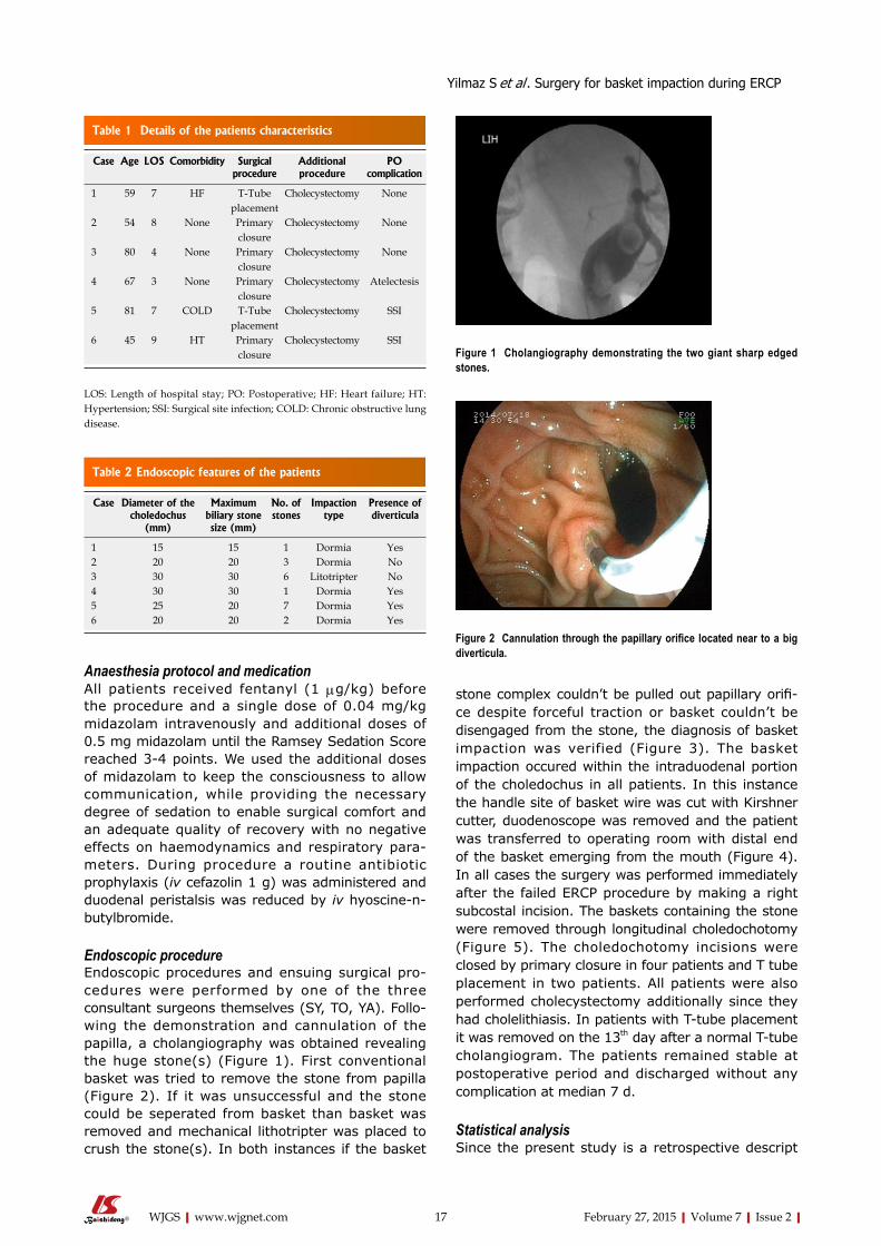
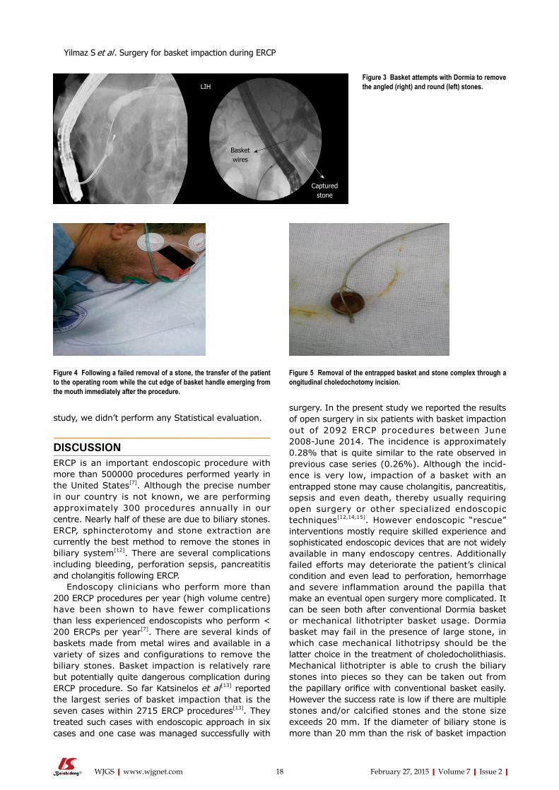
stone complex couldn’t be pulled out papillary orifice despite forceful traction or basket couldn’t be disengaged from the stone, the diagnosis of basket impaction was verified (Figure 3). The basket impaction occured within the intraduodenal portion of the choledochus in all patients. In this instance the handle site of basket wire was cut with Kirshner cutter, duodenoscope was removed and the patient was transferred to operating room with distal end of the basket emerging from the mouth (Figure 4). In all cases the surgery was performed immediately after the failed ERCP procedure by making a right subcostal incision. The baskets containing the stone were removed through longitudinal choledochotomy (Figure 5). The choledochotomy incisions were closed by primary closure in four patients and T tube placement in two patients. All patients were also performed cholecystectomy additionally since they had cholelithiasis. In patients with T-tube placement it was removed on the 13th day after a normal T-tube cholangiogram. The patients remained stable at postoperative period and discharged without any complication at median 7 d.
Statistical analysisSince the present study is a retrospective descript
Anaesthesia protocol and medicationAll patients received fentanyl (1 µg/kg) before the procedure and a single dose of 0.04 mg/kg midazolam intravenously and additional doses of 0.5 mg midazolam until the Ramsey Sedation Score reached 3-4 points. We used the additional doses of midazolam to keep the consciousness to allow communication, while providing the necessary degree of sedation to enable surgical comfort and an adequate quality of recovery with no negative effects on haemodynamics and respiratory para-meters. During procedure a routine antibiotic prophylaxis (iv cefazolin 1 g) was administered and duodenal peristalsis was reduced by iv hyoscine-n-butylbromide.
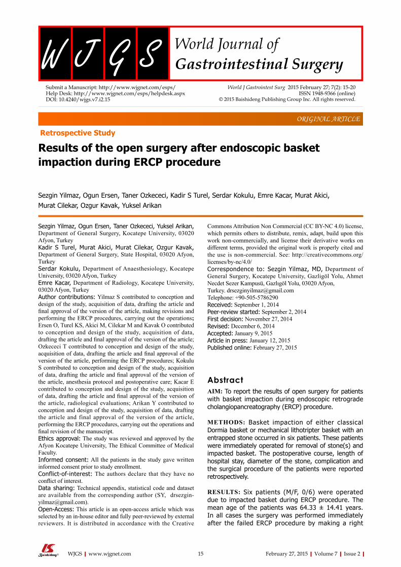
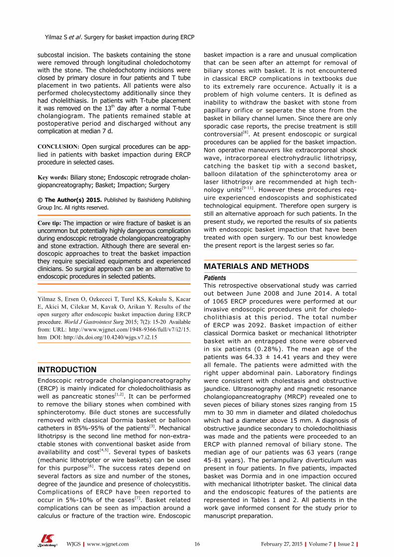
Endoscopic procedureEndoscopic procedures and ensuing surgical pro-cedures were performed by one of the three consultant surgeons themselves (SY, TO, YA). Follo-wing the demonstration and cannulation of the papilla, a cholangiography was obtained revealing the huge stone(s) (Figure 1). First conventional basket was tried to remove the stone from papilla (Figure 2). If it was unsuccessful and the stone could be seperated from basket than basket was removed and mechanical lithotripter was placed to crush the stone(s). In both instances if the basket
February 27, 2015|Volume 7|Issue 2|WJGS|www.wjgnet.com 17
Case Age LOS Comorbidity Surgical procedure
Additional procedure
PO complication
1 59 7 HF T-Tube placement
Cholecystectomy None
2 54 8 None Primary closure
Cholecystectomy None
3 80 4 None Primary closure
Cholecystectomy None
4 67 3 None Primary closure
Cholecystectomy Atelectesis
5 81 7 COLD T-Tube placement
Cholecystectomy SSI
6 45 9 HT Primary closure
Cholecystectomy SSI
Table 1 Details of the patients characteristics
LOS: Length of hospital stay; PO: Postoperative; HF: Heart failure; HT: Hypertension; SSI: Surgical site infection; COLD: Chronic obstructive lung disease.
Case Diameter of the choledochus
(mm)
Maximum biliary stone size (mm)
No. of stones
Impaction type
Presence of diverticula
1 15 15 1 Dormia Yes 2 20 20 3 Dormia No 3 30 30 6 Litotripter No 4 30 30 1 Dormia Yes 5 25 20 7 Dormia Yes 6 20 20 2 Dormia Yes
Table 2 Endoscopic features of the patients
Figure 1 Cholangiography demonstrating the two giant sharp edged stones.
Figure 2 Cannulation through the papillary orifice located near to a big diverticula.
LIH
Yilmaz S et al . Surgery for basket impaction during ERCP
study, we didn’t perform any Statistical evaluation.
DISCUSSIONERCP is an important endoscopic procedure with more than 500000 procedures performed yearly in the United States[7]. Although the precise number in our country is not known, we are performing approximately 300 procedures annually in our centre. Nearly half of these are due to biliary stones. ERCP, sphincterotomy and stone extraction are currently the best method to remove the stones in biliary system[12]. There are several complications including bleeding, perforation sepsis, pancreatitis and cholangitis following ERCP.
Endoscopy clinicians who perform more than 200 ERCP procedures per year (high volume centre) have been shown to have fewer complications than less experienced endoscopists who perform < 200 ERCPs per year[7]. There are several kinds of baskets made from metal wires and available in a variety of sizes and configurations to remove the biliary stones. Basket impaction is relatively rare but potentially quite dangerous complication during ERCP procedure. So far Katsinelos et al[13] reported the largest series of basket impaction that is the seven cases within 2715 ERCP procedures[13]. They treated such cases with endoscopic approach in six cases and one case was managed successfully with
surgery. In the present study we reported the results of open surgery in six patients with basket impaction out of 2092 ERCP procedures between June 2008-June 2014. The incidence is approximately 0.28% that is quite similar to the rate observed in previous case series (0.26%). Although the incid-ence is very low, impaction of a basket with an entrapped stone may cause cholangitis, pancreatitis, sepsis and even death, thereby usually requiring open surgery or other specialized endoscopic techniques[12,14,15]. However endoscopic “rescue” interventions mostly require skilled experience and sophisticated endoscopic devices that are not widely available in many endoscopy centres. Additionally failed efforts may deteriorate the patient’s clinical condition and even lead to perforation, hemorrhage and severe inflammation around the papilla that make an eventual open surgery more complicated. It can be seen both after conventional Dormia basket or mechanical lithotripter basket usage. Dormia basket may fail in the presence of large stone, in which case mechanical lithotripsy should be the latter choice in the treatment of choledocholithiasis. Mechanical lithotripter is able to crush the biliary stones into pieces so they can be taken out from the papillary orifice with conventional basket easily. However the success rate is low if there are multiple stones and/or calcified stones and the stone size exceeds 20 mm. If the diameter of biliary stone is more than 20 mm than the risk of basket impaction
February 27, 2015|Volume 7|Issue 2|WJGS|www.wjgnet.com 18
Figure 3 Basket attempts with Dormia to remove the angled (right) and round (left) stones.
Figure 4 Following a failed removal of a stone, the transfer of the patient to the operating room while the cut edge of basket handle emerging from the mouth immediately after the procedure.
Figure 5 Removal of the entrapped basket and stone complex through a ongitudinal choledochotomy incision.
LIH
Basketwires
Capturedstone
Yilmaz S et al . Surgery for basket impaction during ERCP
as well as fracture of the basket at the junction between the distal and proximal parts may occur[8]. Once the basket catch the stone, there should be enough space between the stone and biliary channel wall to release the stone from basket in case of failed crushing. By definition basket impaction is expressed as inability to withdraw the basket with stone from papillary orifice or separate the stone from the basket in biliary channel lumen. Since there are only sporadic case reports, the precise treatment is still controversial. At present endoscopic or surgical procedures can be recommended after basket impaction. Endoscopic procedures should be tried if there is adequate experience and specialized endoscopic devices. In such a case extension of the sphincterotomy should be attempted first since the most likely cause of impaction is inadequate sphincterotomy and tissue edema. It can be applied when it is clear that the sphincterotomy can be safely extended. The special equipment required is a duodenoscope with a 4.2 mm working channel[16]. However this can lead to duodenal perforation in inexperienced hands. Percutaneous transhepatic route can also be used in suitable cases by using a goose-neck snare in skilled radiology department[3]. Dilating the papillary orifice is sometimes useful to remove the impacted stone-basket complex with the larger balloon[17,18]. These endoscopic procedures are sophisticated and not widely available every-where. Basket impaction represents a surgical emergency unless other non operative maneuvers like extracorporeal shock wave, intracorporeal electrohydraulic lithotripsy or catching the basket tip with a second basket are available[9-11,19]. Addi-tionally since our patients required additional surgical procedures for cholelithiasis, open surg-ery was preferred to treat the current basket impaction problem. In our series all patients also had cholelithiasis thus required cholecystectomy. The basket stone complex was removed through a longitudinal choledochotomy incision. It was repaired with primary closure in four patients and T-tube placement in two patients. In our centre we routinely close choledochotomy incision primarily in patients with previous sphincterotomy. But two patients in the present report were treated with T-tube placement since there are severe inflammation, cholangitis and transmural thickening at the biliary channel. The frequency of diverticula at our 2092 ERCP procedures is approximately 25%, but in the present report we found that 4 patients in 6 basket impaction had duodenal diverticula. This high ratio considered us that the occurence of periampullary diverticula might be a predisposing factor for basket impaction. Small number of patients is our limitation so that the results can not be extrapolated to surgery clinics. However to our knowledge it is the largest series dealing with the open surgery in such patients. So it can suggest an
alternative surgical approach besides endoscopic interventions in otherwise healthy patients without comorbidity. In conclusion impaction or wire fracture of basket is an uncommon complication during ERCP and stone extraction. There are several treatment protocols and it should be tailored to the patient’s clinical condition, endoscopist’s experience and ERCP unit equipment.
COMMENTSBackgroundEndoscopic retrograde cholangiopancreatography (ERCP) is mainly indicated for choledocholithiasis. Endoscopic basket impaction is a rare and unusual complication that can be seen after an attempt for removal of biliary stones with basket during ERCP. It is defined as inability to withdraw the basket with stone from papillary orifice or seperate the stone from the basket in biliary channel lumen. Endoscopic or surgical procedures can be applied for the basket impaction. Non operative endoscopic maneuvers like extracorporeal shock wave, intracorporeal electrohydraulic lithotripsy, catching the basket tip with a second basket, balloon dilatation of the sphincterotomy area or laser lithotripsy are preferable at high technology units. However open surgical procedures can also be applied in selected cases. Research frontiersIn the present study, the authors reported the results of six patients with endoscopic basket impaction that have been treated with open surgery. Innovations and breakthroughsIn literature non-operative endoscopic procedures are widely recommended for ERCP-related basket impactions. These procedures require experienced endoscopists and sophisticated technological equipment. However these techniques and endoscopic devices are not widely available in every endoscopy centre. Therefore open surgery is still an alternative approcah for such patients. ApplicationsOpen surgery by performing choledochotomy can be applied for the patients with basket impaction during ERCP procedure as an alternative to endoscopic interventions in selected cases. TerminologyERCP is abbreviation of endoscopic retrograde cholangiopancreatography and is an endoscopic technique to view the biliary and pancreatic channels. It is also used to remove the stones from these channels. Basket impaction is defined as inability to withdraw the basket with stone from papillary orifice or seperate the stone from the basket in biliary channel lumen. It can be treated with open surgery including choledochotomy (incizing the choledochus and removing the stone and basket together) and closing the choledochus by primary closure or T-tube placement. Peer-reviewThis paper reported the results of six patients with endoscopic basket impaction that have been treated with open surgery. The results are interesting and encouraging, which provided the practical basis that open surgical procedures could be selected when endoscopic basket impaction occurred.
REFERENCES1 Chan CH, Donnellan F, Chan GC, Byrne MF. A novel two-step
approach for retrieval of an impacted biliary extraction basket. Case Rep Gastrointest Med 2012; 2012: 435050 [PMID: 22953076 DOI: 10.1155/2012/435050]
2 Hlaing C, Tarnasky P, Hambrick D. Laser lithotripsy to treat basket impaction during mechanical lithotripsy of a pancreatic duct stone. JOP 2012; 13: 101-103 [PMID: 22233959]
3 Kwon JH, Lee JK, Lee JH, Lee YS. Percutaneous transhepatic release of an impacted lithotripter basket and its fractured traction wire using a goose-neck snare: a case report. Korean J Radiol 2011; 12: 247-251 [PMID: 21430943 DOI: 10.3348/kjr.2011.12.2.247]
4 Hochberger J, Tex S, Maiss J, Hahn EG. Management of difficult common bile duct stones. Gastrointest Endosc Clin N Am 2003; 13:
February 27, 2015|Volume 7|Issue 2|WJGS|www.wjgnet.com 19
COMMENTS
Yilmaz S et al . Surgery for basket impaction during ERCP
623-634 [PMID: 14986790 DOI: 10.1016/S1052-5157(03)00102-8]5 Schneider MU, Matek W, Bauer R, Domschke W. Mechanical
lithotripsy of bile duct stones in 209 patients--effect of technical advances. Endoscopy 1988; 20: 248-253 [PMID: 3168938 DOI: 10.1055/s-2007-1018186]
6 Sezgin O, Tezel A, Sahin B. Dormia basket fracture: an unusual complication of mechanical lithotripsy. J Clin Gastroenterol 2000; 30: 215 [PMID: 10730934 DOI: 10.1097/00004836-200003000-00021]
7 Silviera ML, Seamon MJ, Porshinsky B, Prosciak MP, Doraiswamy VA, Wang CF, Lorenzo M, Truitt M, Biboa J, Jarvis AM, Narula VK, Steinberg SM, Stawicki SP. Complications related to endoscopic retrograde cholangiopancreatography: a comprehensive clinical review. J Gastrointestin Liver Dis 2009; 18: 73-82 [PMID: 19337638]
8 Fukino N, Oida T, Kawasaki A, Mimatsu K, Kuboi Y, Kano H, Amano S. Impaction of a lithotripsy basket during endoscopic lithotomy of a common bile duct stone. World J Gastroenterol 2010; 16: 2832-2834 [PMID: 20533607 DOI: 10.3748/wjg.v16.i22.2832]
9 Schutz SM, Chinea C, Friedrichs P. Successful endoscopic removal of a severed, impacted Dormia basket. Am J Gastroenterol 1997; 92: 679-681 [PMID: 9128323]
10 Sheridan J, Williams TM, Yeung E, Ho CS, Thurston W. Percutaneous transhepatic management of an impacted endoscopic basket. Gastrointest Endosc 1993; 39: 444-446 [PMID: 8514084 DOI: 10.1016/S0016-5107(93)70127-3]
11 Ranjeev P, Goh Kl. Retrieval of an impacted Dormia basket and stone in situ using a novel method. Gastrointest Endosc 2000; 51: 504-506 [PMID: 10744838 DOI: 10.1016/S0016-5107(00)70463-9]
12 Maple JT, Baron TH. Biliary-basket impaction complicated by in
vivo traction-wire fracture: report of a novel management approach. Gastrointest Endosc 2006; 64: 1031-1033 [PMID: 17140927 DOI: 10.1016/j.gie.2006.04.023]
13 Katsinelos P, Lazaraki G, Chatzimavroudis G, Gkagkalis S, Vasiliadis I, Papaeuthimiou A, Terzoudis S, Pilpilidis I, Zavos C, Kountouras J. Risk factors for therapeutic ERCP-related complications: an analysis of 2,715 cases performed by a single endoscopist. Ann Gastroenterol 2014; 27: 65-72 [PMID: 24714755]
14 Katsinelos P, Fasoulas K, Beltsis A, Chatzimavroudis G, Zavos C, Terzoudis S, Kountouras J. Large-balloon dilation of the biliary orifice for the management of basket impaction: a case series of 6 patients. Gastrointest Endosc 2011; 73: 1298-1301 [PMID: 21492853 DOI: 10.16/j.gie.2011.01.034]
15 Nuehaus B, Safrany L. Complications of endoscopic sphinecterotomy and their treatment. Endoscopy 1981; 13: 197-199 [PMID: 7274168]
16 Borgaonkar M. Impacted biliary basket. Gastrointest Endosc 2005; 62: 474 [PMID: 16111986 DOI: 10.1016/j.gie.2005.04.041]
17 Maydeo A, Bhandari S. Balloon sphincteroplasty for removing difficult bile duct stones. Endoscopy 2007; 39: 958-961 [PMID: 17701853]
18 Mabuchi M, Iwashita T, Yasuda I, Okuno M, Uemura S, Nakashima M, Doi S, Adachi S, Shimizu M, Mukai T, Tomita E, Moriwaki H. Endoscopic papillary large balloon dilation as a salvage procedure for basket impaction during retrieval of common bile duct stones. Dig Dis Sci 2014; 59: 220-223 [PMID: 23979442 DOI: 10.1007/s10620-013-2845-0]
19 Kwon CI, Song SH, Hahm KB, Ko KH. Unusual complications related to endoscopic retrograde cholangiopancreatography and its endoscopic treatment. Clin Endosc 2013; 46: 251-259 [PMID: 23767036 DOI: 10.5946/ce.2013.46.3.251]
P- Reviewer: Abu-Zidan FM, Han JH, Sun WB S- Editor: Ji FF L- Editor: A E- Editor: Wu HL
February 27, 2015|Volume 7|Issue 2|WJGS|www.wjgnet.com 20
Yilmaz S et al . Surgery for basket impaction during ERCP
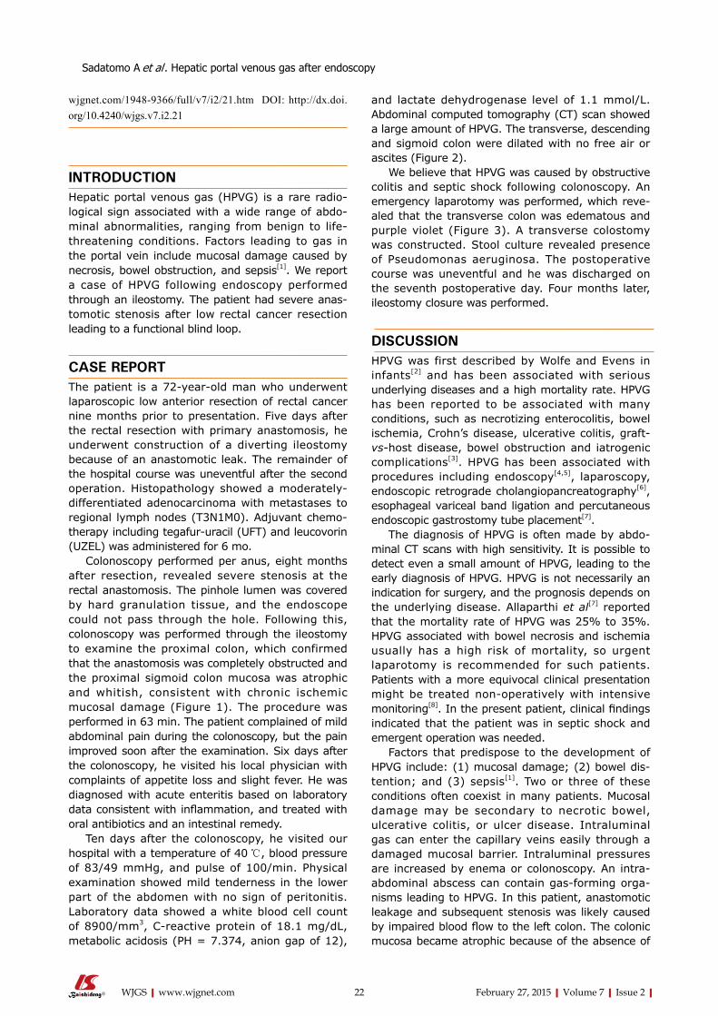
Hepatic portal venous gas after endoscopy in a patient with anastomotic obstruction
Ai Sadatomo, Koji Koinuma, Rihito Kanamaru, Yasuyuki Miyakura, Hisanaga Horie, Alan T Lefor, Yoshikazu Yasuda
Ai Sadatomo, Koji Koinuma, Rihito Kanamaru, Yasuyuki Miyakura, Hisanaga Horie, Alan T Lefor, Yoshikazu Yasuda, Department of Surgery, Jichi Medical University, Shimotsuke, Tochigi 329-0498, JapanAuthor contributions: All authors contributed to this manus-cript.Ethics approval: This study was approved by the Institutional Review Board of Jichi Medical School for ethical issues.Informed consent: The patient provided informed written consent prior to submit the manuscript.Conflict-of-interest: We certify that there is no conflict of interest with any financial organization regarding the material discussed in the manuscript.Open-Access: This article is an open-access article which was selected by an in-house editor and fully peer-reviewed by external reviewers. It is distributed in accordance with the Creative Commons Attribution Non Commercial (CC BY-NC 4.0) license, which permits others to distribute, remix, adapt, build upon this work non-commercially, and license their derivative works on different terms, provided the original work is properly cited and the use is non-commercial. See: http://creativecommons.org/licenses/by-nc/4.0/Correspondence to: Ai Sadatomo, MD, Graduate Student, Department of Surgery, Jichi Medical University, 3311 Yakushiji, Shimotsuke, Tochigi 329-0498, Japan. [email protected]: +81-285-587371 Fax: +81-285-443234Received: July 12, 2014 Peer-review started: July 13, 2014 First decision: September 28, 2014Revised: December 16, 2014 Accepted: January 9, 2015 Article in press: January 12, 201Published online: February 27, 2015
AbstractA 72-year-old male underwent a laparoscopic low anterior resection for advanced rectal cancer. A diverting loop ileostomy was constructed due to an anastomotic leak five days postoperatively. Nine months later,
colonoscopy performed through the stoma showed complete anastomotic obstruction. The mucosa of the proximal sigmoid colon was atrophic and whitish. Ten days after the colonoscopy, the patient presented in shock with abdominal pain. Abdominal computed tomography scan showed hepatic portal venous gas (HPVG) and a dilated left colon. HPVG induced by obstructive colitis was diagnosed and a transverse colostomy performed emergently. His subsequent hospital course was unremarkable. Rectal anastomosis with diverting ileostomy is often performed in patients with low rectal cancers. In patients with anastomotic obstruction or severe stenosis, colonoscopy through diverting stoma should be avoided. Emergent operation to decompress the obstructed proximal colon is necessary in patients with a blind intestinal loop acco-mpanied by HPVG.
Key words: Portal venous gas; Abdominal computed tomography; Colonoscopy; Anastomotic obstruction; Bacterial translocation
© The Author(s) 2015. Published by Baishideng Publishing Group Inc. All rights reserved.
Core tip: A rare case of hepatic portal venous gas (HPVG) is reported. Endoscopy through ileostomy leaded the formation of HPVG induced by obstructive colitis. The anastomosis of rectum was totally obstructed after rectum cancer operation. For nine months, the mucosa of ascending to sigmoid colon has changed atrophy for disuse. The patient’s condition improved after emergent operation of transverse colostomy. In patients with anastomotic obstruction or severe stenosis, colonoscopy through diverting stoma should be avoided.
Sadatomo A, Koinuma K, Kanamaru R, Miyakura Y, Horie H, Lefor AT, Yasuda Y. Hepatic portal venous gas after endoscopy in a patient with anastomotic obstruction. World J Gastrointest Surg 2015; 7(2): 21-24 Available from: URL: http://www.
CASE REPORT
Submit a Manuscript: http://www.wjgnet.com/esps/Help Desk: http://www.wjgnet.com/esps/helpdesk.aspxDOI: 10.4240/wjgs.v7.i2.21
World J Gastrointest Surg 2015 February 27; 7(2): 21-24ISSN 1948-9366 (online)
© 2015 Baishideng Publishing Group Inc. All rights reserved.
February 27, 2015|Volume 7|Issue 2|WJGS|www.wjgnet.com 21
wjgnet.com/1948-9366/full/v7/i2/21.htm DOI: http://dx.doi.org/10.4240/wjgs.v7.i2.21
INTRODUCTIONHepatic portal venous gas (HPVG) is a rare radiological sign associated with a wide range of abdominal abnormalities, ranging from benign to lifethreatening conditions. Factors leading to gas in the portal vein include mucosal damage caused by necrosis, bowel obstruction, and sepsis[1]. We report a case of HPVG following endoscopy performed through an ileostomy. The patient had severe anastomotic stenosis after low rectal cancer resection leading to a functional blind loop.
CASE REPORTThe patient is a 72yearold man who underwent laparoscopic low anterior resection of rectal cancer nine months prior to presentation. Five days after the rectal resection with primary anastomosis, he underwent construction of a diverting ileostomy because of an anastomotic leak. The remainder of the hospital course was uneventful after the second operation. Histopathology showed a moderatelydifferentiated adenocarcinoma with metastases to regional lymph nodes (T3N1M0). Adjuvant chemotherapy including tegafururacil (UFT) and leucovorin (UZEL) was administered for 6 mo.
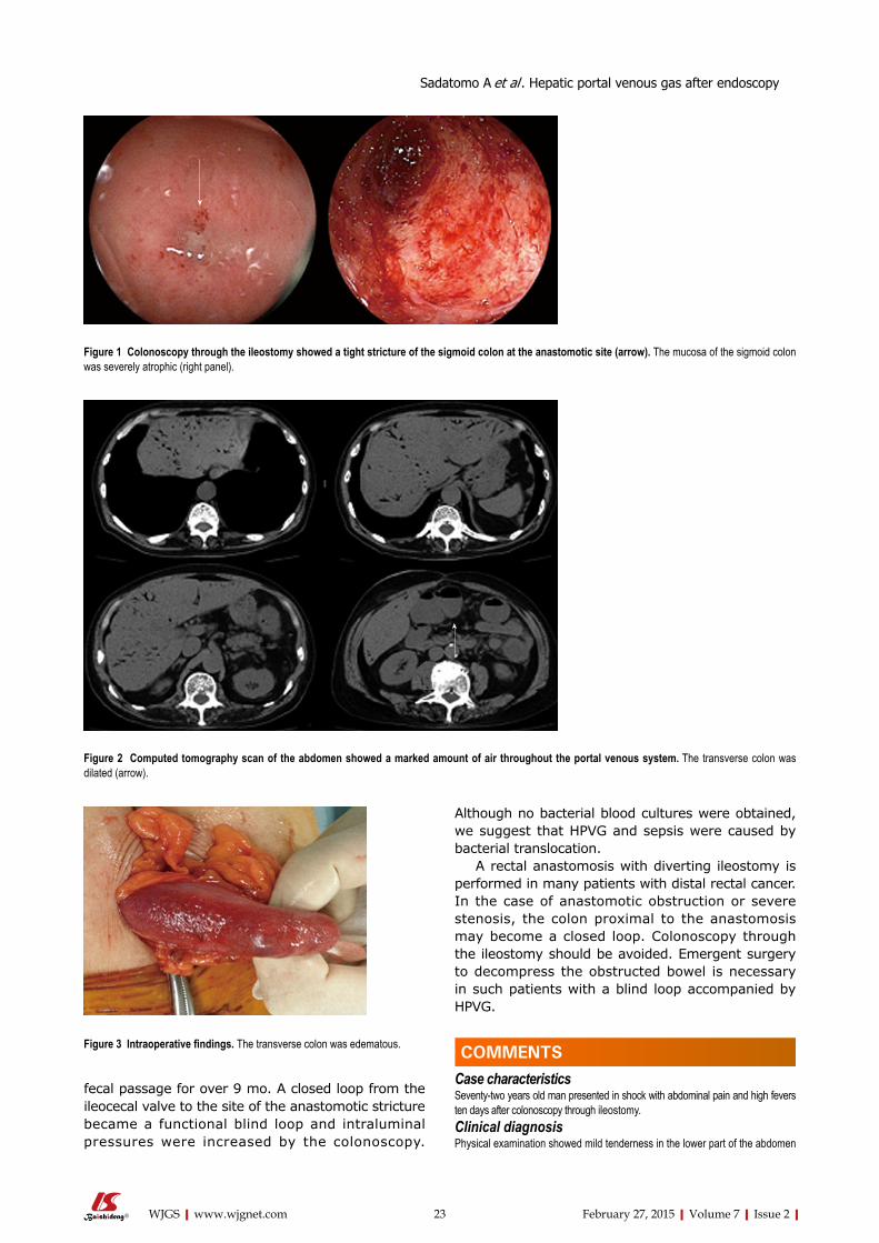
Colonoscopy performed per anus, eight months after resection, revealed severe stenosis at the rectal anastomosis. The pinhole lumen was covered by hard granulation tissue, and the endoscope could not pass through the hole. Following this, colonoscopy was performed through the ileostomy to examine the proximal colon, which confirmed that the anastomosis was completely obstructed and the proximal sigmoid colon mucosa was atrophic and whitish, consistent with chronic ischemic mucosal damage (Figure 1). The procedure was performed in 63 min. The patient complained of mild abdominal pain during the colonoscopy, but the pain improved soon after the examination. Six days after the colonoscopy, he visited his local physician with complaints of appetite loss and slight fever. He was diagnosed with acute enteritis based on laboratory data consistent with inflammation, and treated with oral antibiotics and an intestinal remedy.
Ten days after the colonoscopy, he visited our hospital with a temperature of 40 ℃, blood pressure of 83/49 mmHg, and pulse of 100/min. Physical examination showed mild tenderness in the lower part of the abdomen with no sign of peritonitis. Laboratory data showed a white blood cell count of 8900/mm3, Creactive protein of 18.1 mg/dL, metabolic acidosis (PH = 7.374, anion gap of 12),
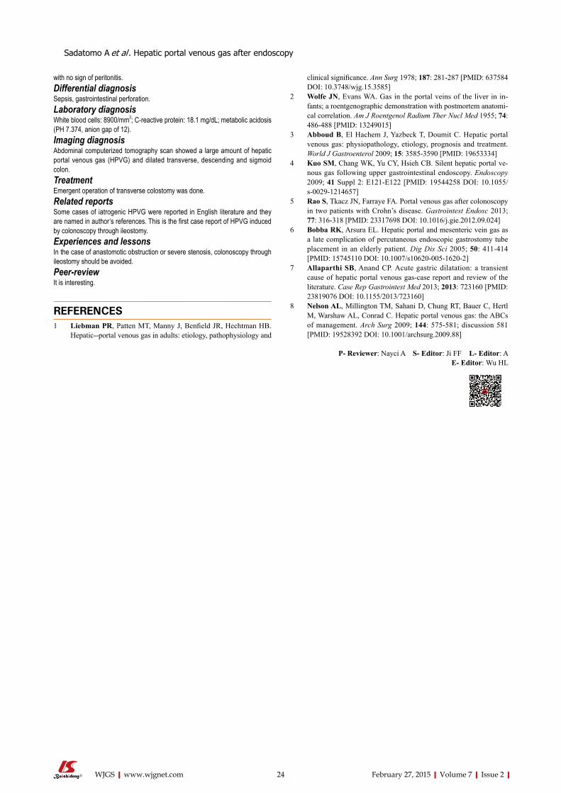
and lactate dehydrogenase level of 1.1 mmol/L. Abdominal computed tomography (CT) scan showed a large amount of HPVG. The transverse, descending and sigmoid colon were dilated with no free air or ascites (Figure 2).
We believe that HPVG was caused by obstructive colitis and septic shock following colonoscopy. An emergency laparotomy was performed, which revealed that the transverse colon was edematous and purple violet (Figure 3). A transverse colostomy was constructed. Stool culture revealed presence of Pseudomonas aeruginosa. The postoperative course was uneventful and he was discharged on the seventh postoperative day. Four months later, ileostomy closure was performed.
DISCUSSIONHPVG was first described by Wolfe and Evens in infants[2] and has been associated with serious underlying diseases and a high mortality rate. HPVG has been reported to be associated with many conditions, such as necrotizing enterocolitis, bowel ischemia, Crohn’s disease, ulcerative colitis, graftvshost disease, bowel obstruction and iatrogenic complications[3]. HPVG has been associated with procedures including endoscopy[4,5], laparoscopy, endoscopic retrograde cholangiopancreatography[6], esophageal variceal band ligation and percutaneous endoscopic gastrostomy tube placement[7].
The diagnosis of HPVG is often made by abdominal CT scans with high sensitivity. It is possible to detect even a small amount of HPVG, leading to the early diagnosis of HPVG. HPVG is not necessarily an indication for surgery, and the prognosis depends on the underlying disease. Allaparthi et al[7] reported that the mortality rate of HPVG was 25% to 35%. HPVG associated with bowel necrosis and ischemia usually has a high risk of mortality, so urgent laparotomy is recommended for such patients. Patients with a more equivocal clinical presentation might be treated nonoperatively with intensive monitoring[8]. In the present patient, clinical findings indicated that the patient was in septic shock and emergent operation was needed.
Factors that predispose to the development of HPVG include: (1) mucosal damage; (2) bowel distention; and (3) sepsis[1]. Two or three of these conditions often coexist in many patients. Mucosal damage may be secondary to necrotic bowel, ulcerative colitis, or ulcer disease. Intraluminal gas can enter the capillary veins easily through a damaged mucosal barrier. Intraluminal pressures are increased by enema or colonoscopy. An intraabdominal abscess can contain gasforming organisms leading to HPVG. In this patient, anastomotic leakage and subsequent stenosis was likely caused by impaired blood flow to the left colon. The colonic mucosa became atrophic because of the absence of
Sadatomo A et al . Hepatic portal venous gas after endoscopy
February 27, 2015|Volume 7|Issue 2|WJGS|www.wjgnet.com 22
Although no bacterial blood cultures were obtained, we suggest that HPVG and sepsis were caused by bacterial translocation.
A rectal anastomosis with diverting ileostomy is performed in many patients with distal rectal cancer. In the case of anastomotic obstruction or severe stenosis, the colon proximal to the anastomosis may become a closed loop. Colonoscopy through the ileostomy should be avoided. Emergent surgery to decompress the obstructed bowel is necessary in such patients with a blind loop accompanied by HPVG.
COMMENTSCase characteristicsSeventy-two years old man presented in shock with abdominal pain and high fevers ten days after colonoscopy through ileostomy.Clinical diagnosisPhysical examination showed mild tenderness in the lower part of the abdomen
fecal passage for over 9 mo. A closed loop from the ileocecal valve to the site of the anastomotic stricture became a functional blind loop and intraluminal pressures were increased by the colonoscopy.
February 27, 2015|Volume 7|Issue 2|WJGS|www.wjgnet.com 23
Figure 1 Colonoscopy through the ileostomy showed a tight stricture of the sigmoid colon at the anastomotic site (arrow). The mucosa of the sigmoid colon was severely atrophic (right panel).
Figure 2 Computed tomography scan of the abdomen showed a marked amount of air throughout the portal venous system. The transverse colon was dilated (arrow).
Figure 3 Intraoperative findings. The transverse colon was edematous. COMMENTS
Sadatomo A et al . Hepatic portal venous gas after endoscopy
with no sign of peritonitis.Differential diagnosisSepsis, gastrointestinal perforation. Laboratory diagnosisWhite blood cells: 8900/mm3; C-reactive protein: 18.1 mg/dL; metabolic acidosis (PH 7.374, anion gap of 12). Imaging diagnosisAbdominal computerized tomography scan showed a large amount of hepatic portal venous gas (HPVG) and dilated transverse, descending and sigmoid colon.TreatmentEmergent operation of transverse colostomy was done. Related reportsSome cases of iatrogenic HPVG were reported in English literature and they are named in author’s references. This is the first case report of HPVG induced by colonoscopy through ileostomy.Experiences and lessonsIn the case of anastomotic obstruction or severe stenosis, colonoscopy through ileostomy should be avoided.Peer-reviewIt is interesting.
REFERENCES1 Liebman PR, Patten MT, Manny J, Benfield JR, Hechtman HB.
Hepatic--portal venous gas in adults: etiology, pathophysiology and
clinical significance. Ann Surg 1978; 187: 281-287 [PMID: 637584 DOI: 10.3748/wjg.15.3585]
2 Wolfe JN, Evans WA. Gas in the portal veins of the liver in in-fants; a roentgenographic demonstration with postmortem anatomi-cal correlation. Am J Roentgenol Radium Ther Nucl Med 1955; 74: 486-488 [PMID: 13249015]
3 Abboud B, El Hachem J, Yazbeck T, Doumit C. Hepatic portal venous gas: physiopathology, etiology, prognosis and treatment. World J Gastroenterol 2009; 15: 3585-3590 [PMID: 19653334]
4 Kuo SM, Chang WK, Yu CY, Hsieh CB. Silent hepatic portal ve-nous gas following upper gastrointestinal endoscopy. Endoscopy 2009; 41 Suppl 2: E121-E122 [PMID: 19544258 DOI: 10.1055/s-0029-1214657]
5 Rao S, Tkacz JN, Farraye FA. Portal venous gas after colonoscopy in two patients with Crohn’s disease. Gastrointest Endosc 2013; 77: 316-318 [PMID: 23317698 DOI: 10.1016/j.gie.2012.09.024]
6 Bobba RK, Arsura EL. Hepatic portal and mesenteric vein gas as a late complication of percutaneous endoscopic gastrostomy tube placement in an elderly patient. Dig Dis Sci 2005; 50: 411-414 [PMID: 15745110 DOI: 10.1007/s10620-005-1620-2]
7 Allaparthi SB, Anand CP. Acute gastric dilatation: a transient cause of hepatic portal venous gas-case report and review of the literature. Case Rep Gastrointest Med 2013; 2013: 723160 [PMID: 23819076 DOI: 10.1155/2013/723160]
8 Nelson AL, Millington TM, Sahani D, Chung RT, Bauer C, Hertl M, Warshaw AL, Conrad C. Hepatic portal venous gas: the ABCs of management. Arch Surg 2009; 144: 575-581; discussion 581 [PMID: 19528392 DOI: 10.1001/archsurg.2009.88]
P- Reviewer: Nayci A S- Editor: Ji FF L- Editor: A E- Editor: Wu HL
February 27, 2015|Volume 7|Issue 2|WJGS|www.wjgnet.com 24
Sadatomo A et al . Hepatic portal venous gas after endoscopy

© 2015 Baishideng Publishing Group Inc. All rights reserved.
Published by Baishideng Publishing Group Inc8226 Regency Drive, Pleasanton, CA 94588, USA
Telephone: +1-925-223-8242Fax: +1-925-223-8243
E-mail: [email protected] Desk: http://www.wjgnet.com/esps/helpdesk.aspx
http://www.wjgnet.com