Isolation of the GFA1 gene encoding glucosamine-6-phosphate synthase of Sporothrix schenckii and its...
8
1 3 Isolation of the GFA1 gene encoding glucosamine-6-phosphate synthase 4 of Sporothrix schenckii and its expression in Saccharomyces cerevisiae 5 6 7 Juan Francisco Sánchez-López a Q1 , Joaquín González-Ibarra a , Aurelio Álvarez-Vargas a , Slawomir Milewski b , 8 Julio César Villagómez-Castro a , Carmen Cano-Canchola a , Everardo López-Romero a,⇑ 9 a Departamento de Biología, División de Ciencias Naturales y Exactas, Universidad de Guanajuato, Apartado Postal 187, Guanajuato, Gto. 36000, Mexico 10 b Department of Pharmaceutical Technology and Biochemistry, Faculty of Chemistry, Gdansk University of Technology, Gdansk, Poland 11 12 14 article info 15 Article history: 16 Received 14 October 2014 17 and in revised form 1 December 2014 18 Available online xxxx 19 Keywords: 20 Sporothrix schenckii 21 Recombinant glucosamine-6-phosphate 22 synthase 23 Purification 24 Biochemical characterization 25 Antifungal target 26 27 abstract 28 Glucosamine-6-phosphate synthase (GlcN-6-P synthase) is an essential enzyme involved in cell wall bio- 29 genesis that has been proposed as a strategic target for antifungal chemotherapy. Here we describe the 30 cloning and functional characterization of Sporothrix schenckii GFA1 gene which was isolated from a geno- 31 mic library of the fungus. The gene encodes a predicted protein of 708 amino acids that is homologous to 32 GlcN-6-P synthases from other sources. The recombinant enzyme restored glucosamine prototrophy of 33 the Saccharomyces cerevisiae gfa1 null mutant. Purification and biochemical analysis of the recombinant 34 enzyme revealed some differences from the wild type enzyme, such as improved stability and less 35 sensitivity to UDP-GlcNAc. The sensitivity of the recombinant enzyme to the selective inhibitor FMDP 36 [N3-(4-methoxyfumaroyl)-L-2,3-diaminopropanoic acid] and other properties were similar to those pre- 37 viously reported for the wild type enzyme. 38 Ó 2014 Elsevier Inc. All rights reserved. 39 40 41 42 Introduction 43 Sporothrix schenckii, now known as the S. schenckii complex, is 44 the etiological agent of sporotrichosis, an acute or chronic subcuta- 45 neous mycosis of humans and other mammals such as cats and 46 dogs. The disease usually follows a benign course but dissemination 47 may occur in HIV infected patients [1–3] and in those undergoing 48 immunosuppressant therapy [4,5]. Fatal cases of sporotrichosis 49 have also been reported [6] and the disease is now considered as 50 an emerging health problem. For reviews of Sporothrix and sporotri- 51 chosis, see Lopes-Bezerra et al. [7] and López-Romero et al. [8]. 52 Gene sequencing has revealed the following species in the 53 S. schenckii complex: Sporothrix albicans, Sporothrix brasiliensis, 54 Sporothrix globosa, Sporothrix luriei, Sporothrix mexicana and 55 S. schenckii Q2 . All these species are considered of medical importance 56 [9,10]. Members of the complex are dimorphic fungi and this prop- 57 erty is controlled by temperature and pH. Accordingly, at 37 °C and 58 at basic pH, the fungus differentiates into the yeast phase, which is 59 the morphotype isolated from infected tissues. Alternatively, at 60 28 °C and an acidic pH, it develops into the mycelial, saprobic 61 phase [11]. The growing number of patients infected with HIV or 62 receiving immunosuppressant therapy and the emergence of 63 fungal pathogens exhibiting multidrug resistance, have seriously 64 limited the clinical use of antifungal agents [12]. In the case of 65 Sporothrix, the presence of strains with different susceptibility to 66 a number of drugs has been described [13] and this seems to 67 correlate with the geographical source of the isolates [14,15]. 68 These facts have led to an increased interest in the developing of 69 new drugs directed to more specific targets in this and other 70 human-pathogenic fungi. In this regard, analysis of the hexosamine 71 biosynthetic pathway (HBP) 1 has become very important in the 72 study of pathogenesis of fungi and protozoa in the mammalian host 73 [16,17]. The first and rate-limiting reaction in HBP is catalyzed by 74 glucosamine-6-phosphate synthase (L-glutamine:D-fructose-6-phos- 75 phate amidotransferase; EC 2.6.1.16; GlcN-6-P synthase) which 76 irreversibly converts Fru-6-P into GlcN-6-P using L-glutamine as 77 the ammonia donor. GlcN-6-P is finally transformed into uridine 78 5-diphospho N-acetylglucosamine (UDP-GlcNAc) by the Leloir route. 79 UDP-GlcNAc is the activated donor of N-acetylglucosamine for the 80 synthesis of a vast diversity of macromolecules in mammals, fungi, 81 bacteria, insects and crustaceans [8]. http://dx.doi.org/10.1016/j.pep.2014.12.002 1046-5928/Ó 2014 Elsevier Inc. All rights reserved. ⇑ Corresponding author. Tel.: +52 (473)7320006x8156; fax: +52 (473)7320006x8153. E-mail address: [email protected] (E. López-Romero). 1 Abbreviations used: HBP, hexosamine biosynthetic pathway; HAP, hydroxyapatite; Ade, adenine; His, histidine; Leu, leucine; GlcN, glucosamine; GlcN-6-P synthase, Glucosamine-6-phosphate synthase; FMDP, N3-(4-methoxyfumaroyl)-L-2,3-diamino- propanoic acid; WDCM, World Data Centre for Microorganisms; LB, Luria–Bertani. Protein Expression and Purification xxx (2014) xxx–xxx Contents lists available at ScienceDirect Protein Expression and Purification journal homepage: www.elsevier.com/locate/yprep YPREP 4614 No. of Pages 8, Model 5G 15 December 2014 Please cite this article in press as: J.F. Sánchez-López et al., Isolation of the GFA1 gene encoding glucosamine-6-phosphate synthase of Sporothrix schenckii and its expression in Saccharomyces cerevisiae, Protein Expr. Purif. (2014), http://dx.doi.org/10.1016/j.pep.2014.12.002
Transcript of Isolation of the GFA1 gene encoding glucosamine-6-phosphate synthase of Sporothrix schenckii and its...

1
3
4
5
6
7 Q1
8
910
1112
1 4
15161718
1920212223242526
2 7
41
42
43
44
45
46
47
48
49
50
51
52
53
54
55 Q2
56
57
58
59
60
61
Protein Expression and Purification xxx (2014) xxx–xxx
YPREP 4614 No. of Pages 8, Model 5G
15 December 2014
Contents lists available at ScienceDirect
Protein Expression and Purification
journal homepage: www.elsevier .com/ locate /yprep
Isolation of the GFA1 gene encoding glucosamine-6-phosphate synthaseof Sporothrix schenckii and its expression in Saccharomyces cerevisiae
http://dx.doi.org/10.1016/j.pep.2014.12.0021046-5928/� 2014 Elsevier Inc. All rights reserved.
⇑ Corresponding author. Tel.: +52 (473)7320006x8156; fax: +52(473)7320006x8153.
E-mail address: [email protected] (E. López-Romero).
1 Abbreviations used: HBP, hexosamine biosynthetic pathway; HAP, hydroxAde, adenine; His, histidine; Leu, leucine; GlcN, glucosamine; GlcN-6-PGlucosamine-6-phosphate synthase; FMDP, N3-(4-methoxyfumaroyl)-L-2,3-propanoic acid; WDCM, World Data Centre for Microorganisms; LB, Luria–B
Please cite this article in press as: J.F. Sánchez-López et al., Isolation of the GFA1 gene encoding glucosamine-6-phosphate synthase of Sporothrix scand its expression in Saccharomyces cerevisiae, Protein Expr. Purif. (2014), http://dx.doi.org/10.1016/j.pep.2014.12.002
Juan Francisco Sánchez-López a, Joaquín González-Ibarra a, Aurelio Álvarez-Vargas a, Slawomir Milewski b,Julio César Villagómez-Castro a, Carmen Cano-Canchola a, Everardo López-Romero a,⇑a Departamento de Biología, División de Ciencias Naturales y Exactas, Universidad de Guanajuato, Apartado Postal 187, Guanajuato, Gto. 36000, Mexicob Department of Pharmaceutical Technology and Biochemistry, Faculty of Chemistry, Gdansk University of Technology, Gdansk, Poland
282930313233343536373839
a r t i c l e i n f o
Article history:Received 14 October 2014and in revised form 1 December 2014Available online xxxx
Keywords:Sporothrix schenckiiRecombinant glucosamine-6-phosphatesynthasePurificationBiochemical characterizationAntifungal target
a b s t r a c t
Glucosamine-6-phosphate synthase (GlcN-6-P synthase) is an essential enzyme involved in cell wall bio-genesis that has been proposed as a strategic target for antifungal chemotherapy. Here we describe thecloning and functional characterization of Sporothrix schenckii GFA1 gene which was isolated from a geno-mic library of the fungus. The gene encodes a predicted protein of 708 amino acids that is homologous toGlcN-6-P synthases from other sources. The recombinant enzyme restored glucosamine prototrophy ofthe Saccharomyces cerevisiae gfa1 null mutant. Purification and biochemical analysis of the recombinantenzyme revealed some differences from the wild type enzyme, such as improved stability and lesssensitivity to UDP-GlcNAc. The sensitivity of the recombinant enzyme to the selective inhibitor FMDP[N3-(4-methoxyfumaroyl)-L-2,3-diaminopropanoic acid] and other properties were similar to those pre-viously reported for the wild type enzyme.
� 2014 Elsevier Inc. All rights reserved.
40
62
63
64
65
66
67
68
69
70
71
72
73
74
75
76
77
78
79
80
81
Introduction
Sporothrix schenckii, now known as the S. schenckii complex, isthe etiological agent of sporotrichosis, an acute or chronic subcuta-neous mycosis of humans and other mammals such as cats anddogs. The disease usually follows a benign course but disseminationmay occur in HIV infected patients [1–3] and in those undergoingimmunosuppressant therapy [4,5]. Fatal cases of sporotrichosishave also been reported [6] and the disease is now considered asan emerging health problem. For reviews of Sporothrix and sporotri-chosis, see Lopes-Bezerra et al. [7] and López-Romero et al. [8].
Gene sequencing has revealed the following species in theS. schenckii complex: Sporothrix albicans, Sporothrix brasiliensis,Sporothrix globosa, Sporothrix luriei, Sporothrix mexicana andS. schenckii. All these species are considered of medical importance[9,10]. Members of the complex are dimorphic fungi and this prop-erty is controlled by temperature and pH. Accordingly, at 37 �C andat basic pH, the fungus differentiates into the yeast phase, which isthe morphotype isolated from infected tissues. Alternatively, at28 �C and an acidic pH, it develops into the mycelial, saprobicphase [11]. The growing number of patients infected with HIV or
receiving immunosuppressant therapy and the emergence offungal pathogens exhibiting multidrug resistance, have seriouslylimited the clinical use of antifungal agents [12]. In the case ofSporothrix, the presence of strains with different susceptibility toa number of drugs has been described [13] and this seems tocorrelate with the geographical source of the isolates [14,15].These facts have led to an increased interest in the developing ofnew drugs directed to more specific targets in this and otherhuman-pathogenic fungi. In this regard, analysis of the hexosaminebiosynthetic pathway (HBP)1 has become very important in thestudy of pathogenesis of fungi and protozoa in the mammalian host[16,17]. The first and rate-limiting reaction in HBP is catalyzed byglucosamine-6-phosphate synthase (L-glutamine:D-fructose-6-phos-phate amidotransferase; EC 2.6.1.16; GlcN-6-P synthase) whichirreversibly converts Fru-6-P into GlcN-6-P using L-glutamine asthe ammonia donor. GlcN-6-P is finally transformed into uridine5-diphospho N-acetylglucosamine (UDP-GlcNAc) by the Leloir route.UDP-GlcNAc is the activated donor of N-acetylglucosamine for thesynthesis of a vast diversity of macromolecules in mammals, fungi,bacteria, insects and crustaceans [8].
yapatite;synthase,diamino-ertani.
henckii
82
83
84
85
86
87
88
89
90
91
92
93
94
95
96
97
98
99
100
101
102
103
104
105
106
107
108
109
110
111
112
113
114
115
116
117
118
119
120
121
122
123
124
125
126
127
128
129
130
131
132
133
134
135
136
137
138
139
140
141
142
143
144
145
146
147
148
149
150
151
152
153
154
155
156
157
158
159
160
161
162
163
164
165
166
167
168
169
Table 2Primers used in this study.
Name Sequence
FAM-D 50-CTAGGTCTGTCTCGGTTGGAATAC-30
FAM-R 50-GACCTCGTGAGGGTAGTGGACAG-30
pYEX-Bam-D 50-GGATCCATGTGTGGCATTTTCGGC-30
pYEX-Eco-R 50-TCTGCGAATTCTTACTCGACCG-30
2 J.F. Sánchez-López et al. / Protein Expression and Purification xxx (2014) xxx–xxx
YPREP 4614 No. of Pages 8, Model 5G
15 December 2014
Purification and detailed characterization of GlcN-6-P synthasehave been seriously hampered due to its well documented instabil-ity [18,19]. The enzyme has been described in a vast diversityof organisms and extensively studied in Candida albicans [19],S. cerevisiae [20] and Escherichia coli [21]. In most cases, it has beenpurified by overexpression of the respective gene in E. coli [22–24].We purified to homogeneity and biochemically characterized thewild type form of the enzyme from the yeast morphotype of S.schenckii and this represented the first report of a non-recombinantGlcN-6-P synthase from a true dimorphic fungus [25]. Here, wedescribe the cloning and characterization of the GFA1 gene fromS. schenckii, its expression in a GlcN-6-P synthase null mutant ofS. cerevisiae and the purification and partial biochemical character-ization of the recombinant enzyme.
Materials and methods
Organisms and culture conditions
S. schenckii, EH-206 strain, was used throughout this study toobtain genomic material. It was kindly provided by Prof. C. Toriello(Universidad Nacional Autónoma de México, México, D.F.) and isregistered in the World Data Centre for Microorganisms (WDCM)as BMFM-UNAM 834 (C. Toriello, director). The E. coli TOP10 strainwas used for plasmid maintenance and propagation in all cloningprocedures. S. cerevisiae 8A, a strain auxotrophic for GlcN-6-Psynthase, was kindly provided by Prof. A.J.P. Brown (University ofAberdeen, Scotland) (Table 1). The strains 8A-pYEX-BX and8A-pYEX-SsGFA1 of S. cerevisiae, were obtained in this study. Allbacteria used in this work were cultured at 37 �C on Luria–Bertani(LB) solid (0.5% NaCl, 1% tryptone, 0.5% yeast extract and 2.0% agar)or liquid media supplemented with 1 lg/ml of ampicillin, whenneeded. When liquid LB medium was used, flasks were shaken at200 rpm. S. schenckii EH-206 was maintained at 28 �C on Petridishes containing YPD solid medium, pH 4.5 (1% yeast extract, 2%peptone and 2% glucose, 2% agar). To obtain DNA or RNA, S. sche-nckii was propagated on YPD liquid medium. S. cerevisiae, strains8A, 8A-pYEX-BX and 8A-pYEX-SsGFA1 were grown in a definedmedium consisting of 0.67% amino acid-free YNB containing 2%glucose supplemented with adenine (30 lg/ml) and L-histidine(20 lg/ml). Depending on the yeast strain, D-glucosamine (5 lg/ml) and L-leucine (20 lg/ml) were added. While strains 8A and8A-pYEX-BX required either both or D-glucosamine only, respec-tively, 8A-pYEX-SsGFA1 did not require any.
Isolation of the S. schenckii GFA1 gene
Genomic DNA was isolated from mycelia of S. schenckii accord-ing to a protocol described elsewhere [26]. A genomic DNA libraryfrom S. schenckii cloned into the bacteriophage kEMBL3 (Promega,GenBank accession numbers U02453 and U02425 for right and leftarms, respectively) was obtained from González-Vite (Bachelor’sdegree Thesis, Universidad de Guanajuato, México). Bacteriophages
Table 1Strains used in this study.
Organism Strain Genotype
S. schenckii EH-206 Clinical isolateS. cerevisiae 8A** MAT gfa1-1, ade2, TRP1, his3, URS. cerevisiae 8A-pYEX-BX MAT gfa1-1, ade2, TRP1, his3, URS. cerevisiae 8A-pYEX-SsGFA1 MAT gfa1-1, ade2, TRP1, his3, URE. coli TOP10 F-, mcrA D(mrr-, hsdRMS-mcrBC
recA1, araD139 D(ara-leu)7697,
* Word Data Center for Microorganism; accession number BMFM-UNAM (834).** This strain grows in YPD medium supplemented with D-glucosamine (5 lg/ml).
Please cite this article in press as: J.F. Sánchez-López et al., Isolation of the GFAand its expression in Saccharomyces cerevisiae, Protein Expr. Purif. (2014), http
were present in E. coli LE392. The library carried DNA fragmentsranging from 12 to 14 Kbp and, according to previous results [27],this allows to find a single copy gene in a genome of 32 Mbp asrecently reported in S. schenckii ATCC 58251 [28]. The primersFAM-D and FAM-R (forward and reverse, respectively) were usedfor amplification of a genomic internal fragment (492 bp) encodingthe GFA1 gene (Table 2).
The genomic DNA was then used as a template for PCR. After a30-cycle amplification (94 �C for 30 s, 62 �C for 1 min, 72 �C for30 s, with a final extension at 72 �C for 10 min), the PCR productwas separated by electrophoresis in a 1.0% agarose gel, purified witha PCR Purification Kit (QIAGEN) and cloned into the plasmid vectorTOPO 2.1 (Invitrogen). The probe was radioactively labeled with[alpha 32P] dCTP during PCR amplification. Infected cells were spreadin LB-maltose plates and processed to obtain positive plaques aspreviously described [27]. Pure bacteriophage was maintained byreinfecting E. coli LE392 cells grown in LB-maltose medium. Purifica-tion of recombinant DNA phage was done as described elsewhere[27]. DNA was digested with SalI, the fragments were cloned intopBluescript-KS II (+) (Stratagene, La Jolla, CA), and colonies werescreened with the specific probe. The positive clone (constructionpKS + 3.7) was subjected to sequencing reactions using severalprimers to perform the genome walking strategy.
Isolation of the cDNA of S. schenckii GFA1 gene
S. schenckii total RNA was isolated with the Trizol reagent(Invitrogen) according to a standard protocol [29], and stored at�80 �C for subsequent use. RT-PCR was performed using thereverse transcriptase SuperScript III� (Invitrogen) using 5 lg totalRNA, according to the manufacturer’s instructions. Primer pairspYEX-Bam-D and pYEX-Eco-R (forward and reverse, respectively)were used to amplify a 2124-bp fragment, corresponding to thecomplete structural region of GFA1 gene (Table 2). The cDNA waspurified from an agarose gel band using a Gel Band Purificationkit (GE Healthcare), and cloned into PCR 2.1 Topo vector (3931pb; Invitrogen). This resulted in a recombinant plasmid of6055 bp whose identity was confirmed by restriction analysisand DNA sequencing.
Construction of expression plasmids
The pYEX-BX plasmid (Clontech) was used to express therecombinant GlcN-6-P synthase of S. schenckii in S. cerevisiae. The
Source
C. Toriello* (UNAM, México)A3, leu2, LYS2 Alistair J.P. Brown (Aberdeen, UK)A3, leu2, LYS2, pYEX-BX This workA3, leu2, LYS2, pYEX-SsGFA1 This work) u80lacZDM15, DlacX74,galU, galK, rpsL (StrR) endA1, nupG
Invitrogen
1 gene encoding glucosamine-6-phosphate synthase of Sporothrix schenckii://dx.doi.org/10.1016/j.pep.2014.12.002

170
171
172
173
174
175
176
177
178
179
180
181
182
183
184
185
186
187
188
189
190
191
192
193
194
195
196
197
198
199
200
201
202
203
204
205
206
207
208
209
210
211
212
213
214
215
216
217
218
219
220
221
222
223
224
225
226
227
228
229
230
231
232
233
234
235
236
237
238
239
240
241
242
243
244
245
246
247
248
249
250
251
252
253
254
255
256
257
258
259
260
261
262
263
264
265
266
267
268
269
270
271
272
273
274
275
276
277
278
279
280
281
282
283
284
285
286
287
288
J.F. Sánchez-López et al. / Protein Expression and Purification xxx (2014) xxx–xxx 3
YPREP 4614 No. of Pages 8, Model 5G
15 December 2014
primers pYEX-Bam-D and pYEX-Eco-R (Table 2) were used foramplification obtaining the recombinant plasmid pYEX-SsGFA1(9224 bp). This construction encodes the S. schenckii GlcN-6-Psynthase that was used to complement the yeast null mutant asdescribed below.
GenBank accession numbers and bioinformatic analysis
The nucleotide sequences of GFA1 genomic DNA and cDNA fromS. schenckii are deposited in the NCBI GenBank under accessionnumbers GU564591.1 and GU477586.1, respectively. Phylogeneticand molecular evolutionary analyses were conducted using theMEGA version 4 [30]. Multiple alignments of protein sequencesand bioinformatic analyses were made using the COBALT [31]and Expasy [32] methods, respectively.
Heterologous expression and complementation of S. schenckii GlcN-6-P synthase in a S. cerevisiae null mutant
The recombinant GlcN-6-P synthase of S. schenckii wasexpressed in S. cerevisiae 8A, a strain auxotrophic for adenine,leucine, histidine and D-glucosamine, as summarized in Table 1.Auxotrophy for D-glucosamine was obtained by mutation with nitr-osoguanidine as the strain does not express a functional GlcN-6-Psynthase. Transformation of the yeast strain 8A with recombinantplasmids was carried out using the lithium acetate method [33].The pYEX-BX plasmid and constructions obtained thereof are underthe control of the copper-inducible CUP1 promoter. The selectablemarker in this vector is the complementation of leucine auxotro-phy. To assess complementation of auxotrophies, three different8A strain derivatives were used: the untransformed strain 8A, and8A transformed with either the empty vector pYEX-BX (8A-pYEX-BX) or with the recombinant plasmid pYEX-SsGFA1 (8A-pYEX-SsGFA1).
Purification of recombinant GlcN-6-P synthase
Yeast cells (8A-pYEX-SsGFA1) were grown overnight at 30 �C inthe defined, liquid YNB medium described above, diluted to anOD600 = 0.5, induced with 0.5 mM or 1 mM CuSO4, and grown asabove for an additional 24 h. The cultures were harvested by cen-trifugation at 6000g for 10 min at 4 �C, cell pellets were washedwith 25 mM potassium phosphate buffer, pH 7.0, containing1 mM EDTA (buffer A), resuspended in the same buffer and usedfor enzyme purification.
The following procedure was carried out at 4 �C. S. cerevisiaecells were disrupted with glass beads (0.45–0.5 mm in diameter)in a MSK cell homogenizer (Braun Melsungen, Germany) cooledwith a stream of liquid CO2. Cell disruption (over 90%) wasassessed by phase contrast microscopy. The cell homogenate wascentrifuged at 10,000g for 10 min and the supernatant was saved.The pellet was resuspended in buffer A supplemented with 1 mMDTT shortly before use (buffer B) and centrifuged in the same con-ditions. The supernatant was collected, combined with the first oneand the mix was further centrifuged at 100,000g for 1 h. The pelletwas discarded and the high-speed supernatant (crude extract,about 17 ml) was recovered and used freshly for enzyme isolationessentially as described earlier for the wild type enzyme [25]except that the step of fractionation with protamine sulfate wasomitted. Accordingly, the crude extract was fractionated with60% ammonium sulfate. After 20 min of gentle shaking, the sus-pension was centrifuged at 15,000g for 20 min and the precipitatedprotein was resuspended in 9 ml of buffer B. After adding 1 ml of100 mM MgCl2, the sample was mixed with 2.5 ml of a solutioncontaining 50% PEG-6000 in buffer B with moderate stirring. After20 min, the suspension was centrifuged at 15,000g for 20 min. The
Please cite this article in press as: J.F. Sánchez-López et al., Isolation of the GFA1and its expression in Saccharomyces cerevisiae, Protein Expr. Purif. (2014), http
ammonium sulfate-free supernatant was discarded and the precip-itate was dissolved in 6 ml of buffer B (PEG fraction).
The PEG fraction was loaded onto a Mono Q HR 10/10 columninstalled in a FPLC (Perkin–Elmer) equipment, equilibrated with25 mM Tris–HCl buffer, pH 7.5, containing 1 mM EDTA, 1 mM DTTand 0.05 mM PMSF, the two latter added right before use (bufferC). The column was washed with buffer C and the bound proteinwas eluted with a linear gradient of 0–0.5 M NaCl in the same bufferat a rate of 2 ml/min. Two-ml fractions were collected and used tomeasure protein and enzyme activity as described below.
The most active fractions eluted from Mono Q were pooled andsubjected to adsorption chromatography in a hydroxyapatite(HAP) column (5 ml) equilibrated with 5 mM potassium phosphatebuffer, pH 7.0, containing 1 mM EDTA and 1 mM DTT (buffer D).The column was washed with the same buffer and the enzymewas eluted with a linear gradient of 0.005–0.3 M potassiumphosphate buffer, pH 7.0, supplemented with 1 mM EDTA, 1 mMDTT and 0.05 PMSF (buffer E). One-ml fractions were collectedand used to determine enzyme activity. The most active fractionswere pooled and used, unless otherwise indicated, to examinesome enzyme properties. In order to quantify protein and carryout the SDS–PAGE analysis, the enzyme preparation obtained afterthe HAP step was concentrated to about 300 ll by ultrafiltration ina Centrifugal Filter Device provided with a regenerated cellulosemembrane of a 50 kDa cutoff limit (Millipore).
Assay of enzyme activity
GlcN-6-P synthase activity was determined essentially asdescribed elsewhere [25] except that enzyme concentration variedbetween 15 and 20 lg protein per assay.
Protein assay and enzyme purity
Protein was measured by the Bradford method [34] usingbovine serum albumin as standard. The purity of enzyme sampleswas evaluated by denaturing electrophoresis in 10% polyacryl-amide gels that were stained with Coomassie Blue [35].
Results
Cloning, sequencing and phylogenetic analysis of S. schenckii GFA1gene
A genomic DNA library was constructed using the mycelialmorphotype of S. schenckii. A SalI fragment of 3.7 Kbp encodedthe S. schenckii GFA1 locus whose organization is illustrated inFig. 1. The S. schenckii GFA1 contains an ORF of 2124 bp encoding708 amino acid residues.
The amino acid sequence of the virtual GlcN-6-P synthase ofS. schenckii, was compared by multiple alignments of sequenceswith those of bacteria, mammals and other fungi, identifying sev-eral consensus regions along the enzyme. Identity ranged from41% to 84% (Table 3). Phylogenetic analysis revealed four separateclades formed by the enzymes of S. schenckii and Aspergillus niger,S. cerevisiae and C. albicans, Homo sapiens and Mus musculus andE. coli and Shigella dysenteriae. The enzyme of S. schenckii was theclosest to fungal enzymes, forming a subgroup.
Bioinformatics analysis of the virtual GlcN-6-P synthase ofS. schenckii revealed a predicted molecular mass of 78.4 kDa andan isoelectric point of 6.1, which are consistent with the real valuepreviously determined for the purified wild type enzyme [25]. Theprotein lacks potential sites for O- and N-glycosylation, it does notcarry a signal peptide and contains several potential phosphoryla-tion sites. These properties are similar to those found previouslyfor other eukaryotic enzyme versions.
gene encoding glucosamine-6-phosphate synthase of Sporothrix schenckii://dx.doi.org/10.1016/j.pep.2014.12.002

289
290
291
292
293
294
295
296
297
298
299
300
301
302
303
304
305
306
307
308
309
310
311
312
313
314
315
316
317
318
319
320
321
322
323
324
325
326
327
328
329
330
331
332
333
334
335
336
337
338
339
340
341
342
343
344
345
346
347
348
349
350
351
352
353
354
355
356
357
358
359
360
361
362
363
364
365
366
Fig. 1. Locus organization of the S. schenckii GFA1 gene. S, restriction endonuclease SalI.
Table 3Amino acid sequence identity of GlcN-6-P synthase proteins of different organisms.
Species Protein length % Identity
S. schenckii 708 100.0A. niger 694 84.3C. albicans 713 63.8S. cerevisiae 717 62.9H. sapiens 681 56.1M. musculus 697 54.5S. dysenteriae 609 41.5E. coli 609 41.4
4 J.F. Sánchez-López et al. / Protein Expression and Purification xxx (2014) xxx–xxx
YPREP 4614 No. of Pages 8, Model 5G
15 December 2014
Heterologous expression and complementation of S. schenckii GlcN-6-P synthase in a S. cerevisiae null mutant
The 8A yeast strain was able to grow on amino acid-free YNBmedium supplemented with adenine (Ade), histidine (His), leucine(Leu) and glucosamine (GlcN) but failed to do so when the mediumlacked Leu or Leu plus GlcN. After transformation with thepYEX-BX vector (8A-pYEX-BX), it grew normally in the absenceof Leu, but was unable to grow in the absence of Leu plus GlcN.On the other hand, the strain transformed with the pYEX-SsGFA1construction (8A-pYEX-SsGFA1) was able to grow in the absenceof Leu plus GlcN, indicating reversion of the amino sugar auxotro-phy and therefore the presence in this strain of a functional GlcN-6-P synthase.
Induction and purification of recombinant enzyme
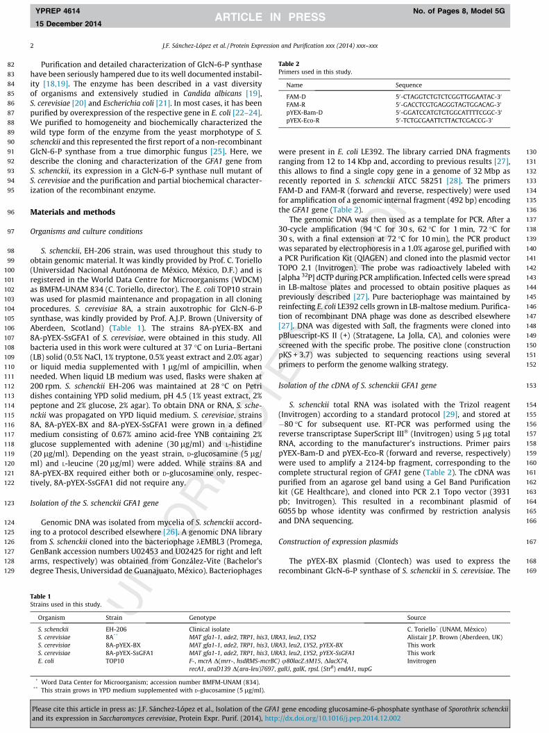
To induce the expression of GlcN-6-P synthase in S. cerevisiae,two concentrations of copper sulfate and different times ofinduction were evaluated. As shown in Fig. 2, the highest level ofinduction was achieved after 24 h of incubation with 1 mM coppersulfate. These conditions were used in further experiments.
Prior to purification, some general properties of the recombi-nant enzyme were determined essentially as described earlier forthe wild type counterpart [25]. Maximum activity was obtainedat pH 7.0 in either 25 mM Tris–HCl or potassium phosphate buf-fers, and at 30 �C. At 18, 37 and 42 �C, enzyme activity represented13.6%, 74% and 9% of maximal activity, respectively (not shown).Recombinant GlcN-6-P synthase was more stable than the wildtype enzyme. Accordingly, the crude extract lost about 17% and5% of the initial activity after 6 days at 4 �C and �20 �C, respec-tively, when stored in 25 mM potassium phosphate buffer, pH7.0, supplemented with 1 mM EDTA, 1 mM DTT and 0.05 mM PMSF(data not shown). This allowed extending the time of purificationto about 3 days where enzyme retained full activity.
Please cite this article in press as: J.F. Sánchez-López et al., Isolation of the GFAand its expression in Saccharomyces cerevisiae, Protein Expr. Purif. (2014), http
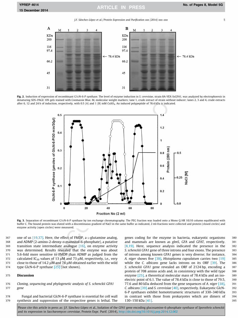
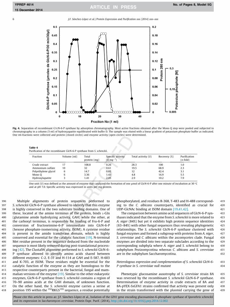
The procedure of purification was essentially similar to thatpreviously described for the wild type enzyme [25] and includedconsecutive enzyme precipitation with ammonium sulfate andpolyethylene glycol followed by anion exchange and adsorptionchromatographies. The elution profiles of the enzyme after the lasttwo steps are shown in Figs. 3 and 4, respectively. Accordingly, theactive protein precipitated with PEG eluted from Mono Q betweenfractions 40 and 60 in the form of a rather broad peak between 0.15and 0.20 M NaCl, well apart from major peaks of spurious proteins(Fig. 3). Further chromatography of the Mono Q fraction in HAPallowed recovery of the recombinant protein as a single peak ofactivity between 0.13 and 0.15 M potassium phosphate (Fig. 4).
Most active fractions from the left side of the peak were pooledand used as the source of purified GlcN-6-P synthase. After the laststep, the expressed enzyme was purified about 8-fold with a recov-ery of 10% with respect to the starting material (Table 4).
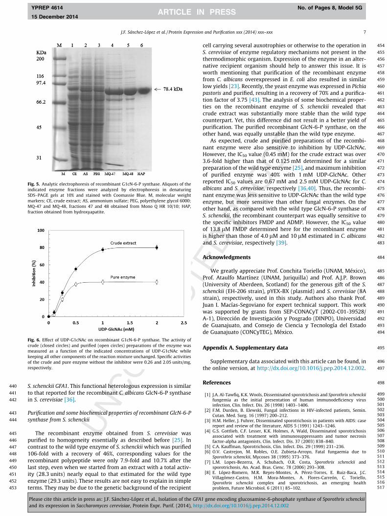
SDS–PAGE (10%) of enzyme preparations obtained after eachstep is depicted in Fig. 5. After HAP, a polypeptide of 79 kDa wasobtained. In addition, a very slight band of lower molecular weightthat migrated close to the active polypeptide was detected. Thiscould not be eliminated in a number of experiments. The degreeof purity of the enzyme was about 90%, as estimated by densito-metric analysis of gels stained with Coomassie Blue (not shown).
Stability, effect of UDP-GlcNAc and Glc-6-P and enzyme sensitivity tospecific inhibitors
When stored in 25 mM potassium phosphate buffer, pH 7.0,containing 1 mM EDTA, 1 mM DTT, 50% glycerol and 10 mMFru-6-P, the purified recombinant enzyme lost 100% activity after24 h at 4 �C but remained fully stable at �20 �C (not shown).
The effect of UDP-GlcNAc, the end product of the Leloir path-way, on recombinant GlcN-6-P synthase, was determined in crudeand purified preparations of the enzyme. Results illustrated inFig. 6 indicate that maximum inhibition of the enzyme occurredat 1 mM, corresponding to 78% and 40% for the crude and purefractions, respectively. The IC50 value for the crude enzyme was0.45 mM. It has been observed that Glc-6-P but not other structur-ally related sugar-phosphates significantly increase the sensitivityof GlcN-6-P synthase from C. albicans to UDP-GlcNAc [36]. On thisbackground, we analyzed the inhibitory effect of UDP-GlcNAc inthe presence and absence of Glc-6-P in crude and purified prepara-tions of recombinant GlcN-6-P synthase. Results in terms of resto-ration of enzyme sensitivity to inhibition by UDP-GlcNAc by thesugar phosphate were essentially similar to those observed forthe wild type enzyme [25] (not shown).
GlcN-6-P synthase has been proposed as a potential target forantifungal agents, a number of which have been developed by
1 gene encoding glucosamine-6-phosphate synthase of Sporothrix schenckii://dx.doi.org/10.1016/j.pep.2014.12.002
367
368
369
370
371
372
373
374
375
376
377
378
379
380
381
382
383
384
385
386
387
388
389
390
391
392
393
394
395
Fig. 2. Induction of expression of recombinant G1cN-6-P synthase. The level of enzyme induction in S. cerevisiae, strain 8A-YEX-SsGFA1, was analyzed by electrophoresis indenaturing SDS–PAGE 10% gels stained with Coomassie Blue. M, molecular weight markers; lane 1, crude extract of strain without inducer; lanes 2, 3 and 4, crude extractsafter 6, 12 and 24 h of induction, respectively, with 0.5 (A) and 1 (B) mM CuSO4. An induced polypeptide of 78.4 kDa is indicated.
Fig. 3. Separation of recombinant G1cN-6-P synthase by ion exchange chromatography. The PEG fraction was loaded onto a Mono Q HR 10/10 column equilibrated withbuffer C. The bound protein was eluted with a discontinuous gradient of NaCl in the same buffer as indicated, 2 ml-fractions were collected and protein (closed circles) andenzyme activity (open circles) were measured.
J.F. Sánchez-López et al. / Protein Expression and Purification xxx (2014) xxx–xxx 5
YPREP 4614 No. of Pages 8, Model 5G
15 December 2014
one of us [19,37]. Here, the effect of FMDP, a L-glutamine analog,and ADMP (2-amino-2-deoxy-D-mannitol-6-phosphate), a putativetransition state intermediate analogue [16], on enzyme activitywas determined. Results revealed that the enzyme was about5.6-fold more sensitive to FMDP than ADMP as judged from thecalculated IC50 values of 13 lM and 73 lM, respectively, i.e., veryclose to those of 14.2 lM and 74 lM obtained earlier with the wildtype GlcN-6-P synthase [25] (not shown).
Discussion
Cloning, sequencing and phylogenetic analysis of S. schenckii GFA1gene
Fungal and bacterial GlcN-6-P synthase is essential for cell wallsynthesis and suppression of the respective genes is lethal. The
Please cite this article in press as: J.F. Sánchez-López et al., Isolation of the GFA1and its expression in Saccharomyces cerevisiae, Protein Expr. Purif. (2014), http
genes coding for the enzyme in bacteria, eukaryotic organismsand mammals are known as glmS, GFA and GFAT, respectively.[8,19]. Here, sequence analysis indicated the presence in theS. schenckii GFA1 gene of three introns and four exons. The presenceof introns among known GFA1 genes is very diverse; for instance,A. niger shows five [38], Histoplasma capsulatum carries two [19]while the C. albicans gene lacks introns on its ORF [39]. TheS. schenckii GFA1 gene revealed an ORF of 2124 bp, encoding aprotein of 708 amino acids and, in consistency with the wild typeenzyme [25], a theoretical molecular mass of 78.4 kDa and an iso-electric point of 6.1. The value of 78.4 kDa is close to those of 79.5,77.6 and 80 kDa deduced from the gene sequences of A. niger [38],C. albicans [39] and S. cerevisiae [40], respectively. Eukaryotic GlcN-6-P synthases exhibit homotetrameric structures of 330–340 kDain contrast with those from prokaryotes which are dimers of130–150 kDa [41].
gene encoding glucosamine-6-phosphate synthase of Sporothrix schenckii://dx.doi.org/10.1016/j.pep.2014.12.002
396
397
398
399
400
401
402
403
404
405
406
407
408
409
410
411
412
413
414
415
416
417
418
419
420
421
422
423
424
425
426
427
428
429
430
431
432
433
434
435
436
437
438
439
Fig. 4. Separation of recombinant G1cN-6-P synthase by adsorption chromatography. Most active fractions obtained after the Mono Q step were pooled and subjected tochromatography in a column (5 ml) of hydroxyapatite equilibrated with buffer D. The sample was eluted with a linear gradient of potassium phosphate buffer as indicated.One ml-fractions were collected and protein (closed circles) and enzyme activity (open circles) were determined.
Table 4Purification of the recombinant GlcN-6-P synthase from S. schenckii.
Fraction Volume (ml) Totalprotein (mg)
Specific activity(U mg�1)
Total activity (U) Recovery (%) Purification(n-fold)
Crude extract 17 108.8 0.26 28.3 100 1.0Ammonium sulfate 10 32 0.61 19.5 68.9 2.3Polyethylene glycol 6 14.7 0.82 12 42.4 3.1Mono Q 6 3.36 1.43 4.8 16.9 5.5Hydroxylapatite 3 1.41 2.05 2.9 10.2 7.9
One unit (U) was defined as the amount of enzyme that catalyzed the formation of one lmol of GlcN-6-P after one minute of incubation at 30 �Cand at pH 7.0. Specific activity was expressed in units per mg protein.
6 J.F. Sánchez-López et al. / Protein Expression and Purification xxx (2014) xxx–xxx
YPREP 4614 No. of Pages 8, Model 5G
15 December 2014
Multiple alignments of protein sequences performed toS. schenckii GlcN-6-P synthase allowed to identify that this enzymeis highly conserved in the two substrate binding domains. One ofthese, located at the amino terminus of the protein, binds L-Gln(glutamine amide hydrolyzing activity, GAH) while the other, atthe carboxyl terminus, is responsible for binding of Fru-6-P andconversion of fructoseimine-6-P intermediate into GlcN-6-P(hexose phosphate-isomerising activity, ISOM). A cysteine residueis present in the amido transferase domain, which is highlyconserved and essential for the catalytic function [19]. N-terminalMet residue present in the sequence deduced from the nucleotidesequence is most likely removed during post-translational process-ing [42]. The ClustalW alignment performed to S. schenckii GlcN-6-P synthase allowed to identify amino acids shared betweendifferent enzymes: C-2, E-31 and H-114 at GAH and E-587, H-603and K-703, at ISOM. These residues might be essential for thecatalytic function of the enzyme as they are homologous to therespective counterparts present in the bacterial, fungal and mam-malian versions of the enzyme [19]. Similar to the other eukaryoticenzymes, GlcN-6-P synthase from S. schenckii contains a region of70–90 amino acids at the GAH domain, of unknown function.On the other hand, the S. schenckii enzyme carries a serine atposition 195 within the 192RKGS195 sequence, a residue potentially
Please cite this article in press as: J.F. Sánchez-López et al., Isolation of the GFAand its expression in Saccharomyces cerevisiae, Protein Expr. Purif. (2014), http
phosphorylated, and residues R-368, T-483 and H-488 correspond-ing to the C. albicans counterparts, identified as crucial forUDP-GlcNAc binding at ISOM domain [19,41,42].
The comparison between amino acid sequences of GlcN-6-P syn-thases indicated that the enzyme from S. schenckii is more related toA. niger (84%) but yet it exhibits high protein sequence identities(63–84%) with other fungal sequences thus revealing phylogeneticrelationships. The S. schenckii GlcN-6-P synthase clustered withfungal enzymes and formed a subgroup with proteins from A. niger,S. cerevisiae and C. albicans within the ascomycetes clade. Fungalenzymes are divided into two separate subclades according to thecorresponding subphyla where A. niger and S. schenckii belong tosubphylum Pezizomycotina whereas C. albicans and S. cerevisiaeare in the subphylum Saccharomycotina.
Heterologous expression and complementation of S. schenckii GlcN-6-P synthase in S. cerevisiae null mutant
Phenotypic glucosamine auxotrophy of S. cerevisiae strain 8Awas reversed by the recombinant S. schenckii GlcN-6-P synthase.Determination of enzyme activity in crude extracts of 8A and8A-pYEX-SsGFA1 strains confirmed that activity was present onlyin the strain transformed with the plasmid carrying the gene of
1 gene encoding glucosamine-6-phosphate synthase of Sporothrix schenckii://dx.doi.org/10.1016/j.pep.2014.12.002
440
441
442
443
444
445
446
447
448
449
450
451
452
453
454
455
456
457
458
459
460
461
462
463
464
465
466
467
468
469
470
471
472
473
474
475
476
477
478
479
480
481
482
483
484
485
486
487
488
489
490
491
492
493
494
495
496
497
498
499500501502503504505506507508509510511512513514515516517
Fig. 5. Analytic electrophoresis of recombinant G1cN-6-P synthase. Aliquots of theindicated enzyme fractions were analyzed by electrophoresis in denaturingSDS–PAGE gels at 10% and stained with Coomassie Blue. M, molecular weightmarkers; CE, crude extract; AS, ammonium sulfate; PEG, polyethylene glycol 6000;MQ-47 and MQ-48, fractions 47 and 48 obtained from Mono Q HR 10/10; HAP,fraction obtained from hydroxyapatite.
Fig. 6. Effect of UDP-G1cNAc on recombinant G1cN-6-P synthase. The activity ofcrude (closed circles) and purified (open circles) preparations of the enzyme wasmeasured as a function of the indicated concentrations of UDP-G1cNAc whilekeeping all other components of the reaction mixture unchanged. Specific activitiesof the crude and pure enzyme without the inhibitor were 0.26 and 2.05 units/mg,respectively.
J.F. Sánchez-López et al. / Protein Expression and Purification xxx (2014) xxx–xxx 7
YPREP 4614 No. of Pages 8, Model 5G
15 December 2014
S. schenckii GFA1. This functional heterologous expression is similarto that reported for the recombinant C. albicans GlcN-6-P synthasein S. cerevisiae [36].
Purification and some biochemical properties of recombinant GlcN-6-Psynthase from S. schenckii
The recombinant enzyme obtained from S. cerevisiae waspurified to homogeneity essentially as described before [25]. Incontrast to the wild type enzyme of S. schenckii which was purified106-fold with a recovery of 46%, corresponding values for therecombinant polypeptide were only 7.9-fold and 10.7% after thelast step, even when we started from an extract with a total activ-ity (28.3 units) nearly equal to that estimated for the wild typeenzyme (29.3 units). These results are not easy to explain in simpleterms. They may be due to the genetic background of the recipient
Please cite this article in press as: J.F. Sánchez-López et al., Isolation of the GFA1and its expression in Saccharomyces cerevisiae, Protein Expr. Purif. (2014), http
cell carrying several auxotrophies or otherwise to the operation inS. cerevisiae of enzyme regulatory mechanisms not present in thethermodimorphic organism. Expression of the enzyme in an alter-native recipient organism should help to answer this issue. It isworth mentioning that purification of the recombinant enzymefrom C. albicans overexpressed in E. coli also resulted in similarlow yields [23]. Recently, the yeast enzyme was expressed in Pichiapastoris and purified, resulting in a recovery of 70% and a purifica-tion factor of 3.75 [43]. The analysis of some biochemical proper-ties on the recombinant enzyme of S. schenckii revealed thatcrude extract was substantially more stable than the wild typecounterpart. Yet, this difference did not result in a better yield ofpurification. The purified recombinant GlcN-6-P synthase, on theother hand, was equally unstable than the wild type enzyme.
As expected, crude and purified preparations of the recombi-nant enzyme were also sensitive to inhibition by UDP-GlcNAc.However, the IC50 value (0.45 mM) for the crude extract was over3.6-fold higher than that of 0.125 mM determined for a similarpreparation of the wild type enzyme [25], and maximum inhibitionof purified enzyme was 40% with 1 mM UDP-GlcNAc. Otherreported IC50 values are 0.67 mM and 2.5 mM UDP-GlcNAc for C.albicans and S. cerevisiae, respectively [36,40]. Thus, the recombi-nant enzyme was less sensitive to UDP-GlcNAc than the wild typeenzyme, but more sensitive than other fungal enzymes. On theother hand, as compared with the wild type GlcN-6-P synthase ofS. schenckii, the recombinant counterpart was equally sensitive tothe specific inhibitors FMDP and ADMP. However, the IC50 valueof 13.8 lM FMDP determined here for the recombinant enzymeis higher than those of 4.0 lM and 10 lM estimated in C. albicansand S. cerevisiae, respectively [39].
Acknowledgments
We greatly appreciate Prof. Conchita Toriello (UNAM, México),Prof. Ataulfo Martínez (UNAM, Juriquilla) and Prof. A.J.P. Brown(University of Aberdeen, Scotland) for the generous gift of the S.schenckii (EH-206 strain), pYEX-BX (plasmid) and S. cerevisiae (8Astrain), respectively, used in this study. Authors also thank Prof.Juan I. Macías-Segoviano for expert technical support. This workwas supported by grants from SEP-CONACyT (2002-C01-39528/A-1), Dirección de Investigación y Posgrado (DINPO), Universidadde Guanajuato, and Consejo de Ciencia y Tecnología del Estadode Guanajuato (CONCyTEG), México.
Appendix A. Supplementary data
Supplementary data associated with this article can be found, inthe online version, at http://dx.doi.org/10.1016/j.pep.2014.12.002.
References
[1] J.A. Al-Tawfiq, K.K. Wools, Disseminated sporotrichosis and Sporothrix schenckiifungemia as the initial presentation of human immunodeficiency virusinfection, Clin. Infect. Dis. 26 (1998) 1403–1406.
[2] F.M. Durden, B. Elewski, Fungal infections in HIV-infected patients, Semin.Cutan. Med. Surg. 16 (1997) 200–212.
[3] H.M. Heller, J. Fuhrer, Disseminated sporotrichosis in patients with AIDS: casereport and review of the literature, AIDS 5 (1991) 1243–1246.
[4] G.S. Gottlieb, C.F. Lesser, K.K. Holmes, A. Wald, Disseminated sporotrichosisassociated with treatment with immunosuppressants and tumor necrosisfactor-alpha antagonists, Clin. Infect. Dis. 37 (2003) 838–840.
[5] C.A. Kauffman, Sporotrichosis, Clin. Infect. Dis. 29 (1999) 231–236.[6] O.V. Castrejon, M. Robles, O.E. Zubieta-Arroyo, Fatal fungaemia due to
Sporothrix schenckii, Mycoses 38 (1995) 373–376.[7] L.M. Lopes-Bezerra, A. Schubach, O.R. Costa, Sporothrix schenckii and
sporotrichosis, An. Acad. Bras. Cienc. 78 (2006) 293–308.[8] E. López-Romero, M.R. Reyes-Montes, A. Pérez-Torres, E. Ruiz-Baca, J.C.
Villagómez-Castro, H.M. Mora-Montes, A. Flores-Carreón, C. Toriello,Sporothrix schenckii complex and sporotrichosis, an emerging healthproblem, Future Microbiol. 6 (2011) 85–102.
gene encoding glucosamine-6-phosphate synthase of Sporothrix schenckii://dx.doi.org/10.1016/j.pep.2014.12.002

518519520521522523524525526527528529530531532533534535536537538539540541542543544545546547548549550551552553554555556557558559560561562563564565566567568569570571572
573574575576577578579580581582583584585586587588589590591592593594595596597598599600601602603604605606607608609610611612613614615616617618619620621622623624625
626
8 J.F. Sánchez-López et al. / Protein Expression and Purification xxx (2014) xxx–xxx
YPREP 4614 No. of Pages 8, Model 5G
15 December 2014
[9] R. Marimon, J. Cano, J. Gené, D.A. Sutton, M. Kawasaki, J. Guarro, Sporothrixbrasiliensis, S. globosa, and S. mexicana, three new Sporothrix species of clinicalinterest, J. Clin. Microbiol. 45 (2007) 3198–3206.
[10] H. Madrid, J. Cano, J. Gené, A. Bonifaz, C. Toriello, J. Guarro, Sporothrix globosa, apathogenic fungus with widespread geographical distribution, Rev. Iberoam.Micol. 26 (2009) 218–222.
[11] N. Rodríguez-del Valle, M. Rosario, G. Torres-Blasini, Effects of pH,temperature, aeration and carbon source on the development of themycelial or yeast forms of Sporothrix schenckii from conidia, Mycopathologia82 (1983) 83–88.
[12] B. DiDomenico, Novel antifungal drugs, Curr. Opin. Microbiol. 2 (1999) 509–515.
[13] V.L. Kan, J.E. Bennett, Efficacies of four antifungal agents in experimentalmurine sporotrichosis, Antimicrob. Agents Chemother. 32 (1988) 1619–1623.
[14] R. Marimon, J. Gené, J. Cano, L. Trilles, M. Dos Santos Lazéra, J. Guarro,Molecular phylogeny of Sporothrix schenckii, J. Clin. Microbiol. 44 (2006) 3251–3256.
[15] C.P. Silveira, J.M. Torres-Rodríguez, E. Alvarado-Ramírez, F. Murciano-Gonzalo,M. Dolande, M. Panizo, V. Reviakina, MICs and minimun fungicidalconcentrations of amphotericin B, itraconazole, posaconazole and terbinafinein Sporothrix schenckii, J. Med. Microbiol. 58 (2009) 1607–1610.
[16] S. Milewski, F. Mignini, L. Micossi, E. Borowski, Antihistoplasmal in vitro andin vivo effect of Lys-Nva-FMDP, Med. Mycol. 36 (1998) 177–180.
[17] T. Naderer, E. Wee, M.J. McConville, Role of hexosamine biosynthesis inLeishmania growth and virulence, Mol. Microbiol. 69 (2008) 858–869.
[18] Q.K. Huynh, E.A. Gulve, T. Dian, Purification and characterization ofglutamine:fructose 6-phosphate amidotransferase from rat liver, Arch.Biochem. Biophys. 379 (2000) 307–313.
[19] S. Milewski, Glucosamine-6-phosphate synthase–the multi-facets enzyme,Biochim. Biophys. Acta 1597 (2002) 173–192.
[20] S. Milewski, I. Gabriel, J. Olchowy, Enzymes of UDP–GlcNAc biosynthesis inyeast, Yeast 23 (2006) 1–14.
[21] N. Floquet, S. Moulleron, R. Daher, B. Maigret, B. Badet, M.A. Badet-Denisot,Ammonia channeling in bacterial glucosamine-6-phophate synthase (glmS):molecular dynamics simulations and kinetic studies of protein mutants, FEBSLett. 581 (2007) 2981–2987.
[22] S. Dutka-Malen, P. Mazodier, B. Badet, Molecular cloning and overexpressionof the glucosamine synthetase gene from Escherichia coli, Biochimie 70 (1988)287–290.
[23] P. Sachadyn, R. Jedrzejczak, S. Milewski, J. Kur, E. Borowski, Purification tohomogeneity of Candida albicans glucosamine-6-phosphate synthaseoverexpressed in Escherichia coli, Protein Expr. Purif. 19 (2000) 343–349.
[24] C. Richez, J. Boetzel, N. Floquet, K. Koteshwar, J. Stevens, B. Badet, M.A. Badet-Denisot, Expression and purification of active human internal His(6)-tagged l-glutamine:d-fructose-6-P amidotransferase I, Protein Expr. Purif. 54 (2007)45–53.
[25] J. González-Ibarra, S. Milewski, J.C. Villagomez-Castro, C. Cano-Canchola, E.Lopez-Romero, Sporothrix schenckii: purification and partial biochemicalcharacterization of glucosamine-6-phosphate synthase, a potentialantifungal target, Med. Mycol. 48 (2010) 110–121.
[26] A. Flores-Martínez, I. Chet, A. Herrera-Estrella, Improved biocontrol activity ofTrichoderma harzianum by over expression of the proteinase encoding geneprb1, Curr. Genet. 31 (1997) 30–37.
[27] A. López-Esparza, A. Álvarez-Vargas, H.M. Mora-Montes, A. Hernández-Cervantes, C. Cano-Canchola, A. Flores-Carreón, Isolation of Sporothrix
Please cite this article in press as: J.F. Sánchez-López et al., Isolation of the GFAand its expression in Saccharomyces cerevisiae, Protein Expr. Purif. (2014), http
schenckii GDA1 and functional characterization of the encoded guanosinediphosphatase activity, Arch. Microbiol. 195 (2013) 499–506.
[28] C.A. Cuomo, N. Rodriguez-Del Valle, L. Perez-Sanchez, A. Abouelleil, J.Goldberg, S. Young, Q. Zeng, B.W. Birren, Genome sequence of thepathogenic fungus Sporothrix schenckii (ATCC 58251), Genome Announc. 2(2014) e00446–14.
[29] P. Chomczynski, K. Mackey, Short technical reports. Modification of the TRIreagent procedure for isolation of RNA from polysaccharide- andproteoglycan-rich sources, Biotechniques 19 (1995) 942–945.
[30] K. Tamura, J. Dudley, M. Nei, S. Kumar, MEGA4: Molecular EvolutionaryGenetics Analysis (MEGA) software version 4.0, Mol. Biol. Evol. 24 (2007)1596–1599.
[31] J.S. Papadopoulos, R. Agarwala, COBALT: constrained-based alignment tool formultiple protein sequences, Bioinformatics 23 (2007) 1073–1079.
[32] P. Artimo, M. Jonnalagedda, K. Arnold, D. Baratin, G. Csardi, E. de Castro, S.Duvaud, V. Flegel, A. Fortier, E. Gasteiger, A. Grosdidier, C. Hernandez, V.Ioannidis, D. Kuznetsov, R. Liechti, S. Moretti, K. Mostaguir, N. Redaschi, G.Rossier, I. Xenarios, H. Stockinger, ExPASy: SIB bioinformatics resource portal,Nucleic Acids Res. 40 (2012) W597–W603.
[33] H. Ito, Y. Fukuda, K. Murata, A. Kimura, Transformation of intact yeast cellstreated with alkali cations, J. Bacteriol. 153 (1983) 163–168.
[34] M.M. Bradford, A rapid and sensitive method for the quantitation ofmicrogram quantities of protein utilizing the principle of protein-dyebinding, Anal. Biochem. 72 (1976) 248–254.
[35] J. Sambrook, D.W. Russell, Molecular Cloning: A Laboratory Manual, third ed.,Cold Spring Harbor Laboratory Press, New York, 2001.
[36] S. Milewski, D. Kuszczak, R. Jedrzejczak, R.J. Smith, A.J.P. Brown, G.W. Gooday,Oligomeric structure and regulation of Candida albicans glucosamine-6-phosphate synthase, J. Biol. Chem. 274 (1999) 4000–4008.
[37] M. Wojciechowski, S. Milewski, J. Mazerski, E. Borowski, Glucosamine-6-phosphate synthase, a novel target for antifungal agents. Molecular modellingstudies in drug design, Acta Biochim. Pol. 52 (2005) 647–653.
[38] A.F. Ram, M. Arentshorst, R.A. Damveld, P.A. vanKuyk, F.M. Klis, C.A. van denHondel, The cell wall stress response in Aspergillus niger involves increasedexpression of the glutamine:fructose-6-phosphate amidotransferase-encodinggene (gfaA) and increased deposition of chitin in the cell wall, Microbiology150 (2004) 3315–3326.
[39] R.J. Smith, S. Milewski, A.J. Brown, G.W. Gooday, Isolation and characterizationof the GFA1 gene encoding the glutamine:fructose-6-phosphateamidotransferase of Candida albicans, J. Bacteriol. 178 (1996) 2320–2327.
[40] G. Watzele, W. Tanner, Cloning of the glutamine:fructose-6-phosphateamidotransferase gene from yeast. Pheromonal regulation of itstranscription, J. Biol. Chem. 264 (1989) 8753–8758.
[41] M.A. Badet-Denisot, H.L.A. Fernandez, J. Berenguer, Characterization of L-glutamine D-fructose-6-phosphate amidotransferase from an extremethermopile Termus thermophilus HB8, Arch. Biochem. Biophys. 337 (1997)129–136.
[42] B. Dummitt, W.S. Micka, Y.H. Chang, Yeast glutamine-fructose-6-phosphateaminotransferase (GFA1) requires methionine aminopeptidase activity forproper function, J. Biol. Chem. 280 (2005) 14356–14360.
[43] S. Wang, P. Li, J. Su, X. Wu, R. Liang, Characterization and expression ofglucosamine-6-phosphate synthase from Saccharomyces cerevisiae in Pichiapastoris, Biotechnol. Lett. 36 (2014) 2023–2026.
1 gene encoding glucosamine-6-phosphate synthase of Sporothrix schenckii://dx.doi.org/10.1016/j.pep.2014.12.002


![Is Sporothrix chilensis circulating outside Chile? Sporothrix... · [1]. Is caused by the thermodimorphic fungi of the genus Sporothrix which are associated, in the environment, with](https://static.fdocuments.net/doc/165x107/5f8537b31c9c5722c234c0e4/is-sporothrix-chilensis-circulating-outside-chile-sporothrix-1-is-caused.jpg)


