Intracellular pathway of Onconase that enables its...
7
1405 Research Article Introduction Onconase ® (Onco, also termed ranpirnase) is selectively cytotoxic to transformed cells (Darzynkiewicz et al., 1988) and possesses potent in vivo antitumour activity (Mikulski et al., 1990). Onco is currently undergoing phase III clinical trials for the treatment of malignant mesothelioma (Favaretto, 2005). Onco toxicity essentially results from its ability to degrade tRNAs (Saxena et al., 2002), whereas intracellular routing that enables Onco delivery to the cytosol remains elusive. Pioneer studies showed that Onco binding to cells was saturable and probably involved membrane receptors. Moreover, metabolic poisons were found to protect cells against Onco toxicity, indicating that this toxin does not access the cytosol by direct transport across the plasma membrane, and that endocytosis is required for cytotoxicity (Wu et al., 1993). Several receptors enter cells using clathrin-mediated endocytosis (CME). This process is dependent on clathrin, adaptors and the GTPase dynamin (Conner and Schmid, 2003). The involvement of dynamin in internalization processes is usually studied using the dynamin-K44A mutant that has a weak affinity for GTP and acts in a dominant-negative manner (Damke et al., 1994). Regarding Onco entry, toxicity was found to be slightly enhanced upon dynamin-K44A expression by stably transfected and inducible HeLa cells, indicating that Onco internalization is dynamin independent (Haigis and Raines, 2003). Following initial uptake, endocytosed tracers are delivered to sorting endosomes. From there they can be directed to the endosome recycling compartment located in the pericentriolar region of the cell. This is the case for recycling ligands such as transferrin (Tf). Alternatively, endocytosed proteins can be transported to late endosomes/lysosomes (Gruenberg and Maxfield, 1995). Most RNases seem to follow this degradation pathway, because neither RNase A (Haigis and Raines, 2003) nor a human pancreatic RNase variant colocalized with Tf upon uptake. The latter mutant indeed accumulated within late endosomes/lysosomes (Bosch et al., 2004). Onco intracellular routing has been evaluated using drugs, such as those able to increase acidic endosomal pH, which is in the pH 5.5–6.5 range (Gruenberg and Maxfield, 1995). Among these molecules, ammonium chloride did not show any effect, whereas monensin enhanced Onco toxicity by 10-fold. Brefeldin A, which prevents retrograde transport to the endoplasmic reticulum (ER), either had no effect (Wu et al., 1993) or potentiated Onco toxicity by 10-fold (Haigis and Raines, 2003). Both studies concluded that Onco probably gains access to the cytosol from endosomes and not from the Golgi or the ER (Haigis and Raines, 2003; Wu et al., 1993). Accordingly, Onco could be visualized within unidentified intracellular vesicles (Haigis and Raines, 2003). The role of endosome acidification in Onco toxicity has yet to be identified. Onconase ® is an RNase with a very specific property because it is selectively toxic to transformed cells. This toxin is thought to recognize cell surface receptors, and the protection conferred by metabolic poisons against Onconase toxicity indicated that this RNase relies on endocytic uptake to kill cells. Nevertheless, its internalization pathway has yet to be unraveled. We show here that Onconase enters cells using AP-2/clathrin- mediated endocytosis. It is then routed, together with transferrin, to the receptor recycling compartment. Increasing the Onconase concentration in this structure using tetanus toxin light chain expression enhanced Onconase toxicity, indicating that recycling endosomes are a key compartment for Onconase cytosolic delivery. This intracellular destination is specific to Onconase because other (and much less toxic) RNases follow the default pathway to late endosomes/lysosomes. Drugs neutralizing endosomal pH increased Onconase translocation efficiency from purified endosomes during cell-free translocation assays by preventing Onconase dissociation from its receptor at endosomal pH. Consistently, endosome neutralization enhanced Onconase toxicity up to 100-fold. Onconase translocation also required cytosolic ATP hydrolysis. This toxin therefore shows an unusual entry process that relies on clathrin-dependent endocytic uptake and then neutralization of low endosomal pH for efficient translocation from the endosomal lumen to the cytosol. Supplementary material available online at http://jcs.biologists.org/cgi/content/full/120/8/1405/DC1 Key words: Onconase, RNase, Coated pits, Recycling endosomes, Endocytosis, Translocation Summary Intracellular pathway of Onconase that enables its delivery to the cytosol Montserrat Rodríguez 1 , Gerard Torrent 1 , Montserrat Bosch 1 , Fabienne Rayne 2 , Jean-François Dubremetz 2 , Marc Ribó 1 , Antoni Benito 1 , Maria Vilanova 1 and Bruno Beaumelle 2, * 1 Laboratori d’Enginyeria de Proteïnes, Departament de Biologia, Facultat de Ciències, Universitat de Girona, Campus de Montilivi s/n E-17071 Girona, Spain 2 UMR 5539 CNRS, Département Biologie-Santé, Université Montpellier II, 34095 Montpellier Cedex 05, France *Author for correspondence (e-mail: [email protected]) Accepted 13 February 2007 Journal of Cell Science 120, 1405-1411 Published by The Company of Biologists 2007 doi:10.1242/jcs.03427 Journal of Cell Science
Transcript of Intracellular pathway of Onconase that enables its...
1405Research Article
IntroductionOnconase® (Onco, also termed ranpirnase) is selectivelycytotoxic to transformed cells (Darzynkiewicz et al., 1988) andpossesses potent in vivo antitumour activity (Mikulski et al.,1990). Onco is currently undergoing phase III clinical trials forthe treatment of malignant mesothelioma (Favaretto, 2005).
Onco toxicity essentially results from its ability to degradetRNAs (Saxena et al., 2002), whereas intracellular routing thatenables Onco delivery to the cytosol remains elusive. Pioneerstudies showed that Onco binding to cells was saturable andprobably involved membrane receptors. Moreover, metabolicpoisons were found to protect cells against Onco toxicity,indicating that this toxin does not access the cytosol by directtransport across the plasma membrane, and that endocytosis isrequired for cytotoxicity (Wu et al., 1993).
Several receptors enter cells using clathrin-mediatedendocytosis (CME). This process is dependent on clathrin,adaptors and the GTPase dynamin (Conner and Schmid, 2003).The involvement of dynamin in internalization processes is usuallystudied using the dynamin-K44A mutant that has a weak affinityfor GTP and acts in a dominant-negative manner (Damke et al.,1994). Regarding Onco entry, toxicity was found to be slightlyenhanced upon dynamin-K44A expression by stably transfectedand inducible HeLa cells, indicating that Onco internalization isdynamin independent (Haigis and Raines, 2003).
Following initial uptake, endocytosed tracers are deliveredto sorting endosomes. From there they can be directed to theendosome recycling compartment located in the pericentriolarregion of the cell. This is the case for recycling ligands suchas transferrin (Tf). Alternatively, endocytosed proteins can betransported to late endosomes/lysosomes (Gruenberg andMaxfield, 1995). Most RNases seem to follow this degradationpathway, because neither RNase A (Haigis and Raines, 2003)nor a human pancreatic RNase variant colocalized with Tfupon uptake. The latter mutant indeed accumulated within lateendosomes/lysosomes (Bosch et al., 2004).
Onco intracellular routing has been evaluated using drugs,such as those able to increase acidic endosomal pH, which is inthe pH 5.5–6.5 range (Gruenberg and Maxfield, 1995). Amongthese molecules, ammonium chloride did not show any effect,whereas monensin enhanced Onco toxicity by 10-fold. BrefeldinA, which prevents retrograde transport to the endoplasmicreticulum (ER), either had no effect (Wu et al., 1993) orpotentiated Onco toxicity by 10-fold (Haigis and Raines, 2003).Both studies concluded that Onco probably gains access to thecytosol from endosomes and not from the Golgi or the ER(Haigis and Raines, 2003; Wu et al., 1993). Accordingly, Oncocould be visualized within unidentified intracellular vesicles(Haigis and Raines, 2003). The role of endosome acidificationin Onco toxicity has yet to be identified.
Onconase® is an RNase with a very specific propertybecause it is selectively toxic to transformed cells. Thistoxin is thought to recognize cell surface receptors, and theprotection conferred by metabolic poisons againstOnconase toxicity indicated that this RNase relies onendocytic uptake to kill cells. Nevertheless, itsinternalization pathway has yet to be unraveled. We showhere that Onconase enters cells using AP-2/clathrin-mediated endocytosis. It is then routed, together withtransferrin, to the receptor recycling compartment.Increasing the Onconase concentration in this structureusing tetanus toxin light chain expression enhancedOnconase toxicity, indicating that recycling endosomes area key compartment for Onconase cytosolic delivery. Thisintracellular destination is specific to Onconase becauseother (and much less toxic) RNases follow the defaultpathway to late endosomes/lysosomes. Drugs neutralizing
endosomal pH increased Onconase translocation efficiencyfrom purified endosomes during cell-free translocationassays by preventing Onconase dissociation from itsreceptor at endosomal pH. Consistently, endosomeneutralization enhanced Onconase toxicity up to 100-fold.Onconase translocation also required cytosolic ATPhydrolysis. This toxin therefore shows an unusual entryprocess that relies on clathrin-dependent endocytic uptakeand then neutralization of low endosomal pH for efficienttranslocation from the endosomal lumen to the cytosol.
Supplementary material available online athttp://jcs.biologists.org/cgi/content/full/120/8/1405/DC1
Key words: Onconase, RNase, Coated pits, Recycling endosomes,Endocytosis, Translocation
Summary
Intracellular pathway of Onconase that enables itsdelivery to the cytosolMontserrat Rodríguez1, Gerard Torrent1, Montserrat Bosch1, Fabienne Rayne2, Jean-François Dubremetz2,Marc Ribó1, Antoni Benito1, Maria Vilanova1 and Bruno Beaumelle2,*1Laboratori d’Enginyeria de Proteïnes, Departament de Biologia, Facultat de Ciències, Universitat de Girona, Campus de Montilivi s/n E-17071Girona, Spain2UMR 5539 CNRS, Département Biologie-Santé, Université Montpellier II, 34095 Montpellier Cedex 05, France*Author for correspondence (e-mail: [email protected])
Accepted 13 February 2007Journal of Cell Science 120, 1405-1411 Published by The Company of Biologists 2007doi:10.1242/jcs.03427
Jour
nal o
f Cel
l Sci
ence
1406
Results and DiscussionTo visualize the internalization of Onco by human cells, weintroduced a Cys residue into Onco for labeling. We selectedposition 72, which is located in a solvent-exposed surface loopremote from the active site (see supplementary material Fig.S1). OncoS72C was fluorescently labeled to obtain Onco-Red,which was as toxic as Onco and therefore biologically active.
To assess the Onco initial uptake pathway, we useddynamin-K44A and other well-established CME inhibitorssuch as a dominant-negative mutant of Eps15 that selectivelyinhibits clathrin/AP-2-dependent endocytosis (Benmerah et al.,1999), and the intersectin SH3 domain that, whenoverexpressed, impairs clathrin-dependent uptake (Simpson etal., 1999).
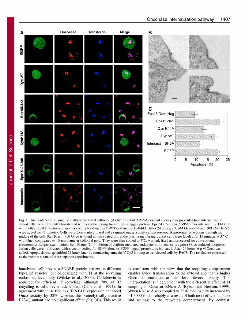
We transiently overexpressed these constructs in Jurkat cellsand monitored the capacity of transfected cells, identified bythe enhanced green fluorescent protein (EGFP) tag of theconstruct (or by cotransfection with EGFP in the case ofdynamin), to internalize Onco-Red. The control version ofEps15 (Eps15D3�2) or dynamin [wild type (WT)] did notaffect the internalization of Onco or the early endosome markerTf, whereas dominant-negative constructions impairing coatedvesicle formation (i.e. Eps15�95/295, the intersectin SH3Adomain and dynamin-K44A) prevented, with almost the sameefficiency, Tf and Onco uptake (Fig. 1A). These resultsindicated that Jurkat cells endocytosed Onco through anEps15-, intersectin- and dynamin-dependent route, andpresumably the well-characterized clathrin/AP-2-mediatedendocytic pathway.
We then examined Onco localization at the plasmamembrane by electron microscopy (EM). Because RNases areconserved proteins it is difficult to raise good antibodiesagainst them (Beintema and Kleineidam, 1998). We thereforedirectly labeled Onco with gold and incubated Jurkat cells at37°C with this conjugate. Onco-gold was found within coatedpits (Fig. 1B). A morphometric analysis showed that more than20% of plasma membrane-associated Onco localized withinthese structures. Because coated pits occupy ~2% of the cellsurface in T cells (Foti et al., 1997), we concluded that Oncois specifically concentrated within coated pits at the plasmamembrane.
To examine whether CME was required by Onco to accessthe cytosol, we tested the ability of the above describedeffectors to protect cells against this toxin. Because Onco killscells by inducing apoptosis (Grabarek et al., 2002; Iordanov etal., 2000), we set up a fluorescence-activated cell sorting(FACS) assay to specifically monitor Onco toxicity totransfected cells using fluorescent annexin-V binding toEGFP-positive cells. Although Onco-induced apoptosis wasnot significantly affected by overexpression of the controlconstructions (Fig. 1C), all the dominant-negative constructsinhibited Onco toxicity with an efficiency ranging from 32%(Dynamin-K44A) to 54% (Eps15 �95/295). Hence, Oncoenters cells using clathrin-mediated uptake, and this enables itssubsequent delivery to the cytosol.
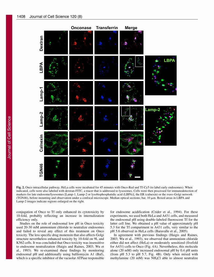
We then examined Onco routing along the endocyticnetwork. This study was performed on HeLa cells that enableeasier visualization and compartment discrimination thanlymphocytes. Onco-Red was internalized in the sameendosomal structures as Tf-Cy5 (Fig. 1A, Fig. 2). In contrastto other RNases that enter cells slowly and are directed to the
degradative pathway (Bosch et al., 2004; Haigis and Raines,2003), Onco-Red was quickly internalized (data not shown)and did not significantly colocalize with established lateendosome/lysosome markers such as lysosomal-associatedmembrane proteins (Lamp-1 and Lamp-2), internalizeddextran, lysobisphosphatidic acid (Fig. 2) or the mannose 6-phosphate receptor (data not shown). Onco-containingstructures were negative for calnexin or TGN46, indicating thatthis protein is not transported to the ER or the trans-Golginetwork. Onco intracellular routing was restrained to Tf-positive structures, and the efficiency of Onco colocalizationwith Tf (75-80%) was the same as that observed when usingtwo Tf bearing different fluorophores (Sabharanjak et al.,2002).
More precisely, Onco seemed to concentrate within acompartment at the center of the cell where fluorescent Tfaccumulates upon labeling, i.e. the recycling compartment(Mallard et al., 1998). Recycling endosomes are the solestructure in the endocytic pathway where the small GTPaseRab11 is present (Sonnichsen et al., 2000). Internalized Oncocolocalized very efficiently with Rab11-EGFP and Tf in thepericentriolar recycling compartment at the center of the cell(see supplementary material Fig. S2). Together, these dataindicated that Onco follows Tf internalization, from coated pitsto the recycling compartment.
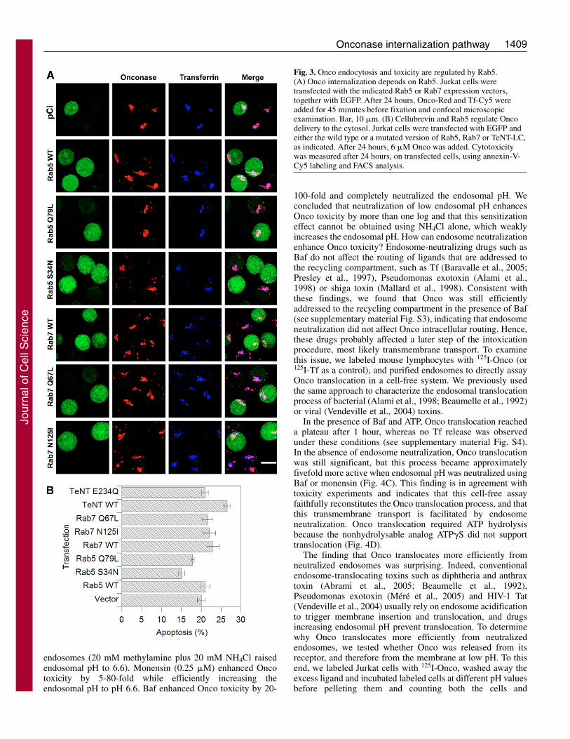
To further show that Onco endocytosis followed a similarpathway to Tf internalization and that Onco was not, as otherRNases, targeted to lysosomes (Bosch et al., 2004), wemonitored the effects of overexpressing Rab5 or Rab7 mutantson Onco endocytosis. Rab5 controls traffic to the sortingendosome compartment (Gruenberg and Maxfield, 1995), anda dominant-negative mutant of Rab5 (Rab5S34N) slows downendocytosis of ligands such as Tf (Stenmark et al., 1994).Active Rab7 is required for transport to late endosomes/lysosomes. Indeed, a dominant-negative version of Rab7(Rab7N125I) prevents tracer delivery to late endosomes, butdoes not significantly inhibit Tf uptake (Bucci et al., 2000;Vitelli et al., 1997). Neither the activated version of Rab5(Ceresa et al., 2001) nor of Rab7 (Bucci et al., 2000; Vitelli etal., 1997) were found to influence uptake of Tf or lysosome-targeted molecules. Accordingly, we observed that the WT orthe activated version of Rab5 and Rab7 did not significantlyaffect Onco or Tf internalization (Fig. 3A). Regardingdominant-negative mutants, Onco and Tf uptake was stronglyinhibited in cells overexpressing Rab5S34N, but was notaffected by Rab7N125I (Fig. 3A).
Similar data were obtained when Onco toxicity totransfected cells was examined, and the sole effector thatsignificantly affected Onco toxicity was dominant-negativeRab5 (Rab5S34N) that partially protected cells against Onco(~25%, Fig. 3B). Together with morphological examination,the Rab5 data showed that Onco internalization and toxicity isregulated by this small GTPase. The result that dominant-negative Rab7 affects neither Onco intracellular routing nor itstoxicity confirmed that the Onco intracellular pathway isrestricted to early endosomes, and that Onco does not needdelivery to late endosomes/lysosomes to reach the cytosol andkill cells.
To examine whether recycling endosomes were implicatedin Onco toxicity we transfected cells with tetanus toxin lightchain (TeNT-LC), a highly specific protease that cleaves and
Journal of Cell Science 120 (8)
Jour
nal o
f Cel
l Sci
ence
1407Onconase internalization pathway
inactivates cellubrevin, a SNARE protein present on differenttypes of vesicles, but colocalizing with Tf at the recyclingendosome level only (Wilcke et al., 2000). Cellubrevin isrequired for efficient Tf recycling, although 70% of Tfrecycling is cellubrevin independent (Galli et al., 1994). Inagreement with these findings, TeNT-LC expression enhancedOnco toxicity by 32%, whereas the proteolytically inactiveE234Q mutant had no significant effect (Fig. 3B). This result
is consistent with the view that the recycling compartmentenables Onco translocation to the cytosol and that a higherOnco concentration at this level favors toxicity. Thisinterpretation is in agreement with the differential effect of Tfcoupling to Onco or RNase A (Rybak and Newton, 1999).When RNase A was coupled to Tf its cytotoxicity increased by~10,000-fold, probably as a result of both more efficient uptakeand routing to the recycling compartment. By contrast,
Fig. 1. Onco enters cells using the clathrin-mediated pathway. (A) Inhibition of AP-2-dependent endocytosis prevents Onco internalization.Jurkat cells were transiently transfected with a vector coding for an EGFP-tagged protein (Eps15D3�2, Eps15�95/295 or intersectin SH3A), orwith both an EGFP vector and another coding for dynamin II-WT or dynamin II-K44A. After 24 hours, 250 nM Onco-Red and 100 nM Tf-Cy5were added for 45 minutes. Cells were then washed, fixed and examined under a confocal microscope. Representative sections through themiddle of the cell. Bar, 10 �m. (B) Onco is found within coated pits at the plasma membrane. Jurkat cells were labeled for 15 minutes at 37°Cwith Onco conjugated to 10-nm diameter colloidal gold. They were then cooled to 4°C, washed, fixed and processed for conventionalelectronmicroscopic examination. Bar, 50 nm. (C) Inhibition of clathrin-mediated endocytosis protects cells against Onco-induced apoptosis.Jurkat cells were transfected with a vector coding for EGFP alone or EGFP-tagged proteins, as indicated. After 24 hours, 6 �M Onco wasadded. Apoptosis was quantified 24 hours later by monitoring annexin-V-Cy5 binding to transfected cells by FACS. The results are expressedas the mean ± s.e.m. of three separate experiments.
Jour
nal o
f Cel
l Sci
ence
1408
conjugation of Onco to Tf only enhanced its cytotoxicity by10-fold, probably reflecting an increase in internalizationefficiency only.
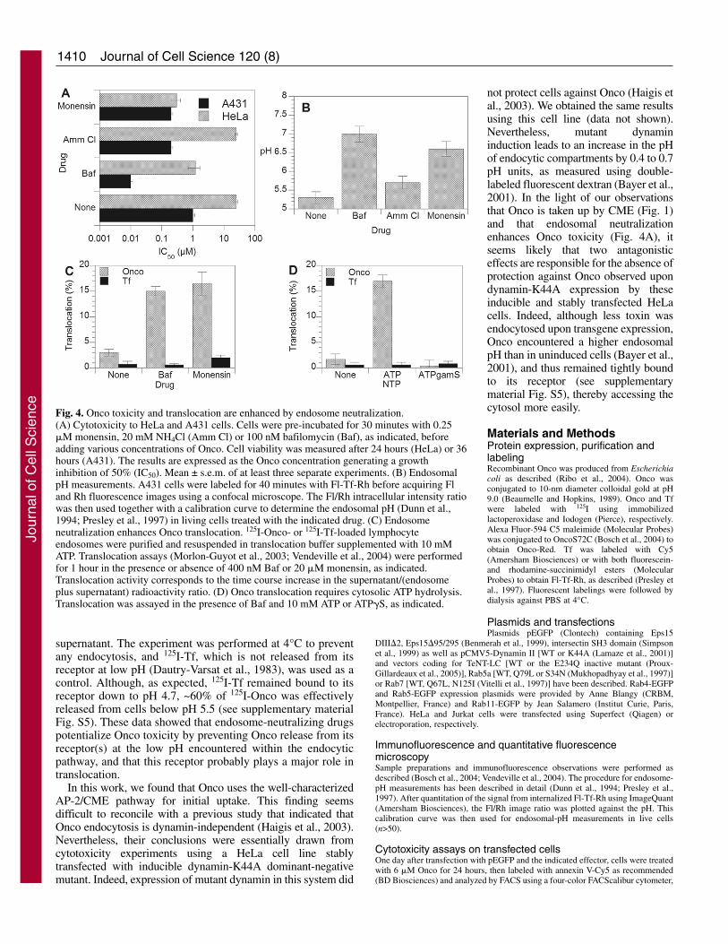
Studies on the role of endosomal low pH in Onco toxicityused 20-30 mM ammonium chloride to neutralize endosomesand failed to reveal any effect of this treatment on Oncotoxicity. The less specific drug monensin that also affects Golgistructure nevertheless enhanced toxicity by 10-fold on 9L andK562 cells. It was concluded that Onco toxicity was insensitiveto endosome neutralization (Haigis and Raines, 2003; Wu etal., 1993). We re-examined these findings by monitoringendosomal pH and additionally using bafilomycin A1 (Baf),which is a specific inhibitor of the vacuolar ATPase responsible
for endosome acidification (Crider et al., 1994). For theseexperiments, we used both HeLa and A431 cells, and measuredthe endosomal pH using double-labeled fluorescent Tf for thelatter cell line. We obtained a pH value of approximately pH5.3 for the Tf compartment in A431 cells, very similar to thepH 5.6 observed in HeLa cells (Baravalle et al., 2005).
In agreement with previous findings (Haigis and Raines,2003; Wu et al., 1993), we observed that ammonium chlorideeither did not affect (HeLa) or moderately sensitized (fivefoldfor A431) cells to Onco (Fig. 4A). Nevertheless, this moleculealone (20 mM) only increased endosomal pH by 0.4 pH units(from pH 5.3 to pH 5.7, Fig. 4B). Only when mixed withmethylamine (20 mM) was NH4Cl able to almost neutralize
Journal of Cell Science 120 (8)
Fig. 2. Onco intracellular pathway. HeLa cells were incubated for 45 minutes with Onco-Red and Tf-Cy5 (to label early endosomes). Whenindicated, cells were also labeled with dextran-FITC, a tracer that is addressed to lysosomes. Cells were then processed for immunodetection ofmarkers for late endosome/lysosomes [Lamp-1, Lamp-2 or lysobisphosphatidic acid (LBPA)], the ER (calnexin) or the trans-Golgi network(TGN46), before mounting and observation under a confocal microscope. Median optical sections; bar, 10 �m. Boxed areas in LBPA andLamp-2 images indicate regions enlarged on the right.
Jour
nal o
f Cel
l Sci
ence
1409Onconase internalization pathway
endosomes (20 mM methylamine plus 20 mM NH4Cl raisedendosomal pH to 6.6). Monensin (0.25 �M) enhanced Oncotoxicity by 5-80-fold while efficiently increasing theendosomal pH to pH 6.6. Baf enhanced Onco toxicity by 20-
100-fold and completely neutralized the endosomal pH. Weconcluded that neutralization of low endosomal pH enhancesOnco toxicity by more than one log and that this sensitizationeffect cannot be obtained using NH4Cl alone, which weaklyincreases the endosomal pH. How can endosome neutralizationenhance Onco toxicity? Endosome-neutralizing drugs such asBaf do not affect the routing of ligands that are addressed tothe recycling compartment, such as Tf (Baravalle et al., 2005;Presley et al., 1997), Pseudomonas exotoxin (Alami et al.,1998) or shiga toxin (Mallard et al., 1998). Consistent withthese findings, we found that Onco was still efficientlyaddressed to the recycling compartment in the presence of Baf(see supplementary material Fig. S3), indicating that endosomeneutralization did not affect Onco intracellular routing. Hence,these drugs probably affected a later step of the intoxicationprocedure, most likely transmembrane transport. To examinethis issue, we labeled mouse lymphocytes with 125I-Onco (or125I-Tf as a control), and purified endosomes to directly assayOnco translocation in a cell-free system. We previously usedthe same approach to characterize the endosomal translocationprocess of bacterial (Alami et al., 1998; Beaumelle et al., 1992)or viral (Vendeville et al., 2004) toxins.
In the presence of Baf and ATP, Onco translocation reacheda plateau after 1 hour, whereas no Tf release was observedunder these conditions (see supplementary material Fig. S4).In the absence of endosome neutralization, Onco translocationwas still significant, but this process became approximatelyfivefold more active when endosomal pH was neutralized usingBaf or monensin (Fig. 4C). This finding is in agreement withtoxicity experiments and indicates that this cell-free assayfaithfully reconstitutes the Onco translocation process, and thatthis transmembrane transport is facilitated by endosomeneutralization. Onco translocation required ATP hydrolysisbecause the nonhydrolysable analog ATP�S did not supporttranslocation (Fig. 4D).
The finding that Onco translocates more efficiently fromneutralized endosomes was surprising. Indeed, conventionalendosome-translocating toxins such as diphtheria and anthraxtoxin (Abrami et al., 2005; Beaumelle et al., 1992),Pseudomonas exotoxin (Méré et al., 2005) and HIV-1 Tat(Vendeville et al., 2004) usually rely on endosome acidificationto trigger membrane insertion and translocation, and drugsincreasing endosomal pH prevent translocation. To determinewhy Onco translocates more efficiently from neutralizedendosomes, we tested whether Onco was released from itsreceptor, and therefore from the membrane at low pH. To thisend, we labeled Jurkat cells with 125I-Onco, washed away theexcess ligand and incubated labeled cells at different pH valuesbefore pelleting them and counting both the cells and
Fig. 3. Onco endocytosis and toxicity are regulated by Rab5.(A) Onco internalization depends on Rab5. Jurkat cells weretransfected with the indicated Rab5 or Rab7 expression vectors,together with EGFP. After 24 hours, Onco-Red and Tf-Cy5 wereadded for 45 minutes before fixation and confocal microscopicexamination. Bar, 10 �m. (B) Cellubrevin and Rab5 regulate Oncodelivery to the cytosol. Jurkat cells were transfected with EGFP andeither the wild type or a mutated version of Rab5, Rab7 or TeNT-LC,as indicated. After 24 hours, 6 �M Onco was added. Cytotoxicitywas measured after 24 hours, on transfected cells, using annexin-V-Cy5 labeling and FACS analysis.
Jour
nal o
f Cel
l Sci
ence
1410
supernatant. The experiment was performed at 4°C to preventany endocytosis, and 125I-Tf, which is not released from itsreceptor at low pH (Dautry-Varsat et al., 1983), was used as acontrol. Although, as expected, 125I-Tf remained bound to itsreceptor down to pH 4.7, ~60% of 125I-Onco was effectivelyreleased from cells below pH 5.5 (see supplementary materialFig. S5). These data showed that endosome-neutralizing drugspotentialize Onco toxicity by preventing Onco release from itsreceptor(s) at the low pH encountered within the endocyticpathway, and that this receptor probably plays a major role intranslocation.
In this work, we found that Onco uses the well-characterizedAP-2/CME pathway for initial uptake. This finding seemsdifficult to reconcile with a previous study that indicated thatOnco endocytosis is dynamin-independent (Haigis et al., 2003).Nevertheless, their conclusions were essentially drawn fromcytotoxicity experiments using a HeLa cell line stablytransfected with inducible dynamin-K44A dominant-negativemutant. Indeed, expression of mutant dynamin in this system did
not protect cells against Onco (Haigis etal., 2003). We obtained the same resultsusing this cell line (data not shown).Nevertheless, mutant dynamininduction leads to an increase in the pHof endocytic compartments by 0.4 to 0.7pH units, as measured using double-labeled fluorescent dextran (Bayer et al.,2001). In the light of our observationsthat Onco is taken up by CME (Fig. 1)and that endosomal neutralizationenhances Onco toxicity (Fig. 4A), itseems likely that two antagonisticeffects are responsible for the absence ofprotection against Onco observed upondynamin-K44A expression by theseinducible and stably transfected HeLacells. Indeed, although less toxin wasendocytosed upon transgene expression,Onco encountered a higher endosomalpH than in uninduced cells (Bayer et al.,2001), and thus remained tightly boundto its receptor (see supplementarymaterial Fig. S5), thereby accessing thecytosol more easily.
Materials and MethodsProtein expression, purification andlabelingRecombinant Onco was produced from Escherichiacoli as described (Ribo et al., 2004). Onco wasconjugated to 10-nm diameter colloidal gold at pH9.0 (Beaumelle and Hopkins, 1989). Onco and Tfwere labeled with 125I using immobilizedlactoperoxidase and Iodogen (Pierce), respectively.Alexa Fluor-594 C5 maleimide (Molecular Probes)was conjugated to OncoS72C (Bosch et al., 2004) toobtain Onco-Red. Tf was labeled with Cy5(Amersham Biosciences) or with both fluorescein-and rhodamine-succinimidyl esters (MolecularProbes) to obtain Fl-Tf-Rh, as described (Presley etal., 1997). Fluorescent labelings were followed bydialysis against PBS at 4°C.
Plasmids and transfectionsPlasmids pEGFP (Clontech) containing Eps15
DIII�2, Eps15�95/295 (Benmerah et al., 1999), intersectin SH3 domain (Simpsonet al., 1999) as well as pCMV5-Dynamin II [WT or K44A (Lamaze et al., 2001)]and vectors coding for TeNT-LC [WT or the E234Q inactive mutant (Proux-Gillardeaux et al., 2005)], Rab5a [WT, Q79L or S34N (Mukhopadhyay et al., 1997)]or Rab7 [WT, Q67L, N125I (Vitelli et al., 1997)] have been described. Rab4-EGFPand Rab5-EGFP expression plasmids were provided by Anne Blangy (CRBM,Montpellier, France) and Rab11-EGFP by Jean Salamero (Institut Curie, Paris,France). HeLa and Jurkat cells were transfected using Superfect (Qiagen) orelectroporation, respectively.
Immunofluorescence and quantitative fluorescencemicroscopySample preparations and immunofluorescence observations were performed asdescribed (Bosch et al., 2004; Vendeville et al., 2004). The procedure for endosome-pH measurements has been described in detail (Dunn et al., 1994; Presley et al.,1997). After quantitation of the signal from internalized Fl-Tf-Rh using ImageQuant(Amersham Biosciences), the Fl/Rh image ratio was plotted against the pH. Thiscalibration curve was then used for endosomal-pH measurements in live cells(n>50).
Cytotoxicity assays on transfected cellsOne day after transfection with pEGFP and the indicated effector, cells were treatedwith 6 �M Onco for 24 hours, then labeled with annexin V-Cy5 as recommended(BD Biosciences) and analyzed by FACS using a four-color FACScalibur cytometer,
Journal of Cell Science 120 (8)
Fig. 4. Onco toxicity and translocation are enhanced by endosome neutralization.(A) Cytotoxicity to HeLa and A431 cells. Cells were pre-incubated for 30 minutes with 0.25�M monensin, 20 mM NH4Cl (Amm Cl) or 100 nM bafilomycin (Baf), as indicated, beforeadding various concentrations of Onco. Cell viability was measured after 24 hours (HeLa) or 36hours (A431). The results are expressed as the Onco concentration generating a growthinhibition of 50% (IC50). Mean ± s.e.m. of at least three separate experiments. (B) EndosomalpH measurements. A431 cells were labeled for 40 minutes with Fl-Tf-Rh before acquiring Fland Rh fluorescence images using a confocal microscope. The Fl/Rh intracellular intensity ratiowas then used together with a calibration curve to determine the endosomal pH (Dunn et al.,1994; Presley et al., 1997) in living cells treated with the indicated drug. (C) Endosomeneutralization enhances Onco translocation. 125I-Onco- or 125I-Tf-loaded lymphocyteendosomes were purified and resuspended in translocation buffer supplemented with 10 mMATP. Translocation assays (Morlon-Guyot et al., 2003; Vendeville et al., 2004) were performedfor 1 hour in the presence or absence of 400 nM Baf or 20 �M monensin, as indicated.Translocation activity corresponds to the time course increase in the supernatant/(endosomeplus supernatant) radioactivity ratio. (D) Onco translocation requires cytosolic ATP hydrolysis.Translocation was assayed in the presence of Baf and 10 mM ATP or ATP�S, as indicated.
Jour
nal o
f Cel
l Sci
ence
1411Onconase internalization pathway
gating on transfected cells using EGFP fluorescence. Apoptosis was monitoredusing annexin-Cy5 binding to these cells. Approximately 20% of the cells becameannexin-V-positive upon Onco treatment. 10,000 events were collected for eachassay, which was performed in duplicate. Results are the mean ± s.e.m. of threeseparate experiments.
Conventional cytotoxicity and cell-free translocation assaysCytotoxicity assays with pharmacological agents were performed using HeLa andA431 cells as described (Morlon-Guyot et al., 2003). For translocation assays,mouse BW lymphocytes were labeled with 125I-Onco (or 125I-Tf as control) andpurified as described (Vendeville et al., 2004). Endosomes were resuspended intranslocation buffer. Translocation was assayed for 0–90 minutes at 37°C, beforecooling and ultracentrifugation. Translocation was monitored using the increase inthe supernatant/(endosome plus supernatant) radioactivity ratio.
We are very grateful to A. Blangy (Montpellier), A. Benmerah, T.Galli, C. Lamaze and J. Salamero (Paris), J. Gruenberg (Geneva) andR. T. Raines (Madison, USA) for their kind gift of reagents. Thanksare due to V. Richard (SCME-UM2) for assistance with electronmicroscopy. This work was supported by a PICS between the CNRS(PICS No. 3067) and the University of Girona (PICS2005-3,Generalitat de Catalunya) and by grants BMC2003-08485-CO2-02and BFU2006-15543-CO2-02 from Ministerio de Educación yCiencia, and SGR2001-00196 from Generalitat de Catalunya. M.Rodríguez and G.T. gratefully acknowledge their predoctoralfellowships from the Ministerio de Educación y Ciencia. F.R. wasfunded by the ANRS and Sidaction.
ReferencesAbrami, L., Reig, N. and van der Goot, F. G. (2005). Anthrax toxin: the long and
winding road that leads to the kill. Trends Microbiol. 13, 72-78.Alami, M., Taupiac, M. P., Reggio, H., Bienvenue, A. and Beaumelle, B. (1998).
Involvement of ATP-dependent Pseudomonas exotoxin translocation from a laterecycling compartment in lymphocyte intoxication procedure. Mol. Biol. Cell 9, 387-402.
Baravalle, G., Schober, D., Huber, M., Bayer, N., Murphy, R. F. and Fuchs, R. (2005).Transferrin recycling and dextran transport to lysosomes is differentially affected bybafilomycin, nocodazole, and low temperature. Cell Tissue Res. 320, 99-113.
Bayer, N., Schober, D., Huttinger, M., Blaas, D. and Fuchs, R. (2001). Inhibition ofclathrin-dependent endocytosis has multiple effects on human rhinovirus serotype 2cell entry. J. Biol. Chem. 276, 3952-3962.
Beaumelle, B. and Hopkins, C. R. (1989). High-yield isolation of functionally competentendosomes from mouse lymphocytes. Biochem. J. 264, 137-149.
Beaumelle, B., Bensammar, L. and Bienvenue, A. (1992). Selective translocation of theA chain of diphtheria toxin across the membrane of purified endosomes. J. Biol. Chem.267, 11525-11531.
Beintema, J. J. and Kleineidam, R. G. (1998). The ribonuclease A superfamily: generaldiscussion. Cell. Mol. Life Sci. 54, 825-832.
Benmerah, A., Bayrou, M., Cerf-Bensussan, N. and Dautry-Varsat, A. (1999).Inhibition of clathrin-coated pit assembly by an Eps15 mutant. J. Cell Sci. 112, 1303-1311.
Bosch, M., Benito, A., Ribo, M., Puig, T., Beaumelle, B. and Vilanova, M. (2004). Anuclear localization sequence endows human pancreatic ribonuclease with cytotoxicactivity. Biochemistry 43, 2167-2177.
Bucci, C., Thomsen, P., Nicoziani, P., McCarthy, J. and van Deurs, B. (2000). Rab7:a key to lysosome biogenesis. Mol. Biol. Cell 11, 467-480.
Ceresa, B. P., Lotscher, M. and Schmid, S. L. (2001). Receptor and membrane recyclingcan occur with unaltered efficiency despite dramatic Rab5(q79l)-induced changes inendosome geometry. J. Biol. Chem. 276, 9649-9654.
Conner, S. D. and Schmid, S. L. (2003). Regulated portals of entry into the cell. Nature422, 37-44.
Crider, B. P., Xie, X. S. and Stone, D. K. (1994). Bafilomycin inhibits proton flowthrough the H+ channel of vacuolar proton pumps. J. Biol. Chem. 269, 17379-17381.
Damke, H., Baba, T., Warnock, D. E. and Schmid, S. L. (1994). Induction of mutantdynamin specifically blocks endocytic coated vesicle formation. J. Cell Biol. 127, 915-934.
Darzynkiewicz, Z., Carter, S. P., Mikulski, S. M., Ardelt, W. J. and Shogen, K. (1988).Cytostatic and cytotoxic effects of Pannon (P-30 Protein), a novel anticancer agent.Cell Tissue Kinet. 21, 169-182.
Dautry-Varsat, A., Ciechanover, A. and Lodisch, H. (1983). pH and the recycling oftransferrin during receptor-mediated endocytosis. Proc. Natl. Acad. Sci. USA 80, 2258-2262.
Dunn, K. W., Park, J., Semrad, C. E., Gelman, D. L., Shevell, T. and McGraw, T. E.(1994). Regulation of endocytic trafficking and acidification are independent of thecystic fibrosis transmembrane regulator. J. Biol. Chem. 269, 5336-5345.
Favaretto, A. (2005). Overview on ongoing or planned clinical trials in Europe. LungCancer 49, S117-S121.
Foti, M., Mangasarian, A., Piguet, V., Lew, D. P., Krause, K. H., Trono, D. andCarpentier, J. L. (1997). Nef-mediated clathrin-coated pit formation. J. Cell Biol. 139,37-47.
Galli, T., Chilcote, T., Mundigl, O., Binz, T., Niemann, H. and de Camili, P. (1994).Tetanus toxin-mediated cleavage of cellubrevin impairs exocytosis of transferrinreceptor-containing vesicles in CHO cells. J. Cell Biol. 125, 1015-1024.
Grabarek, J., Ardelt, B., Du, L. and Darzynkiewicz, Z. (2002). Activation of caspasesand serine proteases during apoptosis induced by onconase (Ranpirnase). Exp. CellRes. 278, 61-71.
Gruenberg, J. and Maxfield, F. R. (1995). Membrane transport in the endocytic pathway.Curr. Opin. Cell Biol. 7, 552-563.
Haigis, M. C. and Raines, R. T. (2003). Secretory ribonucleases are internalized by adynamin-independent endocytic pathway. J. Cell Sci. 116, 313-324.
Haigis, M. C., Kurten, E. L. and Raines, R. T. (2003). Ribonuclease inhibitor as anintracellular sentry. Nucleic Acids Res. 31, 1024-1032.
Iordanov, M. S., Ryabinina, O. P., Wong, J., Dinh, T. H., Newton, D. L., Rybak, S.M. and Magun, B. E. (2000). Molecular determinants of apoptosis induced by thecytotoxic ribonuclease onconase: evidence for cytotoxic mechanisms different frominhibition of protein synthesis. Cancer Res. 60, 1983-1994.
Lamaze, C., Dujeancourt, A., Baba, T., Lo, C. G., Benmerah, A. and Dautry-Varsat,A. (2001). Interleukin 2 receptors and detergent-resistant membrane domains define aclathrin-independent endocytic pathway. Mol. Cell 7, 661-671.
Mallard, F., Antony, C., Tenza, D., Salamero, J., Goud, B. and Johannes, L. (1998).Direct pathway from early/recycling endosomes to the Golgi apparatus revealedthrough the study of shiga toxin B-fragment transport. J. Cell Biol. 143, 973-990.
Méré, J., Morlon-Guyot, J., Bonhoure, A., Chiche, L. and Beaumelle, B. (2005). Acid-triggered membrane insertion of pseudomonas exotoxin A involves an originalmechanism based on pH-regulated tryptophan exposure. J. Biol. Chem. 280, 21194-21201.
Mikulski, S. M., Ardelt, W., Shogen, K., Bernstein, E. H. and Menduke, H. (1990).Striking increase of survival of mice bearing M109 Madison carcinoma treated with anovel protein from amphibian embryos. J. Natl. Cancer Inst. 82, 151-153.
Morlon-Guyot, J., Helmy, M., Lombard-Frasca, S., Pignol, D., Pieroni, G. andBeaumelle, B. (2003). Identification of the ricin lipase site and implication incytotoxicity. J. Biol. Chem. 278, 17006-17011.
Mukhopadhyay, A., Barbieri, A. M., Funato, K., Roberts, R. and Stahl, P. D. (1997).Sequential actions of Rab5 and Rab7 regulate endocytosis in the Xenopus oocyte. J.Cell Biol. 136, 1227-1237.
Presley, J. F., Mayor, S., McGraw, T. E., Dunn, K. W. and Maxfield, F. R. (1997).Bafilomycin A1 treatment retards transferrin receptor recycling more than bulkmembrane recycling. J. Biol. Chem. 272, 13929-13936.
Proux-Gillardeaux, V., Gavard, J., Irinopoulou, T., Mege, R. M. and Galli, T. (2005).Tetanus neurotoxin-mediated cleavage of cellubrevin impairs epithelial cell migrationand integrin-dependent cell adhesion. Proc. Natl. Acad. Sci. USA 102, 6362-6367.
Ribo, M., Bosch, M., Torrent, G., Benito, A., Beaumelle, B. and Vilanova, M. (2004).Quantitative analysis, using MALDI-TOF mass spectrometry, of the N-terminalhydrolysis and cyclization reactions of the activation process of onconase. Eur. J.Biochem. 271, 1163-1171.
Rybak, S. M. and Newton, D. L. (1999). Natural and engineered cytotoxic ribonucleases:therapeutic potential. Exp. Cell Res. 253, 325-335.
Sabharanjak, S., Sharma, P., Parton, R. G. and Mayor, S. (2002). GPI-anchoredproteins are delivered to recycling endosomes via a distinct cdc42-regulated, clathrin-independent pinocytic pathway. Dev. Cell 2, 411-423.
Saxena, S. K., Sirdeshmukh, R., Ardelt, W., Mikulski, S. M., Shogen, K. and Youle,R. J. (2002). Entry into cells and selective degradation of tRNAs by a cytotoxicmember of the RNase A family. J. Biol. Chem. 277, 15142-15146.
Simpson, F., Hussain, N. K., Qualmann, B., Kelly, R. B., Kay, B. K., McPherson, P.S. and Schmid, S. L. (1999). SH3-domain-containing proteins function at distinct stepsin clathrin-coated vesicle formation. Nat. Cell Biol. 1, 119-124.
Sonnichsen, B., De Renzis, S., Nielsen, E., Rietdorf, J. and Zerial, M. (2000). Distinctmembrane domains on endosomes in the recycling pathway visualized by multicolorimaging of Rab4, Rab5, and Rab11. J. Cell Biol. 149, 901-914.
Stenmark, H., Parton, R. G., Steele-Mortimer, O., Lütcke, A., Gruenberg, J. andZerial, M. (1994). Inhibition of rab5 GTPase activity stimulates membrane fusion inendocytosis. EMBO J. 13, 1287-1296.
Vendeville, A., Rayne, F., Bonhoure, A., Bettache, N., Montcourrier, P. andBeaumelle, B. (2004). HIV-1 Tat enters T-cells using coated pits before translocatingfrom acidified endosomes and eliciting biological responses. Mol. Biol. Cell 15, 2347-2360.
Vitelli, R., Santillo, M., Lattero, D., Chiariello, M., Bifulco, M., Bruni, C. B. andBucci, C. (1997). Role of the small GTPase Rab7 in the late endocytic pathway. J.Biol. Chem. 272, 4391-4397.
Wilcke, M., Johannes, L., Galli, T., Mayau, V., Goud, B. and Salamero, J. (2000).Rab11 regulates the compartmentalization of early endosomes required for efficienttransport from early endosomes to the trans-golgi network. J. Cell Biol. 151, 1207-1220.
Wu, Y., Mikulski, S. M., Ardelt, W., Rybak, S. M. and Youle, R. J. (1993). A cytotoxicribonuclease. Study of the mechanism of onconase cytotoxicity. J. Biol. Chem. 268,10686-10693.
Jour
nal o
f Cel
l Sci
ence