Intestinal anastomosis wound healing after platelet- rich ......Hematopatología para el procesado...
118
Departament de Medicina i Cirurgia Animals Facultat de Veterinària Universitat Autònoma de Barcelona Intestinal anastomosis wound healing after platelet- rich plasma (PRP) application on pigs. Macroscopic, microscopic and breaking strength evaluations PhD Thesis By Otilia Rafael Bambo Director Dr. Félix García Arnas
Transcript of Intestinal anastomosis wound healing after platelet- rich ......Hematopatología para el procesado...

Departament de Medicina i Cirurgia Animals Facultat de Veterinària
Universitat Autònoma de Barcelona
Intestinal anastomosis wound healing after platelet- rich plasma (PRP) application on pigs. Macroscopic,
microscopic and breaking strength evaluations
PhD Thesis
By
Otilia Rafael Bambo
Director
Dr. Félix García Arnas

2
3
Félix García Arnas, Profesor Titular del Departament de Medicina i Cirurgia Animals, de
la Facultat de Veterinària de la Universitat Autònoma de Barcelona,
CERTIFICA:
Que la memoria titulada “INTESTINAL ANASTOMOSIS WOUND HEALING
AFTER PLATELET-RICH PLASMA (PRP) APPLICATION ON PIGS.
MACROSCOPIC, MICROSCOPIC AND BREAKING STRENGTH EVALUATIONS”
presentada por Otilia Rafael Bambo para optar al grado de Doctor, ha sido realizada bajo
su dirección y, considerándola finalizada, autoriza su presentación para que ésta sea juzgada
por el tribunal correspondiente.
Y para que así conste, firmo el presente certificado.
Bellaterra, 4 de Mayo de 2009.
Dr. Félix García Arnas
Edifici V-Campus de la UAB / 08193 Bellaterra (Cerdanyola del Vallès) / Barcelona, Spain
Tel: +34935811091 – Fax: +34935813325
[email protected], www.uab.es

4
5
AGRADECIMIENTOS
Deseo expresar mi más sincero agradecimiento a las siguientes personas, sin las cuales no habría sido capaz de llevar a cabo esta tesis doctoral: Al profesor Félix García Arnas por la dirección de esta tesis, señalando su constante estímulo en el campo de la investigación, su entusiasmo por este trabajo y el afecto que siempre me ha mostrado. He tenido la suerte de encontrar en él a un maestro. A la profesora Dolors Fondevila por sus enseñanzas sobre histopatología, colaboración en el procesamiento de las muestras histológicas y su ayuda en la interpretación histológica. A la profesora Laura Fresno por la dirección y redacción de esta tesis; me ha transmitido su ilusión por la investigación y me ha ayudado con su inagotable capacidad de trabajo. Mi especial reconocimiento a su dedicación. A la profesora Anna Andaluz, Cap de Servei de Anestesiología del Hospital Clínic Veterinari - Universitat Autònoma de Barcelona por su orientación durante estos todos años. Al Dr. Antoni Iborra Obiols del Servei de Producció d'Anticossos de l’Institut de Biotecnologia i de Biomedicina Universitat Autònoma de Barcelona por su ayuda en la determinación de los factores de crecimiento. A la Dra. Rafi Cuenca que ha puesto a nuestra disposición su laboratorio de Hematopatología para el procesado de las muestras, así como su inagotable asesoramiento científico. A Ester Bach, Elisabeth Carbonell y Montse Mesalles. A todos mis compañeros del Departamento por su apoyo incondicional y por su colaboración desinteresada en las intervenciones quirúrgicas y procesamiento de las muestras (Asiul Chacaltana, Anna Morist, Carla Fonseca, Laura Santos y Xavi Moll). Al Hospital Clínic Veterinari de la Universitat Autònoma de Barcelona (Berta Juanola, Carmen Nuñez, Sílvia Alonso, Marta Prades, Joaquín Lopera y Jordi Ballester). Al Servei de Bioquímica Veterinària de la UAB, especialmente a la Dra. Yolanda Saco, Mercè Giménez y Raquel Pato. Al Servei de Microscopia electrònica de la UAB, especialmente al señor Àlex Sánchez. Al Servei d’Informàtica de la Facultat de Veterinària de la UAB, los señores Julia Hernández y Jordi Pich. Agradecimientos a la empresa BBraun, especialmente a los señores Jesús López, Mercè y Joaquín. A la Dra. Margarita Arboix y Dr. Juan Antoni Carbonell por la oportunidad que me han ofrecido de realizar una estancia en Barcelona - España. A la profesora Teresa Mora por el trato familiar y toda la ayuda que me ha prestado durante toda mi estancia en Barcelona.

6
A Àlex Peña y Roser Morato de la Unidad de Reprodución Animal de la UAB. A Roser Romaguera del Departament de Producció. A todos los trabajadores del Servei de Granges de la Facultat de Veterinària de la UAB, por su buena disposición y ayuda permanente durante todo el estudio. A Doña María LLunnel, Josep Solá y Ester Solá por su apoyo incondicional y trato familiar durante todo el periodo de mi estancia en Sabadell- Barcelona y los momentos difíciles y felices que hemos compartido. Gracias por teneros como “amigos”. A todos los colegas del Hospital Escolar Veterinário y de la Faculdade de Veterinária de la Universidade Eduardo Mondlane - Maputo - Moçambique. A todos los estudiantes de prácticas de campo de cirugía de la Facultat de Veterinària - UAB de los años lectivos 2006 y 2007 por su apoyo permanente en todos los procedimientos realizados en este estudio. Y a Dércio Taela por su apoyo en la realización de las necropsias y toma de imágenes del estudio. A mis padres Rafael Bata Bambo (fallecido) y Palmira Manuel de Vaz por esta oportunidad única que me habéis ofrecido, no sé cómo agradeceros todo el esfuerzo que habéis tenido que realizar para que pueda estar donde estoy. Gracias. A mis hermanos, primos, sobrinos y cuñados por los momentos únicos llenos de tristezas y felicidad que hemos compartido. A mi esposo Dinis Salomão Paulo, que ha sabido comprender la importancia que para mí ha representado el poder realizar este proyecto a pesar de todas las dificultades, por entender mi ausencia. Gracias por todo su apoyo y comprensión, que me ha ayudado tanto durante este tiempo. A mis amigos de toda la vida, por estar ahí a pesar de la distancia: Alberto Dimande, Ana Cristina Almeida, Ana Elisa Santana Afonso, Arsénio Maposse, Isabel Anacleto, Carla Santos, Carla Cunha, Carmen Garrine, Cesaltina Tchamo, Eduarda Zandamela, Gracinda Mataveia, José Manuel Mota Cardoso, Marielle Esteves, Matilde Matola, Mário Mungói, Momed Harun, Samuel Mabunda, Sérgio Oliveira, Sónia Santana Afonso, Willow Meijer. Y a todos aquellos que han creído en mi y me han apoyado.

7
TABLES OF CONTENTS
Some Common Abbreviations……………………………………………………………9
1. Introduction…………………………………………………………………………..11
2. Literature revision…………………………………………………………………......13
2.1. Platelets……………………………………………………………………..............13
2.1.1. Platelet structure and function…………………………………………………..13
2.1.2. Platelet granules………………………………………………………………....16
2.1.3. Pig Platelets……………………………………………………………………..17
2.2. Pathobiology of wound healing and growth factors………………………………. 18
2.2.1. Wound Healing Process………………………………………………………...18
2.2.2. Regulation of wound repair……………………………………………………..22
2.2.3. Healing of gastrointestinal anastomosis…………………………………………27
2.2.4. Parameters for anastomotic repair evaluation…………………………………...30
a) Mechanical parameters…………………………………………………………....30
b) Biochemical parameters…………………………………………………………..32
c) Histological parameters…………………………………………………………...34
2.2.5. Studies performed in the intestine to improve healing…………………………..35
2.3. Platelet-Rich Plasma (PRP)………………………………………………………...38
2.3.1. Applications on soft and hard tissue, proprieties, contraindications and risks…...41
2.3.2. Methods for preparing PRP…………………………………………………….46
a) Apheresis automated devices……………………………………………………...46
b) Buffy coat devices………………………………………………………………...47
c) Tube method……………………………………………………………………...50
2.3.3. Methods used to trigger GF release from PRP………………………………….53
2.3.4. Evaluation of platelet activation and determination of TGF- β1..……………….54
3. Objectives…………………………………………………………………………….57
4. Material and methods…………………………………………………………………59
4.1. Stage I. PRP obtention and quality evaluation……………………………………...59
4.1.1. PRP obtention……………………………………………………………… …59
a) Pilot study………………………………………………………………………. 59
b) Definitive study…………………………………………………………………..61
8
4.1.2 Evaluation of PRP quality……………………………………………………….63
a) Erythrocyte, platelet and leukocyte count, and quality parameters………………...63
b) Determination of platelet activation by flow cytometry…………………………..64
c) Determination of TGF-β1 by ELISA…………………………………………….64
4.2. Stage II- Surgical procedure………………………………………………………..65
a) Pilot study………………………………………………………………………….65
b) Definitive study…………………………………………………………………….69
4.3. Stage III. Evaluation of the intestinal healing………………………………………69
4.3.1. Macroscopic examination……………………………………………………….69
4.3.2. Histological examination………………………………………………………..69
4.3.3. Anastomotic breaking strength…………………………………………………70
4.4. Statistical analysis…………………………………………………………………..71
5. Results………………………………………………………………………………...73
5.1. Stage I: PRP obtention and quality evaluation……………………………………...73
5.1.1. PRP obtention………………………………………………………………….73
5.1.2. Evaluation of PRP quality……………………………………………………....73
a) Erythrocyte, platelet and leukocyte count, and quality parameters……………......73
b) Determination of platelet activation by flow cytometry………………………....77
c) Determination of TGF-β1 levels………………………………………………...78
5.2. Stage II: Surgical procedure………………………………………………………..78
5.3. Stage III: Evaluation of the intestinal healing……………………………………....79
5.3.1. Macroscopic examination………………………………………………………79
5.3.2. Histological examination……………………………………………………….80
a) Pilot study………………………………………………………………………..80
b) Definitive study………………………………………………………………….81
5.3.3. Determination of the breaking strength………………………………………...91
6. Discussion……………………………………………………………………………93
7. Conclusions………………………………………………………………………......101
8. References…………………………………………………………………………...103
9
Some Common Abbreviations ADP - Adenosine diphosphate
ANOVA - analisys of variance
dpm - Decay per minute
DTS - Dense tubular system
EGF - Epidermal growth factor
ECM -Extracellular matrix
EDTA - Ethylenediamine tetra-acetic acid
FGF - Fibroblast growth factor
fl - Femtoliter
HGF- Hepatocyte growth factor
IGF - Insulin-like growth factor
IL-1- Interleukin-1
g - Gravitational force
GAGs - Glucosaminoglycans
GH - Growth hormone
GI - Gastro intestinal
GDP - Guanosine Diphosphate
GP - Glycoproteins
GTP - Guanosine Triphosphate
H&E - Hematoxylin-eosin
KGF - Keratinocyte growth factor
MPC - Mean platelet component
MCP-1 -Monocyte chemotactic protein-1
mRNA - messenger ribonucleic acid
MMP - Matrix metalloproteinase
MSC - Mesenchymal stem cells
MSSA -Methicillin-sensitive Staphylococcus aureus
OCS - Open canalicular system
PCG - Platelet concentrate gel
PDGF - Platelet-derived growth factor
PED - Persistent epithelial defects
(pg) - Picograms
PMNs - Polymorphonuclear leukocytes
10
PPP - Platelet-poor plasma
PRP - Platelet-rich plasma
TGF- β1 - Transforming growth factor- β1
TNF - Tumor necrosis factor
TLR - toll-like receptors
VEGF - Vascular endothelial growth factor
11
1. Introduction Intestinal wound healing is a process for surgical reconstitution of the digestive tract (Ikeuchi et al., 1999). Rapid and effective wound-healing is of vital importance to the surgeon and to the patient. Failure of wound-healing generally leads to potentially severe complications, additional surgical procedures, prolonged hospitalization, long-term disability, discomfort, diminished quality of life and mortality rates between 2 and 5 percent (Vignali et al., 1997; Pickleman et al., 1999). Despite many recent advances in surgical technologies, patients continue to suffer from anastomotic disruptions and strictures. Attempts to enhance anastomotic healing have included the use of various surgical techniques and materials (improved surgical technique, higher quality of suture threads, use of staples, control of sepsis by specific bowel preparation before surgery, use of parenteral nutrition, use of various sealants, fibrin-collagen patch, etc.), but none is sufficient to prevent the development of complications (Haukipuro et al., 1988; Van der Hamm et al., 1992; Golub et al 1997; Ikeuchi et al., 1999; Ozel et al., 2006). One of the most significant advances in the field of modern molecular biology and biochemistry in the last three decades are certainly growth factors and cytokines. Growth factors are biologically active polypeptides influencing growth, differentiation and metabolism of target cells through specific receptors. Numerous growth factors have been identified so far, taking part in the complex wound healing process, with new ones being identified constantly. In the last several years a number of authors investigated the use of various recombinant growth factors (local or systemic) in order to enhance some of the phases of wound healing process (Ciacci et al., 1993; Dignass et al., 1993; Beck et al., 2003). Some laboratory studies have suggested that there may be a hierarchy in the action of these growth factors and cytokines in stimulating intestinal restitution. In vitro studies using monolayers of intestinal epithelial cell lines indicate that transforming growth factor-β (TGF-β) plays a central role (Ciacci et al., 1993; Dignass et al., 1993; Beck et al., 2003). TGF-β regulates enterocyte proliferation and differentiation and helps to maintain intestinal integrity of the epithelial surface along the villi (Dignass and Sturm, 2001). During injury or disease, TGF-β stimulates epithelial cell migration (Ciacci et al., 1993) and extracellular matrix production (O’Kane and Ferguson, 1997), thereby promoting wound healing. TGF-β is also known as a potent immunoregulator and plays a critical role in maintaining mucosal immune homeostasis (Letterio and Roberts, 1998), and local production of TGF-β in the intestinal mucosa increases in response to mucosal inflammation (Babyatsky et al. 1996). One should also take into account that recombinant growth factors are pure human or animal growth factors, but they are not native growth factors. Human and animal cells such as platelets do not synthesize them. Another disadvantage of these recombinant growth factors is that they are expensive and concerns exist about their safety in human administration (Calabresi et al., 1998). Alternatively, platelet-rich plasma (PRP), which is a volume of autogenous plasma that has a platelet concentration above baseline, is a proven source of growth factors (Marx et al., 1998). The use of PRP is based on the premise that the large number of platelets in PRP release significant quantities of growth factors that promote chemotaxis of precursor cells, cell mitosis, collagen production, initiating vascular in-growth, and inducing cell differentiation (Spencer et al., 1993; Anitua, 1999; Freymiller and Aghaloo, 2004).
12
PRP has been used for hard and soft tissue regeneration, particulary in maxillofacial and oral surgery with predictable clinical outcomes (Marx et al., 1998; Anitua, 1999; Margolis et al., 2001; Crovetti et al., 2004; Mazzuco et al., 2004). In recent times, strategies in clinical treatment plans encourage the production of autologous PRP containing high concentrations of platelet growth factors with the use of whole blood separation devices. PRP mixed with thrombin and calcium chloride will result in the production of platelet gel, which can be exogenously applied to surgical wounds, leading to the degranulation of the platelet α-granules and platelet growth factor release (Frechette et al., 2005). In several studies, investigators have appeared to take advantage of platelet growth factor delivery in support of hemostasis and wound healing (Englert et al., 2005; Everts et al., 2007). In oral and maxillofacial surgery, published results imply that earlier bone graft maturation can be expected when platelet gel is used in mandibular defects (Marx et al., 1998; Steigmann and Garg, 2005). In addition, platelet gel applications have also been reported to improve soft tissue healing in chronic non-healing wounds (Margolis et al., 2001; Crovetti et al., 2004; Mazzuco et al., 2004). PRP was been used in different tissues with promising results, but their application to gastrointestinal tract healing (end-to-end anastomosis) was poorly investigated (Yol et al., 2008). In this study we are investigating if the platelet concentrate gel could have a beneficial effect on healing of small bowel anastomosis such as early cicatritation and decreased incidence of leakage.
13
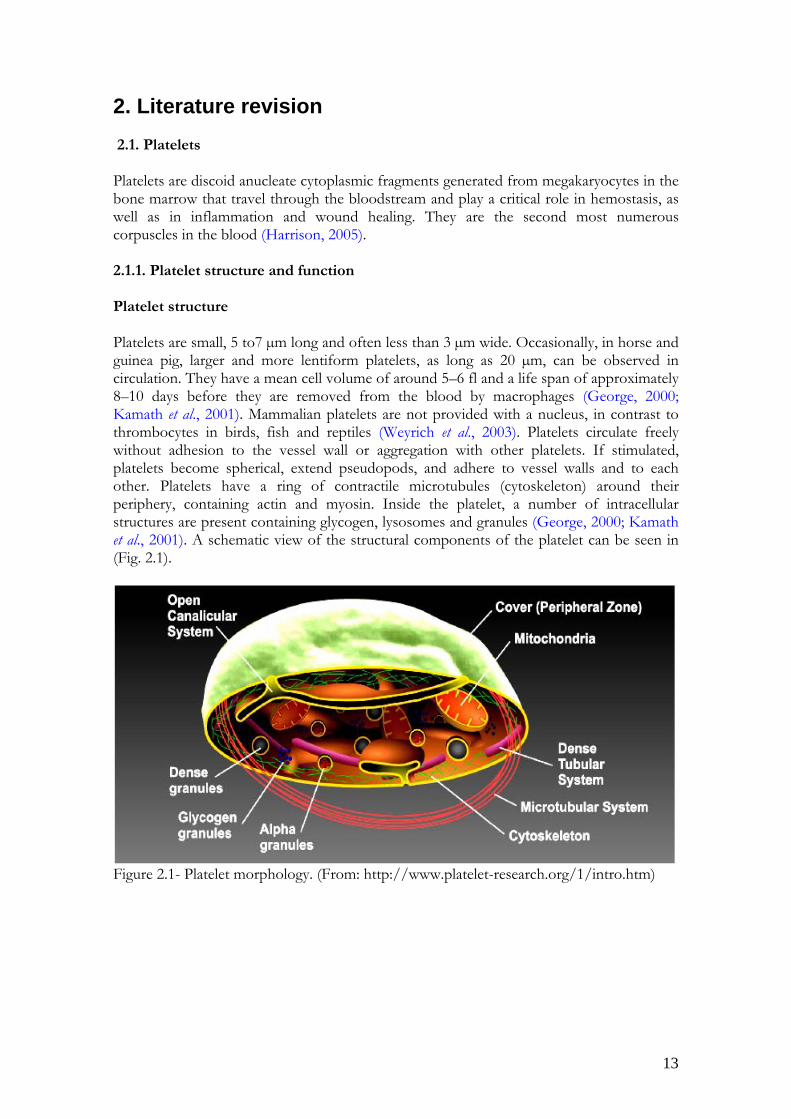
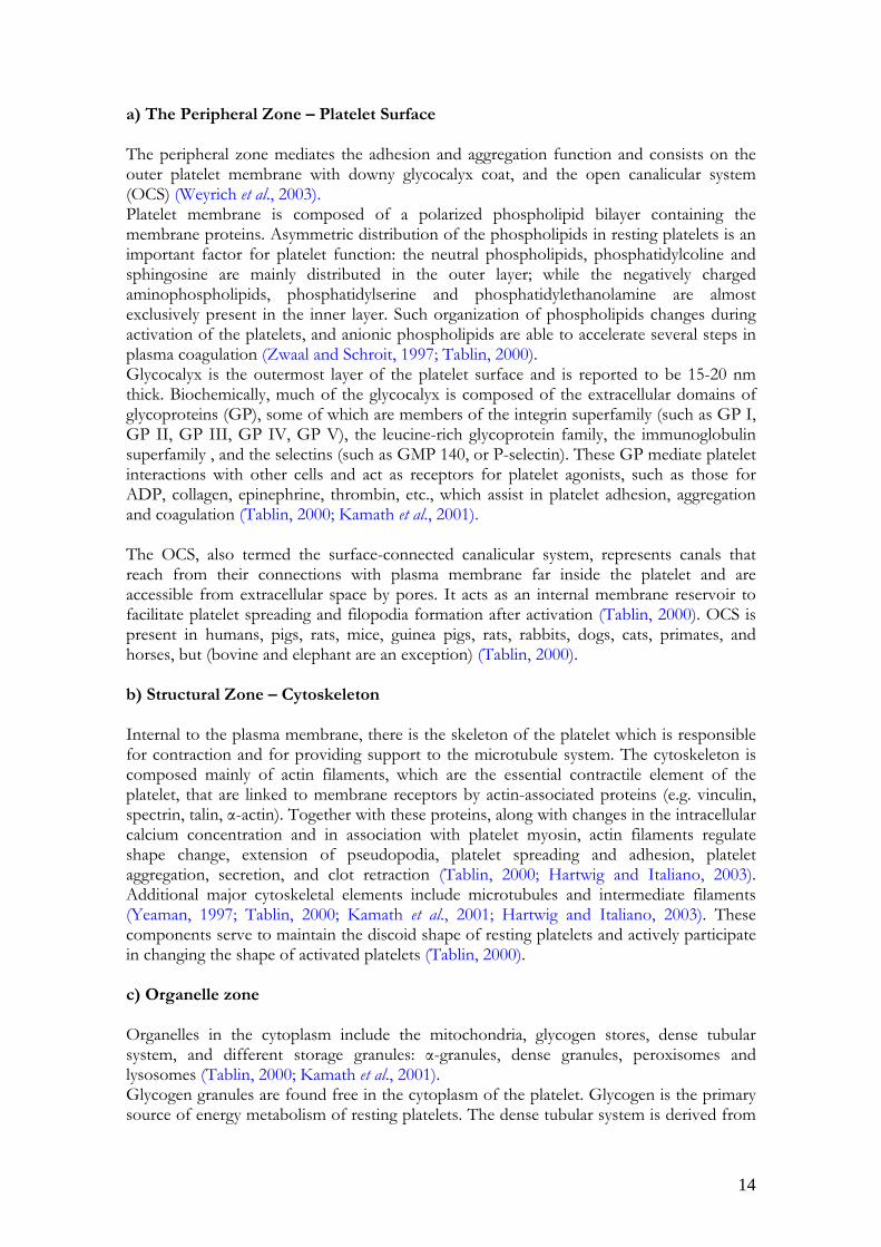
2. Literature revision 2.1. Platelets Platelets are discoid anucleate cytoplasmic fragments generated from megakaryocytes in the bone marrow that travel through the bloodstream and play a critical role in hemostasis, as well as in inflammation and wound healing. They are the second most numerous corpuscles in the blood (Harrison, 2005). 2.1.1. Platelet structure and function Platelet structure Platelets are small, 5 to7 µm long and often less than 3 µm wide. Occasionally, in horse and guinea pig, larger and more lentiform platelets, as long as 20 µm, can be observed in circulation. They have a mean cell volume of around 5–6 fl and a life span of approximately 8–10 days before they are removed from the blood by macrophages (George, 2000; Kamath et al., 2001). Mammalian platelets are not provided with a nucleus, in contrast to thrombocytes in birds, fish and reptiles (Weyrich et al., 2003). Platelets circulate freely without adhesion to the vessel wall or aggregation with other platelets. If stimulated, platelets become spherical, extend pseudopods, and adhere to vessel walls and to each other. Platelets have a ring of contractile microtubules (cytoskeleton) around their periphery, containing actin and myosin. Inside the platelet, a number of intracellular structures are present containing glycogen, lysosomes and granules (George, 2000; Kamath et al., 2001). A schematic view of the structural components of the platelet can be seen in (Fig. 2.1).
Figure 2.1- Platelet morphology. (From: http://www.platelet-research.org/1/intro.htm)
14
a) The Peripheral Zone – Platelet Surface The peripheral zone mediates the adhesion and aggregation function and consists on the outer platelet membrane with downy glycocalyx coat, and the open canalicular system (OCS) (Weyrich et al., 2003). Platelet membrane is composed of a polarized phospholipid bilayer containing the membrane proteins. Asymmetric distribution of the phospholipids in resting platelets is an important factor for platelet function: the neutral phospholipids, phosphatidylcoline and sphingosine are mainly distributed in the outer layer; while the negatively charged aminophospholipids, phosphatidylserine and phosphatidylethanolamine are almost exclusively present in the inner layer. Such organization of phospholipids changes during activation of the platelets, and anionic phospholipids are able to accelerate several steps in plasma coagulation (Zwaal and Schroit, 1997; Tablin, 2000). Glycocalyx is the outermost layer of the platelet surface and is reported to be 15-20 nm thick. Biochemically, much of the glycocalyx is composed of the extracellular domains of glycoproteins (GP), some of which are members of the integrin superfamily (such as GP I, GP II, GP III, GP IV, GP V), the leucine-rich glycoprotein family, the immunoglobulin superfamily , and the selectins (such as GMP 140, or P-selectin). These GP mediate platelet interactions with other cells and act as receptors for platelet agonists, such as those for ADP, collagen, epinephrine, thrombin, etc., which assist in platelet adhesion, aggregation and coagulation (Tablin, 2000; Kamath et al., 2001). The OCS, also termed the surface-connected canalicular system, represents canals that reach from their connections with plasma membrane far inside the platelet and are accessible from extracellular space by pores. It acts as an internal membrane reservoir to facilitate platelet spreading and filopodia formation after activation (Tablin, 2000). OCS is present in humans, pigs, rats, mice, guinea pigs, rats, rabbits, dogs, cats, primates, and horses, but (bovine and elephant are an exception) (Tablin, 2000). b) Structural Zone – Cytoskeleton Internal to the plasma membrane, there is the skeleton of the platelet which is responsible for contraction and for providing support to the microtubule system. The cytoskeleton is composed mainly of actin filaments, which are the essential contractile element of the platelet, that are linked to membrane receptors by actin-associated proteins (e.g. vinculin, spectrin, talin, α-actin). Together with these proteins, along with changes in the intracellular calcium concentration and in association with platelet myosin, actin filaments regulate shape change, extension of pseudopodia, platelet spreading and adhesion, platelet aggregation, secretion, and clot retraction (Tablin, 2000; Hartwig and Italiano, 2003). Additional major cytoskeletal elements include microtubules and intermediate filaments (Yeaman, 1997; Tablin, 2000; Kamath et al., 2001; Hartwig and Italiano, 2003). These components serve to maintain the discoid shape of resting platelets and actively participate in changing the shape of activated platelets (Tablin, 2000). c) Organelle zone Organelles in the cytoplasm include the mitochondria, glycogen stores, dense tubular system, and different storage granules: α-granules, dense granules, peroxisomes and lysosomes (Tablin, 2000; Kamath et al., 2001). Glycogen granules are found free in the cytoplasm of the platelet. Glycogen is the primary source of energy metabolism of resting platelets. The dense tubular system is derived from

15
the endoplasmic reticulum of the parent megakaryocyte and serves as the reservoir for calcium. The secretory granules possess molecules that affect platelet function, coagulation, fibrinolysis, vascular tone, inflammation, and wound healing. Some of the components are synthesized by megakaryocytes, other are taken up from plasma and incorporated into the granules. Upon activation, granules fuse with platelet surface membrane and extrude their content. It is a graded process depending on the number, nature, and concentration of the original stimuli (Reed and Fitzgerald, 2000; Kamath et al., 2001; Hartwig and Italiano, 2003). Platelet functions Platelets are surprisingly multifunctional and are involved in many pathophysiological processes including haemostasis and thrombosis, clot retraction, vessel constriction and repair, inflammation, including promotion of atherosclerosis, host defence and even tumour growth and metastasis (table 2.1). Upon vessel wall damage, platelets undergo a highly regulated set of functional responses including adhesion, spreading, release reactions, aggregation, and exposure of a procoagulant surface, microparticle formation and clot retraction. All of these platelet responses function to rapidly form a haemostatic plug that occludes the site of damage to prevent blood loss (Harrison, 2005).
Platelet main physiological functions
Haemostasis and
thrombosis
Maintenance/regulation of vascular tone
Inflammation Host defence Tumour biology
Adhesion Activation Spreading Secretion Aggregation
Uptake of serotonin when resting
Atheroslerosis Phagocytosis, internalisation of viruses and bacteria
Tumour growth
Procoagulant activity
Release of serotonin, thromboxane, prostaglandins upon activation
Allergic asthma; renal disease
Killing of bacteria
Tumour killing
Clot retraction
Chemotaxis Release of platelet microbicidal proteins
Tumour metastasis
Tissue Repair Platelet-leukocyte interactions
Superoxide production
Table 2.1 - The multifunctional platelet. Platelets are involved in many patophysiological processes, in addition to hemostasis and thrombosis, namely maintenance of vascular tone, inflammation, host defence and tumour biology (adapted from Harrison, 2005).

16
2.1.2. Platelet granules Platelets contain three distinct cytoplasmatic granules types: 1) Dense (σ) granules store mediators of vascular tone including serotonin, ADP, eicosanoids, thromboxane A2, calcium, and magnesium. Of all species studied, pig platelet dense granules release the most magnesium, (Yeaman, 1997; Tablin, 2000). 2) The α-granules are the largest granule population and contain proteins involved in hemostatic functions such as adhesion (e.g., fibrinogen, thrombospondin, vitronectin, laminin, and von Willebrand factor), modulation of coagulation (e.g. plasminogen, α2-plasmin inhibitor, and thrombasthenin), growth factors (such as platelet-derived growth factor (PDGF), transforming growth factor-β (TGF-β), epidermal growth factor (EGF), insuline-like growth factor (IGF-I), vascular endothelial growth factor (VEGF), hepatocyte growth factor (HGF) and fibroblast growth factor (FGF), glycoproteins (GPIb, GPIIb-IIIa), thrombospondin, fibronectin, and α-granule specific protein P-selectin (Tablin, 2000; Anitua et al., 2004). 3) Lysosomal (λ) granules contain acid hidrolases, guanine, phospholipases and kinases which act as proteolytic and hydrolytic enzymes, glycosidases, proteases and cationic proteins. Lysosomal granules are also believed to contain enzymes that principally mediate thrombus dissolution of residual megakaryocyte mRNA templates (Klinger, 1997; Yeaman, 1997; Werner, 2003). These distinct platelet granules are subject to discrete or synchronous release, dependent on agonist specificity and potency. For example, low levels of thrombin or ADP induce dense (σ) and (α) degranulation, while lysosomal (λ) granules are not secreted until these agonists are present at much higher concentrations. From this perspective, platelets may be viewed as vehicles that respond to agonists and ligands expressed at sites of endovascular damage or microbial colonization and release a diverse array of preformed bioactive molecules (Klinger, 1997; Yeaman, 1997; Werner, 2003).

17
2.1.3. Pig Platelets Porcine platelets are morphologically similar to those of other domestic species. They are variable in shape and are generally small, 1 to 3 µm in diameter, with a mean volume of 6,9 to 8,9 fl (Evans, 1994). Platelets are anucleate and have deeply staining purple cytoplasmic granules. Platelets are found in variable-sized clumbs discoid anucleate cytoplasmic fragments generated from megakaryocytes in the bone marrow that travel through the bloodstream and have a critical function in hemostasis (Fig. 2.2).
A
B
A
B
Figure 2.2 - Normal platelet density in peripheral blood smears (Giemsa stain, original magnification x l0). Scattered platelets correlating to a peripheral blood platelet count of 514,000 cells per µL. A - Platelets in different sizes. B - Red blood cells. The spiny crenation is common in pig.
18
2.2. Pathobiology of wound healing and growth factors 2.2.1. Wound Healing Process The wound healing process, or wound repair, is a complex series of events that begins at the moment of injury and can continue for months to years. These events overlap in time and are categorized into separate steps: the inflammatory phase, the proliferative phase, maturation and remodelling phase (table 2.2) (Falanga et al., 1995; Martin, 1997). The inflammatory phase The inflammatory phase is characterized by hemostasis and inflammation. Hemostasis The initial response following tissue injury via an incision is usually bleeding. Collagen exposed during wound formation activates the clotting cascade (both the intrinsic and extrinsic pathways), initiating the inflammatory phase. Normal hemostasis is a coordinated sequence of cellular and biochemical interactions, starting with an injury and ending several minutes later with a stabilized platelet aggregate, a process that conserves blood and enables wound healing (White, 2000; Harrison, 2005). Blood platelets contribute to every aspect of hemostasis, from the initial adhesion of platelets to the vessel wall, spreading over the surface and forming a platelet aggregate, providing an activated cell surface that vastly accelerates coagulation and leads to stabilization of the platelet aggregate by fibrin. All of these processes are regulated, entirely or in part, by specific glycoprotein receptors on the platelet surface. The (GP) IbIX-V complex, a constitutively expressed receptor on the platelet plasma membrane, mediates the initial deposition of platelets on the subendothelium under high shear conditions (Falanga et al., 1995; Martin, 1997). Inflammation Neutrophils are the first cells appear in the wound area; migration of those cells are stimulated by various chemotactic factors, and are important for controlling local infection of bacteria through endocytosis as well as the release of lysosomal enzymes. This causes additional tissue death at the wound site, initiating the debridement processes (Falanga et al., 1995). The neutrophil attain their maximal numbers in 24-48 hours and commence their departure by hour 72. Neutrophil infiltration slows down as monocyte infiltration increases. Upon migration into the wound area, monocytes undergo phenotypic changes to become macrophages (Falanga et al., 1995; Martin, 1997). The macrophages continue the cleansing process during days 3-4 as well as debride the wound, aided by the macrophage secretion of tumor necrosis factor-α and interleukin-1. In addition to cleaning the wound, the macrophages release numerous cytokines that are important for the attraction and proliferation of fibroblasts. These include PDGF, FGF, TGF-β, VEGF and plasma-activated complements (anaphylactic toxins). Other inflammatory cells that interact within the wound area include lymphocytes, plasma cells, and mast cells. These cells also contribute with cytokines, such as interleukin-4, to induce collagen production in fibroblasts (Falanga et al., 1995; Martin, 1997).

19
Proliferative phase The proliferative phase is characterized by angiogenesis, granulation tissue formation, collagen deposition and re-epithelization (Midwood et al., 2004). In angiogenesis, new blood vessels grow from endothelial cells. In fibroplasia and granulation tissue formation, fibroblasts grow and form a new, provisional extracellular matrix (ECM) by excreting collagen and fibronectin. On collagen deposition, fibroblasts begin to commit apoptosis, converting granulation tissue from an environment rich in cells to one that consists mainly of collagen. The reepithelialization involves epithelial cells migration across the new tissue (granulation tissue) to form a barrier between the wound and the environment (Midwood et al., 2004). Angiogenesis The process of angiogenesis occurs concurrently with fibroblast proliferation when endothelial cells migrate to the area of the wound (Kuwahara and Rasberry, 2007). Stem cells called endothelial cells originating from parts of uninjured blood vessels develop pseudopodia and push through the ECM into the wound site. Through this activity, they establish new blood vessels (Greenhalgh, 1998). The most important family of cytokines implicated in the neovascularization processes are VEGF family and FGF (Romo and Pearson, 2005). Endothelial growth and proliferation is also stimulated by hypoxia and presence of lactic acid in the wound (Falanga, 2005). Fibroplasia and granulation tissue formation Simultaneously with angiogenesis, fibroblasts begin entering the wound site two to five days after wounding as the inflammatory phase is ending and their numbers peak at one to two weeks post-wounding being the main cells in wound healing for that time (Stadelmann et al., 1998). Fibroplasia ends two to four weeks after wounding. In the first two or three days after injury, fibroblasts mainly proliferate and migrate, while later, they are the main cells that lay down the collagen matrix in the wound site (Stadelmann et al., 1998). Fibroblasts from normal tissue migrate into the wound area from its margins. Initially fibroblasts use the fibrin scab formed in the inflammatory phase to migrate across, adhering to fibronectin (Romo and Pearson, 2005). Fibroblasts then deposit ground substance into the wound bed, and later collagen, which they can adhere to for migration (de la Torre and Sholar, 2006). Growth factors (PDGF, TGF-β) and fibronectin encourage proliferation, migration to the wound bed, and production of ECM molecules by fibroblasts (Falanga et al., 1995; Martin, 1997). Granulation tissue begins to appear in the wound even during the inflammatory phase; two to five days post wounding, and continues growing until the wound bed is covered. It is needed to fill the void that has been left by a large, open wound that crosses the basement membrane. Basically consist of new blood vessels, fibroblasts, inflammatory cells, endothelial cells, myofibroblasts, and the components of a new, provisional ECM. The provisional ECM is different in composition from the ECM in normal tissue and includes fibronectin, collagen, glycosaminoglycans, and proteoglycans (Romo and Pearson, 2005).

20
Collagen deposition In days 5-7, fibroblasts have migrated into the wound, laying down new collagen of the subtypes I and III. Early in normal wound healing, type III collagen predominates but is later replaced by type I collagen (Falanga et al., 1995; Martin, 1997). One of fibroblasts most important duties is the production of collagen (Kuwahara and Rasberry, 2007). Fibroblasts begin secreting appreciable collagen by the second or third post-wounding day (Romo and Pearson, 2005), and its deposition peaks at one to three weeks (Mercandetti and Cohen 2005). Collagen deposition is important because it increases the strength of the wound; before it is laid down, the only thing holding the wound closed is the fibrin-fibronectin clot, which does not provide much resistance to traumatic injury (Greenhalgh, 1998). FGF-1 and FGF-2 are potent stimulators of fibroblasts, which are essential in extracellular matrix production (Podolsky, 1994). Re-epithelization Re-epithelization occurs with the migration of cells from the periphery of the wound and adnexal structures within 24 hours of injury. Division of peripheral cells occurs in hours 48-72, resulting in a thin epithelial cell layer, which bridges the wound. EGF is believed to play a key role in this aspect of wound healing. This succession of subphases can last up to 4 weeks in the clean and uncontaminated wound (Falanga et al., 1995; Martin, 1997; Werner et al., 2003). Maturation and Remodeling phase When the levels of collagen production and degradation equalize, the maturation phase of tissue repair has begun (Greenhalgh, 1998). The maturation phase can last for a year or longer, depending on the size of the wound and whether it was initially closed or left open (Mercandetti and Cohen, 2005). During maturation, type III collagen, which is prevalent during proliferation, is gradually degraded and the stronger type I collagen is laid down in its place. Originally disorganized collagen fibers are rearranged, cross-linked, and aligned along tension lines (Lorenz and Longaker, 2003).

21
I-
In
flam
mat
ory
ph
ase
a) Immediate to 2-5 days
b) Hemostasis - Instantly after endothelial disruption vascular contraction begins platelet adhesion and formation of a soft aggregate plug. Vasoconstriction (maintained by platelet secretion of serotonin, prostaglandin and thromboxane) slows blood flow, enhancing platelet adhesion and activation. The soft plug is solidified through a complex interaction between platelet membrane, enzymes, and coagulation factors.
c) Inflammation - Neutrophils are the first cells appear in the wound area, stimulated by various chemotactic factors. Monocytes appear approximately 24 hours after injury and peak at 48 hours post-injury, and mature into macrophages. Macrophages secrete cytokines and growth factors (PDGF, FGF, TGF-β, VEGF), in matrix synthesis and degradation, and are able to mediate angiogenesis and fibroplasia.
II-
Pro
lifer
ativ
e p
has
e
a) 2 days to 3 weeks
b) Angiogenesis - Stem cells called endothelial cells originating from parts of uninjured blood vessels develop pseudopodia and push through the ECM into the wound site. Through this activity, they establish new blood vessels. VEGF together with FGF may enhance neovascularization.
c) Fibroplasia and Granulation tissue
Fibroplasia, fibroblast proliferates in the deeper parts of the wound. These fibroblasts begin to synthesize small amounts of collagen which acts as a scaffold for migration and further fibroblast proliferation, stimulated by TGF- β, FGF and other related factors.
Granulation tissue - involve the perfusion of fibrous connective tissue that replaces a fibrin clot in healing wounds. Consists of capillary loops supported in this developing collagen matrix, also appears in the deeper layers of the wound.
Collagen deposition - 4 to 5 days after the injury occurs; fibroblasts begin producing large amounts of collagen and proteoglycans. Collagen fibers are laid down randomly and are cross-linked into large, closely packed bundles. FGF-1 and FGF-2 are potent stimulators of fibroblasts.
d) Reepitelization, migration of cells from the periphery of the wound and adnexal structures within 24 hours of injury. Division of peripheral cells occurs in hours 48-72, resulting in a thin epithelial cell layer, which bridges the wound.
III-
Rem
odel
ling
Ph
ase a) 3 weeks to 2 years
b) During remodelling phase loose provisional matrix is gradually replaced by collagen fibers, the provisional matrix collagen is almost exclusively of type III is main fibrillar collagen and it is degraded by collagenase activity and gradually replaced by collagen type I, the main collagen of the mature scar.
Table 2.2 - Phases of wound healing.

22
2.2.2. Regulation of wound repair Platelets Platelets are unique anucleate mammalian blood cells with specialized molecular repertoires that have evolved to accomplish crucial functions in host integrity, defense and repair (Klinger, 1997; Yeaman, 1997; Werner et al., 2003). Platelets are the principal effectors of cellular hemostasis in humans and other mammals (Weyrich et al., 2003). They adhere to the exposed subendothelial matrix and aggregate in response to prothrombotic signals, contributing to the formation of platelet - fibrin clot that is critical for sealing vascular disruptions and for ultimate vessel and wound repair (White, 2000). The formation of a platelet plug in response to prothrombotic stimuli involves several steps, namely adhesion, shape change, release of mediators, aggregation and development of a pro-coagulant surface, which will be covered more comprehensively later under platelet activation (Harrison, 2005). In addition, platelets have major roles in acute and chronic inflammation including: release of proinflammatory mediators, display of the surface molecules that have inflammatory functions and interactions with polymorphonuclear leukocytes and endothelial cells, regulating their adhesion and activation (McIntyre et al., 2003; Weyrich et al., 2003). Although they are highly differentiated for hemostasis and inflammation, they participate in the wound healing process through the release of cytokines, growth factors and other regulatory molecules together with the interaction with neutrophils and endothelial cells. Platelets also have antimicrobial systems and link clotting and immune cascades (Klinger, 1997; Yeaman, 1997; Werner et al., 2003). Growth factors Growth factors are classified as cytokines, which are proteins that act as intercellular signals to allow cells to communicate with one another. Growth factors are involved in all stages of wound healing and also have the ability to regulate many other functions within cells including protein synthesis, cell proliferation and differentiation. They are specific for attracting useful cells and proteins to the wound, including immune cells to fight against infection and other cells to form connective tissue. Growth factors also stimulate an increased production of connective tissue; create a new supply of blood vessels to nourish the site, thus promoting maturation (Marx et al., 1998; Kassolis et al., 2000; Marx et al., 2001; Appel et al., 2002). These cytokines are released by a variety of activated cells (platelets, macrophages) at the wound site and act on the appropriate target cell or cells to carry out specific action (Giannobile, 1996; Nimni, 1997; Liebarman et al., 2002). Growth factors can exert their effect through three different ways: - Autocrine, in which the growth factor influences the cell of its origin or other cells identical in phenotype to that cell (e.g. a growth factor produced by an osteoblast influences the activity of another osteoblast) (Liebarman et al., 2002). - Paracrine, in which the growth factor influences an adjacent or neighboring cell that is different in phenotype from its cell of origin (e.g. a growth factor produced by an osteoblast stimulates differentiation of an undifferentiated cell) (Liebarman et al., 2002). - Endocrine, in which the growth factor influences a cell that is different in phenotype from its cell of origin and located at a remote anatomical site (e.g. a growth factor produced by neural tissue in the central nervous system stimulates osteoblast activity). Thus, a growth

23
factor may have effects on multiple cell types and may induce an array of cellular functions in a variety of tissues (Giannobile, 1996; Nimni, 1997; Liebarman et al., 2002). Platelets contain several growth factors which are released after injury, including PDGF, TGF, IGF, HGF, VEGF, and FGF (Giannobile, 1996; Liberman et al., 2002).The clinical importance of these growth factors is demonstrated by the finding that recombinant DNA-derived polypeptide growth factors significantly accelerate healing of soft tissue (Appel et al., 2002). Platelet Derived Growth Factor (PDGF) PDGF is released from platelet alpha-granules, macrophages, monocytes, endothelial cells and smooth muscle cells immediately after injury. PDGF is composed of two distinct polypeptide chains, A and B, that form homodimers (AA or BB) or heterodimers (AB). The AA and BB isoforms enhance proliferation of bone cells, increasing the production of PDGF-AA in osteoblast cultures (Martin, 1997; Liebarman et al., 2002; Sánchez et al., 2003). PDGF plays a significant role in the formation of connective tissue during wound healing (Giannobile, 1996; Martin, 1997; Werner, 2003). PDGF is a very powerful regulatory growth factor and a sentinel growth factor that begins nearly all wound healing process. PDGF attracts neutrophils, macrophages, and fibroblasts to the wound and serves as a powerful mitogen. PDGF stimulates fibroblasts to synthesize new extracellular matrix, predominantly noncollagenous components such as glycoproteins, GAGs and adhesion proteins (Giannobile, 1996; Liebarman et al., 2002). PDGF also increases the amount of fibroblast-secreted collagenase, indicating a role for this cytokine in tissue remodelling (Giannobile, 1996; Martin, 1997). PDGF’s main function is to stimulate cell replication (mitogenesis) of healing capable stems and premitotic partially differentiated osteoprogenitor cells, which are part of the connective tissue-bone healing cellular make-up. PDGF also causes replication of endothelial cells, causing budding of new capillaries (angiogenesis) (Martin, 1997; Liebarman et al., 2002). PDGF has also important functions during the embryogenesis, particulary in the development of the kidneys, blood vessels, lungs, and central nervous system (CNS). In these organs, connective tissue-like cell types are dependent on PDGF, including mesangial cells, pericytes, alveolar fibroblasts, and glial cells. Transforming Growth Factor- β (TGF- β) It is released from platelets, T-lymphocytes, macrophages/monocytes and neutrophils after injury. It acts stimulating and inhibiting endothelial, fibroblastic, and osteoblastic mitogenesis; regulating collagen synthesis and collagenase secretion; regulating mitogenic effects of other growth factors; stimulating endothelial chemotaxis and angiogenesis (Giannobile, 1996; Liebarman et al., 2002). TGF- β is a 2-chain polypeptide that is linked together by disulfide bonds. It exists as 3 different isoforms: TGF-β1, TGF-β2, and TGF-β3. TGF-β1 is found in high concentrations in bone and platelets (Giannobile, 1996; Liebarman et al., 2002). When released by platelets or secreted by macrophages, TGF-β exerts its effects on adjacent cells, including fibroblasts, marrow stem cells, endothelial cells, and preosteoblasts.

24
TGF-β stimulates angiogenesis and the production of fibronectin, GAGs, and collagen in connective tissue to aid in restitution and adequate wound closure (Giannobile, 1996; Martin 1997). One of the most important functions of TGF-β seems to be the chemotaxis and mitogenesis of osteoblast precursors. In addition, this polypeptide inhibits osteoclast formation and resorption, favoring bone formation. This local connective tissue response to TGF-β in vivo is strongly anabolic and leads to fibrosis and angiogenesis (Milani and Calabro, 2001). A fundamental mechanism of the antiproliferative (catabolic) action of TGF- β is its ability to antagonize the mitogenic effects of other peptide growth factors such as EGF and PDGF. Even in a single cell type, the nature of growth factor action may depend on the context set by other substances present. For example, TGF-β stimulates growth of certain fibroblasts in vitro in the presence of PDGF but inhibit their growth if EGF is present (Milani and Calabro, 2001). Indeed, TGF-β may play a central role in the regulation of migration of intestinal epithelial cells. TGF-β has several potentially important functions in intestinal bowel disease (Ciacci et al., 1993; Beck et al., 2003). Fibroblast Growth Factor (FGF) It is released from platelets, macrophages, neural tissue, adrenal, corpus luteum, placenta and fibroblasts (Kandel et al., 1991; Blotnick et al., 1994) and can be released from mechanically wounded endothelial cells. FGF acts increasing angiogenesis and vessel permeability and stimulates mitogenesis for endothelial cells. There are at least 19 distinct members of the FGF family of growth factors. The two originally characterized FGFs were identified by biological assay and are termed FGF-1 (acidic-FGF, aFGF) and FGF-2 (basic-FGF, bFGF) (Beck and Podolsky, 1999). Both FGF-1 and FGF-2 stimulate proliferation of all cells of mesodermal origin as well as numerous cell populations of endodermal and ectodermal origin including fibroblasts, keratinocytes, smooth muscle cells, endothelia1 cells, and various cell types within the central and peripheral nervous system (Galzie et al., 1997; Hu et al., 1998). Furthermore, FGF-1 and FGF-2 are both mitogenic and chemotactic for endothelial cell, which contribute to angiogenesis following injury. These factors can induce the production of collagenase and plasminogen activator, which in turn leads to extracellular matrix breakdown. FGF-1 and FGF-2 are also potent stimulators of fibroblasts, which are essential in extracellular matrix production and wound healing (Podolsky, 1994). Vascular Endothelial Growth Factor (VEGF) This growth factor is released from platelets and endothelial, macrophages, fibroblasts, and keratinocytes cells after injury. The VEGF family currently includes VEGF-A, VEGF-B, VEGF-C, VEGF-D, VEGF-E. The biological functions of VEGF-A have been characterized in much detail. Based on a series of in vitro and in vivo studies, VEGF-A has been identified as a major regulator of vasculogenesis and angiogenesis during development (Yeaman, 1997), indicating that it might also be involved in the regulation of angiogenesis during wound healing (Giannobile, 1996; Nimni, 1997). Its expression is also increased in hypoxic conditions, such as those found at the wound site (Lieberman et al., 2002). In wound healing, VEGF promotes the vascularization of injured tissues and thus facilitates the arrival of inflammatory and reparative cells

25
(Giannobile, 1996; Martin, 1997; Yeaman, 1997). VEGF-A is vital for endochondral bone formation and also promotes the proliferation of retinal pigment epithelial, pancreatic duct and Shawn cells in vitro (Martin, 1997). Insulin- like Growth Factor (IGF) IGF is a peptide hormone synthesized and secreted into the circulation primarily by the liver via a growth hormone (GH)-dependent process. It is also released from platelets, osteoblasts, macrophages, monocytes, and chondrocytes. IGF has 2 forms, IGF-I and IGF-II each of which has 2 single chain peptides with structural homology to insulin. IGF was shown to contribute to the regulation of tissue growth and cellular differentiation in vivo and in vitro. IGF-I serves as a systemic anabolic function, it is notable that intestine is also a site of IGF-I synthesis and one of the most sensitive IGF-I target tissues (Read et al., 1992). This growth factor also acts in an autocrine way and as insulin factor. In combination with PDGF can enhance the rate and quality of wound healing. Other functions of this growth factor include cartilage growth stimulating, bone matrix formation, and replication of preosteoblasts and osteoblasts. . IGF-I is important for both the regulation of normal physiology, as well as a number of pathological states, including cancer. The IGF has been shown to play roles in the promotion of cell proliferation and the inhibition of cell death (apoptosis). IGF-II is thought to be a primary growth factor required for early development while IGF-I expression is required for achieving maximal growth (Giannobile, 1996; Lieberman et al., 2002; Sánchez et al., 2003). While IGF-II may be primarily fetal in action it is also essential for development and function of organs such as the brain, liver, and kidney. Beyond general growth, insulin-like growth factors are involved in both pre and postnatal development and function of a large number of organs such as the prostate gland, mammary glands, spleen, pancreatic beta cells, lung, inner ear, sympathetic neurons, thymus and teeth. They are also important factors for muscle development and skeletal growth as well as for skeletal mass maintenance (Sánchez et al., 2003). Epidermal Growth Factor (EGF) It is released from platelets, macrophages and monocytes. EGF stimulates endothelial chemotaxis and angiogenesis; regulates collagenase secretion and stimulates epithelial and mesenchymal mitogenesis. This growth factor is present in various body fluids and tissues, and is continuously secreted into the gastrointestinal lumen in humans by submandibular glands, mucous neck cells of the stomach, Brunner’s glands of the duodenum, Paneth cells of the small intestine, and ulcer-associated cell lineage (a recently identified glandular structure induced at the sites of injury) (Marti et al., 1989; Wright et al., 1990). EGF and EGF family of related peptides are involved as key constituents in the maintenance and repair of gastrointestinal mucosa (Jones et al., 1999). Both EGF and TGF-α has numerous functions within the gastrointestinal tract in addition to stimulation of proliferation. It inhibits gastric acid secretion and increases the release of mucus, also up-regulates intestinal electrolyte and nutrient transport, and induces expression of brush border enzymes such as disaccharidases (Goodlad et al., 1991; Uribe and Barrett, 1997). EGF is also responsible for cell differentiation and stimulates re-epitheliation, angiogenesis
26
and collagenase activity. In wound healing, EGF can induce cell proliferation, differentiation and motility. EGF is highly expressed in the margin of wounds promoting re-epithelization (Martin, 1997; Lieberman et al., 2002) and up-regulates matrix metalloproteinase-1 (MMP-1) expression and regulates type I collagen turnover. MMP-1 facilitates epithelial cell migration across the collagen type I matrix in several tissues, including the dermis (Martin, 1997). Hepatocyte Growth Factor (HGF) It is released from platelets, fibroblasts or other mesenchymal cells. HGF is a multifunctional polypeptide secreted by mesenchymal cells, functions as a mitogen, morphogen, and/or motogen for multiple subsets of epithelial cells, including gastrointestinal epithelial cells (Jones et al., 1999; Tahara et al., 2003). HGF activator, HGF activator inhibitor type-1 and HGF-associated molecules involved in the activation of HGF in injured tissues, are associated with colonic mucosal repair (Tahara et al., 2003). Additionally, HGF expression was reported to be up-regulated in inflamed colonic mucosal tissue in patients with ulcerative colitis (Kitamura et al., 2000), and plasma HGF levels are increased in animal models of acute colitis (Nishimura et al., 1998; Ortega-Cava et al., 2002). This growth factor is also a hepatotrophic factor that promotes liver regeneration. It has also been shown to stimulate the growth of various epithelial cells, such as renal tubular cells, epidermal melanocytes and keratocytes (Nishimura et al., 1998). HGF is a heparin-binding protein that is mitogenic for endothelial and epithelial cells (Nishimura et al., 1998; Ortega-Cava et al., 2002). HGF and VEGF show a synergistic action on endothelial cells and tubulogenesis, a response that does not happen with any of the growth factors alone. HGF is also implicated in cutaneous wound pathologic neovascularization and exuberant granular tissue formation (Martin, 1997). Extracellular Matrix (ECM) The ECM is defined as any material produced by cells and excreted to the extracellular space within the tissues. It takes the form of both ground substance and fibres and is mostly made up of fibrous elements, proteins involved in cell adhesion, GAGs and other space-filling molecules (Agren and Werthen, 2007). The ECM serves as a scaffolding to hold tissues together, its form and its composition help to determine tissue characteristics. In epithelia, it includes the basement membrane. The ECM undergoes dynamic interactions with cells that are pivotal for cell adhesion, motility, growth, differentiation, ECM synthesis, in addition to offering passive support to the cells. The ECM is also a depository of growth factors in latent forms under normal, unwounded conditions. With injury and matrix destruction, the previously bound and inactive growth factors are released in active form and thereby assist in initiating and regulating the repair process (Agren and Werthen, 2007).
27
2.2.3. Healing of gastrointestinal anastomosis Anastomotic dehiscence remains a major problem in gastrointestinal surgery. Even if small bowel anastomoses carry the least risk of complications among the other gastrointestinal anastomoses, the healing sequence of anastomoses are necessary to define the underlying mechanisms and find ways to improve surgical outcome in high risk patients (Hendriks and Mastboom, 1990). For a better understanding of the intestinal healing, knowledge of intestinal histology is essential. The gastrointestinal tract has a uniform general histology with some differences which reflect the specialization in functional anatomy. The intestine is divided in the following layers: 1) the mucosa, 2) the submucosa, 3) the muscularis propria, and 4) the serosa. Mucosa The mucosa is the innermost layer of the gastrointestinal tract that is surrounding the lumen, within the tube. This layer comes in direct contact with bolus, and is responsible for absorption and secretion, important processes in digestion (Hedlund et al., 2002). The mucosa of the small intestine is highly modified. The luminal surface is completely covered by a number of finger or leaf-like projections called villi. The core of a villus is an extension of the lamina propria, and its surface is covered by a simple columnar epithelium (Hedlund et al., 2002). The mucosa has three parts: (1) an epithelium (and of course the associated basement membrane), (2) a layer of loose connective tissue called the lamina propria, and (3) a band of smooth muscle, the muscularis mucosa, denoting the limit of the mucosa (Hedlund et al., 2002). 1) The epithelium acts as a barrier separating the organism’s body from the environment, which is continuous with the lumen of the alimentary canal. In different areas, the epithelium may simply be a route (esophagus, anal canal), may have a secretory function (stomach, goblet cells in intestines) or may have an absorptive function (intestines). The type of epithelium present reflects the function of the particular segment of the alimentary canal. 2) The lamina propria consists of loose connective tissue, with numerous blood and lymphatic vessels, and an abundance of diffuse lymphoid tissue. In addition to lymphocytes, plasma cells, eosinophils and macrophages are present. Not infrequently, lymphocytes are seen to be aggregated into lymph nodules. The lymphatic tissue of the lamina propria is referred to as galt, or gut-associated lymphatic tissue, and acts as an immunological barrier for pathogens (Hedlund et al., 2002). 3) The muscularis mucosa allows for movement of the mucosa independent of the wall of the digestive tract, thereby increasing its contact with food. In most of the alimentary canal, the muscularis mucosa consists of an inner circular and outer longitudinal layer of smooth muscle (Hedlund et al., 2002). Submucosa The submucosa consists of a dense irregular layer of connective tissue with large blood vessels, lymphatics and nerves branching into the mucosa and muscularis (Hedlund et al., 2002). It contains Meisser`s plexus, an enteric nervous plexus, situated on the inner surface of the muscularis externa. The submucosa provides the gastrointestinal tract with most of the tensile strength and is responsible for anchoring the sutures that hold the anastomosed bowel ends together (Halsted, 1887; Greatorex, 1970).

28
Muscularis externa The muscularis externa consists of an inner circular layer and an outer longitudinal layer. The circular muscle layer prevents the food from going backwards and the longitudinal layer shortens the tract. Contractions of these layers are coordinated and called peristalsis, propels the bolus, or balled-up food, through the gastrointestinal tract (Hedlund et al., 2002). Serosa The serosa is the outermost layer which is made up of loose connective tissue and coated in mucus so as to prevent friction damage from the intestine rubbing against other tissue. Serosa is important for forming a quick seal at site of injury or incision (Furst et al., 1994; de Waard et al., 1995; Hedlund et al., 2002). Intestinal wound healing The three overlapping phases of healing in intestine are named: lag phase, proliferative phase and the maturation phase. The lag phase occurs during days 0 to 4 and is associated with inflammation and edema of the healing intestine. A fibrin seal forms during the first few hours. Although the fibrin clot contributes to wound strength, during this phase most of the wound strength is attributed to sutures. During the lag phase macrophages are important in wound debridement and production of growth factors that modulate fibroplasia and angiogenesis (TGF- β, EGF, PDGF, and cytokines) (Jones et al., 1999; Hedlund et al., 2002). Healing is functionally weakest at the end of lag phase, because of fibrinolysis and collagen deposition, therefore dehiscence most commonly occurs 3 to 5 days after intestinal surgery (Hedlund et al., 2002). The proliferative phase begins with the arrival of fibroblasts at the wound site. Fibroblasts become the major cell type by day 4, and their arrival is regulated by various growth factors. Fibroblasts replace the provisional matrix (established through the inflammation phase) with collagen-rich granulation tissue. The strength of the repair site approximates that of normal intestine 10 to 17 days after surgery (Hedlund et al., 2002). The maturation phase occurs between 10 and 180 days. With time, newly formed granulation tissue undergoes remodeling, and the density of macrophages and fibroblasts is reduced. Collagen is reorganized and remodelled during this phase (Hedlund et al., 2002). Thin collagen fibers transform into thick bundles, and the percentage of type III collagen is reduced to 20%. Wound contraction occurs as fibroblasts pack thick collagen bundles into contractile units. Factors that contribute to intestinal anastomosis healing In small animals the reported incidence of small intestinal dehiscence rates 7-16%, with 74 -80% of those patients dying (Hedlund et al., 2002). As for any wound, a compromised anastomosis represents the convergence of a number of factors-systemic, local, and operative that disrupts the normal sequence of wound healing (Gordillo and Sen, 2003). Many factors have been implicated in the success or failure of the anastomotic healing process. The anastomosis oxygenation is of paramount importance and depends primarily on the intrinsic vasculature (Thornton and Barbul, 1997). However, several factors have been described to affect intestinal healing.

29
Operative factors: It is well known that technical factors relating to the construction of an anastomosis (gentle handling of tissue; aseptic technique; sharp anatomic dissection of tissue; careful hemostasis; obliteration of dead space; avoidance of tension; use of fine instruments for sharp dissection; accurate suture placement and blood supply, these are known as Halsted's principles of surgery) are of paramount importance to ensure optimal anastomotic healing, most likely resulting from effects on tissue perfusion and oxygen delivery (Thornton and Barbul, 1997). Optimal intestinal healing depends on a good blood supply, accurate mucosal apposition, and minimal surgical trauma. Approximating suture patterns facilitate a rapid healing. Everting and inverting suture patterns were found to retard intestinal healing and may result in greater stricture formation. Healing is also facilitated by adjacent serosal surfaces and omentum, which help seal wounds and contribute to the blood supply (Hedlund et al., 2002). Good serosal apposition is necessary to minimize the risk of leakage (Furst et al., 1994; de Waard et al., 1995; Hedlund et al., 2002). Unfortunately, even the most experienced and technically proficient surgeons have a certain leakage rate which suggests that there are other factors at play. Systemic factors: Wound healing requires energy and adequate nutritional intake by the patient. Malnourished patients are predisposed to wound healing failure as they lack of the necessary proteins, vitamins and minerals for repair. These include vitamins A, C, and B6, all required for collagen synthesis and cross-linking, as well as zinc, iron and copper. Zinc and iron acts as cofactors to many reactions involved in DNA synthesis, protein synthesis, and cellular proliferation. Zinc or copper deficiencies result in fewer fibroblasts, and impacts collagen synthesis (Thornton and Barbul, 1997). Other systemic factors such as shock, hipoproteinemia, debilitation and concurrent infections may delay healing and increase the risk of incisional breakdown. Tension on the repair caused by accumulation of ingesta, fluid, gas or mobilization of the bowel increases intestinal suture breakdown (Hedlund et al., 2002). Local factors: Such as peritonitis impairs wound healing by prolonging the inflammatory phase, and inducing the increased expression of tissue proteases. Elevated tissue proteases digest growth factors, and thus delay epithelization and collagen deposition. Localized preoperative or perioperative bowel irradiation was not shown to compromise anastomotic healing in animal model (Thornton and Barbul, 1997). The effect of ischemia on intestinal healing It is clear that the availability of oxygen is critical for wound healing. Oxygen is essential for normal cell metabolism and energy production in the absence of tissue injury (Gordillo and Sen, 2003). Oxygen availability becomes especially important during wound healing as a result of the increased metabolic demands for processes such as cellular proliferation and collagen synthesis (Gordillo and Sen, 2003; Tandara and Mustoe, 2004). Adequate tissue oxygenation is required for normal oxidative function of neutrophils, leukocyte activation, fibroblast production, angiogenesis, and reepithelialization, which all are essential in wound healing (Grim et al., 1990; Hirn, 1993). Fibroblasts require oxygen for their energy production, and collagen can not be synthesised in the absence of molecular oxygen (Roberts and Harding, 1994). Oxygen is necessary in the hydroxylation of lysine and proline during collagen synthesis, which is ultimately responsible for conferring tensile strength to the wound (anastomosis) (Gordillo and Sen, 2003; Tandara and Mustoe, 2004).

30
Hypoxemia, caused by disrupted vasculature, is a key factor that limits wound healing. The central area of the wound is the most hypoxic, with a progressive increase in the oxygen gradient toward the uninjured tissue at the periphery (Khanna and Wallace, 2002). Disruption of microcirculatory blood flow at the level of the anastomosis is one of the major factors leading to anastomotic dehiscence and leakage. Decreased tissue perfusion begins intraoperatively, and can lead to necrosis and tissue breakdown in the early postoperative period. Studies showed that the anastomosis was at highest risk during the first 24h, and that microvascular recovery has improved anastomotic perfusion by 96h (Schroder et al., 2004). In addition to oxygen, many growth factors and cytokines are required for wound healing to proceed normally. Considering the numerous different substances produced by the wound, nitrous oxide has a particularly important role. The effect of nitrous oxide on surgical wound infections has been investigated previously. In-vitro evidence indicates that exposure to nitrous oxide inactivates vitamin B12 and thus methionine synthase (Perry et al., 1983). Methionine synthase is the enzyme responsible for conversion of homocysteine to methionine and methyltetrahydrofolate to tetrahydrofolate, which are critical pathways for thymidine formation, which, in turn, is essential for DNA formation. Without functioning methionine synthase, protein cannot be produced. Protein expression is a critical aspect of scar formation and tissue repair (Hassanain et al., 2005). Thus, nitrous oxide toxicity could impair healing. Nitrous oxide also has been shown to depress chemotactic migration by monocytes, apparently by interfering with microtubules. Chemotaxis is a key part of the bacterial killing process, which needs chemotaxis, phagocytosis, and killing (Benhaim and Hunt, 1992; Fleischmann et al., 2005). 2.2.4. Parameters for anastomotic repair evaluation Substantial investigative effort has been dedicated to develop strategies to improve anastomotic healing, including improved surgical technique, bowel preparation, nutritional and pharmacological interventions. Investigating wound healing and attempting to improve its outcome necessitates process quantification. Parameters to evaluate the anastomotic repair may be: a) Mechanical parameters. b) Biochemical parameters. c) Histological parameters. a) Mechanical parameters: two mechanical parameters have been used to evaluate the healing of intestinal anastomosis i.e., bursting pressure and breaking strength (Hendriks and Mastboom, 1990; Ikeuchi et al., 1999, Månsson et al., 2002). Bursting pressure: is a measure of the resistance of the intestinal wall to increasing intraluminal pressure and is considered a good mechanical parameter to monitor the anastomotic repair as long as the rupture takes place within the anastomosis. Experimentally, the bursting pressure is the pressure at which disruption occurs when the segment of bowel (containing the anastomosis) is progressively distended with gas or liquid (Hendriks and Mastboom, 1990; Ikeuchi et al., 1999; Månsson et al., 2002). The bursting pressure measurement apparatus consisted of an air-filled 60-ml syringe placed in an infusion pump and connected via plastic tubing to an in-line sphygomanometer. Generally, bursting pressure is recorded in millimetres of mercury (mmHg). This method of measurement is found to be cheap and easy to perform. The bursting pressure is a more

31
reliable measure to evaluate early postoperative anastomotic mechanical strength, especially within a week of operation (Hendriks and Mastboom, 1990; Ikeuchi et al., 1999; Mansson et al., 2002). In addition, the bursting pressure is generally considered to reflect the physiological strain in the intestine more accurately than the breaking strength (Hendriks and Mastboom, 1990; Månsson et al., 2002). However there are differences in results in diverse studies. The existence of a certain degree of variation between data from these types of experiments is not unexpected, since tissue repair can be affected by a multitude of factors. At first sight, non-uniformity in suturing techniques could be envisaged to contribute to the numerical divergence apparent from the various publications. It is regrettable that the techniques employed and the suture materials used are not always described fully. Evidently, these have differed widely and consequently variations on bursting pressure results (Hendriks and Mastboom, 1990). According to Hendriks and Mastboom, (1990), bursting pressure measurements also depends on the treatment of the bowel after sacrifice. In most cases, the anastomotic segment is isolated, one end is tied off, and the other end is connected to a nipple through which gas or liquid is infused. This procedure requires careful handling. Also, adhesions involved in sealing the closures might be disturbed. It often remains unclear if adhesions are removed or left in place. Most authors only refer to excision of the anastomosis without mentioning adhesions at all. The fact that this factor is indeed of considerable influence, certainly during the first post-operative days, may be illustrated by the bursting pressures measured in anastomoses which have been left in place (Hendriks and Mastboom, 1990). Hendriks and Mastboom (1990) and Månsson et al. (2002) have shown that the rate of inflation of the bowel is an important determinant in the outcome of bursting pressure measurements, and this confounding factor has been suggested to help explain the difficulty in comparing bursting pressure data from different studies. Furthermore, the bursting pressure analysis depends critically on whether the anastomosis is left in situ or is excised (Hendriks and Mastboom 1990), some authors determinate a bursting pressure in situ on the anaesthetised animal to preserve vascular supply of the colon and to minimise the mechanical manipulation and possible post-mortem changes of an anastomosis before testing (Christensen et al., 1991; Petersen et al., 1996; Colak et al., 2003) and other determinate bursting pressure before sacrifice the animal (Ikeuchi et al., 1999; Siemonsma et al., 2003; Posma et al., 2007). This fact can lead some differences in the results of various studies. Few investigators have compared breaking strength and bursting pressure measurements in the same study. Smith et al. (1982) evaluated the impact of drainage materials on anastomosis repair and demonstrated that breaking strength values were similar while data on bursting pressure differed significantly between groups, indicating that bursting pressure measurements are more sensitive than those of breaking strength. In addition, it has been reported that there is no correlation between breaking strength and bursting pressure in colonic anastomosis (Ikeuchi at al., 1999). Breaking strength: reflects the resistance of the intestinal wall to forces exerted in a longitudinal direction. Breaking strength is measured by applying an increasing force in a longitudinal direction to anastomotic segments. The peak force necessary to induce disruption is taken as the breaking strength (Hendriks and Mastboom, 1990; Ikeuchi et al., 1999; Månsson et al., 2002).

32
Generally, for breaking strength determination the intestinal segment with the anastomosis in the middle is placed between the two clamps of the tensiometer with sutures left in place or removed according to the rules of the investigator. Testing force is applied perpendicular to the direction of the anastomosis line. The anastomosis is stretched until rupture at a constant force. The peak force necessary to induce disruption is taken as the breaking strength in kilograms force (Kgf). The minimal tensile strength (MITS) necessary to break part of anastomosis and maximal tensile strength (MATS) needed to disrupt the whole anastomosis are registered for each sample. Unfortunately, many of the tensiometers described in the literature are not well suited to evaluate the tensile strength of intestinal anastomoses because they are not sensitive enough to reproducibly quantify such small forces. In addition, some of these have proven to be sensitive and reproducible enough for this application but frequently prohibitively expensive. However, data may vary between studies. Weiber et al. (1994) found that breaking strength is constant up to 4 days after formation of colonic anastomosis while others have reported that breaking strength may decrease markedly 2 days postoperatively (Hogstrom and Haglund, 1985). The reason for this inconsistency in breaking strength data is not completely known yet, but it may be because breaking strength is very sensitive to the surgical technique, being a critical aspect the distance between the wound edges and the site at which the sutures are placed (Hogstrom and Haglund, 1985). Another possible explanation is related to the fact that some studies use constantly increasing forces (Jiborn et al., 1978), while others use increasing forces by intervals (Hillan et al., 1988). It stands to reason that the outcome of breaking strength measurements is equally dependent on the procedure followed for the isolation of the segment and the actual testing method applied (Hendriks and Mastboom, 1990; Månsson et al., 2002). Anastomotic breaking strength has been reported to increase progressively from day 7 to day 28 after operation, suggesting that breaking strength may be used to study to late healing of colonic anastomoses, although outside the clinically relevant period (Weiber et al., 1994). Interestingly, this late increase in breaking strength is correlated in time with the accumulation of fibrillar collagens (types I and III) (Brasken, 1991), indicating that breaking strength may be of value in later phases of anastomotic healing. b) Biochemical parameters: The biochemical description of anastomotic repair is limited to the behavior of collagen, as represented by its rather unique constituent amino acid hydroxyproline. Usually early anastomotic strength depends on the ability of the existing collagenous network to retain the sutures while newly formed collagen fibrils should restore the original strength to the healing bowel. Thus, postoperative collagen degradation and synthesis are expected to affect anastomotic strength, and the course of various parameters for collagen metabolism has been taken as a measure for anastomotic healing. As yet, the majority of the data reported concern to quantitative aspects of collagen-concentration, content, and synthesis, while its quality, in terms of solubility, crosslinking, or type, has received little attention (Hendriks and Mastboom, 1990). Collagen concentration and content: Quantification of collagen in enteric anastomoses has been synonymous with quantification of hydroxyproline, an amino acid unique to collagenous proteins in most tissues. The hydroxyproline level is always taken as a measure for the amount of collagen present. Anastomotic hydroxyproline concentrations, usually expressed on the basis of dry weight, change massively during the first postoperative
33
period. For collagen determination a sample containing part of the anastomosis is usually stored in liquid nitrogen for subsequent hydroxyproline assay. After weighing, the sample is frozen, lyophilized, and pulverized. The hydroxyproline content, as a measure of the collagen content, is measured by high-performance liquid chromatography after hydrolysis with 6N hydrochloric acid and derivatization with dabsylchloride. The hydroxyproline content is expressed as µg/mg of wet tissue weight (Hendriks and Mastboom, 1990; Posma et al., 2007). Generally, a lower anastomotic hydroxyproline concentration reflects the presence of less collagen and thus poorer healing. However this is not necessarily the case: a changed concentration may be caused by changes in noncollagenous substances and an unchanged concentration may be the result of, for instance, a loss of collagen attended by an increased amount of noncollagenous material. (As part of the healing process, noncollagenous fibrous tissue is always present in the region of an anastomosis site. During removal of the anastomosis, this adhered tissue cannot be separated completely from the anastomotic segment, and this noncollagenous tissue comprises part of the tissue weight). Without this type of precaution, it seems advisable to show some restraint in concluding that a certain treatment impairs or improves anastomotic repair, or anastomotic collagen metabolism (Hendriks and Mastboom, 1990). One way to determine that differences in the postoperative course of hydroxyproline concentrations between various experimental groups are attributable to changes on collagen metabolism is to ensure that changes in total dry weight of the anastomotic segments in the groups are similar. Another precaution must take during postoperative course of collagen content description, a control value should be available that represents the collagen content at the time of surgery. If the anastomotic hydroxyproline content is to be measured in an intestinal segment containing the entire anastomosis, the control cannot simply be a segment of uninjured intestine of similar length (obtained at operation from the same animal or from an unoperated control animal) since the anastomotic segment will always contain more tissue per centimeter than a biopsy from normal intestine (Hendriks and Mastboom, 1990). Collagen synthesis and degradation: The occurrence of collagen synthesis around experimental intestinal anastomoses has been shown in vivo, in both ileum (Jönsson et al., 1987) and colon (Jiborn et al., 1980) from two days after operation onwards. The methodology used for such experiments requires administration of large amounts of radioactive-labeled proline to animals and precludes the measurement of synthesis in the immediate postoperative period as the precursor needs to be injected at least 24 hours before sacrifice. Subsequently, the incorporation of label into hydroxyproline is determined and it’s specific (dpm labeled hydroxyproline per mole hydroxyproline present) or total (dpm labeled hydroxyproline per biopsy) activity is a measure for the amount of newly synthesized collagen. The course of anastomotic collagen synthesis may thus be followed and compared under various conditions thought to affect anastomotic repair. So far, application of this technique in the research on intestinal healing has been limited, possibly because it requires administration of large amounts of (expensive) labeled proline to living animals (Hendriks and Mastboom, 1990).
34
Quality of collagen: Ultimately, collagen mass have been found to be responsible of anastomotic strength. It is important to point out that the strength of collagenous tissue may also depend on the types of collagen present. In this respect, the collagen types present and the stability of the constituting crosslinks are of importance for the strength of the collagenous network (Hendriks and Mastboom, 1990). In this point of view not only the collagen mass determines the anastomotic strength but also the quality of the collagen fibrils. For instance, the major collagen types in soft tissues, type I and type III, form fibrils with large and small diameters, respectively. In addition to the fact that fibril diameter plays a major part in determining its mechanical properties, it has also been suggested that collagen in small fibrils shows a higher turnover rate than collagen in large fibrils (Hendriks and Mastboom, 1990). Thus, a changing type distribution will certainly affect tensile strength. Healing tissue shows changing patterns of collagen types: type III transiently becomes more abundant than in uninjured tissue and an important role has also been suggested for type V (Hendriks and Mastboom, 1990). While it is known that the intestinal wall contains the collagen types I, III, and V, nothing has been reported about its composition after resection and anastomosis. This knowledge might be valuable in the search for disorders on the molecular level, which may explain impaired anastomotic healing (Hendriks and Mastboom 1990). c) Histological parameters: is based on the observation of the sample by means of a conventional binocular light microscopy, after the tissue has been fixated, dehydrated, embedded, cut and stained. Common stained used are haematoxylin-eosin (HE). Histological evaluation certainly it is very useful to describe the course and eventual result of the healing sequence at tissue level, and the successive infiltration of various cell types into the wound area (Hendriks and Mastboom 1990; Verhofstad et al., 2001).Various histological parameters of wound repair have been studied in intestinal anastomosis to compare the healing processes on treated and untreated groups in an experimental model. Those parameters include: healing of the mucosa based on restored glandular epithelium; edema expressed as the ratio of maximum thickness of the wall at the anastomosis to the thickness of the normal intestinal wall; degree of necrosis; creation of fibrin network; sequential appearance and migration of polymorphonuclears (PMNs) and macrophages; proliferation and orientation of fibroblasts. Creation of new extracellular matrix especially, collagen fibres, neoangiogenesis and granulation tissue (Verhofstad et al., 2001). Restoration of tissue strength is an important aspect of wound healing. This process requires the deposition of adequate amounts of extracellular matrix components, particularly collagen fibers, in the wound area. Generally classic (HE) stain is used to determine the number of inflammatory cells and the overall architectural completion of the anastomosis (presence of gaps and healing by layers). However the organization of collagen fibers is essential in understanding the process of anastomotic healing. Special collagen stain (e.g. Masson’s trichrome), rather than routine histopathological stain (e.g. HE), can visualize collagen fiber organization. Masson’s trichrome stain is used for the detection of collagen fibers and their increases in tissues, on formalin-fixed, paraffin-embedded sections, and may be used for frozen sections as well. The collagen fibers will be stained blue, the nuclei will be stained black and the background is stained red (Rabau et al., 1995).
35
2.2.5. Studies performed in the intestine to improve healing Gastrointestinal anastomosis is among the most frequently performed procedures in general surgery clinics. Numerous experimental and clinical studies in the literature have investigated the factors that have an effect on healing of gastrointestinal anastomosis. Although many local and systemic factors affecting anastomotic healing have been determined, anastomotic leaks still cause considerable morbidity and mortality in daily surgical practice. In human medicine and large collective review, general incidence of leakage after gastrointestinal anastomosis was found to be 1.8% with a mortality rate of 24% (Pickleman et al., 1999). On the other hand, overall incidence of leakage for colonic anastomosis is between 5.5% and 9% (Chandramouli et al., 2004). Attempts to enhance anastomotic healing have included the use of various surgical techniques and materials: quality of suture threads, use of staples, control of sepsis by specific bowel preparation before surgery, use of parenteral nutrition; mechanical protection (endoluminal latex prosthesis, biofragmental ring) and local application of fibrin bioadhesive, fibrin and collagen patches, omental flaps with the most promising results. Nowadays a number of authors investigated the (local or systemic) use of various growth factors, hormones (GH) and pharmacological agents such as the antibiotic doxycycline (orally or subcutaneously) in order to enhance some of the phases of the wound healing process (Christensen et al., 1990; Adams et al., 1992; Christensen et al., 1991; Christensen and Oxlund, 1994; Christensen et al.,1995; Harrison et al., 1995; Petersen et al., 1996; Egger et al., 1998; Merad et al., 1998; Egger et al., 1999; Zilling et al., 1999; Pierie et al., 2000; Egger et al., 2001; Kanellos et al., 2002; Kanellos et al., 2003; Siemonsma et al., 2003; Kanellos et al., 2004; Papalambros et al., 2004; Ozel et al., 2006; Nordentoft et al., 2007). Clinical and experimental gastrointestinal anastomoses have been sealed with omental flaps. The omentum has been found to be effective in preventing leakage after the reestablishment of gastrointestinal continuity. Omentum can reabsorb fluid collections as an ideal biologic drain because it is rich in lymphatics and vascularity (Morison et al., 1906; Merad et al., 1998; Pierie et al., 2000). Indeed, Adams et al. (1992), reported an animal model of a vascular anastomosis that developed between the omental blood supply and bowel vessels as early as the third postoperative day to aid in anastomotic healing, and they concluded that the omental wrap can be used to protect a compromised anastomosis by providing both a biologically viable plug to prevent early leakage and neovasculature for later wound repair. Fibrin glue (FG) has many different uses across a broad range of surgeries, where they have proved to be successful in controlling bleeding, supporting angiogenesis and stimulating the macrophages, that secrete the necessary factors for fibroblast proliferation and collagen production, to go to the related region, providing suture support and tissue sealing (Zilling et al., 1999; Kanellos et al., 2002; Kanellos et al., 2003). In gastrointestinal surgery, FG has been used as a protective seal in normal and high risk anastomoses and also has been claimed to strengthen the anastomoses and one study demonstrated a reduction of anastomotic leakage (Kanellos et al., 2004). The combination of suture and FG has been proposed by Hjortrup et al. (1989) in their experiment using dog rectums, anastomoses was made combinating both fibrin adhesive internally and sutures externally. In other experimental studies, sutureless anastomoses have been made successfully with liquid FG on small bowel (Detweiler et al., 1999; Zelling et al., 1999; Bonanomi et al., 2004). Kanellos et al. (2002) also applied FG on the anastomotic colon edges by an applicator tip before suture pattern; and in another study they performed first suture and FG after around the
36
anastomosis line (Kanellos et al., 2003). Ozel et al. (2006) used fibrin-collagen patch to seal intestinal anastomosis in rats and Nordentoft et al. (2007) sealed small bowel anastomosis in pigs with collagen patch coated with FG components with hopeful results. In attempt to improve intestinal wound healing, various pharmacological agents have been used in experimental studies to define their role in the healing of bowel anastomoses. Doxycycline has been demostrated to improve wound strength after intestinal anastomosis in the rat but only at day 3, when it was at its lowest (Siemonsma et al., 2003). Growth hormone (GH) is a very potent anabolic agent released in the physiological situation in a pulsative manner from the pituitary gland. As a result of its anabolic actions, exogenous GH has beneficial effects on protein conservation and has a pronounced stimulatory effect on the wound healing. The gastrointestinal tract is highly responsive to the trophic effect of GH, which stimulates the healing of intestinal anastomoses (Christensen and Oxlund, 1994; Petersen et al., 1996). The effects of GH on healing of bowel anastomosis are currently being evaluated in an attempt to identify agents that may promote and improve the anastomotic healing process, especially in seriously ill patients with altered metabolism and impaired tissue healing (Christensen et al., 1991; Christensen and Oxlund, 1994; Christensen et al., 1995; Gomez de-Segura et al., 1995; Harrison et al., 1995). Zaizen et al. (1990) had demonstrated that GH administration after celiotomy for 5 post-operative days improves wound healing in malnourished rats. The investigators suggest that GH may be clinically applicable in preventing wound complications in malnourished patients requiring urgent or emergency laparotomy. The colon is highly responsive to exogenous GH as demonstrated by increases in weight and collagen content in intact left colon of adult male and female rats after GH treatment for 30 days (Christensen et al., 1990). Wheeless et al. (1998) demonstrated that post-operative recombinant human growth hormone (rhGH) treatment increased the bursting pressure of the ileal anastomotic segment in an irradiated rat model. GH also has a stimulating effect on left colonic anastomoses by increasing the strength and collagen deposition rate in non-irradiated bowel in rats (Christensen et al., 1991; Christensen and Oxlund, 1994; Harrison et al., 1995). For the last decade, it has been acknowledged that growth factors are essential for regulating the cellular events involved in the formation of granulation tissue and in wound healing. Recently, clinical trials were initiated to study the wound healing effect of applying growth factors in wounds. Several growth factors such as FGF, IGF-I, EGF, keratinocyte growth factor (KGF), PDGF have been demonstrated to stimulate intestinal wound healing (Mustoe et al., 1990; Nanney et al., 1990; Mustoe et al., 1991; Slavin et al., 1992). These factors are dependent on being applied directly in the wound or systemically. Intraluminal administration of EGF has been shown to increase tensile strength in intestinal wounds in pigs (Kingsnorth et al., 1990) and has minimised the increase in intestinal permeability and reduced histological changes following ischaemia and reperfusion in the rat (Villa et al., 2002). Parenteral administration of IGF-I reduced catabolism post intestinal ischaemia (Haglind et al., 1998) and restored serum total protein, reduced weight loss, and enhanced intestinal regeneration following small intestine resection (Yamaguchi et al., 2000). Exogenous administration of this growth factor has also been shown to induce intestinal wound healing in dexamethasone-treated rats (Read et al., 1992) and enhance mucosal adaptation after jejuno-ileal resection (Vanderhood et al., 1992). Intra-ileal infusion of IGF-I, has been shown to have trophic effects on the intestine (Olanrewajutt et al., 1992). IGF-I also

37
partially prevents experimental colitis in rats (Hawarth et al., 1998), and has demonstrated to reverse diabetes-induced impairment of wound healing in rats (Bitar, 1997). Treatment of colon-operated rats with IGF-I increased the postoperative body weight and stimulated the collagen deposition of left colonic anastomoses, whereas the anastomotic strength remained unaffected by this treatment (Petersen et al., 1996). In inflammatory bowel disease, increased expression of KGF has been described in mucosa suggesting a potencial role of KGF in mediating mucosal repair mechanisms (Brauchle et al., 1996; Finch et al., 1996). Systemic administration of KGF ameliorated colonic mucosal injury in different models of colitis in rats and mice (Zeeh et al., 1996; Egger et al., 1999). In other study systemic administration of KGF promoted anastomic healing in the left colon in rats (Egger et al., 1998) and increased anastomic bursting pressure compared with values in control animals. As a source of growth factors PRP has been clinically applied for accelerating healing either on hard and soft tissues (Anitua, 1999; Sanchez et al., 2003; Anitua et al., 2004; Marx, 2004), but limited studies have been performed on gastrointestinal healing. Brady et al. (2006) suggested that the use of platelet concentrate gel via an endoscopic delivery system, after a laparoscopic Roux-en Y gastric bypass procedure, contributed to an enhanced hemostatic response, accelerated tissue healing, and improved collagen synthesis, thus preventing anastomotic leaks and thereby improving outcome. Yol et al. (2008) also used PRP in colonic anastomosis on rats and they observed the positive effects of PRP on inflammation, increasing the strength and collagen deposition rate after colonic anastomosis, concluding that PRP may be used in colon anastomosis, especially in patients with impaired wound healing, to obtain a better anastomosis.
38
2.3. Platelet-Rich Plasma (PRP) Before PRP was first introduced by Whitman et al. (1997) and Marx et al. (1998) for its use in oral and maxillofacial surgery, fibrin sealants and biomaterials were being employed to improve tissue healing. Fibrin sealants are any substance with characteristics that allow for polymerization, holding tissues together or serving as a barrier to leakage (Reece et al., 2001). There are three basic types of fibrin sealants: 1) autologous, 2) homologous (both obtained from cryoprecipitate), and 3) synthetic/commercial. The major advantage of a single donor for fibrin sealant is the fact that the donor’s cryoprecipitate is readily available, can be stored and it allows a sufficient time for adequate screening for transmissible diseases. Cryopreserving is a laborious, intensive and costly process that requires several steps to add and to remove the cryoprotective. These reasons seem to contribute to the limitations in its widespread use. The synthetic/commercial fibrin sealants advantage includes their availability and ease of use. However, the disadvantage is related with thrombin component which usually is a commercial bovine one, and is expensive, not readily available, with storage problems and cause adverse reaction (Landsberg et al., 2000). Synthetic/commercial products are slightly more expensive than autologous and homologous fibrin sealants (Matras, 1982; Matras, 1985; Whitman et al., 1997). A biomaterial is defined as any substance synthetic or natural in origin, which can be used for any period of time, as whole or part of a system which treats, augments or replaces any tissue, organ or function of the body and is continuously or intermittently in contact with body fluids. There are currently five types of biomaterials in use: polymers, composites, metals and alloys, ceramics (often used in combination with metals) and biological materials (Lloyd and Cross, 2002). Especially, biodegradable biomaterials play an important role in medical applications (Anitua et al., 2007). The advantages of biomaterial include their degradability and can disappear in the body, before accomplishment its action; it is not necessary to retrieve the material from the body. Disadvantage, is that some are not reasonable cost-effective. PRP is an autologous product that concentrates a high number of platelets in a small volume of plasma, produced from anticoagulated freshly drawn autologous whole blood. In general, this blood is obtained by point-of-care devices and sequestered into different blood components, namely, PRP, platelet-poor plasma (PPP), and erythrocyte concentrate (Whitman et al., 1997; Marx et al., 1998; Anitua, 1999; Zimmerman et al. 2001; Appel et al., 2002; Everst et al., 2006). Platelet rich plasma (PRP), although other terms such as, platelet concentrate (PC), autologous platelet gel (APG), plasma very rich in platelets (PVRP), platelet rich in growth factors (PRGF), are used for the same preparations (Whitman et al., 1997; Marx et al., 1998; Anitua et al., 1999; Marx et al., 2001; Appel et al., 2002; Ervest et al., 2007). In the present investigation we used the term PRP. The advantage of the PRP is to be autologous, disease transmission is not an issue; can be generated in the clinical setting and with small volumes of blood, the patient does not need to incur the expense of the harvesting procedure in hospital or at the blood bank; is easy to handle and actually improves the ease of application of bone substitute materials, bone grafting products and other tissues by making gel-like. Obviously, patients with bleeding disorders or hematologic diseases do not qualify for this in-office procedure (Marx et al.,

39
1998; Anitua et al., 1999; Zimmerman et al., 2001; Appel et al., 2002; Weibich and Kleis, 2002; Weibrich et al., 2003). The crucial step was begun after some investigations (Whitman et al., 1997; Marx et al., 1998), which introduce the use of PRP in oral and maxillofacial surgery. Marx et al. (1998) presented the use of PRP in the reconstruction of mandibular continuity defects with cancellous marrow grafts and described a higher maturation rate and greater bone density radiographically compared with grafts without PRP. Autologous platelet as a source of healing factors have been shown to enhance bone regeneration and soft tissue maturation in the fields of orthopaedics, periodontics, maxillofacial surgery, urology, and cosmetic surgical treatments (Man et al., 2001; Anitua et al., 2004; Anila and Nandakumar, 2006; Murray et al., 2007). PRP contain a number of important growth factors such as PDGF, TGF- β, IGF, EGF, HGF and VEGF. Additionally, PRP also contains proteins (i.e. fibrin, fibronectin, vitronectin) known to act as cell adhesion molecules for osteoconduction and as a matrix for bone, connective tissue and epithelial cell migration (Slater et al., 1995; Marx et al., 1998; Marx et al., 2004; Everts et al., 2006). Thus, it is logical to use platelets as a wound healing stimulator. The most significant benefit of using an autologous growth factors (AGF) concentrate is its being autologous, endogenously derived, and easily available. There are no issues about immunogenicity or transmission of infection. There are no known local or systemic side effects or adverse effects. The process is also considerably cost effective as compared with the use of purified or recombinant growth factors, and also it may be a more physiologically source to provide a combination of all factors in the platelets rather than individual factors (Lowery et al., 1999). Usually, PRP is applied after its activation by platelet agonist to form a gel. The most used PRP activators are calcium chloride (a citrate inhibitor that allows the plasma to coagulate) and thrombin (an activator that allows polymerization of the fibrin into a viscous gel) (Marx et al., 1998; Anitua et al., 1999; Landesberg et al., 2000; Appel et al., 2002). Platelets become immediately activated because of interaction with thrombin and calcium, the most potent platelet activators, and a sticky platelet aggregate is formed. Afterwards, platelet α-granules release growth factors into the extracellular environment, where they bind to specific platelet growth factor receptors. After this, through intracellular tissue signaling, a number of pathways are triggered that initiate the healing process (Harrison, 1993; Kamath et al., 2001). Thereafter, the PRP gel can be exogenously applied in various forms: as biologic membranes, sprayed on soft tissue, with a syringe or as a solid clotted jelly mass applied to soft tissues or applied on the surface of the graft immersion (embedded titanium implant and tendon) (Anitua et al., 2007) or successive local application (diabetis and ulceral wound healing) (Margolis et al., 2004; Saldalamacchia et al., 2004) to surgical wound sites during closure and to soft tissue structures to stimulate tissue regeneration, or it is mixed with bone or bone substitutes to accelerate bone healing (Aghaloo et al., 2004; Schlegel et al., 2004; Suba et al., 2004). In fact, the interest in PRP is increasing as novel potential effects are being discovered. For example, recent reports suggest that PRP exerts potent angiogenic effects (Anitua et al., 2006) and also antibacterial effects against Staphylococcus aureus and Escherichia coli (Bielecki et

40
al., 2007). A reduction in the development of severe postoperative wound infections with the application of PRP during incision wound closure after a cardiac surgical procedure has been reported (Trowbridge et al., 2005). Platelet-rich products act also as anti-inflammatory agents by blocking monocyte chemotactic protein-1 (MCP-1) released from the monocytes and lipoxin A4 generation (El-Sharkawy et al., 2007). In addition, serotonin, a neurotransmitter and hormone present in platelets, has been reported to directly mediate liver regeneration (Lesurtel et al., 2006). The outcome of multiple studies on the efficacy of PRP treatment has been published. Proponents of PRP application refer to improved wound healing and increased bone growth to warrant its use (Marx et al., 1998; Anitua, 1999; Man et al., 2001; Sanchez et al., 2003; Anitua et al., 2004; Anitua et al., 2007). There is a large variety of animal studies on PRP gel research in the literature (Marx et al 1998; Aghaloo et al., 2002; Kim et al., 2002; Li et al., 2004; Weibrich et al., 2004; Intini et al., 2007), however, the results tend to be confusing and the reader might conclude that the animal data on PRP studies is conflicting. One concern is that a variety of different animal species has been used, and often no information of platelet counts or growth factor numbers in the PRP is provided. Furthermore, methods showing how PRP was produced are sometimes lacking. Some investigators even used damaged platelets (activated platelet) whereas others did not activate the PRP at all, as most clinicians would do in a clinical setting to release PRP growth factors (Nikolidakis et al., 2006). Also, “true” autologous PRP is not always achieved in small animals. Conversely, there are publications that have concluded that there was little or no benefit from PRP. For example, in an animal study, Aghaloo et al. (2002) grafted rabbit calvarial defects with autogenous bone, PRP alone, or autogenous bone and PRP; the control had no treatment. The histomorphometric evaluation showed a tendency for slightly more bone when PRP was combined with autogenous bone than for autogenous bone alone, but this difference was not significant. Also, Plachokova et al. (2006) and Plachokova et al. (2007) showed that PRP had no effect on early bone healing in addition to an osteoconductive material in a rat model. On other study, Choi et al. (2004) did not find any effect of PRP on bone formation in the mandible reconstruction of a canine model. Li et al. (2004) also concluded that the combination of beta tricalcium phosphate biomaterial and PRP did not improve the bone-forming capacity in anterior spine fusion on pigs. Therefore, the analysis of the animal studies outlines the controversial nature of this issue and generates confusion regarding the beneficial effect of PRP on other studies. The positive observations of PRP indicate that its application is a promising technique to deliver autologous platelet growth factors to the wound/tissue. However, more scientific evidence and data to support the use of PRP in clinical settings are mandatory for progress in the use of these autologous biotechnology procedures to be achieved. Recently, novel applications have emerged in the field of PRP applications and in a variety of surgical disciplines.

41
2.3.1. Applications on soft and hard tissue, properties, contraindications and risks. - Clinical use in humans and animal models Effects of PRP on chronic nonhealing diabetic foot ulcer wounds Diabetic foot ulcerations are multifactorial in origin and are known for their slow healing rate and difficult to treat because of poor wound healing, persistent sepsis and underlying neuropathy. Complications, such as infection and gangrene, are frequent and amputation is often necessary (Crovetti et al., 2004; Driver et al., 2006). Recently, the topical application of autologous PRP gel provides an ulcer management option that may avoid or prevent loss of a limb by major amputation. Saldalamacchia et al.(2004), in a controlled study of the use of autologous platelet gel for the treatment of diabetic foot ulcers have reported promising results obtained for patients with chronic nonhealing (diabetic) wounds after topical PRP application. Some authors demonstrated that the application of PRP was more effective than standard care methods for wound healing, and that this treatment was even more effective for patients with deeper wounds and chronic non-healing wounds when treated with PRP dressings (Margolis et al., 2004; Mazzucco et al., 2004). Pain reduction following PRP application was observed in a study by Crovetti et al. (2004), an effect that is still not understood. Effects of PRP on tendons and ligaments Soft tissue trauma such as tendon and ligament ruptures and joint capsular injuries, which frequently occur, often require a surgical intervention (Kannus et al., 1997; Sharma et al., 2005). It is supposed that surgical repair combined with the application of biologically active PRP should accelerate healing with an improved outcome. The mechanism of action by which tendon repair is improved with the application of PRP is based on release of VEGF. This growth factor stimulates angiogenesis, leading to an improved blood supply, which is mandatory for the tendon repair process (Sanchez et al., 2003; Anitua et al., 2004). Furthermore, released platelet growth factors induce a proliferation of tendon cells and stimulate production of VEGF and HGF, a potent antifibrotic agent. The latter may be of importance in reducing scar formation and fibrosis in newly reconstructed tendon tissue, which may lead to poor outcomes (Sanchez et al., 2003; Anitua et al., 2004; Everts et al., 2007). Aspenberg and Virchenko (2004) showed, in an in vivo rat model, that PRP applied to traumatize Achilles tendons increased tensile strength and stiffness by about 30% after the first week. The effect persisted for as long as 3 weeks after the injection, suggesting that the use of PRP in tendon repair improved the physiological healing process. Sanchez et al. (2003) used PRP to anterior cruciate reconstructive ligament surgery with an autologous graft and improved fixation of the graft within the bone tunnels. Recently, Mishra and Pavelko (2006) used platelet gel in the treatment of chronic elbow tendinitis. Treated patients had less pain and better function when compared to other non operative or conservative standardized physical therapy protocols (Anitua et al., 2004).
42
Effects of PRP on ophthalmic surgery In vivo ophthalmic clinical studies were performed using autologous platelet-rich plasma to treat diverse ocular surface disorders such as idiopathic macular holes, ocular surface disorders, superior limbal keratoconjunctivitis, persistent epithelial defect, recurrent erosion syndrome and symptomatic dry eye (Gehring et al., 1999; Paques et al., 1999; Hartwig et al., 2004; Liu et al., 2006; Alio et al., 2007a.). Alio et al. (2007b.) also produced autologous PRP to use as topical eyedrops for the treatment of recalcitrant neurotrophic, herpetic and immunologically induced corneal ulceration, and also observed that pain was particularly reduced in all patients. Eye drops made from autologous serum are thought to be superior to artificial tears in several aspects (Tsubota et al., 1999; Geerling et al., 2001). First, their pH, osmolality and biomechanical properties are similar to natural tears. Second, they contain essential ocular surface nutrients, such as growth factors, vitamins, and bacteriostatic components such as IgG, lysozyme, and complement. Third, they are free of preservatives, stabilizers, and other additives that potentially induce toxic or allergic reactions (Liu et al., 2006). Effects of PRP on bone growth and bone surgery Impaired bone healing after fractures, with the development of pseudarthrosis, or fusion operations in the case of nonunions cause pain and disability. Attempts are being made to create bone substitutes and technologies to improve bone healing by adding biological materials, such as PRP which were shown to stimulate osteogenesis and osteoconduction. In bone healing (i.e., callus formation), platelets act as an exogenous source of growth factors stimulating the activity of bone cells on the basis of a unique role in bone growth (Everts et al., 2007). Marx et al. (1998) used platelet concentrates as a source of autologous growth factors in bone and have shown, radiographically and histologically, an increase in bone formation and density after autologous bone grafting in 44 patients. Kim et al., (2002) suggested that platelet concentrate may be useful in accelerating the osseous integration of titanium implants after its use as a dental implant. Another therapeutic application of PRP involves the combination of PRP with different bone substitutes. Aghaloo et al. (2004) used natural deproteinized bovine matrix and measured improved bone growth when it was used with PRP gel. On another study, Suba et al. (2004) used β-tricalcium phosphate in combination with PRP gel and observed more intense bone regeneration. Furthermore, due to the sticky structure of PRP gel, caused by the fibrin strands present in the gel, bone substitutes are kept together, avoiding unwanted migration of bone particles. Recently, the percutaneous application of PRP gel in a diabetic femur fracture model was described. Normalized cellular proliferation and chondrogenesis, with improved mechanical strength, were observed when PRP gel was injected in this model (Gandhi et al., 2006).

43
Further application of PRP includes combining of PRP and biomaterials. Anitua et al. (2007) applied a PRP gel to an embedded titanium implant and created a new dynamic surface that could potentially show biological activity. One potential field of interest is the incorporation of platelet-rich products in bone grafting technology. This development is extremely relevant in different surgical fields including orthopaedic and maxillofacial surgery and oral implantology. In this context a wide array of composite biomaterials can be created by mixing PRP gel with either artificial or natural biomaterials (Fig. 2.3). Figure 2.3 - The liquid PRP used to bio-activate dental implant surfaces. (Adapted from: Anitua et al., 2007) Effects of PRP on cardiovascular surgery Recent work shows that it is now possible to apply activated-PRP (by either calcium or thrombin) to surgical wound sites of patients undergoing cardiac surgery (Vang et al., 2007). PRP enriched growth factor has been directed applied at the sternum before and after re-approximation and before skin closure, to enhance osteointegration and facilitate sternal (bone) healing, thereby reducing the incidence of post-operative sternal disruption, which is a documented complication of thoracic surgery. During cardiopulmonary bypass surgery, withdrawal of PRP by plateletpheresis immediately before start of surgery appears to be promising because it avoids cardiopulmonary bypass-related platelet damage and also limits post surgery blood loss (Boldt, 1995; Misumi et al., 1995; Crowther et al., 2000; Hiramatsu et al., 2002). PRP was also shown to confer beneficial effects on pain, blood loss and bruising in the postoperative phase following cardiac surgery. Apart from the wound healing properties of PRP, the use of PPP is also valuable as a sealant, and haemostatic agent (via fibrin deposition), thereby reducing haematoma formation and fluid collections in dead spaces (Vang et al., 2007).

44
- Clinical use of PRP in the pig model In general pigs are, because of their size and short cycles of reproduction, suitable experimental animals for many surgery techniques requiring the dimensions of a human (e.g., liver, kidney, heart, blood circulation, and intestine) (Swindle et al., 1988; Clark et al., 1999). Furthermore there is a strong morphological and functional resemblance of the aforementioned organic systems between human and pigs. New surgical techniques can first be evaluated and investigated in pigs for feasibility, safety, and efficiency. For many of these reasons, the pig becomes the animal of choice in our study. Also, pigs can become very adaptable, and willing to cooperate if they are maintained in low-stress conditions and they are especially suitable for the obtention of platelets concentrate. Pig platelets were found to be similar in biochemical properties as well in terms of platelet count and clotting parameters to human platelets than platelets from other non-primate species (Hönig and Merten, 1993). Frequently, platelet concentrate requires a relatively high whole blood volume. However, this volume can vary according to the lesion/tissue in which PRP will be applied and its application technique (gel, liquid, or with autologous bone graft, etc). Generally, pigs dispose of an overall blood volume of 65 mL/kg (6.5% of the body weight). So with this volume of blood per body weight, sampling to obtain PRP can be made without disturbing of the pig hematologically (Smith et al., 1991; Brundage et al., 2003). Pigs have been used as an animal model in platelet concentrate application in different clinical areas see table 2.3.
Tissue/lesion, studied PLT-derived product Recovery or regeneration
Maxillary bone (Schlegel et al., 2004)
PRP and autogenous bone graft
Not significant
Spinal bone (Li et al., 2004) PRP and tricalcium phosphate biomaterial
Poor
Maxillary bone(Thorwarth et al.,2005)
PRP Significant (early phase)
Anterior cruciate ligament (Murray et al., 2007)
Collagen-PRP Hydrogel Significant (early phase)
Superficial skin burns (Henderson et al., 2003)
PRP held in place by a standard OpSite dressing
Poor
To stimulate articular chondrocyte proliferation and matrix biosynthesis (Akeda et al., 2006)
PRP Significant
Table 2.3 - Some studies in which pigs were used as a model in platelet concentrate application. PLT - Platelet

45
- Application of PRP on tissue engineering A diversity of methods have been used for the restoration of bone or soft tissue defects in different surgical settings, including orthopaedic surgery, maxillofacial surgery, and reconstructive surgery (Langer and Vacanti, 1993; Yamada et al., 2004). However, the manipulation and reinforcement of biocompatible materials in surgery is not always easy to achieve. Mixtures of autologous tissues have been used to accomplish restoration of defects. Tissue engineering, a technology involving the morphogenesis of new tissues using isolated cells with biocompatible matrices, is often combined with growth factors. Mesenchymal stem cells are multipotent cells that can replicate as undifferentiated cells with the possibility of differentiating into mesenchymal tissues (i.e., bone, cartilage, tendon, muscle). This ability has made mesenchymal stem cells a potential component of tissue engineering concepts. Recently, several research groups have been studying activated PRP as a matrix for tissue-engineering models because the activated platelet gel releases numerous platelet-derived growth factors (Lucarelli et al., 2003; Zhu et al., 2006). The recent interest in obtaining higher regeneration rates for injured mesenchymal tissues has lead to the development of therapeutic protocols based on the non transfusional use of hemocomponents, including platelet lysate. Platelet lysate is a proteic extract obtained from the lysis of concentrated purified platelets. Platelets lysate contains very high levels of growth and chemotactic factors and, when applied to injured mesenchymal tissues, can stimulate proliferation and differentiation of stem cells which are locally present (Anitua et al., 2004). - Other PRP properties: Infection prevention Clinical studies have shown that PRP may have advantageous effects on wound healing due to the growth factors platelets release when activated. Since PRP also contains leukocytes, or white blood cells, it is hypothesized that application of the gel to surgical wounds will reduce or possibly eliminate bacterial growth. However, the mechanism of antibacterial effect of PRP gel is not yet fully discovered (Bielecki et al., 2007). Dohan et al. (2006) suggested that PRP gel is also an immune node able to stimulate defense mechanisms. Bielecki et al. (2007) demonstrated in vitro a strong activity comparable to gentamicin and oxacillin for PRP gel against methicillin-sensitive Staphylococcus aureus and Escherichia coli. In a study evaluating the effect of PRP on the postoperative wound healing process in patients receiving total knee prosthesis, 5% of patients not treated with PRP developed a superficial wound infection compared to the patients in the PRP group (Everts et al. 2006). Finally, PRP was demonstrated to avoid superficial and deep wound infections in a study on the use of PRP in cardiac surgery (Trowbridge et al., 2005). - Risks and contraindications of using PRP gel We are not currently aware of any serious adverse effects that have occurred when PRP was used for wound healing and bone grafting. Still, a possible risk arises from bovine thrombin that is used to activate PRP. Landsberg et al. (1998) and Marx, (2004) described that the use of bovine thrombin may be associated with the development of antibodies to factors V, XI, and thrombin, resulting in the risk of life-threatening coagulopathies.
46
- Contraindications of PRP In the literature there are reported contraindications for the use of autologous platelet gel in patients with: critical thrombocytopenia; hypofibrinogenemia; platelet dysfunction syndrome; sensitivity to bovine thrombin; unstable patients and pregnancy. The use of PRP are also contraindicated in patients with acute and chronic infections; chronic liver pathology; anti coagulation therapy and sepsis that cause significant bleeding disorders, and, therefore, inhibit platelet viability, may not benefit from such therapy. Some pharmacologic agents may inhibit platelet viability and also may hinder the quality of PRP (Landesberg et al., 1998; Floryan and Berghoff, 2004). 2.3.2. Methods for preparation of PRP PRP was first introduced to the oral surgery community by Whitman et al., (1997). The authors thought that through activation of the platelets within the gel and the resultant release of growth factors, enhanced wound healing should be expected. Platelet gel is formed by activating PRP, derived from differential centrifugation of autologous whole blood, with thrombin and calcium chloride. Platelets, once activated in the presence of thrombin, release a myriad of factors and begin to form the scaffold for the developing fibrin clot (Whitman et al., 1997). Various protocols have been used to obtain PRP, but many of these techniques have been criticized for their inability to obtain an adequate concentration of platelets. These variations in the procedures used to obtain PRP, undoubtedly modified both the amount of PRP obtained and the corresponding GF concentrations. All the devices use the general principle of centrifuge technology. Among the existing procedures, we can distinguish between single and two-step centrifugation techniques (Whitman et al., 1997; Anitua, 1999; Marx, 2001; Landsberg et al., 2000; Appel et al., 2002; Weibrich et al., 2004). Preparation of PRP in human The preparation of PRP in humans is well known and different methods for the preparation of locally usable platelet-rich plasma (PRP) components have been described (Marx et al., 1998; Anitua, 1999; Landsberg et al., 2000; Zimmerman et al., 2001; Appel et al., 2002; Weibich and Kleis, 2002; Weibrich et al., 2003). These methods include: apheresis automated devices; buffy coat devices and tube methods. a) Apheresis automated devices Apheresis, from the Greek "to take away", means that platelets are collected using a device which draws blood from the donor and centrifuges the collected blood to separate out the platelets and other components to be collected (Westphal, 1984; Weibrich and Kleis, 2002; Anila and Nandakumar, 2006). The advantage of this method is that the remaining blood is returned to the donor and single donation provides at least one therapeutic dose, as opposed to the multiple donations for whole blood. This means that a recipient is not exposed to as many different donors and has less risk of transfusion transmitted disease and other complications. PRP obtained by the apheresis technique have been extensively assessed for their platelet collection efficiency and quantity of some growth factors (Weibrich and Kleis, 2002). With this technique, PRP can be produced at a concentration of 300% of normal blood levels (Marx et al., 1998; Sonnleitner et al., 2000). The most advantage of this technique is its low risk of bacterial contamination during PRP

47
preparation (Zimmerman et al., 2001). It seems reasonable that this method is used when both red cell salvage and PRP application are both indicated. However the disadvantage of the apheresis system is the high technology and experienced personnel required. This technique is not applicable for small clinical settings and larger predonation volumes (250 to >500 mL) of whole blood are needed, resulting in a PRP volume ranging from (20 to >50 mL) compared to the buffy coat and tube methods (Zimmerman et al., 2001; Appel et al., 2002; Weibrich and Kleis, 2002). Limitations of this method are the high cardiovascular stress caused to the recipient, known health risks and high production costs (Westphal, 1984; Weibrich and Kleis, 2002; Anila and Nandakumar, 2006). b) Buffy coat devices Several commercial systems are available for preparing PRP, using buffy coat procedure have been developed and evaluated for use in human medicine. Generally double spin centrifugation is required. Some of those specialized custumized divices include: (i) Platelet Concentrate Collection System (PCCS®, Biomet-Merck, Bridgend) (fig.2.4). (ii) SmartPrep Platelet Concentrate System (Harvest Technologies, Plymouth, MA) (fig.2.5). (iii) Gravitational Platelet Separation System (GPS System, Biomet Merck Biomaterials, Darmstadt, Germany) (Fig.2.6). (iv) Secquire PRP System® (PPAI Medical, FL, USA). (v) Magellan®Autologous Platelet Separator System, and amongst others. The buffy coat systems have important clinical advantages in comparison with the apheresis such as permitting the procurement of PRP using smaller volumes of blood, increasing the platelet concentrations and avoiding the need for red blood cells (RBC) and platelet poor plasma (PPP) reinfusion (Zimmerman et al., 2001; Weibrich and Kleis, 2002; Anila and Nandakumar, 2006). A higher number of platelets and growth factors are collected when compared with apheresis and tube methods. The risk of contamination is lower than with the manual method and can be used in small clinical settings (Zimmerman et al., 2001; Appel et al., 2002). Buffy coat devices platelet concentrating systems also provide higher platelet levels (>1000x103 PLTs/μl) (Zimmermann et al., 2001; Weibrich and Kleis, 2002) than both the apheresis procedure (Whitman et al., 1997; Marx, 1998) and the tube method, when single spin is performed (Weibrich et al., 2005). However, the disadvantage of those devices is that concentrate a high number of leukocytes, and they require expensive and cumbersome centrifuges and collection/preparation kits. It is important to point out that the exact role of leukocytes in PRP intended for regenerative objectives has not been established, but it is thought that a high number of inflammatory cells could be detrimental for some treated tissues (Zimmerman et al., 2001). Among of these buffy coat methods mentionated above, the most studied ones regarding centrifugation steps (rpm, g/min), amount of blood collected preoperatively (immediately before surgery), baseline number of platelets, amount of PRP obtained, number of platelets increase above baseline and number of growth factors on PRP are: SmartPrep Platelet Concentrate System (Harvest Technologies, Plymouth, MA), Platelet Concentrate Collection System (PCCS®, Biomet-Merck, Bridgend, UK) and Gravitational Platelet Separation System (GPS System, Biomet Merck Biomaterials, Darmstadt, Germany) (Sonnleitner et al., 2000; Weibrich and Kleis, 2002; Floryan and Berghoff, 2004; Marlovits et al., 2004; Weibrich et al., 2005; Everts, 2006).
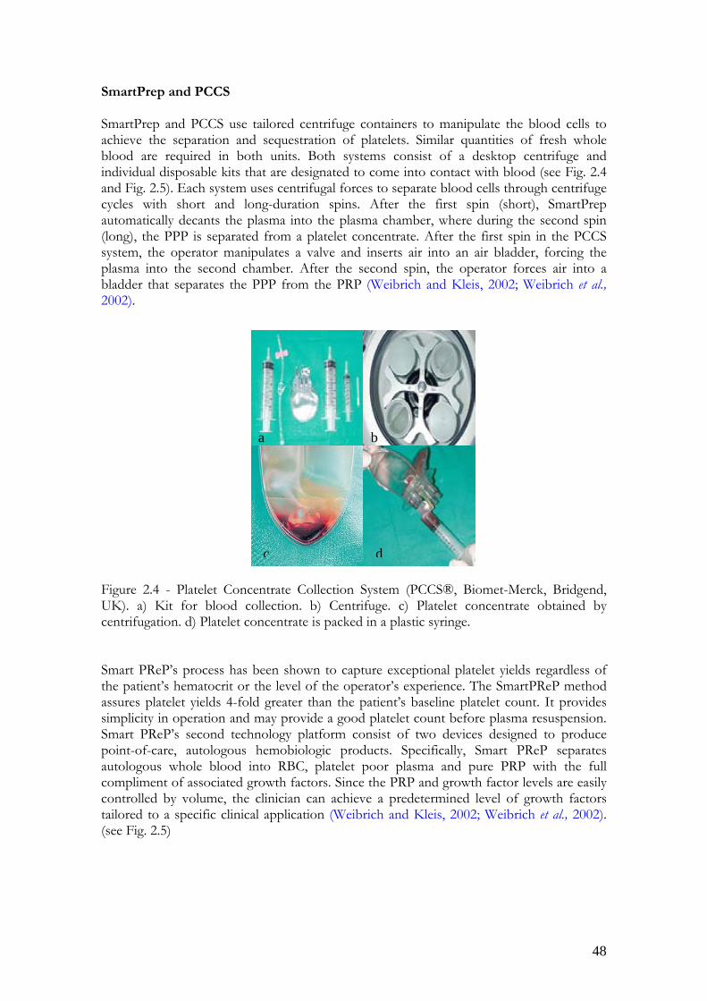
48
SmartPrep and PCCS SmartPrep and PCCS use tailored centrifuge containers to manipulate the blood cells to achieve the separation and sequestration of platelets. Similar quantities of fresh whole blood are required in both units. Both systems consist of a desktop centrifuge and individual disposable kits that are designated to come into contact with blood (see Fig. 2.4 and Fig. 2.5). Each system uses centrifugal forces to separate blood cells through centrifuge cycles with short and long-duration spins. After the first spin (short), SmartPrep automatically decants the plasma into the plasma chamber, where during the second spin (long), the PPP is separated from a platelet concentrate. After the first spin in the PCCS system, the operator manipulates a valve and inserts air into an air bladder, forcing the plasma into the second chamber. After the second spin, the operator forces air into a bladder that separates the PPP from the PRP (Weibrich and Kleis, 2002; Weibrich et al., 2002). Figure 2.4 - Platelet Concentrate Collection System (PCCS®, Biomet-Merck, Bridgend, UK). a) Kit for blood collection. b) Centrifuge. c) Platelet concentrate obtained by centrifugation. d) Platelet concentrate is packed in a plastic syringe. Smart PReP’s process has been shown to capture exceptional platelet yields regardless of the patient’s hematocrit or the level of the operator’s experience. The SmartPReP method assures platelet yields 4-fold greater than the patient’s baseline platelet count. It provides simplicity in operation and may provide a good platelet count before plasma resuspension. Smart PReP’s second technology platform consist of two devices designed to produce point-of-care, autologous hemobiologic products. Specifically, Smart PReP separates autologous whole blood into RBC, platelet poor plasma and pure PRP with the full compliment of associated growth factors. Since the PRP and growth factor levels are easily controlled by volume, the clinician can achieve a predetermined level of growth factors tailored to a specific clinical application (Weibrich and Kleis, 2002; Weibrich et al., 2002). (see Fig. 2.5)
a b
c d
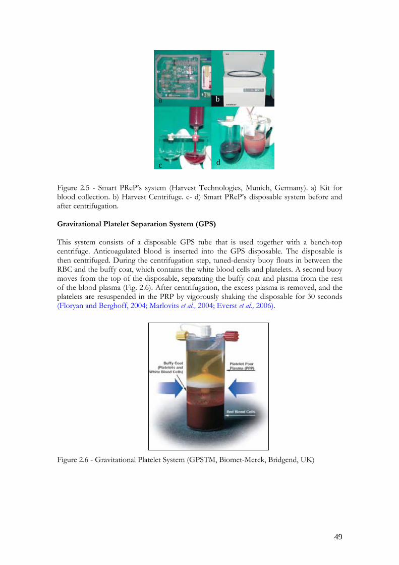
49
Figure 2.5 - Smart PReP’s system (Harvest Technologies, Munich, Germany). a) Kit for blood collection. b) Harvest Centrifuge. c- d) Smart PReP’s disposable system before and after centrifugation. Gravitational Platelet Separation System (GPS) This system consists of a disposable GPS tube that is used together with a bench-top centrifuge. Anticoagulated blood is inserted into the GPS disposable. The disposable is then centrifuged. During the centrifugation step, tuned-density buoy floats in between the RBC and the buffy coat, which contains the white blood cells and platelets. A second buoy moves from the top of the disposable, separating the buffy coat and plasma from the rest of the blood plasma (Fig. 2.6). After centrifugation, the excess plasma is removed, and the platelets are resuspended in the PRP by vigorously shaking the disposable for 30 seconds (Floryan and Berghoff, 2004; Marlovits et al., 2004; Everst et al., 2006). Figure 2.6 - Gravitational Platelet System (GPSTM, Biomet-Merck, Bridgend, UK)
a b
c d

50
c) Tube method. This method is also termed chair side technique, platelet rich in growth factors (PRGF) procedure. Since 2001 the PRGF system (G.A.C. Medicale San Antonio, 15-5°, 01005 Vitoria-España) is available (Fig. 2.7) (Anitua et al., 2006). Figure 2.7- Platelet rich growth factors (PRGF kit®). a) 4.5mL of whole blood (combined with 0.5 mL of ACD-A, anticoagulant). b) PRGF centrifuge. c- d) Platelet concentrate is packed in a tube. The advantages of this technique are the low cost and the minimal technical requirements in comparison to the apheresis and buffy coat methods. Only a simple centrifuge machine, 5 mL citrated tubes and 1 butterfly catheter are all that is needed to prepare PRGF (Marx et al., 1998; Zimmermann et al., 2001; Appel et al., 2002; Weibrich and Kleis, 2002; Weibrich et al., 2005). It is important to point out that with this technique the lowest number of leukocytes is collected when compared with other techniques such as buffy coat and apheresis devices (Zimmernan et al., 2001). The clinical use of autologous growth factors using this method has obtained good results regarding the number of platelets and leucocytes content in PRP (Anitua et al., 1999; Sanchez et al., 2003; Anitua et al., 2004). The main disadvantage of this system for perioperative platelet preparation is to be open-system devices that force the maintenance of a maximal 8 hours interval between platelet preparation and use on surgical setting needs sterile conditions (Marx et al., 2001, Zimmerman et al., 2001). However, the PRP must be separated from the PPP soon after centrifugation because the concentrated platelets will slowly diffuse into the PPP over time and would reduce the platelet count of the PRP preparation. When single spin centrifugation is performed, this technique concentrates a mean lower number of platelets (400-500x103 PLTs/μl) (Weibrich et al., 2005) than the apheresis and buffy coat techniques and tube method double spin centrifugation (Whitman et al., 1997; Marx et al., 1998, Zimmerman et al., 2001; Appel et al., 2002). The question of the actual quantity of platelets required is often put forward by clinicians who need to know the minimal therapeutic level of the platelets in the PRP that would result in a significant improvement, using PRP compared with standard treatments (Everts, et al., 2007). At present, not many data are available to answer this question directly, and only indirect information exists. Marx et al. (1998) performed the first study showing a significant improvement in mandibular continuity defects when PRP was mixed with autogenous bone grafts. Their PRP contained three to four times higher platelet count
a b
c d

51
compared with baseline values, although the average PRP platelet count found in their patients was just below 8 × 10⁵/µL, a number that is lower than in most other studies. Nevertheless, they observed a significantly faster radiographic maturation and histomorphometrically denser bone regeneration. Nowadays, the latest separation devices produce PRP platelet counts in excess of 6–10 times the baseline platelet count values. Manufacturers tend to interpret a high platelet concentration as a quality performance indicator of their separation devices, regardless of the fact that these high concentrations may not be necessary or might even contribute to a negative outcome (Everts et al., 2007). Weibrich et al. (2004) observed an advantageous effect with platelet concentrations of approximately 106/µL. Furthermore, they state that higher concentrations might have a paradoxically inhibitory effect. Comparison between different methods In humans some studies have been published comparing these methods with regard to PRP volume, platelet count on PRP and platelet yields (see table 2.4) (Zimmerman et al., 2001; Appel et al., 2002; Weibrich and Kleis, 2002; Weibrich et al., 2002; Everts et al., 2006). Device Mean PRP
Volume (mL)
Mean platelet Concentrate
(x 103)
Platelet Yield (%)
Increase above
baseline (%) Anitua protocol 9.5±4.1 433±129 35±16.8 190 Landsberg protocol 10.6±2.4 336±141 30±10.3 150 Clinaseal Sealed 7.6±1.5 401±267 39±16.3 164 ACE Surgical 7.8±0.6 493±245 33±10.2 180 AG Curasan 7.6±1.5 344±192 29±14.1 139 3i PCCS 7.0±1.5 939±284 61±8.9 324 Smart PReP 7.4±0.5 1.086±227 62±4.4 404 Table 2.4 - Platelet yields per device (adapted from Marx, 2004). The clinical effectiveness of each type of PRP preparation method can vary within the technique and efficiency, and current methods to evaluate the platelet concentration efficiency of PRP systems have several limitations. Different techniques of preparation have been known to lead to substantially different amounts of cells, i.e. platelets and leucocytes, as well as different levels of growth factors (Zimmermann et al., 2001; Appel et al., 2002; Weibrich and Kleis, 2002; Weibrich et al., 2002; Zimmermann et al., 2003; Schlegel et al., 2004; Wiltfang et al., 2004). Some studies have questioned the effect of differential centrifugation on the integrity of the remaining platelets. Ledent et al. (1995) showed that whole blood centrifuged at 1,500 g for 90 seconds resulted in a minimal release of growth factors from the platelets. However, PRP subjected to centrifugation forces of 2,500 g resulted in a significant release of growth factors from the platelet alpha granules. Therefore in other study Dugrillon et al. (2002) demonstrated that spins greater than 800 g may decrease the amount of TGF- β in the PRP released.

52
There are also several choices of anticoagulants that can be used during PRP preparation. Anticoagulant citrate dextrose-A is a preferable agent (Marx, 2001). The citrate binds calcium and prevents coagulation, whereas the dextrose and other ingredients support platelet metabolism and viability. Citrate phosphate dextrose is also useful for PRP preparation. It is similar to anticoagulant citrate dextrose-A, but it has fewer supportive ingredients and therefore may be less effective at maintaining platelet viability (Marx, 2001). The use of ethylenediaminetetraacetic acid (EDTA) is potentially more harmful than citrate in the preparation of PRP, and a large number of damaged platelets have been observed (Landesberg et al., 2000). Trisodium citrate solution is suggested as an anticoagulant with no negative effects on PRP preparation (Anitua, 1999). Preparation of PRP in Swine To date, various devices for obtaining porcine PRP have been described, such as apheresis or discontinous platelet concentrate system which include reinfusion of red cells and remainder plasma (Henderson et al., 2003). On pigs three commercial devices for obtaining PRP have been experimentally assessed. And also have been analyzed for platelet and leukocyte counts as well as for TGF-β, PDGF-AB, and IGF-I levels (Li at al., 2004; Schlegel et al., 2004; Thorwarth et al., 2005, Akeda et al., 2006) (table 2.5). Device
PCCS (Thorwarth et al., 2005)
AG Curasan (Thorwarth et al., 2005)
Symphony system (Li et al., 2004)
Platelets in WB (x103) 118±12 118±12
398,4
Platelets in PRP (x103) 767±108,7
483,8±97,2
1786,6
WBC in WB (x103) 4,3±1,9
4,3±1,9
18,7
WBC in PC (x 103) 14,7±68
24,8±8,9
29,5
TGF- β (ng/mL)
467,1
79,7
ND
PDGF-AB (ng/mL) 251,8
314.1
ND
IGF-I (ng/mL) 91
69
ND
Table 2.5 - Devices used to prepare platelet concentrate in pigs. ND- Non determined The buffy coat and apheresis techniques are acceptable, but they present some economical restrictions according to the kind of study and size of the wound for PRP application. Platelet concentrate methods, like the apheresis method, can only be performed in a specialized laboratory (Whitman et al., 1997; Marx et al., 1998; Lowery et al., 1999).
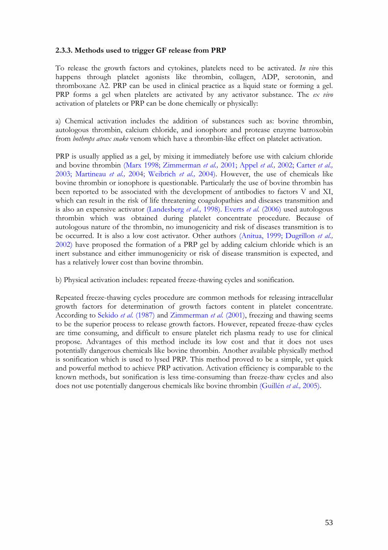
53
2.3.3. Methods used to trigger GF release from PRP To release the growth factors and cytokines, platelets need to be activated. In vivo this happens through platelet agonists like thrombin, collagen, ADP, serotonin, and thromboxane A2. PRP can be used in clinical practice as a liquid state or forming a gel. PRP forms a gel when platelets are activated by any activator substance. The ex vivo activation of platelets or PRP can be done chemically or physically: a) Chemical activation includes the addition of substances such as: bovine thrombin, autologous thrombin, calcium chloride, and ionophore and protease enzyme batroxobin from bothrops atrax snake venom which have a thrombin-like effect on platelet activation. PRP is usually applied as a gel, by mixing it immediately before use with calcium chloride and bovine thrombin (Marx 1998; Zimmerman et al., 2001; Appel et al., 2002; Carter et al., 2003; Martineau et al., 2004; Weibrich et al., 2004). However, the use of chemicals like bovine thrombin or ionophore is questionable. Particularly the use of bovine thrombin has been reported to be associated with the development of antibodies to factors V and XI, which can result in the risk of life threatening coagulopathies and diseases transmition and is also an expensive activator (Landesberg et al., 1998). Everts et al. (2006) used autologous thrombin which was obtained during platelet concentrate procedure. Because of autologous nature of the thrombin, no imunogenicity and risk of diseases transmition is to be occurred. It is also a low cost activator. Other authors (Anitua, 1999; Dugrillon et al., 2002) have proposed the formation of a PRP gel by adding calcium chloride which is an inert substance and either immunogenicity or risk of disease transmition is expected, and has a relatively lower cost than bovine thrombin. b) Physical activation includes: repeated freeze-thawing cycles and sonification. Repeated freeze-thawing cycles procedure are common methods for releasing intracellular growth factors for determination of growth factors content in platelet concentrate. According to Sekido et al. (1987) and Zimmerman et al. (2001), freezing and thawing seems to be the superior process to release growth factors. However, repeated freeze-thaw cycles are time consuming, and difficult to ensure platelet rich plasma ready to use for clinical propose. Advantages of this method include its low cost and that it does not uses potentially dangerous chemicals like bovine thrombin. Another available physically method is sonification which is used to lysed PRP. This method proved to be a simple, yet quick and powerful method to achieve PRP activation. Activation efficiency is comparable to the known methods, but sonification is less time-consuming than freeze-thaw cycles and also does not use potentially dangerous chemicals like bovine thrombin (Guillén et al., 2005).

54
2.3.4. Evaluation of platelet activation and determination of transforming growth factor-β1 (TGF-β1) Platelet activation during PRP preparation Two main requirements must be met to avoid platelet activation ex vivo during platelet collection (a) a venipuncture procedure must be used that minimizes spontaneous platelet activation and (b) blood must be collected into a medium that will not only prevent coagulation, but will also preserve the activation status of platelets until the samples can be analyzed (Wu, 1994; Anitua, 1999; Landesberg et al., 2000; Marx, 2001). Platelets can also be activated by excessive pipetting, the use of metallic or glass instruments and the use of a tourniquet over the vein to be punctured for blood extraction, or when not eliminating the first 2 mm of the extracted blood (Michelson, 1996). This premature platelet activation leads to an early release of growth factors. So when the blood sample is centrifuged, the growth factors will move to the most superior part. This way, the obtained PRP will be poor in growth factors (Tamimi et al., 2007). Another critical step in PRP preparation is the high spin centrifugation to concentrate the platelets. The mechanical forces may activate platelets, with the consequence of loosing the granular load that contains the growth factors into the plasma supernatant (Ledent et al 1995; Dugrillon et al., 2002). Determination of platelet activation (platelet activation markers) Direct and indirect measures of platelet activation have been described. As a direct measure of platelet activation, detection and quantification of antigens exposed during platelet degranulation can be determined. The cell adhesion molecule P-selectin (CD62P) is one of such activation antigens. P-selectin is restricted to the inner surface of the alpha granule membranes of resting platelets. Platelet activation and granule release results in an increase of P-selectin (CD62P) expression on the external surface of the platelet. The increase in P-selectin may be rapidly and conveniently detected by immunofluorescent flow cytometry and used as a reliable marker of platelet activation (Macey et al., 1999; Zimmerman et al., 2001; Lalko et al., 2003). Recently new platelet activation markers are available and are named toll-like receptors (TLR). TLR family recognizes pathogen-associated molecular patterns not found on host cells. TLR1 and TLR6 have been observed on human platelets (Shiraki et al., 2004); and TLR2 and TLR4 have been observed on murine platelets (Semple et al., 2004; Andonegui et al., 2005). TLR4 expression on resting human and rodent platelets has been previously demonstrated (Andonegui et al., 2005; Aslam et al., 2006; Ward et al., 2005). Despite these findings, the functional significance of platelet TLR expression is unknown. However, Andonegui et al. (2005) demonstrated that megariocytes express TL4 and Coppinger et al. (2004) showed that TLR is released from human platelets on thrombin activation, suggesting that at least some of platelet TLR expression may originate from megakaryocytopoiesis and from alpha granules. That suppose that during platelet activation, α-granule content release may results in an increase of TLR expression on the external surface of the platelet, conveniently detected by flow cytometry and used as a reliable marker of platelet activation (Coppinger et al., 2004; Andonegui et al., 2005; Ward et al., 2005; Aslam et al., 2006).

55
As an indirect measure, the mean platelet component (MPC) parameter has been proposed as a potentially useful screening test for platelet activation (Macey et al., 1999; Ifran et al., 2005). MPC is a new platelet parameter detected by current complete blood counter (CBC) ADVIA 120 ™, which represents the mean refractive index of the platelets. MPC is linearly related to platelet density and is reduced when platelets degranulate, thus indicating that platelets have undergone activation. Several studies in human medicine have recently demonstrated that MPC values are inversely correlated with platelet membrane P-selectin expression, (Macey et al., 1999; Ahnadi et al., 2004) and has been used as an indicator of platelet activation in different disease and physiological states in people (Ahnadi et al., 2004). In animals, MPC has been evaluated in dogs (Moritz et al., 2003; Moritz et al., 2005) and cats (Zelmanovic and Hetherington, 1998) and has been shown to decrease with platelet activation in both species and to be inversely related to platelet P-selectin expression (Moritz et al., 2003; Moritz et al., 2005). Thus, determination of the MPC by the ADVIA 120™ is becoming a quick method for the easy detection of platelet activation. Although no studies are available demonstrating the relationship between MPC and platelet activation in pigs, we hypothesized that MPC may also be an indicator of platelet activation in this specie and that it may be reduced in PRP obtained by tube method and double spin centrifugation in pigs if platelets become activated during procedure. Determination of transforming growth factor-β1 (TGF-β1) Wound healing, whether in bone or soft tissue, involves a complex chain of events. During this wound-healing cascade, platelets are trapped and activated, resulting in the release of growth factors present in the α-granules of the platelets and a fibrin clot generation in which a matrix for migration of tissue-forming cells will be formed (Slater et al., 1995; Everts et al., 2006; Everts et al., 2007). The platelet released growth factors have mitogenic and chemotactic properties that may contribute to improve wound healing. Of particular importance is the TGF-β1 which most likely promotes the early influx of cells and stimulation of proliferation (Landesberg et al., 2000). Usually, the TGF-β1 concentrations are determined by enzyme-linked immunosorbent sandwich assay (ELISA) developed with commercial antibodies for TGF-β1.

56
57
3. Objectives The healing of intestinal anastomoses remains a topic of ongoing research interest because clinical practice shows it to be disturbed rather frequently. Compromised healing may lead to dehiscence and leakage, which is attended by high morbidity and mortality. The occurrence of anastomotic leakage appears to be unavoidable in large bowel surgery in contrast with surgery of the small bowel (Hesp et al., 1984). In small animals the reported incidence of small intestinal dehiscence rates 7-16%, with 74-80% of those patients dying (Hedlund et al., 2002). Such a situation has led many surgeons throughout the world to devise and investigate new and more effective methods, techniques and materials to prevent dehiscence. Local protective methods to "protect" intestinal anastomoses during their complex biologic healing process have been the interest of many surgeons. Those methods included the use of various surgical techniques and materials, as well the use of various sealants, but none is sufficient to prevent the development of complications (Haukipuro et al., 1988; Van der Hamm et al., 1991). Using these facts, in the last several years a number of authors have investigated the use of various growth factors (local or systemic) in order to enhance the intestinal wound healing with some encouraging results (Mustoe et al., 1990; Slavin et al., 1992). The disadvantage of these growth factors is that they are expensive and concerns exist about their safety in human administration (Calabresi et al., 1998). Alternatively, PRP, which is a volume of autogenous plasma that has a platelet concentration above baseline, is a proven source of growth factors (Marx et al., 1998). However, the most significant benefit of using the autologous growth factors (AGF) concentrate is its being autologous, endogenously derived, and easily available. There are no issues about immunogenicity or transmission of infection. There are no known local or systemic side effects or adverse effects. The process is also considerably cost effective as compared with use of purified or recombinant growth factors, and also it may be a more physiologically source to provide a combination of all factors in the platelets rather than individual factors (Lowery et al., 1999). Recently, PRP has been exogenously applied in many different clinical applications, demonstrating the effectiveness and importance of the product for a variety of medical procedures. Even with those related promising results the use of platelet concentrate on gastrointestinal tract healing, especially for end-to-end anastomosis, was poorly investigated. In our knowledge only one study is available in colonic rats and with promising results by Yol et al. (2008). Therefore this study was performed in rat model and only 7 days after anastomosis construction. On other hand any study are available on the lag phase (3 days after anastomosis construction) when clinically the anastomotic dehiscence occurs.

58
Based on the considerations above, the aims of the present study are: 1. To validate the tube method technique double spin centrifugation for obtaining autologous PRP derived from pig blood. 2. To evaluate platelet activation during double spin method procedure to obtain platelet concentrates in pigs, by monitored MPC values on whole blood and PRP obtained by flow cytometry haematology system. 3. To validate the TGF-β1 level determination technique with an enzyme-linked immunosorbent sandwich assay (ELISA) in pig PRP obtained by double centrifugation tube method. 4. To evaluate the quality of PRP obtained by tube method technique double spin centrifugation, determination TGF-β1 level with an ELISA technique. 5. To comprehensively characterize the effects of PRP gel at 24h, 48h, 72h, 96h, and 7 days after anastomotic construction on the development of wound histology. 6. To quantify the anastomotic resistance using the breaking strength at days 3 and 7 post anastomosis construction.

59
4. Material and methods Animals Large White breed pigs, males and females, weighting 55±5 Kg, were recruited in the present study. All animals were used in accordance with the appropriate standard for the use of experimental animals in research studies and in accordance with the Spanish Code of Practice for the Care and Use of Animals for Scientific Purposes, published by the Spanish Government Publishing Service, (Directiva CE 86/609- BOE RD 1201/05- 21/11/05). All procedures were approved by the Ethical Commission of Animal and Human Experimentation (CEEAH 644) of the Autonomous University of Barcelona. Upon arrival, the animals were adapted for 14 days before surgery. The pigs were housed in groups of 2 or 3 in pens ranging from 3,6 - 5,4 m2. During the acclimatization period, the environment was controlled to remain within a temperature range of 20 - 24 ºC and 65% of humidity. The light/dark cycle was 13 and 11 h, respectively, with the photoperiod between 6.30 a.m. and 7.30 p.m. with 30 min of dusk and dawn. After surgery each pig was kept in a separate box (1,83 - 3,92 m2). They were fed with standard swine diet according to their weight, before and after the surgery, and received an uninterrupted supply of clean, fresh water. The pigs were monitored 24 h/day throughout the observation period. This monitoring included observation of general behaviour, food intake, fecal production, and temperature. The pre and post-operative care and observation were performed by trained animal technicians, supervised by veterinarians, who attended the animals daily. Experimental design The present study was divided into 3 main stages: - Stage I: PRP obtention and quality evaluation - Stage II: Surgical procedure - Stage III: Evaluation of the intestinal healing 4.1. Stage I – PRP obtention and quality evaluation 4.1.1. PRP obtention In order to design an appropriate technique for PRP obtention in pigs, a pilot study was carried out. a) Pilot study On this preliminary study, pigs were used only to prepare PRP in order to obtain the best method to concentrate platelets in pigs. Thirty pigs were used to collect 80 mL of whole blood for PRP preparation. For all these procedures, digital centrifuge (Ortoalresa unicent 20) was used. At the beginning, Anitua’s (1999) protocol was used (460g for 8 minutes and single spin centrifugation), but attempts to produce PRP using the same parameters failed to yield satisfactory enrichment percentages. For this reason, a large group of animals was used. Blood obtained was spun

60
at different cycles of centrifugations forces, different times, and pellet obtained ressuspended in different volumes of plasma, in order to obtain an optimal concentration. The PRP preparation technique was considered to be a success when concentration of the platelets within the PRP was around 1.000.000 platelets/µl, “pellet” volume of 400-500 µL of viable undamaged platelets, and an increase in concentrate of platelets just about 3-fold above the physiologic levels. The preparation of PRP, including the amount of blood collected, the centrifugal forces, and the final volume of the obtained PRP was optimized for each bioassay in order to obtain that concentration. The successful on PRP preparation technique was repeated twice to test reproducibility, and when the results appeared to be reproducible, the experiments for definitive purpose were begun (application of PRP gel on intestinal anastomosis wound). To evaluate the efficacy of PRP tube method and double spins centrifugation, several quality parameters were taken into consideration. The essential parameter was the total number of platelets obtained in the PRP, from the whole blood volume drew from the animal. The total number of platelets in the PRP preparation was the most meaningful parameter because it is a measure of the ability of the system to obtain as many of the platelets within the PRP as possible. The total number of the platelets within the PRP (number of platelets per µL) was related to the concentration within the original whole blood. The volume of the PRP were compared to the volume of blood drew from the animal. These last two parameters are proportional and both give an indication of the volume of the plasma fraction, in which the platelets are yielded. Thus, the quality parameters used to evaluate the tube method and double spin centrifugation on PRP preparation in pig were: increase in platelet concentrate, platelet increase over baseline, and collection efficiency. These parameters where calculated using the following equations (Marx et al., 1998; Carter et al., 2003; Efeoglu et al., 2004; Weibrich et al., 2005; Akeda et al., 2006):
Increase in platelet concentrate (%) = 100×WBofCountPlateletPRPofCountPlatelet
PLT increase over baseline (fold) =WBofCountPlatelet
WBofCountPlateletPRPofCountPlatelet −
Collection efficiency (%) = 100×××
WBofVolumeWBofCountPlateletPRPofVolumePRPofCountPlatelet
PLT- platelet; PRP- platelet rich-plasma; WB- whole blood, PPP- platelet poor- plasma After this preliminar study, a double spin centrifugation was chosen using the following spin method: for the initial spin 786g for 10 minutes, and for the second spin, 526 g for 10 minutes. Final pellet was ressuspended in 500μL of plasma. This combination yielded the best results in large white breed pigs.

61
b) Definitive study Based on the pilot study, PRP obtention in pigs which were going to be submitted to the surgical procedure was performed as follows:
Collection of blood samples
Whole blood collection was done immediately prior to anaesthetic induction and surgery. Sixty-five cubic centimetres of arterial blood were withdrawn from the femoral artery of each pig into 13 (65mL) 5 mL-glass tubes containing 0.5 mL buffered sodium citrate (Venoject®, Terumo Europe N.V.) (Fig. 4.1) using a butterfly needle. One of the tubes was used for baseline measurements. An additional blood sample was taken in heparin tubes for biochemistry evaluation. The heparinised whole blood was centrifuged at 1000g for 15 minutes and no less than 1 ml of plasma was placed into Eppendorf tubes that were held in a conventional freezer at -20ºC for further biochemistry analysis. Figure 4.1-Withdrawal of whole blood on citrated tubes using a butterfly needle, from the femoral artery in the pig.
Double spin centrifugation
Platelet-rich plasma was prepared using the 12 tubes (60mL) via a 2-step centrifugation procedure. The citrated whole blood samples were first centrifuged at 786g during 10 minutes at room temperature. The blood was then separated into its three basic components; red blood cells, buffy coat, and platelet-poor plasma (PPP). The first supernatant plasma fraction, adjacent to the buffy coat, composed of platelets and plasma was collected under aseptic conditions, and transferred into silicone-coated glass tubes (Venoject®, Terumo Europe N.V.) and centrifuged again in order to pellet the platelets. This second centrifugation was 526 g for 10 minutes. The resulting supernatant was removed and platelet pellet was resuspended in about 500 μL of plasma per tube. The “pellet” was resuspended in the residual plasma by carefully ‘hand shaking’ the siliconed
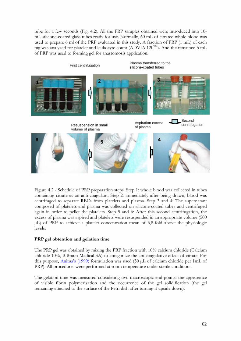
62
tube for a few seconds (Fig. 4.2). All the PRP samples obtained were introduced into 10-mL silicone-coated glass tubes ready for use. Normally, 60 mL of citrated whole blood was used to prepare 6 ml of the PRP evaluated in this study. A fraction of PRP (1 mL) of each pig was analyzed for platelet and leukocyte count (ADVIA 120TM). And the remained 5 mL of PRP was used to forming gel for anastomosis application. Figure 4.2 - Schedule of PRP preparation steps. Step 1: whole blood was collected in tubes containing citrate as an anti-coagulant. Step 2: immediately after being drawn, blood was centrifuged to separate RBCs from platelets and plasma. Step 3 and 4: The supernatant composed of platelets and plasma was collected on silicone-coated tubes and centrifuged again in order to pellet the platelets. Step 5 and 6: After this second centrifugation, the excess of plasma was aspired and platelets were resuspended in an appropriate volume (500 µL) of PRP to achieve a platelet concentration mean of 3,8-fold above the physiologic levels. PRP gel obtention and gelation time The PRP gel was obtained by mixing the PRP fraction with 10% calcium chloride (Calcium chloride 10%, B.Braun Medical SA) to antagonize the anticoagulative effect of citrate. For this purpose, Anitua’s (1999) formulation was used (50 µL of calcium chloride per 1mL of PRP). All procedures were performed at room temperature under sterile conditions. The gelation time was measured considering two macroscopic end-points: the appearance of visible fibrin polymerization and the occurrence of the gel solidification (the gel remaining attached to the surface of the Petri dish after turning it upside down).
First centrifugation
Second cenrtifugationResuspension in small
volume of plasma
Aspiration excess of plasma
Plasma transferred to the silicone-coated tubes
2 3
456
1

63
4.1.2. Evaluation of PRP quality Once the PRP obtention technique was designed, quality evaluation of all the PRPs performed was assessed by means of the following determinations: a) Erythrocyte, platelet and leukocyte count, and quality parameters b) Determination of platelet activation by flow cytometry c) Determination of TGF- β1 a) Erythrocyte, platelet and leukocyte count, and quality parameters Erythrocyte, platelet and leukocyte count was determined in samples of all animals submitted to surgical procedure (35 pigs) and in animals used for TGF-β1 determination (12 pigs), representing a total of 47 pigs. Quality parameters were calculated in all animals using the formulas described above. Hematological and platelet parameters analysis in all studied pigs were performed few minutes after blood collection and before PRP obtention. Erythrocytic parameters, leukocytic series count, and platelet parameters, on whole blood and PRP, were automatically determined using a flow cytometry hematology system (ADVIA 120 Analyzer, Bayer Lab, NY, USA). All samples were measured using the same automatic cell counter, to ensure that the evaluations of the PRP preparations were comparable and not the result of measurement error; as a control, double measurements were made in all specimens, which showed repeatable results. Evaluated hematological parameters included: red blood cell count (RBC, 106/µL), hemoglobin concentration (HGB, g/dL), haematocrit (Ht %), mean corpuscular volume (MCV, fl), mean corpuscular haemoglobin (MCH, pg), red cell distribution width (RDW %), mean corpuscular hemoglobin concentration (MCHC, g/L). Total leukocyte count (WBC, 103 cell/µL), as well as the relative and absolute numbers of neutrophils, lymphocytes, monocytes, eosinophils, basophils (103 cell/µL ;%), and large unstained cell (LUC, 103 cell/µL; %) counts were determined. Platelet parameters: absolute platelets count (PLT, 103/µL), number of large platelets (103/ µL), mean platelet component (MPC, g/dL), mean corpuscular volume (MCV, fl); mean platelet volume(MPV, fl), mean platelet mass (MPM, pg), platelet distribution width (PDW, %), platelet mass distribution width (PMDW, pg), platelet component distribution width (PCDW, g/dL), plateletcrit (PCT, %), large platelets (LP, 103 cell/µL), amongst others were determined. For biochemical analysis plasma specimens obtained from heparinised whole blood were analyzed for glucose, urea, alanine aminotransferase, lactate dehydrogenase, and total protein, to further confirm their health status. Biochemical analysis were undertaken on serum using Olympus® AU400 Analyzer (serial number 3112676 Germany) and reagents from OSR (Olympus System Reagent®, Olympus®, Ireland). For serum proteins determination Hydrasys and Hyrys Analyzer (serial number 517; Sebia, French) and Hydragel protein (E) gels, (Sebia) were used.

64
b) Determination of platelet activation by flow cytometry The MPC values were determined in the 47 animals mentioned above. The MPC values on citrated whole blood and PRP were obtained during routinely determination of the fundamental platelets parameters on whole blood and PRP using the ADVIA 120TM Analyser. c) Determination of TGF-β1 by ELISA Twelve animals not submitted to the surgical procedure were included in this group. In this animals, sampling and PRP obtention was performed as described before, and TGF- β1 levels were determined on different PRP fractions (PPP, PRP, and PRP gel) using an enzyme-linked immunosorbent assay (ELISA). All PRP fractions samples were obtained by centrifugation of citrated whole blood at 756g for 10 min first spin and the supernatant were centrifuged at 526g for 10 min second spin. The upper half of the PRP preparation after the second centrifugation was designated as platelet-poor plasma (PPP) and, after collecting a volume for analysis, the rest was subsequently discarded. The platelet pellet was resuspended in 500μL of plasma resulting in the PRP fraction. PRP gel was obtained by adding calcium chloride on fresh PRP. All PRP fractions (PPP, PRP and PRP gel) were frozen at -80ºC before growth factor analysis. The PRP fractions were thawed and mixed well prior to growth factor analysis. TGF-β1 concentrations were determined by enzyme-linked immunosorbent sandwich assay (ELISA) developed with commercial antibodies for TGF-β1 (Human TGFbeta DuoSet®, R&D Systems) according to manufacturer instructions. The ELISA was performed in triplicate for each sample. Briefly, a capture antibody specific to TGF-β1 was added to a polystyrene 96-well plate (MaxiSorp, Nunc, Roskilde, Denmark) and incubated overnight at 4° C. After incubating the plate with blocking solution (phosphate buffered saline (PBS) containing 1% bovine serum albumin and 0.05% Tween 20), for 45 minutes at 37ºC, test samples and the standard recombinant cytokines (R&D Systems) were added, and the plate was incubated at 37ºC for 2 hours. For total TGF-β1 levels, the samples were diluted 1:4 in Dulbecco's (PBS) and treated with 1N HCl for 15 min (to obtaining a sample pH of approximately 2.6); afterwards samples were neutralized with 1N NaOH to a pH of approximately 7.6. After washing four times with PBS containing Tween 20, a detection antibody specific for TGF-β1 was added, and the reactions were allowed to proceed for 1 hour at 37ºC. After washing three times, OPD (Sigma, USA) was added to induce the colour reaction for 30 minutes. A microplate reader (Victor3, Perkin Elmer) was used to measure the optical density at 450nm. A standard curve four parameter logistic (4 -PL) and logaritmic curve was drawn by plotting optical density versus the log of the concentration. The increase in TGF-β1 concentrate was calculated using the following equation:
Increase in TGF-β1 levels (pg/mL) = PPPoflevelsTGFPRPoflevelsTGF
11
ββ

65
4.2. Stage II- Surgical procedure A preliminar study was carried out in order to decide the PRP gel application technique on intestinal anastomoses. a) Pilot study Six pigs divided into 2 groups of three animals each were used for macroscopic and histological evaluation of intestinal wound healing and evaluation of PRP gel application technique. The animals were euthanized at 24 and 48 hours after surgery. These animals underwent a laparatomy and a double enterectomy and anastomosis of the jejunum. One of the anastomosis was closed with suture repair alone (control), while the other was treated with PRP gel using a “sandwich technique”. Another six pigs, divided into the same groups as describe above, were treated in a similar fashion, but PRP gel application technique was modified, as an “immersion technique” was performed. Pre-operative protocol and anesthesia The pre-operative protocol included bowel preparation which consisted of a 24 - hour period of fasting before operation. Free access of water was allowed. The pigs were premedicated with a combination of ketamine 10 mg/kg IM (Imalgene 1000®, Merial Laboratories), azaperone 2 mg/kg IM (Stressnil®, Janssen Pharmaceutical) and buprenorphine 20 μg/kg IM (Buprex®, Schering-Plough Laboratories). Immediately prior to induction, 65 mL of whole blood were drawn for PRP obtention. Then, a single dose of 4 mg/kg of propofol (Propofol Lipuro 1%, B.Braun Medical SA) was given IV for anaesthetic induction through an 18-gauge polyurethane catheter (Vasocan, B.Braun Medical SA) placed in the peripheral auricular vein. The trachea was intubated and anesthesia was maintained with 1.5-2.5% isofluorane (IsoFlo, Abbott Laboratories) in 100% oxygen through a semi-closed circular anaesthetic system. To compensate fluid loss, all animals received an infusion of Ringer Lactate, at a rate of 10 mL/kg/h, during the perioperative period, and until completely recovered from anaesthesia, perioperative IV antibiotic therapy (cephalexin, 20 mg/kg) were administered via the auricular vein. Heart rate, pulse oximetry, respiratory rate and capnography, and temperature, were monitored during anaesthesia, using a Datex Ohmeda Cardiocap II Monitor. Surgical procedure Each pig was positioned in dorsal recumbency on the operating room table and the ventral abdominal region was prepared aseptically. After the surgical field was draped, a ventral midline abdominal incision was made and the linea alba was incised from 5 cm cranial to the umbilicus and extending 7-8 cm caudally. The jejunum was exteriorized from the abdomen, the lumen was occluded using digital pressure, and a complete transversal section was made with Metzenbaum scissors (Fig 4.3.a).
66
“Sandwich technique” Autologous PRP gel was activated with calcium chloride in a glass Petri dish, and in few minutes PRP gel was obtained (Fig. 4.3.b). Thus the intestinal edges were opposed and a film of PRP gel was applied between the two intestinal edges. Intestinal anastomosis was performed suturing the gel in between (“sandwich technique”). Intestinal end-to-end anastomosis was performed using synthetic monofilament glyconate 3/0 USP suture material (Monosyn®, B.Braun Medical SA) and a simple interrupted pattern (Fig. 4.3.c). A second enterectomy (control) was performed in the mid-jejunum at about 50 cm from the first enterectomy and was sutured using the same pattern as describe above without any treatment. When the intestinal suture was completed, tightness was checked for leakage and the anastomotic site was omentalized. Both anastomosis (treated and untreated) were performed on the same pig. The laparatomy was closed in a routine manner. During surgery, the body temperature was kept at 38 °C by using a heating pad and warm Lactated Ringer’s solution given intravenously during the operation. The animals were euthanized at 24 and 48 hours after surgery for histological evaluation. Figure 4.3- Application of calcium activated platelet concentrate “sandwich technique”. (a) end-to-end intestinal anastomosis; (b) Petri dish with calcium chloride activated PRP; (c) application of PRP gel between the intestinal edges (“sandwich technique”); (d) intestinal lumen after performing the suture of the opposite side.
ba
c d
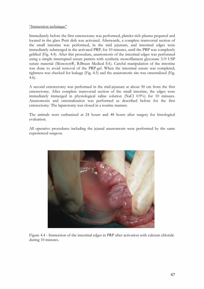
67
“Immersion technique” Immediately before the first enterectomy was performed, platelet-rich plasma prepared and located in the glass Petri dish was activated. Afterwards, a complete transversal section of the small intestine was performed, in the mid jejunum, and intestinal edges were immediately submerged in the activated PRP, for 10 minutes, until the PRP was completely gelified (Fig. 4.4). After this procedure, anastomosis of the intestinal edges was performed using a simple interrupted suture pattern with synthetic monofilament glyconate 3/0 USP suture material (Monosyn®, B.Braun Medical SA). Careful manipulation of the intestine was done to avoid removal of the PRP-gel. When the intestinal suture was completed, tightness was checked for leakage (Fig. 4.5) and the anastomotic site was omentalized (Fig. 4.6). A second enterectomy was performed in the mid-jejunum at about 50 cm from the first enterectomy. After complete transversal section of the small intestine, the edges were immediately immerged in physiological saline solution (NaCl 0.9%) for 10 minutes. Anastomosis and omentalization was performed as described before for the first enterectomy. The laparotomy was closed in a routine manner. The animals were euthanized at 24 hours and 48 hours after surgery for histological evaluation. All operative procedures including the jejunal anastomosis were performed by the same experienced surgeon. Figure 4.4 - Immersion of the intestinal edges in PRP after activation with calcium chloride during 10 minutes.

68
Figure 4.5 - Checking leakage before omentoplasty. Figure 4.6 - Technique for mobilizing the omentum based on the epiploic arcade. Post-operative period All animals recovered from surgery without any complication and were allowed ad libitum activity. After the operation, the animals were monitored at least thrice-daily for subjects like daily activity, inquisitiveness, vocalization, gait, urinary and bowel habits, oral intake and examination of the abdomen wound incision. Animals were gradually fed with a standard diet 12-24 hours postoperatively and given water ad libitum. Buprenorphine 20 μg/kg IM, was provided thrice-daily for postoperative analgesia. Antibiotic therapy consisted of 20 mg/kg cephalexin IV every 12 hours until endpoint day. Weights were obtained preoperatively, at midsurvival, and before end point. To evaluate the occurrence of anastomotic leakage the following indicators were assessed: (i) Clinical indicators (abdominal pain, abdomen distension, accumulation of gas or fluids and vomiting).

69
(ii) Biochemical and physiologic parameters or observation abnormalities (fever, tachycardia, hypopnea or shallow breathing, rapid breathing, low blood pressure, rapid pulse, polydipsia and increased white cell count). b) Definitive study Since the results obtained with the “sandwich technique” were not good enough (see Results section below), the “immersion technique” was chosen for definitive study purposes. A total of 35 pigs (including those employed in the pilot study) were operated following the “immersion technique” for PRP gel application. From those 35 pigs, 15 pigs were assigned to evaluation of the intestinal healing through histological analysis, while 20 pigs were used for evaluation of the intestinal anastomotic strength. 4.3. Stage III. Evaluation of the intestinal healing Evaluation of the intestinal healing following PRP application was done at different times by means of a macroscopic and histological examination and through determination of the breaking strength. Thus, all the animals were sacrificed without pain by overdose with a lethal intravenous injection of Phenobarbital sodium 20 mg (Dolethal®) Vetoquinol- Madrid) preceded by a combination of ketamine 10 mg/kg IM, azaperone 2 mg/kg IM sedation, at respective times. All animals received care in accordance with the Spanish Code of Practice for the Care and Use of Animals for Scientific Purposes, published by Spanish Government Publishing Service, (Directiva CE 86/609- BOE RD 1201/05 - 21/11/05). 4.3.1. Macroscopic examination In all operated animals (35 pigs), at appropriate endpoint day for each group respectively, macroscopic examination was performed. Immediately after euthanasia, the previous incision was reopened, anastomosis was examined in situ and healing was visually assessed as normal or abnormal. The intestines were carefully exteriorized and the anastomotic sites identified, the tightness was checked for leakage occurrence. Abnormal healing was defined as the presence of stricture formation, anastomotic leakage, perianastomotic abscess formation, and anastomotic dehiscence or obstruction. Normal healing was defined as apparent anastomotic integrity with no evidence of leakage, perianastomotic collection, obstruction or distension of the bowel. 4.3.2. Histological examination Histological examination was first performed in animals included in the pilot study in order to decide which surgical technique of PRP gel application was more suitable. Intestinal samples were evaluated at 24 and 48 hours. After that, histological examination was performed in the definitive study, which included 15 animals divided in five groups of 3 animals each: group 1 was euthanized after 24 hours from surgery, group 2 after 48 hours, group 3 after 72 hours, group 4 after 96 hours, and group 5 after 7 days. Animals from to group 1 and group 2 belong to the pilot study using the “immersion technique” of PRP gel application.

70
Segments of approximately 10 cm in length containing the anastomosis in the middle were carefully resected. Intestinal samples were opened at the mesenteric side, gently washed with saline solution, and fixed in 10% neutral buffered formalin. Each intestinal sample was cut in 6-8 pieces and routinely processed for further histopathological study. Four µm sections routinely stained with hematoxylin and eosin (H-E) and with Masson trichrome were evaluated. Microscopic observations focused on healing of intestinal layers and thickness of granulation tissue formed. Sections were then examined by the same trained pathologist who was blinded to the tissue source. 4.3.3. Anastomotic breaking strength Unwounded intestinal samples were taken from the animals included in the pilot study in order to check the feasibility of the dynamometer (Instron Tensile Machine 1122 TM, Instron Corporation, MA) for evaluation of intestinal breaking strength. For definitive purposes, 20 pigs were used for the anastomotic strength measurement. These animals were subdivided in two groups of 10 animals each: group 1 - In those animals the measurement of breaking strength was made at 3 days after surgery. Group 2 - The measurement of breaking strength was made at 7 days after surgery. For comparison purposes, 10 samples of unwounded intestine from the same animals were also taken and measured for breaking strength. The intestines were carefully exteriorized and the anastomotic sites identified. Segments of approximately 10 cm in length containing the anastomosis in the middle were carefully resected, and adhesions were cut as far as possible without injury to the intestine. Intestines were opened at the mesenteric side and gently washed with saline solution. The segments were placed in the dynamometer, which provided a force of 10 kgf (0.1 kN) increasing at a rate of 300 mm/min (Fig. 4.7). The peak force necessary to induce disruption was taken as the breaking strength, as well as the site of rupture (within or outside the anastomotic line) was recorded. Figure 4.7- Anastomotic breaking strength evaluation. a) Instron Tensile Machine 1122 TM; b) The specimen was attached to the gripping component; c) Distraction of the tissue until rupture.
a b c
71
4.4. Statistical analysis
Data were analysed using a statistical computer software program (SPSS v15.0). Nonparametric tests were selected for all variables because they were either not normally distributed. All quantitative measurements were described using summary statistics (number, mean, and standard deviation, minimum, maximum). All duplicate values were within 10% of each other, and the mean of duplicates was used for data analysis. The results are presented as mean±SD, unless stated otherwise, n represents number of animals.
The means of platelets number in arterial blood, means of platelets number in PRP, means of the enrichment percentages, and means of hematological values, means of MPC values on PRP and whole blood; means of TGF-β1 concentrations, on PPP, PRP and PRP gel, of experimental groups were compared, using Wilcoxon statically analysis to compare means. Significance in all instances was set at a value p<0.05. A paired Student’s t-test statistical analysis was employed to compare breaking strength between PRP and control groups. Breaking strength at 3 days, 7 days and unwounded intestine was compared using a one- way ANOVA. Significance in all instances was set at a value p<0.05.

72

73
5. Results 5.1 Stage I: PRP obtention and quality evaluation
5.1.1. PRP obtention After the realization of different trials, it was concluded that the PRP obtention technique in pigs consisting in a tube method, using a double spin centrifugation (786g for 10 minutes for first spin, and 526g for 10 minutes for second spin, and resuspending the final pellet in 500 μL), yielded the best results. PRP obtention, using that technique, was prepared in approximately 30-40 minutes. 5.1.2. Evaluation of PRP quality Erythrocytic parameters, leukocytic series, and platelet parameters, including monitorization of MPC, on whole blood and PRP samples of the 35 pigs submitted to the surgical procedure were assessed. Calculation of the quality parameters was also performed. The same parameters were evaluated in the 12 animals used for TGF-β1 levels determination, as well as concentration of this growth factor in the different fractions (PPP, PRP, PRP gel). Mean, standard deviation (SD) and range values in statistical analyses were used. a) Erythrocyte, platelet and leukocyte count, and quality parameters The values of erythrocytic and platelet parameters, before and after centrifugation on operated animals are resumed on table 5.1. Statistically significant differences (P<0.05), were observed between erythrocytic parameters on whole blood, when comparing those parameters on PRP. However, on some platelet parameters MPV, MCV, MPM and LP no significant differences (P<0.05) were observed. In contrast, in other platelet parameters (PDW) significant differences were observed on whole blood, when comparing with (PDW) count on PRP. The leukocytic series (neutrophils, lymphocytes, monocytes, eosinophils and basophils) values in 103/µL and %, before and after centrifugation on operated animals, are resumed on table 5.2 and 5.3, respectively. Statistically significant differences (P<0.05), were observed between leucocytic series count on whole blood and PRP.

74
Citrated whole blood Platelet - rich plasma Parameter Mean SD Range Mean SD Range RBC a (103/µL) 5,7 0,4 4,8-6,6 0,01 0,04 0,01-0,02Hemoglobin (g/dL) 9,7 0,9 8,1-11,6 0,01 0,07 0-0,4 Hematocrit (%) 30 2,8 24,1-35,1 0,09 0,02 0-0,1 MCH b (pg) 16,9 0,9 14,6-18,7 0 0 0 MCHC c (g/dL) 32,5 1,1 30,2-36,9 0 0 0 RDW d (%) 16,7 1,2 15,3-20,3 26,2 6,64 17,3-43,5HDW e (g/dL) 1,9 0,09 1,73-2,24 3,46 0,86 2,4-6,56 MPV f (fl) 8,96 0,74 7,1-10,2 8,44 0,69 6,8-9,3 MCV g (fl) 51,94 2,5 46,6-56,1 50,5 4,72 35,1-64,5MPM h (g/dL) 1,72 0,12 1,51-1,84 1,56 0,14 1,31-1,76PDW i (%) 56,4 5,32 48,5-69,4 42,58 3,5 36,6-50,4PCT j (%) 0,72 0,09 0,26-0,69 1,3 0,43 0,69-2,34LP l (103/µL) 13,37 4,37 8-26 16,3 10,1 3-49 Table 5.1 - Mean, SD and range values of erythrocyte and platelet parameters on citrated whole blood and PRP of operated animals (n=35). a, RBC-red blood cells; b, MCH-mean corpuscular hemoglobin; c, MCHC-mean corpuscular hemoglobin concentration; d, RDW-red blood cell distribution width; e, HDW-hemoglobin distribution width; f, MPV-mean platelet volume; g, MCV-mean corpuscular volume; h, MPM-mean platelet mass; i, PDW-platelet distribution width; j, PCT-Plateletcrit; l, LP-large platelets. Citrated whole blood Platelet - rich plasma Parameter Mean SD Range Mean SD Range WBC (103/µL) 15,33 3,45 9,02-23,63 1,83 1,03 0,37- 4,29Neutrophils (103/µL) 4,34 1,56 2,03-7,99 0,06 0,04 0,01-0,24 Lymphocytes (103/µL) 9,47 2,77 3,12-16,18 1,64 0,94 0,32-3,95 Monocytes (103/µL) 0,97 1,28 0,27-8,11 0,08 0,06 0,01-0,3 Eosinophils (103/µL) 0,24 0,2 0,03-1,18 0,01 0,01 0-0,1 Basophils (103/µL) 0,2 0,08 0,08-0,48 0,01 0,02 0-0,1 LUC (103/µL) 0,26 0,29 0,03-1,7 0,03 0,04 0-0,2 Table 5.2 - Mean, SD and range values of leucocytic series (103/µL) on citrated whole blood and PRP of operated animals (n=35). LUC - large unstained cells; WBC- white blood cells
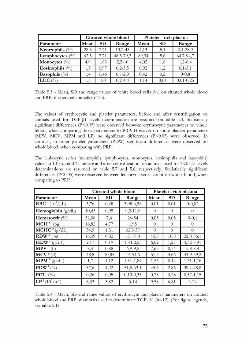
75
Citrated whole blood Platelet - rich plasma Parameter Mean SD Range Mean SD Range Neutrophils (%) 28,3 7,71 13,2-43 4,13 5,1 0,4-28,9 Lymphocytes (%) 62,5 7,71 48,5-79,3 89,34 5,6 64,7-94,7 Monocytes (%) 4,9 1,63 2,5-10 4,02 1,8 1,2-8,4 Eosinophils (%) 1,5 0,97 0,2-5,5 0,92 1,2 0,1-5,1 Basophils (%) 1,4 0,46 0,7-2,9 0,42 0,2 0-0,8 LUC (%) 1,5 1,0 0,2-4,4 1,14 0,04 0,01-0,25
Table 5.3 - Mean, SD and range values of white blood cells (%) on citrated whole blood and PRP of operated animals (n=35). The values of erythrocytic and platelet parameters, before and after centrifugation on animals used for TGF-β1 levels determination are resumed on table 5.4. Statistically significant differences (P<0.05) were observed between erythrocytic parameters on whole blood, when comparing those parameters to PRP. However on some platelet parameters (MPV, MCV, MPM and LP) no significant differences (P<0.05) were observed. In contrast, in other platelet parameters (PDW) significant differences were observed on whole blood, when comparing with PRP. The leukocytic series (neutrophils, lymphocytes, monocytes, eosinophils and basophils) values in 103/µL and %, before and after centrifugation, on animals used for TGF-β1 levels determinations are resumed on table 5.7 and 5.8, respectively. Statistically significant differences (P<0.05) were observed between leucocytic series count on whole blood, when comparing to PRP. Citrated whole blood Platelet - rich plasma Parameter Mean SD Range Mean SD Range RBC a (103/µL) 5,76 0,48 5,08-6,58 0,01 0,01 0-0,02 Hemoglobin (g/dL) 10,45 0,95 9,2-11,9 0 0 0 Hematocrit (%) 32,08 7,4 26-54 0,05 0,05 0-0,1 MCH b (pg) 16,82 4,77 1,95 0 0 0 MCHC c (g/dL) 34,9 1,31 32,5-37 0 0 0 RDW d (%) 16,39 0,82 15-17,8 43,5 10,8 22,8-56,1HDW e (g/dL) 2,17 0,19 1,84-2,53 6,02 1,27 4,32-8,91MPV f (fl) 8,4 0,88 6,9-9,5 7,65 0,74 5,8-8,8 MCV g (fl) 48,8 10,85 15-54,6 51,5 4,66 44,9-59,2MPM h (g/dL) 1,7 1,12 1,51-1,84 1,56 0,14 1,31-1,76PDW i (%) 57,6 4,22 51,8-63,1 45,6 2,86 39,4-48,8PCT j (%) 0,26 0,05 0,13-0,33 0,75 0,28 0,37-1,15LP l (103/µL) 8,33 3,82 3-14 9,58 6,81 2-24 Table 5.4 - Mean, SD and range values of erythrocyte and platelet parameters on citrated whole blood and PRP of animals used to determinate TGF- β1 (n=12). (For figure legends, see table 5.1)
76
Citrated whole blood Platelet - rich plasma Parameter Mean SD Range Mean SD Range WBC (103/µL) 19,9 3,12 14,6-24,5 5,57 3,73 1,81-13,1 Neutrophils (103/µL) 6,54 2,65 3,87-10,9 0,31 0,51 0,03-1,89 Lymphocytes(103/µL) 11,98 2,42 8,18-16,5 4,9 3,2 1,68-12,2 Monocytes (103/µL) 0,74 0,35 0,3-1,54 0,27 0,32 0,03-1,19 Eosinophils (103/µL) 0,25 0,12 0,11-0,56 0,06 0,11 0,01-0,4 Basophils (103/µL) 0,35 0,46 0,09-1,8 0,03 0,03 0-0,12 LUC (103/µL) 0,15 0,13 0,04-0,54 0,03 0,03 0-0,09 Table 5.5 - Mean, SD and range values of white blood cells and leukocytic series on citrated whole blood and PRP of animals used to determinate a TGF- β1 levels (n=12). Citrated whole blood Platelet - rich plasma Parameter Mean SD Range Mean SD Range Neutrophils (%) 32,55 10,4 17-45,6 4,49 4,36 1-16,2 Lymphocytes (%) 60,47 9,58 46,9-72,6 89,45 6,86 76-96,4 Monocytes (%) 3,63 1,49 1,7-6,8 4,1 4,1 1,2-16,3 Eosinophils (%) 1,33 0,72 0,5-3 0,78 0,87 0,2-3,4 Basophils (%) 1,22 0,37 0,6-1,8 0,46 0,28 0,1-0,9 LUC (%) 0,75 0,73 0,2-3 0,68 0,74 0,2-2,9 Table 5.6 - Mean, SD and range values of white blood cells (%) on citrated whole blood and PRP of animals used to determinate a TGF- β1 levels (n=12). The parameters used to evaluate the tube method and double spin centrifugation to obtain PRP on operated animals, and on animals used for TGF-β1 levels determination, are resumed on tables 5.7 and 5.8, respectively. Statistically significant differences (P<0.05) were observed between platelet count on whole blood, when comparing platelet count on PRP.
Variables Mean SD Range Platelets in whole blood (103/µL) 424,97 107,54 192-814 Platelets in PRP (103/µL) 1620,4 536,69 894 -3173 Increase in PLT concentration (%) 388 88 254 -547 PLT increase over baseline (%) 2,8 0,94 1,5 - 4,5 Collection efficiency (%) 38,4 9,4 25,4 -54,8
Table 5.7 - Mean, SD and range values of platelet yields on citrated whole blood and PRP of operated animals (n=35). PLT: platelet.

77
Variables Mean SD Range
Platelets in whole blood(103/µL) 308,2 70,4 145 - 402 Platelets in PRP(103/µL) 991 343,5 461- 1436 PLT increase baseline (%) 3,16 0,61 2,2 - 3,9 PLT increase over baseline (%) 2,2 0,61 1,2 - 2,9 Collection efficiency (%) 31,6 6,1 21,8- 39,8
Table 5.8 - Mean, SD and range values of platelet yields on citrated whole blood and PRP of animals used to determinate a TGF- β1 levels (n=12). b) Determination of platelet activation by flow cytometry The platelet activation evaluation on operated animals and on animals used for TGF-β1 levels determination, by means of MPC assessment on PRP and whole blood, are represented on table 5.9 and 5.10, respectively.
Mean platelet component (MPC)- g/dL Whole blood PRP Mean 21,30 20,03 SD 1,58 1,39 Min 19,2 18,1 Max 24,8 24,2
Table 5.9 - Mean, SD and range values of mean platelet component (MPC) on citrated whole blood and PRP of operated animals (n= 35).
Mean platelet component (MPC) - g/dL Whole blood PRP PPP Mean 22,4 21,88 20,78 SD 1,59 1,68 1,95 Min 18,9 19,4 18,8 Max 25 25 18,8
Table 5.10 - Mean, SD and range values of mean platelet component (MPC) on citrated whole blood, PRP and PPP determinate by citometry (n= 12). In the operated animals, statistically no significant differences (P<0.05) were observed between whole blood values, when comparing to PRP values. In the animals used for TGF-β1 determination, statistically no significant differences (P<0.05) were observed between whole blood values, when comparing to PRP values. However, statically significant differences (P<0.05) were observed between whole blood values, when comparing with PPP values.
78
c) Determination of TGF-β1 levels The TGF-β1 concentrations on the different fractions (PPP, PRP, PRP gel) are resumed on table 5.11. Statistically significant differences (P<0.05) were observed between TGF-β1 levels in all PRP fractions.
Transforming growth factor beta- TGF- β1 (pg/mL) PPP PRP PRP gel Mean 3715 24312 15504 SD 2188 21755 11279 Min 210 3521 5092 Max 8336 67815 36345
Table 5.11 - Mean, SD and range values of TGF-β1 on PRP, PPP, and PRP gel (n=12). PPP - platelet poor plasma after second centrifugation; PRP - platelet rich plasma Frozen and thawed once; PRP gel - calcium chloride activated platelet rich plasma. 5.2. Stage II: Surgical procedure Any animal died during the surgery either on pilot or the definitive study. Heart rate, peripheral oxygen saturation, and body temperature remained within the normal limits. All pigs survived after surgery and recovered quickly from the operation, resulting in normal postoperative behaviour already after a few hours with no postoperative complications. Pigs drank water immediately after recovery from general anesthesia. The solid meal was gradually restored 24 hours after surgery. Any animal presented classic features of peritonitis characterized by abdominal pain, pyrexia, leucocytosis, dehiscence or other complications.

79
5.3. Stage III: Evaluation of the intestinal healing 5.3.1. Macroscopic examination Macroscopically, the dissected bowel appeared thickened in PRP treated animals as compared with control animals, in both pilot and definitive study. The anastomosis line was completely healed and without visible perforation since 24 hours after surgery until 7 days after surgery on PRP and control anastomoses samples. In all observed days, the anastomosis line was completely closed. (Fig. 5.1) shows the macroscopic appearance of the intestinal healing at 24h and 7 days after the immersion technique of PRP gel application. Figure 5.1- Pig small intestinal mucosa. a) PRP at 24 hours; b) Control at 24 hours; c) PRP at 7 days; d) Control at 7 days. (Arrows indicate the anastomosis line)
a b
c d
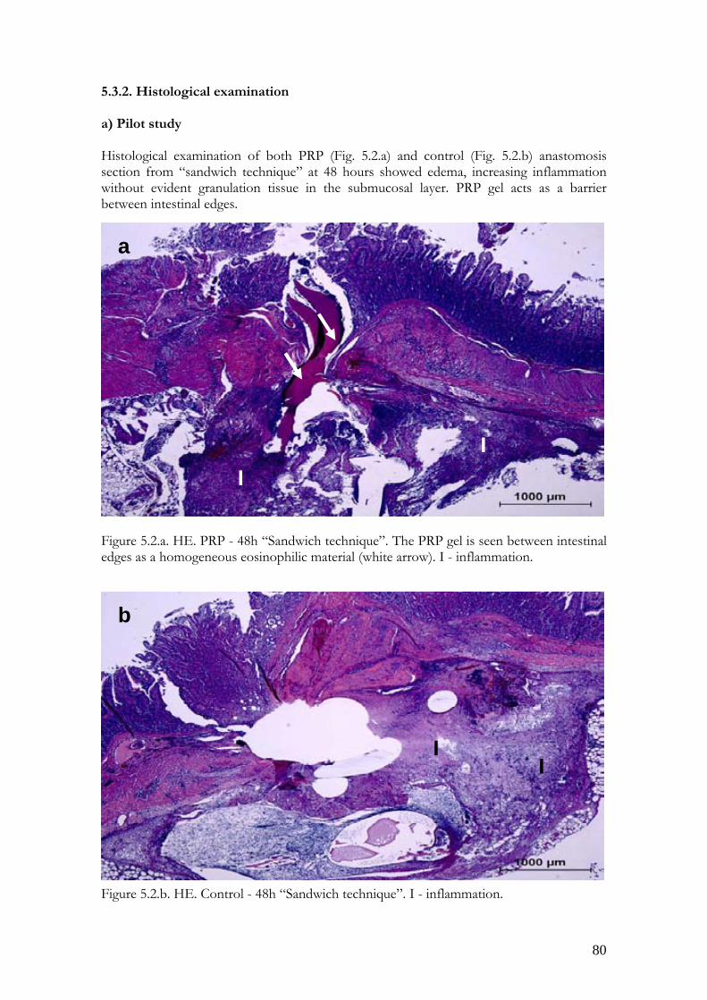
80
5.3.2. Histological examination a) Pilot study Histological examination of both PRP (Fig. 5.2.a) and control (Fig. 5.2.b) anastomosis section from “sandwich technique” at 48 hours showed edema, increasing inflammation without evident granulation tissue in the submucosal layer. PRP gel acts as a barrier between intestinal edges. Figure 5.2.a. HE. PRP - 48h “Sandwich technique”. The PRP gel is seen between intestinal edges as a homogeneous eosinophilic material (white arrow). I - inflammation. Figure 5.2.b. HE. Control - 48h “Sandwich technique”. I - inflammation.
a
II
b
I I
81
Since PRP gel application in a “sandwich technique” acts as a physical barrier, blocking the contact between both intestinal wound edges, and thus preventing a rapid healing, this technique was ruled as a method for PRP gel positioning. Results of the histological examination of the “immersion technique” PRP gel application in the pilot study at 24 and 48 hours are included below, in the definitive study. b) Definitive study Based on the results obtained with the sandwich technique, the “immersion technique” was used for definitive purposes. Microscopically, the lumen surface of the anastomotic line at 24 hours (Fig. 5.3) was covered by the epithelium. At 48 hours PRP (Fig. 5.4.a) and control (Fig. 5.4.b) intestinal anastomosis samples showed hemorrhage and inflammation, this was replaced by an immature granulation tissue at 72 hours. On PRP intestinal anastomosis samples at 72 hours high production of immature granulation tissue with neoangiogenesis, immature fibroblast and intense inflammation (Fig. 5.5.a and Fig. 5.6.a) were observed, whereas on control intestinal anastomosis samples discreet presence of granulation tissue and low inflammation (Fig. 5.5.b and Fig. 5.6.b) have been appreciated. With Masson`s stain for collagen fibers, at 72 hours on PRP (Fig. 5.7.a) and control (Fig. 5.7.b) intestinal anastomosis samples, more fibroblasts and less collagen fibres were observed, without evident differences between control and PRP anastomosis sample. Histological examination of PRP (Fig. 5.8.a) and control (Fig. 5.8.b) intestinal anastomosis samples at 96 hours also revealed an increased in immature granulation tissue, with less neovascularization and inflammation. At 7 days, PRP (Fig. 5.9.a) anastomosis samples showed a marked increase of thickness of intestinal wall wereas a control samples showed a low thickness of intestinal wall (Fig.5.9.b). Presence of mature granulation tissue with fibrosis and deposition of collagen was observed in both PRP (Fig. 5.10.a) and control (Fig. 5.10.b) intestinal anastomosis samples without evident differences. With Masson`s stain for collagen fibers, at day 7 showed a collagen deposition but no evident differences between PRP (Fig. 5.11.a) and control (Fig. 5.11.b) intestinal anastomosis samples were observed. In line with this is the observation that the amount of collagen increases rapidly between days 3 and 7.

82
On histological examination of PRP anastomosis samples from “immersion technique” at 24 hours (Fig. 5.3), the lumen surface of the anastomotic line was covered by the epithelium. No granulation tissue has appeared yet in the submucosal layer. Images from control samples are not shown because not evident differences were appreciated compared to the PRP samples. Figure 5.3. HE. PRP - 24 h.
a

83
On histological examination of both, PRP (Fig. 5.4.a) and control anastomosis (Fig. 5.4.b) samples at 48 hours, showed hemorrhage and inflammation whithout evident differences, no granulation tissue has appeared yet in the submucosal layer. Figure 5.4.a. HE. PRP - 48 h. (I- inflammation, H- hemorrhage) Figure 5.4.b. HE. Control - 48 h. (I- inflammation, H- hemorrhage).
I
HI
H
a
b
I
IH

84
Histological assessment of the PRP samples at 72 hours revealed a much more extended inflammatory activity at the anastomotic site (Fig. 5.5.a), with marked increase of thickness in intestinal wall, while a lower thickness of the healing reaction was observed in the control samples (Fig. 5.5.b) at the same time period. Figure 5.5.a. HE. PRP - 72 h. Thickness of the healing reaction is shown (dotted line). Figure 5.5.a. HE. Control - 72 h. Thickness of the healing reaction is shown (dotted line).
b
a

85
Higher magnification images of the granulation tissue in both, PRP and control samples at 72h are shown in figures 5.6.a and 5.6.b. Figure 5.6.a. HE. PRP - 72 h. Figure 5.6.b. HE. Control - 72 h.
a
b

86
On Masson’s trichrome stain sections at high magnification intestinal anastomosis showed evident neovascularisation and fibroblast proliferation and only few collagen fibers. Figure 5.7.a. Masson’s trichrome. PRP - 72 h. Figure 5.7.b. Masson’s trichrome. Control - 72 h.
a
b

87
At 96 hours, PRP anastomosis samples (Fig. 5.8.a.) showed high thickness of the healing reaction consisting on an immature granulation tissue with immature fibroblasts, less neoangiogenesis and inflammation. Control anastomosis samples (Fig. 5.8.b.) showed the lower thickness of the healing reaction is shown with the dotted line. Fig. 5.8.a.. HE. PRP - 96 h. Thickness of the healing reaction is shown (dotted line) Fig. 5.8.b. HE. Control - 96 h. Thickness of the healing reaction is shown (dotted line)
b
a

88
At 7 days, PRP anastomosis samples (Fig. 5.9.a.) showed more mature granulation tissue that control anastomosis samples (Fig. 5.9.b.) Fig. 5.9.a. HE. PRP - 7 days. Thickness of the healing reaction is shown (dotted line). Fig. 5.9.b. HE. Control - 7 days. Thickness of the healing reaction is shown (dotted line).
a
b
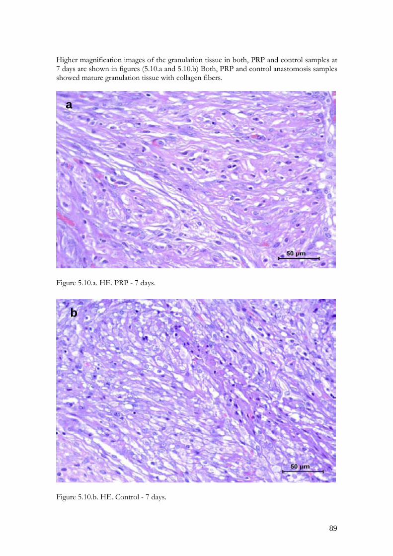
89
Higher magnification images of the granulation tissue in both, PRP and control samples at 7 days are shown in figures (5.10.a and 5.10.b) Both, PRP and control anastomosis samples showed mature granulation tissue with collagen fibers. Figure 5.10.a. HE. PRP - 7 days. Figure 5.10.b. HE. Control - 7 days.
a
b

90
On Masson’s trichrome stain sections at high magnification, at 7 days both, PRP (Fig. 5.11.a) and control (Fig. 5.11.b) samples showed a pattern of collagen deposition. Figure 5.11.a. Masson’s trichrome. PRP - 7 days. Figure 5.11.b. Masson’s trichrome. Control - 7 days.
a
b

91
5.3.3. Determination of the breaking strength Trials on breaking strength of unwounded intestine using the Instron Tensile Machine 1122 yielded satisfactory results (data not shown). Based on this, the dynamometer was employed for definitive purposes. The results of anastomotic breaking strength at 72 hours, 7 days and those of the unwounded intestine are represented on table 5.12. The lowest values were obtained in control samples, while the highest were observed in PRP samples. Differences in breaking strength between control and PRP samples at 72 hours and 7 days were not significant. Breaking strength values of control and PRP samples never reached those of the unwounded tissue with statistically significant differences (P<0.05).
Mean ± SD (kgf)
Range (kgf)
Control 72 hours 1,21± 0,27 0,64 - 1,65 PRP 72 hours 1,34± 0,22 1,02 - 1,83 Control 7 days 1,10± 0,27 0,56 - 1,56 PRP 7 days 1,08± 0,41 0,44 - 1,83 Unwounded sample 1,89±0,18 1,57 - 2,15
Table 5.12 - Mean, SD and range values of breaking strength of anastomotic wound on PRP and control samples at 72 hours, 7 days and unwounded tissue. Tearing of the tissue breaking site occurred within the anastomoses (Fig 5.12.a), except for 2/10 PRP samples at 72 hours, and 2/10 PRP samples and 1/10 control samples at 7 days, where tearing occurred outside the anastomotic site (Fig 5.12.b). Figure 5.12 - Tearing of the tissue breaking site at 72 hours and 7 days (black arrows show the anastomotic line).
a b
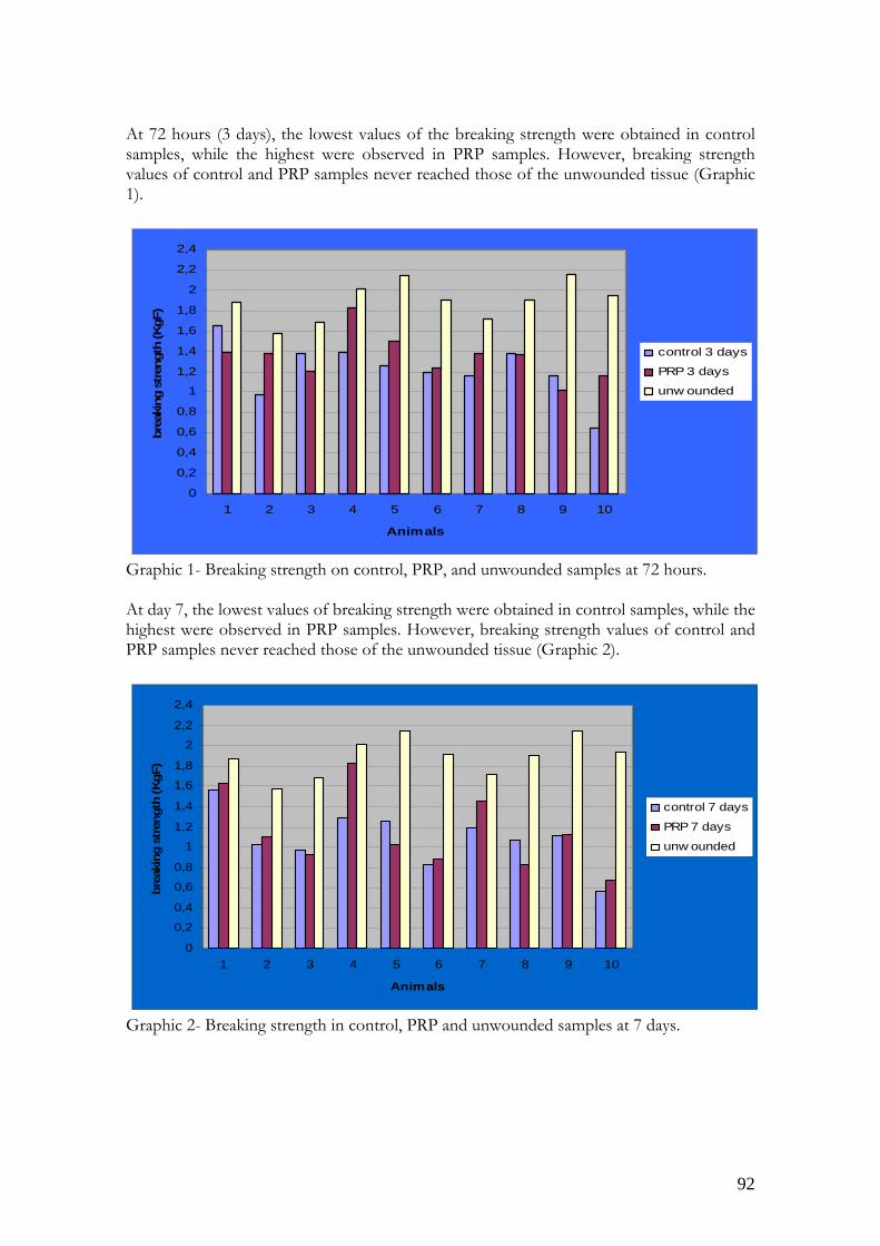
92
At 72 hours (3 days), the lowest values of the breaking strength were obtained in control samples, while the highest were observed in PRP samples. However, breaking strength values of control and PRP samples never reached those of the unwounded tissue (Graphic 1).
0
0,2
0,4
0,6
0,8
1
1,2
1,4
1,6
1,8
2
2,2
2,4
1 2 3 4 5 6 7 8 9 10
Animals
brea
king
str
engt
h (K
gF)
control 3 days
PRP 3 days
unw ounded
Graphic 1- Breaking strength on control, PRP, and unwounded samples at 72 hours. At day 7, the lowest values of breaking strength were obtained in control samples, while the highest were observed in PRP samples. However, breaking strength values of control and PRP samples never reached those of the unwounded tissue (Graphic 2).
0
0,2
0,4
0,6
0,8
1
1,2
1,4
1,6
1,8
2
2,2
2,4
1 2 3 4 5 6 7 8 9 10
Animals
brea
king
str
engt
h (K
gF)
control 7 days
PRP 7 days
unw ounded
Graphic 2- Breaking strength in control, PRP and unwounded samples at 7 days.
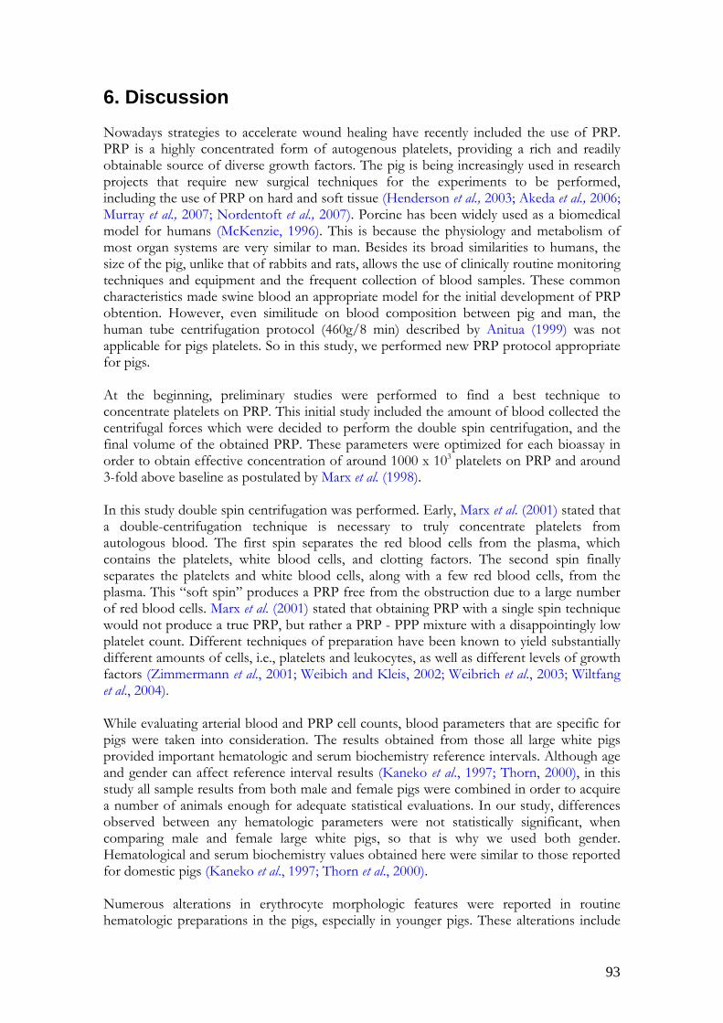
93
6. Discussion Nowadays strategies to accelerate wound healing have recently included the use of PRP. PRP is a highly concentrated form of autogenous platelets, providing a rich and readily obtainable source of diverse growth factors. The pig is being increasingly used in research projects that require new surgical techniques for the experiments to be performed, including the use of PRP on hard and soft tissue (Henderson et al., 2003; Akeda et al., 2006; Murray et al., 2007; Nordentoft et al., 2007). Porcine has been widely used as a biomedical model for humans (McKenzie, 1996). This is because the physiology and metabolism of most organ systems are very similar to man. Besides its broad similarities to humans, the size of the pig, unlike that of rabbits and rats, allows the use of clinically routine monitoring techniques and equipment and the frequent collection of blood samples. These common characteristics made swine blood an appropriate model for the initial development of PRP obtention. However, even similitude on blood composition between pig and man, the human tube centrifugation protocol (460g/8 min) described by Anitua (1999) was not applicable for pigs platelets. So in this study, we performed new PRP protocol appropriate for pigs. At the beginning, preliminary studies were performed to find a best technique to concentrate platelets on PRP. This initial study included the amount of blood collected the centrifugal forces which were decided to perform the double spin centrifugation, and the final volume of the obtained PRP. These parameters were optimized for each bioassay in order to obtain effective concentration of around 1000 x 103 platelets on PRP and around 3-fold above baseline as postulated by Marx et al. (1998). In this study double spin centrifugation was performed. Early, Marx et al. (2001) stated that a double-centrifugation technique is necessary to truly concentrate platelets from autologous blood. The first spin separates the red blood cells from the plasma, which contains the platelets, white blood cells, and clotting factors. The second spin finally separates the platelets and white blood cells, along with a few red blood cells, from the plasma. This “soft spin” produces a PRP free from the obstruction due to a large number of red blood cells. Marx et al. (2001) stated that obtaining PRP with a single spin technique would not produce a true PRP, but rather a PRP - PPP mixture with a disappointingly low platelet count. Different techniques of preparation have been known to yield substantially different amounts of cells, i.e., platelets and leukocytes, as well as different levels of growth factors (Zimmermann et al., 2001; Weibich and Kleis, 2002; Weibrich et al., 2003; Wiltfang et al., 2004). While evaluating arterial blood and PRP cell counts, blood parameters that are specific for pigs were taken into consideration. The results obtained from those all large white pigs provided important hematologic and serum biochemistry reference intervals. Although age and gender can affect reference interval results (Kaneko et al., 1997; Thorn, 2000), in this study all sample results from both male and female pigs were combined in order to acquire a number of animals enough for adequate statistical evaluations. In our study, differences observed between any hematologic parameters were not statistically significant, when comparing male and female large white pigs, so that is why we used both gender. Hematological and serum biochemistry values obtained here were similar to those reported for domestic pigs (Kaneko et al., 1997; Thorn et al., 2000). Numerous alterations in erythrocyte morphologic features were reported in routine hematologic preparations in the pigs, especially in younger pigs. These alterations include

94
Howell-Jolly bodies, polychromasia, poikilocytosis, crenation, and rouleaux formation. Although anisocytosis is also seen in adults, is more prominent in young pigs (Thorn, 2000). In our study, crenation was evident on whole blood smear. In the present experiment pigs showed a hematocrit of 30 ± 2.8% (mean±SD) and an initial platelet count of 420 ± 105 x 103 cells/μL (range: 292-814 x 103cells/µL), which is slightly higher than that among humans. The amount of citrated whole blood was about 60 mL and yielded a harvest of 6 mL of PRP; this volume is 10% of the whole blood volume drawn from each pig, as proposed by Marx et al. (2000). In the PRP samples, the platelet concentration was significantly higher than that of native blood (p<0.05), being the mean platelet count in PRP of 1620,6 x 103cells/µL. This concentration is comparable to the effective concentration of 1000 x 103 reported in the literature (Weibrich et al., 2004). With PRP tube method used in this study, concentration of 383% (3,8-fold) mean and range of 250 to 550% (2,5 to 5,5-fold) were obtained. These results are compared with the pig values reported for Curasan system 410% (4,1-fold) and buffy coat system 390% (3,9-fold) (Schlegel et al., 2004; Wiltfang et al., 2004; Thorwarth et al., 2005; Akeda et al., 2006). The average increase in platelet increase above baseline was 280% (2,8-fold) mean and range of 150 to 450% (1,5 to 4,5-fold). The number of white blood cells on whole blood was 15,33 ± 3,45 x 10³ cells/µL (range: 9,02 to 23,63 x10³ cells/µL). In the PRP samples, the number of white blood cells was significantly lower than that of citrated whole blood (p<0.05), the mean count of white blood cells was 1,83 cells x103/µL (range: 0,37 to 4,29 cells x 103 cells/µL). This values are lower than those reported by other authors (Li et al., 2004; Thorwarth et al., 2005) on pig model using Symphony II (29,5 x10³ cells/µL); PCCS (14,7±68 x 103 cells/µL), and Curasan (24,8±8,9 x 10³ cells/µL). As occurs with Anitua’s tube technique (Weibrich et al., 2005), in the technique described here a very low quantity of leukocytes are concentrated in comparison with the buffy coat and tube methods (pig and human). The real role of the leukocytes present in human and pig PRP prepared for wound healing has never been defined very well, and their true effect is completely unknown because there is lack of reliable research data (Weibrich et al., 2005). To evaluate the double spin centrifugation method to prepare PRP we also calculated an enrichment percentage which was 38,4 ±6,1% (range: 25,4 to 54,8%). This values are within the range reported in human PRP concentrate systems 19,0±16,6% (laboratory system), 41,9±9,7% (Curasan system) and 49,6±21,0% (PCCS) (Appel et al., 2002), and 30-39% using the manual systems for platelet concentrate (Marx et al., 2001). This parameter confirms the ability of the method to collect platelets within a certain volume of plasma. Generally, PRP clot are formed by mixing autologous PRP and bovine thrombin dissolved in calcium chloride solution. Those substances are added to PRP to initiate fibrin polymerization and to release platelet growth factors and cytokines, depending on the surgical requirements (Landsberg et al., 2000). But for some purposes only calcium chloride is used (Anitua, 1999; Dugrillon et al., 2002). In this study, to produce a PRP clot sterile 10% calcium chloride (CaCl2) solution was added. For this purpose Anitua’s (1999) procedure was used (50 µL per 1mL of PRP, and the time of forming PRP clot was approximately 4 minutes and ranged between 3-5 minutes).
95
The PRP obtention tecnhique used in this study was analyzed not only for cellular content but also for platelet activation during PRP procedure, and content of the growth factors, especially TGF-β1. In our study, the viability and structural changes of the pig platelets were investigated by assessing the platelet parameter supplied by a flow cytometry hematology system, such as MPC. It is known that this parameter, are correlated with platelet activation in human beings (Macey et al., 1999) dogs (Stanworth et al., 1999) and cats (Zelmanovic and Hetherington, 1998). We monitored MPC values on citrated whole blood and on PRP (a decrease in the MPC value occurs when platelets are activated and degranulated in vitro). The MPC values were not statistically significantly different (p<0.05) on citrated whole blood 21,3±1,58 g/dL (range:19,2-24,8 g/dL) when compared to PRP 20,3±1,39 g/dL (range:18,1-24,2 g/dL) values. In our opinion, this finding might be the result of the non activation of platelets during PRP preparation process. TGF-β1 is produced by a variety of cells including platelets that are normally recruited to an injury site. TGF-β1 regulates cellular differentiation, proliferation, chemotaxis, and synthesis of many ECM components (Roberts et al., 1988; Kane et al., 1991). In vivo studies have confirmed that exogenous TGF-β1 increases granulation tissue, collagen formation, and wound tensile strength when applied locally in animal models (Ashcroft et al., 1999). Because it was found to be pivotal on intestinal wound healing (Ciacci et al., 1993; Dignass et al., 1993; Beck et al., 2003), we analyzed the amount of TGF-β1 by enzyme-linked immunosorbent sandwich assay (ELISA) on PRP (obtained after double spin centrifugation), PPP (supernatant after second centrifugation) and PRP gel (obtained by mixing fresh PRP and calcium chloride). In our study, concentrations of TGF-β1 on PRP fractions were different. The concentration of TGF-β1 on PRP (24312 pg/mL) was significantly increased compared to PPP (3715 pg/mL). The concentration of TGF-β1 was 6,5-fold increased on PRP compared to PPP. This concentration is higher than that obtained by Akeda et al. (2006) on mini pigs using the Symphony II Platelet Concentration System (DePuy Spine, Raynham, MA) in which they obtained TGF-β1 concentrations two-fold higher than PPP values. It is important to point out that if platelets become activated by the preparation before a final centrifugation step, growth factors may be lost with a discarded supernatant finding high concentrations of growth factors in the PPP while low concentrations in the activated PRP. In our study, the concentration of TGF-β1 on supernatant (PPP) was low, and after a freeze/thawing activation, concentration of TGF-β1 were high. This supposes that the centrifugation method used in this study does not activate the platelets to release their growth factors which are also in accordance with the results obtained by the MPC values. As freezing and thawing activates platelets (Sekido et al., 1987; Weibrich et al., 2003; Zimmerman et al., 2003), the TGF-β1 levels in calcium chloride activated PRP (gel) are expected to be the same or higher than those in frozen and thawed PRP. However, on this study, the concentration of TGF-β1 was significantly increased on frozen and thawed PRP (24312 pg/mL) than in calcium activated PRP (15504 pg/mL). Two possible hypotheses could explain the results obtained. On one hand, during platelet activation by calcium chloride, a clot or gel formation occurs and released growth factors are recruited on this gel, resulting in a few growth factors left into the surrounding liquid which will be analysed (Tsay et al., 2005). On the other hand, because growth factors are not stable in plasma,

96
growth factors released by calcium chloride activation and then frozen and thawed may be degraded by this overall process and thus not detected. Nevertheless, we obtained higher TGF-β1 levels on PRP (24312±21755 pg/mL) and in PRP gel (15504±11279 pg/mL). In vitro concentrations of 5000 pg/mL TGF-β1 are known to induce chondrocyte proliferation and an increase of cartilage ECM synthesis (Fortier et al., 1997; Carmona et al., 2007). Perhaps supraphysiologic concentrations of TGF-β1 can be found in these PRP, and could control catabolic and inflammatory processes occurring during intestinal wound healing. TGF-β1 has been found, in vitro, to exert potent effects on the intestinal epithelium, which appear to stimulate production of extracellular matrix components. Furthermore, TGF-β1 has potent effects on many other cell populations present in the intestinal mucosa that could dominate the overall outcome of mucosal injury including lymphocytes, macrophages, fibroblasts, neutrophils. TGF-β1 has also been proposed to play a key role in regulating intestinal epithelial cell migration and wound repair (Ciacci et al., 1993; Dignass et al., 1993; Beck et al., 2003). Despite ongoing improvements in surgical technique, anastomotic leakage remains a relatively frequent complication of gastrointestinal surgery (Pickleman et al., 1999). This complication is much feared, because it has a profound negative impact on clinical outcome as shown by the significant increase in morbidity and in mortality. Experimental studies have shown that in the intestine the immediate postoperative anastomotic strength is low and decreases even further during the early postoperative period (Hogstrom and Haglund, 1985; Hendriks and Mastboom, 1990; Siemonsma et al., 2003). After the third day, when the lowest levels are encountered, wound strength increases rapidly. This postoperative pattern occurs even when the anastomosis is constructed under ideal circumstances and has been attributed to physiologic changes in matrix metabolism during this period. In the immediate postoperative period, wound strength is mainly determined by the suture holding capacity of the submucosa, in which collagen is the major structural protein. After a few days, newly formed matrix deposited in the anastomotic area will restore tissue strength. The capacity to synthesize collagen is increased in anastomotic tissue soon after operation (Martens and Hendriks, 1991). However, matrix degradation, e.g., to remove damaged tissue and to permit vessel formation, also is an intrinsic feature of wound repair. In the intestine, localized and limited degradation of the existing submucosal matrix may lead to transient loss of anastomotic strength during the first postoperative days. Therefore, it seems reasonable to promote intestinal healing to reduce the dehiscence-risk in this period as much as possible. Nowadays basic clinical research on gastro-intestinal anastomosis impaired wound healing has focused on the application of growth factors. Platelet concentrates such as platelet-rich plasma (PRP) have been used in a variety of clinical applications based on the premise that higher growth factor content should promote better healing (Fréchette et al., 2005). Autologous platelets as a source of healing factors have been shown to enhance bone regeneration and soft tissue maturation in the fields of orthopedics, periodontal and maxillofacial surgery, urology and plastic surgery (Marx et al., 1998; Anitua, 1999; Anitua et al., 2005; Fréchette et al., 2005; Murray et al., 2007).
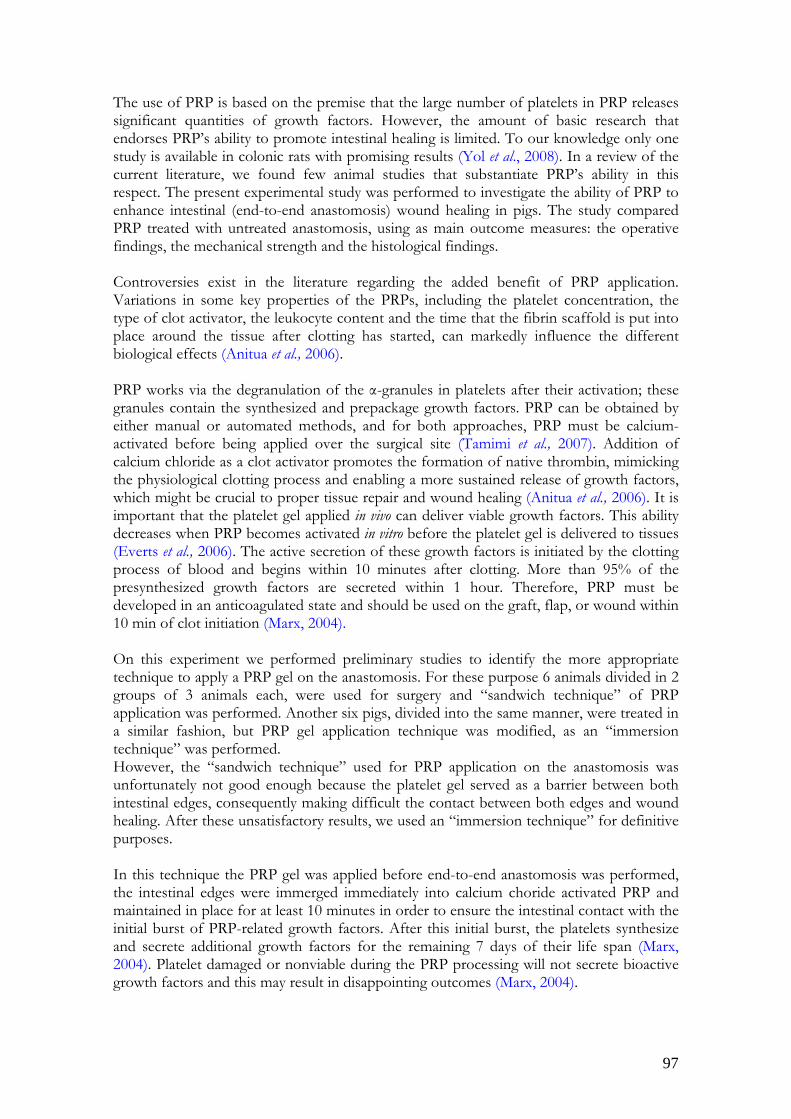
97
The use of PRP is based on the premise that the large number of platelets in PRP releases significant quantities of growth factors. However, the amount of basic research that endorses PRP’s ability to promote intestinal healing is limited. To our knowledge only one study is available in colonic rats with promising results (Yol et al., 2008). In a review of the current literature, we found few animal studies that substantiate PRP’s ability in this respect. The present experimental study was performed to investigate the ability of PRP to enhance intestinal (end-to-end anastomosis) wound healing in pigs. The study compared PRP treated with untreated anastomosis, using as main outcome measures: the operative findings, the mechanical strength and the histological findings. Controversies exist in the literature regarding the added benefit of PRP application. Variations in some key properties of the PRPs, including the platelet concentration, the type of clot activator, the leukocyte content and the time that the fibrin scaffold is put into place around the tissue after clotting has started, can markedly influence the different biological effects (Anitua et al., 2006). PRP works via the degranulation of the α-granules in platelets after their activation; these granules contain the synthesized and prepackage growth factors. PRP can be obtained by either manual or automated methods, and for both approaches, PRP must be calcium-activated before being applied over the surgical site (Tamimi et al., 2007). Addition of calcium chloride as a clot activator promotes the formation of native thrombin, mimicking the physiological clotting process and enabling a more sustained release of growth factors, which might be crucial to proper tissue repair and wound healing (Anitua et al., 2006). It is important that the platelet gel applied in vivo can deliver viable growth factors. This ability decreases when PRP becomes activated in vitro before the platelet gel is delivered to tissues (Everts et al., 2006). The active secretion of these growth factors is initiated by the clotting process of blood and begins within 10 minutes after clotting. More than 95% of the presynthesized growth factors are secreted within 1 hour. Therefore, PRP must be developed in an anticoagulated state and should be used on the graft, flap, or wound within 10 min of clot initiation (Marx, 2004). On this experiment we performed preliminary studies to identify the more appropriate technique to apply a PRP gel on the anastomosis. For these purpose 6 animals divided in 2 groups of 3 animals each, were used for surgery and “sandwich technique” of PRP application was performed. Another six pigs, divided into the same manner, were treated in a similar fashion, but PRP gel application technique was modified, as an “immersion technique” was performed. However, the “sandwich technique” used for PRP application on the anastomosis was unfortunately not good enough because the platelet gel served as a barrier between both intestinal edges, consequently making difficult the contact between both edges and wound healing. After these unsatisfactory results, we used an “immersion technique” for definitive purposes. In this technique the PRP gel was applied before end-to-end anastomosis was performed, the intestinal edges were immerged immediately into calcium choride activated PRP and maintained in place for at least 10 minutes in order to ensure the intestinal contact with the initial burst of PRP-related growth factors. After this initial burst, the platelets synthesize and secrete additional growth factors for the remaining 7 days of their life span (Marx, 2004). Platelet damaged or nonviable during the PRP processing will not secrete bioactive growth factors and this may result in disappointing outcomes (Marx, 2004).
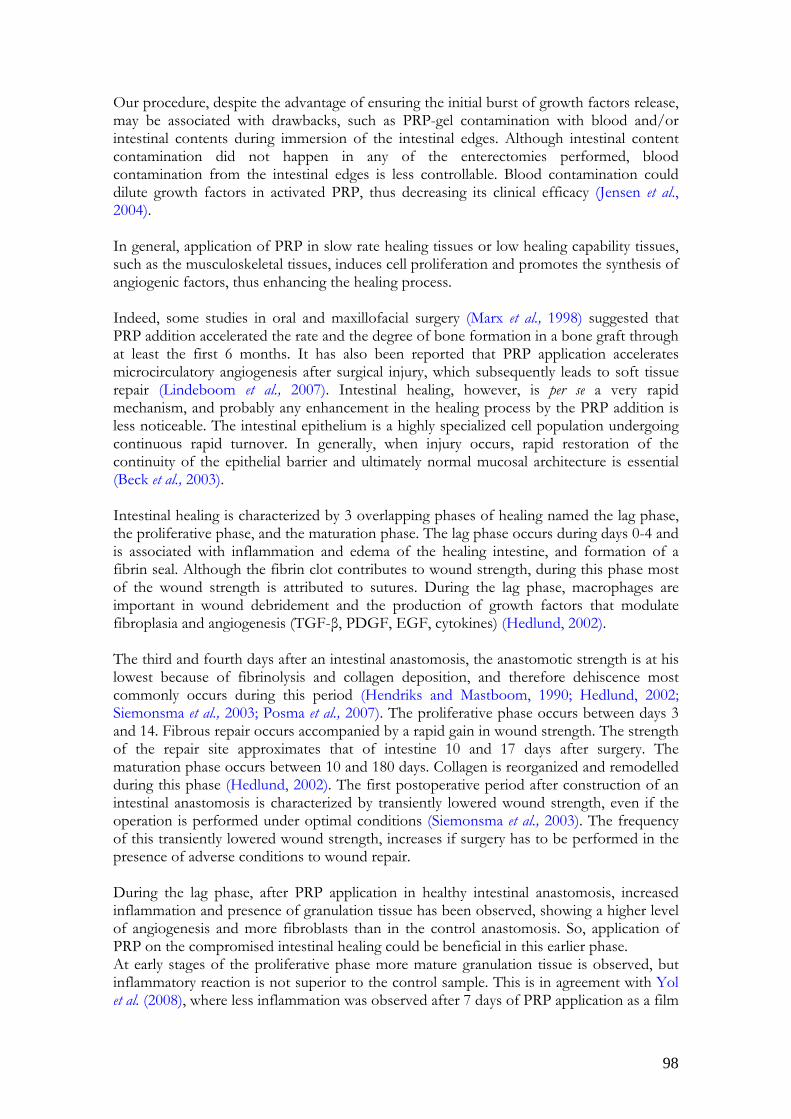
98
Our procedure, despite the advantage of ensuring the initial burst of growth factors release, may be associated with drawbacks, such as PRP-gel contamination with blood and/or intestinal contents during immersion of the intestinal edges. Although intestinal content contamination did not happen in any of the enterectomies performed, blood contamination from the intestinal edges is less controllable. Blood contamination could dilute growth factors in activated PRP, thus decreasing its clinical efficacy (Jensen et al., 2004). In general, application of PRP in slow rate healing tissues or low healing capability tissues, such as the musculoskeletal tissues, induces cell proliferation and promotes the synthesis of angiogenic factors, thus enhancing the healing process. Indeed, some studies in oral and maxillofacial surgery (Marx et al., 1998) suggested that PRP addition accelerated the rate and the degree of bone formation in a bone graft through at least the first 6 months. It has also been reported that PRP application accelerates microcirculatory angiogenesis after surgical injury, which subsequently leads to soft tissue repair (Lindeboom et al., 2007). Intestinal healing, however, is per se a very rapid mechanism, and probably any enhancement in the healing process by the PRP addition is less noticeable. The intestinal epithelium is a highly specialized cell population undergoing continuous rapid turnover. In generally, when injury occurs, rapid restoration of the continuity of the epithelial barrier and ultimately normal mucosal architecture is essential (Beck et al., 2003). Intestinal healing is characterized by 3 overlapping phases of healing named the lag phase, the proliferative phase, and the maturation phase. The lag phase occurs during days 0-4 and is associated with inflammation and edema of the healing intestine, and formation of a fibrin seal. Although the fibrin clot contributes to wound strength, during this phase most of the wound strength is attributed to sutures. During the lag phase, macrophages are important in wound debridement and the production of growth factors that modulate fibroplasia and angiogenesis (TGF-β, PDGF, EGF, cytokines) (Hedlund, 2002). The third and fourth days after an intestinal anastomosis, the anastomotic strength is at his lowest because of fibrinolysis and collagen deposition, and therefore dehiscence most commonly occurs during this period (Hendriks and Mastboom, 1990; Hedlund, 2002; Siemonsma et al., 2003; Posma et al., 2007). The proliferative phase occurs between days 3 and 14. Fibrous repair occurs accompanied by a rapid gain in wound strength. The strength of the repair site approximates that of intestine 10 and 17 days after surgery. The maturation phase occurs between 10 and 180 days. Collagen is reorganized and remodelled during this phase (Hedlund, 2002). The first postoperative period after construction of an intestinal anastomosis is characterized by transiently lowered wound strength, even if the operation is performed under optimal conditions (Siemonsma et al., 2003). The frequency of this transiently lowered wound strength, increases if surgery has to be performed in the presence of adverse conditions to wound repair. During the lag phase, after PRP application in healthy intestinal anastomosis, increased inflammation and presence of granulation tissue has been observed, showing a higher level of angiogenesis and more fibroblasts than in the control anastomosis. So, application of PRP on the compromised intestinal healing could be beneficial in this earlier phase. At early stages of the proliferative phase more mature granulation tissue is observed, but inflammatory reaction is not superior to the control sample. This is in agreement with Yol et al. (2008), where less inflammation was observed after 7 days of PRP application as a film

99
layer to the anastomotic line. However, in the latter study, the histopathological examination and bursting pressure was only evaluated on postoperative day 7, and effects of PRP were not determined during the lag phase. The increased granulation tissue present in PRP samples at 72 hours may lead to a higher fibrous tissue formation and thus to a major gain in wound strength. These facts may reflect a higher anastomotic resistance in PRP samples compared to control samples during this phase. Indeed, these results were in accordance with the results in breaking strength. Although differences were not significant and dehiscence most commonly occurs 3-4 days after intestinal surgery, breaking strength have been found to be highest at 72 hours after PRP application. At 7 days breaking strength measurements demonstrated that, rupture occurred primarily within anastomosis line in the control groups whereas on 2/10 tested samples occurred outside the wound area in PRP treated animals which indicates that at this point, the anastomosis had grown stronger than the adjacent bowel wall. In this case the highest values were observed on treated samples but statistically the differences were not significant. Thus, although differences were not significant, PRP application on intestinal anastomosis seemed to increase the wound strength. This is in accordance to the study reported by Yol et al. (2008) in rats, where topical PRP gel applied to the anastomotic line as a film layer obtained a better anastomotic strength than the group subjected to colonic anastomosis only. However, in contrast to the latter study, the higher anastomotic strength on PRP samples observed in the present study is speculated to be caused by the increased granulation tissue and fibrosis caused by the PRP application.
The findings of the present study are based on macroscopic and microscopic analysis of tissue sections. This approach enables a more accurate study of the various cell types and extracellular matrix components involved in the repair process as it has previously proposed by Verhofstand et al. (2001). We used the classic haematoxylin-eosin stain to evaluate the overall architectural completion of the anastomosis (healing of intestinal layers and thickness of granulation tissue formed), and Masson’s trichrome stain to visualize collagen fiber organization in all tissue sections at 3 and 7 days.
Despite we did not evaluate growth factor content neither metalloprotease matrix activity, an increase on thickness of the intestinal wall with high presence of granulation tissue were observed on PRP treated animals on 72 hours, 96 hours and 7 days. This result is in agreement with Yol et al. (2008), who considered that growth factors in PRP are responsible for better anastomotic strength and wound healing. However, they did not evaluate the inflammatory reaction and granulation tissue during the first days of intestinal healing (lag phase), so we do not compare the initial inflammatory reaction caused by PRP application observed in our study with Yol et al. (2008)’s investigation. An anastomosis should be able to withstand both intraluminal and longitudinal forces caused by movements of the intestine (Posma et al., 2007). Anastomotic bursting pressure and breaking strength are valid criteria for assessing anastomotic strength and integrity during its healing (Rabau et al., 1998). Breaking strength measures the resistance of the intestinal wall to longitudinal forces and evaluates the biological aspect of the healing process; while bursting pressure determines the resistance of the intestinal wall to intraluminal pressure and can evaluate the overall anastomotic integrity (Hendriks and Mastboom, 1990; Rabau et al., 1998; Ikeuchi et al., 1999). However, Ikeuchi et al. (1999) reported that the MITS (minimal tensile strength) appears to be a
100
better standard to evaluate the biological aspect of anastomotic healing including suture-holding capacity. In the present study, measurements performed by the Instron machine approached the minimal tensile strength. Several studies have been performed on improvement of intestinal healing based on the factors suspected of influencing anastomotic healing (Yagci et al., 2006). Although the effects of growth hormone and growth factors applied systemically and topically on healing of bowel anastomosis are currently being evaluated (Christensen et al., 1991; Christensen et al., 1993; Christensen et al., 1995; Gomez de-Segura et al., 1995; Egger et al., 1998; Dinc et al., 2002), the effects of PRP application have been poorly documented (Yol et al., 2008). The present investigation evaluates the effects of PRP on intestinal healing during the lag phase. However, it is a short-term study that does not address long-term effects or complications. Additionally, further investigations on different PRP application techniques on intestinal anastomosis (rather than the immersion technique) as well as evaluations at longer time intervals are required. Although differences were subtle, PRP application on intestinal anastomosis seemed to slightly improve the anastomotic resistance due to an increase in inflammatory infiltrates and fibrosis. PRP use on intestine does not appear to have obvious disadvantages. Whether PRP application in intestinal anastomosis can reduce the frequency of anastomotic leakage needs to be evaluated in future studies.

101
7. Conclusions From the results obtained in this experimental study we may conclude: 1. The protocol for PRP obtention using a double spin centrifugation procedure employed in this study results in a high platelet concentrate and maintains a high quality biological product as measured by MPC values and TGF-β1 levels. 2. The anti-human TGF-β1 antibodies for TGF-β1 detection by enzyme linked immunosorbent assay (ELISA) have been proved to be suitable for the detection of porcine TGF-β1. 3. The immersion technique for the application of PRP in the intestine seems to be easy to perform and may guarantee the initial burst of growth factors. 4. The dynamometer Instron 1122 seems useful as a method for breaking strength evaluation during the lag phase as it better measures the suture-holding capacity. 5. Microscopic study revealed more inflammatory infiltrate and tissue granulation formation in PRP samples. 6. Although differences were not significant, PRP samples showed a higher breaking strength values in agreement to microscopic findings, especially in the lag phase. 7. At 7 days the breaking strength values, neither of PRP or control samples, never reached those of the unwounded intestine. 8. The slightly improvement of intestinal resistance by PRP gel application on healthy intestinal anastomoses does not seem to compensate for the technical and surgical effort required for PRP obtention and application. However, future studies in compromised intestine healing will be required.

102

103
8. References
1. Adams W, Ctercteko G, Bilous M. Effect of an omental wrap on the healing and vascularity of compromised intestinal anastomoses. Dis Colon Rectum 1992; 35:731-738.
2. Aghaloo TL, Moy PK, Freymiller EG. Investigation of platelet-rich plasma in rabbit cranial defects: a pilot study. J Oral Maxillofac Surg 2002; 60: 1176-1181.
3. Aghaloo TL, Moy PK, Freymiller EG. Evaluation of platelet-rich plasma in combination with an organic bovine bone in the rabbit cranium: a pilot study. Int J Oral Maxillofac Implants 2004; 19: 59-65.
4. Ågren MS, Werthén M. The Extracellular Matrix in Wound Healing: A Closer Look at Therapeutics for Chronic Wounds Lower Extremity Wounds 2007; 6: 82-97.
5. Ahnadi CE, Boughrassa FF, Chapman-Montgomery ES, Poisson V, Gervais A, Okrongly D, Grant AM. Comparison of two methods to assess variability of platelet response to anti-platelet therapies in patients with acute coronary syndrome undergoing angioplasty. Thromb Haemost 2004; 92:1207-1213.
6. Akeda K. An HS, Pichika R, Attavia M, Thonar EJ, Lenz ME, Uchida A. Platelet-rich plasma (PRP) stimulates the extracellular matrix metabolism of porcine nucleus pulposus and annulus fibrosus cells cultured in alignate beads. Spine 2006; 31:959-966.
7. a.Alio JL, Abad M, Artola A, Rodriguez-Prats JL, Pastor S, Ruiz- Colecha J. Use of autologous platelet-rich plasma in the treatment of dormant corneal ulcers. Ophthalmology 2007; 114:1286–1293.
8. b.Alio JL, Colecha JR, Pastor S, Rodriguez A, Artola A. Symptomatic dry eye treatment with autologous platelet-rich plasma. Ophthalmic Res 2007; 19:124-129.
9. Andonegui G, Kerfoot SM, McNagny K, Ebbert KJ, Patel KD, Kubes P. Platelets express functional Toll-like receptor-4. Blood 2005; 106, 2417–2423.
10. Anila S and Nandakumar K. Applications of Platelet Rich Plasma for Regenerative Therapy in Periodontics. Trends Biomater. Artif. Organs 2006; 20: 78-83.
11. Anitua E. Plasma rich in growth factors: preliminary results of use in the preparation of sites for implants. Int. J. Oral Maxillofac. Implants 1999; 14: 529-535.
12. Anitua E, Andia I, Sanchez M; Azofra J, del Mar Zalduendo M, de la Fuente M, Nurden P, Nurden AT. Autologous preparations rich in growth factors promote proliferation and induce VEGF and HGF production by human tendon cells in culture. J Orthop Res 2004; 23:281-286.
13. Anitua E, Sanchez M, Nurden AT, Nurden P, Orive G, Andia I. New insights into and novel applications for platelet-rich fibrin therapies. Trends Biotechnol 2006; 24:227-234.
14. Anitua E, Sanchez M, Orivean G, Andıa I. The potential impact of the preparation rich in growth factors (PRGF) in different medical fields. Biomaterials 2007; 28: 4551-4560.
15. Appel TR, Potzsch B, Muller J, von Lindern JJ, Berge SJ, Reich RH. Comparison of three different preparations of platelet concentrates for growth factor enrichment. Clin Oral Implants Res 2002; 13:522-528.
16. Ashcroft GS, Yang X, Glick AB, Weinstein M, Letterio JL, Mizel DE, Anzano M, Greenwell-Wild T, Wahl SM, Deng C, Roberts AB. Mice lacking Smad3 show

104
accelerated wound healing and an impaired local inflammatory response. Nat Cell Biol 1999; 1:260 -266.
17. Aslam R, Aslam R, Speck ER, Kim M, Crow AR, Bang B, Nestel FP, Ni H, Lazarus AH, Freedman J, Semple JW. Platelet Toll-like receptor expression modulates lipopolysaccharide induced thrombocytopenia and tumor necrosis factor-a production in vivo. Blood 2006; 107:637-641.
18. Aspenberg P, Virchenko O. Platelet concentrate injection improves Achilles tendon repair in rats. Acta Orthop Scand 2004; 75:93-99.
19. Babyatsky MW, Rossiter G, Podolsky DK: Expression of transforming growth factors alpha and beta in colonic mucosa in inflammatory bowel disease. Gastroenterology 1996; 110:975-984.
20. Beck PL, Podolsky DK. Growth factors in inflammatory bowel disease. Inflamm Bowel Dis 1999; 5:44-60.
21. Beck PL, Rosenberg IM, Xavier RJ, Koh T, Wong JF, Podolsky DK. Transforming growth factor-β mediates intestinal healing and susceptibility to injury in vitro and in vivo through epithelial cells. American Journal of Pathology 2003; 162:597-608.
22. Benhaim P, Hunt TK. Natural resistance to infection: leukocyte functions. J Burn Care Rehabil 1992; 13: 287-292.
23. Bielecki TM, Gazdzik TS, Arendt J, Szczepanski T, Krol W, Wielkoszynski T. antibacterial effect of autologous platelet gel enriched with growth factors and other active substances. J Bone Joint Surg 2007; 89:417–420.
24. Bitar MS. Insulin like growth factor-I reverses diabetes- induced wound healing impairment in rats. Horm Metab res 1997; 29: 383-386.
25. Blotnick S, Peoples GE, Freeman MR, Eberlein TJ, Klagsbrun M. T lymphocytes synthesize and export heparin-binding epidermal growth factor-like growth factor and basic fibroblast growth factor, mitogens for vascular cells and fibroblasts: differential production and release by CD4+ and CD8+ T cells. Proc Natl Acad Sci USA 1994; 91:2890-2894.
26. Boldt J. Acute Platelet-Rich Plasmapheresis for Cardiac Surgery Journal of Card/othoractc and Vascular Anesthesia 1995; 9: 79-88.
27. Bonanomi G, Prince J, McSteen F, Schauer PR, Hamad GG. Sealing effect of fibrin glue on the healing of gastrointestinal anastomoses: implications for the endoscopic treatment of leaks. Surg Endosc 2004; 18:1620-1624.
28. Brady C, Vang S, Christensen K, Isler J, Vollstedt K, Holt D. Use of platelet gel in bariatric surgery. J Extracorp Technol 2006; 38: 161-164.
29. Brasken P. Healing of experimental colon anastomosis. Eur J Surg1991; 566 (Suppl):1-51.
30. Brauchle M, Madlener M, Wagner AD, Angermayer K, Lauer U, Hofschneider PH, Gregor M, Werner S. Keratinocyte growth factor is highly over expressed in inflammatory bowel disease Am J Pathol 1996; 149:521-529.
31. Browder W, Williams D, Lucore P, Pretus H, Jones E, McNamee R. Effects of enhanced function on early wound healing. Surgery 1988; 104:224.
32. Brundage SI, Schreiber MA, Holcomb JB. Amplification of the proinflammatory transcription factor cascade increases with severity of uncontrolled hemorrhage in swine. J. Surg. Res. 2003; 113:74.
33. Calabresi PA, Fields NS, Maloni HW, Hanham A, Carlino J, Moore J, Levin MC, Dhib-Jalbut S, Tranquill LR, Austin H, McFarland HF, Racke MK. Phase I trial of transforming growth factor beta 2 in chronic progressive MS. Neurology 1998; 51:289.
105
34. Carmona JU, Argüelles D, Climent F, Prades M. Autologous Platelet Concentrates as a Treatment of Horses with Osteoarthritis: A Preliminary Pilot Clinical Study. Journal of Equine Veterinary Science. 2007; 27:167-170.
35. Carter CA, Jolly DG, Worden CE, Hendren DG, Kane CJ. Platelet-rich plasma gel promotes di.erentiation and regeneration during equine wound healing. Experimental and Molecular Pathology 2003; 74: 244–255.
36. Chandramouli B, Srinivasan K, Jagdish S, Ananthakrishnan N. Morbidity and mortality of colostomy and its closure in children. J Pediatr Surg 2004; 39:596-599.
37. Choi BH, IM CJ, Huh JY, Suh JJ, Lee SH. Effect of platelet-rich plasma on bone regeneration in autogenous bone graft. International Journal of Oral and Maxillofacial Surgery 2004; 33:56-59.
38. Christensen H, Oxlund H, Laurberg S. Growth hormone increases the bursting strength of colonic anastomoses. Int J Colorect Dis 1990; 5:130-134.
39. Christensen H, Oxlund H, Laurberg S. Postoperative biosynthetic human growth hormone increased the strength and collagen deposition of experimental colonic anastomoses. Int J Colorect Dis 1991; 6:133-138.
40. Christensen H, Flyvbjerg A, Ørskov H, Laurberg S. Effect of growth hormone on the inflammatory activity of experimental colitis in rats. Stand J Gasteroenterol 1993; 28:503-511.
41. Christensen HN, Oxlund H. Growth hormone increases the collagen deposition rate and breaking strength of left colonic anastomosis in rats. Surgery 1994; 116: 550-556.
42. Christensen H, Chemnitz J, Christensen BC, Oxlund H. Collagen structural organization of healing anastomoses and the effect of growth hormone treatment. Dis Colon Rectum 1995; 38:1200-1205.
43. Ciacci C, Lind SE, Podolsky DK. Transforming growth factor regulation of migration in wounded rat intestinal epithelial monolayers. Gastroenterology 1993; 105:93-101.
44. Clark SC, Sudarshan, CD, Khanna R, Roughan JV, Flecknell PA, Dark JH. A New porcine model of reperfusion injury after lung transplantation. Lab. Anim 1999; 33: 135-142.
45. Colak T, Nayci A, Polat G, Polat A, Comelekoglu U, Kanik A, Turkmenoglu O, Aydin S. Effects of Trapidil on the healing of colonic anastomoses in an experimental rat model ANZ J. Surg 2003; 73: 916-921.
46. Coppinger JA, Cagney G, Toomey S, Kislinger T, Belton O, McRedmond JP, Cahill DJ, Emili A, Fitzgerald DJ, Maguire PB. Characterization of the proteins released from activated platelets leads to localization of novel platelet proteins in human atherosclerotic lesions. Blood 2004; 103:2096-2104.
47. Crovetti G, Martinelli G, Issi M, Barone M, Guizzardi M, Campanati B, Moroni M, Carabelli A. Platelet gel for healing cutaneous chronic wounds. Transf Apheresis Sci 2004; 30:145-151.
48. Crowther M, Ford I, Jeffrey RR, Urbaniak SJ, Greaves M. Quality of harvested autologous platelets compared with stored donor platelets for use after cardiopulmonary bypass procedures. Br J Haematol 2000; 111:175-181.
49. de la Torre J, Sholar A. Wound healing: Chronic wounds. 2006. Emedicine.com 50. Detweiler MB, Detweiler JG, Fenton J. Sutureless and reduced suture
anastomosis of hollow vessels with fibrin glue: a review. J Invest Surg 1999; 12: 245-262.
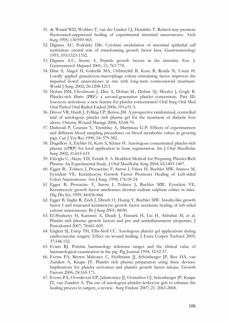
106
51. de Waard WD, Wobbes T, van der Linden CJ, Hendriks T. Retinol may promote fluorouracil-suppressed healing of experimental intestinal anastomoses. Arch Surg 1995; 130:959-965.
52. Dignass AU, Podolsky DK: Cytokine modulation of intestinal epithelial cell restitution: central role of transforming growth factor beta. Gastroenterology 1993, 105:1323-1332.
53. Dignass A.U, Sturm A. Peptide growth factors in the intestine. Eur. J. Gastroenterol. Hepatol 2001; 13, 763-770.
54. Dinc S, Alagol H, Gulcelik MA, Ozbirecikli B, Kuru B, Renda N, Ustun H. Locally applied granulocyte-macrophage colony-stimulating factor improves the impaired bowel anastomoses in rats with long-term corticosteroid treatment. World J Surg. 2002; 26:1208-1213.
55. Dohan DM, Choukroun J, Diss A, Dohan SL, Dohan AJ, Mouhyi J, Gogly B. Platelet-rich fibrin (PRF): a second-generation platelet concentrate. Part III: leucocyte activation: a new feature for platelet concentrates? Oral Surg Oral Med Oral Pathol Oral Radiol Endod 2006; 101:e51-5.
56. Driver VR, Hanft J, Fylling CP, Beriou JM. A prospective randomized, controlled trial of autologous platelet rich plasma gel for the treatment of diabetic foot ulcers. Ostomy Wound Manage 2006; 52:68-70.
57. Dubreuil P, Couture Y, Tremblay A, Martineau G-P. Effects of experimenters and different blood sampling procedures on blood metabolite values in growing pigs. Can J Vet Res 1990; 54: 379-382.
58. Dugrillon A, Eichler H, Kern S, Kluter H. Autologous concentrated platelet-rich plasma (cPRP) for local application in bone regeneration. Int J Oral Maxillofac Surg 2002; 31:615-619.
59. Efeoglu C, Akçay YD, Ertürk S. A Modified Method for Preparing Platelet-Rich Plasma: An Experimental Study. J Oral Maxillofac Surg 2004; 62:1403-1407.
60. Egger B, Tolmos J, Procaccino F, Sarosi I, Friess H, Buchler MW, Stamos M, Eysselein VE. Keratinocyte Growth Factor Promotes Healing of Left-sided Colon Anastomoses. Am J Surg. 1998; 176:18-24.
61. Egger B, Procacino F, Sarosi I, Tolmos J, Buchler MW, Eysselein VE. Keratinocyte growth factor ameliorates dextran sodium sulphate colites in mice. Dig Dis Sci; 1999; 44:836-844.
62. Egger B, Inglin R, Zeeh J, Dirsch O, Huang Y, Buchler MW. Insulin-like growth factor I and truncated keratinocyte growth factor accelerate healing of left-sided colonic anastomoses. Br J Surg 2001; 88:90.
63. El-Sharkawy H, Kantarci A, Deady J, Hasturk H, Liu H, Alshahat M, et al. Platelet rich plasma: growth factors and pro and antiinflammatory properties. J Periodontol 2007; 78:661-669.
64. Englert SJ, Estep TH, Ellis-Stoll CC. Autologous platelet gel applications during cardiovascular surgery: Effect on wound healing. J Extra Corpor Technol 2005; 37:148-152.
65. Evans RJ. Porcine haematology reference ranges and the clinical value of haematological examination in the pig. Pig Journal 1994; 32:52-57.
66. Everts PA, Brown Mahoney C, Hoffmann JJ, Schönberger JP, Box HA, van Zundert A, Knape JT. Platelet rich plasma preparation using three devices: Implications for platelet activation and platelet growth factor release. Growth Factors 2006; 24:165-171.
67. Everts PA, Overdevest EP, Jakimowicz JJ, Oosterbos CJ, Schonberger JP, Knape JT, van Zundert A. The use of autologous platelet–leukocyte gels to enhance the healing process in surgery, a review. Surg Endosc 2007; 21: 2063-2068.

107
68. Falanga V, Grinnell F, Gilchrest B, Maddox YT, Moshell A. Experimental approaches to chronic wounds. Wound Rep Reg 1995; 3:132-140.
69. Falanga V. Wound Healing. American Academy of Dermatology (AAD) 2005. 70. Finch PW, Pricolo V, Wu A, Finkelstein Sd. Increased expression of Keratinocyte
growth factor messenger RNA associated with inflammatory bowel disease. Gastroenterology 1996; 110:441-451.
71. Fleischmann E, Lenhardt R, Kurz A, Herbst F, Fülesdi B, Greif R, ISessler D. Nitrous oxide and risk of surgical wound infection: a randomised trial. Lancet 2005; 366: 1101-1107.
72. Floryan KM, Berghoff WJ. Intraoperative use of autologous platelet-rich and platelet-poor plasma for orthopedic surgery patients. Aorn Journal 2004; 80: 667-674.
73. Fortier LA, Nixon AJ, Mohammed HO, Lust G. Altered biological activity of equine chondrocytes cultured in a three-dimensional fibrin matrix and supplemented with transforming growth factor beta-1. Am J Vet Res 1997; 58:66-70.
74. Fréchette JP, Martineau I, Gagnon G. Platelet-rich plasma: growth factor content and roles in wound healing. J Dent Res 2005; 84:434-439.
75. Freymiller EG, Aghaloo TL. Platelet-rich plasma: ready or not? J Oral Maxillofac Surg 2004; 62:484-488.
76. Furst MB, Stromberg BV, Blatchford GJ, Christensen MA, Thorson AG. Colonic anastomoses: Bursting strength after corticosteroid treatment Dis Colon Rectum. 1994; 37: 12-15.
77. Galzie Z, Kinsella AR, Smith JA. Fibroblast growth factors and their receptors. Eiochem Cell Biol 1997; 75:669-685.
78. Gandhi A, Doumas C, O'Connor JR, Parsons JR, Lin SS. The effects of local platelet rich plasma delivery on diabetic fracture healing. Bone 2006; 38:540-546.
79. Geerling G, Daniels JT, Dart JK, Cree IA, Khaw PT. Toxicity of natural tear substitutes in a fully defined culture model of human corneal epithelial cells. Invest Ophthalmol Vis Sci 2001; 42:948-956.
80. Gehring S, Hoerauf H, Laqua H, Kirchner H, Klueter H. Preparation of autologous platelets for the ophthalmologic treatment of macular holes. Transfusion 1999; 39:144-148.
81. George JN. Platelets. Lancet 2000; 355:1531-1539. 82. Giannoble WV. Periodontal tissue engineering by growth factors. Bone 1996;
S19:S23-37. 83. Golub R, Golub RW, Cantu R Jr, Stein HD. A multivariate analysis of factors
contributing to leakage of intestinal anastomoses. J Am Coll Surg 1997; 184:364-372.
84. Gomez de-Segura IA, Aguilera MJ, Codesal J, De-Miguel E. Administration of growth hormone enhances the intestinal adaptive response after resection of small intestine in rats. Rev Esp Enferm Dig 1995; 87:288-293.
85. Goodlad RA, Raja KB, Peters TJ, Wright NA. Effects of urogas-trone-epidermal growth factor on intestinal brush border enzymes and mitotic activity. Gut 1991; 32:994-998.
86. Gordillo GM, Sen CK. Revisting the essential role of oxygen in wound healing. Am J Surg 2003; 186:259-263.
87. Greatorex G, Whitaker BL, Dixon RA. Anastomotic failure in relation to blood transfusion and blood loss. Proc R Soc Med 1970; 63 :751.
88. Greenhalgh DG. The role of apoptosis in wound healing. The International Journal of Biochemistry & Cell Biology 1998; 30:1019-1030.
108
89. Grim PS, Gottlieb LJ, Boddie A, Batson E. Hyperbaric oxygen therapy. JAMA. 1990; 263:2216-2220.
90. Guillén MI, Mirabet LV, Sopena JJ, Fernández FP, Segura GP, Corpa AJM. Rediferenciación de condrocitos en plasma rico en factores de crecimiento plaquetarios para condroplastia articular. Patología del Aparato Locomotor, 2005; 3:13-23.
91. Haglind E, Malmlof K, Fan J, Lang CH. Insulin-like growth factor-I and growth hormone administration in intestinal shock in the rat Shock 1998; 10:62-68.
92. Halstead WS. Circular suture of the intestine-an experimental study. Am J Med Sci 1887; 94:436-461.
93. Harrison P, Cramer EM. Platelet alpha-granules. Blood Reviews.1993; 7:52-62. 94. Harrison LE, Port JL, Hochwald S, Blumberg D. Perioperative growth hormone
improves wound healing and immunologic function in rats receiving adriamycin. J Surg Res 1995; 58:646-650.
95. Harrison P. Platelet function analysis. Blood Reviews 2005; 19:111-123. 96. Hartwig J, Italiano Jr. The birth of the platelet. J Thromb Haemost 2003; 1:1580-
1586. 97. Hartwig D, Harloff S, Liu L, Schlenke P, Wedel T, Geerling G. Epitheliotrophic
capacity of growth factor preparation produced from platelet concentrates on corneal epithelial cells: a potential agent for the treatment of ocular surface defects? Transfusion 2004; 44:1724-1731.
98. Hassanain HH, Irshaid F, Wisel S, Sheridan J, Michler RE, Goldschmidt-Clermont PJ. Smooth muscle cell expression of a constitutive active form of human Rac 1 accelerates cutaneous wound repair. Surgery 2005; 137:92-101.
99. Haukipuro KA, Hulkko OA, Alavaikko MJ, Laitinen ST. Sutureless colon anastomosis with FG in the rat. Dis. Colon Rectum 1988; 31:601-604.
100. Hawarth GS, Xian CJ, Read LC. Insulin like growth factor-I partially attenuates colonic damage in rats with experimental colitis induced by oral dextran Sulphate sodium. Scand J Gastroenterol 1998; 33:189-190.
101. Hedlund CS. Surgery of the small intestine. Surgery of the digestive system. In: Fossum TW, Hedlund CS, Hulse DA, Johnson AL, Seim HB, Willard MD, Carroll WL, editors. Small Animal Surgery. 2nd Ed. St Louis (Missouri): Mosby, Inc. 2002:369-398.
102. Henderson JL, Cupp CL, Ross EV, Shick PC, Keef MA, wester DC, Hannan T, McConnell. The effects of autologous platelet gel on wound healing. Ear Nose Throat J 2003; 82:598-602.
103. Hendriks T, Mastboom WJB. Healing of experimental intestinal anastomoses: parameters for repair. Dis Colon Rectum 1990; 33:891-901.
104. Hesp FL, Hendriks T, Lubbers EJ, de Boer HH. Wound healing in the intestinal wall. A comparison between experimental ileal and colonic anastomoses. Dis Colon Rectum 1984; 27:99-104.
105. Hillan K, Nordlinger B, Ballet F, Puts JP, Infante R The healing of colonic anastomoses after early intraperitoneal chemotherapy: an experimental study in rats. J Surg Res 1988; 44:166-171.
106. Hiramatsu T, Okamura T, Imai Y, Kurosawa H, Aoki M, Shinoka T. Effects of autologous platelet concentrate reinfusion after open heart surgery in patients with congenital heart disease. Ann Thor Surg 2002; 73:1282-1285.
107. Hirn M. Hyperbaric oxygen in the treatment of gas gangrene and perineal necrotizing fasciitis. A clinical and experimental study. Eur J Surg 1993; 570(Suppl):1-36.
109
108. Hjortrup A, Nordkild P, Cristensen T, Sjontoft E, Kjaergaard J. Rectal anastomosis with application of luminal fibrin adhesive in the rectum of dogs. An experimental study. Dis Colon Rectum 1989; 32:422-425.
109. Hogstrom H, Haglund U. Post operative decrease in suture holding capacity in laparotomy wounds and anastomoses. Acta Chir Scand 1985 151:533-535.
110. Hönig JF, Merten HA. Das Göttinger Miniaturschwein (GMS) als Versuchstier in der humanmedizinischen osteologischen Grundlagenforschung. Z Zahnärztl Implantol 1993; 2:237-243.
111. Hu MC, Qiu WR, Wang YP, Hill D, Ring BD, Scully S, Bolon B, DeRose M, Luethy R, Simonet WS, Arakawa T, Danilenko DM. FGF-18, a novel member of the fibroblast growth factor family, stimulates hepatic and intestinal proliferation. Mol Cell Biol 1998; 18:6063-6074.
112. Ifran A, Haoimi A, Kaptan K, Nevruz O, Beyan C, Erbil K. Evaluation of platelet parameters in healthy apheresis donors using the ADVIA 120™ Transfusion and Apheresis Science 2005;33:87-90.
113. Ikeuchi D, Onodera H, Aung T, Kan S, Kawamoto K, Imamura M, Maetani S. Correlation of tensile strength with bursting pressure in the evaluation of intestinal anastomosis. Dig Surg 1999; 16:478-485.
114. Intini G, Andreana S, Intini F, Buhite R, Bobek L. Calcium Sulfate and Platelet-Rich Plasma make a novel osteoinductive biomaterial for bone regeneration. Journal of Translational Medicine 2007; 5:1-13.
115. Jensen TB, Rahbek O, Overgaard S, Soballe K. Platelet rich plasma and fresh frozen bone allograft as enhancement of implant fixation. An experimental study in dogs. Journal of Orthopedic Research 2004; 22:653-658.
116. Jiborn H, Ahonen J, Zederfeldt B. Healing of experimental colonic anastomoses. II. Breaking strength of the colon after left colon resection and anastomosis. Am J Surg 1978; 136:595-599.
117. Jiborn H, Ahonen J, Zederfeldt B. Healing of experimental colonic anastomoses IV. Effect of suture technique on collagen metabolism in the colonic wall. Am J Surg 1980; 139:398-405.
118. Jones MK, Tomikawa M, Mohajer B, Tarnawski AS. Gastrointestinal Mucosal Regeneration: Role of Growth Factors. Frontiers in Bioscience 1999; 4:d303-309.
119. Jonsson K, Jiborn H, Zederfeldt B. Collagen metabolism in small intestinal anastomosis, Am. J. Surg. 154; 1987:288-291.
120. Kamath S, Blann AD, Lip GY. Platelet activation: assessment and quantification Eur Heart J 2001; 22:1561-1571.
121. Kane CJM, Hebda PA, Mansbridge JN, Hanawalt PC. Direct evidence for spatial and temporal regulation of transforming growth factor β1 expression during cutaneous wound healing. J. Cell. Physiol. 1991; 148:157-173.
122. Kandel J, Bossy-Wetzel E, Radvanyi F, Klagsbrun M, Folkman J, Hanahan D. Neovascularization is associated with a switch to the export of bFGF in the multistep development of fibrosarcoma. Cell 1991; 66:1095-1104.
123. Kaneko JJ, Harvey JW, Bruss ML Appendix VIII; Blood analyte reference values in large animals. In: Kaneko JJ, Harvey JW, Bruss ML (eds) Clinical biochemistry of domestic animals, 5th edn. Academic, San Diego, (1997) pp. 890-894.
124. Kanellos I, Mantzoros I, Demetriades H, Kalfadis S, Sakkas L, Kelpis T, Betsi D. Sutureless colonic anastomosis in the rat: a randomized controlled study. Tech Coloproctol 2002; 6:143-146.

110
125. Kanellos I, Mantzoros I, Goulimaris I, Zacharakis E, Zavitsanakis A, Betsis D. Effects of the use of fibrin glue around the colonic anastomosis of the rat Tech Coloproctol 2003; 7:82-84.
126. Kanellos I, Mantzoros I, Demetriades H, Kalfadis S, Kelpis T, Sakkas L, Betsis D. Healing of Colon Anastomoses Covered With Fibrin Glue After Immediate Postoperative Intraperitoneal Administration of 5-Fluorouracil. Dis Col Rectum 2004; 47:510-515.
127. Kannus P, Natri A. Etiology and pathophysiology of tendon ruptures in sports. Scan. J. Med. Sci. Sports 1997; 7:107-112.
128. Kassolis JD, Rosen PS, Reynolds MA. Alveolar ridge and sinus augmentation utilizing platelet-rich plasma in combination with freeze-dried bone allograft: case series.J Periodontol 2000; 71:1654-661.
129. Khanna S, Wallace WA. Wound healing: oxygen and emerging therapeutics. Antiox Redox Signal 2002; 4:961-963.
130. Kim SG, Kim WK, Park JC, Kim HJ. A comparative study of osseointegration of Avana implants in a demineralized freeze-dried bone alone or with platelet-rich plasma. J Oral Maxillofac Surg 2002; 60:1018-1025.
131. Kingsnorth AN, Vowles R, Nash JRG Epidermal growth factor increases tensile strength in intestinal wounds in pigs. Br J Surg 1990; 77:409-412.
132. Kitamura S, Kondo S, Shimomura Y, Isozaki K, Kanayama S, Higashimoto Y, Minami T, Kiyohara T, Yasunaga Y, Ishikawa H, Ohtani T, Ishiguro S, Matsuzawa Y. Expression of hepatocyte growth factor and c-met in ulcerative colitis. Inflamm Res 2000; 49:320-324.
133. Klinger MF. Platelets and inflammation Anat Embryol 1997; 196:1-11. 134. Kuwahara RT and Rasberry R. Chemical Peels. 2007. Emedicine.com. 135. Lalko CC, Deppea E, Ulatowskia D, Lutgena A, Harta AP, Elisabeth A,
Pattona D, Lunna P, Sureshb M, Dariena BJ. Equine platelet CD62P (P-selectin) expression: a phenotypic and morphologic study. Veterinary Immunology and Immunopathology 2003; 91:119-134.
136. Landesberg R, Moses M, Karpatkin M. Risk of using platelet-rich plasma gel. J. Oral Maxillofac. Surg. 1998; 56:1116-1117.
137. Landesberg R, Roy M, Glickman RS. Quantification of growth factor levels using a simplified method of platelet-rich plasma gel preparation. J Oral Maxillofac Surg 2000; 58:297-300.
138. Langer R, Vacanti J. Tissue engineering. Science 1993; 260:920-926. 139. Ledent E, Wasteson A, Berlin G: Growth factor release during preparation and
storage of platelet concentrates. Vox Sang 1995; 68:205-209. 140. Letterio JJ, Roberts AB. TGF-β Regulation of Immune Responses. Annual
Review of Immunology 1998; 16:137-161. 141. Lesurtel M, Graf R, Aleil B, Walther DJ, Tian Y, Jochum W, et al. Platelet-
derived serotonin mediates liver regeneration. Science 2006; 312:104-107. 142. Li H, Zou X, Xue Q, Egund N, Lind M, Bunger C. Anterior lumbar interbody
fusion with carbon fiber cage loaded with bioceramics and platelet-rich plasma: An experimental study on pigs. Eur Spine J 2004; 13:354-358.
143. Lieberman JR, Daluiski A, Einhorn TA. The role of growth factors in the repair of bone. Biology and clinical applications. J Bone Joint Surg Am 2002; 84-A: 1032-1044.
144. Lindeboom JH, Mathura KR, Aartman IA, Kroon FM, Milstein DM, Ince C. influence of the application of platelet-enriched plasma in oral mucosal wound healing. Clin Oral Impl Res 2007; 18:133-139.

111
145. Lipska MA, Bissett IP, Parry BR, Merrie AH. Anastomotic leakage after lower gastrointestinal anastomosis: men are at a higher risk. ANZ J. Surg. 2006; 76:579-585.
146. Liu L, Hartwig D, Harloff S, Herminghaus P, Wedel T, Kasper K, Geerling G. Corneal Epitheliotrophic Capacity of Three Different Blood-Derived Preparations. Invest Ophthalmol Vis Sci. 2006; 47:2438-2444.
147. Lloyd SN and Cross W. the Current use of biomaterials in urology. European Urology Supplements. 2002; (Suppl) 1:2-6.
148. Lorenz HP, Longaker MT. Wounds: Biology, Pathology, and Management. 2003. Stanford University Medical Center.
149. Lozano M, Estebanell E, Cid J, DiazRicart M, Mazzara R, Ordinas A, Escolar G. Platelet concentrates prepared and stored under currently optimal conditions: minor impact on platelet adhesive and cohesive functions after storage. Transfusion 1999; 39:951-959.
150. Lowery GL, Kulkarni S, Pennisi AE. Use of autologous growth factors in lumbar spinal fusion. Bone 1999; 25:47S-50S.
151. Lucarelli E, Beccheroni A, Donati D, Sangiorgi L, Cenacchi A, Del Vento AM, Meotti C, Bertoja AZ, Giardino R, Fornasari PM, Mercuri M, Picci P. Platelet-derived growth factors enhance proliferation of human stromal stem cells Biomaterials 2003; 24:3095-3100.
152. Macey MG, Carty E, Webb L, Chapman ES, Zelmanovic D, Okrongly D, Rampton DS, Newland AC. Use of mean platelet component to measure platelet activation on the ADVIA 120 haematology system. Cytometry 1999; 38:250-255.
153. McKenzie JE.Advances in Swine in Biomedical Research in: M.E. Tumbleson, L.B. Schook (Eds.), Vol. 1, Plenum Press, New York, 1996, pp.7-17.
154. Man D, Plosker H, Winland-Brown JE. The use of autologous platelet-rich plasma (platelet gel) and autologous platelet-poor plasma (fibrin glue) in cosmetic surgery. Plast Reconstr Surg 2001; 107:229-237.
155. Mannaioni PF, Di bello GM, Masini E. Platelets and inflammation: role of platelet-derived growth factor, adhesion molecules and histamine. Inflammation Research 1997; 46: 4-18.
156. Månsson P, Zhang XW, Jeppsson B, Thorlacius H. Anastomotic healing in the rat colon: comparison between a radiological method, breaking strength and bursting pressure. Int J Colorectal Dis 2002; 17:420-425.
157. Margolis DJ, Kantor J, Santanna J, Strom BL, Berlin JA. Effectiveness of platelet releasate for the treatment of diabetic neuropathic foot ulcers. Diabetes Care 2001; 24:483-488.
158. Marlovits S, Mousavi M, Gabler C, Erdos J, Vecsei V. A new simplified technique for producing platelet-rich plasma: a short technical note. Eur Spine J 2004; 13 (Suppl) 1:102-106.
159. Martens MF, Hendriks T. Postoperative changes in collagen synthesis in intestinal anastomoses of the rat: Differences between small and large bowel. Gut 1991; 32:1482-1487.
160. Marti U, Burwen SJ, Jones AL. Biological effects of epidermal growth factor, with emphasis on the gastrointestinal tract and liver: an update. Hepatology 1989; 9: 126-138.
161. Martin P. Wound healing, aiming for perfect skin regeneration. Science 1997; 276:75-81.
162. Martineau I, Lacoste E, Gagnon G. Effects of calcium and thrombin on growth factor release from platelet concentrates: kinetics and regulation of endothelial cell proliferation. Biomaterials 2004; 25:4489-4502.
112
163. Marx RE, Carlson ER, Eichstaedt RM, Schimmele SR, Strauss JE, Georgeff KR. Platelet-rich plasma: Growth factor enhancement for bone grafts. Oral Surg Oral Med Oral Pathol Oral Radiol Endod. 1998; 85:638-646.
164. Marx RE. Platelet-rich plasma (PRP): what is PRP and what is not PRP? Implant Dent 2001; 10:225-228.
165. Marx RE. Platelet-rich plasma: evidence to support its use. J Oral Maxillofac Surg 2004; 62:489-496.
166. Matras H. The use of fibrin sealant in oral and maxillofacial surgery. J Oral Maxillofac Surg 1982; 40:617.
167. Matras H. Fibrin seal: the state of the art. J Oral Maxillofac Surg 1985; 43:605-611.
168. Mazzuco L, Medici D, Serra M, Panizza R, Rivara G, Orecchia S, Libener R, Cattana E, Levis A, Betta PG, Borzini P. The use of autologous platelet gel to treat difficult-to-heal wounds: A pilot study.Transfusion 2004; 44:1013-1018.
169. McIntyre TM, Pescott SM, Weyrich AS, Zimmerman GA. Cell-Cell interactions: Leukocyte- Endothelial interactions. Cur Opin Hematol 2003; 10:150-158.
170. Merad F, Hay J, Fingerhut A, Flamant Y, Molkhou J, Laborde Y. Omentoplasty in the prevention of anastomotic leakage after colonic or rectal resection: a prospective randomized study in 712 patients. Ann Surg 1998; 227:179-186.
171. Mercandetti M, Cohen AJ. Wound Healing: Healing and Repair. 2005. Emedicine.com.
172. Michelson AD. Flow cytometry: a clinical test of platelet function-a review. Blood 1996; 87: 4925-4936.
173. Midwood KS, Williams LV, Schwarzbauer JE. Tissue repair and the dynamics of the extracellular matrix. The International Journal of Biochemistry & Cell Biology 2004; 36:1031-1037.
174. Milani S, Calabrò A. Role of Growth Factors and Their Receptors in Gastric Ulcer Healing. Microscopy Research and Technique 2001; 53:360-371.
175. Mishra A, Pavelko T. Treatment of Chronic Elbow Tendinosis with Buffered Platelet- Rich Plasma. Am J Sports Med 2006; 34:1774-1778.
176. Misumi T, Kiso I, Nagumo M, Takahashi R, Onoguchi K, Itoh T. Effect of autologous platelet rich plasma on adult open heart surgery. Nippon Kyobu Geka.1995; 43:6-9.
177. Morison R. Remarks on some functions of the omentum. BMJ 1906; 1:76-78. 178. Moritz A, Walcheck BK, Weiss DJ. Flow Cytometric Detection of Activated
Platelets in the Dog. Vet Clin Pathol 2003; 32:6-12. 179. Moritz A, Walcheck BK, Weiss DJ. Evaluation of flow cytometric and
automated methods for detection of activated platelets in dogs with inflammatory disease. Am J Vet Res 2005; 66:325-329.
180. Murray M, Spindler K, Abreu E, Muller JA, Nedder A, Kelly M, Frino J, Zurakowski D, Valenza M, Snyder BD, Connolly SA. Collagen-platelet rich plasma hydrogel enhances primary repair of the porcine anterior cruciate ligament. J Orthop Res 2007; 25:81-91.
181. Mustoe TA, Andrew L, Cromack TD, Mistry D, Griffin A, Deuel TF, Pierce GF. Differential acceleration of healing of surgical incisions in the rabbit gastrointestinal tract by platelet-derived growth factor, type beta. Surgery 1990; 108:324-330.
182. Mustoe TA, Pierce GF, Morishima C, Deuel TF. Growth factor induced acceleration of tissue repair through direct and inductive activities. J Clin Invest 1991; 87:694-703.
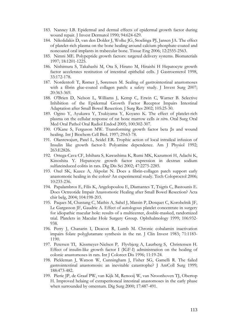
113
183. Nanney LB. Epidermal and dermal effects of epidermal growth factor during wound repair. J Invest Dermatol 1990; 94:624-629.
184. Nikolidakis D, van den Dolder J, Wolke JG, Stoelinga PJ, Jansen JA. The effect of platelet-rich plasma on the bone healing around calcium phosphate-coated and noncoated oral implants in trabecular bone. Tissue Eng 2006; 12:2555-2563.
185. Nimni ME. Polypeptide growth factors: targeted delivery systems. Biomaterials 1997; 18:1201-1225.
186. Nishimura S, Takahashi M, Ota S, Hirano M, Hiraishi H Hepatocyte growth factor accelerates restitution of intestinal epithelial cells. J Gastroenterol 1998, 33:172-178.
187. Nordentoft T, Rømer J, Sørensen M. Sealing of gastrointestinal anastomoses with a fibrin glue-coated collagen patch: a safety study. J Invest Surg 2007; 20:363-369.
188. O'Brien D, Nelson L, Williams J, Kemp C, Erwin C, Warner B. Selective Inhibition of the Epidermal Growth Factor Receptor Impairs Intestinal Adaptation after Small Bowel Resection. J Surg Res 2002; 105:25-30.
189. Ogino Y, Ayukawa Y, Tsukiyama Y, Koyano K. The effect of platelet-rich plasma on the cellular response of rat bone marrow cells in vitro. Oral Surg Oral Med Oral Pathol Oral Radiol Endod 2005; 100:302-307.
190. O'Kane S, Ferguson MW. Transforming growth factor beta βs and wound healing. Int J Biochem Cell Biol. 1997; 29:63-78.
191. Olanrewajutt, Patel L, Seidel ER. Trophic action of local intraileal infusion of Insulin like growth factor-I: Polyamine dependence. Am J Physiol 1992; 263:E2826.
192. Ortega-Cava CF, Ishihara S, Kawashima K, Rumi MK, Kazumori H, Adachi K, Kinoshita Y. Hepatocyte growth factor expression in dextran sodium sulfateinduced colitis in rats. Dig Dis Sci 2002; 47:2275-2285.
193. Ozel SK, Kazez A, Akpolat N. Does a fibrin-collagen patch support early anastomotic healing in the colon? An experimental study. Tech Coloproctol 2006; 10:233-236.
194. Papalambros E, Filis K, Angelopoulou E, Diamantes T, Tsigris C, Bastounis E. Does Octreotide Impair Anastomotic Healing after Small Bowel Resection? Acta chir belg, 2004; 104:198-203.
195. Paques M, Chastang C, Mathis A, Sahel J, Massin P, Dosquet C, Korobelnik JF, Le Gargasson JF, Gaudric A. Effect of autologous platelet concentrate in surgery for idiopathic macular hole: results of a multicenter, double-masked, randomized trial. Platelets in Macular Hole Surgery Group. Ophthalmology 1999; 106:932-938.
196. Perry J, Chanarin I, Deacon R, Lumb M. Chronic cobalamin inactivation impairs folate polyglutamate synthesis in the rat. J Clin Invest 1983; 71:1183-1190.
197. Petersen TI, Kissmeyer-Nielsen P, Flyvbjerg A, Laurberg S, Christensen H. Effect of insulin-like growth factor I (IGF-I) administration on the healing of colonic anastomoses in rats. Int J Colorect Dis 1996; 11:19-24.
198. Pickleman J, Watson W, Cunningham J, Fisher SG, Gamelli R. The failed gastrointestinal anastomosis: an inevitable catastrophe? J AmColl Surg 1999; 188:473-482.
199. Pierie JP, de Graaf PW, van Kijk M, Renooij W, van Nroonhoven TJ, Obertop H. Improved helaing of extraperitoneal intestinal anastomoses in the early phase when surrounded by omentum. Dig Surg 2000; 17:487-491.

114
200. Plachokova AS, van den Dolder J, Stoelinga PJ, Jansen JA. The bone regenerative effect of platelet-rich plasma in combination with an osteoconductive material in rat cranial defects. Clin Oral Implants Res 2006; 17:305-311.
201. Plachokova AS, van den Dolder J, Stoelinga PJ, Jansen JA. Early effect of platelet-rich plasma on bone healing in combination with an osteoconductive material in rat cranial defects. Clin Oral Implants Res 2007; 18:244-251.
202. Platelet Research Laboratory. Importance of its study. Social interest.http://www platelet-research org/1/interest_soci htm.
203. Podolsky DK. Peptide growth factors in the gastrointestinal tract. In: Johnson LR, Ed. Physiology of the gastrointestinal tract. New York: Raven Press; 1994: 129-167.
204. Posma LE, Bleichtodt RP, van Goor H, Hendriks T. Transient profound mesenteric ischemia strongly affects the strength of intestinal anastomoses in the rat. Dis Colon Rectum 2007; 50:1070-1079.
205. Rabau M, Hirshberg A, Hiss Y, Dayan D. Intestinal anastomosis healing in rats: Collagen concentration and histochemical characterization by picrosirius red staining and polarizing microscopy. Exp Mol Pathol, 1995, 62:160-165.
206. Rabau M, Eyal A, Kluger Y, Dayan D. Bursting pressure in anastomotic healing in experimentally induced colitis in rats. Diseases of the Colon and Rectum 1998; 41, 468-472.
207. Read LC, Tomas FM, Howard GS, Martin AA, Edson KJ, Gillespie C, Owens PC, Ballard FJ. Insulin like growth factor and its N-terminal modified analogues induce marked gut growth in dexamethasone-treated rats. J Endocrinol 1992; 133:421- 431.
208. Reece TB, Maxey TS, Kron IL. A prospectus on tissue adhesives. Am J Surg 2001; 182:40S-44S.
209. Reed GL, Fitzgerald ML, Polgar J. Molecular mechanisms of platelet exocytosis: insights into the ‘secrete’ life of thrombocytes. Blood 2000; 96:3334-3342.
210. Roberts AB, Flanders KC, Kondaiah P, Thompson NL, Van Obberghen- Schilling E, Wakefield L, Rossie P, De Crumbrugghe B, Heine U, Sporn MB. Transforming growth factor β: biochemistry and roles in embryogenesis, tissue repair and remodeling, and carcinogenesis. Rec Prog Horm Res 1988; 44:157-197.
211. Roberts GP, Harding KG. Stimulation of glycosaminoglycan synthesis in cultured fibroblasts by hyperbaric oxygen. Br J Dermatol. 1994; 131:630-633.
212. Romo T and Pearson JM. Wound Healing, Skin. 2005. Emedicine.com. 213. Ruszczak Z. Effect of collagen matrices on dermal wound healing. Advanced
Drug Delivery Reviews 2003; 55:1595-1611. 214. Saldalamacchia G, Lapice E, Cuomo V, De Feo E, D’Agostino E, Rivellese
AA, Vaccaro O. A controlled study of the use of autologous platelet gel for the treatment of diabetic ulcers. Nutr Metab Cardiovasc Dis 2004; 14:395-396.
215. Sanchez AR, Sheridan PJ, Kupp LI. Is platelet-rich plasma the perfect enhancement factor? A current review. Int J Oral Maxillofac Implants 2003; 18:93-103.
216. Sanchez M, Anitua E, Andia I. Use of autologous plasma rich in growth factors in arthroscopic surgery. Cuader Artroscopia 2003;10:12-19.
217. Schlegel KA, Donath K, Rupprech S, Falka S, Zimmermann R, Felszegh E, Wiltfang J. De novo bone formation using bovine collagen and platelet-rich plasma Biomaterials 2004; 25:5387-5393.

115
218. Schmidt DA, Tumbleson ME. Swine hematology. In Swine in Biomedical Research, Volume 2 (New York: Plenum Press), pp.767-782.
219. Schröder W, Stippel D, Gutschow C, Leers J, Holscher AH. Postoperative recovery of microcirculation after gastric tube formation. Langenbecks Arch Surg 2004; 389:267-271.
220. Sekido Y, Morishima Y, Ohya K. Activity of platelet-derived growth factor (PDGF) in platelet concentrates and cryopreserved platelets determined by PDGF bioassay. Vox Sang 1987; 52:27-30.
221. Semple J, Aslam R, Freedman J. Murine platelets express Toll like receptor 2: a potential regulator of innate and adaptive immunity [abstract]. Platelets. 2004; 15:267.
222. Sharma P. and Mafulli N. Tendon injury and tendinopathy: healing and repair. J. Bone Joint Surg. Am 2005; 87:187-202.
223. Shiraki R, Inoue N, Kawasaki S, Takei A, Kadotani M, Ohnishi Y, Ejiri J, Kobayashi S, Hirata K, Kawashima S, Yokoyama M. Expression of Toll-like receptors on human platelets. Thromb Res 2004; 113:379-385.
224. Siemonsma MA, de Hingh IT, de Man BM, Lomme RM, Verhofstad AJ, Hendriks T. Doxycycline improved wound strength after intestinal anastomosis in the rat. Surgery 2003; 133:268-276.
225. Slater M, Patava J, Kingham K, Mason RS. Involvement of platelets in stimulating osteogenic activity. J Orthop Res 1995; 13:655-663.
226. Slavin J, Nash J, Kingsnorth A. Effect of transforming growth factors β and basic fibroblast growth factor on steroid impaired healing intestinal wounds. Br J Surg 1992; 79:69-72.
227. Smith CA, Ficken MD. Non-surgical cannulation of the vena cava for chronic blood collection in mature swine. Lab Anim 1991; 41:274-287.
228. Smith SR, Connolly JC, Crane PW, Gilmore OJ. The effect of surgical drainage materials on colonic healing. Br J Surg 1982; 69:153-155.
229. Sonnleitner D, Huemer P, Sullivan DY. A simplified technique for producing platelet-rich plasma and platelet concentrate for intraoral bone grafting techniques: a technical note. Int J Oral Maxillofac Implants 2000; 15:879–882.
230. Spencer EM, Tokunaga A, Hunt KT. Insulin-like growth factor binding protein-3 is present in the α-granules of platelets. Endocrinology 1993; 132:996-1001.
231. Stadelmann WK, Digenis AG, Tobin GR. Physiology and healing dynamics of chronic cutaneous wounds. The American Journal of Surgery 1998; 176:26S-38S.
232. Stanworth SJ, Denton K, Monteath J, Patton WN. Automated counting of platelets on the Bayer ADVIA 120 analyser. Clinical and Laboratory Haematology 1999; 21:113-117.
233. Steigmann M, Garg AK. A comparative study of bilateral sinus lifts performed with platelet-rich plasma alone versus alloplastic graft material reconstituted with blood. Implant Dent 2005 14:261-266.
234. Suba, Z. Takacs D, Gyulai-Gaal S, Kovacs K. Facilitation of b-tricalcium phosphate-induced alveolar bone regeneration by platelet-rich plasma in beagle dogs: a histologic and histomorphometric study. Int. J. Oral Maxillofac. Implants 2004; 19:832-838.
235. Swindle MM, Smith AC, Hepburn BJ. Swine as models in experimental surgery. J Invest Surg 1988; 1:65-79.
236. Tabata Y. Tissue regeneration based on growth factor release. Tissue Eng 2003; 9:S5-S15.

116
237. Tablin F. Platelet structure and function. In: Feldman BF, Zinkl JG, Jain NC, eds. Schalm's Veterinary Hematology, 5th ed. Philadelphia: Lippincot Williams & Wilkins; 2000:448-452.
238. Tahara Y, Ido A, Yamamoto S, Miyata Y, Uto H, Hori T, Hayashi K, Tsubouchi H. Hepatocyte Growth Factor Facilitates Colonic Mucosal Repair in Experimental Ulcerative Colitis in Rats. The Journal of Pharmacology and Experimental Therapeutics 2003; 307:146-151.
239. Tamimi F, Montalvo S, Tresguerres I, Blanco Jerez L. A Comparative Study of 2 Methods for Obtaining Platelet-Rich Plasma. Journal of Oral and Maxillofacial Surgery 2007; 65:1084-1093.
240. Tandara AA, Mustoe TA. Oxygen in wound healing- more than a nutrient. World J Surg 2004; 28:294-300.
241. Thorn C. Normal haematology of the Pig Venous blood platelet counts and haematocrit values of all pigs were within normal limits. In: Feldman BF, Zinkl JG, Jain NC, et al. (Eds). Schulman’s veterinary hematology. Philadelphia, Lippincott Williams and Wilkins, 2000; pp. 1089-1095.
242. Thornton FJ, Barbul A. Healing in the gastrointestinal tract. Surg Clin North Am 1997; 77:549-573.
243. Thorwarth M, Rupprecht S, Falk S, Felszeghy E, Wiltfang J, Schlegel KA. Expression of bone matrix proteins during de novo bone formation using bovine collagen and platelet-rich plasma (PRP)—an immunihistochemical analysis. Biomatherials 2005; 26:2575-2584.
244. Trowbridge CC, Stammers AH, Woods E, Yen BR, Klayman M, Gilbert C. Use of platelet gel and its effects on infection in cardiac surgery. J Extra Corpor Technol. 2005; 37:381-386.
245. Tsay RC, Burke A, Eisig SB, Lu HH, Landsberg R. Differential growth factor retention by platelet-rich plasma composites. J Oral Maxillofac Surg. 2005; 63:521-528.
246. Tsubota K, Goto E, Fujita H, Ono M, Inoue H, Saito I, Shimmura S. Treatment of dry eye by autologous serum application in Sjögren’s syndrome. Br J Ophthalmol. 1999; 83:390-305.
247. Uribe JM, Barrett KE. Nonmitogenic actions of growth factors: an integrated view of their role in intestinal physiology and pathophysiology. Gastroenterology 1997; 112:255-268.
248. van der Hamm AC, Kort WJ, Weijama IM, van der Ingh HM, Jeekel J. Effect of fibrin sealant on the healing colonic anastomosis in the rat. Br J Surg 1991; 78:49-53.
249. van der Ham AC, Kort WJ, Weijma IM, van den Ingh HM, Jeekel J. Healing of ischemic colon anastomosis: fibrin sealant does not improve wound healing. Dis Colon Rectum. 1992; 35:884-891.
250. Vang SN, Brady CP, Christensen KA, Allen KR, Anderson JE, Isler JR, Holt DW, Smith LM. Autologous platelet gel in coronary artery bypass grafting: effects on surgical wound healing. J Extra Copor Technolog 2007; 29:31-38.
251. Vanderhood JA, Mc Cusker RH, Clark K, Mohammadpour H, Blackwood DJ, Harty RF, Park JY. Truncated and native Insulin like growth factor I enhance mucosal adaptation after jejunoileal resection. Gastroenterology 1992; 102:1949-1956.
252. Verhofstad MJ, Lange WL, van der Laak JM, Verhofstad AJ, Hendriks T. Microscopic analysis of anastomotic healing in the intestine of normal and diabetic rats. Dis Colon Rectum, 2001, 44:423-431.
117
253. Vignali A, Fazio VW, Lavery IC, Milsom JW, Church JM, Hull TL, Strong SA, Oakley JR. Factors associated with the occurrence of leaks in stapled rectal anastomoses: a review of 1014 patients. J Am Coll Surg 1997; 185:105-113.
254. Villa X, Kuluz JW, Schleien CL, Thompson JF. Epidermal growth factor reduces ischemia-reperfusion injury in rat small intestine. Crit. Care Med 2002; 30:1576-1580.
255. Ward JR, Bingle L, Judge HM, Brown SB, Stoley RF, Whyte MK, Dower SK, Buttle DJ, Sabroe I. Agonists of toll-like receptor TLR2 and TLR4 are unable to modulate platelet activation by adenosine diphosphate and platelet activating factor. Thromb. Haemost. 2005; 94, 831-838.
256. Weiber S, Jiborn H, Zederfeldt B. Preoperative irradiation and colonic healing. Eur J Surg. 1994; 160:47-51.
257. Weibrich G, Kleis WK, Hafner G. Growth factor levels in the platelet-rich plasma produced by 2 different methods: curasan- type PRP kit versus PCCS PRP system. International Journal of Oral and Maxillofacial Implants 2002; 17:184-190.
258. Weibrich G, Kleis WK. Curasan PRP kit vs. PCCS PRP system. Collection efficiency and platelet counts of two different methods for the preparation of platelet-rich plasma. Clin Oral Implants Res 2002; 13:437-443.
259. Weibrich G, Kleis WK, Hafner G, Hitzler WE, Wagner W. Comparison of platelet, leukocyte, and growth factor levels in point-of-care platelet-enriched plasma, prepared using a modified Curasan kit, with preparations received from a local blood bank. Clin Oral Implants Res 2003; 14:357-362.
260. Weibrich G, Hansen T, Kleis W, Buch R, Hitzler WE. Effect of platelet concentration in platelet-rich plasma on peri-implant bone regeneration. Bone 2004; 34:665- 671.
261. Weibrich G, Kleis WK, Hitzler WE, Hafner G. Comparison of the platelet concentrate collection system with the plasma-rich-in-growth-factors kit to produce platelet-rich plasma: a technical report. Int J Oral Maxillofac Implants 2005; 20:118-123.
262. Weiner BK, Walker M. Efficacy of autologous growth factors in lumbar intertransverse fusions. Spine 2003; 28:1968-1971.
263. Werner S and Grose R. Regulation of Wound Healing by Growth Factors and Cytokines. Physiol Rev 2003; 83:835-870.
264. Westphal RG. Health risks to cytapheresis donors. Clin Haematol 1984; 13:289-301.
265. Weyrich AS, Lindermann S, Zimmerman GA. The evolving role of platelets in inflammation 2003; 1:1897-1905.
266. Wheeless CR Jr, Zanagnolo V, Bowers D, Brenner MJ, and Lilley R. The effect of growth hormone on the bursting strength of ileal anastomotic segments in radiation-injured rat bowel. Gynecol Oncol 1998; 70:121-122.
267. White GC. Platelet physiology and function. Blood coagul fibrinolysis 2000; 11(Suppl 1): S53.
268. Whitman DH, Berry RL, Green DM. Platelet gel: an autologous alternative to fibrin glue with applications in oral and maxillofacial surgery. J Oral Maxillofac Surg 1997; 55:1294-1299.
269. Wiltfang J, Kloss FR, Kessler P, Nkenke E, Schultze-Mosgau S, Zimmermann R, Schlegel KA. Effects of platelet-rich plasma on bone healing in combination with autogenous bone and bone substitutes in critical-size defects. An animal experiment. Clinical Oral Implants Research 2004; 15:187-193.

118
270. Witte MB, Barbul A. General principles of wound healing. Surg Clin North Am 1997; 77:509-528.
271. Wright NA, Pike C, Elia G. Induction of a novel epidermal growth factor- secreting cell lineage by mucosal ulceration in human gastrointestinal stem cells. Nature 1990; 343: 82-85.
272. Wu KK. Platelet activation and arterial thrombosis. Lancet 1994; 344:991-995. 273. Yagci G, Ozturk E, Ozgurtas T, Gorgulu S, Kutlu OC, Topal T, Cetiner S,
Tufan T. Preoperative and postoperative administration of hyperbaric oxygen improves biochemical and mechanical parameters on ischemic and normal colonic anastomoses. J Invest Surg 2006; 19:237-244.
274. Yamada Y, Ueda M, Hibi H, Nagasaka T. Translational research for injectable tissue engineered bone regeneration using mesenchymal stem cells and platelet-rich plasma: from basic research to clinical case study. Cell Transplant 2004; 13:343-355.
275. Yamaguchi M, Kuzume M, Asakawa K, Matsumiya A, Matsumoto T, Shimura H, Midorikawa T, Narihara K, Sanada Y, Kumada K. Effects of insulin-like growth factor-1 on massive resection of small intestine with or without ileocecal resection in rats. Transplant Proc 2000; 32:2486-2487.
276. Yeaman MR. The role of platelets in antimicrobial host defense. Clin Infect Dis 1997; 25:951-970.
277. Yamamoto K, Hayashi J, Miyamura H, Eguchi S. A comparison study of the effect of autologous platelet rich plasma and fresh whole blood on haemostasis after cardiac surgery. Cardiovascular Surgery 1996; 4:9-14.
278. Yol S, Tekin A, Yilmaz H, Küçükkartallar T, Esen H, Çaglayan O, Tatkan Y. Effects of platelet Rich plasma on colonic anastomosis. J Surg Res 2008; 146:130-194.
279. Zaizen Y, Ford EG, Costin G, Atkinson JB. Stimulation of wound bursting strength during protein malnutrition. J Surg Res 1990; 49:333-336.
280. Zeeh JM, ProccacinoF, Hoffman P, Aukerman SL, Mc Roberts JA, Soltani S, Pierce GF, Lakshmanan J, Lacey D, Eysselein VE. Keratinocyte growth factor ameliorates mucosal injury in an experimental model colitis in rats. Gastroenterology 1996; 110:1077-1083.
281. Zelmanovic D, Hetherington EJ. Automated analyses of feline platelets in whole blood, including platelet count, mean platelet volume, and activation state. Vet Clin Pathol 1998; 27:2-9.
282. Zilling TL, Jansson O, Walther BS, Ottosson A. Sutureless small bowel anastomoses: Experimental study in pigs. Eur J Surg 1999; 165:61-68.
283. Zimmermann R, Jakubietz R, Jakubietz M, Strasser E, Schlegel A, Wiltfang J, Eckstein R. Different preparation methods to obtain platelet components as a source of growth factors for local application. Transfusion 2001; 41:1217-1224.
284. Zimmermann R, Arnold D, Strasser E, Ringwald J, Schlegel A, Wiltfang J, Eckstein R. Sample preparation technique and with cell content influence the detectable levels of growth factors in platelet concentrates. Vox Sanguinis 2003; 85:283-289.
285. Zhu SJ, Choi BH, Jung JH, Lee SH, Huh JY, You TM, Lee HJ, Li J. A comparative histologic analysis of tissue-engineered bone using platelet-rich plasma and platelet-enriched fibrin glue. Oral Surg Oral Med Oral Pathol Oral Radiol Endod 2006; 102:175-179.
286. Zwaal RF, Schroit AJ. Pathophysiologic implications of membrane phospholipid asymmetry in blood cells. Blood 1997; 89:1121-1132.