![국내SDN 시장활성화를위한비즈니스전략에관한연구web.yonsei.ac.kr/bkgsi/pdf_paper/이봉규교수님... · · 2014-03-04를가지고있다[9]. 따라서본논문은국내sdn](https://static.fdocuments.net/doc/165x107/5ae89e367f8b9a08778fedca/sdn-web-2014-03-049.jpg)
International Journal of Food Microbiology -...
8
Analysis of yeast and archaeal population dynamics in kimchi using denaturing gradient gel electrophoresis Ho-Won Chang a , Kyoung-Ho Kim a , Young-Do Nam a,c , Seong Woon Roh a,c , Min-Soo Kim a , Che Ok Jeon d , Hee-Mock Oh a , Jin-Woo Bae a,b,c, ⁎ a Biological Resource Center, KRIBB, Daejeon 305-806, South Korea b Environmental Biotechnology National Core Research Center, Gyeongsang National University, Jinju 660-701, South Korea c Korea University of Science & Technology, 52, Eoeun-dong, Yuseong-gu, Daejeon, 305-333, South Korea d Department of Life Science, Chung-Ang University, Seoul 156-756, South Korea ABSTRACT ARTICLE INFO Article history: Received 10 April 2008 Received in revised form 14 May 2008 Accepted 14 May 2008 Keywords: Kimchi DGGE Archaea Yeast Kimchi is a traditional Korean food that is fermented from vegetables such as Chinese cabbage and radish. Many bacteria are involved in kimchi fermentation and lactic acid bacteria are known to perform significant roles. Although kimchi fermentation presents a range of environmental conditions that could support many different archaea and yeasts, their molecular diversity within this process has not been studied. Here, we use PCR-denaturing gradient gel electrophoresis (DGGE) targeting the 16S and 26S rRNA genes, to characterize bacterial, archaeal and yeast dynamics during various types of kimchi fermentation. The DGGE analysis of archaea expressed a change of DGGE banding patterns during kimchi fermentation, however, no significant change was observed in the yeast DGGE banding patterns during kimchi fermentation. No significant difference was indicated in the archaeal DGGE profile among different types of kimchi. In the case of yeasts, the clusters linked to the manufacturing corporation. Haloarchaea such as Halococcus spp., Natronococcus spp., Natrialba spp. and Haloterrigena spp., were detected as the predominant archaea and Lodderomyces spp., Trichosporon spp., Candida spp., Saccharomyces spp., Pichia spp., Sporisorium spp. and Kluyveromyces spp. were the most common yeasts. © 2008 Elsevier B.V. All rights reserved. 1. Introduction Fermentation is a well-known and ancient technique that uses microorganisms to process and preserve food (Ross et al., 2002). Kimchi is a Korean food prepared as a result of fermentation and in recent years it has been recognized as a health-promoting functional food (Song, 2004). Since it is interesting to know what microbes are involved by kimchi fermentation, the fermentation process has been studied extensively by microbiologists with respect to its ecology, proteomics, genetics and physiology (Kim and Chun, 2005; Li et al., 2006; Nan et al., 2005). These studies have reported that kimchi is a healthy food, rich in β-carotene, chlorophylls, dietary fiber and various minerals (Park et al., 2003). It supports a wide range of microorgan- isms, including lactic acid bacteria (LAB), which perform significant roles during fermentation (Bae et al., 2005; Kim and Chun, 2005). Bacterial isolates from kimchi have been investigated with respect to production and characterization of beneficial enzymes such as dex- transucrase and alcohol/acetaldehyde dehydrogenase (Eom et al., 2007; Koo et al., 2005), and for biodegradation of toxic compounds such as sodium nitrite and bisphenol (Oh et al., 2004; Yamanaka et al., 2007). Several halophilic archaea have been isolated from jeotgal, tradi- tional Korean fermented seafood used in kimchi as an ingredient (Roh et al., 2007a; Roh et al., 2007b). Since the average NaCl concentration of kimchi is 3% (Mheen and Kwon, 1984), we reasoned that it could support haloarchaea. Yeasts such as Pichia spp. have been isolated from kimchi at low pH (about pH 4) that have been fermented over a long period of time (Oh and Han, 2003). However, the overall molecular diversity of yeasts and archaea in kimchi has not yet been reported. Molecular biology techniques are used frequently to explore the diversity and structure of microbial communities, and microorgan- isms are identified using certain molecular markers such as 16S or 26S rRNA. In particular, the study of microbial ecology has benefited greatly from the introduction of denaturing gradient gel electrophor- esis (DGGE), which provided a molecular fingerprinting technique for studying community structure (Yeates et al., 1998). In DGGE analysis, PCR amplicons of the same size but different sequences can be separated (Muyzer and Smalla, 1998) and this technique has been applied widely for studying microbial dynamics in complex environments such as soil International Journal of Food Microbiology 126 (2008) 159–166 ⁎ Corresponding author. Biological Resource Center, KRIBB, Daejeon 305-806, South Korea. Tel.: +82 42 860 4628; fax: +82 42 860 4677. E-mail address: [email protected] (J.-W. Bae). 0168-1605/$ – see front matter © 2008 Elsevier B.V. All rights reserved. doi:10.1016/j.ijfoodmicro.2008.05.013 Contents lists available at ScienceDirect International Journal of Food Microbiology journal homepage: www.elsevier.com/locate/ijfoodmicro
Transcript of International Journal of Food Microbiology -...

International Journal of Food Microbiology 126 (2008) 159–166
Contents lists available at ScienceDirect
International Journal of Food Microbiology
j ourna l homepage: www.e lsev ie r.com/ locate / i j foodmicro
Analysis of yeast and archaeal population dynamics in kimchi using denaturinggradient gel electrophoresis
Ho-Won Chang a, Kyoung-Ho Kim a, Young-Do Nam a,c, Seong Woon Roh a,c, Min-Soo Kim a, Che Ok Jeon d,Hee-Mock Oh a, Jin-Woo Bae a,b,c,⁎a Biological Resource Center, KRIBB, Daejeon 305-806, South Koreab Environmental Biotechnology National Core Research Center, Gyeongsang National University, Jinju 660-701, South Koreac Korea University of Science & Technology, 52, Eoeun-dong, Yuseong-gu, Daejeon, 305-333, South Koread Department of Life Science, Chung-Ang University, Seoul 156-756, South Korea
⁎ Corresponding author. Biological Resource Center, KKorea. Tel.: +82 42 860 4628; fax: +82 42 860 4677.
E-mail address: [email protected] (J.-W. Bae).
0168-1605/$ – see front matter © 2008 Elsevier B.V. Aldoi:10.1016/j.ijfoodmicro.2008.05.013
A B S T R A C T
A R T I C L E I N F OArticle history:
Kimchi is a traditional Kore Received 10 April 2008Received in revised form 14 May 2008Accepted 14 May 2008Keywords:KimchiDGGEArchaeaYeast
an food that is fermented from vegetables such as Chinese cabbage and radish.Many bacteria are involved in kimchi fermentation and lactic acid bacteria are known to perform significantroles. Although kimchi fermentation presents a range of environmental conditions that could support manydifferent archaea and yeasts, their molecular diversity within this process has not been studied. Here, we usePCR-denaturing gradient gel electrophoresis (DGGE) targeting the 16S and 26S rRNA genes, to characterizebacterial, archaeal and yeast dynamics during various types of kimchi fermentation. The DGGE analysis ofarchaea expressed a change of DGGE banding patterns during kimchi fermentation, however, no significantchange was observed in the yeast DGGE banding patterns during kimchi fermentation. No significantdifference was indicated in the archaeal DGGE profile among different types of kimchi. In the case of yeasts,the clusters linked to the manufacturing corporation. Haloarchaea such as Halococcus spp., Natronococcusspp., Natrialba spp. and Haloterrigena spp., were detected as the predominant archaea and Lodderomycesspp., Trichosporon spp., Candida spp., Saccharomyces spp., Pichia spp., Sporisorium spp. and Kluyveromycesspp. were the most common yeasts.
© 2008 Elsevier B.V. All rights reserved.
1. Introduction
Fermentation is a well-known and ancient technique that usesmicroorganisms to process and preserve food (Ross et al., 2002).Kimchi is a Korean food prepared as a result of fermentation and inrecent years it has been recognized as a health-promoting functionalfood (Song, 2004). Since it is interesting to know what microbes areinvolved by kimchi fermentation, the fermentation process has beenstudied extensively by microbiologists with respect to its ecology,proteomics, genetics and physiology (Kim and Chun, 2005; Li et al.,2006; Nan et al., 2005). These studies have reported that kimchi is ahealthy food, rich in β-carotene, chlorophylls, dietary fiber and variousminerals (Park et al., 2003). It supports a wide range of microorgan-isms, including lactic acid bacteria (LAB), which perform significantroles during fermentation (Bae et al., 2005; Kim and Chun, 2005).Bacterial isolates from kimchi have been investigated with respect to
RIBB, Daejeon 305-806, South
l rights reserved.
production and characterization of beneficial enzymes such as dex-transucrase and alcohol/acetaldehyde dehydrogenase (Eom et al., 2007;Koo et al., 2005), and for biodegradation of toxic compounds such assodium nitrite and bisphenol (Oh et al., 2004; Yamanaka et al., 2007).
Several halophilic archaea have been isolated from jeotgal, tradi-tional Korean fermented seafood used in kimchi as an ingredient (Rohet al., 2007a; Roh et al., 2007b). Since the average NaCl concentration ofkimchi is 3% (Mheen and Kwon, 1984), we reasoned that it couldsupport haloarchaea. Yeasts such as Pichia spp. have been isolated fromkimchi at low pH (about pH 4) that have been fermented over a longperiod of time (Oh and Han, 2003). However, the overall moleculardiversity of yeasts and archaea in kimchi has not yet been reported.
Molecular biology techniques are used frequently to explore thediversity and structure of microbial communities, and microorgan-isms are identified using certain molecular markers such as 16S or 26SrRNA. In particular, the study of microbial ecology has benefitedgreatly from the introduction of denaturing gradient gel electrophor-esis (DGGE), which provided a molecular fingerprinting technique forstudying community structure (Yeates et al., 1998). In DGGE analysis,PCRamplicons of the samesizebut different sequences canbe separated(Muyzer and Smalla, 1998) and this technique has been applied widelyfor studying microbial dynamics in complex environments such as soil
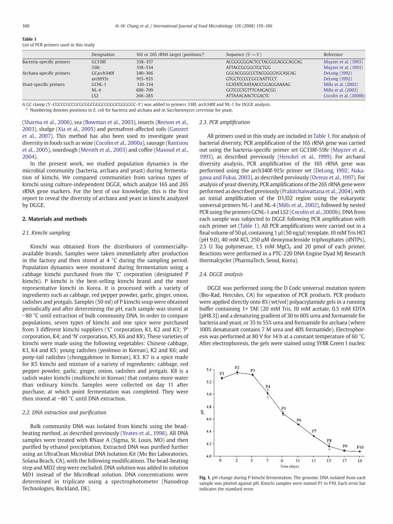
Table 1List of PCR primers used in this study
Designation 16S or 26S rRNA target (positions)a Sequence (5′→3′) Reference
Bacteria-specific primers GC338f 338–357 ACGGGGGGACTCCTACGGGAGGCAGCAG Muyzer et al. (1993)518r 518–534 ATTACCGCGGCTGCTGG Muyzer et al. (1993)
Archaea-specific primers GCarch340f 340–366 GGCACGGGCCCTACGGGGYGCASCAG DeLong (1992)arch915r 915–935 GTGCTCCCCCGCCAATTCCT DeLong (1992)
Yeast-specific primers GCNL-1 110–134 GCATATCAATAAGCGGAGGAAAAG Mills et al. (2002)NL-4 690–709 GGTCCGTGTTTCAAGACGG Mills et al. (2002)LS2 266–285 ATTAAACAACTCGACTC Cocolin et al. (2000b)
A GC clamp (5′-CGCCCGCCGCGCGGCGGGCGGGGCGGGGGC-3′) was added to primers 338f, arch340f and NL-1 for DGGE analysis.a Numbering denotes positions in E. coli for bacteria and archaea and in Saccharomyces cerevisiae for yeast.
Fig. 1. pH change during P kimchi fermentation. The genomic DNA isolated from eachsample was plotted against pH. Kimchi samples were named P1 to P10. Each error barindicates the standard error.
160 H.-W. Chang et al. / International Journal of Food Microbiology 126 (2008) 159–166
(Sharma et al., 2006), sea (Bowman et al., 2003), insects (Reeson et al.,2003), sludge (Xia et al., 2005) and permafrost-affected soils (Ganzertet al., 2007). This method has also been used to investigate yeastdiversity in foods such aswine (Cocolin et al., 2000a), sausage (Rantsiouet al., 2005), sourdough (Meroth et al., 2003) and coffee (Masoud et al.,2004).
In the present work, we studied population dynamics in themicrobial community (bacteria, archaea and yeast) during fermenta-tion of kimchi. We compared communities from various types ofkimchi using culture-independent DGGE, which analyze 16S and 26SrRNA gene markers. For the best of our knowledge, this is the firstreport to reveal the diversity of archaea and yeast in kimchi analyzedby DGGE.
2. Materials and methods
2.1. Kimchi sampling
Kimchi was obtained from the distributors of commercially-available brands. Samples were taken immediately after productionin the factory and then stored at 4 °C during the sampling period.Population dynamics were monitored during fermentation using acabbage kimchi purchased from the ‘C’ corporation (designated Pkimchi). P kimchi is the best-selling kimchi brand and the mostrepresentative kimchi in Korea. It is processed with a variety ofingredients such as cabbage, red pepper powder, garlic, ginger, onion,radishes and jeotgals. Samples (50ml) of P kimchi soupwere obtainedperiodically and after determining the pH, each sample was stored at−80 °C until extraction of bulk community DNA. In order to comparepopulations, seven types of kimchi and one spice were purchasedfrom 3 different kimchi suppliers (‘C’ corporation, K1, K2 and K3; ‘P’corporation, K4; and ‘N’ corporation, K5, K6 and K8). These varieties ofkimchi were made using the following vegetables: Chinese cabbage,K1, K4 and K5; young radishes (yeolmoo in Korean), K2 and K6; andpony-tail radishes (chonggakmoo in Korean), K3. K7 is a spice madefor K5 kimchi and mixture of a variety of ingredients: cabbage, redpepper powder, garlic, ginger, onion, radishes and jeotgals. K8 is aradish water kimchi (mulkimchi in Korean) that contains more waterthan ordinary kimchi. Samples were collected on day 11 afterpurchase, at which point fermentation was completed. They werethen stored at −80 °C until DNA extraction.
2.2. DNA extraction and purification
Bulk community DNA was isolated from kimchi using the bead-beating method, as described previously (Yeates et al., 1998). All DNAsamples were treated with RNase A (Sigma, St. Louis, MO) and thenpurified by ethanol precipitation. Extracted DNA was purified furtherusing an UltraClean Microbial DNA Isolation Kit (Mo Bio Laboratories,Solana Beach, CA), with the following modifications. The bead-beatingstep andMD2 stepwere excluded. DNA solutionwas added to solutionMD1 instead of the MicroBead solution. DNA concentrations weredetermined in triplicate using a spectrophotometer (NanodropTechnologies, Rockland, DE).
2.3. PCR amplification
All primers used in this study are included in Table 1. For analysis ofbacterial diversity, PCR amplification of the 16S rRNA gene was carriedout using the bacteria-specific primer set GC338f-518r (Muyzer et al.,1993), as described previously (Henckel et al., 1999). For archaealdiversity analysis, PCR amplification of the 16S rRNA gene wasperformed using the arch340f-915r primer set (DeLong, 1992; Naka-gawa and Fukui, 2003), as described previously (Ovreas et al., 1997). Foranalysis of yeast diversity, PCR amplifications of the 26S rRNA genewereperformed asdescribed previously (Prakitchaiwattana et al., 2004),withan initial amplification of the D1/D2 region using the eukaryoticuniversal primers NL-1 and NL-4 (Mills et al., 2002), followed by nestedPCRusing the primers GCNL-1 and LS2 (Cocolin et al., 2000b). DNA fromeach sample was subjected to DGGE following PCR amplification witheach primer set (Table 1). All PCR amplifications were carried out in afinal volume of 50 µl, containing 1 µl (50 ng/µl) template,10mMTris HCl(pH 9.0), 40 mM KCl, 250 µM deoxynucleoside triphosphates (dNTPs),2.5 U Taq polymerase, 1.5 mM MgCl2 and 20 pmol of each primer.Reactions were performed in a PTC-220 DNA Engine Dyad MJ Researchthermalcycler (PharmaTech, Seoul, Korea).
2.4. DGGE analysis
DGGE was performed using the D Code universal mutation system(Bio-Rad, Hercules, CA) for separation of PCR products. PCR productswere applied directly onto 8% (wt/vol) polyacrylamide gels in a runningbuffer containing 1× TAE (20 mM Tris, 10 mM acetate, 0.5 mM EDTA[pH8.3]) and a denaturing gradient of 30 to 60% urea and formamide forbacteria and yeast, or 35 to 55% urea and formamide for archaea (where100% denaturant contains 7 M urea and 40% formamide). Electrophor-esis was performed at 80 V for 14 h at a constant temperature of 60 °C.After electrophoresis, the gels were stained using SYBR Green I nucleic
161H.-W. Chang et al. / International Journal of Food Microbiology 126 (2008) 159–166
acid stain (Bioneer) and photographed under UV transillumination.Sterile blades were used to excise bands from the gels, and these werethenmixedwith 20µl of 0.1× TEbuffer solution and incubated overnightat 4 °C. These solutions were then used for PCR amplification with theappropriate primer set.
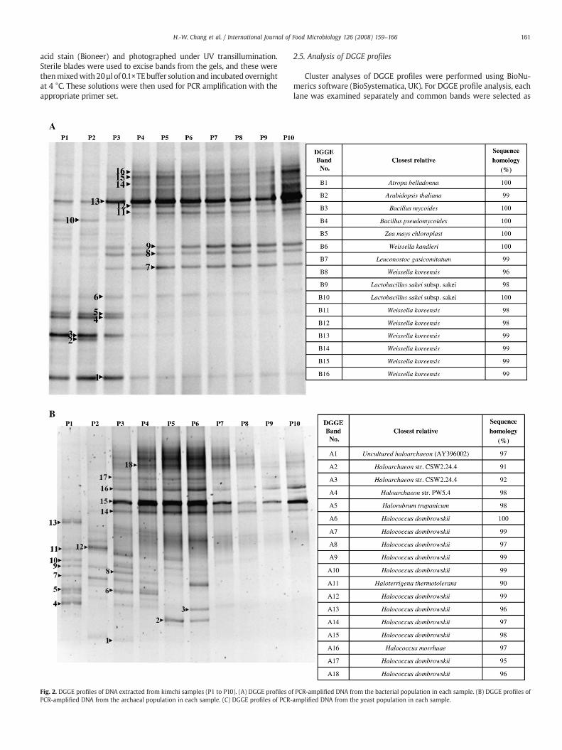
Fig. 2. DGGE profiles of DNA extracted from kimchi samples (P1 to P10). (A) DGGE profiles oPCR-amplified DNA from the archaeal population in each sample. (C) DGGE profiles of PCR-
2.5. Analysis of DGGE profiles
Cluster analyses of DGGE profiles were performed using BioNu-merics software (BioSystematica, UK). For DGGE profile analysis, eachlane was examined separately and common bands were selected as
f PCR-amplified DNA from the bacterial population in each sample. (B) DGGE profiles ofamplified DNA from the yeast population in each sample.
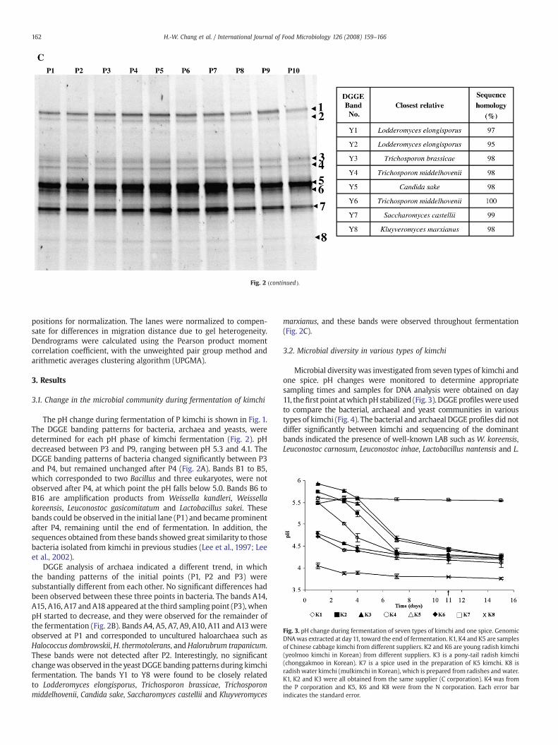
Fig. 2 (continued).
Fig. 3. pH change during fermentation of seven types of kimchi and one spice. GenomicDNAwas extracted at day 11, toward the end of fermentation. K1, K4 and K5 are samplesof Chinese cabbage kimchi from different suppliers. K2 and K6 are young radish kimchi(yeolmoo kimchi in Korean) from different suppliers. K3 is a pony-tail radish kimchi(chonggakmoo in Korean). K7 is a spice used in the preparation of K5 kimchi. K8 isradish water kimchi (mulkimchi in Korean), which is prepared from radishes and water.K1, K2 and K3 were all obtained from the same supplier (C corporation). K4 was fromthe P corporation and K5, K6 and K8 were from the N corporation. Each error barindicates the standard error.
162 H.-W. Chang et al. / International Journal of Food Microbiology 126 (2008) 159–166
positions for normalization. The lanes were normalized to compen-sate for differences in migration distance due to gel heterogeneity.Dendrograms were calculated using the Pearson product momentcorrelation coefficient, with the unweighted pair group method andarithmetic averages clustering algorithm (UPGMA).
3. Results
3.1. Change in the microbial community during fermentation of kimchi
The pH change during fermentation of P kimchi is shown in Fig. 1.The DGGE banding patterns for bacteria, archaea and yeasts, weredetermined for each pH phase of kimchi fermentation (Fig. 2). pHdecreased between P3 and P9, ranging between pH 5.3 and 4.1. TheDGGE banding patterns of bacteria changed significantly between P3and P4, but remained unchanged after P4 (Fig. 2A). Bands B1 to B5,which corresponded to two Bacillus and three eukaryotes, were notobserved after P4, at which point the pH falls below 5.0. Bands B6 toB16 are amplification products from Weissella kandleri, Weissellakoreensis, Leuconostoc gasicomitatum and Lactobacillus sakei. Thesebands could be observed in the initial lane (P1) and became prominentafter P4, remaining until the end of fermentation. In addition, thesequences obtained from these bands showed great similarity to thosebacteria isolated from kimchi in previous studies (Lee et al., 1997; Leeet al., 2002).
DGGE analysis of archaea indicated a different trend, in whichthe banding patterns of the initial points (P1, P2 and P3) weresubstantially different from each other. No significant differences hadbeen observed between these three points in bacteria. The bands A14,A15, A16, A17 and A18 appeared at the third sampling point (P3), whenpH started to decrease, and they were observed for the remainder ofthe fermentation (Fig. 2B). Bands A4, A5, A7, A9, A10, A11 and A13wereobserved at P1 and corresponded to uncultured haloarchaea such asHalococcus dombrowskii,H. thermotolerans, andHalorubrum trapanicum.These bands were not detected after P2. Interestingly, no significantchangewas observed in the yeast DGGE banding patterns during kimchifermentation. The bands Y1 to Y8 were found to be closely relatedto Lodderomyces elongisporus, Trichosporon brassicae, Trichosporonmiddelhovenii, Candida sake, Saccharomyces castellii and Kluyveromyces
marxianus, and these bands were observed throughout fermentation(Fig. 2C).
3.2. Microbial diversity in various types of kimchi
Microbial diversity was investigated from seven types of kimchi andone spice. pH changes were monitored to determine appropriatesampling times and samples for DNA analysis were obtained on day11, thefirst point atwhichpHstabilized (Fig. 3). DGGEprofileswere usedto compare the bacterial, archaeal and yeast communities in varioustypes of kimchi (Fig. 4). The bacterial and archaeal DGGE profiles did notdiffer significantly between kimchi and sequencing of the dominantbands indicated the presence of well-known LAB such as W. koreensis,Leuconostoc carnosum, Leuconostoc inhae, Lactobacillus nantensis and L.

163H.-W. Chang et al. / International Journal of Food Microbiology 126 (2008) 159–166
sakei (Fig. 4A), as well as halophilic archaea such as Natronococcusjeotgali, Natronococcus zabuyensis, Natrialba aegyptiaca, Halosimplexcarlsbadense and Halobiforma nitratireducens (Fig. 4B). DGGE bandingpatterns for the yeast populations in various types of kimchi are shownin Fig. 4C. Their sequences corresponded to Trichosporon domesticum,Trichosporon loubieri, T. brassicae, Trichosporon cutaneum, Sporisoriumcutaneum, Saccharomyces unisporus and Pichia kluyveri.
3.3. Cluster analysis of DGGE profiles
Cluster analysis of the bacterial DGGE profile indicated that thespice (K7) formed an out-group and that the two clusters (K1, K2, K5,K6 and K3, K4) showed N90% similaritywith each other (Fig. 5A). Thesedata confirm that the bacterial populations do not differ significantlybetween kimchi. Archaeal DGGE profiles divided into three clusters,group 1 (K1 andK2), group 2 (K3 andK4) and group 3 (K5 andK6), withmembers of each group showing about 85%, 98% and 78% similarity to
Fig. 4. DGGE profiles of PCR products amplified from various types of kim
each other, respectively. The cluster K1, K2, K5 and K6 exhibited about72% similarity (Fig. 5B). In analysis of yeasts, the clusters linked to themanufacturing corporation, with different kimchi samples from thesame supplier showing higher similarities to each other than to thosefrom other suppliers; similarities between K5, K6 and K8 were N80%and between K1, K2 and K3 were N75% (Fig. 5C).
4. Discussion
In general, the major ingredient of kimchi is Chinese cabbage anddoes not require the use of a starter culture. It is ripened by lacticfermentation and alcohol fermentation, a process performed primar-ily by LAB at low temperatures. Its main ingredients are vegetablessuch as cabbage or radish, and it can include additional ingredientssuch as onion, garlic, ginger and pepper. Currently, kimchi is in thehealth food spotlight because it is rich in nutrients and is recognizedas a health-promoting functional food (Song, 2004). Although it has
chi using specific primers for bacteria (A), archaea (B) and yeast (C).

Fig. 4 (continued).
164 H.-W. Chang et al. / International Journal of Food Microbiology 126 (2008) 159–166
been studied by food microbiologists, there has been no investigationof its yeast and archaeal diversity. Here, we examined the populationdynamics of yeasts and archaea during fermentation of various typesof kimchi using DGGE.
We determined that the pH of kimchi represented a significantfactor in the diversity of bacteria and archaea. For example, the LABdetected by DGGEwere found to proliferate during the initial period ofripening, producing lactic acid and hydrogen ions as byproducts offermentation (Cheigh and Park, 1994). Decreasing pH suppressedgrowth of other bacteria and the population of bacteria did not changesignificantly after its decrease. W. koreensis, L. sakei and L. inhae wereidentified as the predominant bacteria in kimchi and they have alsobeen found in various other fermented vegetable foods (Kim andChun, 2005). Since it appears that these microorganisms performessential roles in many food fermentations, they could also affect thetaste and flavor of kimchi (Kim and Chun, 2005).
Some archaea disappeared as the pH decreased, while others weredetected throughout the fermentation process. Even though mosthaloarchaea have been reported to grow well at near neutral pH, itwould appear that some can survive at low pH (4–5) (Grant et al.,2001). Although the breakdown of proteins and other macromole-cules occurs in high salinities, halophilic archaea inhabiting hypersa-line environments are capable of living in high NaCl concentration(Kushner, 1985). In order for halophilic archaea to survive in halophilicenvironments, organic compounds such as amino acids and sugarsaccumulate in the cytoplasm, and osmotic adaptations can involve theselective influx of K+ ions into the cytoplasm (Santos and da Costa,
2002). Most of the halophilic archaea studied, have been isolated fromsalt crystals, soda lakes and salty soil (Kanal et al., 1995; Vreeland et al.,2002; Xu et al., 2005) and these organisms require at least 9% NaCl forgrowth (Oren, 2000). However, some haloarchaea from coastal saltmarshes have been shown to grow on 2.5% NaCl (Purdy et al., 2004)and kimchi also has a comparatively low concentration of NaCl (3%).
Most yeasts detected in the DGGE bands had been foundpreviously in fermented foods such as cheese (K. marxianus and S.unisporus; Callon et al., 2006), cider and olives (C. sake; Coton et al.,2006), orange juice (P. kluyveri; Arias et al., 2002) and variousfermented foods (Pichia spp., Saccharomyces spp. and Kluyveromycesspp.; Coton et al., 2006; Mills et al., 2002). However, Trichosporon spp.have rarely been identified from fermented foods and they weredetected frequently in all types of kimchi. Furthermore, some of thesewere not closely related to commonly cultured strains (94–98%).
Different species of Candida, Pichia and Trichosporonwere detectedin kimchi and since these can grow in 2 to 4MNaCl (Lages et al., 1999),many have been isolated previously from hypersaline environments(Butinar et al., 2005). A number of food spoilage yeasts were detectedin kimchi including Pichia spp., Saccharomyces spp. and Kluyveromycesspp., all of which have been reported to growwell between pH 4.0 and7.0 (Praphailong and Fleet, 1997). However, the diversity of yeastspecies did not change according to pH. This finding implies thatgrowth of yeasts was suppressed during kimchi fermentation and thatindigenous yeast might not be involved in or affect kimchi fermenta-tion. However, it has been reported that foreign yeasts can affect thecharacteristics of kimchi, i.e. when yeast was used as starter, a high-

Fig. 5. Cluster analysis of the profiles obtained fromDGGE banding patterns for bacteria,archaea and yeast. Dendrograms were calculated using the Pearson product momentcorrelation coefficient of similarity with the unweighted pair group method and thearithmetic averages clustering algorithm (UPGMA). Each kimchi sample was describedin Fig. 3.
165H.-W. Chang et al. / International Journal of Food Microbiology 126 (2008) 159–166
quality kimchi was produced that had less of a sour taste and smellthan would be produced without starter culture (Kim et al., 1997).
This study also demonstrated that microbial diversity in kimchi isassociated with supplier. Manufacturers made kimchi with mixturesof the main ingredients, as well as several additional ingredients.Although the different kinds of kimchi alter with respect to the mainingredient, the additional ingredients are usually the same. In the caseof bacteria and archaea, it seems that the population differs a littlebetween suppliers because of the generalized manufacturing processand ingredients. However, the yeast population was affected more bysupplier than by type of kimchi.
The DGGE technique was effective and a convenient means fordetermining population structures. The combination of DNA-basedmolecular techniques allowed us to monitor changes in overallmicrobial composition during fermentation and show relationshipsbetween samples. From this study, we have identified members of themicrobial community in kimchi and investigated the associationbetween pH and population dynamics of bacteria, archaea and yeastduring fermentation. Moreover, we detected novel archaea and yeastthat could be important for inclusion in starter cultures and may havea capacity for production of beneficial compounds. Although bacteria
such as LAB are isolated easily from kimchi, it is necessary to identifythe other microorganisms in the community in order to establish theirrelationships and functional significance for the beneficial character-istics kimchi.
Acknowledgements
This workwas supported by the KRIBB Research Initiative Program,the Environmental Biotechnology National Core Research CenterProgram (KOSEF: R15-2003-012-02002-0) and the ConservationTechnology Research and Development project hosted by the NationalResearch Institute of Cultural Heritage (of the Cultural HeritageAdministration). The first author was supported by the Korea ResearchFoundation Grant funded by the Korean Government (MOEHRD, BasicResearch Promotion Fund) (KRF-2007-512-C00016).
References
Arias, C.R., Burns, J.K., Friedrich, L.M., Goodrich, R.M., Parish, M.E., 2002. Yeast speciesassociated with orange juice: evaluation of different identification methods.Applied and Environmental Microbiology 68, 1955–1961.
Bae, J.W., Rhee, S.K., Park, J.R., Chung, W.H., Nam, Y.D., Lee, I., Kim, H., Park, Y.H., 2005.Development and evaluation of genome-probing microarrays for monitoring lacticacid bacteria. Applied and Environmental Microbiology 71, 8825–8835.
Bowman, J.P., McCammon, S.A., Gibson, J.A., Robertson, L., Nichols, P.D., 2003.Prokaryotic metabolic activity and community structure in Antarctic continentalshelf sediments. Applied and Environmental Microbiology 69, 2448–2462.
Butinar, L., Santos, S., Spencer-Martins, I., Oren, A., Gunde-Cimerman, N., 2005. Yeastdiversity in hypersaline habitats. FEMS Microbiology Letters 244, 229–234.
Callon, C., Delbes, C., Duthoit, F., Montel, M.C., 2006. Application of SSCP-PCRfingerprinting to profile the yeast community in raw milk Salers cheeses.Systematic and Applied Microbiology 29, 172–180.
Cheigh, H.S., Park, K.Y., 1994. Biochemical, microbiological, and nutritional aspects ofkimchi (Korean fermented vegetable products). Critical Reviews in Food Scienceand Nutrition 34, 175–203.
Cocolin, L., Bisson, L.F., Mills, D.A., 2000a. Direct profiling of the yeast dynamics in winefermentations. FEMS Microbiology Letters 189, 81–87.
Cocolin, L., Manzano, M., Cantoni, C., Comi, G., 2000b. Development of a rapid methodfor the identification of Lactobacillus spp. isolated from naturally fermented Italiansausages using a polymerase chain reaction-temperature gradient gel electrophor-esis. Letters in Applied Microbiology 30, 126–129.
Coton, E., Coton, M., Levert, D., Casaregola, S., Sohier, D., 2006. Yeast ecology in Frenchcider and black olive natural fermentations. International Journal of FoodMicrobiology 108, 130–135.
DeLong, E.F., 1992. Archaea in coastal marine environments. Proceedings of the NationalAcademy of Sciences of the United States of America 89, 5685–5689.
Eom, H.J., Seo, D.M., Han, N.S., 2007. Selection of psychrotrophic Leuconostoc spp.producing highly active dextransucrase from lactate fermented vegetables.International Journal of Food Microbiology 117, 61–67.
Ganzert, L., Jurgens, G., Munster, U., Wagner, D., 2007. Methanogenic communities inpermafrost-affected soils of the Laptev Sea coast, Siberian Arctic, characterized by16S rRNA gene fingerprints. FEMS Microbiology Ecology 59, 476–488.
Grant, W.D., Kamekura, M., McGenity, T.J., Ventosa, A., 2001. Class III. Halobacteria class.nov. In: Boone, D.R., Castenholz, R.W. (Eds.), Bergey's Manual of SystematicBacteriology. Springer, New York, USA, pp. 294–334.
Henckel, T., Friedrich, M., Conrad, R., 1999. Molecular analyses of the methane-oxidizingmicrobial community in rice field soil by targeting the genes of the 16S rRNA,particulate methane monooxygenase, and methanol dehydrogenase. Applied andEnvironmental Microbiology 65, 1980–1990.
Kanal, H., Kobayashi, T., Aono, R., Kudo, T., 1995. Natronococcus amylolyticus sp. nov., ahaloalkaliphilic archaeon. International Journal of Systematic Bacteriology 45,762–766.
Kim, M., Chun, J., 2005. Bacterial community structure in kimchi, a Korean fermentedvegetable food, as revealed by 16S rRNA gene analysis. International Journal of FoodMicrobiology 103, 91–96.
Kim, H.J., Kang, S.M., Yang, C.B., 1997. Effects of yeast addition as starter on fermentationof Kimchi. Korean Journal of Food Science Technology 29, 790–799.
Koo, O.K., Jeong, D.W., Lee, J.M., Kim, M.J., Lee, J.H., Chang, H.C., Kim, J.H., Lee, H.J., 2005.Cloning and characterization of the bifunctional alcohol/acetaldehyde dehydro-genase gene (adhE) in Leuconostoc mesenteroides isolated from kimchi. Biotechnol-ogy Letters 27, 505–510.
Kushner, D.J., 1985. The Halobacteriaceae. In: Woese, C.R., Wolfe, R.S. (Eds.), Thebacteria, vol. 8. Academic Press, Inc., New York, pp. 171–214.
Lages, F., Silva-Graca, M., Lucas, C., 1999. Active glycerol uptake is a mechanismunderlying halotolerance in yeasts: a study of 42 species. Microbiology 145,2577–2585.
Lee, J.S., Chun, C.O., Hector, M., Kim, S.B., Kim, H.J., Park, B.K., Joo, Y.J., Lee, H.J., Park, C.S.,Ahn, J.S., Park, Y.H., Mheen, T.I., 1997. Identification of Leuconostoc strains isolatedfrom Kimchi using carbon-source utilization patterns. Journal of Microbiology 35,10–14.

166 H.-W. Chang et al. / International Journal of Food Microbiology 126 (2008) 159–166
Lee, J.S., Lee, K.C., Ahn, J.S., Mheen, T.I., Pyun, Y.R., Park, Y.H., 2002.Weissella koreensis sp.nov., isolated from kimchi. International Journal of Systematic and EvolutionaryMicrobiology 52, 1257–1261.
Li, S.J., Paik, H.Y., Joung, H., 2006. Dietary patterns are associated with sexual maturationin Korean children. British Journal of Nutrition 95, 817–823.
Masoud, W., Cesar, L.B., Jespersen, L., Jakobsen, M., 2004. Yeast involved in fermentationof Coffea arabica in East Africa determined by genotyping and by directdenaturating gradient gel electrophoresis. Yeast 21, 549–556.
Meroth, C.B., Hammes, W.P., Hertel, C., 2003. Identification and population dynamics ofyeasts in sourdough fermentation processes by PCR-denaturing gradient gelelectrophoresis. Applied and Environmental Microbiology 69, 7453–7461.
Mheen, T.I., Kwon, T.W., 1984. Effect of temperature and salt concentration on kimchifermentation. Korean Journal of Food Science Technology 16, 443–450.
Mills, D.A., Johannsen, E.A., Cocolin, L., 2002. Yeast diversity and persistence in botrytis-affected wine fermentations. Applied and Environmental Microbiology 68,4884–4893.
Muyzer, G., Smalla, K., 1998. Application of denaturing gradient gel electrophoresis(DGGE) and temperature gradient gel electrophoresis (TGGE) in microbial ecology.Antonie Leeuwenhoek 73, 127–141.
Muyzer, G., De Wall, E.C., Uitterlinden, A.G., 1993. Profiling of complex microbialpopulations by denaturing gradient gel electrophoresis analysis of polymerasechain reaction-amplified genes coding for 16S rRNA. Applied and EnvironmentalMicrobiology 59, 695–700.
Nakagawa, T., Fukui, M., 2003. Molecular characterization of community structures andsulfur metabolismwithin microbial streamers in Japanese hot springs. Applied andEnvironmental Microbiology 69, 7044–7057.
Nan, H.M., Park, J.W., Song, Y.J., Yun, H.Y., Park, J.S., Hyun, T., Youn, S.J., Kim, Y.D., Kang,J.W., Kim, H., 2005. Kimchi and soybean pastes are risk factors of gastric cancer.World Journal of Gastroenterology 11, 3175–3181.
Oh, J.Y., Han, Y.S., 2003. Purification and characterization of L-galactono-γ-lactoneoxidase in Pichia sp. isolated from kimchi. Korean Journal of Food ScienceTechnology 35, 1135–1142.
Oh, C.K., Oh, M.C., Kim, S.H., 2004. The depletion of sodium nitrite by lactic acid bacteriaisolated from kimchi. Journal of Medicinal Food 7, 38–44.
Oren, A., 2000. The order Halobacteriales. In: Dworkin, M. (Ed.), The Prokaryotes: anEvolving Electronic Resource for the Microbiological Community. Springer-Verlag,New York.
Ovreas, L., Forney, L., Daae, F.L., Torsvik, V., 1997. Distribution of bacterioplankton inmeromictic Lake Saelenvannet, as determined by denaturing gradient gelelectrophoresis of PCR-amplified gene fragments coding for 16S rRNA. Appliedand Environmental Microbiology 63, 3367–3373.
Park, K.Y., Cho, E.J., Rhee, S.H., Jung, K.O., Yi, S.J., Jhun, B.H., 2003. Kimchi and an activecomponent, beta-sitosterol, reduce oncogenic H-Ras(v12)-induced DNA synthesis.Journal of Medicinal Food 6, 151–156.
Prakitchaiwattana, C.J., Fleet, G.H., Heard, G.M., 2004. Application and evaluation ofdenaturing gradient gel electrophoresis to analyse the yeast ecology of wine grapes.FEMS Yeast Research 4, 865–877.
Praphailong,W., Fleet, G.H., 1997. The effect of pH, sodium chloride, sucrose, sorbate andbenzoate on the growth of food spoilage yeasts. Food Microbiology 14, 459–468.
Purdy, K.J., Cresswell-Maynard, T.D., Nedwell, D.B., McGenity, T.J., Grant, W.D., Timmis,K.N., Embley, T.M., 2004. Isolation of haloarchaea that grow at low salinities.Environmental Microbiology 6, 591–595.
Rantsiou, K., Urso, R., Iacumin, L., Cantoni, C., Cattaneo, P., Comi, G., Cocolin, L., 2005.Culture-dependent and -independent methods to investigate the microbial ecologyof Italian fermented sausages. Applied and Environmental Microbiology 71,1977–1986.
Reeson, A.F., Jankovic, T., Kasper, M.L., Rogers, S., Austin, A.D., 2003. Application of 16SrDNA-DGGE to examine themicrobial ecology associated with a social wasp Vespulagermanica. Insect Molecular Biology 12, 85–91.
Roh, S.W., Nam, Y.D., Chang, H.W., Sung, Y., Kim, K.H., Lee, H.J., Oh, H.M., Bae, J.W., 2007a.Natronococcus jeotgali sp. nov., a halophilic archaeon isolated from shrimp jeotgal, atraditional fermented seafood from Korea. International Journal of Systematic andEvolutionary Microbiology 57, 2129–2131.
Roh, S.W., Nam, Y.D., Chang, H.W., Sung, Y., Kim, K.H., Oh, H.M., Bae, J.W., 2007b.Halalkalicoccus jeotgali sp. nov., a halophilic archaeon from shrimp jeotgal, atraditional Korean fermented seafood. International Journal of Systematic andEvolutionary Microbiology 57, 2296–2298.
Ross, R.P., Morgan, S., Hill, C., 2002. Preservation and fermentation: past, present andfuture. International Journal of Food Microbiology 79, 3–16.
Santos, H., da Costa, M.S., 2002. Compatible solutes of organisms that live in hot salineenvironments. Environmental Microbiology 4, 501–509.
Sharma, S., Szele, Z., Schilling, R., Munch, J.C., Schloter, M., 2006. Influence of freeze–thaw stress on the structure and function of microbial communities anddenitrifying populations in soil. Applied and Environmental Microbiology 72,2148–2154.
Song, Y.O., 2004. The functional properties of kimchi for the health benefits. Journal ofFood Science and Nutrition 9, 27–33.
Vreeland, R.H., Straight, S., Krammes, J., Dougherty, K., Rosenzweig, W.D., Kamekura, M.,2002. Halosimplex carlsbadense gen. nov., sp. nov., a unique halophilic archaeon,with three 16S rRNA genes, that grows only in defined medium with glycerol andacetate or pyruvate. Extremophiles 6, 445–452.
Xia, S., Shi, Y., Fu, Y., Ma, X., 2005. DGGE analysis of 16S rDNA of ammonia-oxidizingbacteria in chemical–biological flocculation and chemical coagulation systems.Applied and Environmental Microbiology 69, 99–105.
Xu, X.W., Wu, M., Zhou, P.J., Liu, S.J., 2005. Halobiforma lacisalsi sp. nov., isolated from asalt lake in China. International Journal of Systematic and Evolutionary Micro-biology 55, 1949–1952.
Yamanaka, H., Moriyoshi, K., Ohmoto, T., Ohe, T., Sakai, K., 2007. Degradation ofbisphenol A by Bacillus pumilus isolated from kimchi, a traditionally fermentedfood. Applied Biochemistry and Biotechnology 136, 39–51.
Yeates, C., Gillings, M.R., Davison, A.D., Altavilla, N., Veal, D.A., 1998. Methods formicrobial DNA extraction from soil for PCR amplification. Biological ProceduresOnline 1, 40–47.