In Vitro Insertion of Leader Peptidase into Escherichia coli
4
JOURNAL OF BACTERIOLOGY, Sept. 1988, p. 4395-4398 Vol. 170, No. 9 0021-9193/88/094395-04$02.00/0 Copyright © 1988, American Society for Microbiology In Vitro Insertion of Leader Peptidase into Escherichia coli Membrane Vesicles KAREN E. MOORE, ROSS E. DALBEY,t AND WILLIAM WICKNER* Molecular Biology Institute and Department of Biological Chemistry, University of California, Los Angeles, California 90024 Received 24 March 1988/Accepted 15 June 1988 Leader peptidase is an integral protein of the Escherichia coli cytoplasmic membrane whose topology is known. We have taken advantage of this knowledge and available mutants of this enzyme to develop a genetic test for a cell-free protein translocation reaction. We report that leader peptidase inserted into inverted plasma membrane vesicles in its correct transmembrane orientation. We have examined the in vitro membrane assembly characteristics of a variety of leader peptidase mutants and found that domains required for insertion in vivo are also necessary for insertion in vitro. These data demonstrate the physiological validity of the in vitro insertion reaction and strengthen the use of this in vitro protein translocation reaction for the dissection of this complex sorting pathway. Cell-free protein translocation reactions have provided important insight into the metabolic requirements for the translocation of proteins across membranes (17). Within recent years, a complex translocation reaction for Esche- richia coli has been described (3, 14). Using this system, Chen and Tai have made the important finding (4) that ATP is needed for translocation into inverted inner membrane vesicles (IMVs). Geller et al. (10) have shown that both the electrochemical potential and ATP are required for optimal assembly of the precursor form of the outer membrane protein A (pro-OmpA) into E. coli IMVs. In recent reports, Crooke and Wickner (5, 6) have demonstrated that trigger factor, a cytosolic protein, must interact with pro-OmpA to assure its proper folding into a translocation-competent conformation. Since this in vitro reaction is a major new avenue for investigating bacterial protein export, we thought it necessary to establish that it faithfully reflects the proper- ties of the intact cell. We chose leader peptidase as a model protein to test the in vitro membrane assembly reaction for several reasons: (i) abundant information is available from in vivo studies on the biogenesis of this protein (7-9, 18-20); (ii) the topology of leader peptidase across the cytoplasmic membrane has been determined (13, 20); (iii) a number of mutants are available whose in vivo membrane insertion characteristics have been investigated (7-9); and (iv) in addition to an internal, uncleaved signal sequence, leader peptidase contains novel "translocation poison" (16) and "hydrophobic helper" (7, 8, 16) regions. An analysis of in vitro translocation of mutant leader peptidase molecules which lack these functions may indicate whether these functions can be studied in such a cell-free system. Leader peptidase has three hydrophobic domains (resi- dues 1 through 22, 62 through 76, and 83 through 98) and is an integral protein of the cytoplasmic membrane. Residues 23 through 61 of leader peptidase comprise a polar, cytoplas- mic domain, while the majority of the polypeptide chain (residues 77 through 323) is exposed to the periplasmic space. The second hydrophobic region (residues 62 through 76) anchors the protein to the membrane (13) and serves as * Corresponding author. t Present address: Department of Chemistry, Ohio State Univer- sity, Columbus, OH 43210. the internal, uncleaved signal sequence for membrane as- sembly (7-9). The topology of leader peptidase in inverted IMVs is shown in Fig. lc. Previous studies (19) have demonstrated that when inverted IMVs are treated with trypsin, a proteolytic fragment of approximately 32,000 daltons remains protected from complete digestion (Fig. ld). This fragment has been named TRF for trypsin resistant fragment. The 5,000-dalton fragment degraded during this treatment is derived from the amino terminus of leader peptidase (19). Treatment of vesicles with proteinase K yields approximately the same TRF as treatment with tryp- sin (K. Moore and W. Wickner, unpublished results). Our assay (Fig. 1) consists of an in vitro transcription-translation- translocation reaction carried out in the presence of ["SI methionine, followed by incubation with proteinase K to generate TRF. IMVs used in these translocation assays were prepared from E. coli K-12 D10 (rna-10 relAl spoTI metBI) by the method of Chang et al. (2) including the modifications suggested by Goodman et al. (11). To assess the proportion of vesicles in these reactions which were sealed in an inverted orientation, we incubated vesicles in buffer alone (Fig. 2, lane 2) or with increasing concentrations of trypsin (lanes 3 to 5). Following a 1-h digestion at 0°C, trypsin was inactivated with soybean trypsin inhibitor and the samples were subjected to sodium dodecyl sulfate-polyacrylamide gel electrophoresis (SDS-PAGE; 12) and immunoblot analy- sis (15). A polyclonal antibody to leader peptidase was used as a probe for these blots. At the highest concentration of trypsin (10-mg/ml final concentration; Fig. 2, lane 5), all of the full-length leader peptidase (37,000 daltons) was con- verted to the TRF form. When right-side-out, sealed vesicles were treated with trypsin, a protected fragment of 11,000 daltons was detected (20). The 11,000-dalton protected frag- ment was not observed in this vesicle preparation. In addi- tion, the TRF was not detected when the nonionic detergent Triton X-100 was included in the reaction (Fig. 2, lane 6). We conclude that these membranes are sealed in an inverted orientation. The mutant leader peptidases included in the following experiments have been studied with respect to their in vivo insertion into the E. coli cytoplasmic membrane (7-9, 16). Their characteristics are summarized in Fig. 3. These pro- 4395 Downloaded from https://journals.asm.org/journal/jb on 11 January 2022 by 31.170.63.212.
Transcript of In Vitro Insertion of Leader Peptidase into Escherichia coli

JOURNAL OF BACTERIOLOGY, Sept. 1988, p. 4395-4398 Vol. 170, No. 90021-9193/88/094395-04$02.00/0Copyright © 1988, American Society for Microbiology
In Vitro Insertion of Leader Peptidase into Escherichia coliMembrane Vesicles
KAREN E. MOORE, ROSS E. DALBEY,t AND WILLIAM WICKNER*Molecular Biology Institute and Department ofBiological Chemistry, University of California,
Los Angeles, California 90024
Received 24 March 1988/Accepted 15 June 1988
Leader peptidase is an integral protein of the Escherichia coli cytoplasmic membrane whose topology isknown. We have taken advantage of this knowledge and available mutants of this enzyme to develop a genetictest for a cell-free protein translocation reaction. We report that leader peptidase inserted into inverted plasmamembrane vesicles in its correct transmembrane orientation. We have examined the in vitro membraneassembly characteristics of a variety of leader peptidase mutants and found that domains required for insertionin vivo are also necessary for insertion in vitro. These data demonstrate the physiological validity of the in vitroinsertion reaction and strengthen the use of this in vitro protein translocation reaction for the dissection of thiscomplex sorting pathway.
Cell-free protein translocation reactions have providedimportant insight into the metabolic requirements for thetranslocation of proteins across membranes (17). Withinrecent years, a complex translocation reaction for Esche-richia coli has been described (3, 14). Using this system,Chen and Tai have made the important finding (4) that ATPis needed for translocation into inverted inner membranevesicles (IMVs). Geller et al. (10) have shown that both theelectrochemical potential and ATP are required for optimalassembly of the precursor form of the outer membraneprotein A (pro-OmpA) into E. coli IMVs. In recent reports,Crooke and Wickner (5, 6) have demonstrated that triggerfactor, a cytosolic protein, must interact with pro-OmpA toassure its proper folding into a translocation-competentconformation. Since this in vitro reaction is a major newavenue for investigating bacterial protein export, we thoughtit necessary to establish that it faithfully reflects the proper-ties of the intact cell. We chose leader peptidase as a modelprotein to test the in vitro membrane assembly reaction forseveral reasons: (i) abundant information is available from invivo studies on the biogenesis of this protein (7-9, 18-20); (ii)the topology of leader peptidase across the cytoplasmicmembrane has been determined (13, 20); (iii) a number ofmutants are available whose in vivo membrane insertioncharacteristics have been investigated (7-9); and (iv) inaddition to an internal, uncleaved signal sequence, leaderpeptidase contains novel "translocation poison" (16) and"hydrophobic helper" (7, 8, 16) regions. An analysis of invitro translocation of mutant leader peptidase moleculeswhich lack these functions may indicate whether thesefunctions can be studied in such a cell-free system.Leader peptidase has three hydrophobic domains (resi-
dues 1 through 22, 62 through 76, and 83 through 98) and isan integral protein of the cytoplasmic membrane. Residues23 through 61 of leader peptidase comprise a polar, cytoplas-mic domain, while the majority of the polypeptide chain(residues 77 through 323) is exposed to the periplasmicspace. The second hydrophobic region (residues 62 through76) anchors the protein to the membrane (13) and serves as
* Corresponding author.t Present address: Department of Chemistry, Ohio State Univer-
sity, Columbus, OH 43210.
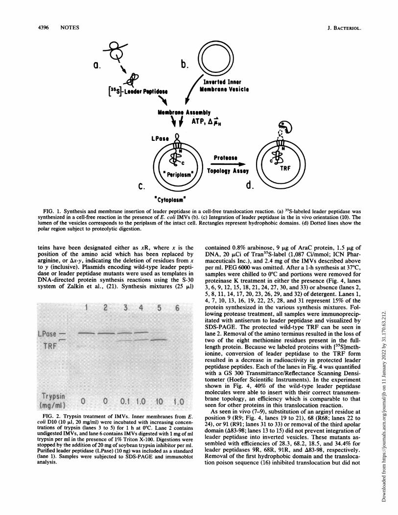
the internal, uncleaved signal sequence for membrane as-sembly (7-9). The topology of leader peptidase in invertedIMVs is shown in Fig. lc. Previous studies (19) havedemonstrated that when inverted IMVs are treated withtrypsin, a proteolytic fragment of approximately 32,000daltons remains protected from complete digestion (Fig. ld).This fragment has been named TRF for trypsin resistantfragment. The 5,000-dalton fragment degraded during thistreatment is derived from the amino terminus of leaderpeptidase (19). Treatment of vesicles with proteinase Kyields approximately the same TRF as treatment with tryp-sin (K. Moore and W. Wickner, unpublished results). Ourassay (Fig. 1) consists of an in vitro transcription-translation-translocation reaction carried out in the presence of ["SImethionine, followed by incubation with proteinase K togenerate TRF.IMVs used in these translocation assays were prepared
from E. coli K-12 D10 (rna-10 relAl spoTI metBI) by themethod of Chang et al. (2) including the modificationssuggested by Goodman et al. (11). To assess the proportionof vesicles in these reactions which were sealed in aninverted orientation, we incubated vesicles in buffer alone(Fig. 2, lane 2) or with increasing concentrations of trypsin(lanes 3 to 5). Following a 1-h digestion at 0°C, trypsin wasinactivated with soybean trypsin inhibitor and the sampleswere subjected to sodium dodecyl sulfate-polyacrylamidegel electrophoresis (SDS-PAGE; 12) and immunoblot analy-sis (15). A polyclonal antibody to leader peptidase was usedas a probe for these blots. At the highest concentration oftrypsin (10-mg/ml final concentration; Fig. 2, lane 5), all ofthe full-length leader peptidase (37,000 daltons) was con-verted to the TRF form. When right-side-out, sealed vesicleswere treated with trypsin, a protected fragment of 11,000daltons was detected (20). The 11,000-dalton protected frag-ment was not observed in this vesicle preparation. In addi-tion, the TRF was not detected when the nonionic detergentTriton X-100 was included in the reaction (Fig. 2, lane 6). Weconclude that these membranes are sealed in an invertedorientation.The mutant leader peptidases included in the following
experiments have been studied with respect to their in vivoinsertion into the E. coli cytoplasmic membrane (7-9, 16).Their characteristics are summarized in Fig. 3. These pro-
4395
Dow
nloa
ded
from
http
s://j
ourn
als.
asm
.org
/jour
nal/j
b on
11
Janu
ary
2022
by
31.1
70.6
3.21
2.
4396 NOTES
a.
4,[^5s]-Leeder Peptidese
b.Inverted Inner
Membrane Vesicle
Membrane Assembly>; ATP,A"
C.
Protease
Topology Assay
d.Cytoplasm
FIG. 1. Synthesis and membrane insertion of leader peptidase in a cell-free translocation reaction. (a) I'S-labeled leader peptidase wassynthesized in a cell-free reaction in the presence of E. coli IMVs (b). (c) Integration of leader peptidase in the in vivo orientation (10). Thelumen of the vesicles corresponds to the periplasm of the intact cell. Rectangles represent hydrophobic domains. (d) Dotted lines show thepolar region subject to proteolytic digestion.
teins have beenposition of the aarginine, or Ax-y,to y (inclusive). Idase or leader pelDNA-directed prsystem of Zalkin
LPase- -TRF
Trypsin c(mg/ml)FIG. 2. Trypsin
coli D10 (10 ,ul, 20trations of trypsin (undigested IMVs, artrypsin per ml in thestopped by the additPurified leader peptii(lane 1). Samples w
analysis.
designated either as xR, where x is the contained 0.8% arabinose, 9 ,ug of AraC protein, 1.5 ,ug ofimino acid which has been replaced by DNA, 20 puCi of Tran35S-label (1,087 Ci/mmol; ICN Phar-,indicating the deletion of residues from x maceuticals Inc.), and 2.4 mg of the IMVs described abovePlasmids encoding wild-type leader pepti- per ml. PEG 6000 was omitted. After a 1-h synthesis at 37°C,ptidase mutants were used as templates in samples were chilled to 0°C and portions were removed forotein synthesis reactions using the S-30 proteinase K treatment in either the presence (Fig. 4, laneset al., (21). Synthesis mixtures (25 ,LI) 3, 6, 9, 12, 15, 18, 21, 24, 27, 30, and 33) or absence (lanes 2,
5, 8, 11, 14, 17, 20, 23, 26, 29, and 32) of detergent. Lanes 1,4, 7, 10, 13, 16, 19, 22, 25, 28, and 31 represent 15% of the
2 3 4 5 6 protein synthesized in the various synthesis mixtures. Fol-lowing protease treatment, all samples were immunoprecip-itated with antiserum to leader peptidase and visualized bySDS-PAGE. The protected wild-type TRF can be seen inlane 2. Removal of the amino terminus resulted in the loss oftwo of the eight methionine residues present in the full-length protein. Because we labeled proteins with [35S]meth-ionine, conversion of leader peptidase to the TRF formresulted in a decrease in radioactivity in protected leaderpeptidase peptides. Each of the lanes in Fig. 4 was quantifiedwith a GS 300 Transmittance/Reflectance Scanning Densi-tometer (Hoefer Scientific Instruments). In the experimentshown in Fig. 4, 40% of the wild-type leader peptidasemolecules were able to insert with their correct transmem-
0 0. 1.0 10 1.0 brane topology, an efficiency which is comparable to thatseen for other proteins in this translocation reaction.As seen in vivo (7-9), substitution of an arginyl residue at
treatment of IMVs. Inner membranes from E. position 9 (R9; Fig. 4, lanes 19 to 21), 68 (R68; lanes 22 tomg/ml) were incubated with increasing concen- 24), or 91 (R91; lanes 31 to 33) or removal of the third apolar
rid lane 6 contains IMVs digested with 1 mg of ml domain (A83-98; lanes 13 to 15) did not prevent integration ofpresence of 1% Triton X-100. Digestions were leader peptidase into inverted vesicles. These mutants as-
ion of 20 mg of soybean trypsin inhibitor per ml. sembled with efficiencies of 28.3, 68.2, 18.5, and 34.4% foridase (LPase) (10 ng) was included as a standard leader peptidases 9R, 68R, 91R, and A83-98, respectively.vere subjected to SDS-PAGE and immunoblot Removal of the first hydrophobic domain and the transloca-
tion poison sequence (16) inhibited translocation but did not
J. BACTERIOL.
Dow
nloa
ded
from
http
s://j
ourn
als.
asm
.org
/jour
nal/j
b on
11
Janu
ary
2022
by
31.1
70.6
3.21
2.
NOTES 4397
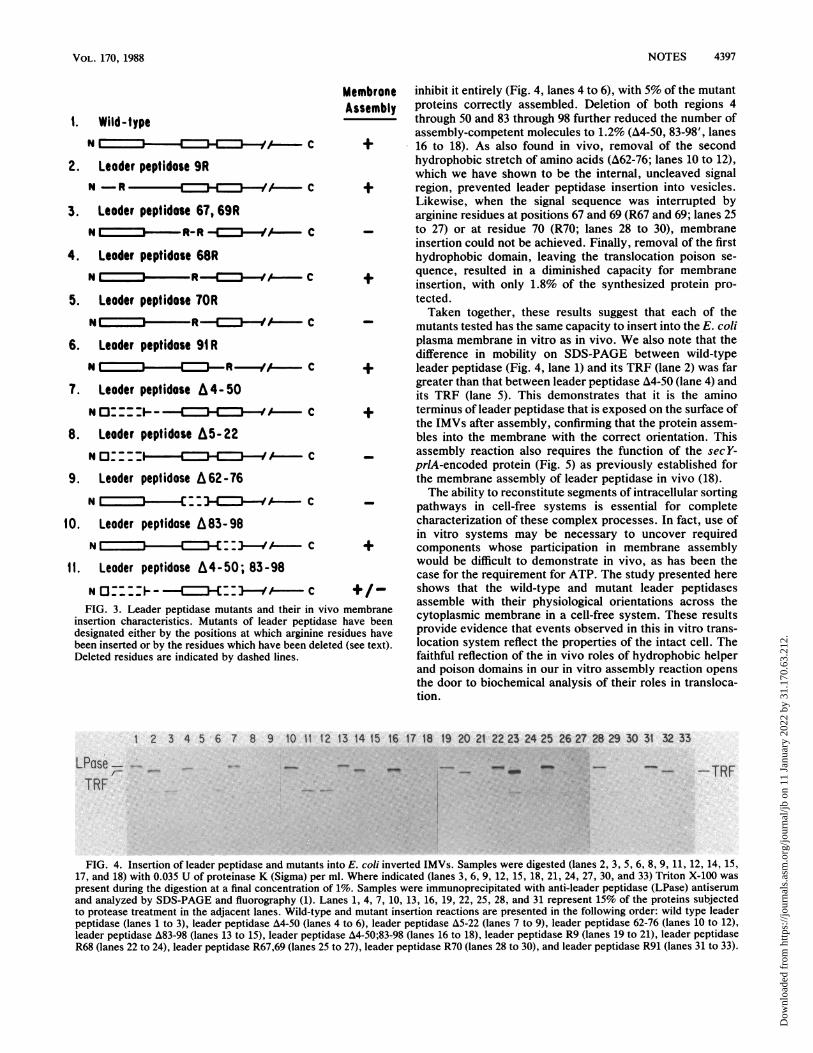
1. Wild-typeN I - C +
2. Leoder peptidose 9RN -R =3 C +
3. Leoder peptidose 67, 69RN R- R
4. Leoder peptidose 68RN I R If C +
5. Leoder peptidose 70RNIRJ- C -
6. Leoder peptidose 91RN I = R {EC== -R-/A C +
7. Leoder peptidose A4-50N0 __ E -c +
8. Leoder peptidose A5-.22N0 _ ___!CH:3--A_
9. Leoder peptidose A62-76N I::-C=--n-if A C _
10. Leoder peptidose A83-98N = =_3.C _3C::} /--AE C +
11. Leoder peptidose A4-50; 83-98
N O _ t: {EC--1-4A+/FIG. 3. Leader peptidase mutants and their in vivo membrane
insertion characteristics. Mutants of leader peptidase have beendesignated either by the positions at which arginine residues havebeen inserted or by the residues which have been deleted (see text).Deleted residues are indicated by dashed lines.
inhibit it entirely (Fig. 4, lanes 4 to 6), with 5% of the mutantproteins correctly assembled. Deletion of both regions 4through 50 and 83 through 98 further reduced the number ofassembly-competent molecules to 1.2% (A4-50, 83-98', lanes16 to 18). As also found in vivo, removal of the secondhydrophobic stretch of amino acids (A62-76; lanes 10 to 12),which we have shown to be the internal, uncleaved signalregion, prevented leader peptidase insertion into vesicles.Likewise, when the signal sequence was interrupted byarginine residues at positions 67 and 69 (R67 and 69; lanes 25to 27) or at residue 70 (R70; lanes 28 to 30), membraneinsertion could not be achieved. Finally, removal of the firsthydrophobic domain, leaving the translocation poison se-quence, resulted in a diminished capacity for membraneinsertion, with only 1.8% of the synthesized protein pro-tected.Taken together, these results suggest that each of the
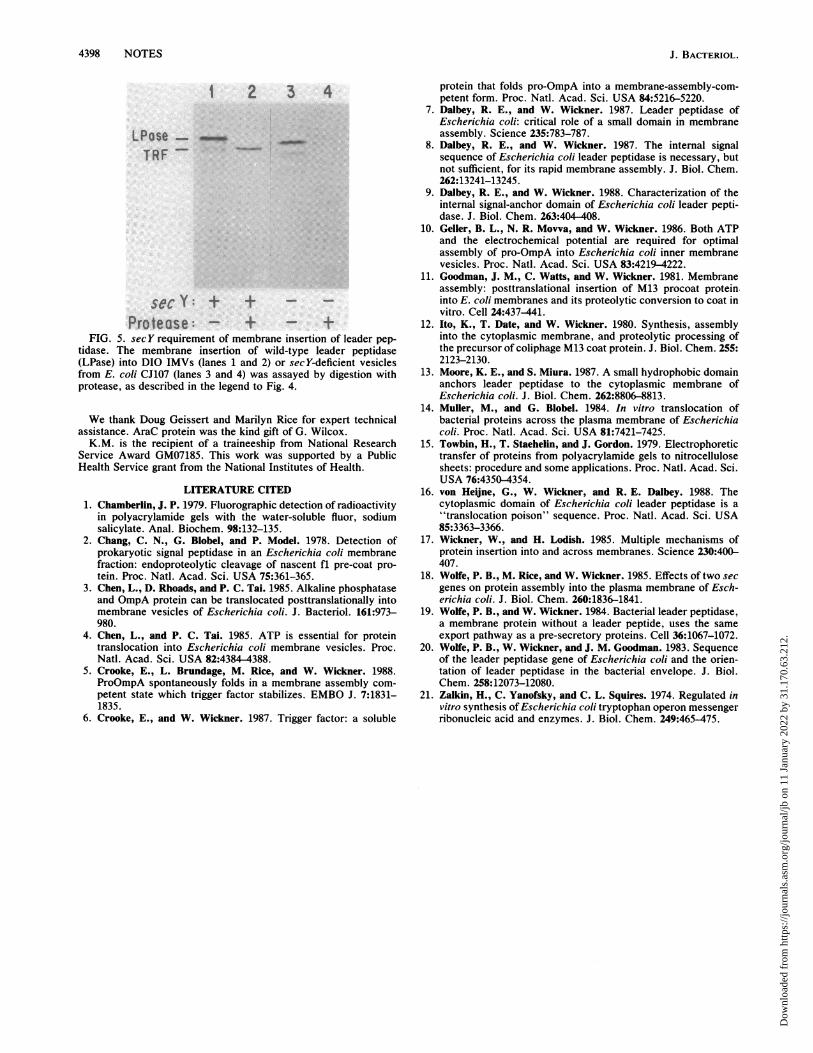
mutants tested has the same capacity to insert into the E. coliplasma membrane in vitro as in vivo. We also note that thedifference in mobility on SDS-PAGE between wild-typeleader peptidase (Fig. 4, lane 1) and its TRF (lane 2) was fargreater than that between leader peptidase A4-50 (lane 4) andits TRF (lane 5). This demonstrates that it is the aminoterminus of leader peptidase that is exposed on the surface ofthe IMVs after assembly, confirming that the protein assem-bles into the membrane with the correct orientation. Thisassembly reaction also requires the function of the sec Y-prlA-encoded protein (Fig. 5) as previously established forthe membrane assembly of leader peptidase in vivo (18).The ability to reconstitute segments of intracellular sorting
pathways in cell-free systems is essential for completecharacterization of these complex processes. In fact, use ofin vitro systems may be necessary to uncover requiredcomponents whose participation in membrane assemblywould be difficult to demonstrate in vivo, as has been thecase for the requirement for ATP. The study presented hereshows that the wild-type and mutant leader peptidasesassemble with their physiological orientations across thecytoplasmic membrane in a cell-free system. These resultsprovide evidence that events observed in this in vitro trans-location system reflect the properties of the intact cell. Thefaithful reflection of the in vivo roles of hydrophobic helperand poison domains in our in vitro assembly reaction opensthe door to biochemical analysis of their roles in transloca-tion.
1 2 3 4 5 6 7 8 9 10 11 12 13 14 15 16 17 18 19 20 21 22 23 24 25 26 27 28 29 30 31 32 33
"n_mi~~~~~~~~~~~~~~~~~~~~~~~~~~~~~~~
i
-_ -TRF
FIG. 4. Insertion of leader peptidase and mutants into E. coli inverted IMVs. Samples were digested (lanes 2, 3, 5, 6, 8, 9, 11, 12, 14, 15,17, and 18) with 0.035 U of proteinase K (Sigma) per ml. Where indicated (lanes 3, 6, 9, 12, 15, 18, 21, 24, 27, 30, and 33) Triton X-100 was
present during the digestion at a final concentration of 1%. Samples were immunoprecipitated with anti-leader peptidase (LPase) antiserumand analyzed by SDS-PAGE and fluorography (1). Lanes 1, 4, 7, 10, 13, 16, 19, 22, 25, 28, and 31 represent 15% of the proteins subjectedto protease treatment in the adjacent lanes. Wild-type and mutant insertion reactions are presented in the following order: wild type leaderpeptidase (lanes 1 to 3), leader peptidase A4-50 (lanes 4 to 6), leader peptidase A5-22 (lanes 7 to 9), leader peptidase 62-76 (lanes 10 to 12),leader peptidase A83-98 (lanes 13 to 15), leader peptidase A4-50;83-98 (lanes 16 to 18), leader peptidase R9 (lanes 19 to 21), leader peptidaseR68 (lanes 22 to 24), leader peptidase R67,69 (lanes 25 to 27), leader peptidase R70 (lanes 28 to 30), and leader peptidase R91 (lanes 31 to 33).
LPase- -..
TRF
VOL. 170, 1988
Dow
nloa
ded
from
http
s://j
ourn
als.
asm
.org
/jour
nal/j
b on
11
Janu
ary
2022
by
31.1
70.6
3.21
2.
4398 NOTES
1 2 3 4
LPose ",,TRF
secY: + +
Protease: - + +FIG. 5. sec Y requirement of membrane insertion of leader pep-
tidase. The membrane insertion of wild-type leader peptidase(LPase) into DIO IMVs (lanes 1 and 2) or secY-deficient vesiclesfrom E. coli CJ107 (lanes 3 and 4) was assayed by digestion withprotease, as described in the legend to Fig. 4.
We thank Doug Geissert and Marilyn Rice for expert technicalassistance. AraC protein was the kind gift of G. Wilcox.K.M. is the recipient of a traineeship from National Research
Service Award GM07185. This work was supported by a PublicHealth Service grant from the National Institutes of Health.
LITERATURE CITED1. Chamberlin, J. P. 1979. Fluorographic detection of radioactivity
in polyacrylamide gels with the water-soluble fluor, sodiumsalicylate. Anal. Biochem. 98:132-135.
2. Chang, C. N., G. Blobel, and P. Model. 1978. Detection ofprokaryotic signal peptidase in an Escherichia coli membranefraction: endoproteolytic cleavage of nascent fl pre-coat pro-tein. Proc. Natl. Acad. Sci. USA 75:361-365.
3. Chen, L., D. Rhoads, and P. C. Tai. 1985. Alkaline phosphataseand OmpA protein can be translocated posttranslationally intomembrane vesicles of Escherichia coli. J. Bacteriol. 161:973-980.
4. Chen, L., and P. C. Tai. 1985. ATP is essential for proteintranslocation into Escherichia coli membrane vesicles. Proc.Natl. Acad. Sci. USA 82:4384-4388.
5. Crooke, E., L. Brundage, M. Rice, and W. Wickner. 1988.ProOmpA spontaneously folds in a membrane assembly com-petent state which trigger factor stabilizes. EMBO J. 7:1831-1835.
6. Crooke, E., and W. Wickner. 1987. Trigger factor: a soluble
protein that folds pro-OmpA into a membrane-assembly-com-petent form. Proc. Natl. Acad. Sci. USA 84:5216-5220.
7. Dalbey, R. E., and W. Wickner. 1987. Leader peptidase ofEscherichia coli: critical role of a small domain in membraneassembly. Science 235:783-787.
8. Dalbey, R. E., and W. Wickner. 1987. The internal signalsequence of Escherichia coli leader peptidase is necessary, butnot sufficient, for its rapid membrane assembly. J. Biol. Chem.262:13241-13245.
9. Dalbey, R. E., and W. Wickner. 1988. Characterization of theinternal signal-anchor domain of Escherichia coli leader pepti-dase. J. Biol. Chem. 263:404-408.
10. Geller, B. L., N. R. Movva, and W. Wickner. 1986. Both ATPand the electrochemical potential are required for optimalassembly of pro-OmpA into Escherichia coli inner membranevesicles. Proc. Natl. Acad. Sci. USA 83:4219-4222.
11. Goodman, J. M., C. Watts, and W. Wickner. 1981. Membraneassembly: posttranslational insertion of M13 procoat protein.into E. coli membranes and its proteolytic conversion to coat invitro. Cell 24:437-441.
12. Ito, K., T. Date, and W. Wickner. 1980. Synthesis, assemblyinto the cytoplasmic membrane, and proteolytic processing ofthe precursor of coliphage M13 coat protein. J. Biol. Chem. 255:2123-2130.
13. Moore, K. E., and S. Miura. 1987. A small hydrophobic domainanchors leader peptidase to the cytoplasmic membrane ofEscherichia coli. J. Biol. Chem. 262:8806-8813.
14. Muller, M., and G. Blobel. 1984. In vitro translocation ofbacterial proteins across the plasma membrane of Escherichiacoli. Proc. Natl. Acad. Sci. USA 81:7421-7425.
15. Towbin, H., T. Staehelin, and J. Gordon. 1979. Electrophoretictransfer of proteins from polyacrylamide gels to nitrocellulosesheets: procedure and some applications. Proc. Natl. Acad. Sci.USA 76:4350-4354.
16. von Heine, G., W. Wickner, and R. E. Dalbey. 1988. Thecytoplasmic domain of Escherichia coli leader peptidase is a"translocation poison" sequence. Proc. Natl. Acad. Sci. USA85:3363-3366.
17. Wickner, W., and H. Lodish. 1985. Multiple mechanisms ofprotein insertion into and across membranes. Science 230:400-407.
18. Wolfe, P. B., M. Rice, and W. Wickner. 1985. Effects of two secgenes on protein assembly into the plasma membrane of Esch-erichia coli. J. Biol. Chem. 260:1836-1841.
19. Wolfe, P. B., and W. Wickner. 1984. Bacterial leader peptidase,a membrane protein without a leader peptide, uses the sameexport pathway as a pre-secretory proteins. Cell 36:1067-1072.
20. Wolfe, P. B., W. Wickner, and J. M. Goodman. 1983. Sequenceof the leader peptidase gene of Escherichia coli and the orien-tation of leader peptidase in the bacterial envelope. J. Biol.Chem. 258:12073-12080.
21. Zalkin, H., C. Yanofsky, and C. L. Squires. 1974. Regulated invitro synthesis of Escherichia coli tryptophan operon messengerribonucleic acid and enzymes. J. Biol. Chem. 249:465-475.
J. BACTERIOL.
Dow
nloa
ded
from
http
s://j
ourn
als.
asm
.org
/jour
nal/j
b on
11
Janu
ary
2022
by
31.1
70.6
3.21
2.